Embed Size (px)
Citation preview

THE ROLE OF GUT PERMEABILITY AND SERINE-PROTEASES
IN THE PATHOMECHANISM OF
IRRITABLE BOWEL SYNDROME AND INFLAMMATORY BOWEL DISEASES
Krisztina Gecse, MD
First Department of Internal Medicine
University of Szeged
PhD thesis
2011

LIST OF FULL PAPERS THE THESIS IS BASED ON
I. Gecse K, Roka R, Ferrier L, Leveque M, Eutamene H, Cartier C, Ait-Belgnaoui A,
Rosztoczy A, Izbeki F, Fioramonti J, Wittmann T, Bueno L. Increased fecal serine-protease
activity in diarrheic IBS patients: a colonic luminal factor impairing colonic permeability and
sensitivity.
Gut 2008; 57: 591-9.
IF: 9.766
II. M. Dabek, L. Ferrier, R. Róka, K. Gecse, A. Annaházi, J. Moreau, J. Escourrou, C. Cartier,
G. Chaumaz, M. Leveque, A. Ait-Belgnaoui, T. Wittmann, V. Theodoru, L. Bueno. Luminal
cathepsin G and protease-activated receptor 4: a duet involved in alterations of colonic
epithelial barrier in ulcerative colitis.
Am J Path 2009; 175:207-214.
IF: 5.673
III. Gecse K, Róka R, Séra T, Rosztóczy A, Annaházi A, Izbéki F, Nagy F, Molnár T, Szepes
Z, Pávics L, Bueno L, Wittmann T. Leaky gut in patients with diarrhea-predominant irritable
bowel syndrome and inactive ulcerative colitis.
Digestion 2011; accepted, ahead of print.
IF: 2.146 (2010)
LIST OF FULL PAPERS RELATED TO THE SUBJECT OF THE THESIS
IV. Annaházi A, Gecse K, Dabek M, Ait-Belgnaoui A, Rosztóczy A, Róka R, Molnár T,
Theodorou V, Wittmann T, Bueno L, Eutamene H. Fecal proteases from diarrheic-IBS and
ulcerative colitis patients exert opposite effect on visceral sensitivity in mice.
Pain 2009, 144: 209-217.
IF: 6.030

V. R. Róka, K. Gecse, T. Wittmann. Novel strategies and future landmarks in the treatment of
irritable bowel syndrome.
Therapy 2009; 6: 603-613.
IF: -
VI. Róka R, Gecse K, Wittmann T. Recent Observations Related to the Pathogenesis of
Irritable Bowel Syndrome.
European Gastroenterology & Hepatology Review. 2011;7:26-30.
IF: -
TABLE OF CONTENTS
LIST OF ABBREVIATIONS
I. SUMMARY 1
II. INTRODUCTION 3
III. AIMS 7
IV. PATIENTS AND METHODS 8
1. PATIENT SELECTION
1.1. Fecal serine proteases in IBS and in UC, their role in mediating increased colonic permeability and sensitivity in murine model
8
1.2. In vivo gut permeability in IBS and in inactive ulcerative colitis 8
2. METHODS
2.1. Fecal samples 9
2.2. Measurement of fecal enzymatic activities 10
2.3. Animals 10
2.4. Visceral sensitivity model 11
2.5. In vitro permeability model 11
2.6. Immunohistochemistry of pMLC and ZO-1 12
2.7. Western blot for pMLC 13
2.8. In vivo permeability measurement with 51Cr-EDTA 13
2.9. Evaluation of symptoms in IBS-D patients 14
3. STATISTICAL ANALYSIS 14
V. RESULTS
1. Fecal serine protease activity 14
2. Visceral hypersensitivity is triggered by elevated serine protease activity of IBS-D supernatants and is dependent on mucosal PAR-2 expression
17

3. Increase in colonic paracellular permeability (CPP) is evoked by elevated serine protease activity of IBS-D fecal supernatants and is PAR-2 dependent
20
4. Increased rapid phosphorylation of MLC and delayed redistribution of ZO-1 in epithelial cells after mucosal exposure to IBS-D fecal supernatants
22
5. Increase in colonic paracellular permeability (CPP) is evoked by cathepsin-G of UC fecal supernatants and is PAR-4 mediated 24
6. In vivo intestinal and colonic permeability in IBS and in inactive UC patients 26
7. Correlation between increased gut permeability and clinical symptoms in IBS-D patients 29
VI. DISCUSSION 30
VII. ACKNOWLEDGEMENTS 37
VIII. REFERENCES 38
IX. ANNEXES I. 46
X. ANNEXES II. 49

LIST OF ABBREVIATIONS
AEBSF: 4-(2-aminoethyl) benzenesulphonyl fluoride hydrochloride
Cat-G: cathepsin G
CD: Crohn’s disease
CPP: colonic paracellular permeability
EMG: electromyography
FITC-dextrane: fluorescein isothiocyanate dextrane
IBD: inflammatory bowel diseases
IBS: irritable bowel syndrome
IBS-D: diarrhea-predominant irritable bowel syndrome
IBS-C: constipation-predominant irritable bowel syndrome
IBS-M: IBS patients with mixed pattern
IBS-U: unsubtyped IBS
INF: acute infectious diarrhea
MLC: myosin light chain
MLCK: myosin light chain kinase
MPO: myeloperoxidase
PARs: protease-activated receptors
pMLC: phosphorylated myosin light chain
PMN: polymorphonuclear neutrophil
SBTI: soybean trypsin inhibitor
SCGI: specific cathepsin-G inhibitor
SLIGRL: (H-serine-leucine-isoleucine-glycine-arginine-leucine-OH)
SLPI: human secretory leucocyte protease inhibitor
SPA: serine-protease activity
UC: ulcerative colitis
ZO-1: zonula occludens-1

SUMMARY
Introduction Inflammatory bowel diseases (IBD) and diarrhea-predominant irritable bowel
syndrome (IBS-D) are characterized by elevated colonic luminal serine protease activity.
Luminal serine proteases act on protease-activated receptors (PARs), whose activation has
been associated with increased paracellular permeability. Defective epithelial barrier has been
implicated in the pathogenesis of both in irritable bowel syndrome (IBS) and in IBD.
Aims The studies were conducted (i) to investigate the origin of elevated serine protease
activity in IBS-D patients, (ii) to evaluate if it may be sufficient to trigger alterations in
colonic permeability and sensitivity in IBS-D, (iii) to examine the possible involvement of
PAR-2 activation in this process and (iv) to investigate the underlying molecular mechanisms,
(v) to evaluate whether high colonic luminal serine-protease activity in UC may contribute to
increased gut permeability and (vi) to examine the possible involvement of cathepsin-G, a
neutrophil-derived serine-protease, and PAR-4 in this process. The studies also aimed (vii) to
measure intestinal and colonic permeability in vivo in patients with IBS and (viii) with UC in
remission, and (ix) to investigate possible correlation between increased gut permeability and
clinical symptoms in IBS-D patients.
Patients and Methods Fecal enzymatic activities were assayed in healthy subjects and in
patients with IBS, ulcerative colitis and acute infectious diarrhea. Visceral sensitivity was
evaluated following mucosal exposure to supernatants from control subjects and IBS patients
by recording electromyographic response to colorectal balloon distension in wild-type and
PAR-2–/– mice. Colonic paracellular permeability (CPP) was evaluated on murine colonic
strips in Ussing chambers. Tight junction protein zonula occludens-1 (ZO-1) and
phosphorylated myosin light chain (pMLC) were detected by immunohistochemistry. In vivo
gut permeability was evaluated by measuring 24-hour urine excretion of orally administered 51Cr-EDTA in IBS and UC patients and in control subjects. Clinical symptoms were evaluated
in IBS-D patients and correlated to colonic permeability.
Results The threefold increase in fecal serine protease activity seen in IBS-D patients is of
neither epithelial nor inflammatory cell origin, nor is it coupled with decreased antiprotease
activity of endogenous origin. Mucosal application of fecal supernatants from IBS-D patients

evoked allodynia and increased CPP in mice, both of which effects were prevented by serine
protease inhibitors and dependent on PAR-2 expression. Colonic exposure to supernatants
from IBS-D patients resulted in rapid phosphorylation of myosin light chain and a delayed
redistribution of ZO-1 in murine colonocytes. Mucosal application of UC fecal supernatants
increased CPP in mice, an effect that was prevented by a cathepsin-G inhibitor and PAR-4
antagonist. In vivo gut permeability is significantly decreased in the proximal small intestine
in IBS-C patients, however distal small intestinal permeability showed no significant
difference in the studied group of patients compared to controls. Colonic permeability of IBS-
D and inactive UC patients was significantly increased compared to controls. Colonic
permeability of IBS-D patients showed correlation with stool frequency.
Conclusions Elevated colonic luminal serine protease activity in IBS-D patients evokes a
PAR-2-mediated colonic epithelial barrier dysfunction and subsequent allodynia in mice,
suggesting a novel organic background in the pathogenesis of IBS. Increased luminal
cathepsin-G may in fact contribute to the development or aggravation of defective epithelial
barrier in a PAR-4 dependent pathway in active UC. Elevated gut permeability is restricted to
the IBS-D subgroup of IBS patients and is localized to the colon both in IBS-D and in
inactive UC patients. Even though the question still persists whether altered barrier function
makes a primary or secondary contribution to IBS and IBD pathogenesis, restoring barrier
function remains a future therapeutic objective.

INTRODUCTION
The intestinal epithelium is faced with the dual task of providing a barrier while also allowing
nutrient and water absorption, therefore its integrity is crucial to maintain physiological
function and prevent diseases. Defective epithelial barrier function, which can be measured as
increased gut permeability, has been implicated in the pathogenesis of both irritable bowel
syndrome (IBS) and inflammatory bowel diseases (IBD).
Irritable bowel syndrome is a gastrointestinal disorder characterized by abdominal pain and
altered bowel habit, for which there is no apparent structural basis. The diagnosis of irritable
bowel syndrome is symptom-based according to the Rome criteria, since 2006 Rome III being
the most recent1. “Red flag” symptoms for organic disorders are ruled out by careful history
taking and thorough physical examination. Rome III defines IBS as recurrent abdominal pain
or discomfort lasting for at least 3 days per month in the last 3 months, which is associated
with 2 or more of the following characteristics: improvement with defecation, onset
associated with change in stool frequency or onset associated with change in stool form.
Irritable bowel syndrome affects 5–20% of the population worldwide2, thus increasing interest
has recently been shown towards its poorly understood pathophysiology and to possible
therapeutic approaches of the disease. Due to intensive research of recent decades, there has
been a paradigm shift in IBS pathophysiology from considering it a purely psychosocial
disturbance to finding organic backgrounds of the disease. By now, genetic factors, altered
“brain-gut axis” - both in terms of altered sensory afferent function and central processing
resulting in visceral hypersensitivity - abnormal serotonerg neurotransmission, altered gut
motility, changes in the gut microflora, stress, intestinal hyperpermeability and mucosal
immune activation have all been implicated in the pathogenesis of IBS. In a preliminary pilot
study, it has been shown that fecal supernatants of diarrhea-predominant IBS (IBS-D) patients
have a substantially higher serine protease activity, similar to that of patients with ulcerative
colitis (UC), when compared with healthy subjects or other subgroups of IBS patients3.
Certain serine proteases are signaling molecules that cleave protease-activated receptors
(PARs), a family of G-protein coupled receptors with a widespread distribution (Figure 1).

Figure 1. Mechanism of PAR activation. Proteinases cleave the extracellular N terminal domain (1) releasing a
new N terminal tail which acts as a “tethered ligand”, that binds the receptor itself (2) to induce an intracellular signal (3) 4.
Four members of the PAR family have been identified so far in human tissues, of which
PAR-2 and 4 are highly expressed on intestinal epithelial cells5-7. The primary activating
protease of PAR-2 is trypsin, however, serine proteases of other endogenous and/or bacterial
origin are also able to cleave the receptor8. Activation of PAR-2 modulates several
gastrointestinal functions, such as motility, ionic exchange, paracellular permeability, sensory
functions and inflammation4. Intracolonic but not intraperitoneal administration of the
synthetic selective PAR-2 agonist, SLIGRL (H-serine-leucine-isoleucine-glycine-arginine-
leucine-OH), produces delayed visceral hyperalgesia in rats associated with increased
paracellular permeability9–11. In the intact epithelium the paracellular space between adjacent
cells is sealed by dynamically changing tight junctions at the luminal aspect of the apical
junction complex (Figure 2), which forms a selectively permeable barrier and is structurally
related to the perijunctional actomyosin ring. The integrity of the epithelial barrier is
dependent upon the contraction of this perijunctional actomyosin ring and subsequent
physical tension on the tight junction, an event that involves the phosphorylation of myosin
light chain (MLC), initiated by MLC kinase (MLCK)12,13.

Figure 2. The tight junction. Immunofluorescence demonstrates the restricted location of the tight junction. Nuclei are stained blue, actin is
green and zonula occludens-1 (ZO-1) tight junction protein is red; all of the ZO-1 co-localizes with perijunctional actin, producing a yellow
color at the tight junction. The claudin family of proteins form the actual paracellular pore within the tight junction, and is associated with
another transmembrane protein, occludin. ZO-1, 2 and 3 are attached to this complex14.
Interestingly, both visceral hypersensitivity15-17 and impaired intestinal permeability are
commonly observed features of IBS. Gut permeability has been reported to be enhanced in
50% of post-infectious IBS patients, in agreement with the study showing increased small
intestinal permeability in both the post-infectious and sporadic forms of IBS,
characteristically in the diarrhea-predominant subtype18-20, the same subpopulation where
increased serine protease activity has been detected3. In accordance, the report on the
“Walkerton epidemic” - a waterborne outbreak of acute gastroenteritis in Walkerton, Ontario -
proved subtle increase in small intestinal permeability in a large number of patients with IBS,
however in vitro studies suggest enhanced permeability in colonic biopsies of IBS patients
compared to healthy subjects21-22. Therefore it seems that gut permeability in IBS is altered,
though the data on the subgroup of IBS patients affected and the exact localization of the
defective barrier are still contradictory. It is also well established that impaired intestinal
barrier function could facilitate the passage of luminal antigens and lead to mucosal immune
response23. Furthermore, there is growing evidence for microinflammation of the intestinal
and colonic mucosa to play a role in IBS pathogenesis24-27. Thus identifying the role of
defective mucosal barrier in IBS pathomechanism and symptom generation may be an
important landmark in better understanding of the disease.

Inflammatory bowel disease is a chronic inflammatory condition of the intestines that is
characterized by remission and relapses and distills clinically into one of the two major
subtypes of disease: ulcerative colitis and Crohn‘s disease (CD). Current understanding
regarding the underlying pathophysiological mechanisms in IBD is that dysregulated mucosal
immune response leads to barrier defect triggered by antigenic components of the normal
commensal microbiota that reside within the intestine in a genetically susceptible host.
Epithelial barrier impairment is considered important in IBD as it leads to increased luminal
antigen exposition of the lamina propria, i.e. immune cells, including large number of
neutrophils28, which further aggravate both the inflammatory process and the
hyperpermeability (Figure 3).
Figure 3. A model for the pathogenesis of
inflammatory bowel disease. (1) Initial barrier disruption, which leads to a (2) mixing of luminal
content (ie. bacteria) with lamina propria content, ie. antigen presenting cells (3) that promote (4) T-cell
response and secretion of the pro-inflammatory cytokine IL-10 (5). T cells respond by secreting IFNγ
that induce macrophag activation (6), which in turn secrete TNFα. Both IFNγ and TNFα are known to
affect epithelial barrier function (7). Through unknown mechanism, these cytokines activate MLCK (8) leading
to MLC phosphorylation, actomyosin contraction and opening of the tight junction, which leads to a vicious
cycle in disease progression14.
Neutrophils are characterized by two major granule populations, primary (azurophil) and
secondary (specific) granules, formed at different stages of neutrophil maturation. Cathepsin-
G (Cat-G), a serine protease, makes up approximately 20% of the neutrophil azurophil
granule proteins and it also plays an important role in neutrophil function during
inflammatory processes, including degradation of extracellular matrix components and
cytokines, modulation of integrin clustering on neutrophils and direct chemoattraction of T
cells and other leukocytes29. Besides trypsin and thrombin, proteinase-activated receptor 4 is
also activated by the neutrophil derived serine protease cathepsin-G30.
There is strong evidence for barrier dysfunction in IBD31: hyperpermeability in non-involved
segments of the intestine of CD patients as well as in first degree relatives has been
reported32-34 and increased permeability have also been associated with an increased risk of
relapse35,36. Data are however less abundant on paracellular permeability regarding UC.
Increase in gut permeability has previously been reported in clinically active UC, which was
also shown to correlate with disease severity31,37,38. Still, gut permeability has not yet been
evaluated in remission of the disease.
AIMS
The studies were conducted (i) to investigate the origin of elevated fecal serine protease
activity in IBS-D patients, (ii) to evaluate if this elevated colonic luminal serine protease
activity may be sufficient to trigger alterations in colonic permeability and sensitivity in mice,
(iii) to examine the possible involvement of PAR-2 activation in this process and (iv) to
analyze the underlying molecular mechanisms in the tight junction. We also aimed to (v)
evaluate whether the high colonic luminal serine-protease activity in UC may contribute to
increased gut permeability and (vi) to examine the possible involvement of cathepsin-G and
PAR-4 in this process. Furthermore, we aimed (vii) to measure intestinal and colonic
permeability of patients with IBS of the diarrhea and of the constipation-predominant subtype
in vivo, (viii) to investigate possible correlation between increased gut permeability and
clinical symptoms in IBS-D patients and (ix) to measure gut permeability in patients with
ulcerative colitis in remission.

PATIENTS AND METHODS
1. Patient selection
1.1. Fecal serine proteases in IBS and in UC, their role in mediating increased colonic
permeability and sensitivity in murine model
Demographic data of the patients enrolled in the study are summarized in Table 1. Patients
fulfilling the Rome II criteria for IBS (patient screening was carried out in 2005-2006)
participated in the study39. All participants provided medical history and underwent physical
examination. In IBS patients other gastrointestinal disorders were excluded by detailed blood
and stool analyses, serological assays for coeliac disease, lactose–hydrogen breath test and
colonoscopy. Active UC was assessed clinically and endoscopically. The study protocol was
approved by the Ethical Committee of the University of Szeged. All subjects provided written
and informed consent to participate.
Group Number of patients Age mean (range) Sex ratio (M/F)IBS-D 24 49 (19-75) 9/42IBS-C 18IBS-A 10UC 17 41 (18-79) 6/11INF 23 52 (19-90) 12/11Healthy subjects 25 44 (30-65) 4/21
Table 1. Demographic data. IBS-D: diarrhea-predominant IBS patients, IBS-C: constipation-predominant IBS
patients, IBS-A: IBS patients with altered bowel habit, UC: patients with ulcerative colitis, INF: patients with acute infectious diarrhea.
1.2. In vivo gut permeability in IBS and in inactive ulcerative colitis
Demographic data of the patients enrolled in the study are summarized in Table 2. Thirty
patients fulfilling the Rome III criteria for IBS participated in the study, none of which related
the onset of their symptoms to infectious gastroenteritis. According to the Rome III criteria
introduced in 2006, symptoms are expected to originate 6 months before diagnosis, which is a
less restrictive timeframe compared to the Rome II criteria (12 weeks of symptoms over 12
months). In addition, according to Rome III criteria IBS subtyping should be based on stool

consistency, which results in four subgroups, namely diarrhoea predominant (IBS-D),
constipation predominant (IBS-C), subjects with mixed pattern (IBS-M) and unsubtyped IBS
(IBS-U). Still, bowel subtyping used in Rome II for IBS-D and IBS-C remained acceptable.
Other gastrointestinal disorders were excluded by detailed blood and stool analyses,
serological assays for celiac disease, lactose-hydrogen breath test and colonoscopy. Patients
with inactive ulcerative colitis (partial Mayo score ± SEM: 1.3±0.2; CRP (mg/dl) ± SEM:
3.8±1.3) were previously shown to have either left-sided colitis or pancolitis. Voluntary
subjects, free of any gastrointestinal symptoms, served as controls. Patients and voluntary
subjects with impaired renal function, alcohol consumption, using NSAIDs, prokinetics,
antihistamines or immunosuppressive agents were excluded from the study. UC patients were
required to be exclusively on 5-ASA maintenance therapy. The study protocol was approved
by the Human Investigation Review Board, University of Szeged. All subjects provided
written and informed consent to participate.
Group Number of patients Age mean (range) Sex ratio (M/F)IBS-D 18 49 (25-68) 6/12IBS-C 12 56 (37-65) 2/10UC 13 47 (29-72) 3/10Healthy subjects 10 49 (38-65) 2/8
Table 2. Demographic data
2. Methods
2.1. Fecal samples
Only feces collected in situ, or collected at home and transported within 1 h after defecation
to the First Department of Internal Medicine in Szeged, Hungary, were used. Samples were
stored at -80°C until transportation on dry ice to INRA Toulouse, France. Upon arrival, 1g of
fecal sample was thawed, dissolved, and homogenised in 7ml of Tris buffer, centrifuged (4500
rpm, 10 min, +4C) and filtered (0.2 µm, Nalgene, (Adventure 16, San Diego, California,
USA)). The acquired supernatants were used for measuring total protease activity, serine
protease activity, polymorphonuclear neutrophil (PMN) and pancreatic elastase,

myeloperoxidase (MPO), calprotectin and human secretory leucocyte protease inhibitor
(SLPI) activity.
2.2. Measurement of fecal enzymatic activities
To measure total fecal serine protease activity, supernatants of fecal homogenates (25 ml)
were incubated with 1 ml of reaction buffer (0.15 M NaCl and 20 mM Tris-HCl, pH 8.3) and
1ml of 0.5% (w/v) azocasein (Sigma, St Quentin Fallavier, France) at 40°C. The reaction was
stopped after 20 minutes with 1ml of 10% (v/v) trichloracetic acid (TCA, Sigma). Following
centrifugation, absorption of the clear supernatant was measured at 366 nm. Enzymatic
activities of the supernatants were normalized to protein content. To determine whether
protease activity was dependent upon serine proteases, measurements were done after
preincubation for 30 min with selective serine protease inhibitors, AEBSF (4-(2-aminoethyl)
benzenesulphonyl fluoride hydrochloride) and a mixture of soybean trypsin inhibitor (SBTI)
and aprotinin (Sigma). Protease activity was expressed as units per milligram of protein,
standardized against activity elicited by 1 U of standard trypsin. Pancreatic elastase-1, SLPI,
PMN elastase and calprotectin concentrations were assayed by ELISA (Schebo-Tech,
Giessen, Germany; R& D Systems, Lille, France; Immundiagnostik AG, Bensheim, Germany;
HyCult Biotechnology, Uden, The Netherlands, respectively). Fecal MPO activity was
measured as described earlier40. Cat-G activity was measured in fecal supernatants from
patients with UC and healthy subjects, using N-succinyl-Ala-Ala-Pro-Phe p Nitroanilide
(Sigma) as a substrate. Enzymatic activity was measured at 410 nm for 5 minutes at 37°C.
2.3. Animals
Congenic 6–9-week old male C57BL/6J wild type (Janvier, Le Genest St-Isle, France) and
PAR-2-deficient mice (The Jackson Laboratory, Bar Harbor, Maine, USA) were used. The
genetic status of the PAR-2-deficient mice was confirmed by PCR. Mice were housed in
polypropylene cages in a light- and temperature-controlled room (12 h/12 h cycles; 20±2°C),
were fed standard pellets (Harlan Teklad, Bicester, Oxon, UK), and water was provided ad
libitum. The experimental protocols described in the study were approved by the local
Institutional Animal Care and Use Committee.

2.4. Visceral sensitivity model
Under xylazine/ketamine anaesthesia (both 1.2 mg, subcutaneously), two nickel–chrome
electrodes were implanted into the abdominal external oblique muscle and a third into the
abdominal skin, and were exteriorized on the back of the neck. On the fifth to seventh
postoperative day, colorectal distensions were used as noxious stimuli to evaluate visceral
hyperalgesia by electromyographic (EMG) recording. Under sodium pentobarbital
anaesthesia (10mg, intraperitoneally), polyethylene perfusion and distension catheters
(Fogarty catheter for arterial embolectomy, 4F, Edwards Lifesciences, Nijmegen, The
Netherlands) were inserted into the colon. Animals received 0.3 ml of fecal supernatants of
IBS-D or IBS-C (constipation-predominant IBS) patients, of healthy subjects or of IBS-D
patients previously incubated with serine protease inhibitors, SBTI and aprotinin. The
colorectal distension procedure started 60 min after the infusion of the supernatants had
finished with volumes progressively increasing in 0.02 ml steps, from 0 to 0.12 ml, each step
lasting 10 s with 5 min non-distension periods in between. During the distension periods, the
striated muscle’s EMG activity was recorded and analysed according to Larsson et al41. Basal
EMG activity was subtracted from the EMG activity registered during the periods of
distension.
2.5. In vitro permeability model
Mice were sacrificed by cervical dislocation and the distal part of the colon was removed.
Colonic strips were mounted with a flux area of 0.3 cm2 in Easymount Ussing-type chambers
(Physiologic Instruments, San Diego, California, USA), bathed in Krebs solution and
oxygenated at a maintained temperature of 37°C. After allowing 15 min for equilibrium, one-
fifth of the initial volume of the buffer solution (1ml) of the apical compartment (mucosal
side) was replaced with physiological saline or supernatants (500ul) and fluorescein
isothiocyanate (FITC)-labelled dextran (500ul) (4000 Da, 0.022 g/ml, Sigma). 60 minutes
later fluorescent intensity was measured on the serosal side of the chamber. Supernatants
derived from healthy subjects, from IBS patients or from UC patients. IBS-D supernatants
were alternatively pre-incubated with protease-inhibitors soybean trypsin inhibitor (SBTI) and
aprotinin for 30 minutes on ice and than added to the mucosal side of the colonic strip. To

assess the potential role of PAR-1, PAR-2 and PAR-4 in the effect of UC fecal supernatants on
colonic paracellular permeability, selective receptor antagonists were used: PAR1 antagonist
FLLRN (Phe-Leu-Leu-Arg-Asn, 10µmol/L, Peptides International, Louisville, KY)42,43
(3/13,14), PAR2 antagonist FSLLRY (Phe-Ser-Leu-Arg-Tyr, 10µmol/L, Bachem, Weil am
Rhein, Germany)44 and P4-pal10 pepducin (final concentration 1µmol/L) (NeoMPS,
Strasbourg, France) were added to the mucosal side of the chamber prior to the administration
of fecal supernatants to the mucosal side. The effect of the PAR-4 agonist peptide (Ala-Tyr-
Pro-Gly-Lys-Phe-NH2, 50 µmol/L, Sigma) was also assessed, as well as the effect of a
selective Cathepsin-G inhibitor (SCGI): UC fecal supernatants were pre-incubated for 30
minutes on ice with the inhibitor (0.2mmol/L) and then the mixture was added to the mucosal
side of the colonic strip.
2.6. Immunohistochemistry of pMLC and ZO-1
At 1 and 4 h after the intracolonic infusion of supernatants of IBS-D patients or healthy
controls, saline, SLIGRL (5 mg) or IBS-D supernatants previously incubated with serine
protease inhibitors, the mice were sacrificed and the distal colon was removed. For both
phosphorylated MLC (pMLC) and ZO-1 immunolabelling, samples were fixed in buffered
paraformaldehyde (4%), incubated in 30% sucrose (24 h, +4°C), embedded (Tissue Tek
medium) and frozen in isopentane at -45°C. Cryostat sections (7 um) were fixed with acetone
(10min, -20°C), hydrated in phosphate-buffered saline (PBS) and treated with 4 mg/ml
sodium borohydride (45min, +4°C). Sections were permeabilized with PBS–0.5% Triton
X-100 and incubated in blocking solution (PBS containing 1% bovine serum albumin). For
pMLC staining, samples were then incubated with goat anti-pMLC antibodies (1/100,
SantaCruz, Santa Cruz, California, USA) followed by incubation with biotin-conjugated IgG
donkey anti-goat antibody (1/1000, Interchim, Montlucon, France). Sections were rinsed in
NaHCO3 (0.1M, pH 8.2) and incubated with FITC-conjugated avidin (1/500) diluted in the
same solution. For ZO-1 labelling, sections were incubated with rabbit anti-ZO-1 antibodies
(1/500, Zymed, San Francisco, California, USA) followed by incubation with Alexa fluor
488-conjugated IgG donkey anti-rabbit antibodies (1/2000, Molecular Probes, Cergy-
Pontoise, France). All sections were mounted in Vectashield HardSet Mounting Medium with

4’,6-diamidino-2-phenylindole (DAPI; Vector Laboratories, Burlingame, California, USA)
and examined under a Nikon 90i fluorescent microscope.
2.7.Western blot for pMLC
Colonic mucosa was collected from mice 1h after intracolonic infusion with fecal
supernatants of healthy subjects or IBS-D patients, or saline. Proteins were extracted with
RIPA buffer and quantified. Following the Laemmli method45, equal amounts of protein
extracts were electrophoresed by 12% SDS–PAGE (sodium dodecyl sulphate–polyacrylamide
gel electrophoresis) and then electrotransferred onto Hybond-P membrane (GE Healthcare,
Bordeaux, France). After saturation, the membrane was incubated with anti-pMLC primary
antibody (1/1000, Biosource, Worcester, Massachusetts, USA) and peroxidase-conjugated
goat anti-rabbit secondary antibody (1/1000, Millipore, St Quentin en Yvelines, France). The
membrane was developed with SuperSignal Reagent (Pierce, Prebieres, France). Integrated
density values were assessed by ImageJ 1.37 software (NIH, Bethesda, Maryland, USA).
2.8. In vivo permeability measurement with 51Cr-EDTA
To measure intestinal and colonic permeability after an overnight of fasting participants
emptied their bladders and consumed 51Cr-EDTA (Perkin Elmer Life Sciences, Boston, MA,
USA) of 1.8 MBq activity dissolved in 100 ml of water, followed by 200 ml of standard meal
(Nutridrink, Nutricia, Budapest, Hungary) containing 300 kcal. Study participants were
requested to restrain from drinking for 3 hours and from eating for 5 hours. Gut permeability
was evaluated by measuring 24-hour urine excretion of orally administered 51Cr-EDTA,
where time periods were chosen to relate to permeability within the proximal (0-3h) and distal
(3-5h) small intestine and the large bowel (5-24h)38,46,47. Urinary output was recorded for each
period and the radioactivity of 1 ml aliquots were counted by a gamma-counter (Packard
Cobra, Canberra Packard, UK) in duplicates. Gut permeability was expressed as percentage of
urinary excretion of the orally administered dose of 51Cr-EDTA (%).

2.9. Evaluation of symptoms in IBS-D patients
IBS-D patients were asked to fill out a questionnaire, regarding their clinical symptoms at the
time of the permeability measurement. Stool frequency (/week) and consistency (according to
the Bristol stool scale, which evaluates stool consistency according to a visual scale graded 1:
very hard-7: watery), frequency of abdominal pain, distension and bloating (/week), intensity
of abdominal pain, distension and bloating and quality of life (visual analogue scale /VAS/;
%) were the symptoms being evaluated and correlated to colonic permeability.
3. Statistical analysis
All data are presented as means ± SEM. For statistical analysis, Prism 4.0 (GraphPad, San
Diego, California, USA) was used. Multiple comparisons for fecal enzymatic activities of
different patient groups and integrated optical density values of pMLC western blots were
analyzed by repeated measures of one-way analysis of variance (ANOVA), followed by
Tukey’s posttest or Kruskal–Wallis posttest (for MPO activity). Statistical significance for
visceral hypersensitivity results was established by using two-way ANOVA, followed by
Bonferroni posttest. In vitro permeability results were analyzed with one-way ANOVA,
followed by Tukey posttest or two-tailed unpaired t test. Multiple comparisons for in vivo
permeability of different patient groups were analyzed by repeated measures of one-way
ANOVA, followed by Tukey’s posttest. Unpaired t-test was used to evaluate colonic
permeability data in subgroups of UC patients. Linear regression was applied to establish
correlation between clinical symptoms and permeability. Statistical significance was accepted
at p <0.05.
RESULTS
1. Fecal serine protease activity
In healthy subjects the total fecal serine protease activity was 698 U/mg of protein. This
serine protease activity was significantly greater in IBS-D patients (2079 U/mg, p<0.001) and
in UC patients (2193 U/mg, p<0.01) compared with healthy controls; a similar increase was

not present regarding the IBS-C, IBS-A subgroups or patients with acute infectious diarrhea
(INF) (Figure 4A). Addition of the serine protease inhibitor AEBSF abolished this increased
protease activity in IBS-D and UC supernatants. A similar inhibition was obtained with
preincubation with SBTI and aprotinin, two common serine protease inhibitors validated for
human use (Figure 4B). Regarding the potential origin of the elevated serine protease activity,
no significant difference was observed in fecal pancreatic elastase-1 concentration in UC or in
any subgroups of IBS patients when compared with controls (Figure 4C). However,
pancreatic elastase-1 concentration was significantly decreased in INF patients (p<0.05),
which might be attributed to the diluted fecal content (i.e. profuse watery diarrhea) and
restricted diet. PMN-derived elastase had a significantly elevated fecal concentration in UC
and INF patients (p<0.05), but not in any subgroup of IBS patients (Figure 4D). Human fecal
SLPI activity showed no significant difference in any of the studied groups of patients when
comparing them with control subjects (Figure 4E). Fecal inflammatory markers, such as
human calprotectin and MPO, showed a significant increase in UC (p<0.001 and p<0.05,
respectively) and INF patients (p<0.05 and p<0.001, respectively), but we found no increase
in any of the IBS subgroups (Figure 4F and G).

Figure 4.

Figure 4. The increase in total fecal serine protease activity in diarrhea-predominant irritable bowel syndrome (IBS-D) is neither accompanied by elevated enzymatic activity of pancreatic or inflammatory cell origin nor
coupled with decreased antiprotease activity. (A) Fecal total protease activity in healthy controls, in IBS-D patients, IBS-C(constipation-predominant) patients, those with alternating bowel habits (IBS-A), patients with
active ulcerative colitis (UC) and those with acute infectious diarrhea (INF). (B) Fecal protease activity after incubation with serine protease inhibitors AEBSF(4-(2-aminoethyl) benzenesulphonyl fluoride hydrochloride),
or a mixture of soybean trypsin inhibitor and aprotinin. (C) Fecal pancreatic elastase-1 concentration. (D) Fecal polymorphonuclear neutrophil (PMN) -elastase concentration. (E) Fecal human secretory leucocyte protease
inhibitor (SLPI) concentration. (F) Fecal calprotectin concentration. (G) Fecal myeloperoxidase (MPO) activity. *p<0.05, **p<0.01, compared with healthy controls, #p<0.05, compared with IBS-D (one-way analysis of
variance). Error bars represent SEM.
2. Visceral hypersensitivity is triggered by elevated serine protease activity of IBS-D
supernatants and is dependent on mucosal PAR-2 expression
Intracolonic infusion of fecal supernatants from IBS-D patients administered prior to
colorectal distensions in mice significantly increased the abdominal muscle EMG response (a
valid criterion representing nociception, Figure 5A) at low distension volumes, namely at
0.02, 0.04 and 0.06 ml, compared with the intensity of muscle contractions in animals treated
with fecal supernatants of control subjects (0.02 ml, 30.6 (5.9) mV/s vs 3.5 (1.7) mV/s; 0.04
ml, 73.4 (7.9) mV/s vs 18.9 (7.5) mV/s; 0.06 ml, 107.4 (4.4) mV/s vs 53.2 (14.3) mV/s;
p<0.05, p<0.001 and p<0.001, respectively, Figure 5B). Conversely, colonic instillation of
IBS-C supernatants did not evoke alteration of visceral sensitivity to colorectal distension
(Figure 5C). Incubation of fecal supernatants from IBS-D patients with serine protease
inhibitors prior to colonic infusion, prevented the increased EMG response to low volumes of
distension (0.02 ml, 2.8 (1.4)mV/s, 0.04 ml, 49.8 (2.9) mV/s; p<0.01, p<0.05, respectively,
Figure 5B), while protease inhibitors per se had no effect (data not shown). Furthermore, IBS-
D supernatants failed to induce allodynia or visceral hypersensitivity in PAR-2-deficient mice
(0.02 ml, 4.6 (3.5) mV/s; 0.04 ml, 41.4 (7.0) mV/s; 0.06 m, 59.4 (14.2) mV/s, Figure 5D).

Figure 5A. Representative electromyographic recordings of abdominal muscle contractions (as a valid criterion
for visceral nociception) evoked by colorectal balloon distension 60min after intracolonic infusion of fecal supernatants from diarrhea-predominant irritable bowel syndrome (IBS-D) patients (n=5), control subjects (n=5)
and IBS-D supernatants previously incubated with serine protease inhibitors (n=3) in wild type or PAR-2 deficient mice (n=5) (each supernatant was tested on an average of five wild type mice).
Figure 5B. Supernatants of IBS-D patients induced allodynia and visceral hypersensitivity in wild-type mice at
low distension volumes compared with control supernatants (0.02, 0.04, 0.06 ml; ap<0.05, aaap<0.001, aaap<0.001, respectively), an effect that was partially prevented by the administration of serine protease inhibitors
(at 0.02 and 0.04 ml distension volumes, bbp<0.01 and bp<0.05, respectively). APRO, aprotinin; SBTI, soybean
trypsin inhibitor.

Figure 5C. Constipation-predominant irritable bowel syndrome (IBS-C) supernatants were devoid of any effect
on visceral sensitivity.
Figure 5D. IBS-D supernatants did not trigger visceral hyperalgesia in PAR-2-deficient mice (0.02, 0.04,
0.06ml; aap<0.01, ap<0.05, ap<0.05, respectively).

3. Increase in colonic paracellular permeability (CPP) is evoked by elevated serine protease
activity of IBS-D fecal supernatants and is PAR-2 dependent
Administration of fecal supernatants from healthy subjects to the mucosal side of the murine
colon mounted in Ussing chambers did not significantly alter CPP compared with saline
(128% vs. 100%; ns.). In contrast, addition of fecal supernatants from IBS-D patients
significantly increased the FITC–dextran flux compared with the administration of
supernatants from healthy subjects or saline (192%, p<0.05). Comparatively, supernatants
from IBS-C patients failed to induce such an increase in colonic permeability (137%; ns.,
Figure 6A). Previous incubation of the supernatants from IBS-D patients with SBTI and
aprotinin prior to administration significantly decreased the elevated CPP evoked by
supernatants from IBS-D patients (111% vs. 202%, p<0.01, Figure 6B). No increase in CPP
was observed in response to fecal supernatants from IBS-D patients on colonic strips of
PAR-2-deficient mice in contrast to their wild-type strain (127% vs. 248%, p<0.05, Figure
6C).
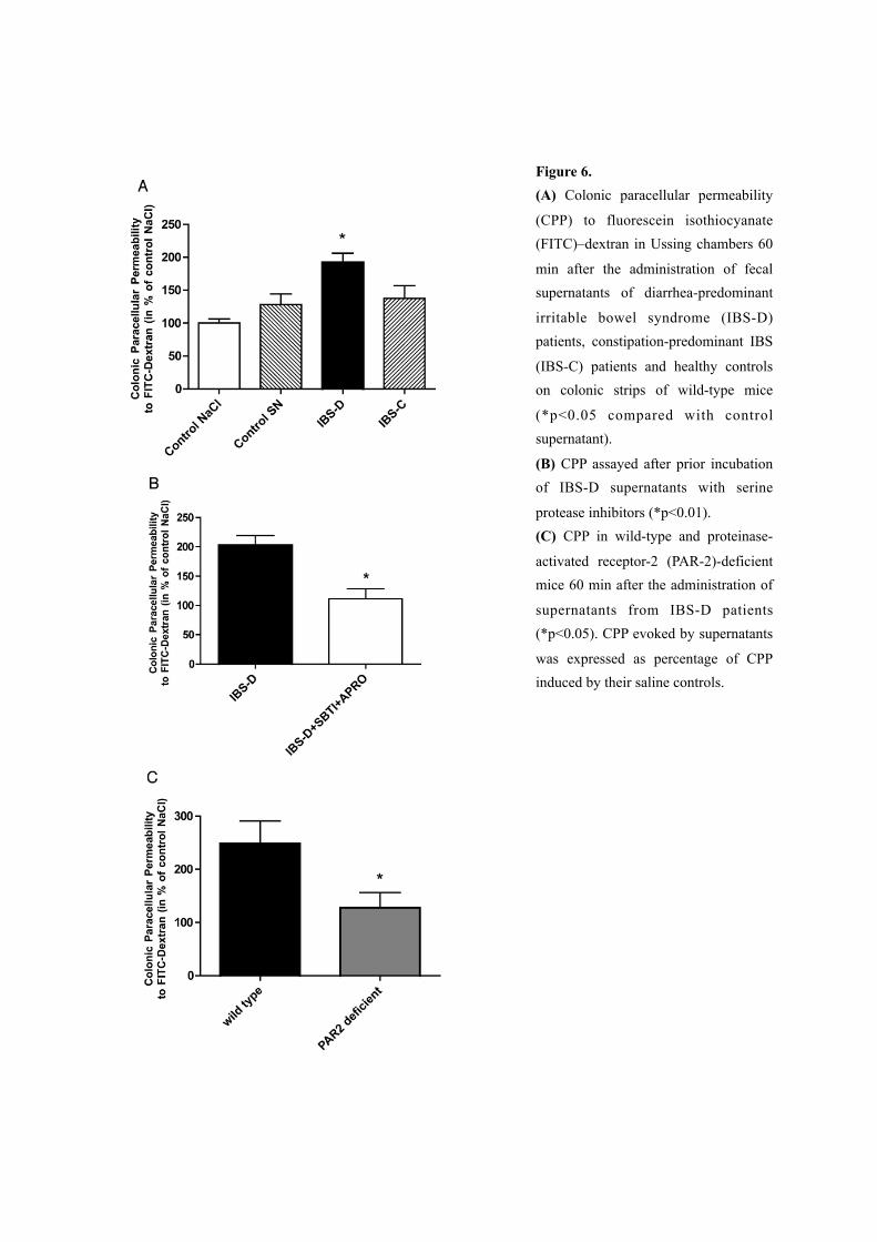
Figure 6. (A) Colonic paracellular permeability
(CPP) to fluorescein isothiocyanate (FITC)–dextran in Ussing chambers 60
min after the administration of fecal supernatants of diarrhea-predominant
irritable bowel syndrome (IBS-D) patients, constipation-predominant IBS
(IBS-C) patients and healthy controls on colonic strips of wild-type mice
(*p<0.05 compared with control supernatant).
(B) CPP assayed after prior incubation of IBS-D supernatants with serine
protease inhibitors (*p<0.01). (C) CPP in wild-type and proteinase-
activated receptor-2 (PAR-2)-deficient mice 60 min after the administration of
supernatants from IBS-D patients (*p<0.05). CPP evoked by supernatants
was expressed as percentage of CPP induced by their saline controls.

4. Increased rapid phosphorylation of MLC and delayed redistribution of ZO-1 in epithelial
cells after mucosal exposure to IBS-D fecal supernatants
One hour after intracolonic infusion with supernatants from IBS-D patients there was a
pronounced and diffuse labelling of pMLC in epithelial cells, similar to that observed after the
infusion of SLIGRL used as a positive control for PAR-2 activation, and inhibited by
preincubation with serine protease inhibitors. Administration of supernatants from healthy
subjects or saline failed to evoke an increase in pMLC immunostaining, which remained
restricted to the tight junction area (Figure 7A).
Four hours after intracolonic infusion of supernatants from healthy subjects or saline,
selective immunostaining of the tight junction protein ZO-1 showed labelling that was
restricted to the surface of epithelial cells. In contrast, intracolonic infusion of supernatants
from IBS-D patients resulted in a marked labelling of the intracellular compartment, similarly
to that experienced after the infusion of SLIGRL, suggesting intensive internalisation of the
protein. Previous incubation of fecal supernatants from IBS-D patients with serine protease
inhibitors prevented the occurrence of a marked intracellular labelling of colonocytes (Figure
7B). Western blotting showed an increased level of pMLC in the colonic mucosa of mice 1 h
after the intracolonic infusion of supernatants from IBS-D patients compared with those
infused with supernatants from healthy controls (p<0.001). There was no significant
difference in the level of pMLC in the colonic mucosa of mice that were infused with
supernatants from healthy subjects or IBS-D supernatants previously incubated with a mixture
of serine protease inhibitors (Figure 7C).

Figure 7.

Figure 7. (A) Immunolabelling of phosphorylated myosin light chain (pMLC, green) and nuclear labelling with 4,6-diamidino-2-phenylindole (DAPI, blue) in enterocytes 1 h after intracolonic infusion with supernatants from
healthy controls, diarrhea-predominant irritable bowel syndrome (IBS-D) patients, SLIGRL, control and IBS-D supernatants previously incubated with serine protease inhibitors and saline. (B) Immunostaining of zonula
occludens-1 (ZO-1, green) and nuclear labelling with DAPI (blue) in enterocytes 4 h after intracolonic infusion with supernatants from healthy controls, IBS-D patients, SLIGRL, control and IBS-D supernatants previously
incubated with serine protease inhibitors and saline. (C) Western blotting of pMLC in mice colonic mucosa after intracolonic infusion with supernatants from IBS-D patients, healthy controls and IBS-D supernatants previously
incubated with serine protease inhibitors. pMLC bands can be identified at 20 kDa. Quantitative analysis of the blots showed a significantly higher level of pMLC in colonic mucosa infused with IBS-D supernatants compared
with healthy controls, ***p<0.001 (PI: protease inhibitors).
5. Increase in colonic paracellular permeability (CPP) is evoked by cathepsin-G of UC fecal
supernatants and is PAR-4 mediated
Administration of fecal supernatants from healthy subjects to the mucosal side of murine
colon mounted in Ussing chambers did not significantly alter CPP, as compared with saline
(1.58 ± 0.29 vs. 2.00 ± 0.28nmol/h/cm2, ns., Figure 8A). In contrast, addition of fecal
supernatants from UC patients significantly increased the FITC-dextran flux compared with
supernatants from healthy subjects (3.34 ± 0.47nmol/h/cm2; p<0.05) and saline (p<0.01)
(Figure 8A). The addition of PAR-1 antagonist FLLRN (10µmol/L) and PAR-2 antagonist
FSLLRY (10µmol/L) did not significantly alter the increased CPP triggered by UC fecal
supernatants (Figure 8B). In contrast, pepducin P4pal-10 significantly reduced the effect of
UC fecal supernatants on permeability (1.84 ± 0.20nmol/h/cm2; p<0.05, Figure 8A). Finally,
PAR4 activating peptide (AYPGKFNH2; 50 µmol/L) increased the dextran flux across mice
colonic epithelium by 199% in comparison with vehicle (2.69 ± 0.17 vs. 0.90 ± 0.17nmol/h/
cm2, p<0.001, Figure 8C). Fecal supernatant from patients with UC showed high activity,
whereas those from healthy subjects presented weak ability to cleave the substrate N-
succinyl-Ala-Ala-Pro-Phe p-Nitroanilide, which is widely used to assess Cat-G activity (28.34
± 3.05 vs. 2.02 ± 0.22, p<0.001, Figure 9A). Moreover, SCGI decreased enzymatic activity in
UC supernatants by 43% (16.14 ± 2.02, p<0.01, Figure 9A), showing that Cat-G is present in
significant amounts within the lumen in UC, but not in healthy subjects. Preincubation of UC
fecal supernatants with SCGI abolished the effect of UC fecal supernatants by 77% on
paracellular permeability (3.49 ± 0.53 vs. 2.01 ± 0.30, p<0.05, Figure 9B).

Figure 8.
(A) Colonic paracellular permeability (CPP) to FITC-Dextran in Ussing chambers 60 minutes
after the administration of fecal supernatants of UC patients (n =7), healthy controls (n=6), and
saline (n=10) on colonic strips of wild-type mice, and CPP after prior incubation of fecal supernatant
of UC patients (n =15), healthy controls (n=12), and saline (n=12) with pepducin P4pal-10
targeting PAR4 (N-palmitodyl-SGRRYGHALR-NH2, 1µmol/L). +p<0.05 compared with fecal
supernatants of UC patients, *p<0.05 compared with saline and **p<0.01 compared with saline.
(B) CPP after prior incubation of fecal supernatant of UC patients (n=6) with the PAR1 antagonist,
FLLRN (10µmol/L) and the PAR2 antagonist, FSLLRY-amide (10µmol/L). No significant
differences were observed. (C) CPP 60 minutes after the administration of PAR4 agonist (AY-
NH2, 50µmol/L) (n =10) and saline controls (n=7) on colonic strips of wild-type mice. Data are
expressed as means SEM, ***p<0.001.

Figure 9. (A) Cat-G activity assessed with the substrate N-Suc-Ala-Ala-Pro-Phe-pNA, in fecal supernatants from healthy
subjects (n=6) and UC patients (n=9), and UC supernatants pretreated with a specific Cat-G inhibitor (SCGI) (1 µmol/L). Data are expressed as means ± SEM, ***p<0.001 compared with fecal supernatants from healthy
subjects, and ++p<0.01 compared with fecal supernatants from UC patients. (B) Colonic paracellular permeability to FITC-Dextran after prior incubation of fecal supernatants from UC patients with SCGI (0.2
µmol/L). Data are expressed as means ± SEM (n=10), *p<0.05, **p<0.01, compared with fecal supernatants from UC patients.
6. In vivo intestinal and colonic permeability in IBS and in inactive UC patients
Twenty-four hours urinary excretion of orally administered 51Cr-EDTA showed significant
increase in the IBS-D and UC groups of patients compared to control subjects (3.93±0.43 and
5.39±0.61 vs. 1.97±0.33%, p<0.05 and p<0.001 respectively). Gut permeability in IBS-C
patients remained as low as those of controls showing no significant difference (1.34±0.2%)
(Figure 10A).
Results were consistent with the above when time periods were chosen to relate to
permeability within the proximal (0-3h) and distal (3-5h) small intestine and the large bowel
(5-24h) during twenty-four-hour urine excretion of orally administered 51Cr-EDTA. There was
no significant difference in the proximal small intestinal permeability in IBS-D and inactive
UC patients compared to controls (0.63±0.08 and 0.82±0.09 vs. 0.63±0.1%, respectively).
However, proximal small intestinal permeability of IBS-C patients was significantly

decreased compared to controls (0.26±0.05%; p<0.05, Figure 10B). Gut permeability did not
show any significant difference regarding the distal small intestine in the diarrhea- and
constipation-predominant subgroups of IBS patients, and patients with inactive UC compared
to control subjects (0.61±0.12, 0.39±0.08, 0.83±0.09 vs. 0.43±0.07%, respectively, Figure
10C). Colonic permeability of IBS-C patients remained as low (0.69±0.12%), as those of
control subjects, showing no significant difference. On the contrary, colonic permeability of
IBS-D patients proved to be significantly higher compared to healthy controls (2.68±0.35 vs.
1.04±0.18%; p<0.05). Furthermore, colonic permeability of patients with inactive UC was
also found to be significantly elevated compared to control subjects (3.74±0.49 vs.
1.04±0.18%; p<0.001, Figure 10D). There was no significant difference in colonic
permeability between patients with previous endoscopic diagnosis of left-sided colitis or
pancolitis (3.26±0.43 vs. 4.31±0.94%, ns., Figure 10E).
Figure 10. (A) Twenty-four-hour excretion of 51Cr-EDTA in subgroups of IBS and inactive UC patients compared to control subjects. (B) Excretion of 51Cr-EDTA measured between 0-3 hours after ingestion in
subgroups of IBS and inactive UC patients compared to control subjects, which represents proximal small intestinal permeability.
Control
IBS-D
IBS-C UC
0
5
10
***
Per
mea
bilit
y of
51C
r-E
DTA
(%)
A
Control
IBS-D
IBS-C UC
0
1
2
*
Per
mea
bilit
y to
51C
r-E
DTA
(%)
B

Figure 10. (C) Excretion of 51Cr-EDTA measured between 3-5 hours after ingestion in subgroups of IBS and
inactive UC patients compared to control subjects, which represents distal small intestinal permeability. (D) Excretion of 51Cr-EDTA measured between 5-24 hours after ingestion in subgroups of IBS and inactive UC
patients compared to control subjects, which represents colonic permeability.
Figure 10. (E) Comparison of colonic permeability in patients with inactive left-sided colitis and pancolitis. Data are expressed as means ± SEM, *p<0.05, **p<0.01, compared with healthy controls.
UC
UC -
left s
ided
UC - pan
colit
is0
2
4
6
8
Per
mea
bilit
y of
51C
r-E
DTA
(%)E

7. Correlation between increased gut permeability and clinical symptoms in IBS-D patients
Stool consistency, frequency of abdominal pain, distension and bloating, intensity of
abdominal pain, distension and bloating or quality of life did not show correlation with
increased colonic permeability in IBS-D patients (Table 3). Nevertheless, stool frequency
showed good correlation with colonic permeability in IBS-D patients (r=0.62; p=0.005,
Figure 11). Colonic permeability of inactive UC patients did not show correlation with stool
frequency.
IBS-D patientsmean ± SEM
UC patientsmean ± SEM
Correlation (Pearson r)
Correlation (p value)
Number of stools (/week) 19.5 ± 3.34 0.62 0.0057
Stool consistency (Bristol) 4.94 ± 0.26 -0.23 ns.
Frequency of abdominal pain (/week) 8.44 ± 2.04 0.27 ns.
Intensity of abdominal pain (VAS, %) 53.33 ± 6.3 -0.22 ns.
Frequency of abdominal distension (/week) 7.83 ± 2.03 0.24 ns.
Intensity of abdominal distension (VAS, %) 56.94 ± 5.33 -0.05 ns.
Frequency of bloating (/week) 5.0 ± 0.97 -0.3 ns.
Intensity of bloating (VAS, %) 50 ± 5.17 -0.11 ns.
Quality of life (VAS, %) 45.28 ± 5.24 0.02 ns.
Number of stools (/week) 1.92 ± 0.33 -0.13 ns.
Table 3. Correlation between clinical symptoms and colonic permeability in IBS-D patients.

Figure 11. Stool frequency showing good correlation with colonic permeability in IBS-D patients.
DISCUSSION
In the present study we have shown that serine protease activity can be used as a marker to
distinguish between IBS and acute infectious diarrhea, and the absence of increased fecal
inflammatory markers permits the differentiation of IBS-D patients from UC patients. Our
present investigations provide evidence that fecal supernatants from IBS-D patients are able
to evoke visceral hypersensitivity in mice when applied intracolonically, and to increase
colonic paracellular permeability in vitro. Both of these effects are prevented by the
administration of serine protease inhibitors and are dependent on PAR-2 expression, since
they are absent in PAR-2-deficient mice. We also showed that intracolonic infusion of
supernatants from IBS-D patients promptly increases the phosphorylation of MLC, a process
that is known to be involved in disrupting the integrity of epithelial tight junctions12.
Furthermore, the tight junction protein ZO-1 showed delayed internalisation in vivo in
colonocytes in response to mucosal exposure to IBS-D fecal supernatants. We have also
shown that fecal supernatants from UC patients are able to increase colonic paracellular
permeability in vitro, an event that is induced by cathepsin-G and mediated by PAR-4.
Experiments on assessing gut permeability in patients with IBS and inactive UC in vivo
showed that there is no significant difference in the proximal intestinal permeability in IBS-D
0 1 2 3 4 5 60
10
20
30
40
50
60
(r=0.62; p=0.005)
Colonic permeability to 51Cr-EDTA (%)in IBS-D patients
Num
ber o
f sto
ols/w
eek

and in inactive UC patients compared to controls, although in IBS-C patients a significant
decrease was found. Distal small intestinal permeability is similar in all studied groups of
IBS, inactive UC patients and healthy controls. Colonic permeability of IBS-C patients
remains as low as those of control subjects, however colonic permeability of IBS-D patients
and UC patients in remission is significantly higher than those of healthy controls. We also
establish that among clinical symptoms evaluated, increased stool frequency correlates with
the increase in colonic permeability in IBS-D patients.
As it has previously been shown, the increase in fecal serine protease activity is similar in
patients with IBS-D and those with active UC3. Interestingly, patients with acute transient
infectious diarrhea are characterised by a low level of serine protease activity, suggesting that
in IBS-D patients elevated enzymatic activity is not due to an accelerated intestinal transit. In
agreement with previous studies48-50, we found elevated levels of inflammatory markers in the
fecal samples of UC and INF patients, but none of these markers were increased in any of the
IBS subgroups. The high luminal serine protease activity in the fecal samples of IBS-D
patients is neither associated with elevated enzymatic activity of pancreatic or inflammatory
cell origin nor coupled with decreased antiprotease activity (SLPI), which could disturb the
luminal balance between proteases and antiproteases. This is in contrast with results on
elevated mast cell tryptase activity found in biopsy samples from IBS patients51. In this study
an increased proteolytic activity was found in colonic washes, however its origin was not
characterised, therefore we cannot exclude that the elevated activity was due to exposure to
hyperosmotic laxatives used for colonoscopy preparation. In a report using fecal material and
not colonic washes, no increase in mast cell tryptase was detected in fecal supernatants from
IBS-D patients3. The origin of the elevated serine protease level needs further evaluation,
though our data suggests microbial origin. Indeed bacteria contribute substantially to the
production of colonic serine proteases52 and quantitative as well as qualitative alterations in
fecal microbiota were described in IBS-D patients53,54, namely a decrease in lactobacilli,
which have high antiprotease activity. Moreover, reduced colonic microflora obtained by oral
antibiotic treatment resulted in a lower serine protease activity and was associated with a
decreased expression of PAR-2 on the colonic epithelial cells of mice55. PAR-2 has been
reported to be activated by pathogen proteases as well, such as Porphyromonas gingivalis-

produced gingipain that mediates inflammatory events in the pathogenesis of periodontitis
through PAR-256.
Enhanced colonic sensitivity to distension in IBS patients was first demonstrated in 197316
and since then numerous studies have confirmed altered gut sensitivity in IBS patients15,17,57.
In rats, PAR-2 activation with intracolonic infusion of PAR-2 activating peptide SLIGRL or
trypsin provokes delayed long-lasting colorectal hypersensitivity to distension, which is
associated with increased CPP11. Our present results show that in a murine model allodynia is
evoked by fecal supernatants of the IBS-D subgroup of patients, already by low volumes of
distension. This effect was completely prevented by the administration of serine protease
inhibitors at 0.02 ml, and partially at 0.04 ml. In accordance, IBS-D supernatants were not
able to evoke hypersensitivity in PAR-2-deficient mice, further supporting the role of serine
proteases in the initiation of visceral hypersensitivity. However, we cannot completely
exclude the presence of other luminal factors besides serine proteases, which might also be
involved in alterations of visceral sensitivity.
Altered intestinal permeability has been described by several clinical studies as a
characteristic feature of both IBS and IBD13-14,18-22. The intestinal barrier is composed of the
secreted mucus layer, the structural barrier of epithelial cells and the underlying nonepithelial
mucosal cells, mainly leukocytes with regulatory function58. The main constituent of the
intestinal barrier is the single layer of epithelial cells, where the paracellular space between
adjacent cells is sealed by intercellular tight junctions, which represent the rate-limiting step
for paracellular transit. Naturally, the barrier is severely compromised when epithelial cells
are lost, as it occurs in erosions and ulcerations of active IBD, however recent data spotlight
on less striking alterations, namely on altered tight junction function both in IBS18-22 and in
IBD59,60, which may serve as a structural basis for altered gut permeability. Intracolonic
administration of a serine protease inhibitor, aprotinin, resulted in reduced CPP in mice55. In
agreement with this, we have shown that fecal supernatants of IBS-D patients are able to
evoke increased CPP in colonic strips of mice, an effect inhibited by serine protease
inhibitors, which further supports the role of serine proteases in impaired gut permeability.
Accordingly, supernatants of IBS-C patients or healthy subjects, which lack elevated serine
protease activity, failed to induce such permeability changes. Some studies have reported that

PAR-2 participates directly in the pathogenesis of IBS4. Our present data on in vitro
permeability studies in PAR-2 deficient mice demonstrates that the permeability changes
induced by supernatants from IBS-D patients with high serine protease activity are mediated
through PAR-2. Increased intestinal permeability when triggered by intracolonic PAR-2
activation was shown to be due to MLC phosphorylation51. Our immunohistochemical studies
revealed that colonic exposure to supernatants of IBS-D patients, with high protease activity,
triggers a rapid phosphorylation of MLC and a subsequent, delayed internalisation of ZO-1 in
colonocytes in vivo. This supports the hypothesis of a serine protease-mediated mechanism in
the alteration of tight junction permeability. Our in vivo permeability measurements show that
epithelial barrier dysfunction is localized to the colon and is restricted to the diarrhea-
predominant subtype of IBS patients. This is in agreement with previous observations that
fecal supernatants of IBS-D patients with high serine-protease activity were able to evoke
immediate increase in paracellular permeability on colonic strips of mice. Therefore, we may
speculate that the high concentration of serine-proteases in the colonic luminal content of
IBS-D patients is also able to induce permeability changes in vivo. Our data are in contrast
with a study, showing no difference in gut permeability between IBS patients and healthy
controls measured by the lactulose/mannitol test and polyethylene glycols (PEGs) of different
molecular weigh61. Evidence shows that saccharides are degraded by colonic bacteria and
PEG recovery in ileostomy patients is similar to those of healthy controls62,63. Thus in contrast
to 51Cr-EDTA64, neither of these compounds can be considered ideal to measure colonic
permeability, where we localized the barrier dysfunction. Our results show that 51Cr-EDTA
excretion of IBS-C patients is significantly decreased in the first three hours of the experiment
compared to controls, which we rather attribute to the fact that in healthy subjects 51Cr-EDTA
reaches its peak concentration in the serum within 1-2 hours after administration47, however
in constipation-predominant IBS patients who are known to bear with delayed gastric
emptying marker absorption may be delayed65. In support, there was no significant decrease
in the twenty-four-hour 51Cr-EDTA excretion between IBS-C patients and controls.
Dysregulation of epithelial barrier function leads to increased exposure to luminal antigens,
bacterial translocation and activation of the mucosal immune system. Of interest, low grade
inflammation of the intestinal mucosa, increased number of mast cells, T-cells and

proinflammatory cytokines have lately been verified by several studies on IBS, mostly being
present in the ileocoecum and in the colon24-27. This is in agreement with our results regarding
the localization of increased permeability of IBS-D patients. We may also speculate that the
release of inflammatory mediators, as a consequence of low-grade mucosal inflammation, is
sufficient to sensitise sensory neuron nerve terminals, possibly resulting in a decreased
threshold for visceral sensitivity in IBS patients. Until recently, reports are contradictory on
correlation between gut permeability and IBS symptoms19,20,66. In our present study, we add
new information that among several clinical symptoms evaluated, stool frequency correlates
well with colonic permeability in IBS-D patients. Similar correlation between colonic
permeability and stool frequency cannot be observed in UC patients with low partial Mayo
score, which also supports the theory of different underlying pathophysiology.
Epithelial barrier defect in UC is characterized by three mechanisms: in moderate-to-severe
inflammation leaks correlate with epithelial erosions or ulcers and in mild forms leaks are
considered to be either foci of epithelial apoptosis or altered epithelial tight junction
structure67,68. It is also well-known that neutrophil transmigration across mucosal epithelium
is a hallmark of inflammatory conditions, such as IBD69. Moreover, neutrophil accumulation
within epithelial crypts and in the intestinal lumen directly correlates with clinical disease
severity and epithelial injury70,71. Even though catepsin-G is one of the most abundant
proteins found in human and mouse neutrophils72, no clinical studies have so far evaluated its
involvement in the pathogenesis of IBD. Our present findings show that UC fecal
supernatants increased paracellular permeability in murine colon. Pepducin P4pal-10, a
lipopeptide that specifically blocks PAR-4 signaling73, abolished the increased paracellular
permeability caused by UC fecal supernatants. Moreover, PAR-1 and PAR-2 antagonists had
no effect on increased paracellular permeability triggered by UC fecal supernatants. In
addition, the synthetic peptide AYPGKF-NH2, which can activate PAR-4 but no other PARs74,
was able to reproduce the effect of UC fecal supernatants on CPP in mice. Accordingly, we
concluded that fecal supernatants from patients with UC contained an amount of serine-
proteases, which is able to activate PAR-4 localized on epithelial cells, to initiate an increase
in CPP, a phenomenon that was abolished by a selective cathepsin-G inhibitor. Thus, Cat-G
appears to be a major colonic luminal factor present in UC to stimulate PAR-4 to trigger

epithelial barrier alternations in active disease. So far, little information has been available on
the mechanism of epithelial barrier defect in UC in remission. Our in vivo data have shown
that colonic permeability is impaired in inactive UC irrespective of the extension of the
disease, when comparing patients with left-sided colitis or pancolitis. This novel finding
regarding increased colonic permeability in inactive UC is in agreement with the fact that
myosin light chain kinase (MLCK) expression, which is a key enzyme in regulating
cytoskeletal contractility and thus tight junction permeability, is also increased in patients
with histologically inactive UC75. In inflamed mucosa of patients with UC upregulation of
pore-forming claudin-2 tight junction protein has been reported, however no such changes
were seen in inactive disease59,60. Though little is yet known about structural alterations in the
epithelial tight junction in inactive UC and one might speculate that it offers a plausible
explanation to the persistent “leaky gut”, it needs further evaluation.
To summarize the new results of the thesis, we have shown for the first time that (i) serine
protease activity can be used as a biomarker to distinguish between IBS-D and acute
infectious diarrhea, and permits differentiation of IBS-D and UC patients in the absence of
increased fecal inflammatory markers; (ii) elevated luminal serine protease activity seen in
IBS-D patients is sufficient to trigger an increase in colonic permeability and subsequent
visceral hypersensitivity in mice, suggesting similar effects in humans; (iii) this effect of
luminal serine proteases on colonic permeability and subsequent visceral hypersensitivity is
PAR-2-mediated; (iv) colonic exposure to supernatants of IBS-D patients triggers a rapid
phosphorylation of MLC and a subsequent, delayed internalisation of ZO-1 in colonocytes in
vivo; (v) elevated serine-protease activity seen in UC is able to trigger epithelial barrier
disruption; (vi) in active UC, a neutrophil-derived mediator, cathepsin-G is responsible for the
barrier disruption able via PAR-4 activation; (vii) impaired epithelial barrier function is
localized to the colon and is restricted to the diarrhea-predominant subtype of IBS patients;
(viii) increased colonic permeability in IBS-D patients correlates with stool frequency and (ix)
that colonic epithelial barrier is also compromised in patients with UC in remission.
In conclusion, these data support an organic background for IBS offering novel therapeutic
approaches in the treatment of the disease considering luminal serine protease inhibition or
PAR-2 antagonism as valid therapeutic targets. IBS is a highly prevalent functional

gastrointestinal disease whose heterogeneous nature is coupled with a poorly understood
pathogenesis, which hinders both patient diagnosis and treatment. Substantial efforts are
ongoing in an attempt to bridge the gaps of current knowledge on the pathophysiology of the
disease in the hope that the success will ease the diagnosis of IBS and result in more efficient
patient management. Considering inflammatory bowel diseases, our data also implicate that
there is no complete restoration of epithelial barrier function even in remission of UC. Since
cathepsin-G and PAR-4 have been shown to be responsible for epithelial barrier disruption,
they may represent a novel, promising therapeutic approach in the treatment of IBD. Even
though the question whether altered barrier function makes a primary or secondary
contribution to IBS and IBD pathogenesis still persists, restoring barrier function remains a
future therapeutic objective.

ACKNOWLEDGEMENTS
I am deeply grateful to Prof. Dr. Gábor Jancsó and Dr. Péter Sántha, who introduced me to the
field of research during my university years at the Department of Physiology. I would like to
express my gratitude to Prof. Dr. Lonovics János, the former head the First Department of
Internal Medicine, of who gave me the opportunity to start working at the institute. I am
especially grateful to my supervisors Prof. Dr. Tibor Wittmann, the head of the First
Department of Internal Medicine, and Dr. András Rosztóczy for their support and guidance.
Very special thanks to Prof. Lionel Bueno for his personal guidance, ideas and the outstanding
collaboration of his research group. I truly appreciate the contribution of time and ideas of my
colleges Laurent Ferrier, Richárd Róka, Ferenc Izbéki, Emese Séra, Anita Annaházi, Marta
Dabek and Viktor Horváth. I would also like to thank to my colleges Ferenc Nagy, Tamás
Molnár, Zoltán Szepes, Zsuzsanna Lénárt, Domonka Fodor and Ildikó Kovács for helping me
with patient screening, and to assistants Mathilde Leveque and Klára Vadászi for their
invaluable technical assistance. I dedicate the thesis to my family for their long-standing and
never ending patience, support and love.
The work was supported by an institutional grant from INRA and by TÁMOP-4.2.-08.

REFERENCES
1. Drossmann DA. The functional gastrointestinal disorders and the Rome III process.
Gastroenterology 2006; 130: 1377-90.
2. Corazziari E. Definition and epidemiology of functional gastrointestinal disorders. Best
Pract Res Clin Gastroenterol 2004;18:613–31.
3. Roka R, Rosztoczy A, Leveque M, Izbéki F, Nagy F, Molnár T, Lonovics J, Garcia-Villar R,
Fioramonti J, Wittmann T, Bueno L. A pilot study of fecal serine-protease activity: a
pathophysiologic factor in diarrhea-predominant irritable bowel syndrome. Clin Gastroenterol
Hepatol 2007;5:550–5.
4. Vergnolle N. Clinical relevance of proteinase activated receptors (PARs) in the gut. Gut
2005;54:867–74.
5. Amadesi S, Bunnett N. Protease-activated receptors: protease signaling in the
gastrointestinal tract. Curr Opin Pharmacol 2004;4:551–6.
6. Bohm SK, Kong W, Bromme D, Smeekens SP, Anderson DC, Connolly A, Kahn M,
Nelken NA, Coughlin SR, Payan DG, Bunnett NW. Molecular cloning, expression and
potential functions of the human proteinase-activated receptor-2. Biochem J 1996;314:1009–
16.
7. Mulè F, Pizzuti R, Capparelli A, Vergnolle N. Evidence for the presence of functional
protease activated receptor 4 (PAR4) in the rat colon. Gut 2004 Feb;53(2):229-34.
8. Kong W, McConalogue K, Khitin LM, Hollenberg MD, Payan DG, Böhm SK, Bunnett
NW. Luminal trypsin may regulate enterocytes through proteinase-activated receptor 2. Proc
Natl Acad Sci USA 1997;94:8884–9.
9. Ait-Belgnaoui A, Bradesi S, Fioramonti J, Theodorou V, Bueno L. Acute stress-induced
hypersensitivity to colonic distension depends upon increase in paracellular permeability: role
of myosin light chain kinase. Pain 2005;113:141–7.
10. Cenac N, Andrews CN, Holzhausen M, Chapman K, Cottrell G, Andrade-Gordon P,
Steinhoff M, Barbara G, Beck P, Bunnett NW, Sharkey KA, Ferraz JG, Shaffer E, Vergnolle

N. Role for protease activity in visceral pain in irritable bowel syndrome. J Clin Invest
2007;117:636–47.
11. Coelho AM, Vergnolle N, Guiard B, Fioramonti J, Bueno L. Proteinases and proteinase-
activated receptor 2: a possible role to promote visceral hyperalgesia in rats. Gastroenterology
2002;122:1035–47.
12. Turner JR, Rill BK, Carlson SL, Carnes D, Kerner R, Mrsny RJ, Madara JL. Physiological
regulation of epithelial tight junctions is associated with myosin light-chain phosphorylation.
Am J Physiol 1997;273:C1378–85.
13. Weber CR, Turner JR. Inflammatory bowel disease: is it really just another break in the
wall? Gut 2007;56:6–8.
14. Clayburgh DR, Shen L, Turner JR. A porous defense: the leaky epithelial barrier in
intestinal disease. Lab Invest 2004;84:282-91.
15. Mertz H, Naliboff B, Munakata J, Niazi N, Mayer EA. Altered rectal perception is a
biological marker of patients with irritable bowel syndrome. Gastroenterology 1995;109:40–
52.
16. Ritchie J. Pain from distension of the pelvic colon by inflating a balloon in the irritable
colon syndrome. Gut 1973;14:125–32.
17. Trimble KC, Farouk R, Pryde A, Douglas S, Heading RC. Heightened visceral sensation
in functional gastrointestinal disease is not site-specific. Evidence for a generalized disorder
of gut sensitivity. Dig Dis Sci 1995;40:1607–13.
18. Spiller RC, Jenkins D, Thornley JP, Hebden JM, Wright T, Skinner M, Neal KR. Increased
rectal mucosal enteroendocrine cells, T lymphocytes, and increased gut permeability
following acute Campylobacter enteritis and in post-dysenteric irritable bowel syndrome. Gut
2000;47:804-11.
19. Dunlop SP, Hebden J, Campbell E, Naesdal J, Olbe L, Perkins AC, Spiller RC. Abnormal
intestinal permeability in subgroups of diarrhea-predominant irritable bowel syndromes. Am J
Gastroenterol. 2006;101:1288-94.

20. Zhou Q, Zhang B, Verne GN. Intestinal membrane permeability and hypersensitivity in
the irritable bowel syndrome. Pain 2009;146:41-6.
21. Marshall JK, Thabane M, Garg AX, Clark W, Meddings J, Collins SM; WEL
Investigators: Intestinal permeability in patients with irritable bowel syndrome after a
waterborne outbreak of acute gastroenteritis in Walkerton, Ontario. Aliment Pharmacol Ther
2004;20:1317-22.
22. Piche T, Barbara G, Aubert P, Bruley des Varannes S, Dainese R, Nano JL, Cremon C,
Stanghellini V, De Giorgio R, Galmiche JP, Neunlist M. Impaired intestinal barrier integrity in
the colon of patients with irritable bowel syndrome: involvement of soluble mediators. Gut
2009; 58:196-201.
23. Porras M, Martín MT, Yang PC, Jury J, Perdue MH, Vergara P. Correlation between
cyclical epithelial barrier dysfunction and bacterial translocation in the relapses of intestinal
inflammation. Inflamm Bowel Dis 2006,12:843-52.
24. Chadwick VS, Chen W, Shu D, Paulus B, Bethwaite P, Tie A, Wilson I. Activation of the
mucosal immune system in irritable bowel syndrome. Gastroenterology 2002;122:1778-83.
25. Barbara G, Stanghellini V, De Georgio R, Cremon C, Cottrell GS, Santini D, Pasquinelli
G, Morselli-Labate AM, Grady EF, Bunnett NW, Collins SM, Corinaldesi R. Activated mast
cells in proximity to colonic nerves correlate with abdominal pain in irritable bowel
syndrome. Gastroenterology 2004;126:693-702.
26. Park JH, Rhee PL, Kim HS, Lee JH, Kim YH, Kim JJ, Rhee JC. Mucosal mast cell counts
correlate with visceral hypersensitivity in patients with diarrhea predominant irritable bowel
syndrome. J Gastroenterol Hepatol 2006;21:71-78.
27. Tornblom H, Lindberg G, Nyberg B, Veress B. Full-thickness biopsy of the jejunum
reveals inflammation and enteric neuropathy in irritable bowel syndrome. Gastroenterology
2002;123:1972-1979.
28. Robinson CE, Kottapalli V, D’Astice M, Fields JZ, Winship D, Keshavarzian A.
Regulation of neutrophils in ulcerative colitis by colonic factors: a possible mechanism of
neutrophil activation and tissue damage. J Lab Clin Med 1997, 130:590–602.

29. Shimoda N, Fukazawa N, Nonomura K, Fairchild RL. Cathepsin g is required for
sustained inflammation and tissue injury after reperfusion of ischemic kidneys. Am J Pathol
2007, 170:930-940.
30. Sambrano GR, Huang W, Faruqi T, Mahrus S, Craik C, Coughlin SR. Cathepsin G
activates protease-activated receptor-4 in human platelets. J Biol Chem 2000, 275:6819–6823.
31. Jenkins RT, Ramage JK, Jones DB, Collins SM, Goodacre RL, Hunt RH. Small bowel and
colonic permeability to 51Cr-EDTA in patients with active inflammatory bowel disease. Clin
Invest Med. 1988;11:151-5.
32. Söderholm JD, Peterson KH, Olaison G, Franzén LE, Weström B, Magnusson KE,
Sjödahl R. Epithelial permeability to proteins in the noninflamed ileum of Crohn's disease?
Gastroenterology 1999;117:65-72.
33. Peeters M, Ghoos Y, Maes B, Hiele M, Geboes K, Vantrappen G, Rutgeerts P. Increased
permeability of macroscopically normal small bowel in Crohn's disease. Dig Dis Sci.
1994;39:2170-6.
34. Hollander D, Vadheim CM, Brettholz E, Petersen GM, Delahunty T, Rotter JI. Increased
intestinal permeability in patients with Crohn's disease and their relatives. A possible etiologic
factor. Ann Intern Med 1986;105:883-5.
35. Wyatt J, Vogelsang H, Hübl W, Waldhöer T, Lochs H. Intestinal permeability and the
prediction of relapse in Crohn's disease. Lancet 1993;341:1437-9.
36. Tibble JA, Sigthorsson G, Bridger S, Fagerhol MK, Bjarnason I. Surrogate markers of
intestinal inflammation are predictive of relapse in patients with inflammatory bowel disease.
Gastroenterology 2000;119:15-22.
37. Arslan G, Atasever T, Cindoruk M, Yildirim IS. (51)CrEDTA colonic permeability and
therapy response in patients with ulcerative colitis. Nucl Med Commun 2001;22:997-1001.
38. Teahon K, Somasundaram S, Smith T, Menzies I, Bjarnason I. Assessing the site of
increased intestinal permeability in coeliac and inflammatory bowel disease. Gut
1996;38:864-9.

39. Thompson WG, Longstreth GF, Drossman DA, Heaton KW, Irvine EJ, Müller-Lissner
SA. Functional bowel disorders and functional abdominal pain. Gut 1999;45(Suppl 2):II43–7.
40. Bradley PP, Priebat DA, Christensen RD, Rothstein G. Measurement of cutaneous
inflammation: estimation of neutrophil content with an enzyme marker. J Invest Dermatol
1982;78:206-9.
41. Larsson M, Arvidsson S, Ekman C, Bayati A. A model for chronic quantitative studies of
colorectal sensitivity using balloon distension in conscious mice - effects of opioid receptor
agonists. Neurogastroenterol Motil 2003;15:371–81.
42. Day JR, Haskard DO, Taylor KM, Landis RC. Effect of aprotinin and recombinant
variants on platelet protease-activated receptor 1 activation. Ann Thorac Surg 2006,
81:619-624.
43. Poullis M, Manning R, Laffan M, Haskard DO, Taylor KM, Landis RC. The
antithrombotic effect of aprotinin: actions mediated via the protease activated receptor 1. J
Thorac Cardiovasc Surg 2000, 120:370–378.
44. Sethi AS, Lees DM, Douthwaite JA, Corder R: Factor VIIa stimulates endothelin-1
synthesis in TNF-primed endothelial cells by activation of protease-activated receptor 2. Clin
Sci (Lond) 2005;108:255–263.
45. Laemmli UK. Cleavage of structural proteins during the assembly of the head of
bacteriophage T4. Nature 1970;227:680–5.
46. Bjarnason I, Peters TJ, Veall N. A persistent defect in intestinal permeability in coeliac
disease demonstrated by a 51Cr-labelled EDTA absorption test. Lancet 1983;1(8320):323-5.
47. Bjarnason I, O'Morain C, Levi AJ, Peters TJ. Absorption of 51chromium-labeled
ethylenediaminetetraacetate in inflammatory bowel disease. Gastroenterology 1983;85(2):
318-22.
48. Carroccio A, Iacono G, Cottone M, Di Prima L, Cartabellotta F, Cavataio F, Scalici C,
Montalto G, Di Fede G, Rini G, Notarbartolo A, Averna MR. Diagnostic accuracy of fecal
calprotectin assay in distinguishing organic causes of chronic diarrhea from irritable bowel
syndrome: a prospective study in adults and children. Clin Chem 2003;49:861–7.

49. Lettesjo H, Hansson T, Peterson C, Ung KA, Ringström G, Abrahamsson H, Simrén M.
Detection of inflammatory markers in stools from patients with irritable bowel syndrome and
collagenous colitis. Scand J Gastroenterol 2006;41:54-9.
50. Silberer H, Kuppers B, Mickisch O, Baniewicz W, Drescher M, Traber L, Kempf A,
Schmidt-Gayk H. Fecal leukocyte proteins in inflammatory bowel disease and irritable bowel
syndrome. Clin Lab 2005;51:117–26.
51. Cenac N, Chin AC, Garcia-Villar R, Salvador-Cartier C, Ferrier L, Vergnolle N, Buret AG,
Fioramonti J, Bueno L. PAR2 activation alters colonic paracellular permeability in mice via
IFN-gamma-dependent and -independent pathways. J Physiol 2004;558:913-25.
52. Macfarlane GT, Allison C, Gibson SA, Cummings JH. Contribution of the microflora to
proteolysis in the human large intestine. J Appl Bacteriol 1988;64:37-46.
53. Malinen E, Rinttila T, Kajander K, Mättö J, Kassinen A, Krogius L, Saarela M, Korpela
R, Palva A. Analysis of the fecal microbiota of irritable bowel syndrome patients and healthy
controls with real-time PCR. Am J Gastroenterol 2005;100:373-82.
54. Kassinen A, Krogius-Kurikka L, Makivuokko H, Rinttilä T, Paulin L, Corander J, Malinen
E, Apajalahti J, Palva A. The fecal microbiota of irritable bowel syndrome patients differs
significantly from that of healthy subjects. Gastroenterology 2007;133:24-33.
55. Roka R, Demaude J, Cenac N, Ferrier L, Salvador-Cartier C, Garcia-Villar R, Fioramonti
J, Bueno L. Colonic luminal proteases activate colonocyte proteinase-activated receptor-2 and
regulate paracellular permeability in mice. Neurogastroenterol Motil 2007;19:57-65.
56. Lourbakos A, Chinni C, Thompson P, Potempa J, Travis J, Mackie EJ, Pike RN. Cleavage
and activation of proteinase activated receptor-2 on human neutrophils by gingipain-R from
Porphyromonas gingivalis. FEBS Lett 1998;435:45-8.
57. Kwan CL, Diamant NE, Mikula K, Davis KD. Characteristics of rectal perception are
altered in irritable bowel syndrome. Pain 2005;113:160-71.
58. McGuckin MA, Eri R, Simms LA, Florin TH, Radford-Smith G. Intestinal barrier
dysfunction in inflammatory bowel diseases. Inflamm Bowel Dis. 2009;15:100-13.

59. Schmitz H, Barmeyer C, Fromm M, Runkel N, Foss HD, Bentzel CJ, Riecken EO,
Schulzke JD. Altered tight junction structure contributes to the impaired epithelial barrier
function in ulcerative colitis. Gastroenterology. 1999;116:301-9.
60. Zeissig S, Bürgel N, Günzel D, Richter J, Mankertz J, Wahnschaffe U, Kroesen AJ, Zeitz
M, Fromm M, Schulzke JD. Changes in expression and distribution of claudin 2, 5 and 8 lead
to discontinuous tight junctions and barrier dysfunction in active Crohn's disease. Gut.
2007;56:61-72.
61. Kerckhoffs AP, Akkermans LM, de Smet MB, Besselink MG, Hietbrink F, Bartelink IH,
Busschers WB, Samsom M, Renooij W. Intestinal permeability in irritable bowel syndrome
patients: effects of NSAIDs. Dig Dis Sci. 2010;55:716-23.
62. Camilleri M, Gorman H. Intestinal permeability and irritable bowel syndrome.
Neurogastroenterol Motil. 2007;19:545-52.
63. Philipsen EK, Batsberg W, Christensen AB. Gastrointestinal permeability to polyethylene
glycol: an evaluation of urinary recovery of an oral load of polyethylene glycol as a parameter
of intestinal permeability in man. Eur J Clin Invest. 1988;18:139-45.
64. Jenkins AP, Nukajam WS, Menzies IS, Creamer B. Simultaneous administration of
lactulose and 51Cr-ethylenediaminetetraacetic acid. A test to distinguish colonic from small-
intestinal permeability change. Scand J Gastroenterol. 1992;27:769-73.
65. van Wijk HJ, Smout AJ, Akkermans LM, Roelofs JM, ten Thije OJ. Gastric emptying and
dyspeptic symptoms in the irritable bowel syndrome. Scand J Gastroenterol. 1992;27:99-102.
66. Shulman RJ, Eakin MN, Czyzewski DI, Jarrett M, Ou CN. Increased gastrointestinal
permeability and gut inflammation in children with functional abdominal pain and irritable
bowel syndrome. J Pediatr. 2008;153:646-50.
67. Gitter AH, Wullstein F, Fromm M, Schulzke JD. Epithelial barrier defects in ulcerative
colitis: characterization and quantification by electrophysiological imaging. Gastroenterology.
2001;121:1320-8.
68. Sträter J, Wellisch I, Riedl S, Walczak H, Koretz K, Tandara A, Krammer PH, Möller P:
CD95 (APO-1/Fas)-mediated apoptosis in colon epithelial cells: a possible role in ulcerative
colitis. Gastroenterology. 1997;113:160-7.
69. Ando T, Nobata K, Watanabe O, Ina K, Kusugami K, Maeda O, Ishiguro K, Ohmiya N,
Niwa Y, Goto H. Abnormalities in the upper gastrointestinal tract in inflammatory bowel
disease. Inflammopharmacology. 2007,15:101–104.
70. Chin AC, Parkos CA: Neutrophil transepithelial migration and epithelial barrier function
in IBD: potential targets for inhibiting neutrophil trafficking. Ann NY Acad Sci. 2006,
1072:276–287.
71. Hanai H, Takeuchi K, Iida T, Kashiwagi N, Saniabadi AR, Matsushita I, Sato Y, Kasuga
N, Nakamura T: Relationship between fecal calprotectin, intestinal inflammation, and
peripheral blood neutrophils in patients with active ulcerative colitis. Dig Dis Sci 2004,
49:1438–1443.
72. MacIvor DM, Shapiro SD, Pham CT, Belaaouaj A, Abraham SN, Ley TJ. Normal
neutrophil function in cathepsin G-deficient mice. Blood 1999, 94:4282–4293.
73. Covic L, Misra M, Badar J, Singh C, Kuliopulos A: Pepducin-based intervention of
thrombin-receptor signaling and systemic platelet activation. Nat Med 2002,8:1161–1165.
74. Houle S, Papez MD, Ferazzini M, Hollenberg MD, Vergnolle N: Neutrophils and the
kallikrein-kinin system in proteinase-activated receptor 4-mediated inflammation in rodents.
Br J Pharmacol 2005,146:670–678.
75. Blair SA, Kane SV, Clayburgh DR, Turner JR. Epithelial myosin light chain kinase
expression and activity are upregulated in inflammatory bowel disease. Lab Invest.
2006;86:191-201.

ANNEXES I.

ÖSSZEFOGLALÓ
Bevezetés A gyulladásos bélbetegségeket (IBD) és az irritabilis bél szindróma hasmenés-
predomináns formáját (IBS-D) magas luminális szerin-proteáz aktivitás jellemzi. Ismert, hogy
a szerin-proteázok proteáz-aktivált receptorokon (PAR) keresztül emelkedett paracelluláris
permeabilitás kiváltására képesek. Irodalmi adatok alapján mind az irritábilis bél szindróma
(IBS), mind pedig az IBD patogenezisében az epitheliális barrier sérülése szerepet játszhat.
Célkitűzések Vizsgálatainkban célul tűztük ki, hogy (i) meghatározzuk az emelkedett
luminális szerin-proteáz aktivitás eredetét IBS-D-ben, (ii) megvizsgáljuk, hogy az emelkedett
szerin-proteáz aktivitás képes-e kiváltani emelkedett vastagbél permeabilitást illetve
colorectalis hiperszenzitivitást IBS-D-ben, (iii) tanulmányozzuk a PAR-2 lehetséges szerepét
a fenti folyamatokban (iv) illetve, hogy felderítsük az annak hátterében álló molekuláris
mechanizmusokat, (v) megvizsgáljuk, hogy a colitis ulcerosas (UC) betegekben tapasztalt
emelkedett luminalis szerin-proteáz aktivitás hozzájárulhat-e az emelkedett bélpermeabilitás
kiváltásához illetve fenntartásához és (vi) elemezzük egy neutrophil szerin-proteáz, a
cathepsin-G illetve a PAR-4 lehetséges szerepét a fenti folyamatban. Továbbá célul tűztük ki,
hogy (vii) meghatározzuk IBS-ben és (viii) remiszióban lévő UC-s betegekben a vékony- és
vastagbél permeabilitást in vivo, és (ix) megvizsgáljuk, hogy van-e korreláció az emelkedett
bélpermeabilitás és a klinikai tünetek között IBS-D-s betegekben.
Betegek és Módszerek Meghatároztuk a széklet szerin-proteáz, a pankreász elasztáz-1, a
polimorfonukleáris-elasztáz, a szekretoros leukocyta proteáz inhibitor, a calprotectin és a
myeloperoxidáz aktivitást egészséges egyénekben, valamint IBS-ben, UC-ben és akut fertőző
hasmenésben szenvedő betegekben. A viszcerális szenzitivitást kolorektalis ballon dilatációra
adott elektromyográfiás válasszal vizsgáltuk kontroll és IBS széklet szupernatánsok
intrakolonikus infúzióját követően vad típusú illetve PAR-2-/- egerekben. A vastagbél
paracellularis permeabilitás (CPP) vizsgálata egér vastagbél preparátumokon történt Ussing
kamrában. A tight junction fehérje zonula occludens-1 (ZO-1) és a foszforilált myosin könnyű
lánc (MLC) detektálása immunhisztokémiával történt. Az in vivo bélpermeabilitás
meghatározására per os elfogyasztott 51Cr-EDTA-t használtunk, melynek a vizeletbe történő
kiválasztását 24 órán keresztül mértük IBS-ben és UC-ban szenvedő betegekben, valamint

kontroll egyénekben. Az IBS-D betegcsoportban a klinikai tüneteket értékeltük, majd a
vastagbél permeabilitáshoz korreláltattuk.
Eredmények IBS-D alcsoportban tapasztalt magas széklet szerin-proteáz aktivitás nem
tulajdonítható sem epithelialis, sem pedig gyulladásos eredetűnek, valamint kizárható, hogy
csökkent endogén antiprotáz aktivitással társulna. Az IBS-D széklet szupernatáns
intrakolonikus alkalmazása allodyniát és emelkedett CPP-t váltott ki egérben, mely jelenségek
szerin-proteáz gátlókkal kivédhetők és fennállásuk PAR-2 expresszióhoz kötött. Az IBS-D
széklet szupernatáns intracolonikus alkalmazása egyúttal a colonocytákban korai MLC
foszforilációhoz vezet, valamint kései ZO-1 internalizációt okoz. UC széklet szupernatáns
emelkedett CPP-t vált ki egér vastagbél preparátumon, mely jelenség cathepsin-G inhibitorral
illetve PAR-4 antagonista alkalmazásával kivédhető. In vivo, a proximalis vékonybél
permeabilitás szignifikánsan csökken az IBS székrekedéssel járó altípusában, ugyanakkor
viszont a distalis vékonybél permeabilitás nem mutat szignifikáns különbséget az egyes
betegcsoportokban a kontrollhoz képest. A vastagbél permeabilitás IBS-D-ben és
remisszióban lévő UC-s betegekben szignifikánsan magasabb a kontroll egyénekéhez képest.
Az IBS-D alcsoportban az emelkedett vastagbél permeabilitás a vizsgált klinikai tünetek
közül a székletszámmal mutat korrelációt.
Konklúziók Az IBS-D-s betegekben tapasztalt magas luminalis szerin-proteáz aktivitás
PAR-2 mediált epithelialis barrier károsodást valamint allodyniát okoz egérben, mely felveti
egy új organikus ok lehetőségét az IBS pathogenezisében. Az emelkedett luminális cathepsin-
G aktivitás PAR-4 mediált mechanizmus révén kiválthatja illetve hozzájárulhat az epithelialis
barrier sérüléséhez UC-ben. IBS-ben a bélpermeabilitás növekedése az IBS-D betegcsoportra
korlátozódik és a vastagbélre lokalizálható. A barrier diszfunkció remisszióban lévő UC-s
betegekben is fennáll, mely szintén a vastagbélre lokalizálódik. Bár továbbra is kérdés, hogy a
megváltozott barrier funkció primer vagy szekunder szerepet tölt be az IBS illetve az IBD
pathogenezisében, annak helyreállítása a jövőben is terápiás cél lehet.

ANNEXES II.

ÖSSZEFOGLALÓ
Bevezetés A gyulladásos bélbetegségeket (IBD) és az irritabilis bél szindróma hasmenés-
predomináns formáját (IBS-D) magas luminális szerin-proteáz aktivitás jellemzi. Ismert, hogy a
szerin-proteázok proteáz-aktivált receptorokon (PAR) keresztül emelkedett paracelluláris
permeabilitás kiváltására képesek. Irodalmi adatok alapján mind az irritábilis bél szindróma (IBS),
mind pedig az IBD patogenezisében az epitheliális barrier sérülése szerepet játszhat.
Célkitűzések Vizsgálatainkban célul tűztük ki, hogy (i) meghatározzuk az emelkedett luminális
szerin-proteáz aktivitás eredetét IBS-D-ben, (ii) megvizsgáljuk, hogy az emelkedett szerin-proteáz
aktivitás képes-e kiváltani emelkedett vastagbél permeabilitást illetve colorectalis
hiperszenzitivitást IBS-D-ben, (iii) tanulmányozzuk a PAR-2 lehetséges szerepét a fenti
folyamatokban (iv) illetve, hogy felderítsük az annak hátterében álló molekuláris mechanizmusokat,
(v) megvizsgáljuk, hogy a colitis ulcerosas (UC) betegekben tapasztalt emelkedett luminalis szerin-
proteáz aktivitás hozzájárulhat-e az emelkedett bélpermeabilitás kiváltásához illetve fenntartásához
és (vi) elemezzük egy neutrophil szerin-proteáz, a cathepsin-G illetve a PAR-4 lehetséges szerepét a
fenti folyamatban. Továbbá célul tűztük ki, hogy (vii) meghatározzuk IBS-ben és (viii) remiszióban
lévő UC-s betegekben a vékony- és vastagbél permeabilitást in vivo, és (ix) megvizsgáljuk, hogy
van-e korreláció az emelkedett bélpermeabilitás és a klinikai tünetek között IBS-D-s betegekben.
Betegek és Módszerek Meghatároztuk a széklet szerin-proteáz, a pankreász elasztáz-1, a
polimorfonukleáris-elasztáz, a szekretoros leukocyta proteáz inhibitor, a calprotectin és a
myeloperoxidáz aktivitást egészséges egyénekben, valamint IBS-ben, UC-ben és akut fertőző
hasmenésben szenvedő betegekben. A viszcerális szenzitivitást kolorektalis ballon dilatációra adott
elektromyográfiás válasszal vizsgáltuk kontroll és IBS széklet szupernatánsok intrakolonikus
infúzióját követően vad típusú illetve PAR-2-/- egerekben. A vastagbél paracellularis permeabilitás
(CPP) vizsgálata egér vastagbél preparátumokon történt Ussing kamrában. A tight junction fehérje
zonula occludens-1 (ZO-1) és a foszforilált myosin könnyű lánc (MLC) detektálása
immunhisztokémiával történt. Az in vivo bélpermeabilitás meghatározására per os elfogyasztott 51Cr-EDTA-t használtunk, melynek a vizeletbe történő kiválasztását 24 órán keresztül mértük IBS-
ben és UC-ban szenvedő betegekben, valamint kontroll egyénekben. Az IBS-D betegcsoportban a
klinikai tüneteket értékeltük, majd a vastagbél permeabilitáshoz korreláltattuk.
Eredmények IBS-D alcsoportban tapasztalt magas széklet szerin-proteáz aktivitás nem
tulajdonítható sem epithelialis, sem pedig gyulladásos eredetűnek, valamint kizárható, hogy
csökkent endogén antiprotáz aktivitással társulna. Az IBS-D széklet szupernatáns intrakolonikus

alkalmazása allodyniát és emelkedett CPP-t váltott ki egérben, mely jelenségek szerin-proteáz
gátlókkal kivédhetők és fennállásuk PAR-2 expresszióhoz kötött. Az IBS-D széklet szupernatáns
intracolonikus alkalmazása egyúttal a colonocytákban korai MLC foszforilációhoz vezet, valamint
kései ZO-1 internalizációt okoz. UC széklet szupernatáns emelkedett CPP-t vált ki egér vastagbél
preparátumon, mely jelenség cathepsin-G inhibitorral illetve PAR-4 antagonista alkalmazásával
kivédhető. In vivo, a proximalis vékonybél permeabilitás szignifikánsan csökken az IBS
székrekedéssel járó altípusában, ugyanakkor viszont a distalis vékonybél permeabilitás nem mutat
szignifikáns különbséget az egyes betegcsoportokban a kontrollhoz képest. A vastagbél
permeabilitás IBS-D-ben és remisszióban lévő UC-s betegekben szignifikánsan magasabb a
kontroll egyénekéhez képest. Az IBS-D alcsoportban az emelkedett vastagbél permeabilitás a
vizsgált klinikai tünetek közül a székletszámmal mutat korrelációt.
Konklúziók Az IBS-D-s betegekben tapasztalt magas luminalis szerin-proteáz aktivitás PAR-2
mediált epithelialis barrier károsodást valamint allodyniát okoz egérben, mely felveti egy új
organikus ok lehetőségét az IBS pathogenezisében. Az emelkedett luminális cathepsin-G aktivitás
PAR-4 mediált mechanizmus révén kiválthatja illetve hozzájárulhat az epithelialis barrier
sérüléséhez UC-ben. IBS-ben a bélpermeabilitás növekedése az IBS-D betegcsoportra korlátozódik
és a vastagbélre lokalizálható. A barrier diszfunkció remisszióban lévő UC-s betegekben is fennáll,
mely szintén a vastagbélre lokalizálódik. Bár továbbra is kérdés, hogy a megváltozott barrier
funkció primer vagy szekunder szerepet tölt be az IBS illetve az IBD pathogenezisében, annak
helyreállítása a jövőben is terápiás cél lehet.


![TITRE IN AATD AND COPD · 2010-06-04 · proteases, such as cathepsin-B, and the matrix metalloproteases (MMP™s) [4]. In general the serine and cysteine proteases are capable of](https://img.pdfslide.us/doc/110x75/5e7dd6a4e473de6de66ce452/titre-in-aatd-and-copd-2010-06-04-proteases-such-as-cathepsin-b-and-the-matrix.jpg)


![Recent advances in biotechnology for heparin and …...thrombotic disorders[10].Heparin bindstoantithrombinIII(AT),an inhibitor of serine proteases, factorXa and thrombin, resulting](https://img.pdfslide.us/doc/110x75/5fdc78c52eea304975049842/recent-advances-in-biotechnology-for-heparin-and-thrombotic-disorders10heparin.jpg)