Embed Size (px)
Citation preview
GABA and Glutamate and Hypoxic Ventilatory Response 219
219
Tohoku J. Exp. Med., 2004, 203, 219-232
Received January 26, 2004; revision accepted for publication May 12, 2004.Address for reprints: Associate Prof. Nermin K. Yelmen, University of Istanbul, Cerrahpasa Medical School,
Department of Physiology, 34303, Istanbul, Turkey.e-mail: [email protected]
The Role of Gamma-Aminobutyric Acid and Glutamate for Hypoxic Ventilatory Response in Anesthetized Rabbits
NERMIN KARATURAN YELMEN
University of Istanbul, Cerrahpasa Medical School, Department of Physiology, Istanbul, Turkey
YELMEN, N.K. The Role of Gamma-Aminobutyric Acid and Glutamate for Hypoxic Ventilatory Response in Anesthetized Rabbits. Tohoku J. Exp. Med., 2004, 203 (3), 219-232 ── Acute hypoxia produces an increase in ventilation. When the hypoxia is sustained, the initial increase in ventilation is followed by a decrease in ventilation. Hypoxia causes changes in brain neurotransmitters depending on its severity and durations. The purpose of this study was to investigate the role of gamma-aminobutyric acid (GABA) and glutamate for hypoxic ventilatory response in rabbits. The experiments were performed in peripheral chemoreceptors intact and denervated rabbits anesthetized with Na-pentobarbitate. For intracerebroventricular (ICV) injections of reagents in each animal, cannula was placed in left lateral cerebral ventricle by stereotaxic method. After ICV injection of GABA (0.48 mg/kg), air breathing in both groups caused a depression of respiratory activity. On the other hand, after ICV injection of GABA, breathing of hypoxic gas mixture (8% O2-92% N2) in both groups produced the hypoxic hyperventilation. After ICV injection of GABA, blockade of GABAA receptors with bicuculline (0.2 mg/kg) did not prevent the hypoxic hyperventilation. In contrast, after ICV GABA injection, blockade of glutamate NMDA receptors with MK-801 (0.09 mg/kg) completely abolished the hypoxic hyperventilation observed while the animals were breathing hypoxic gas mixture. Our findings suggest that ICV injection of GABA causes respiratory depression in normoxic conditions, and that it increases ventilation in hypoxic conditions with or without peripheral chemoreceptor impulses by increasing glutamate. ──── GABA; hypoxia; control of breathing; bicuculline; MK-801© 2004 Tohoku University Medical Press
N.K. Yelmen
220 GABA and Glutamate and Hypoxic Ventilatory Response 221
are disrupted and respiratory-related neurons are depressed. However, the underlying mechanism of this biphasic response is unclear. Our previ-ous studies showed that central dopaminergic pathways played an important role in the occur-rence of acute hypoxic ventilatory depression (Güner et al. 2002) and that adenosine antagonist (theophylline) prevents the hypoxic ventilatory decline (Karaturan et al. 1996). The prevention of “roll-off” by GABA antagonists (Melton et al. 1990), the increase of GABA concentration dur-ing acute hypoxia (Wood et al. 1968; Iversen et al. 1983; Madl et al. 2000), and the observation of an increase in GABA along with glutamate in extracellular fluid of ventral respiratory group of neurons in the initial hyperventilation phase of acute sustained hypoxia (Richter et al. 1999) make this subject still more interesting. Thus, there is a controversy on whether GABA has a role in the “initial hyperventilation phase” or in the “ventilatory depression phase” of the biphasic response.
In the present study, we aimed to investigate whether GABA had an effect on the occurrence of central ventilatory depression seen in acute hypoxia in animals whose peripheral chemorecep-tors were denervated. We administered GABA directly using the intracerebroventricular route to investigate:
a- whether GABA induces respiratory de-pression in rabbits with intact and denervated peripheral chemoreceptors in normoxia.
b- whether GABA affects respiratory re-sponses to acute hypoxia in rabbits with intact and denervated peripheral chemoreceptors.
c- whether inhibitory GABAergic or excit-atory glutamatergic mechanisms are functional in response to acute hypoxia when GABA and glu-tamate antagonists are used in rabbits with intact and denervated peripheral chemoreceptors.
MATERIALS AND METHODS
Experiments were carried out on twenty albino rabbits with a mean weight of 2.5±0.3 kg. Animals were anesthetized with Na-
Gamma-aminobutyric acid (GABA), the most important inhibitory neurotransmitter in the central nerve system, is produced from glutamate by the effect of the enzyme, glutamate decarbox-ylase (Wu 1976). GABA and glutamate have adverse effects on the neuronal activity (Mueller et al. 1982). It has been shown that GABA recep-tors are localized in the brain stem and cardiore-spiratory nuclei (Taveira-Da Silva et al. 1987).
Cen t r a l l y ad min i s t e r ed G A BA an d GABAergic agonists depress respiration in nor-moxic conditions (Yamada et al. 1981, 1982; Kneussl et al. 1986; Taveira-Da Silva et al. 1987). Respiratory depression may be caused by disappearence of the activity of dorsal and ventral respiratory group of neurons as a result of neuronal hyperpolarization (Anderson et al. 1980; Champagnat et al. 1982; Bennet et al. 1987). Central administration of bicuculline, a GABAA receptor antagonist, causes an increase in ventila-tion (Melton et al. 1990; Soto-Arape et al. 1995). These results suggest that GABA is released from synapses of the neuronal circuits related to central respiratory activity in the central nervous system (CNS). Therefore, GABAergic pathways are im-portant in the regulation of spontaneous respira-tory activity.
It has been shown that hypoxia increases GABA concentration in brain tissues, depending on severity and duration of hypoxia (Wood et al. 1968; Iversen et al. 1983). Furthermore, the depletion of ATP due to severe hypoxia causes an increase in the levels of GABA and its precursor glutamate in the brain (Madl et al. 2000). While ventilation increases in acute hypoxia, the ventila-tory response to acute sustained hypoxia is bi-phasic. Initial hyperventilation phase is followed by ventilatory depression which is also called “roll-off ” (Bureau et al. 1984; Vizek et al. 1987; Karaturan et al. 1996). Acute sustained hypoxia is belived to change the release and turnover of neurotransmitters such as glutamate, GABA, dopamine, adenosine, and serotonin in the brain (Richter et al. 1999). Therefore, the balance be-tween inhibitory and excitatory neurotransmitters
N.K. Yelmen
220 GABA and Glutamate and Hypoxic Ventilatory Response 221
pentobarbitone (initial dose, 25 mg/kg i.v. thereafter 5 mg/kg/h i.v.) to produce a steady level of anesthesia. The level of anesthesia was determined that animals had reached a surgical plane of anesthesia when they became flaccid and no longer exhibited either an eye-blink or limb-withdrawal. Supplementary doses of anaesthetic were given when mean arterial blood pressure increased above the initial value, the respiratory frequency above 60 min-1, and when the irregular fluctuations occurred in heart rate. Animals were killed by i.v. injection of an overdose (500 mg/kg) of sodium pentobarbitone that caused rapid and ir-reversible cardiac arrest. Our protocol and meth-ods were approved by the Animal Care and Use Committee of the Laboratory Animal Service of the University of Istanbul, Turkey. Tracheotomy was performed and the tracheal cannula con-nected to an inspiratory-expiratory valve was inserted into the trachea. The tidal volume (VT) and respiratory frequency (fR) were recorded on a polygraph (Grass Model 7, West Warwick, RI, USA) by means of a pneumatocograph and a Grass PT-5 volumetric pressure transducer. Tidal volume was measured by way of integrated air flow from expiratory outlet of the valve. From these parameters, respiratory minute volume (VE) was calculated. The right vena jugularis and right arteria femoralis were isolated. All rabbits were given heparin (500 U/kg i.v.) in order to prevent thrombosis which could occur before the experiments.
Denervation of peripheral chemoreceptorsIn order to denervate the carotid chemo-
receptors, bifurcation regions of arteria carotis communis were isolated bilaterally. N. caroticuses were cut. These regions were firstly flushed with phenol and then with alcohol, after which the sites were rinsed thoroughly with physiologic serum. To denervate the aortic chemoreceptors, N. aorticuses were isolated and cut bilaterally in the middle cervical region. Chemodenervation was tested by the absence of respiratory response to NaCN (40 μg/kg i.v.) injection.
ICV catheter placementA cannula was inserted intracerebroventricu-
larly (ICV) to the left lateral ventricle with stereo-taxic method. Skulls of the animals were fixed to the stereotaxy device (Stoelting Co., Stellar Cat. No: 51400, Wood Dale IL, USA), after which the scalps were incised at eye- level and the perios-teum covering the bone was peeled. Skull was penetrated at 13 mm anterior to Lamda and at 2.5 mm to the left of the midline with a dentist’s drill without damaging the duramater. A screw was placed (to the point approximately 2 mm away from the hole) into the skull in order to fixate the cannula after placement. The tip of the cannula fixed to the stereotaxy device was positioned op-posite the hole in the skull. The cannula was placed in the left lateral ventricle at 9 mm depth and at the angle of 90o. Acrylic cement (Croform acrylic powder+cold liquid) was used to fixate the cannula.
DrugsCentral administrations of substances were
made through the ICV catheter placed in the left lateral ventricle, using a Hamilton injector. The concentration of ICV GABA (Sigma) administra-tion was determined by an assessment of the for-mer studies which had used GABAergic agonists (Soto-Arape et al. 1995) and antagonists (Melton et al. 1990), and by the help of dose-response curves. The dose of GABA, which caused respi-ratory depression but which did not give rise to apnea, was determined as 0.48 mg/kg. GABAA antagonist bicuculline was obtained in crystalline form from Sigma Chemical Company. NMDA antagonist, MK-801 was obtained in crystalline form from Merck Pharmaceuticals. The doses of bicuculline and MK-801 were performed as 0.2 mg/kg and 0.09 mg/kg respectively. These doses were selected on bases of earlier studies that showed complete antagonism of GABA and glu-tamate effects of the respiratory responses from the central nervous system (Yamada et al. 1981; Ang et al. 1992). These agents were dissolved in physiologic serum. All injections were performed
N.K. Yelmen
222 GABA and Glutamate and Hypoxic Ventilatory Response 223
in 0.1 ml volume. All solutions were freshly pre-pared on the study day.
Isolation of phrenic nervePhrenic nerve was carefully isolated on the
right side of the cervical regions by a stereomi-croscope. The nerve was severed at the 1/3 lower part of the cervical region, at the level above which branches from the 5th and 6th nerve roots interconnect. The sheath of a portion of phrenic nerve coming from the 5th root was bared under microscope, after which the nerve was placed on bipolar platinum electrode to record nerve action potentials. This region was held under neutral paraffin in order to preserve the activity of the nerve during the test procedure.
Experimental procedureThe test animals were divided into two
groups. Intact group: rabbits with intact periph-eral chemoreceptors (10); Chemodenervated group: rabbits with denervated peripheral che-moreceptors (10). Tidal volume (VT), respiratory frequency (fR/min.), systemic arterial blood pres-sure, and integrated phrenic nerve activity (IPA) which shows both the amplitude and frequency of phrenic nerve action potentials were recorded with a polygraph (Grass Model 7, West Warwick, RI, USA) at indicated experiment phases in the control and chemodenervated rabbits. Expired ventilation (VE) and mean systemic arterial pres-sure (BP) were calculated by the recorded param-eters. Arterial blood samples obtained at each experiment phase were taken and PaO2, PaCO2, and pHa values were measured with an instrument for blood gas analysis (Blood Gas Ciba Corning 860). At the beginning of the experimental phase, when the animals were allowed to breathe air and hypoxic gas mixture (8% O2-92% N2), respiratory parameters and systemic arterial pressure in the both groups were determined. Then, GABA (0.48 mg/kg) was injected through the ICV catheter in one minute while the animal breathed room air and the indicated parameters were recorded. After the respiratory parameters and BP returned
to the normal levels (in 30 minutes), which were measured before GABA administration, the same concentration of ICV GABA was administrated in one minute. Then the test animals were breathed hypoxic gas mixture (8% O2-92% N2). GABAA antagonist (bicuculline, 0.2 mg/kg) and gluta-mate n-methyl-d-aspartate (NMDA) antagonist, (MK-801, 0.09 mg/kg) were used to determine the reason for the hypoxic response in both ex-perimental groups after GABA administration. After GABA+hypoxic phase, bicuculline was administrated and hypoxic phase was applied to the test animals following air phase. Then, ani-mals were allowed to breathe air (normoxia) for 30 minutes until respiratory parameters and BP return to normal levels. After GABA+hypoxia phase and air phase, MK- 801 was administrated and animals were allowed to breathe hypoxic gas mixture. After the experiment, 1/10 N HCl was administered through a ICV catheter in order to confirm that the catheter was actually at the right localization during the experiment. Methylene blue was then administered through the catheter, after which craniotomy was done. Cereberum was divided at the midline and methylene blue was seen in the ventricle.
Statistical analysisAnalysis of statistical significance of differ-
ences in VT, fR, VE, BP, PaO2, PaCO2, and pHa be-fore and after ICV injection of GABA, GABAA, and glutamate antagonists (bicuculline and MK-801) during normoxia and hypoxia was performed with the Wilcoxon-Matched Pairs test. In all phases, p<0.05 was considered significant.
RESULTS
Intact groupHypoxia. Significant increases in fR, VT,
and VE were detected while intact animals were breathed hypoxic gas mixture (8% O2, 92% N2) for 3 minutes (p<0.05, p<0.01, p<0.01). Integrated phrenic nerve activity (IPA), which reflects the activation status of the respiratory centers, increased significantly during hypoxia
N.K. Yelmen
222 GABA and Glutamate and Hypoxic Ventilatory Response 223
(p<0.01) (Table 1). Mean systemic arterial blood pressure (BP) values also showed a significant increase in this phase (p<0.01) (Table 2).
ICV GABA. As expected, significant de-creases in fR, VT, VE and IPA values were observed in the intact group of rabbits after the first few minutes of ICV GABA (0.48 mg/kg) administra-tion (p<0.05, p<0.01, p<0.01, p<0.01, respective-ly) (Table 1). The decrease in BP was found to be significant (p<0.01) (Table 2).
ICV GABA+Hypoxia. Following the admin-istration of the ICV GABA (0.48 mg/kg), when the intact animals were allowed to breathe hy-poxic gas mixture, significant increases in fR, VT, and VE were obtained (p<0.01, p<0.05, p<0.01, respectively). Meanwhile, a similar increase in IPA, reflecting the activation of respiratory centers (p<0.01) supports our finding (Table 1). Significant increases in BP values were also de-tected in this phase (p<0.05) (Table 2).
Following GABA Bicuculline+Hypoxia. To determine the
reason for the hypoxic hyperventilation seen in the intact group in spite of GABA administration, ICV bicuculline (GABAA antagonist) (0.2 mg/kg) was administrated following GABA administra-tion. After bicuculline administration, during hypoxic gas mixture (8% O2-92% N2) breathing fR, VT, VE, IPA, and BP significantly increased (p<0.001, p<0.01, p<0.001, p<0.001, p<0.001, respectively) (Tables 1 and 2). The responses of the indicated parameters were higher than those in ICV GABA+ Hypoxia.
MK-801+Hypoxia. As indicated above, bicuculline did not prevent the hypoxic hyperven-tilation in rabbits with intact peripheral chemore-ceptors. This is why we attempted to test whether glutamate antagonist prevented the hypoxic hyperventilation. Following glutamate NMDA antagonist, MK-801 (0.09 mg/kg), when the ani-mals were allowed to breathe hypoxic gas mixture (8% O2-92% N2), no significant responses in fR, VT, VE, IPA were obtained (Table 1). Hypoxic hy-perventilation seen despite GABA administration
was completely abolished. On the other hand, the response of BP was found to be significantly decreased (Table 2).
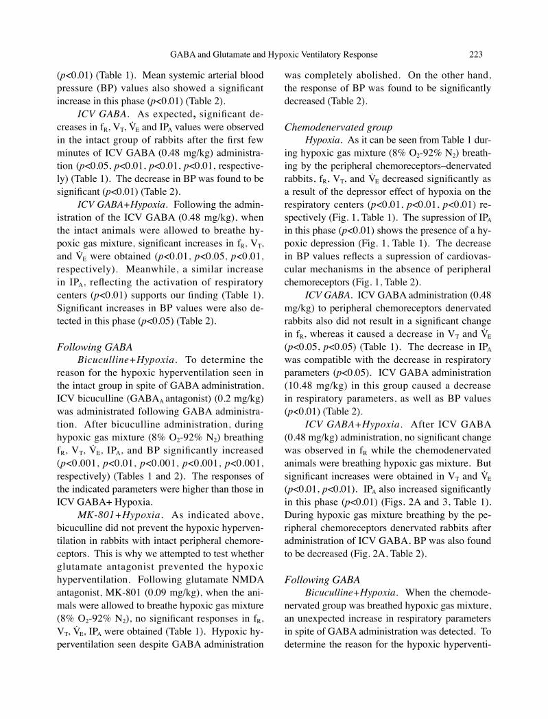
Chemodenervated groupHypoxia. As it can be seen from Table 1 dur-
ing hypoxic gas mixture (8% O2-92% N2) breath-ing by the peripheral chemoreceptors–denervated rabbits, fR, VT, and VE decreased significantly as a result of the depressor effect of hypoxia on the respiratory centers (p<0.01, p<0.01, p<0.01) re-spectively (Fig. 1, Table 1). The supression of IPA in this phase (p<0.01) shows the presence of a hy-poxic depression (Fig. 1, Table 1). The decrease in BP values reflects a supression of cardiovas-cular mechanisms in the absence of peripheral chemoreceptors (Fig. 1, Table 2).
ICV GABA. ICV GABA administration (0.48 mg/kg) to peripheral chemoreceptors denervated rabbits also did not result in a significant change in fR, whereas it caused a decrease in VT and VE (p<0.05, p<0.05) (Table 1). The decrease in IPA was compatible with the decrease in respiratory parameters (p<0.05). ICV GABA administration (10.48 mg/kg) in this group caused a decrease in respiratory parameters, as well as BP values (p<0.01) (Table 2).
ICV GABA+Hypoxia. After ICV GABA (0.48 mg/kg) administration, no significant change was observed in fR while the chemodenervated animals were breathing hypoxic gas mixture. But significant increases were obtained in VT and VE
(p<0.01, p<0.01). IPA also increased significantly in this phase (p<0.01) (Figs. 2A and 3, Table 1). During hypoxic gas mixture breathing by the pe-ripheral chemoreceptors denervated rabbits after administration of ICV GABA, BP was also found to be decreased (Fig. 2A, Table 2).
Following GABABicuculline+Hypoxia. When the chemode-
nervated group was breathed hypoxic gas mixture, an unexpected increase in respiratory parameters in spite of GABA administration was detected. To determine the reason for the hypoxic hyperventi-
N.K. Yelmen
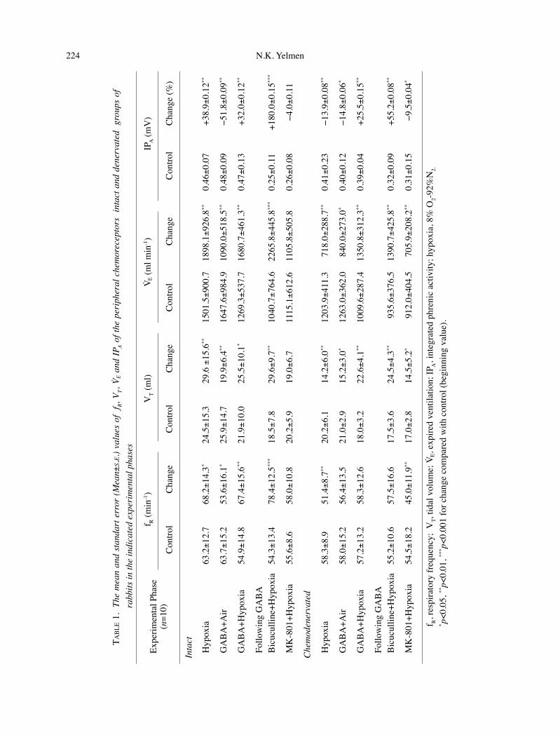
224 GABA and Glutamate and Hypoxic Ventilatory Response 225
TAB
LE 1
. Th
e m
ean
and
stan
dart
err
or (M
ean±
S.E.)
valu
es o
f f R,
V T, V
E an
d IP
A of
the
peri
pher
al c
hem
orec
epto
rs i
ntac
t and
den
erva
ted
gro
ups
of
rabb
its in
the
indi
cate
d ex
peri
men
tal p
hase
s
Expe
rimen
tal P
hase
(n=1
0)
f R (m
in-1)
VT (
ml)
V E (m
l min
-1)
IPA (m
V)
Con
trol
Cha
nge
Con
trol
Cha
nge
Con
trol
Cha
nge
Con
trol
Cha
nge
(%)
Inta
ct
Hyp
oxia
63.2±1
2.7
68.2±1
4.3*
24.5±1
5.3
29.6
±15
.6**
1501
.5±9
00.7
1898
.1±9
26.8
**0.
46±0
.07
+38
.9±0
.12**
GA
BA
+Air
63.7±1
5.2
53.6±1
6.1*
25.9±1
4.7
19.9±6
.4**
1647
.6±9
84.9
1090
.0±5
18.5
**0.
48±0
.09
−51
.8±0
.09**
GA
BA
+Hyp
oxia
54.9±1
4.8
67.4±1
5.6**
21.9±1
0.0
25.5±1
0.1*
1269
.3±5
37.7
1680
.7±4
61.3
**0.
47±0
.13
+32
.0±0
.12**
Follo
win
g G
AB
AB
icuc
ullin
e+H
ypox
ia54
.3±1
3.4
78.4±1
2.5**
*18
.5±7
.829
.6±9
.7**
1040
.7±7
64.6
2265
.8±4
45.8
***
0.25±0
.11
+180
.0±0
.15**
*
MK
-801
+Hyp
oxia
55.6±8
.658
.0±1
0.8
20.2±5
.919
.0±6
.711
15.1±6
12.6
1105
.8±5
05.8
0.26±0
.08
−
4.0±
0.11
Che
mod
ener
vate
d
Hyp
oxia
58.3±8
.951
.4±8
.7**
20.2±6
.114
.2±6
.0**
1203
.9±4
11.3
718
.0±2
88.7
**0.
41±0
.23
−13
.9±0
.08**
GA
BA
+Air
58.0±1
5.2
56.4±1
3.5
21.0±2
.915
.2±3
.0*
1263
.0±3
62.0
840
.0±2
73.0
*0.
40±0
.12
−14
.8±0
.06*
GA
BA
+Hyp
oxia
57.2±1
3.2
58.3±1
2.6
18.0±3
.222
.6±4
.1**
1009
.6±2
87.4
1350
.8±3
12.3
**0.
39±0
.04
+25
.5±0
.15**
Follo
win
g G
AB
AB
icuc
ullin
e+H
ypox
ia55
.2±1
0.6
57.5±1
6.6
17.5±3
.624
.5±4
.3**
935
.6±3
76.5
1390
.7±4
25.8
**0.
32±0
.09
+55
.2±0
.08**
MK
-801
+Hyp
oxia
54.5±1
8.2
45.0±1
1.9**
17.0±2
.814
.5±5
.2*
912
.0±4
04.5
705
.9±2
08.2
**0.
31±0
.15
−
9.5±
0.04
*
f R, r
espi
rato
ry fr
eque
ncy;
VT,
tidal
vol
ume;
VE,
expi
red
vent
ilatio
n; IP
A, i
nteg
rate
d ph
reni
c ac
tivity
; hyp
oxia
, 8%
O2-9
2%N
2.* p
<0.0
5, **
p<0.
01, **
* p<0
.001
for c
hang
e co
mpa
red
with
con
trol (
begi
nnin
g va
lue)
.
N.K. Yelmen
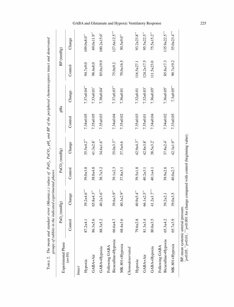
224 GABA and Glutamate and Hypoxic Ventilatory Response 225
TAB
LE 2
. Th
e m
ean
and
stan
dart
err
or (
Mea
n±S.E
.) va
lues
of
PaO
2, Pa
CO
2, pH
a and
BP
of th
e pe
riph
eral
che
mor
ecep
tors
inta
ct a
nd d
ener
vate
d gr
oups
of r
abbi
ts in
the
indi
cate
d ex
peri
men
tal p
hase
s
Expe
rimen
tal P
hase
(n=1
0)
PaO
2 (m
mH
g)Pa
CO
2 (m
mH
g)pH
aB
P (m
mH
g)
Con
trol
Cha
nge
Con
trol
Cha
nge
Con
trol
Cha
nge
Con
trol
Cha
nge
Inta
ct
Hyp
oxia
87.2±4
.139
.2±4
.6**
*39
.0±3
.835
.5±4
.2**
7.34±0
.05
7.37±0
.04**
94.
7±9.
010
9.0±
8.0**
GA
BA
+Air
86.3±5
.665
.8±4
.3**
38.0±4
.841
.3±2
.8*
7.35±0
.05
7.33±0
.01*
98.
4±8.
0 6
9.0±
11.9
**
GA
BA
+Hyp
oxia
88.3±5
.240
.2±3
.6**
*38
.7±3
.234
.6±1
.6**
7.35±0
.03
7.38±0
.04*
85.
0±19
.910
0.2±
15.0
*
Follo
win
g G
AB
AB
icuc
ullin
e+H
ypox
ia66
.6±4
.739
.6±3
.9**
39.1±2
.335
.0±3
.3**
7.34±0
.04
7.37±0
.03**
75.
0±9.
212
7.6±
12.5
***
MK
-801
+Hyp
oxia
68.4±3
.640
.3±2
.9**
37.8±3
.137
.5±4
.67.
35±0
.02
7.36±0
.01
70.
0±16
.5 5
0.5±
9.0**
Che
mod
ener
vate
d
Hyp
oxia
79.6±2
.840
.9±5
.4**
39.5±1
.842
.9±4
.3**
7.35±0
.03
7.32±0
.01
116.
5±27
.1 9
3.2±
23.8
**
GA
BA
+Air
81.3±3
.466
.1±2
.5**
40.2±3
.142
.8±3
.8*
7.35±0
.01
7.32±0
.03*
124.
2±17
.5 9
5.5±
22.5
**
GA
BA
+Hyp
oxia
80.6±3
.541
.2±1
.7**
*40
.1±4
.138
.5±3
.2*
7.34±0
.04
7.36±0
.05*
111.
5±23
.0 7
5.5±
15.2
**
Follo
win
g G
AB
AB
icuc
ullin
e+H
ypox
ia65
.3±4
.239
.2±2
.139
.9±2
.637
.8±2
.4*
7.34±0
.02
7.36±0
.05*
85.
8±17
.313
5.9±
22.5
***
MK
-801
+Hyp
oxia
65.7±3
.939
.0±3
.540
.0±2
.742
.3±1
.9**
7.33±0
.05
7.3±0
.05**
90.
7±19
.2 5
5.0±
23.4
***
BP,
mea
n sy
stem
ic a
rteria
l pre
ssur
e.* p<
0.05
, **p<
0.01
, *** p<
0.00
1 fo
r cha
nge
com
pare
d w
ith c
ontro
l (be
ginn
ing
valu
e).
N.K. Yelmen
226 GABA and Glutamate and Hypoxic Ventilatory Response 227
lation, ICV bicuculline (0.2 mg/kg), the GABA-A
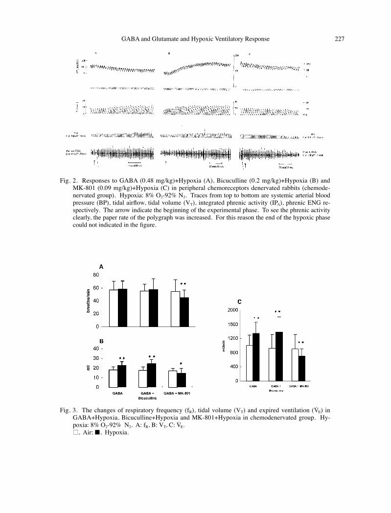
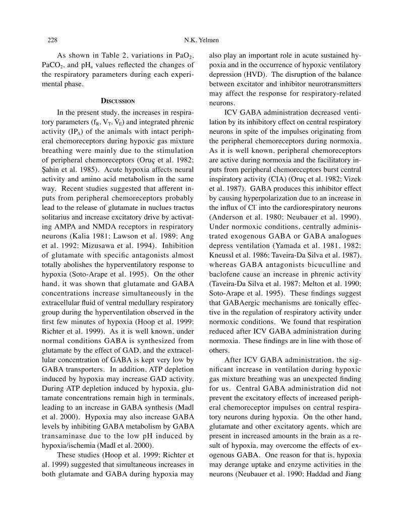
antagonist, was administrated and hypoxic phase was repeated. During hypoxic gas mixture (8% O2-92% N2) breathing after bicuculline adminis-tration, VT, VE, IPA, and BP significantly increased (p<0.01, p<0.01, p<0.01, p<0.001, respectively) (Figs. 2B and 3, Tables 1 and 2). There was no significant change in fR (Fig. 3, Table 1).
MK-801+Hypoxia. As shown above, be-cause bicuculline did not eliminate the hypoxic hyperventilation in the chemodenervated rabbits as in the intact group, glutamate NMDA antago-
nist (MK-801) (0.09 mg/kg) was used to inves-tigate the reason for this response. Following glutamate antagonist, MK-801, when the che-modenervated animals were allowed to breathe hypoxic gas mixture (8% O2-92% N2), significant decreases in fR, VT, VE, and IPA were obtained (p<0.01, p<0.01, p<0.05, p<0.05, respectively) (Figs. 2C and 3, Table 1). Hypoxic hyperventila-tion seen despite GABA administration was com-pletely abolished as in the intact group. There was a significant decrease in BP (p<0.001) (Fig. 2C, Table 2).
Fig. 1. Responses to hypoxia in chemodenervated group. Hypoxia: 8% O2-92% N2. Traces from top to bottom are systemic arterial blood pressure (BP), tidal airflow, tidal volume (VT), integrated phrenic activity (IPA), phrenic electroneurogram (ENG) respectively. The arrow indicates the beginning of the hypoxic phase. The decreases in VT and IPA show hypoxic depression. To see the phrenic activ-ity clearly, the paper rate of the polygraph was increased. For this reason the end of the hypoxic phase could not indicated in the figure.
N.K. Yelmen
226 GABA and Glutamate and Hypoxic Ventilatory Response 227
Fig. 2. Responses to GABA (0.48 mg/kg)+Hypoxia (A), Bicuculline (0.2 mg/kg)+Hypoxia (B) and MK-801 (0.09 mg/kg)+Hypoxia (C) in peripheral chemoreceptors denervated rabbits (chemode-nervated group). Hypoxia: 8% O2-92% N2. Traces from top to bottom are systemic arterial blood pressure (BP), tidal airflow, tidal volume (VT), integrated phrenic activity (IPA), phrenic ENG re-spectively. The arrow indicate the beginning of the experimental phase. To see the phrenic activity clearly, the paper rate of the polygraph was increased. For this reason the end of the hypoxic phase could not indicated in the figure.
Fig. 3. The changes of respiratory frequency (fR), tidal volume (VT) and expired ventilation (VE) in GABA+Hypoxia, Bicuculline+Hypoxia and MK-801+Hypoxia in chemodenervated group. Hy-poxia: 8% O2-92% N2. A: fR, B: VT, C: VE.
□, Air;■, Hypoxia.
N.K. Yelmen
228 GABA and Glutamate and Hypoxic Ventilatory Response 229
As shown in Table 2, variations in PaO2, PaCO2, and pHa values reflected the changes of the respiratory parameters during each experi-mental phase.
DISCUSSION
In the present study, the increases in respira-tory parameters (fR, VT, VE) and integrated phrenic activity (IPA) of the animals with intact periph-eral chemoreceptors during hypoxic gas mixture breathing were mainly due to the stimulation of peripheral chemoreceptors (Oruç et al. 1982; Sahin et al. 1985). Acute hypoxia affects neural activity and amino acid metabolism in the same way. Recent studies suggested that afferent in-puts from peripheral chemoreceptors probably lead to the release of glutamate in nuclues tractus solitarius and increase excitatory drive by activat-ing AMPA and NMDA receptors in respiratory neurons (Kalia 1981; Lawson et al. 1989; Ang et al. 1992; Mizusawa et al. 1994). Inhibition of glutamate with specific antagonists almost totally abolishes the hyperventilatory response to hypoxia (Soto-Arape et al. 1995). On the other hand, it was shown that glutamate and GABA concentrations increase simultaneously in the extracellular fluid of ventral medullary respiratory group during the hyperventilation observed in the first few minutes of hypoxia (Hoop et al. 1999; Richter et al. 1999). As it is well known, under normal conditions GABA is synthesized from glutamate by the effect of GAD, and the extracel-lular concentration of GABA is kept very low by GABA transporters. In addition, ATP depletion induced by hypoxia may increase GAD activity. During ATP depletion induced by hypoxia, glu-tamate concentrations remain high in terminals, leading to an increase in GABA synthesis (Madl et al. 2000). Hypoxia may also increase GABA levels by inhibiting GABA metabolism by GABA transaminase due to the low pH induced by hypoxia/ischemia (Madl et al. 2000).
These studies (Hoop et al. 1999; Richter et al. 1999) suggested that simultaneous increases in both glutamate and GABA during hypoxia may
also play an important role in acute sustained hy-poxia and in the occurrence of hypoxic ventilatory depression (HVD). The disruption of the balance between excitator and inhibitor neurotransmitters may affect the response for respiratory-related neurons.
ICV GABA administration decreased venti-lation by its inhibitory effect on central respiratory neurons in spite of the impulses originating from the peripheral chemoreceptors during normoxia. As it is well known, peripheral chemoreceptors are active during normoxia and the facilitatory in-puts from peripheral chemoreceptors burst central inspiratory activity (CIA) (Oruç et al. 1982; Vizek et al. 1987). GABA produces this inhibitor effect by causing hyperpolarization due to an increase in the influx of Cl- into the cardiorespiratory neurons (Anderson et al. 1980; Neubauer et al. 1990). Under normoxic conditions, centrally adminis-trated exogenous GABA or GABA analogues depress ventilation (Yamada et al. 1981, 1982; Kneussl et al. 1986; Taveira-Da Silva et al. 1987), whereas GABA antagonists bicuculline and baclofene cause an increase in phrenic activity (Taveira-Da Silva et al. 1987; Melton et al. 1990; Soto-Arape et al. 1995). These findings suggest that GABAergic mechanisms are tonically effec-tive in the regulation of respiratory activity under normoxic conditions. We found that respiration reduced after ICV GABA administration during normoxia. These findings are in line with those of others.
After ICV GABA administration, the sig-nificant increase in ventilation during hypoxic gas mixture breathing was an unexpected finding for us. Central GABA administration did not prevent the excitatory effects of increased periph-eral chemoreceptor impulses on central respira-tory neurons during hypoxia. On the other hand, glutamate and other excitatory agents, which are present in increased amounts in the brain as a re-sult of hypoxia, may overcome the effects of ex-ogenous GABA. One reason for that is, hypoxia may derange uptake and enzyme activities in the neurons (Neubauer et al. 1990; Haddad and Jiang
N.K. Yelmen
228 GABA and Glutamate and Hypoxic Ventilatory Response 229
1993). The increase in exogenous GABA may prevent the rise in endogenous GABA during hy-poxia. The increase in the GABA content of the brain may prevent the conversion of glutamate to GABA by inhibiting GAD activity, thereby caus-ing an increase in glutamate. Excitatory and in-hibitory synaptic connections both have a role in the regulation of the respiratory rhythm (Richter et al. 1999). It is known that reciprocal inhibitory synapses are a characteristic of the respiratory circuit. Administration of GABA along with hy-poxic stimulation may produce a balance between inhibitory and excitatory neurotransmitters, how-ever it does not cause a disruption in the hypoxic response, as in the present study. The increase in glutamate and GABA in the phase in which venti-lation increases during hypoxia (Hoop et al. 1999; Richter et al. 1999) and the increase in GABA in vivo and in vitro in hypoxia (Wood et al. 1968; Iversen et al. 1983; Madl et al. 2000) also support this unexpected finding. Thus, the hypothesis that glutamate is solely responsible for the in-crease in acute sustained hypoxic response while GABA is responsible of acute sustained hypoxic depression (Chiang et al. 1986; McCrimmon et al. 1989; Kazemi and Hoop 1991) seems to weaken.
In the rabbits with denervated peripheral che-moreceptors, acute hypoxia gave rise to respira-tory depression, as expected. As it is well known, peripheral chemoreceptors are essential in the hypoxic ventilatory response. Chemodenervation abolishes the hypoxic hyperventilatory response. Hence, hypoxia depresses ventilation following chemodenervation in many species (Robbins 1995; Weil 1999). On the other hand, the oxygen-ation of the respiratory centers plays an important role in the regulation of VT in the absence of impulses originating from the peripheral chemo-receptors during hypoxia (Oruç et al. 1982).
After ICV GABA administration, when the peripheral chemoreceptors-denervated animals were allowed to breathe hypoxic gas mixture, in-creased ventilation was another unexpected find-ing. In our experiment, GABA was injected into the lateral ventricle. This administration method
may supress not only the medulla oblongata but also higher brainstem and other brain regions. It was reported that supramedullary brain regions are involved in hypoxic ventilatory depression (Monin et al. 1999) and send inhibitory signals to the medulla oblongata during hypoxic expo-sure due to release and local accumulation of inhibitory neurotransmitters (GABA, dopamine, adenosine). Under these conditions, injection of GABA into the lateral ventricle may suppress hypoxia- induced ventilatory inhibitory mecha-nisms of the midbrain and pons; this suppression may reverse hypoxic ventilatory depression and even increase ventilation during hypoxia. But in our experiment, the hypoxic hyperventilation seen after GABA administration occur not only in peripheral chemoreceptors intact animals, but also in peripheral chemoreceptors- denervated animals (Fig. 3). Therefore, the increased ventilation fol-lowing exogenous ICV GABA administration may be attributed either to GABA-evoked block of the cerebellar inhibitory input during hypoxia (Monin et al. 1999) or to the releasing of the glutamate by the effect of both exogenous GABA and hypoxia, independent of the stimulation of peripheral chemoreceptor afferents. On the other hand, the response of BP was found to be signifi-cantly decreased, as expected. The depression in BP in this phase shows again the importance of the impulses from the peripheral chemoreceptors on cardiovascular regulation.
In the present study, in order to identify better underlying mechanism of the increased hy-poxic ventilatory responses in spite of GABA administration in the intact and peripheral chem-oreceptors-denervated rabbits, the antagonists, GABAA (bicuculline) and glutamate NMDA (MK-801) were used. Following GABA admin-istration, when intact and peripheral chemodener-vated animals were breathed hypoxic gas mixture after ICV bicuculline administration, both VT, and IPA increased. Bicuculline is a well- studied an-tagonist of the GABAA receptor, which is linked to chloride gating. It decreases chloride flux, re-sulting in depolarization. These findings show hy-
N.K. Yelmen
230 GABA and Glutamate and Hypoxic Ventilatory Response 231
poxia- induced ventilatory inhibitor mechanisms are prevented because GABA administration in to the lateral ventricule is not responsible for the hy-poxic hyperventilation in our experimental proto-col. Furthermore, we did not observe a reduction in VT due to bicuculline administration during hy-poxia in both groups (Fig. 3). On the other hand, the increase in the GABA content of the brain in hypoxia may prevent the conversion of glutamate to GABA by inhibiting GAD activity, thereby causing an increase in glutamate. One can specu-late that GABA may influence respiration during hypoxia indirectly via other transmitter systems as it takes place in the neuronal network of the central nervous system. It was also suggested that the increased activity seen in hypoxia, at least partly, is also due to the increased release of glu-tamate by directly activating the ventral respira-tory neuron circuits, since early hypoxic increases in respiratory discharges also occur in peripheral chemodenervated animals (Richter et al. 1991). For this reason, the increase in ventilation with acute hypoxia in both groups in spite of GABA administration is thought to be mediated with such excitatory neurotransmitters as glutamate. In our study, when ICV glutamate antagonist (MK-801) was administrated, the hypoxic hyperventilatory responses seen in both groups were abolished. It was shown that acute hypoxia increases glutamate turnover (Hoop et al. 1999; Richter et al. 1999). Respiratory neurons receive glutamergic tonic inputs, which activate both NMDA and AMPA receptors (Haji et al. 2000). Furthermore, it was suggested that short-term potentiation is mediated by NMDA receptors, which augment EPSPs and prolong depolarization of phrenic motorneurons (McCrimmon et al. 1997). NMDA mechanisms are also responsible for phase transition from in-spiration to expiration. Thus, exogenous GABA may increase ventilation by increasing the level of glutamate in the brain, independent of peripheral chemoreceptors in hypoxia as shown in Fig. 3.
The responses of BP after bicuculline and MK-801 administrations show that cardiovascular mechanisms are under both GABAergic and glu-
tamatergic control during hypoxia.In conclusion, our findings suggest that
ICV GABA injection supresses cardiorespira-tory centers both in the presence and absence of peripheral chemoreceptors in accordance with its inhibitory characteristics in normoxic conditions. In contrast, ICV GABA causes an increase in ven-tilation during hypoxic conditions with or without peripheral chemoreceptor impulses by increasing glutamate which may be synthesized or released by the effect of GABA via a mechanism not clear-ly understood. For this reason, our results suggest that exogenous GABA can not be held responsible for acute hypoxic ventilatory depression. On the contrary, both GABA and glutamate may be re-sponsible for acute hypoxic hyperventilation.
AcknowledgmentsI gratefully thank Prof. Dr. G. Sahin and Prof.
Dr. T. Oruc for their advice and helpful comments. I also thank Dr. I. Güner for his technical assistance.
ReferencesAnderson, P., Dingledine, R., Gyerstad, L., Langmoen,
I. & Mosfeldt Laursen, A. (1980) Two different responses of hippocampal pyramidal cell to apli-cation of gamma-amino butyric acid. J. Physiol. (Lond)., 305, 279-296.
Ang, R.C., Hoop, B. & Kazemi, H. (1992) Role of glutamate as the central neurotransmitter in the hypoxic ventilatory response. J. Appl. Physiol., 72, 1480-1487.
Bennet, J.A., Mc William, P.N. & Shepheard, S.L. (1987) γ -amino butyric acid-mediated inhibi-tion of neurones in the nucleus tractus solitarius of the cat. J. Physiol. Lond., 392, 417-430.
Bureau, M.A., Zinman, R., Foulon, P. & Begin, R. (1984) Diphasic ventilatory response to hy-poxia in the newborn Lamb. J. Appl. Physiol., 56, 84-90.
Champagnat, J., Denavit-Saubie, M., Moyanova, S. & Rondouın, G. (1982) Involvement of amino ac-ids in periodic inhibitions of bulbar respiratory neurones. Brain Res., 237, 351-365.
Chiang, C.H., Pappas, P., Hoop, B. & Kazemi, H. (1986) Central cardiorespiratory effects of glutamate in dogs. J. Appl. Physiol., 60, 2056-2062.
Güner, I., Yelmen, N., Sahin, G. & Oruc, T. (2002)
N.K. Yelmen
230 GABA and Glutamate and Hypoxic Ventilatory Response 231
The effect of intracerebroventricular dopamine administration on the respiratory response to hypoxia. Tohoku. J. Exp. Med., 196, 219-230.
Haddad, G.G. & Jiang, C. (1993) O2 deprivation in the central nervous system. On mechanisms of neuronal response, differential sensitivity and injury. Progress. Neurobiol., 40, 277-318.
Haji, A., Takeda, R. & Okazaki, M. (2000) Neurophar-macology of control of respiratory rhythm and pattern in mature mammals. Pharmacol. Ther., 86, 277-304.
Hoop, B., Beagle, J.L., Maher, T.J. & Kazemi, H. (1999) Brainstem amino acid neurotransmitters and hypoxic ventilatory response. Respir. Physiol., 118, 117-129.
Iversen, K., Hedner, T. & Lundberg, P. (1983) GABA concentrations and turnover in neonatal rat brain during asphyxia and recovery. Acta Physiol. Scand., 118, 91-94.
Kalia, M.P. (1981) Anatomical organization of central respiratory neurons. Ann. Rev. Physiol., 43, 105-120.
Karaturan, N., Turgut, G., Sahin, G. & Oruç, T. (1996) Response of respiratory control mechanisms to isocapnic sustained moderate hypoxia. Eur. Respir. J., 23, 47.
Kazemi, H. & Hoop, B. (1991) Glutamic acid and γ -aminobutyric acid neurotransmitters in central control of breathing. J. Appl. Physiol., 70, 1-7.
Kneussl, M.P., Pappagianopoulos, P., Hoop, B. & Kazemi, H. (1986) Reversible depression of ventilation and cardiovascular function by ventriculocisternal perfusion with gamma-aminobutyric acid in dogs. Am. Rev. Respir. Dis., 133, 1024-1028.
Lawson, E.E., Richter, D.W., Ballantyne, D. & Lalley, P.M. (1989) Peripheral chemoreceptor inputs to medullary inspiratory and postinspiratory neurons of cats. Pflügers. Archiv ., 414, 523-533.
Madl, J.E. & Royer, S.M. (2000) Glutamate dependence of GABA levels in neurons of hypoxic and hypoglycemic rat hippocampal slices. Neurosci., 96, 657-664.
McCrimmon, O.R., Smith, J.C & Feldman, J.L. (1989) Involvement of excitatory amino acid in neurotransmission of inspiratory drive to spinal respiratory motoneuronal. J. Neurosci., 9, 1910-1921.
McCrimmon, D.R., Zuperku, E.J., Hayashi, F., Dogas, Z., Hinrichsen, C.F., Stuth, E.A., Tonkovic-Capin, M., Krolo, M. & Hopp, F.A. (1997)
Modulation of the synaptic drive to respiratory premotor and motor neurons. Respir. Physiol., 110, 161-176.
Melton, J.E., Neubauer, J.A. & Edelman, N.H. (1990) GABA antagonism reverses hypoxic respiratory depression in the cat. J. Appl. Physiol., 69, 1296-1301.
Mizusawa, A., Ogawa, H., Kikuchi, Y., Hida, W., Kurosawa, H., Okabe, S., Takishima, T. & Shirato, K. (1994) Invivo release of glutamate in nucleus tractus solitarii of the rat during hypoxia. J. Physiol., 478, 55-66.
Monin, P., Aranda, J., Bansal, R. & Trippenbach, T. (1999) Effect of increased brain GABA concentrations on breathing in unanesthetized newborn rabbits. Biol. Neonate., 76, 168-180.
Mueller, R.A., Lundberg, D.B.A., Breese, G.R., Hedner, J., Hedner, T. & Jonason, J. (1982) The neuropharmacology of respiratory control. Pharmacol. Rev., 34, 255-278.
Neubauer, J.A., Melton, J.E & Edelman, N.H. (1990) Modulation of respiration during brain hypoxia (Brief review). J. Appl. Physiol., 68, 441-451.
Oruç, T., Terzioğlu, M., Sahin, G. & Dursun, S. (1982) Response of central mechanisms to hyperoxia and hypoxia. Bull. Europ. Physiopath. Resp., 18, 439-447.
Richter, D.W., Bischoff, A., Anders, K., Bellingham, M. & Windhorst, U. (1991) Response of the medullary respiratory network of the cat to hypoxia. J. Physiol., 443, 231-256.
Richter, D.W., Schmidt-Garcon, P., Pierrefiche, O., Bischoff, A.M. & Lalley, P.M. (1999) Neurotransmitters and neuromodulators controlling the hypoxic respiratory response in anaesthetized cats. J. Physiol., 514, 567-578.
Robbins, P.A. (1995) Hypoxic ventilatory decline: site of action. J. Appl. Physiol., 79, 373-374.
Soto-Arape, I., Burton, M.D. & Kazemi, H. (1995) Central amino acid neurotransmitters and the hypoxic ventilatory response. Am. J. Respir. Crit. Care. Med., 151, 1113-1120.
Sahin, G. & Terzioğlu, M. (1985) The influence of chronic hypoxia on erythrocytic 2, 3-diphosphoglycerate and the sensitivity of peripheral chemoreceptors of rabbits. Cer. Med. Rev., 4, 46-56.
Taveira-Da Silva, A.M., Hartley, B., Hamosh, P., Quest, J.A. & Gillis, R. (1987) Respiratory depressant effect of GABA alpha and beta-receptor agonists in the cat. J. Appl. Physiol., 62, 2264-2272.
N.K. Yelmen
232
Vizek, M., Pickett, C.K. & Weil, J.V. (1987) Biphasic ventilatory response of adult cats to sustained hypoxia has central origin. J. Appl. Physiol., 63, 1658-1664.
Weil, J.V. (1999) Ventilatory responses to CO2 and hypoxia after sustained hypoxia in awake cats. J. Appl. Physiol., 76, 2251-2252.
Wood, J.D., Watson, W.J. & Drucker, A.J. (1968) The effect of hypoxia on brain gamma-amino butyric acid levels. J. Neurochem . , 15 , 603-608.
Wu, J.Y. (1976) Purification, characterization and
kinetic studies of GAD and GABA-T from mouse brain. In: GABA in Nervous System Function, edited by E. Roberts, T. Chase & D. Tower. New York: Raven, pp. 7-55.
Yamada, K.A., Hamosh, P. & Gillis, R.A. (1981) Respiratory depression produced by activation of GABA receptors in hindbrain of cat. J. Appl. Physiol., 5, 1278-1286.
Yamada, K.A., Norman, W.P., Hamosh, P. & Gillis, R.A. (1982) Medullary ventral surface GABA receptors affect respiratory and cardiovascular function. Brain Res., 248, 71-78.


![Rama Kamal GHB addiction treatment in the Netherlands · Present Naturally Precursor and metabolite of Gamma-aminobutyric acid [GABA] and visa versa. Extern precursors GBL and 1.4](https://img.pdfslide.us/doc/110x75/5af87c847f8b9a5f588cd54b/rama-kamal-ghb-addiction-treatment-in-the-naturally-precursor-and-metabolite-of.jpg)