Embed Size (px)
Citation preview

___________________________________________________________________________________________
*Corresponding author: Email: [email protected];
British Journal of Medicine & Medical Research3(4): 962-970, 2013
SCIENCEDOMAIN internationalwww.sciencedomain.org
The Role of Electron Microscopy in theAssessment of Dermatomyositis: A
Retrospective Pilot Study on Skeletal MuscleBiopsies
Hisham Alkhalidi1*
1Department of Pathology (32), College of Medicine, King Saud University, P.O. Box 2925,Riyadh 11461, Saudi Arabia.
Author’s contribution
The author performed the whole research work. Author HA wrote the first draft of the paper.Author HA read and approved the final manuscript.
Received 21st December 2012Accepted 3rd March 2013
Published 13th March 2013
ABSTRACT
Aims: To assess the contribution of electron microscopy in the process of musclebiopsies evaluation for dermatomyositis.Study Design: Retrospective review of muscle biopsy cases.Place and Duration of Study: Pathology Department of King Khalid University Hospital,King Saud University, Riyadh, Saudi Arabia from January 2008 to January 2012.Methodology: Samples from cases suspected to have dermatomyositis were reviewedfor light and ultrastructural morphological examination. Tubuloreticular inclusions (TRI)were considered present if these undulating tubules were detected in the endothelialcells of the capillaries.Results: Out of ten cases that were suspected for dermatomyositis, three cases showedclassical light microscopic features of dermatomyositis, two of which showed TRI.Among four cases with non-specific light microscopic features that can be seen indermatomyositis, TRI were detected in two of these four cases. Among three cases withnon-contributory light microscopy, TRI were found in all of these three cases.Conclusion: Electron microscopy -if feasible- may be useful in the screening of musclebiopsies, when clinically or morphologically suspected inflammatory myopathies areconsidered. Further studies to assess the significance of TRI with a larger number of
Research Article
British Journal of Medicine & Medical Research, 3(4): 962-970, 2013
963
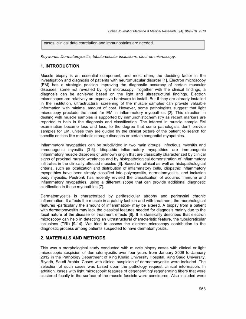
cases, clinical data correlation and immunostains are needed.
Keywords: Dermatomyositis; tubuloreticular inclusions; electron microscopy.
1. INTRODUCTION
Muscle biopsy is an essential component, and most often, the deciding factor in theinvestigation and diagnosis of patients with neuromuscular disorder [1]. Electron microscopy(EM) has a strategic position improving the diagnostic accuracy of certain musculardiseases, some not revealed by light microscopy. Together with the clinical findings, adiagnosis can be achieved based on the light and ultrastructural findings. Electronmicroscopes are relatively an expensive hardware to install. But if they are already installedin the institution, ultrastructural screening of the muscle samples can provide valuableinformation with minimal amount of cost. However, some pathologists suggest that lightmicroscopy preclude the need for EM in inflammatory myopathies [2]. This direction indealing with muscle samples is supported by immunohistochemistry as recent markers arereported to help in the diagnosis and classification. The interest in muscle sample EMexamination became less and less, to the degree that some pathologists don’t providesamples for EM, unless they are guided by the clinical picture of the patient to search forspecific entities like metabolic storage diseases or certain congenital myopathies.
Inflammatory myopathies can be subdivided in two main groups: infectious myositis andimmunogenic myositis [3-5]. Idiopathic inflammatory myopathies are immunogenicinflammatory muscle disorders of unknown origin that are classically characterized by clinicalsigns of proximal muscle weakness and by histopathological demonstration of inflammatoryinfiltrates in the clinically affected muscles [6]. Based on clinical as well as histopathologicalcriteria, such as localization and distribution of inflammatory cells, idiopathic inflammatorymyopathies have been simply classified into polymyositis, dermatomyositis, and inclusionbody myositis. Pestronk has recently revised the classification of acquired immune andinflammatory myopathies, using a different scope that can provide additional diagnosticclarification in these myopathies [7].
Dermatomyositis is characterized by perifascicular atrophy and perimysial chronicinflammation. It affects the muscle in a patchy fashion and with treatment, the morphologicalfeatures -particularly the amount of inflammation- may be altered. A biopsy from a patientwith dermatomyositis may lack the classical features needed for diagnosis mainly due to thefocal nature of the disease or treatment effects [8]. It is classically described that electronmicroscopy can help in detecting an ultrastructural characteristic feature, the tubuloreticularinclusions (TRI) [9-14]. We tried to assess the electron microscopy contribution to thediagnostic process among patients suspected to have dermatomyositis.
2. MATERIALS AND METHODS
This was a morphological study conducted with muscle biopsy cases with clinical or lightmicroscopic suspicion of dermatomyositis over four years from January 2008 to January2012 in the Pathology Department of King Khalid University Hospital, King Saud University,Riyadh, Saudi Arabia. Cases with clinical suspicion of dermatomyositis were included. Theselection of such cases was based upon the pathology request clinical information. Inaddition, cases with light microscopic features of degenerating/ regenerating fibers that wereclustered focally in the surface of the muscle fascicle were considered. Also included were

British Journal of Medicine & Medical Research, 3(4): 962-970, 2013
964
cases with mild chronic inflammation that was predominantly perimysial. The age, the sex,the creatine kinase level and the status of immunosuppressive recent therapy at the time ofthe biopsy were documented. The clinical diagnosis after the biopsy was recorded.
Samples from each case were submitted for light and ultrastructural examination. Threemicrons thick sections were made using the formalin fixed, paraffin embedded tissue of theskeletal muscle samples. They were stained using standard hematoxylin and eosin stainingprocedure (H&E). The different sections were studied under the optic routine microscope bya neuropathologist. The routine stains that were reviewed include Gomori trichrome, NADH,SDH, COX, PAS, ORO and the ATPases. Congo red was carefully assessed for thepresence of inclusions. The sample was considered “typical” on light microscopy if itexhibited perifascicular atrophy and perimysial chronic inflammation. The sample wasconsidered “not specific” if the light microscopy revealed random (rather than perifascicular)fiber atrophy with mild perimysial inflammation only. The sample was considered “nothelpful” if it was almost normal with no inflammation or minimal random atrophy only.
Tissues submitted for electron microscopy examination were fixed in 3% glutaraldehyde.Tissues were embedded in osmium tetroxide and semi-thin sections were stained withtoluidine blue. The adequacy of each sample was checked on the semi thin sections. Thethin sections were stained with uranyl acetate and lead citrate. Careful screening of thesamples skeletal muscle cells and blood vessels walls was performed. TRI were consideredpresent if these undulating tubules were identified in the endothelial cells of the capillaries.
3. RESULTS
A total of ten cases were retrieved (Table 1).
Table 1. Light microscopy and EM findings of 10 cases studied
Case no. Age Sex Clinical suspicion Light microscopy TRI on EM1 47 F Dermatomyositis Not specific Negative2 25 F Dermatomyositis Typical Negative3 63 F Myopathy Not specific Present4 38 F Dermatomyositis Not helpful Present5 29 M Dermatomyositis Typical Present6 34 M Myopathy Not specific Negative7 58 F Myopathy Typical Present8 30 M Dermatomyositis Not helpful Present9 33 F Dermatomyositis Not helpful Present10 45 F Dermatomyositis Not specific Present
Of these, three cases showed classical light microscopic features for dermatomyositis. Inparticular, these cases exhibited the perifascicular atrophy (Fig. 1). Two of these casescontained TRI on ultrastructural examination. Four cases showed non-specific morphologicalfeatures that can be seen in dermatomyositis (Fig. 2). TRI were detected in two out of thesefour cases (Figs. 3A and 3E). TRI were detected in three cases out of four (Figs. 3B, 3C and3D) where the light microscopy was not helpful (Fig. 4). None of the cases showed nuclearor cytoplasmic sarcoplasmic inclusions or pseudo-myelinic membranes.

British Journal of Medicine & Medical Research, 3(4): 962-970, 2013
965
Fig. 1. Muscle biopsy showing a characteristic perifascicular atrophy (arrow), which isa classical feature of dermatomyositis. The atrophic area exhibits myofiber necrosis
and regeneration , myofiber splitting, occasional internal nuclei and irregularsarcoplasmic vacuolation (H&E, X100)
Fig. 2. In this muscle biopsy, there was mild perivascular perimysial inflammation inaddition to random fiber atrophy (arrow) within the fascicle (H&E, X100)

British Journal of Medicine & Medical Research, 3(4): 962-970, 2013
966
Fig. 3. TRI (arrows) in endothelial cells lining capillaries. A) Case 3 (X4000). B) Case 4(X4000). C) Case 8 (X8000). D) Case 9 (X4000). E) Case 10 (X4000)
Fig. 4. Almost unremarkable muscle biopsy on light microscopy (H&E, X100)
Screening Trichrome and Congo red stained sections didn’t reveal the presence ofinclusions in any of these cases. NAD, SDH and COX showed uneven staining or lobulationin some patients (cases 2,3,5 and 7). Otherwise, they were non-contributory. ATPasesstains revealed type II atrophy in one of the patients who received steroids before the biopsy(case 3). PAS and ORO were non contributory.

British Journal of Medicine & Medical Research, 3(4): 962-970, 2013
967
Creatine kinase level was increased at the time of the biopsy three to ten folds the normalcontrol levels in seven patients (1,4-7,9 and 10). It was normal in two patients (2 and 3). Thedata about one of the patients (case 8) is not available. It was an outside referral for tissueassessment only. Six of the patients (cases 1,3,4,6,7 and 9) received an immunomodulatingtreatment prior to the biopsy (usually in the form of oral steroids). This is attributed due todelays in the muscle biopsy procedure. Three patients (cases 2,5 and 10) didn’t receive anytreatment. The data about one of the patients (case 8) was not available.
The clinical final diagnosis was dermatomyositis in 8 patients. One patient (case 6) wasfound to have scleroderma. One patient (case 8) clinical data was not available.
4. DISCUSSION
Several studies have described patients with myositis with pronounced muscle weaknessand fatigue but without detectable infiltration of inflammatory cells in muscle tissues [7,8]. Asmentioned above, it is classically described that the presence of TRI can be an importantclue to dermatomyositis. This could be useful in patients whose biopsies could not provideenough light microscopic features. Our findings suggest that without electron microscopy,some of these biopsies may not add much to the patient management course (cases 3,4,9and10). This is particularly true if the immunohistochemical stains are not available. Hence,with careful ultrastructural screening, these undulating tubules in the endothelial cells of thecapillaries served its use to assess a well-described feature in dermatomyositis. Suchfeature may help to support the diagnosis of dermatomyositis in the right clinical context. Ifan electron microscope is already available in the institution, screening for TRI could beapplied routinely on each sample, considering that the volume of the muscle tissue isappropriate.
However, TRI are not entirely specific to dermatomyositis. They can also be seen in somecollagen-vascular disease [15]. These include connective tissue disorders-related myositisthat can be induced by SLE, scleroderma and Sjogren syndrome [16]. They are also well-described in patients receiving zidovudine-associated myopathies [17-19]. TRI also havebeen rarely reported in inclusion body myositis [16,20]. This differential diagnosis is limitedand can be narrowed by clinical correlation. For example, inclusion body myositis ispredominantly a distal myopathy while dermatomyositis is a proximal one. Inclusion bodymyositis exhibits Congo red-positive inclusions on light microscopy, in addition tocharacteristic intranuclear and perinuclear filaments and perinuclear myelin figures onelectron microscopy.
Immunohistochemistry provides an important tool that can assist in the process of thediagnosis. The analysis of inflammatory cells, MHC class I expressions and MAC depositsmay help to rule in an idiopathic inflammatory myopathy [21,22]. This role can be limited bya) non-specificity of the procedure, b) the difficulty of optimization of some antibodies and c)the difficulty in reproducibility of the results. The major issues are the non-specificity of theprocedure and the difficulty of the optimization. In dermatomyositis, one of the commonantibodies used to evaluate the disease is MHC class I immunostain [23,24]. MHC class I isfound to be expressed in all classic forms of inflammatory myopathy [25,26]. MHC class I isreported to be not specific for categorization of inflammatory myopathies [22,27]. It isexpressed on any regenerating fibers [10], and multiple other disorders including xp21muscle dystrophy [10,26]. Similarly, MAC immunostaining was found to lack the ability todifferentiate the individual subtypes of inflammatory myopathy [22].

British Journal of Medicine & Medical Research, 3(4): 962-970, 2013
968
Our study suggests that electron microscopy may play a role in the screening of musclebiopsies, particularly in clinically suspected inflammatory myopathies. Further studiesassessing the clinical pattern and outcome and comparing TRI with theimmunohistochemical staining pattern are needed before assuming any solid conclusions.Considering the high cost of an electron microscope installation; screening for TRI could beconsidered practical if the institution has one already (e.g. for renal biopsies examination).
5. CONCLUSION
Electron microscopy (if feasible) may be useful in the screening of muscle biopsies,particularly when clinically suspected inflammatory myopathies are considered, since thepresence of TRI may aid in the diagnosis of dermatomyositis. Further studies to assess thesignificance of TRI with a larger number of cases, clinical data and immunostains arerequired.
CONSENT
Not applicable.
ETHICAL APPROVAL
Not applicable.
COMPETING INTERESTS
Authors have declared that no competing interests exist.
REFERENCES
1. Fernandez C, Figarella-Branger D, Meyronet D, Cassote E, Tong S, Pellissier JF.Electron microscopy in neuromuscular disorders. Ultrastruct Pathol. 2005;29:437-450.
2. Cenacchi G, Tarantino L, Corbu A, De Giorgi LB, Fanin M, Pegoraro E, et al. Electronmicroscopy and neuromuscular pathology. Basic Applied Myology. 2007;17(3&4):167-171.
3. Dalakas MC. Autoimmune muscular pathologies. Neurol Sci. 2004;25:S7-S8.4. Dalakas MC, Hohlfeld. Polymyositis and dermatomyositis. Lancet. 2003;362:971-982.5. Miller FW, Rider LG, Plotz PH, Isemberg DA, Oddis CV. Diagnostic criteria for
polymyositis and dermatomyositis. Lancet. 2003;362:1762-1763.6. Mantegazza R, Bernasconi P, Confalonieri P, Cornelio F. Inflammatory myopathies
and systemic disorders: a review of immunopathogenetic mechanisms and clinicalfeatures. J Neurol. 1997;244:277-287.
7. Pestronk A. Acquired immune and inflammatory myopathies: pathologic classification.Curr Opin Rheumatol. 2011;23(6):595-604.
8. Nyberg P, Wikman A-L, Nennessmo I, Lundberg I. Increased expression of interleukin-1 alpha and MHC class I in muscle tissue of patients with chronic, inactivepolymyositis and dermatomyositis. J Rheumatol. 2000;27:940-948.
9. Olsen NJ, Park JH. Inflammatory myopathies: issues in diagnosis and management.Arthritis Care Res. 1997;10:200-207.

British Journal of Medicine & Medical Research, 3(4): 962-970, 2013
969
10. Dubowitz V, Sewry CA. Muscle biopsy: a practical approach: expert consult; ThirdEdition. Saunders: Elsevier; 2006.
11. Peloro TM, Miller OF 3rd, Hahn TF, Newman ED. Juvenile dermatomyositis: aretrospective review of a 30-year experience. J Am Acad Dermatol. 2001;45(1):28-34.
12. Pourmand R. Inflammatory myopathies. J Clin Neuromuscul Dis. 2006;7(3):158-63.13. Greenberg SA. Proposed immunologic models of the inflammatory myopathies and
potential therapeutic implications. Neurology. 2007;69(21):2008-19.14. Carpenter S, Karpati G. Pathology of Skeletal Muscle. 2nd ed. Oxford:Oxford
University Press; 2001.15. Palungwachira P, Palungwachira P. Electron microscopy: application and progress in
diagnostic methods on skin disease. J Med Assoc Thai. 1993;76(10):564-9.16. Bronner IM, Hoogendijk JE, Veldman H, Ramkema M, van den Bergh Weerman MA,
Rozemuller AJ, de Visser M. Tubuloreticular structures in different types of myositis:implications for pathogenesis. Ultrastruct Pathol. 2008;32:123-6.
17. Pezeshkpour G, Illa I, Dalakas MC. Ultrastructural characteristics and DNAimmunocytochemistry in human immunodeficiency virus and zidovudine-associatedmyopathies. Hum Pathol. 1991;22:1281-8.
18. Lane RJ, McLean KA, Moss J, Woodrow DF. Myopathy in HIV infection: the role ofzidovudine and the significance of tubuloreticular inclusions. Neuropathol ApplNeurobiol. 1993;19:406-13.
19. Cupler EJ, Danon MJ, Jay C, Hench K, Ropka M, Dalakas MC. Early featuresof zidovudine-associated myopathy: histopathological findings and clinical correlations.Acta Neuropathol. 1995;90:1-6.
20. Katzberg HD, Munoz DG. Tubuloreticular inclusions in inclusion body myositis. ClinNeuropathol. 2010;29:262-6.
21. Choi JH, Park YE, Kim SI, Kim JI, Lee CH, Park KH, Kim DS. Differentialimmunohistological features of inflammatory myopathies and dysferlinopathy. JKorean Med Sci. 2009;24(6):1015-23.
22. Panicker JB, Chacko G, Patil AK, Alexander M, Muliyil J. Immunohistochemicaldifferentiation of inflammatory myopathies. Neurol India. 2011;59:513-20.
23. Pedrol E, Grau JM, Casademont J, Cid MC, Masanés F, Fernandez-Sola J, Urbano-Márquez A. Idiopathic inflammatory myopathies. Immunohistochemical analysis of themajor histocompatibility complex antigen expression, inflammatory infiltratesphenotype and activation cell markers. Clin Neuropathol. 1995;14:179-84.
24. Civatte M, Schleinitz N, Krammer P, Fernandez C, Guis S, Veit V, Pouget J, Harlé JR,Pellissier JF, Figarella-Branger D. Class I MHC detection as a diagnostic tool innoninformative muscle biopsies of patients suffering from dermatomyositis (DM).Neuropathol Appl Neurobiol. 2003;29:546-52.
25. Appleyard ST, Dunn MJ, Dubowitz V, Rose ML. Increased expression of HLA ABCclass I antigens by muscle disorders. Lancet. 1985;1:361-3.
26. Karpati G, Pouliot Y, Carpenter S. Expression of immunoreactive majorhistocompatibility complex products in human skeletal muscles. Ann Neurol.1988;23:64-72.

British Journal of Medicine & Medical Research, 3(4): 962-970, 2013
970
27. Englund P, Lindroos E, Nennesmo I, Klareskog L, Lundberg IE. Skeletal muscle fibersexpress major histocompatibility complex class II antigens independently ofinflammatory infiltrates in inflammatory myopathies. Am J Pathol. 2001;159:1263-73.
© 2013 Alkhalidi; This is an Open Access article distributed under the terms of the Creative Commons AttributionLicense (http://creativecommons.org/licenses/by/3.0), which permits unrestricted use, distribution, and reproductionin any medium, provided the original work is properly cited.
Peer-review history:The peer review history for this paper can be accessed here:
http://www.sciencedomain.org/review-history.php?iid=205&id=12&aid=1097