Embed Size (px)
Citation preview
University of Groningen
The role of effector T-cells in the pathogenesis op lupus nephritisDolff, Sebastian Conrad Johannes
IMPORTANT NOTE: You are advised to consult the publisher's version (publisher's PDF) if you wish to cite fromit. Please check the document version below.
Document VersionPublisher's PDF, also known as Version of record
Publication date:2011
Link to publication in University of Groningen/UMCG research database
Citation for published version (APA):Dolff, S. C. J. (2011). The role of effector T-cells in the pathogenesis op lupus nephritis Groningen: s.n.
CopyrightOther than for strictly personal use, it is not permitted to download or to forward/distribute the text or part of it without the consent of theauthor(s) and/or copyright holder(s), unless the work is under an open content license (like Creative Commons).
Take-down policyIf you believe that this document breaches copyright please contact us providing details, and we will remove access to the work immediatelyand investigate your claim.
Downloaded from the University of Groningen/UMCG research database (Pure): http://www.rug.nl/research/portal. For technical reasons thenumber of authors shown on this cover page is limited to 10 maximum.
Download date: 06-04-2018
CHAPTER 4
Increased expression of costimulatory markers CD134 and CD80 on IL-17 producing T-cells in
patients with systemic lupus erythematosus
Sebastian Dolff1,2*, Daniel Quandt1*, Benjamin Wilde1, Thorsten
Feldkamp1, Fan Hua1, Xin Cai1, Christof Specker3, Andreas Kribben1, Cees G.M. Kallenberg2, Oliver Witzke1
1 Department of Rheumatology and Clinical Immunology, University Medical Center Groningen, University of Groningen, The Netherlands 2 Department of Nephrology, University Hospital Essen, University Duisburg-Essen, Germany * these authors contributed equally
3 Department of Rheumatology and Clinical Immunology, Kliniken Essen Süd, Germany
Arthritis Res Ther. 2010; 12(4): R150.
Chapter 4
58
Abstract Objectives: There is growing evidence that interleukin-17 (IL-17) producing T-
cells are involved in the pathogenesis of systemic lupus erythematosus (SLE).
Previous studies showed that increased percentages of T-cell subsets
expressing the costimulatory molecules CD80 and CD134 are associated with
disease activity and renal involvement in SLE. The aim of this study was to
investigate the distribution and phenotypical characteristics of IL-17 producing
T-cells in SLE, in particular in patients with lupus nephritis, with emphasis on
the expression of CD80 and CD134.
Patients and Methods: Thirty-four patients (3 male, 31 female, mean age
41 ±15 years) fulfilling at least four of the American College of Rheumatology
(ACR) revised criteria for the diagnosis of SLE and 24 healthy controls were
enrolled. T-cells from the peripheral blood were analysed by fluorescence
activated cell sorting (FACS) for their expression levels of CD80, CD134 and
CCR6. In vitro stimulated CD3+IL17+ cells were also investigated for the
expression of these costimulatory markers. Finally, renal biopsies from SLE
patients were evaluated for the presence of CD134 expressing T-cells.
Results: Percentages of IL-17 expressing T-cells were significantly increased in
patients with active disease as compared to healthy controls (1.46 ±0.58 %
versus 0.93 ±0.30 %, p=0.007). The percentage of IL-17 producing T-cells was
correlated with disease activity as assessed by systemic lupus erythematosus
disease activity index (SLEDAI) (r=0.53, p=0.003). In patients, most of the IL-17
producing T-cells were confined to the CCR6+ T-cell subset (80 ±13 %).
Expression of CD80 and CD134 on the IL-17 producing T-cell subset was
higher in SLE than in healthy controls (HC) (CD134: 71.78 ±14.51 % versus
51.45 ±16.58 %, p=0.002; CD80: 25.5 ±14.99 % versus 14.99 ±5.74 %,
p=0.02). Also, patients with lupus nephritis expressed higher levels of CD134+
on CD3+IL-17+ cells as compared to HC (72.69 ±11.54 % versus 51.45
±16.58 %, p=0.006). Furthermore, renal biopsies of lupus nephritis patients
showed infiltration of CD134+ T cells.
CD134+ IL-17 producing T-cells in lupus nephritis
59
Conclusion: Percentages IL-17 expressing T-cells correlate with disease
activity. Further, these cells show increased expression of costimulatory
markers such as CD134 and CD80. The presence of CD134+ T-cells in renal
biopsies of lupus nephritis patients suggest that these cells migrate to the
kidney and might contribute to inflammatory processes through IL-17 secretion.
Chapter 4
60
Introduction Systemic lupus erythematosus (SLE) is a multiorgan autoimmune disease
characterized by an imbalanced T-cell homeostasis with a shift towards
activated effector T-cell subsets. Two major subsets of CD4+ T helper cells, Th1
and Th2, have been shown to be involved in the pathogenesis of SLE. Th1 cells
secrete interferon gamma (INF-γ) and are induced by IL-12, whereas Th2 cells
secrete IL-4, IL-5 and IL-13 and are induced by IL-4.1;2
More recently, another subset of cytokine producing T-cells, so called
Th17 cells producing the cytokine IL-17, were described. IL-17 exerts its
function through recruiting neutrophils and monocytes, upregulating local
chemokine expression, facilitating T-cell migration into tissues, and inducing
immune responses.3-6 There is increasing evidence that IL-17 producing T-cells
play an important role in various autoimmune diseases including multiple
sclerosis, psoriasis, rheumatoid arthritis, inflammatory bowel disease, ANCA
associated vasculitis and systemic lupus erythematosus.7-11
Recent studies demonstrated the importance of IL-17 produced by
different T-cell subsets such as CD4+ T-cells, CD8+ T-cells, CD3+CD4-CD8- T-
cells, and γδ T-cells in human SLE.12;13 Furthermore, Yang et al. reported an
association of IL17-producing T-cells and clinical features as disease activity
assessed by systemic lupus erythematodes disease activity index (SLEDAI).14
Studies investigating the role of IL-17 producing T-cells in the pathogenesis of
lupus nephritis (LN) are rare. However, studies in mice support the idea that IL-
17 may contribute to renal disease, in particular lupus nephritis.14-17 Crispin et
al. demonstrated infiltration of IL-17+ double negative T-cell in kidneys of lupus
nephritis patients. More recently, single-cell analysis of laser-microdissected
lupus nephritis sections showed a skewing towards IL-17.18 Further evidence
comes from urine analysis in lupus nephritis patients where IL-17 gene
expression was inversely correlated with disease activity.19 These studies
demonstrated the pivotal role of IL-17 cells in the pathogenesis of lupus
nephritis. In humans an overwhelming amount of IL-17 cells express the
CD134+ IL-17 producing T-cells in lupus nephritis
61
chemokine receptor CCR6.20 Therefore, CCR6 might be a useful phenotypic
marker for the analysis of IL-17 T-cells.
Previously, we reported an association of increased levels of
costimulatory markers on CD4+ cells with lupus nephritis.21 Especially,
expression of CD134 was associated with disease activity and renal
involvement, but a functional analysis of these CD134 expressing cells is still
lacking. As a member of the tumour necrosis factor (TNF) superfamily, CD134
(OX40) provides co-stimulatory signals upon ligation to the CD134Ligand.
Moreover, it is possible that CD134+ T-cells infiltrate kidneys and cause
inflammation after ligation with CD134L which has been shown to be present on
glomerular endothelial cells in SLE patients.15 Thus, CD134 might be a pivotal
surface marker to enable effector cell migration towards the kidney. The
significance of CD134 for effector functions has been shown by the observation
that treatment with a stimulatory anti-CD134 antibody enhances T-cell
expansion and differentiation to effector cells in mice.22;23 This stimulation,
apparently, promotes the secretion of IFN-γ and the upregulation of various
interleukin (IL)-receptors with might lead subsequently to cytokine-mediated
kidney cell damage.24 Remarkably, increased percentages of CD134
expressing T-cells as well as Th17 cells have been found amongst effector cells
in several autoimmune diseases such as Wegener’s Granulomatosis,
rheumatoid arthritis and myasthenia gravis.7;25-28
To elucidate the role and phenotype of IL-17 producing effector T-cells
in patients with systemic lupus erythematosus, in particular with lupus nephritis,
we investigated their presence and phenotypic characteristics in the present
study. We analysed not only peripheral blood but also tissue from patients with
lupus nephritis. We tested the hypothesis that increased expression of
costimulatory molecules on these cells may promote their infiltrations into the
kidney due to interaction with ligands on resident renal cells.
Chapter 4
62
Patients and methods Study population
34 patients (3 male, 31 female, mean age 41 ±15 years) fulfilling at least four of
the American College of Rheumatology (ACR) revised criteria for the diagnosis
of SLE and 24 healthy controls (mean age 46 ±14 years) were enrolled in the
study. 13 patients had lupus nephritis (WHO class II: 2 patients, class III: 1
patient, class IV: 5 patients, class V: 4 patients, unclassified: 2 patients) while
22 patients had no clinical evidence of lupus nephritis (absence of proteinuria
and/or glomerular hematuria). Clinical disease activity at the time of
measurement was assessed according to the systemic lupus erythematosus
disease activity index (SLEDAI) (mean SLEDAI 4 ±5). Patients with a SLEDAI ≥
4 were defined as active, 19 active and 15 inactive patients were included into
the study. 26 patients were treated with prednisone (mean 27 ±91 mg/d), 8
patients were without prednisone, and 23 patients were on a constant dose of
immunomodulating drugs (azathioprine (n=8), mycophenolate mofetil (n=10),
cyclosporine A (n=2), hydroxychloroquine (n=9), leflunomide (n=1), rituximab
(n=2)), 2 patients had no medication at the time of the study. 13 patients had
been treated with cyclophosphamide (mean dose: 2003 ±4193 mg) during their
disease course. Consecutive patients were included from the University
Hospital of Essen and the Medical Center Essen Süd. The study protocol was
approved by the institutional review board. All patients gave informed consent
for participation in this study.
Flow cytometry
Expression level of the surface molecules on lymphocytes was assessed by
four-colour surface staining. Phycoerythrin (PE), fluorenscein isothiocyanate
(FITC), peridin chlorophyll protein (PerCP) and Allophycocyanin (APC)-labelled
antibodies were used: CD3 (mouse IgG1, PerCP), CD4 (mouse IgG1, PerCP),
CD8 (mouse IgG1 FITC), CCR6 (mouse igG1, FITC), CD134 (mouse IgG1,
PE), CD80 (mouse IgG1, PE, R&D Systems, Wiesbaden, Germany) and IL-17
(mouse IgG1, APC). All antibodies were purchased from Becton Dickinson,
CD134+ IL-17 producing T-cells in lupus nephritis
63
Heidelberg, Germany, except for IL-17, purchased from eBioscience, San
Diego, USA, and CCR6 purchased from R&D Systems, Minneapolis, USA.
Appropriate isotype controls (Becton Dickinson, Heidelberg) were used. Briefly,
peripheral blood was stained with labelled monoclonal antibodies for 20 min. in
the dark at room temperature. The cell suspension was incubated with lysis
buffer for 15 min and prepared as indicated. Analysis was performed with a
fluorescence activated cell sorter (FACS) Calibur™ from Becton Dickinson.
Immunostaining for intracellular cytokines Peripheral blood mononuclear cells (PBMCs) of patients were separated by
standard Ficoll-Paque density gradient centrifugation. The cells were
resuspended in RPMI 1640 medium (Gibco BRL, Karlsruhe, Germany)
supplemented with 10 % heat inactivated fetal calf serum (Biochrom, Berlin,
Germany). The cells were cultured in the absence or presence of PMA (5 ng/ml)
and Ionomycin (1 µM) (Sigma-Aldrich, Seelze, Germany) for 5 hours. Cytokine
secretion was inhibited by Brefeldin A (Ebioscience, Frankfurt, Germany). Then
surface staining was performed with CD3, CD134, CD80, CCR6 and
appropriate isotype controls. Cells were fixed and permeabilized by using a
Cytofix/Cytoperm kit purchased from Becton Dickinson (Heidelberg, Germany).
Finally, the samples were intracellulary stained with IL-17 or an appropriate
isotype control.
Immunohistochemistry Renal biopsies were provided by the Institute of Pathology University Hospital of
Essen. Specimens were fixed in 10 % neutral buffered formaline and paraffin-
embedded. 5 µm thick sections were deparaffinized in xylene and rehydrated in
a series of ethanol with different concentrations (100 %, 95 %, 70 % and 50 %).
Citrate buffer pH 6.0 (Zytomed, Berlin) was applied for heat induced epitope
retrieval, followed by neutralization of endogenous peroxidase with 0.3 % H2O2.
Primary antibodies (CD3 obtained from DCS, Hamburg, Germany and CD134
obtained from Becton Dickinson) and HRP-conjugated secondary antibodies
Chapter 4
64
(Zytomed) were incubated on slides (each for 30 minutes) at room temperature.
Washing with PBS was performed after each incubation step. A DAB substrate
Kit (Zytomed) was used for visualization. Finally, the slides were slightly
counterstained with hematoxylin.
Immunofluorescence double staining Tissues were fixed, embedded in paraffin and sectioned as indicated above.
Epitope retrieval was performed with citrate buffer pH 6.0 (Zytomed). Primary
antibodies against CD3 (rabbit IgG1, DCS) and CD134 (mouse IgG1, Becton
Dickinson) were used and incubated for 60 minutes at room temperature
simultaneously. Secondary antibodies conjugated to Cy2 and Cy3 (Dianova,
Hamburg) were applied for 30 minutes. Finally, the slides were mounted with
Immu Mount™ (Thermo Fisher, Kehl).
Statistics All values are expressed as mean ±SD. Significance for the differences
between groups was determined by the Mann-Whitney U-test. Spearman´s rank
correlation test was applied for detecting correlations between different study
parameters. A p value less than 0.05 was considered significant.
CD134+ IL-17 producing T-cells in lupus nephritis
65
Results Expression of CCR6 and phenotypic features of CCR6+ peripheral blood
CD4+ cells
In order to characterise IL-17 producing cells with a suitable marker expressed
on the surface we analysed T-cells for the expression of CCR6.20 Peripheral
blood circulating CD4+CCR6+ cells were analysed in 28 patients and 11 healthy
individuals. There were no differences in the percentages of CD4+CCR6+ cells
between patients with SLE and healthy controls (19.74 ±8.57 % vs. 16.97
±5.67 %, p=0.34). Furthermore, there were no significant differences between
active (n=10) and inactive (n=18) patients (18.63 ±8.01 % vs. 20.36 ±9.04 %,
p=0.46) or between patients with or without lupus nephritis (22.39 ±8.05 % vs.
17.86 ±8.08 %, p=0.24). There were also no differences in of CCR6 expression
between patients with and without lupus nephritis and healthy controls (22.39
±8.05 % vs. 16.97 ±5.67 % and 17.86 ±8.08 % vs. 16.97 ±5.67 %, p=0.08 and
p=0.76, respectively). Active and inactive patients showed no difference in
levels of CCR6+ cells as compared to healthy controls (18.63 ±8.01 % vs. 16.97
±5.67 % and 20.36 ±9.04 % vs. 16.97 ±5.67 %, p=0.6 and p=0.31,
respectively).
The CCR6+ subset was analysed for the expression of costimulatory
markers CD80 and CD134. Remarkably, percentages of CD134 and CD80
expressing CCR6+ cells were significantly increased in patients with SLE in
comparison to healthy controls (CD134: 67.87 ±12.23 % vs. 59.27 ±8.18 %,
p=0.02; CD80: 26.46 ±12.28 % vs. 13.86 ±3.82 %, p=0.001).
Expression of costimulatory markers CD134 and CD80 on CD4+CCR6+
cells in active and inactive patients and patients with and without lupus
nephritis
Patients with and without active disease as assessed by SLEDAI, and patients
with and without lupus nephritis proven by renal biopsy, were analysed for
CD80 expression on CD4+CCR6+ cells. The percentages of CD80 expressing
on CD4+CCR6+ cells showed no difference between active and inactive patients
Chapter 4
66
(23.58 ±13.41 % vs. 28.06 ±11.07 %, p=0.37). There was a significant
difference between inactive SLE patients and healthy controls (28.06 ±11.07 %
vs. 13.86 ±3.82 %, p=0.0006). Expression of CD80 on CD4+CCR6+ cells in
active patients tended to be increased as compared to healthy controls (23.58
±13.41 % vs. 13.86 ±3.82 %, p=0.062). Patients with lupus nephritis showed
significantly higher levels of CD80 expression on CD4+CCR6+ cells in
comparison to healthy controls (22.13 ±10.58 % vs. 18.86 ±3.82 %, p=0.02).
The same applied for patients without lupus nephritis (30.66 ±12.75 % vs. 18.86
±3.82 %, p=0.0009). Comparing patients with lupus nephritis and patients
without lupus nephritis no difference could be observed (22.13 ±10.58 % vs.
30.66 ±12.75 %, p=0.11).
Expression of CD134 on CD4+CCR6+ cells was significantly higher in
patients with inactive disease compared to healthy controls (70.89± 10.35 % vs.
59.27 ±8.18 %, p=0.0097) but not different from active patients (70.89± 10.35 %
vs. 62.14 ±14.16 %, p=0.17). Patients with lupus nephritis expressed higher
levels of CD134 on peripheral blood CD4+ T-cells as compared to HC (68.08
±7.52 % vs. 59.27 ±8.18 %, p=0.027). Patients without lupus nephritis showed
no difference in comparison to patients with lupus nephritis and healthy
individuals (67.75 ±13.46 % vs. 68.08 ±7.52 % p=0.91; 67.75 ±13.46 % vs.
59.27 ±8.18 %, p=0.05).
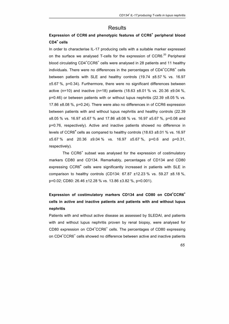
SLE Patients with active disease show increased levels of IL-17 producing
T-cells in peripheral blood
The percentage of IL-17 producing T-cells was analysed in the peripheral blood
of 30 SLE patients and 16 healthy controls. There was no significant difference
between SLE patients and healthy controls (1.17 ±0.61 % vs. 0.93 ±0.30 %;
p=0.37). Patients with active disease had significantly elevated levels of IL-17
expressing T-cells in the peripheral blood in comparison to healthy controls
(1.46 ±0.58 % vs. 0.93 ±0.30 %, p=0.007). Active patients had also increased
levels of IL17+ T-cells as compared to inactive patients. (1.46 ±0.58 % vs. 0.88
CD134+ IL-17 producing T-cells in lupus nephritis
67
±0.5 %, p=0.002) (Figure 1). The expression of IL-17 within CD8 cells revealed
an expression of 0.9 ±0.5 % (n=5).
Figure 1. (a) Percentages of IL-17 producing CD3+ cells in patients with SLE (n=30) and healthy controls (HC) (n=16). (b) A representative two colour immunofluorescence dot plot of CD3+ cells showing expression levels of IL-17 from an SLE patient and a healthy control. Cells positive for both antibodies are represented in the right upper quadrant with the percentage indicated. (c) Percentages of IL-17 producing CD3+ cells in active patients (n=15), inactive patients (n=15) and healthy controls (n=16). (d) Percentages of IL-17 producing CD3+ cells in patients with lupus nephritis (with LN) (n=13), patients without lupus nephritis (without LN) (n=14) and healthy controls (n=16). Data are presented as mean value. Significance was tested by the Mann-Whitney U-test. A p value less than 0.05 was considered significant.
a b
c d
Chapter 4
68
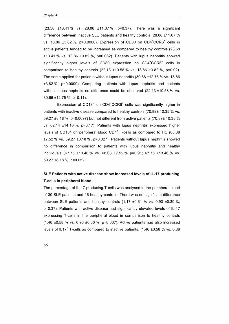
Ex vivo IL-17 production of CD3+ cells correlates with disease activity and
is independent of renal involvement
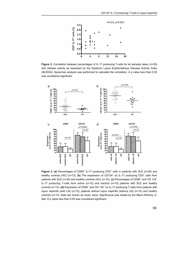
The percentage of IL-17 producing CD3+ cells correlated significantly with
disease activity (p=0.003, r=0.53) (Figure 2). To study the influence of
medication we compared the proportion of IL-17 expressing cells in patients on
prednisone alone versus patients on a combination of immunosuppressants.
There was no difference between these groups. This observation could be
confirmed in a follow-up in 12 patients. Changes over time (21 ±13 weeks) of IL-
17 expression were associated with changes in disease activity (r=0.81,
p=0.001). Expression of IL-17 in patients with lupus nephritis (n=13) was not
different as compared to patients without lupus nephritis (n=14) (1.29 ±0.65 %
vs. 1.08 ±0.57 %, p=0.34). A subanalysis revealed also no difference regarding
the the expression of IL-17 between patients with a class IV LN and patients
with class V (0.94 ±0.53% vs. 1.62 ±0.94%, p=0.25). There was also no
association with other histological features. Further, there was no correlation
between expression of IL-17 and anti-dsDNA titres or complement levels.
Patients with lupus nephritis showed no difference to healthy controls (0.93
±0.30 %, p=0.15).
Phenotypic features of IL-17+ T-cells in patients with SLE
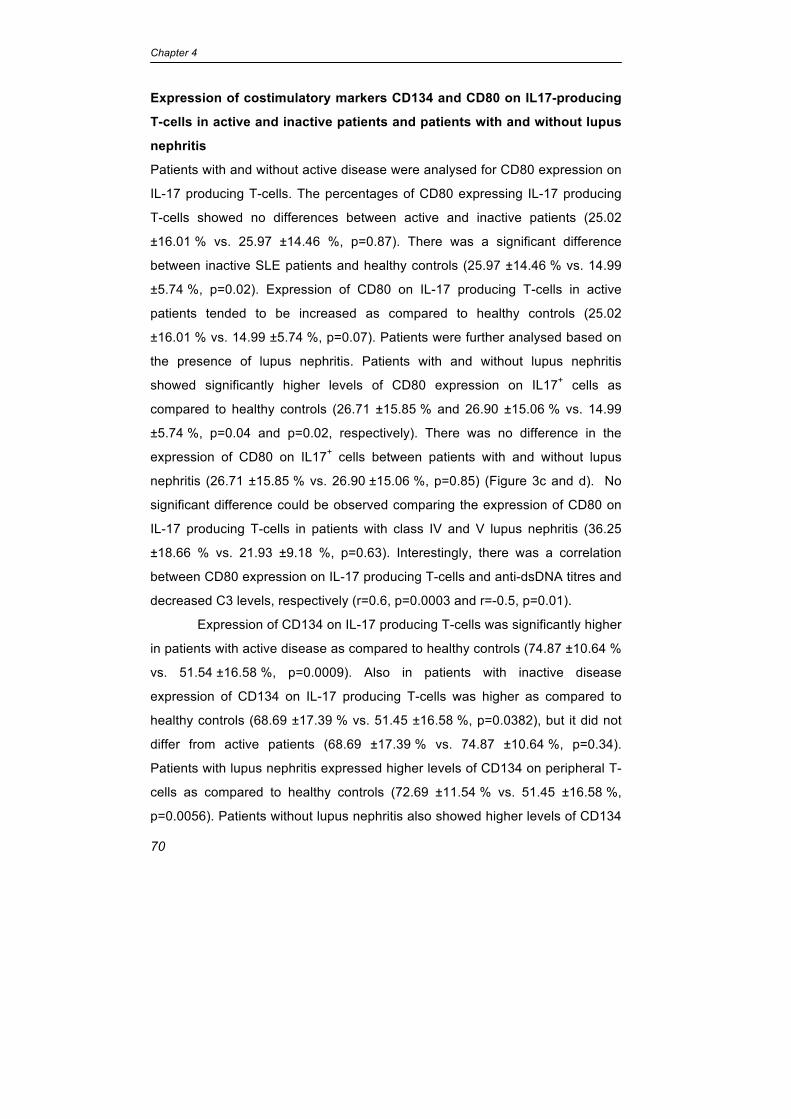
The expression of costimulatory markers CD80 and CD134 was analysed within
the IL17+ subset in patients and healthy controls. The percentages of CD134
and CD80 expressing IL17 producing T-cells were significantly increased in
SLE patients in comparison to healthy controls (CD134: 71.78 ±14.51 % vs.
51.45 ±16.58 %, p=0.002; CD80: 25.5 ±14.99 % vs. 14.99 ±5.74 %, p=0.02)
(figure 3a and b). A subanalysis in SLE patients (n=5) showed that only
1.7 ±1.8 % IL-17 producing T-cells were double positive for CD80 and CD134.
There was no significant correlation between the proportion of CD134 and
CD80 and IL-17 expression (p=0.8 and p=0.6, respectively).
CD134+ IL-17 producing T-cells in lupus nephritis
69
Figure 2. Correlation between percentages of IL-17 producing T-cells for all samples taken (n=30) and disease activity as assessed by the Systemic Lupus Erythematosus Disease Activity Index (SLEDAI). Spearman analysis was performed to calculate the correlation. A p value less than 0.05 was considered significant.
Figure 3. (a) Percentages of CD80+ IL-17 producing CD3+ cells in patients with SLE (n=30) and healthy controls (HC) (n=13). (b) The expression of CD134+ on IL-17 producing CD3+ cells from patients with SLE (n=30) and healthy controls (HC) (n=13). (c) Percentages of CD80+ and CD 134+ IL-17 producing T-cells form active (n=15) and inactive (n=15) patients with SLE and healthy controls (n=13). (d) Expression of CD80+ and CD 134+ on IL-17 producing T-cells from patients with lupus nephritis (with LN) (n=13), patients without lupus nephritis (without LN) (n=14) and healthy controls (n=13). Data are shown as mean value. Significance was tested by the Mann-Whitney U-test. A p value less than 0.05 was considered significant.
a b
c d
Chapter 4
70
Expression of costimulatory markers CD134 and CD80 on IL17-producing
T-cells in active and inactive patients and patients with and without lupus
nephritis
Patients with and without active disease were analysed for CD80 expression on
IL-17 producing T-cells. The percentages of CD80 expressing IL-17 producing
T-cells showed no differences between active and inactive patients (25.02
±16.01 % vs. 25.97 ±14.46 %, p=0.87). There was a significant difference
between inactive SLE patients and healthy controls (25.97 ±14.46 % vs. 14.99
±5.74 %, p=0.02). Expression of CD80 on IL-17 producing T-cells in active
patients tended to be increased as compared to healthy controls (25.02
±16.01 % vs. 14.99 ±5.74 %, p=0.07). Patients were further analysed based on
the presence of lupus nephritis. Patients with and without lupus nephritis
showed significantly higher levels of CD80 expression on IL17+ cells as
compared to healthy controls (26.71 ±15.85 % and 26.90 ±15.06 % vs. 14.99
±5.74 %, p=0.04 and p=0.02, respectively). There was no difference in the
expression of CD80 on IL17+ cells between patients with and without lupus
nephritis (26.71 ±15.85 % vs. 26.90 ±15.06 %, p=0.85) (Figure 3c and d). No
significant difference could be observed comparing the expression of CD80 on
IL-17 producing T-cells in patients with class IV and V lupus nephritis (36.25
±18.66 % vs. 21.93 ±9.18 %, p=0.63). Interestingly, there was a correlation
between CD80 expression on IL-17 producing T-cells and anti-dsDNA titres and
decreased C3 levels, respectively (r=0.6, p=0.0003 and r=-0.5, p=0.01).
Expression of CD134 on IL-17 producing T-cells was significantly higher
in patients with active disease as compared to healthy controls (74.87 ±10.64 %
vs. 51.54 ±16.58 %, p=0.0009). Also in patients with inactive disease
expression of CD134 on IL-17 producing T-cells was higher as compared to
healthy controls (68.69 ±17.39 % vs. 51.45 ±16.58 %, p=0.0382), but it did not
differ from active patients (68.69 ±17.39 % vs. 74.87 ±10.64 %, p=0.34).
Patients with lupus nephritis expressed higher levels of CD134 on peripheral T-
cells as compared to healthy controls (72.69 ±11.54 % vs. 51.45 ±16.58 %,
p=0.0056). Patients without lupus nephritis also showed higher levels of CD134
CD134+ IL-17 producing T-cells in lupus nephritis
71
on IL-17 producing T-cells in comparison to healthy individuals (70.27 ±17.18 %
vs. 51.45 ±16.58 %, p=0.01). No difference could be observed between
patients with and without lupus nephritis (72.69 ±11.54 % vs.70.27 ±17.18 %,
p=0.91) (Figure 3d). Further, expression of CD134 on IL-17 producing T-cells
showed no significant differences between class IV and V lupus nephritis (69.05
±10.97 % vs. 78.27 ±6.51 %, p=0.23).
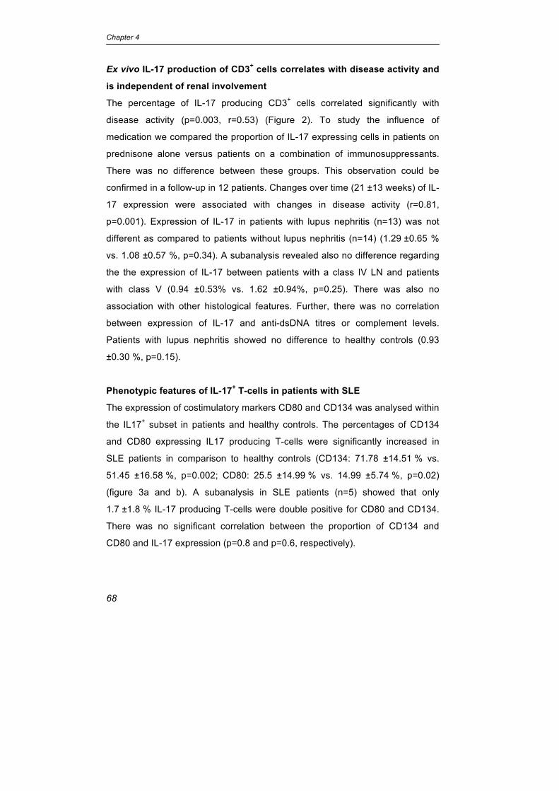
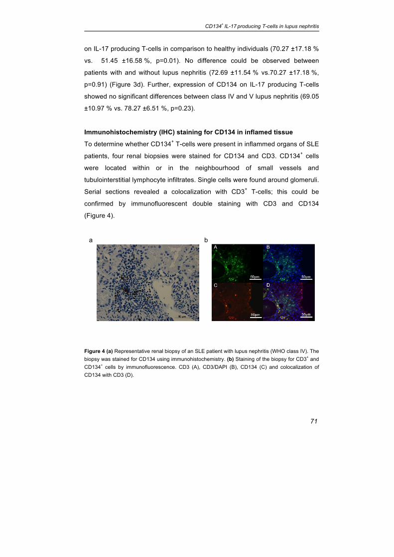
Immunohistochemistry (IHC) staining for CD134 in inflamed tissue
To determine whether CD134+ T-cells were present in inflammed organs of SLE
patients, four renal biopsies were stained for CD134 and CD3. CD134+ cells
were located within or in the neighbourhood of small vessels and
tubulointerstitial lymphocyte infiltrates. Single cells were found around glomeruli.
Serial sections revealed a colocalization with CD3+ T-cells; this could be
confirmed by immunofluorescent double staining with CD3 and CD134
(Figure 4).
Figure 4 (a) Representative renal biopsy of an SLE patient with lupus nephritis (WHO class IV). The biopsy was stained for CD134 using immunohistochemistry. (b) Staining of the biopsy for CD3+ and CD134+ cells by immunofluorescence. CD3 (A), CD3/DAPI (B), CD134 (C) and colocalization of CD134 with CD3 (D).
a b
Chapter 4
72
Discussion The results of this study suggest that IL-17 producing cells play a pivotal role in
the pathogenesis of systemic lupus erythematosus. In vitro stimulated CD3+
cells from active SLE patients produced significantly higher levels of IL-17 as
compared to healthy controls. Remarkably, only T-cells isolated from active
patients produced higher levels of IL-17 than controls. The association between
IL-17 production and disease activity was further supported by a significant
correlation of IL-17 expression and disease activity as assessed by SLEDAI.
These findings are in accordance with a large previous study by Yang et al. in
which 50 patients were enrolled.14 In addition, Wong et al. demonstrated
increased circulating plasma concentrations of IL-17 in SLE patients as
compared to healthy controls. In contrast to our study a correlation of plasma
levels of IL-17 and disease activity could only be found in patients without renal
disease.29 Both studies lack a detailed subanalysis of patients with and without
lupus nephritis. Therefore, a subanalysis was performed on the presence of
lupus nephritis in this study. Patients with biopsy proven lupus nephritis were
compared to patients without renal involvement. However, a significant
difference in the amount of IL17-producing peripheral CD3+ cells could not be
found between these groups which could be related to the rather small size and
heterogeneous composition of this group of biopsy proven lupus nephritis
patients. Moreover, the impact of immunosuppressive drugs remains uncertain
although we found no difference in patients on prednisone versus patients with
a combination of immunosuppressive drugs regarding either IL-17 expression
nor expression of costimulatory molecules (data not shown). There is growing
evidence in murine models that IL-17 plays a crucial role in the pathogenesis of
renal diseases such as lupus nephritis.16;17 These results can be explained by
the hypothetical migration of IL-17 cells into inflamed kidneys.
The present study reveals that IL-17 cells express significantly higher
amounts of the costimulatory markers CD80 and CD134. Ex vivo stimulation
has been shown to upregulate the expression of CD134 over time but the
significant difference between active patients and controls seem to be
CD134+ IL-17 producing T-cells in lupus nephritis
73
associated with disease activity according to our findings.30 In addition, we
detected CD134+ T-cells in lupus nephritis biopsies. These findings have been
confirmed by Zhou et al. in a larger analysis of 40 kidney biopsies.31 Possibly, in
humans these CD134+ T-cells infiltrate the kidney after ligation with CD134L
expressed by endothelial cells which could subsequently lead to IL-17 mediated
renal injury. Blocking CD134/CD134Ligand interaction as a therapeutic
intervention has been successfully used in lupus mice.30 However, the role of
IL-17 has not been investigated in that study. Interestingly, a recent report
investigating the influence of CD134/CD134Ligand interaction on IL-17 cytokine
production suggests that IL-17 production is downregulated after ligation of
CD134.32 This could be interpreted as a negative feedback loop for the effector
function of CD134+ cells but the detailed mechanisms remain unclear. An
important regulatory role of Th17 cells through the CD28/CD80 pathway was
also discussed in a murine model.33
Taken together we demonstrated that IL-17 producing cells are closely
linked to disease activity in SLE patients and express high levels of the
costimulatory markers CD80 and CD134. These new subsets of IL-17 cells
might be important in human lupus nephritis. The presence of CD134+ T-cells in
renal biopsies of lupus nephritis patients suggest that these cells migrate to the
kidney and might contribute to inflammatory processes through IL-17 secretion.
Further studies are necessary to dissect pathogenic role of IL-17 in lupus in
order to establish IL-17 as a therapeutic target in SLE.
Chapter 4
74
References 1. Abbas,A.K., K.M.Murphy, and A.Sher. 1996. Functional diversity of helper T
lymphocytes. Nature 383:787-793.
2. Viallard,J.F., J.L.Pellegrin, V.Ranchin, T.Schaeverbeke, J.Dehais, M.Longy-
Boursier, J.M.Ragnaud, B.Leng, and J.F.Moreau. 1999. Th1 (IL-2, interferon-
gamma (IFN-gamma)) and Th2 (IL-10, IL-4) cytokine production by peripheral
blood mononuclear cells (PBMC) from patients with systemic lupus
erythematosus (SLE). Clin. Exp. Immunol. 115:189-195.
3. Annunziato,F., L.Cosmi, V.Santarlasci, L.Maggi, F.Liotta, B.Mazzinghi, E.Parente,
L.Fili, S.Ferri, F.Frosali, F.Giudici, P.Romagnani, P.Parronchi, F.Tonelli, E.Maggi,
and S.Romagnani. 2007. Phenotypic and functional features of human Th17 cells.
J. Exp. Med. 204:1849-1861.
4. Laan,M., Z.H.Cui, H.Hoshino, J.Lotvall, M.Sjostrand, D.C.Gruenert, B.E.Skoogh,
and A.Linden. 1999. Neutrophil recruitment by human IL-17 via C-X-C chemokine
release in the airways. J. Immunol. 162:2347-2352.
5. Witowski,J., K.Pawlaczyk, A.Breborowicz, A.Scheuren, M.Kuzlan-Pawlaczyk,
J.Wisniewska, A.Polubinska, H.Friess, G.M.Gahl, U.Frei, and A.Jorres. 2000. IL-
17 stimulates intraperitoneal neutrophil infiltration through the release of GRO
alpha chemokine from mesothelial cells. J. Immunol. 165:5814-5821.
6. Woltman,A.M., S.de Haij, J.G.Boonstra, S.J.Gobin, M.R.Daha, and C.van Kooten.
2000. Interleukin-17 and CD40-ligand synergistically enhance cytokine and
chemokine production by renal epithelial cells. J. Am. Soc. Nephrol. 11:2044-
2055.
7. Abdulahad,W.H., C.A.Stegeman, P.C.Limburg, and C.G.Kallenberg. 2008.
Skewed distribution of Th17 lymphocytes in patients with Wegener's
granulomatosis in remission. Arthritis Rheum. 58:2196-2205.
8. Fitch,E., E.Harper, I.Skorcheva, S.E.Kurtz, and A.Blauvelt. 2007. Pathophysiology
of psoriasis: recent advances on IL-23 and Th17 cytokines. Curr. Rheumatol.
Rep. 9:461-467.
9. Kebir,H., K.Kreymborg, I.Ifergan, A.Dodelet-Devillers, R.Cayrol, M.Bernard,
F.Giuliani, N.Arbour, B.Becher, and A.Prat. 2007. Human TH17 lymphocytes
promote blood-brain barrier disruption and central nervous system inflammation.
Nat. Med. 13:1173-1175.
10. Schmechel,S., A.Konrad, J.Diegelmann, J.Glas, M.Wetzke, E.Paschos, P.Lohse,
B.Goke, and S.Brand. 2008. Linking genetic susceptibility to Crohn's disease with
Th17 cell function: IL-22 serum levels are increased in Crohn's disease and
CD134+ IL-17 producing T-cells in lupus nephritis
75
correlate with disease activity and IL23R genotype status. Inflamm. Bowel. Dis.
14:204-212.
11. Shen,H., J.C.Goodall, and J.S.Hill Gaston. 2009. Frequency and phenotype of
peripheral blood Th17 cells in ankylosing spondylitis and rheumatoid arthritis.
Arthritis Rheum. 60:1647-1656.
12. Crispin,J.C., M.Oukka, G.Bayliss, R.A.Cohen, C.A.Van Beek, I.E.Stillman,
V.C.Kyttaris, Y.T.Juang, and G.C.Tsokos. 2008. Expanded double negative T
cells in patients with systemic lupus erythematosus produce IL-17 and infiltrate
the kidneys. J. Immunol. 181:8761-8766.
13. Crispin,J.C. and G.C.Tsokos. 2009. Human TCR-alpha beta+ CD4- CD8- T cells
can derive from CD8+ T cells and display an inflammatory effector phenotype. J.
Immunol. 183:4675-4681.
14. Yang,J., Y.Chu, X.Yang, D.Gao, L.Zhu, X.Yang, L.Wan, and M.Li. 2009. Th17 and
natural Treg cell population dynamics in systemic lupus erythematosus. Arthritis
Rheum. 60:1472-1483.
15. Aten,J., A.Roos, N.Claessen, E.J.Schilder-Tol, I.J.Ten Berge, and J.J.Weening.
2000. Strong and selective glomerular localization of CD134 ligand and TNF
receptor-1 in proliferative lupus nephritis. J. Am. Soc. Nephrol. 11:1426-1438.
16. Paust,H.J., J.E.Turner, O.M.Steinmetz, A.Peters, F.Heymann, C.Holscher,
G.Wolf, C.Kurts, H.W.Mittrucker, R.A.Stahl, and U.Panzer. 2009. The IL-23/Th17
axis contributes to renal injury in experimental glomerulonephritis. J. Am. Soc.
Nephrol. 20:969-979.
17. Zhang,Z., V.C.Kyttaris, and G.C.Tsokos. 2009. The Role of IL-23/IL-17 Axis in
Lupus Nephritis. J. Immunol.
18. Wang,Y., S.Ito, Y.Chino, D.Goto, I.Matsumoto, H.Murata, A.Tsutsumi, T.Hayashi,
K.Uchida, J.Usui, K.Yamagata, and T.Sumida. 2010. Laser microdissection-based
analysis of cytokine balance in the kidneys of patients with lupus nephritis. Clin.
Exp. Immunol. 159:1-10.
19. Kwan,B.C., L.S.Tam, K.B.Lai, F.M.Lai, E.K.Li, G.Wang, K.M.Chow, P.K.Li, and
C.C.Szeto. 2009. The gene expression of type 17 T-helper cell-related cytokines
in the urinary sediment of patients with systemic lupus erythematosus.
Rheumatology. (Oxford) 48:1491-1497.
20. Singh,S.P., H.H.Zhang, J.F.Foley, M.N.Hedrick, and J.M.Farber. 2008. Human T
cells that are able to produce IL-17 express the chemokine receptor CCR6. J.
Immunol. 180:214-221.
Chapter 4
76
21. Patschan,S., S.Dolff, A.Kribben, J.Durig, D.Patschan, B.Wilde, C.Specker,
T.Philipp, and O.Witzke. 2006. CD134 expression on CD4+ T cells is associated
with nephritis and disease activity in patients with systemic lupus erythematosus.
Clin. Exp. Immunol. 145:235-242.
22. Lathrop,S.K., C.A.Huddleston, P.A.Dullforce, M.J.Montfort, A.D.Weinberg, and
D.C.Parker. 2004. A signal through OX40 (CD134) allows anergic, autoreactive T
cells to acquire effector cell functions. J. Immunol. 172:6735-6743.
23. Huddleston,C.A., A.D.Weinberg, and D.C.Parker. 2006. OX40 (CD134)
engagement drives differentiation of CD4+ T cells to effector cells. Eur. J.
Immunol. 36:1093-1103.
24. Williams,C.A., S.E.Murray, A.D.Weinberg, and D.C.Parker. 2007. OX40-mediated
differentiation to effector function requires IL-2 receptor signaling but not CD28,
CD40, IL-12Rbeta2, or T-bet. J. Immunol. 178:7694-7702.
25. Wilde,B., S.Dolff, X.Cai, C.Specker, J.Becker, M.Totsch, U.Costabel, J.Durig,
A.Kribben, J.W.Tervaert, K.W.Schmid, and O.Witzke. 2009. CD4+CD25+ T-cell
populations expressing CD134 and GITR are associated with disease activity in
patients with Wegener's granulomatosis. Nephrol. Dial. Transplant. 24:161-171.
26. Xiaoyan,Z., R.Pirskanen, V.Malmstrom, and A.K.Lefvert. 2006. Expression of
OX40 (CD134) on CD4+ T-cells from patients with myasthenia gravis. Clin. Exp.
Immunol. 143:110-116.
27. Giacomelli,R., A.Passacantando, R.Perricone, I.Parzanese, M.Rascente,
G.Minisola, and G.Tonietti. 2001. T lymphocytes in the synovial fluid of patients
with active rheumatoid arthritis display CD134-OX40 surface antigen. Clin. Exp.
Rheumatol. 19:317-320.
28. Chabaud,M., J.M.Durand, N.Buchs, F.Fossiez, G.Page, L.Frappart, and
P.Miossec. 1999. Human interleukin-17: A T cell-derived proinflammatory cytokine
produced by the rheumatoid synovium. Arthritis Rheum. 42:963-970.
29. Wong,C.K., L.C.Lit, L.S.Tam, E.K.Li, P.T.Wong, and C.W.Lam. 2008.
Hyperproduction of IL-23 and IL-17 in patients with systemic lupus erythematosus:
implications for Th17-mediated inflammation in auto-immunity. Clin. Immunol.
127:385-393.
30. Zhou,Y.B., R.G.Ye, Y.J.Li, and C.M.Xie. 2009. Targeting the CD134-CD134L
interaction using anti-CD134 and/or rhCD134 fusion protein as a possible strategy
to prevent lupus nephritis. Rheumatol. Int. 29:417-425.
CD134+ IL-17 producing T-cells in lupus nephritis
77
31. Zhou,Y.B., R.G.Ye, Y.J.Li, and C.M.Xie. 2007. Expression and role of CD134 and
NF-KB in rena tissue of lupus nephritis. Ann Rheum Dis Int. 66(Suppl II):318,
(abstract).
32. Li,J., L.Li, X.Shang, J.Benson, M.Merle Elloso, A.Schantz, M.Bracht, Y.Orlovsky,
and R.Sweet. 2008. Negative regulation of IL-17 production by OX40/OX40L
interaction. Cell Immunol. 253:31-37.
33. Bouguermouh,S., G.Fortin, N.Baba, M.Rubio, and M.Sarfati. 2009. CD28 co-
stimulation down regulates Th17 development. PLoS. One. 4:e5087.

Chapter 4
78
![Burden of Lupus Nephritis among Patients Managed in ... · Keywords: Lupus Nephritis, Burden, Flares, Hospitalizations, Europe . 1. ... compared with Caucasians (14%) [5].Approximately](https://img.pdfslide.us/doc/110x75/5f08e9627e708231d42452c4/burden-of-lupus-nephritis-among-patients-managed-in-keywords-lupus-nephritis.jpg)