Embed Size (px)
Citation preview
REVIEW
The role of echocardiography in the
diagnosis and management of patients
with pulmonary hypertensionG. Habib* and A. Torbicki#
ABSTRACT: Pulmonary hypertension (PH) is defined as an increased mean pulmonary artery
pressure (Ppa) .25 mmHg at rest as assessed by right heart catheterisation (RHC). However,
this technique is invasive and noninvasive alternatives are desirable for early diagnosis
of PH. Although estimation of systolic pulmonary arterial pressure is easily obtained using
Doppler echocardiography, cases of under- and over-estimations are not rare and direct
measurement of Ppa is not possible using this method. Therefore, echocardiography should
be considered as a tool for assessment of the likelihood rather than the definite presence or
absence of PH. Transthoracic echocardiography may be useful for noninvasive screening of
patients at risk of PH. On the basis of an echocardiographic assessment, patients showing
signs suggestive of PH can be referred for a confirmatory RHC. A number of variables measured
during echocardiography reflect the morphological and functional consequences of PH and
have prognostic value. The presence of pericardial effusion, reduced tricuspid annular plane
excursion and right atrial enlargement are associated with a poorer prognosis. Echocardiography
is also an important procedure for monitoring the response of patients to therapy, and is recom-
mended 3-4 months after initiation of, or a change in, therapy. Echocardiographic assessment as
part of a goal-oriented approach to therapy is essential for the effective management of PH
patients.
KEYWORDS: Diagnosis, echocardiography, prognosis, pulmonary hypertension
Definitive diagnosis of pulmonary hyper-tension (PH) requires an elevated meanpulmonary arterial pressure (Ppa) of
o25 mmHg at rest measured by right heartcatheterisation (RHC) [1]. Although RHC is nowa relatively safe procedure, it is invasive andimpractical to perform in patients for whom it isnot clearly indicated. Consequently there is aclear need for noninvasive procedures that aiddiagnosis and allow identification of patients forwhom diagnostic RHC is warranted, because of ahigh likelihood of the existence of PH.
Transthoracic echocardiography (TTE) is onesuch tool and is widely available and safe. Inaddition to its role in diagnosis, it can be used toscreen for high-risk patient populations, to assessprognosis and to monitor disease stability andresponse to treatment. These applications for TTEare presented in this article and the limitations ofthe procedure are discussed.
DIAGNOSTIC VALUE OFECHOCARDIOGRAPHY AND ITS ROLE INSCREENING PATIENTS AT RISK OFDEVELOPING PULMONARY ARTERIALHYPERTENSIONAssessment of pulmonary arterial pressureA number of echocardiographic variables corre-late with right heart haemodynamics, includ-ing pulmonary arterial pressure (Ppa), and aretherefore of interest in the diagnosis of PH.Noninvasive assessment of Ppa has always beendifficult. Most of the methods used to estimatePpa before the era of Doppler echocardiographywere indirect. Such methods took advantage ofthe detectable consequences of right ventricularpressure overload on right ventricular morphol-ogy and function. For example, echocardio-graphic measurement of right ventricular end-diastolic dimension indexed for body surfacearea was reported to correlate with Ppa inpatients with chronic PH, but this correlation
AFFILIATIONS
*Hopital La Timone, Marseille,
France.#Institute of Tuberculosis and Lung
Diseases, Warsaw, Poland.
CORRESPONDENCE
G. Habib
Service de Cardiologie
Hopital La Timone
Boulevard Jean Moulin
13005 Marseille
France
E-mail: [email protected]
Received:
Aug 24 2010
Accepted after revision:
Sept 11 2010
PROVENANCE
Publication of this peer-reviewed
article was supported by Actelion
Pharmaceuticals Ltd, Switzerland
(unrestricted grant, European
Respiratory Review issue 118).
European Respiratory Review
Print ISSN 0905-9180
Online ISSN 1600-0617
288 VOLUME 19 NUMBER 118 EUROPEAN RESPIRATORY REVIEW
Eur Respir Rev 2010; 19: 118, 288–299
DOI: 10.1183/09059180.00008110
Copyright�ERS 2010
was not sufficiently robust to allow for Ppa estimation inindividual patients [2–4]. Moreover, right ventricular dilationcould be caused by its primary failure or increased preload.
Two principal approaches based on Doppler echocardiogra-phy were suggested to predict Ppa; assessment of rightventricular systolic and diastolic time intervals and measure-ments of peak velocities of valvular regurgitations and shuntsaffecting the right heart. Initial reports from clinical trials wereenthusiastic both for pulsed wave Doppler estimates of Ppa
based particularly on the interval from the start to peakvelocity of right ventricular ejection (acceleration time (AcT))as well as continuous wave Doppler predictions of systolic anddiastolic pressures derived from tricuspid and pulmonaryvalvular regurgitation velocity, respectively.
AcT was reported to decrease with increasing Ppa [5]. Inaddition, mid-systolic deceleration of right ventricular ejectioninto the pulmonary artery, whenever present, occurred earlierwith higher Ppa [6]. Reports suggested excellent correlationsbetween AcT and Ppa, particularly when measured simulta-neously [7], and offered regression equations for noninvasivepressure calculations. However, significant differencesbetween these equations precluded their universal application[8]. Moreover, acutely induced changes in Ppa were notaccurately reflected by respective changes in AcT [9]. Whileheart rate [10] and the presence of tricuspid regurgitationapparently did not affect AcT [11], several other factors did; atthe same level of Ppa AcT time tended to be longer in patientswith low cardiac index [12] and with increased pulmonaryflow due to pre-tricuspid shunts [11]. In addition, AcT tended tobe shorter with more distal Doppler sample volume positions[13, 14] in individuals with body surface area .2.0 m2 and inadults .30 yrs old [15]. Due to these limitations, the measure-ment of time intervals with pulsed wave Doppler as a method ofestimation of Ppa was practically eliminated from clinical practice,while their potential role in assessment of components of pulsatilehaemodynamics still remain largely unexplored [16, 17].
In contrast to time intervals, estimation of Ppa using contin-uous Doppler and jet velocity methods were based on simplehaemodynamic laws. The pressure gradient (PG) driving bloodthrough an orifice can be calculated according to the simplifiedBernoulli equation as PG54V2 (mmHg), where V representsthe peak jet velocity, not accounting for orifice geometry andblood viscosity [16, 18]. Consequently, the peak jet velocity ofpulmonary insufficiency [19] as well as peak flow velocitiesacross a ventricular septal defect [20, 21] or patent ductusarteriosus [22] allow for calculations of Ppa. However, tricuspidvalve regurgitant jet is the most widely used for this purpose[23]. With modern equipment it is detectable in the majority ofpatients and its Doppler spectrum can be enhanced byintravenous injection of echocardiographic contrast, furtherincreasing the number of tracings suitable for evaluation [24, 25].
For calculation of systolic Ppa (Ppa,syst), stenosis of rightventricular outflow must be excluded, and an estimate of theright atrial pressure (Pra) should be added to the tricuspidinsufficiency pressure gradient (TIPG): Ppa,syst 5 TIPG +estimated Pra. Estimation of Pra based on echocardiographicinferior vena cava (IVC) collapsibility index has beensuggested. This index is calculated by subtracting inspiratory
from expiratory IVC diameter, and dividing the result byexpiratory IVC diameter [26, 27].
While reported correlations between Ppa,syst and its Dopplerestimate were excellent, the standard error of estimationremained above 5 mmHg, affecting the precision of Ppa
estimation in an individual patient [28]. In an early landmarktrial, the reported inter- and intra-observer variabilities werelow [23], contrasting with both everyday clinical experienceand results of a recent re-evaluation of the method [29]. In arecent trial, although Ppa and Doppler measurements wereobtained within 1 h of each other in 65 clinically stable patientswith PH and there was no systematic over- or under-estimation of directly measured Ppa,syst with the tricuspid jetmethod, 95% limits of agreement showed a very broad range ofpotential error (+38.8 to -40.0 mmHg). Doppler echocardio-graphy resulted in over- or under-estimation of Ppa,syst bymore than 10 mmHg in 48% of cases [30]. Pressure under-estimation in 12 out of 15 patients exceeded 20 mmHg whilepressure overestimates exceeded 20 mmHg in six patients.Half of all the cases of Ppa,syst overestimation were related toPra overestimation by echocardiography but disagreementbetween systolic right ventricle-right atrial gradient by twomethods was also significant [30].
A correlation was observed between diastolic Ppa andDoppler-derived end-diastolic pressure gradient (r50.91)[31]. However, when directly compared in the same patients,Doppler-derived pressure calculations based on diastolicvelocities across the pulmonary valve were less accurate thanthose based on tricuspid jet velocity measurements (r50.83versus 0.98, respectively) [32].
Joint evaluation of tricuspid and pulmonary regurgitant jetsallows for complete reconstruction of the Ppa curve from whichPpa could be theoretically calculated [32]. A simplified strategybased on measurement of the peak (early) diastolic pulmonaryto right ventricular pressure gradient derived from theDoppler flow profiles has also been suggested [31].
Extrapolation of Ppa from the Doppler estimate of Ppa,syst
assessed from TIPG can also be considered. Data suggesting astrong correlation between Ppa,syst and Ppa, regardless of theclinical diagnosis of a patient, seem encouraging. Measuredwith high-fidelity tip catheters, systolic Ppa equalled0.61(Ppa,syst)+2 mmHg, with negligible error [33, 34]. Whethersuch direct measurement of Ppa,syst might be substituted in theequation by its Doppler estimate remains to be verified.
Echocardiographic diagnosis of PHNoninvasive diagnosis of PH is tempting but risky in view ofthe discussed limitations of all available echocardiographicand Doppler methods. Recent clinical practice guidelines havesuggested that echocardiographic conclusions should belimited to assignment of the level of probability of PH ratherthan confirmation or exclusion of its presence in individualsubjects [1].
One of the obvious problems with noninvasive diagnosis of PHis that echocardiography provides us in most cases with anestimate of Ppa,syst rather than Ppa, on which definition of PH isactually based (i.e. Ppa o25 mmHg at rest). The suggestedcriteria for the three levels of probability of PH acknowledged
G. HABIB AND A. TORBICKI REVIEW: ECHOCARDIOGRAPHY IN PH
cEUROPEAN RESPIRATORY REVIEW VOLUME 19 NUMBER 118 289
existing data on upper limits of Doppler-derived Ppa estimatesin normal people. It also attempted to identify a cut-off valuefor the Doppler estimate of Ppa,syst corresponding to the RHCdefinition of PH.
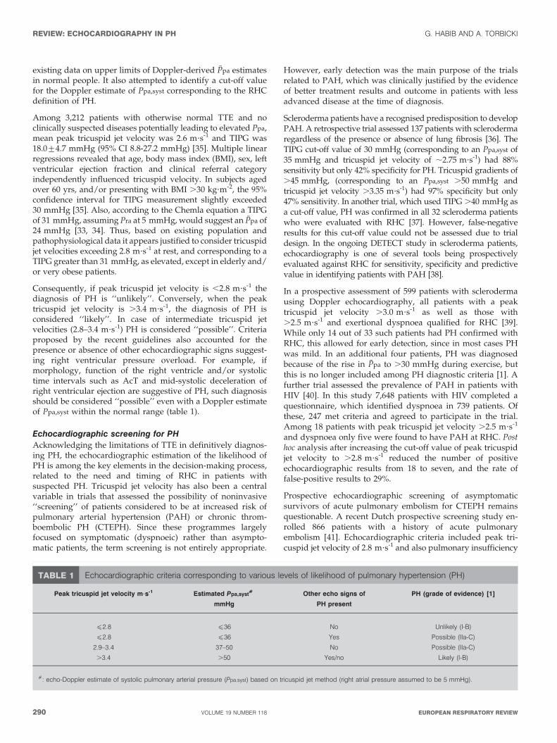
Among 3,212 patients with otherwise normal TTE and noclinically suspected diseases potentially leading to elevated Ppa,mean peak tricuspid jet velocity was 2.6 m?s-1 and TIPG was18.0¡4.7 mmHg (95% CI 8.8-27.2 mmHg) [35]. Multiple linearregressions revealed that age, body mass index (BMI), sex, leftventricular ejection fraction and clinical referral categoryindependently influenced tricuspid velocity. In subjects agedover 60 yrs, and/or presenting with BMI .30 kg?m-2, the 95%confidence interval for TIPG measurement slightly exceeded30 mmHg [35]. Also, according to the Chemla equation a TIPGof 31 mmHg, assuming Pra at 5 mmHg, would suggest an Ppa of24 mmHg [33, 34]. Thus, based on existing population andpathophysiological data it appears justified to consider tricuspidjet velocities exceeding 2.8 m?s-1 at rest, and corresponding to aTIPG greater than 31 mmHg, as elevated, except in elderly and/or very obese patients.
Consequently, if peak tricuspid jet velocity is ,2.8 m?s-1 thediagnosis of PH is ‘‘unlikely’’. Conversely, when the peaktricuspid jet velocity is .3.4 m?s-1, the diagnosis of PH isconsidered ‘‘likely’’. In case of intermediate tricuspid jetvelocities (2.8–3.4 m?s-1) PH is considered ‘‘possible’’. Criteriaproposed by the recent guidelines also accounted for thepresence or absence of other echocardiographic signs suggest-ing right ventricular pressure overload. For example, ifmorphology, function of the right ventricle and/or systolictime intervals such as AcT and mid-systolic deceleration ofright ventricular ejection are suggestive of PH, such diagnosisshould be considered ‘‘possible’’ even with a Doppler estimateof Ppa,syst within the normal range (table 1).
Echocardiographic screening for PHAcknowledging the limitations of TTE in definitively diagnos-ing PH, the echocardiographic estimation of the likelihood ofPH is among the key elements in the decision-making process,related to the need and timing of RHC in patients withsuspected PH. Tricuspid jet velocity has also been a centralvariable in trials that assessed the possibility of noninvasive‘‘screening’’ of patients considered to be at increased risk ofpulmonary arterial hypertension (PAH) or chronic throm-boembolic PH (CTEPH). Since these programmes largelyfocused on symptomatic (dyspnoeic) rather than asympto-matic patients, the term screening is not entirely appropriate.
However, early detection was the main purpose of the trialsrelated to PAH, which was clinically justified by the evidenceof better treatment results and outcome in patients with lessadvanced disease at the time of diagnosis.
Scleroderma patients have a recognised predisposition to developPAH. A retrospective trial assessed 137 patients with sclerodermaregardless of the presence or absence of lung fibrosis [36]. TheTIPG cut-off value of 30 mmHg (corresponding to an Ppa,syst of35 mmHg and tricuspid jet velocity of ,2.75 m?s-1) had 88%sensitivity but only 42% specificity for PH. Tricuspid gradients of.45 mmHg, (corresponding to an Ppa,syst .50 mmHg andtricuspid jet velocity .3.35 m?s-1) had 97% specificity but only47% sensitivity. In another trial, which used TIPG .40 mmHg asa cut-off value, PH was confirmed in all 32 scleroderma patientswho were evaluated with RHC [37]. However, false-negativeresults for this cut-off value could not be assessed due to trialdesign. In the ongoing DETECT study in scleroderma patients,echocardiography is one of several tools being prospectivelyevaluated against RHC for sensitivity, specificity and predictivevalue in identifying patients with PAH [38].
In a prospective assessment of 599 patients with sclerodermausing Doppler echocardiography, all patients with a peaktricuspid jet velocity .3.0 m?s-1 as well as those with.2.5 m?s-1 and exertional dyspnoea qualified for RHC [39].While only 14 out of 33 such patients had PH confirmed withRHC, this allowed for early detection, since in most cases PHwas mild. In an additional four patients, PH was diagnosedbecause of the rise in Ppa to .30 mmHg during exercise, butthis is no longer included among PH diagnostic criteria [1]. Afurther trial assessed the prevalence of PAH in patients withHIV [40]. In this study 7,648 patients with HIV completed aquestionnaire, which identified dyspnoea in 739 patients. Ofthese, 247 met criteria and agreed to participate in the trial.Among 18 patients with peak tricuspid jet velocity .2.5 m?s-1
and dyspnoea only five were found to have PAH at RHC. Posthoc analysis after increasing the cut-off value of peak tricuspidjet velocity to .2.8 m?s-1 reduced the number of positiveechocardiographic results from 18 to seven, and the rate offalse-positive results to 29%.
Prospective echocardiographic screening of asymptomaticsurvivors of acute pulmonary embolism for CTEPH remainsquestionable. A recent Dutch prospective screening study en-rolled 866 patients with a history of acute pulmonaryembolism [41]. Echocardiographic criteria included peak tri-cuspid jet velocity of 2.8 m?s-1 and also pulmonary insufficiency
TABLE 1 Echocardiographic criteria corresponding to various levels of likelihood of pulmonary hypertension (PH)
Peak tricuspid jet velocity m?s-1 Estimated Ppa,syst#
mmHg
Other echo signs of
PH present
PH (grade of evidence) [1]
f2.8 f36 No Unlikely (I-B)
f2.8 f36 Yes Possible (IIa-C)
2.9–3.4 37–50 No Possible (IIa-C)
.3.4 .50 Yes/no Likely (I-B)
#: echo-Doppler estimate of systolic pulmonary arterial pressure (Ppa,syst) based on tricuspid jet method (right atrial pressure assumed to be 5 mmHg).
REVIEW: ECHOCARDIOGRAPHY IN PH G. HABIB AND A. TORBICKI
290 VOLUME 19 NUMBER 118 EUROPEAN RESPIRATORY REVIEW
jet velocity, AcT and the presence of morphological andfunctional consequences of PH. All patients who met one ormore of the six predefined echocardiographic criteria weresuspected of having PH and underwent further standardisedwork-up including perfusion lung scintigraphy and RHC forpressure measurements. The final diagnosis assessed by anindependent expert panel according to predefined criteriarevealed 0.57% (95% CI 0.02-1.2%) prevalence of CTEPH.However, most of the patients with CTEPH were alreadyidentified because of clinical symptoms and signs. Thisoccurred before they were invited for formal echocardio-graphic screening, which had very low additional diagnosticyield for CTEPH, and was not found practically useful by theauthors [41]. A similar conclusion was recently extended topatients who reported new or increased dyspnoea after aprevious episode of acute pulmonary embolism. SymptomaticCTEPH was considered if the Ppa,syst exceeded 40 mmHg onechocardiography and the perfusion scan showed a perfusiondefect on the lobar or segmental levels of the lungs. If thesefindings were positive, patients underwent further diagnos-tic workup, including pulmonary angiography with directmeasurement of Ppa, to confirm or refute the diagnosis ofCTEPH. No additional patients were diagnosed as a resultof echocardiographic examinations performed within thisprogramme [42].
Screening appears to be more successful when higher cut-offlevels for Ppa,syst are used to identify patients who should bereferred for RHC. Among 101 candidates for liver transplantwith Doppler estimate of Ppa,syst .50 mmHg, all-cause PHwas documented at RHC in 90 out of 101 (90%) patients andPAH in 66 out of 101 (65%) patients [43].
In summary, echocardiographic screening for PAH andCTEPH is not effective in asymptomatic patients at increasedrisk and requires better definition of both clinical andechocardiographic enrolment criteria in those patients at risk,who are mildly symptomatic. In the recent European Society ofCardiology (ESC)/European Respiratory Society (ERS) guide-lines [1], echocardiographic screening is recommended insymptomatic patients with scleroderma (evidence level I-B),symptomatic patients with liver disease (evidence level I-B) orHIV (I-C) and may be considered in asymptomatic patientswith scleroderma (evidence level IIb-C). The potential role ofechocardiographic stress testing in the early diagnosis of PHwill be addressed later in the article.
ECHOCARDIOGRAPHIC EVALUATION OF THECONSEQUENCES OF PHAssessment of the consequences of PH on left and right heartremodelling is one of the most important parts of theechocardiographic examination, because of its potential prog-nostic value. Since PH is a very severe disease with highmortality rate [44–48], the identification of noninvasivemarkers of a poor prognosis will be useful for patientmanagement and follow-up. Several echocardiographic indiceshave been proposed [26, 49–51] and have the commonobjective of assessing the three main consequences of PH:right heart remodelling, impact on the left heart and RVdysfunction.
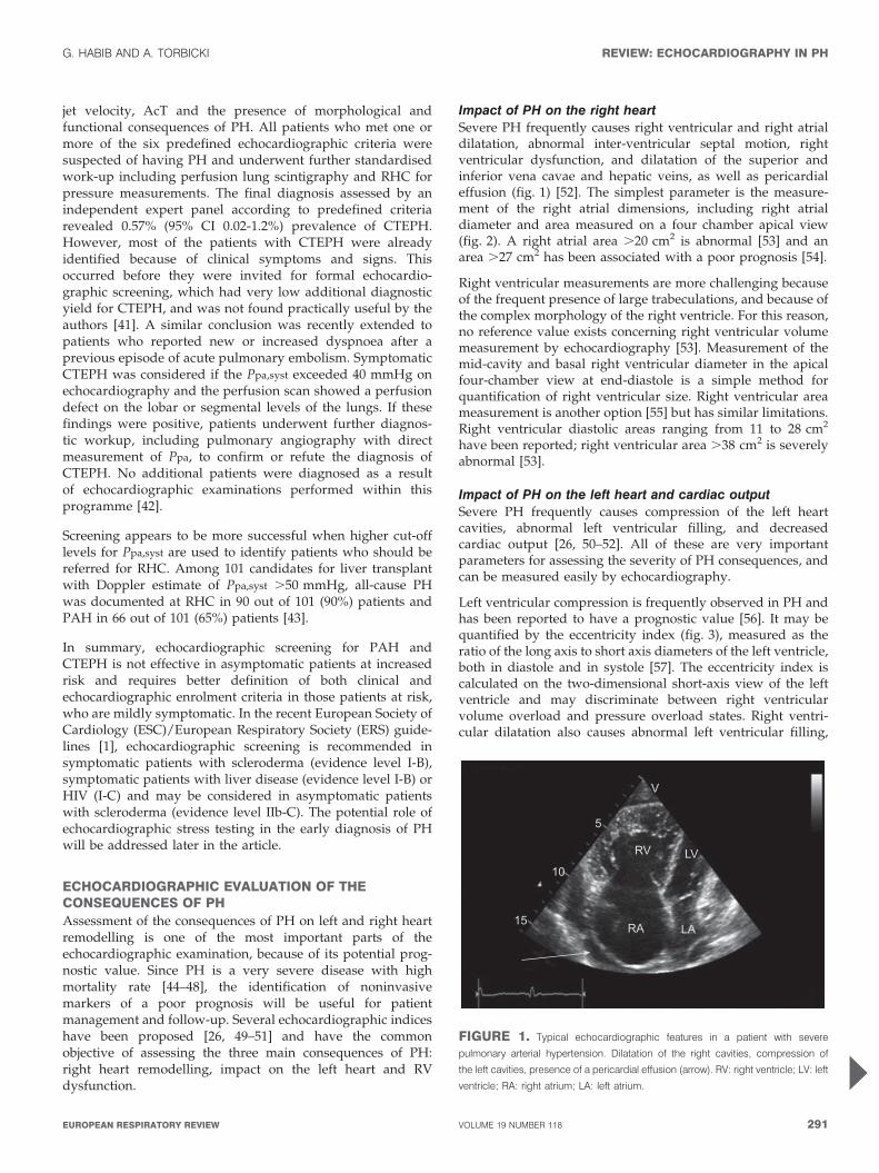
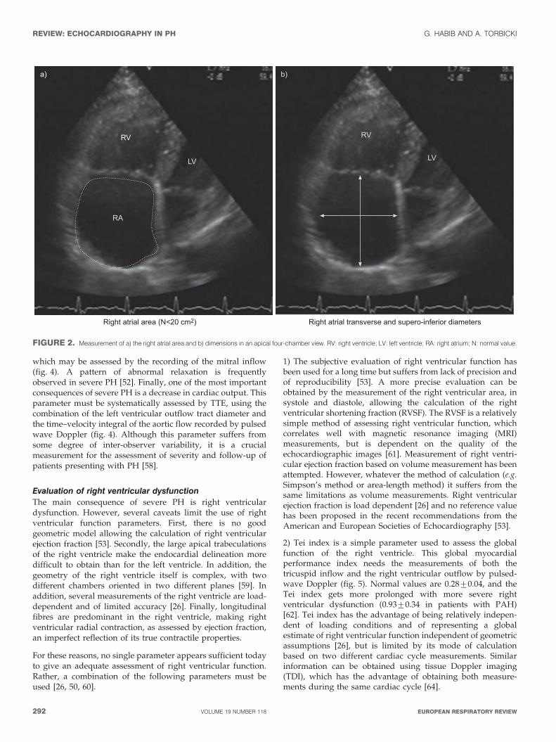
Impact of PH on the right heartSevere PH frequently causes right ventricular and right atrialdilatation, abnormal inter-ventricular septal motion, rightventricular dysfunction, and dilatation of the superior andinferior vena cavae and hepatic veins, as well as pericardialeffusion (fig. 1) [52]. The simplest parameter is the measure-ment of the right atrial dimensions, including right atrialdiameter and area measured on a four chamber apical view(fig. 2). A right atrial area .20 cm2 is abnormal [53] and anarea .27 cm2 has been associated with a poor prognosis [54].
Right ventricular measurements are more challenging becauseof the frequent presence of large trabeculations, and because ofthe complex morphology of the right ventricle. For this reason,no reference value exists concerning right ventricular volumemeasurement by echocardiography [53]. Measurement of themid-cavity and basal right ventricular diameter in the apicalfour-chamber view at end-diastole is a simple method forquantification of right ventricular size. Right ventricular areameasurement is another option [55] but has similar limitations.Right ventricular diastolic areas ranging from 11 to 28 cm2
have been reported; right ventricular area .38 cm2 is severelyabnormal [53].
Impact of PH on the left heart and cardiac outputSevere PH frequently causes compression of the left heartcavities, abnormal left ventricular filling, and decreasedcardiac output [26, 50–52]. All of these are very importantparameters for assessing the severity of PH consequences, andcan be measured easily by echocardiography.
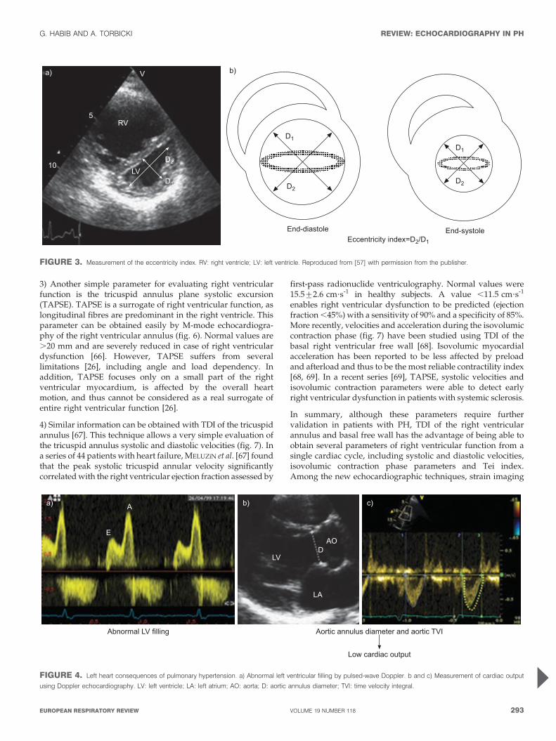
Left ventricular compression is frequently observed in PH andhas been reported to have a prognostic value [56]. It may bequantified by the eccentricity index (fig. 3), measured as theratio of the long axis to short axis diameters of the left ventricle,both in diastole and in systole [57]. The eccentricity index iscalculated on the two-dimensional short-axis view of the leftventricle and may discriminate between right ventricularvolume overload and pressure overload states. Right ventri-cular dilatation also causes abnormal left ventricular filling,
V
RV
RA
10
15
5
LA
LV
FIGURE 1. Typical echocardiographic features in a patient with severe
pulmonary arterial hypertension. Dilatation of the right cavities, compression of
the left cavities, presence of a pericardial effusion (arrow). RV: right ventricle; LV: left
ventricle; RA: right atrium; LA: left atrium.
G. HABIB AND A. TORBICKI REVIEW: ECHOCARDIOGRAPHY IN PH
cEUROPEAN RESPIRATORY REVIEW VOLUME 19 NUMBER 118 291
which may be assessed by the recording of the mitral inflow(fig. 4). A pattern of abnormal relaxation is frequentlyobserved in severe PH [52]. Finally, one of the most importantconsequences of severe PH is a decrease in cardiac output. Thisparameter must be systematically assessed by TTE, using thecombination of the left ventricular outflow tract diameter andthe time–velocity integral of the aortic flow recorded by pulsedwave Doppler (fig. 4). Although this parameter suffers fromsome degree of inter-observer variability, it is a crucialmeasurement for the assessment of severity and follow-up ofpatients presenting with PH [58].
Evaluation of right ventricular dysfunctionThe main consequence of severe PH is right ventriculardysfunction. However, several caveats limit the use of rightventricular function parameters. First, there is no goodgeometric model allowing the calculation of right ventricularejection fraction [53]. Secondly, the large apical trabeculationsof the right ventricle make the endocardial delineation moredifficult to obtain than for the left ventricle. In addition, thegeometry of the right ventricle itself is complex, with twodifferent chambers oriented in two different planes [59]. Inaddition, several measurements of the right ventricle are load-dependent and of limited accuracy [26]. Finally, longitudinalfibres are predominant in the right ventricle, making rightventricular radial contraction, as assessed by ejection fraction,an imperfect reflection of its true contractile properties.
For these reasons, no single parameter appears sufficient todayto give an adequate assessment of right ventricular function.Rather, a combination of the following parameters must beused [26, 50, 60].
1) The subjective evaluation of right ventricular function hasbeen used for a long time but suffers from lack of precision andof reproducibility [53]. A more precise evaluation can beobtained by the measurement of the right ventricular area, insystole and diastole, allowing the calculation of the rightventricular shortening fraction (RVSF). The RVSF is a relativelysimple method of assessing right ventricular function, whichcorrelates well with magnetic resonance imaging (MRI)measurements, but is dependent on the quality of theechocardiographic images [61]. Measurement of right ventri-cular ejection fraction based on volume measurement has beenattempted. However, whatever the method of calculation (e.g.Simpson’s method or area-length method) it suffers from thesame limitations as volume measurements. Right ventricularejection fraction is load dependent [26] and no reference valuehas been proposed in the recent recommendations from theAmerican and European Societies of Echocardiography [53].
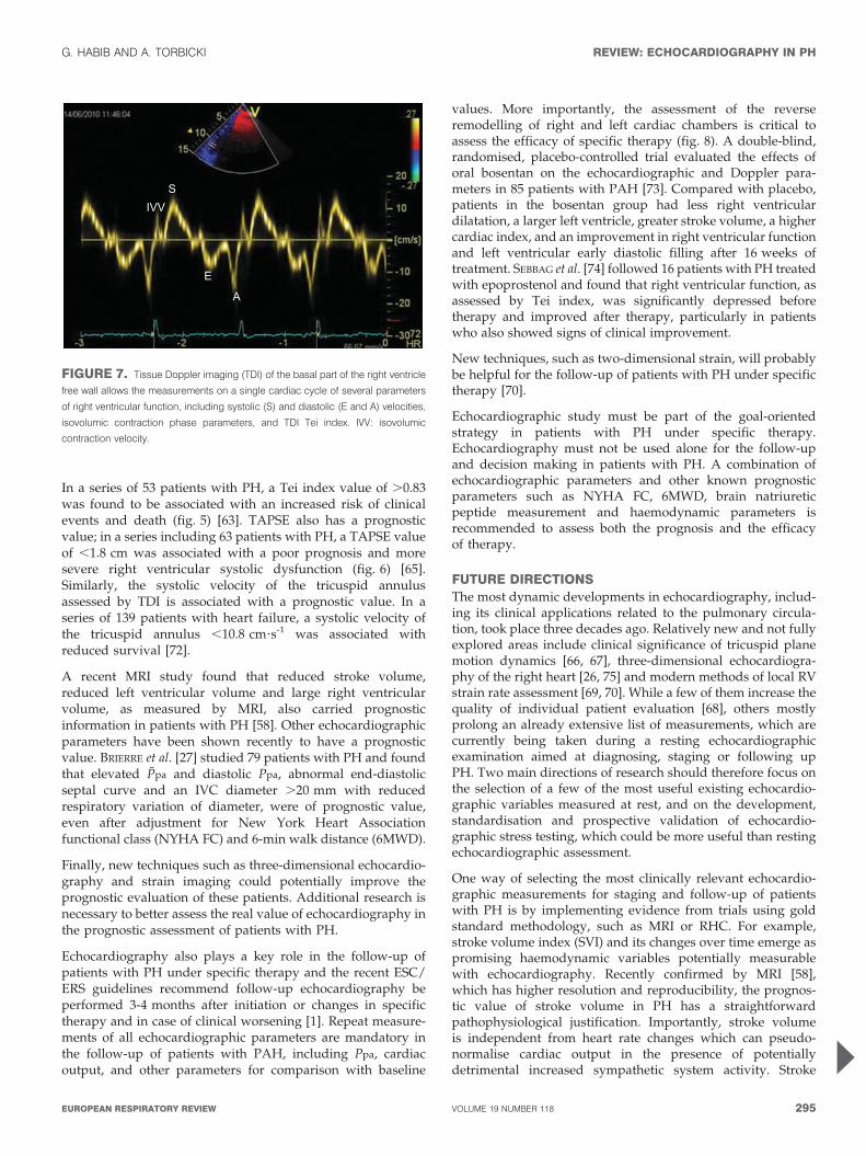
2) Tei index is a simple parameter used to assess the globalfunction of the right ventricle. This global myocardialperformance index needs the measurements of both thetricuspid inflow and the right ventricular outflow by pulsed-wave Doppler (fig. 5). Normal values are 0.28¡0.04, and theTei index gets more prolonged with more severe rightventricular dysfunction (0.93¡0.34 in patients with PAH)[62]. Tei index has the advantage of being relatively indepen-dent of loading conditions and of representing a globalestimate of right ventricular function independent of geometricassumptions [26], but is limited by its mode of calculationbased on two different cardiac cycle measurements. Similarinformation can be obtained using tissue Doppler imaging(TDI), which has the advantage of obtaining both measure-ments during the same cardiac cycle [64].
RV
LV
RA
Right atrial area (N<20 cm2)
a)
RV
LV
Right atrial transverse and supero-inferior diameters
b)
FIGURE 2. Measurement of a) the right atrial area and b) dimensions in an apical four-chamber view. RV: right ventricle; LV: left ventricle; RA: right atrium; N: normal value.
REVIEW: ECHOCARDIOGRAPHY IN PH G. HABIB AND A. TORBICKI
292 VOLUME 19 NUMBER 118 EUROPEAN RESPIRATORY REVIEW
3) Another simple parameter for evaluating right ventricularfunction is the tricuspid annulus plane systolic excursion(TAPSE). TAPSE is a surrogate of right ventricular function, aslongitudinal fibres are predominant in the right ventricle. Thisparameter can be obtained easily by M-mode echocardiogra-phy of the right ventricular annulus (fig. 6). Normal values are.20 mm and are severely reduced in case of right ventriculardysfunction [66]. However, TAPSE suffers from severallimitations [26], including angle and load dependency. Inaddition, TAPSE focuses only on a small part of the rightventricular myocardium, is affected by the overall heartmotion, and thus cannot be considered as a real surrogate ofentire right ventricular function [26].
4) Similar information can be obtained with TDI of the tricuspidannulus [67]. This technique allows a very simple evaluation ofthe tricuspid annulus systolic and diastolic velocities (fig. 7). Ina series of 44 patients with heart failure, MELUZıN et al. [67] foundthat the peak systolic tricuspid annular velocity significantlycorrelated with the right ventricular ejection fraction assessed by
first-pass radionuclide ventriculography. Normal values were15.5¡2.6 cm?s-1 in healthy subjects. A value ,11.5 cm?s-1
enables right ventricular dysfunction to be predicted (ejectionfraction ,45%) with a sensitivity of 90% and a specificity of 85%.More recently, velocities and acceleration during the isovolumiccontraction phase (fig. 7) have been studied using TDI of thebasal right ventricular free wall [68]. Isovolumic myocardialacceleration has been reported to be less affected by preloadand afterload and thus to be the most reliable contractility index[68, 69]. In a recent series [69], TAPSE, systolic velocities andisovolumic contraction parameters were able to detect earlyright ventricular dysfunction in patients with systemic sclerosis.
In summary, although these parameters require furthervalidation in patients with PH, TDI of the right ventricularannulus and basal free wall has the advantage of being able toobtain several parameters of right ventricular function from asingle cardiac cycle, including systolic and diastolic velocities,isovolumic contraction phase parameters and Tei index.Among the new echocardiographic techniques, strain imaging
V
RV5
a)
10LV
D2
D1
b)
D1
D2
End-diastole End-systoleEccentricity index=D2/D1
D2
D1
FIGURE 3. Measurement of the eccentricity index. RV: right ventricle; LV: left ventricle. Reproduced from [57] with permission from the publisher.
A
LV
AOD
LA
E
a) b) c)
Abnormal LV filling Aortic annulus diameter and aortic TVI
Low cardiac output
FIGURE 4. Left heart consequences of pulmonary hypertension. a) Abnormal left ventricular filling by pulsed-wave Doppler. b and c) Measurement of cardiac output
using Doppler echocardiography. LV: left ventricle; LA: left atrium; AO: aorta; D: aortic annulus diameter; TVI: time velocity integral.
G. HABIB AND A. TORBICKI REVIEW: ECHOCARDIOGRAPHY IN PH
cEUROPEAN RESPIRATORY REVIEW VOLUME 19 NUMBER 118 293
and three-dimensional echocardiography have been shown tobe potentially useful to assess right ventricular function [70].
Other consequences of PH assessed by echocardiographyTwo other important consequences of PH may be assessed byechocardiography. Pericardial effusion is usually of minorimportance. However, in some circumstances it can contributeto abnormal left and right ventricular function [71] and is ofprognostic value in PH. Right to left shunting through a patentforamen ovale is observed frequently in severe PH withelevated Pra. It may be easily diagnosed by intravenousinjection of microbubbles using a peripheral line. Severe rightto left atrial shunting may explain hypoxemia observed insome patients with PH.
ECHOCARDIOGRAPHY FOR PROGNOSTICASSESSMENT AND FOLLOW-UP OF PATIENTS WITH PHThe natural history of PH is marked by frequent complicationsand a high mortality rate [44–48]. Besides its diagnostic value andits role in the assessment of consequences of PH, echocardio-graphy is also useful for the prognostic assessment of PH.
Several of the parameters measurable with echocardiographydiscussed above have been shown by multivariate analyses tocarry a prognostic value. These include right atrial area [54],eccentricity index [56], presence of a pericardial effusion [71],Tei index [63] and TAPSE [65]. A multicentre study found thatthe most potent predictive markers of death in PH were rightatrial size, pericardial effusion and eccentricity index [56]. In therecent European guidelines however, only pericardial effusionand TAPSE have been selected as major prognostic indicators onthe basis that they can be measured in the majority of patients[1], although several other indices have the potential to help theclinician in the prognostic assessment of patients with PH.
Tricuspidinflow
Right ventricularoutflow RVMPI=
a-bb
a
bICT IRT
ET
TRT
0.8
0.6
0.4
0.2
1.0
0
Sur
viva
l fre
e of
adv
erse
outc
omes
%
0 4321 5Time yrs
2528
24 (96%)20 (71%)
19 (87%)7 (28%)
8 (73%)1 (4%)
Index ≥ 0.83
R index ≥ 0.83
Index < 0.83
R index < 0.83b)
a)
FIGURE 5. Assessment of the right ventricle global performance by the Tei
index. a) Method of measurement of Tei index. Right ventricle myocardial
performance index (RVMPI) ([a-b]/b) is calculated by measuring two intervals:
1) a is interval between cessation and onset of tricuspid inflow; and 2) b is ejection
time (ET) of right ventricular outflow. ICT: isovolumetric contraction time; IRT:
isovolumetric relaxation time; TRT: tricuspid regurgitation time. Reproduced from
[62] with permission from the publisher. b) Prognostic value of Tei index.
Reproduced from [63] with permission from the publisher.
16 mm
0.25
00
18126 24
17 151617 15TAPSE ≥ 1.8 cm n30 161823 13TAPSE <1.8 cm n
TAPSE ≥ 1.8 cm
TAPSE <1.8 cm
p=0.009Log rank Chi-square 6.8
Time months
Pro
babi
lity
of s
urvi
val
0.75
0.50
1.00
a)
b)
FIGURE 6. Measurement of the tricuspid annulus plane systolic excursion
(TAPSE). a) Method of measurement of TAPSE by M-mode echocardiography.
b) Prognostic value of TAPSE. Reproduced from [65] with permission from the
publisher.
REVIEW: ECHOCARDIOGRAPHY IN PH G. HABIB AND A. TORBICKI
294 VOLUME 19 NUMBER 118 EUROPEAN RESPIRATORY REVIEW
In a series of 53 patients with PH, a Tei index value of .0.83was found to be associated with an increased risk of clinicalevents and death (fig. 5) [63]. TAPSE also has a prognosticvalue; in a series including 63 patients with PH, a TAPSE valueof ,1.8 cm was associated with a poor prognosis and moresevere right ventricular systolic dysfunction (fig. 6) [65].Similarly, the systolic velocity of the tricuspid annulusassessed by TDI is associated with a prognostic value. In aseries of 139 patients with heart failure, a systolic velocity ofthe tricuspid annulus ,10.8 cm?s-1 was associated withreduced survival [72].
A recent MRI study found that reduced stroke volume,reduced left ventricular volume and large right ventricularvolume, as measured by MRI, also carried prognosticinformation in patients with PH [58]. Other echocardiographicparameters have been shown recently to have a prognosticvalue. BRIERRE et al. [27] studied 79 patients with PH and foundthat elevated Ppa and diastolic Ppa, abnormal end-diastolicseptal curve and an IVC diameter .20 mm with reducedrespiratory variation of diameter, were of prognostic value,even after adjustment for New York Heart Associationfunctional class (NYHA FC) and 6-min walk distance (6MWD).
Finally, new techniques such as three-dimensional echocardio-graphy and strain imaging could potentially improve theprognostic evaluation of these patients. Additional research isnecessary to better assess the real value of echocardiography inthe prognostic assessment of patients with PH.
Echocardiography also plays a key role in the follow-up ofpatients with PH under specific therapy and the recent ESC/ERS guidelines recommend follow-up echocardiography beperformed 3-4 months after initiation or changes in specifictherapy and in case of clinical worsening [1]. Repeat measure-ments of all echocardiographic parameters are mandatory inthe follow-up of patients with PAH, including Ppa, cardiacoutput, and other parameters for comparison with baseline
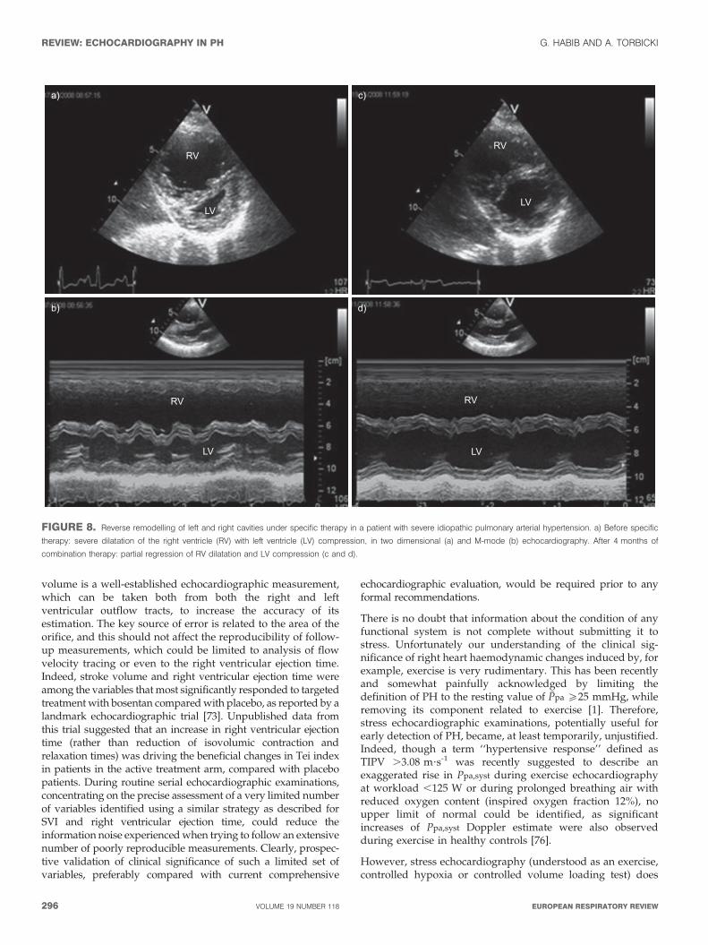
values. More importantly, the assessment of the reverseremodelling of right and left cardiac chambers is critical toassess the efficacy of specific therapy (fig. 8). A double-blind,randomised, placebo-controlled trial evaluated the effects oforal bosentan on the echocardiographic and Doppler para-meters in 85 patients with PAH [73]. Compared with placebo,patients in the bosentan group had less right ventriculardilatation, a larger left ventricle, greater stroke volume, a highercardiac index, and an improvement in right ventricular functionand left ventricular early diastolic filling after 16 weeks oftreatment. SEBBAG et al. [74] followed 16 patients with PH treatedwith epoprostenol and found that right ventricular function, asassessed by Tei index, was significantly depressed beforetherapy and improved after therapy, particularly in patientswho also showed signs of clinical improvement.
New techniques, such as two-dimensional strain, will probablybe helpful for the follow-up of patients with PH under specifictherapy [70].
Echocardiographic study must be part of the goal-orientedstrategy in patients with PH under specific therapy.Echocardiography must not be used alone for the follow-upand decision making in patients with PH. A combination ofechocardiographic parameters and other known prognosticparameters such as NYHA FC, 6MWD, brain natriureticpeptide measurement and haemodynamic parameters isrecommended to assess both the prognosis and the efficacyof therapy.
FUTURE DIRECTIONSThe most dynamic developments in echocardiography, includ-ing its clinical applications related to the pulmonary circula-tion, took place three decades ago. Relatively new and not fullyexplored areas include clinical significance of tricuspid planemotion dynamics [66, 67], three-dimensional echocardiogra-phy of the right heart [26, 75] and modern methods of local RVstrain rate assessment [69, 70]. While a few of them increase thequality of individual patient evaluation [68], others mostlyprolong an already extensive list of measurements, which arecurrently being taken during a resting echocardiographicexamination aimed at diagnosing, staging or following upPH. Two main directions of research should therefore focus onthe selection of a few of the most useful existing echocardio-graphic variables measured at rest, and on the development,standardisation and prospective validation of echocardio-graphic stress testing, which could be more useful than restingechocardiographic assessment.
One way of selecting the most clinically relevant echocardio-graphic measurements for staging and follow-up of patientswith PH is by implementing evidence from trials using goldstandard methodology, such as MRI or RHC. For example,stroke volume index (SVI) and its changes over time emerge aspromising haemodynamic variables potentially measurablewith echocardiography. Recently confirmed by MRI [58],which has higher resolution and reproducibility, the prognos-tic value of stroke volume in PH has a straightforwardpathophysiological justification. Importantly, stroke volumeis independent from heart rate changes which can pseudo-normalise cardiac output in the presence of potentiallydetrimental increased sympathetic system activity. Stroke
SIVV
E
A
FIGURE 7. Tissue Doppler imaging (TDI) of the basal part of the right ventricle
free wall allows the measurements on a single cardiac cycle of several parameters
of right ventricular function, including systolic (S) and diastolic (E and A) velocities,
isovolumic contraction phase parameters, and TDI Tei index. IVV: isovolumic
contraction velocity.
G. HABIB AND A. TORBICKI REVIEW: ECHOCARDIOGRAPHY IN PH
cEUROPEAN RESPIRATORY REVIEW VOLUME 19 NUMBER 118 295
volume is a well-established echocardiographic measurement,which can be taken both from both the right and leftventricular outflow tracts, to increase the accuracy of itsestimation. The key source of error is related to the area of theorifice, and this should not affect the reproducibility of follow-up measurements, which could be limited to analysis of flowvelocity tracing or even to the right ventricular ejection time.Indeed, stroke volume and right ventricular ejection time wereamong the variables that most significantly responded to targetedtreatment with bosentan compared with placebo, as reported by alandmark echocardiographic trial [73]. Unpublished data fromthis trial suggested that an increase in right ventricular ejectiontime (rather than reduction of isovolumic contraction andrelaxation times) was driving the beneficial changes in Tei indexin patients in the active treatment arm, compared with placebopatients. During routine serial echocardiographic examinations,concentrating on the precise assessment of a very limited numberof variables identified using a similar strategy as described forSVI and right ventricular ejection time, could reduce theinformation noise experienced when trying to follow an extensivenumber of poorly reproducible measurements. Clearly, prospec-tive validation of clinical significance of such a limited set ofvariables, preferably compared with current comprehensive
echocardiographic evaluation, would be required prior to anyformal recommendations.
There is no doubt that information about the condition of anyfunctional system is not complete without submitting it tostress. Unfortunately our understanding of the clinical sig-nificance of right heart haemodynamic changes induced by, forexample, exercise is very rudimentary. This has been recentlyand somewhat painfully acknowledged by limiting thedefinition of PH to the resting value of Ppa o25 mmHg, whileremoving its component related to exercise [1]. Therefore,stress echocardiographic examinations, potentially useful forearly detection of PH, became, at least temporarily, unjustified.Indeed, though a term ‘‘hypertensive response’’ defined asTIPV .3.08 m?s-1 was recently suggested to describe anexaggerated rise in Ppa,syst during exercise echocardiographyat workload ,125 W or during prolonged breathing air withreduced oxygen content (inspired oxygen fraction 12%), noupper limit of normal could be identified, as significantincreases of Ppa,syst Doppler estimate were also observedduring exercise in healthy controls [76].
However, stress echocardiography (understood as an exercise,controlled hypoxia or controlled volume loading test) does
RV
a) c)
b) d)
LV
RV
LV
RV
LV
RV
LV
FIGURE 8. Reverse remodelling of left and right cavities under specific therapy in a patient with severe idiopathic pulmonary arterial hypertension. a) Before specific
therapy: severe dilatation of the right ventricle (RV) with left ventricle (LV) compression, in two dimensional (a) and M-mode (b) echocardiography. After 4 months of
combination therapy: partial regression of RV dilatation and LV compression (c and d).
REVIEW: ECHOCARDIOGRAPHY IN PH G. HABIB AND A. TORBICKI
296 VOLUME 19 NUMBER 118 EUROPEAN RESPIRATORY REVIEW
have great potential not only in screening for PH but also infollow-up and assessment of PH treatment. Again, implemen-tation of new methods of functional assessment [77] standar-disation of the tests and their validation should be undertakenin properly designed clinical trials.
Finally, echocardiography should also be prepared to face newchallenges currently emerging in the field of PH and rightventricular function. This includes, for example, evaluation ofright ventricular synchrony in preparation for the possibility ofright ventricular resynchronisation therapy. Echocardiographyshould also allow for serial assessment of pulmonary arterydiameter, recently suggested as related to the risk of suddendeath in patients with PH. Hopefully, the PH community willunderstand the remaining, hidden potential of echocardiogra-phy and will contribute to further development of this methodin the field of pulmonary circulation [26].
CONCLUSIONDespite its several limitations, echocardiography remains themost clinically useful noninvasive method allowing for multi-dimensional assessment of pulmonary circulation. Echocardio-graphy has a key role to play in the diagnosis of PH byidentifying patients for whom RHC is warranted, facilitatingearlier diagnosis and earlier medical management. Theprognostic value of certain echocardiographic parameters isalso recognised and the regular assessment of these, as part ofa goal-oriented approach to therapy, is critical to monitor theprogression of PH and the response of patients to specifictreatment.
STATEMENT OF INTERESTA. Torbicki has served as a consultant for Actelion, Eli Lilly,GlaxoSmithKline and mondoBIOTECH; received honoraria fromBayer Schering, Eli Lilly and Sanofi Aventis; and conducted researchsupported by Actelion, Bayer Schering, Bristol-Myers Squibb, Eli Lilly,GlaxoSmithKline, mondoBIOTECH and Pfizer.
ACKNOWLEDGEMENTSWe received minor editorial assistance from J. Heagerty, ElementsCommunications Ltd (Westerham, UK), supported by ActelionPharmaceuticals Ltd (Allschwil, Switzerland).
REFERENCES1 Galie N, Hoeper MM, Humbert M, et al. Guidelines for the
diagnosis and treatment of pulmonary hypertension: the TaskForce for the Diagnosis and Treatment of PulmonaryHypertension of the European Society of Cardiology (ESC) andthe European Respiratory Society (ERS), endorsed by theInternational Society of Heart and Lung Transplantation(ISHLT). Eur Respir J 2009; 34: 1219–1263.
2 Danchin N, Cornette A, Henriquez A, et al. Two-dimensionalechocardiographic assessment of the right ventricle in patientswith chronic obstructive lung disease. Chest 1987; 92: 229–233.
3 Zenker G, Forche G, Harnoncourt K. Two-dimensional echocardio-graphy using a subcostal approach in patients with COPD. Chest1985; 88: 722–725.
4 Oswald-Mammosser M, Oswald T, Nyankiye E, et al. Non-invasive diagnosis of pulmonary hypertension in chronic obstruc-tive pulmonary disease. Comparison of ECG, radiological mea-surements, echocardiography and myocardial scintigraphy. Eur JRespir Dis 1987; 71: 419–429.
5 Kitabatake A, Inoue M, Asao M, et al. Noninvasive evaluation of
pulmonary hypertension by a pulsed Doppler technique.
Circulation 1983; 68: 302–309.
6 Turkevich D, Groves BM, Micco A, et al. Early partial systolic
closure of the pulmonic valve relates to severity of pulmonary
hypertension. Am Heart J 1988; 115: 409–418.
7 Marangoni S, Quadri A, Dotti A, et al. Noninvasive assessment ofpulmonary hypertension: a simultaneous echo-Doppler hemody-
namic study. Cardiology 1988; 75: 401–408.
8 Robinson PJ, Macartney FJ, Wyse RK. Non-invasive diagnosis ofpulmonary hypertension. Int J Cardiol 1986; 11: 253–259.
9 Torbicki A, Tramarin R, Fracchia F, et al. Reliability of pulsed wave
Doppler monitoring of acute changes in pulmonary artery
pressure in patients with chronic obstructive pulmonary disease.Progr Respir Res 1990; 26: 133–141.
10 Mallery JA, Gardin JM, King SW, et al. Effects of heart rate and
pulmonary artery pressure on Doppler pulmonary artery accel-
eration time in experimental acute pulmonary hypertension. Chest
1991; 100: 470–473.
11 Matsuda M, Sekiguchi T, Sugishita Y, et al. Reliability of non-
invasive estimates of pulmonary hypertension by pulsed Dopplerechocardiography. Br Heart J 1986; 56: 158–164.
12 Isobe M, Yazaki Y, Takaku F, et al. Prediction of pulmonary arterial
pressure in adults by pulsed Doppler echocardiography. Am J
Cardiol 1986; 57: 316–321.
13 Okamoto M, Miyatake K, Kinoshita N, et al. Analysis of blood flow
in pulmonary hypertension with the pulsed Doppler flowmeter
combined with cross sectional echocardiography. Br Heart J 1984;
51: 407–415.
14 Panidis IP, Ross J, Mintz GS. Effect of sampling site on assessment
of pulmonary artery blood flow by Doppler echocardiography.
Am J Cardiol 1986; 58: 1145–1147.
15 Gardin JM, Davidson DM, Rohan MK, et al. Relationship betweenage, body size, gender, and blood pressure and Doppler flow
measurements in the aorta and pulmonary artery. Am Heart J 1987;
113: 101–109.
16 Naeije R, Torbicki A. More on the noninvasive diagnosis of
pulmonary hypertension: Doppler echocardiography revisited.
Eur Respir J 1995; 8: 1445–1449.
17 Torbicki A, Kurzyna M, Ciurzynski M, et al. Proximal pulmonaryemboli modify right ventricular ejection pattern. Eur Respir J 1999;
13: 616–621.
18 Skjaerpe T, Hatle L. Noninvasive estimation of systolic pressure in
the right ventricle in patients with tricuspid regurgitation. Eur
Heart J 1986; 7: 704–710.
19 Masuyama T, Kodama K, Kitabatake A, et al. Continuous-wave
Doppler echocardiographic detection of pulmonary regurgitationand its application to noninvasive estimation of pulmonary artery
pressure. Circulation 1986; 74: 484–492.
20 Matsuoka Y, Hayakawa K. Noninvasive estimation of right
ventricular systolic pressure in ventricular septal defect by acontinuous wave Doppler technique. Jpn Circ J 1986; 50: 1062–1070.
21 Marx GR, Allen HD, Goldberg SJ. Doppler echocardiographic
estimation of systolic pulmonary artery pressure in pediatric
patients with interventricular communications. J Am Coll Cardiol
1985; 6: 1132–1137.
22 Musewe NN, Poppe D, Smallhorn JF, et al. Doppler echocardio-
graphic measurement of pulmonary artery pressure from ductalDoppler velocities in the newborn. J Am Coll Cardiol 1990; 15:
446–456.
23 Yock PG, Popp RL. Noninvasive estimation of right ventricular
systolic pressure by Doppler ultrasound in patients with tricuspidregurgitation. Circulation 1984; 70: 657–662.
24 Beard JT, Byrd BF III. Saline contrast enhancement of trivial
Doppler tricuspid regurgitation signals for estimating pulmonary
artery pressure. Am J Cardiol 1988; 62: 486–488.
G. HABIB AND A. TORBICKI REVIEW: ECHOCARDIOGRAPHY IN PH
cEUROPEAN RESPIRATORY REVIEW VOLUME 19 NUMBER 118 297
25 Himelman RB, Struve SN, Brown JK, et al. Improved recognition ofcor pulmonale in patients with severe chronic obstructivepulmonary disease. Am J Med 1988; 84: 891–898.
26 Badano L, Ginghina C, Easaw J, et al. Right ventricle in pulmonaryarterial hypertension: haemodynamics, structural changes, ima-ging, and proposal of a study protocol aimed to assess remodel-ling and treatment effects. Eur J Echocardiography 2010; 11: 27–37.
27 Brierre G, Blot-Souletie N, Degano B, et al. New echocardiographicprognostic factors for mortality in pulmonary arterial hyperten-sion. Eur J Echocardiogr 2010; 11: 516–522.
28 Hinderliter AL, Willis PW, Barst RJ, et al. Effects of long-terminfusion of prostacyclin (epoprostenol) on echocardiographicmeasures of right ventricular structure and function in primarypulmonary hypertension. Primary Pulmonary HypertensionStudy Group. Circulation 1997; 95: 1479–1486.
29 Liem RI, Young LT, Lay AS, et al. Reproducibility of tricuspidregurgitant jet velocity measurements in children and youngadults with sickle cell disease undergoing screening for pulmon-ary hypertension. Am J Hematol 2010; 85: 741–745.
30 Fisher MR, Forfia PR, Chamera E, et al. Accuracy of Dopplerechocardiography in the hemodynamic assessment of pulmonaryhypertension. Am J Respir Crit Care Med 2009; 179: 615–621.
31 Lei MH, Chen JJ, Ko YL, et al. Reappraisal of quantitativeevaluation of pulmonary regurgitation and estimation of pulmon-ary artery pressure by continuous wave Doppler echocardiogra-phy. Cardiology 1995; 86: 249–256.
32 Ensing G, Seward J, Darragh R, et al. Feasibility of generatinghemodynamic pressure curves from noninvasive Doppler echo-cardiographic signals. J Am Coll Cardiol 1994; 23: 434–442.
33 Chemla D, Castelain V, Humbert M, et al. New formula forpredicting mean pulmonary artery pressure using systolicpulmonary artery pressure. Chest 2004; 126: 1313–1317.
34 Chemla D, Castelain V, Provencher S, et al. Evaluation of variousempirical formulas for estimating mean pulmonary arterypressure by using systolic pulmonary artery pressure in adults.Chest 2009; 135: 760–768.
35 McQuillan BM, Picard MH, Leavitt M, et al. Clinical correlates andreference intervals for pulmonary artery systolic pressure amongechocardiographically normal subjects. Circulation 2001; 104: 2797–2802.
36 Mukerjee D, St George D, Knight C, et al. Echocardiography andpulmonary function as screening tests for pulmonary arterialhypertension in systemic sclerosis. Rheumatology (Oxford) 2004; 43:461–466.
37 Launay D, Mouthon L, Hachulla E, et al. Prevalence andcharacteristics of moderate to severe pulmonary hypertension insystemic sclerosis with and without interstitial lung disease.J Rheumatol 2007; 34: 1005–1011.
38 Vonk M, Coghlan G, Bonderman D, et al. The DETECT study: atwo-stage, prospective, observational, cohort study in sclerodermapatients to evaluate screening tests and the incidence ofpulmonary arterial hypertension and pulmonary hypertension.Clin Exp Rheumatol 2010; 28: Suppl., 55.
39 Hachulla E, Gressin V, Guillevin L, et al. Early detection ofpulmonary arterial hypertension in systemic sclerosis: a Frenchnationwide prospective multicenter study. Arthritis Rheum 2005;52: 3792–3800.
40 Sitbon O, Lascoux-Combe C, Delfraissy JF, et al. Prevalence ofHIV-related pulmonary arterial hypertension in the currentantiretroviral therapy era. Am J Respir Crit Care Med 2008; 177:108–113.
41 Klok FA, van Kralingen KW, van Dijk AP, et al. Prospectivecardiopulmonary screening program to detect chronic throm-boembolic pulmonary hypertension in patients after acutepulmonary embolism. Haematologica 2010; 95: 970–975.
42 Surie S, Gibson NS, Gerdes VE, et al. Active search for chron-ic thromboembolic pulmonary hypertension does not appear
indicated after acute pulmonary embolism. Thromb Res 2010; 125:e202–5.
43 Krowka MJ, Swanson KL, Frantz RP, et al. Portopulmonaryhypertension: results from a 10-year screening algorithm.Hepatology 2006; 44: 1502–1510.
44 D’Alonzo GE, Barst RJ, Ayres SM, et al. Survival in patients withprimary pulmonary hypertension: results from a national pro-spective registry. Ann Intern Med 1991; 115: 343–349.
45 Kuhn KP, Byrne DW, Arbogast PG, et al. Outcome in 91consecutive patients with pulmonary arterial hypertension receiv-ing epoprostenol. Am J Respir Crit Care Med 2003; 167: 580–586.
46 Humbert M, Sitbon O, Chaouat A, et al. Pulmonary arterialhypertension in France: results from a national registry. Am J
Respir Crit Care Med 2006; 173: 1023–1030.
47 Humbert M, Sitbon O, Chaouat A, et al. Survival in patients withidiopathic, familial, and anorexigen-associated pulmonary arterialhypertension in the modern management era. Circulation 2010;122: 156–163.
48 Kawut SM, Taichman DB, Archer-Chicko CL, et al. Hemodynamicsand survival in patients with pulmonary arterial hypertensionrelated to systemic sclerosis. Chest 2003; 123: 344–350.
49 Bossone E, Bodini BD, Maza A, et al. Pulmonary arterialhypertension. The key role of echocardiography. Chest 2005; 127:1836–1843.
50 Burgess MI, Bright-Thomas RJ, Ray SG. Echocardiographicevaluation of right ventricular function. Eur J Echocardiography
2002; 3: 252–262.
51 Haddad F, Hunt SA, Rosenthal DN, et al. Right ventricularfunction in cardiovascular disease, part I: anatomy, physiology,aging, and functional assessment of the right ventricle. Circulation
2008; 117: 1436–1448.
52 Bossone E, Duong-Wagner TH, Paciocco G, et al. Echocardiographicfeatures of primary pulmonary hypertension. J Am Soc Echocardiogr
1999; 12: 655–662.
53 Lang RM, Bierig M, Devereux RB, et al. Recommendations forchamber quantification. Eur J Echocardiogr 2006; 7: 79–108.
54 Perrone S, De La Fuente RL, et al. Right atrial size and tricuspidregurgitation severity predict mortality or transplantation inprimary pulmonary hypertension. J Am Soc Echocardiogr 2002; 15:1160–1164.
55 Jiang L, Levine RA, Weyman AE. Echocardiographic assessmentof right ventricular volume and function. Echocardiography 1997;14: 189–205.
56 Raymond RJ, Hinderliter AL, Willis PW, et al. Echocardiographicpredictors of adverse outcomes in primary pulmonary hyperten-sion. J Am Coll Cardiol 2002; 39: 1214–1219.
57 Ryan T, Petrovic O, Dillon JC, et al. An echocardiographic indexfor separation of right ventricular volume and pressure overload.J Am Coll Cardiol 1985; 5: 918–927.
58 van Wolferen SA, Marcus JT, Boonstra A, et al. Prognostic value ofright ventricular mass, volume, and function in idiopathicpulmonary arterial hypertension. Eur Heart J 2007; 28: 1250–1257.
59 Naito H, Arisawa J, Harada K, et al. Assessment of rightventricular regional contraction and comparison with the leftventricle in normal humans: a cine magnetic resonance study withpresaturation myocardial tagging. Br Heart J 1995; 74: 186–191.
60 Jurcut R, Giusca S, La Gerche A, et al. The echocardiographicassessment of the right ventricle: what to do in 2010? Eur JEchocardiogr 2010; 11: 81–96.
61 Anavekar NS, Gerson D, Skali H, et al. Two dimensionalassessment of right ventricular function: an echocardiographic-MRI correlative study. Echocardiography 2007; 24: 452–456.
62 Tei C, Dujardin KS, Hodge DO, et al. Doppler echocardiographicindex for assessment of global right ventricular function. J Am Soc
Echocardiogr 1996; 9: 838–847.
63 Yeo TC, Dujardin KS, Tei C, et al. Value of a Doppler-derivedindex combining systolic and diastolic time intervals in predicting
REVIEW: ECHOCARDIOGRAPHY IN PH G. HABIB AND A. TORBICKI
298 VOLUME 19 NUMBER 118 EUROPEAN RESPIRATORY REVIEW
outcome in primary pulmonary hypertension. Am J Cardiol 1998;81: 1157–1161.
64 Harada K, Tamura M, Toyono M, et al. Comparison of the rightventricular Tei index by tissue Doppler imaging to that obtainedby pulsed Doppler in children without heart disease. Am J Cardiol2002; 90: 566–569.
65 Forfia PR, Fisher MR, Mathai SC, et al. Tricuspid annulardisplacement predicts survival in pulmonary hypertension. Am J
Respir Crit Care Med 2006; 174: 1034–1041.66 Ueti OM, Camargo EE, Ueti Ade A, et al. Assessment of right
ventricular function with Doppler echocardiographic indicesderived from tricuspid annular motion: comparison with radio-nuclide angiography. Heart 2002; 88: 244–248.
67 Meluzın J, Spinarova L, Bakala J, et al. Pulsed Doppler tissueimaging of the velocity of tricuspid annular systolic motion; a new,rapid, and non-invasive method of evaluating right ventricularsystolic function. Eur Heart J 2001; 22: 340–348.
68 Vogel M, Schmidt MR, Kristiansen SB, et al. Validation ofmyocardial acceleration during isovolumic contraction as a novelnoninvasive index of right ventricular contractility: comparisonwith ventricular pressure–volume relations in an animal model.Circulation 2002; 105: 1693–1699.
69 Schattke S, Knebel F, Grohmann A, et al. Early right ventricularsystolic dysfunction in patients with systemic sclerosis withoutpulmonary hypertension: a Doppler tissue and speckle trackingechocardiography study. Cardiovascular Ultrasound 2010; 8:3–11.
70 Borges AC, Knebel F, Eddicks S, et al. Right ventricular func-tion assessed by two-dimensional strain and tissue Doppler
echocardiography in patients with pulmonary arterial hypertensionand effect of vasodilator therapy. Am J Cardiol 2006; 98: 530–534.
71 Hinderliter AL, Willis PW 4th, Long W, et al. Frequency andprognostic significance of pericardial effusion in primary pul-monary hypertension. Am J Cardiol 1999; 84: 481–484.
72 Meluzın J, Spinarova L, Dusek L, et al. Prognostic importance ofthe right ventricular function assessed by Doppler tissue imaging.Eur J Echocardiogr 2003; 4: 262–271.
73 Galie N, Hinderliter Al, Torbicki A, et al. Effects of the oralendothelin-receptor antagonist bosentan on echocardiographicand Doppler measures in patients with pulmonary arterialhypertension. J Am Coll Cardiol 2003; 41: 1380–1386.
74 Sebbag I, Rudski LG, Therrien J, et al. Effect of chronic infusion ofepoprostenol on echocardiographic right ventricular myocardial per-formance index and its relation to clinical outcome in patients withprimary pulmonary hypertension. Am J Cardiol 2001; 88: 1060–1063.
75 Grapsa J, O’Regan DP, Pavlopoulos H, et al. Right ventricularremodelling in pulmonary arterial hypertension with three-dimensional echocardiography: comparison with cardiac magneticresonance imaging. Eur J Echocardiogr 2010; 11: 64–73.
76 Grunig E, Weissmann S, Ehlken N, et al. Stress Dopplerechocardiography in relatives of patients with idiopathic andfamilial pulmonary arterial hypertension: results of a multicenterEuropean analysis of pulmonary artery pressure response toexercise and hypoxia. Circulation 2009; 119: 1747–1757.
77 Yang HS, Mookadam F, Warsame TA, et al. Evaluation of rightventricular global and regional function during stress echocardio-graphy using novel velocity vector imaging. Eur J Echocardiogr
2010; 11: 157–164.
G. HABIB AND A. TORBICKI REVIEW: ECHOCARDIOGRAPHY IN PH
EUROPEAN RESPIRATORY REVIEW VOLUME 19 NUMBER 118 299