Embed Size (px)
Citation preview
The role of EBV in the pathogenesis of Burkitt’s Lymphoma:
a four Italian hospital based survey.
Authors:
Giuseppe Pannone1, Angela Santoro2, Mirella Pace1, Maria Carmela Pedicillo1
Simona Cagiano1, Pasquale Somma 3, Maria Elena Errico4, Vittoria Donofrio4,
Rosanna Zamparese5, and Pantaleo Bufo6.
Affiliations:
1 Department of Clinical and Experimental Medicine, Institute of Pathological Anatomy,
University of Foggia; Foggia – Italy;
2 Department of Laboratory, Institute of Histopatology and Diagnostic Cytopathology, Fondazione di Ricerca e Cura ‘
Giovanni Paolo II’-UCSC, Campobasso, Italy;
3 Section of Pathological Anatomy, Ospedale dei Colli – Monaldi, Napoli, Italy;
4 Section of Pathological Anatomy, Paediatric Oncological Hospital Pausillipon, Napoli - Italy;
5 Section of Pathological Anatomy, Ospedale di Ascoli, Ascoli Piceno - Italy;
6 Department of Clinical and Experimental Medicine University of Foggia; Foggia - Italy and IRCCS CROB
-Basilicata Cancer Institute, Rionero in Vulture, Potenza – Italy.
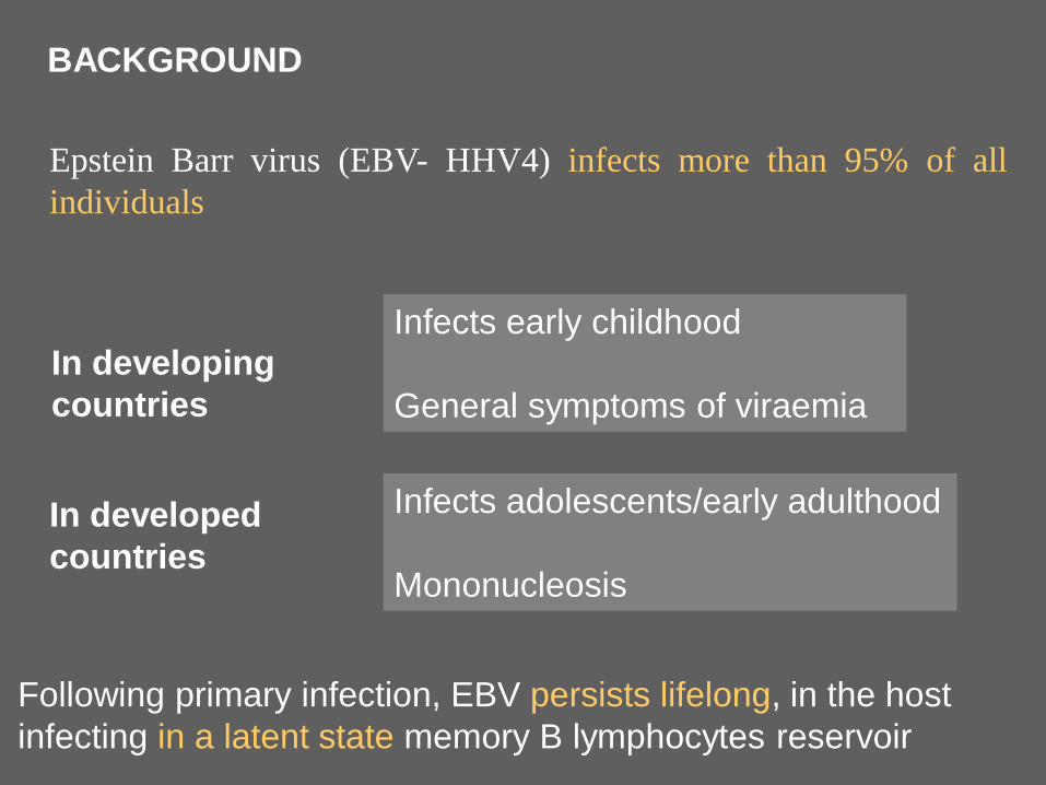
BACKGROUND
Epstein Barr virus (EBV- HHV4) infects more than 95% of all
individuals
In developing
countries
Following primary infection, EBV persists lifelong, in the host
infecting in a latent state memory B lymphocytes reservoir
Infects early childhood
General symptoms of viraemia
In developed
countries
Infects adolescents/early adulthood
Mononucleosis
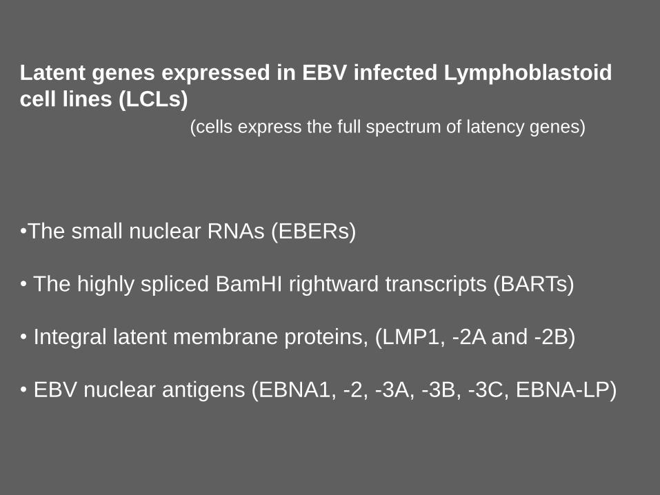
Latent genes expressed in EBV infected Lymphoblastoid
cell lines (LCLs)
(cells express the full spectrum of latency genes)
•The small nuclear RNAs (EBERs)
• The highly spliced BamHI rightward transcripts (BARTs)
• Integral latent membrane proteins, (LMP1, -2A and -2B)
• EBV nuclear antigens (EBNA1, -2, -3A, -3B, -3C, EBNA-LP)
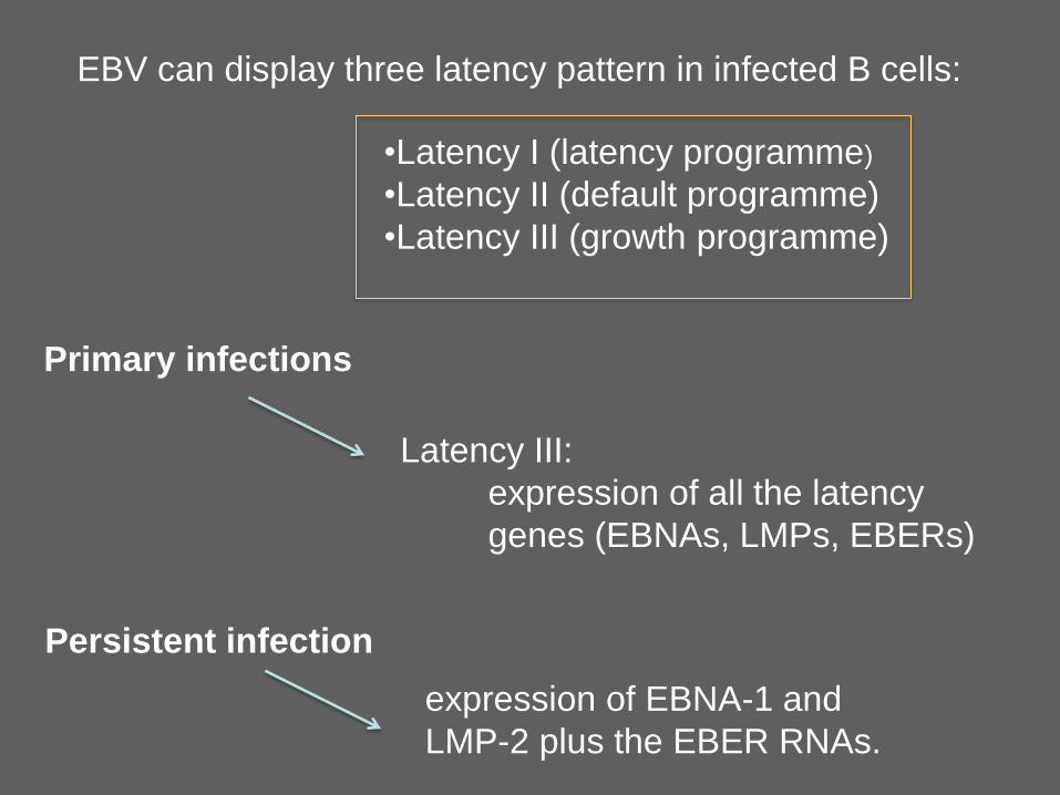
EBV can display three latency pattern in infected B cells:
•Latency I (latency programme)
•Latency II (default programme)
•Latency III (growth programme)
Primary infections
Latency III:
expression of all the latency
genes (EBNAs, LMPs, EBERs)
Persistent infection
expression of EBNA-1 and
LMP-2 plus the EBER RNAs.
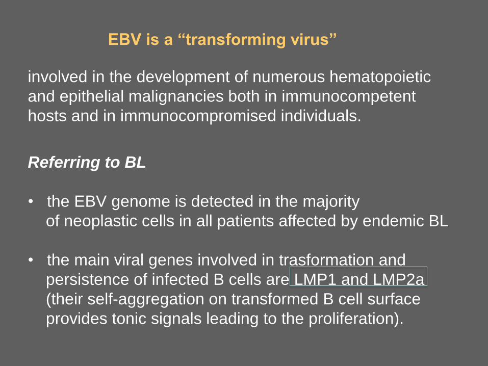
EBV is a “transforming virus”
involved in the development of numerous hematopoietic
and epithelial malignancies both in immunocompetent
hosts and in immunocompromised individuals.
Referring to BL
• the EBV genome is detected in the majority
of neoplastic cells in all patients affected by endemic BL
• the main viral genes involved in trasformation and
persistence of infected B cells are LMP1 and LMP2a
(their self-aggregation on transformed B cell surface
provides tonic signals leading to the proliferation).
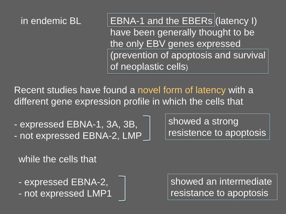
in endemic BL EBNA-1 and the EBERs (latency I)
have been generally thought to be
the only EBV genes expressed
(prevention of apoptosis and survival
of neoplastic cells)
Recent studies have found a novel form of latency with a
different gene expression profile in which the cells that
- expressed EBNA-1, 3A, 3B,
- not expressed EBNA-2, LMP
showed a strong
resistence to apoptosis
while the cells that
- expressed EBNA-2,
- not expressed LMP1
showed an intermediate
resistance to apoptosis
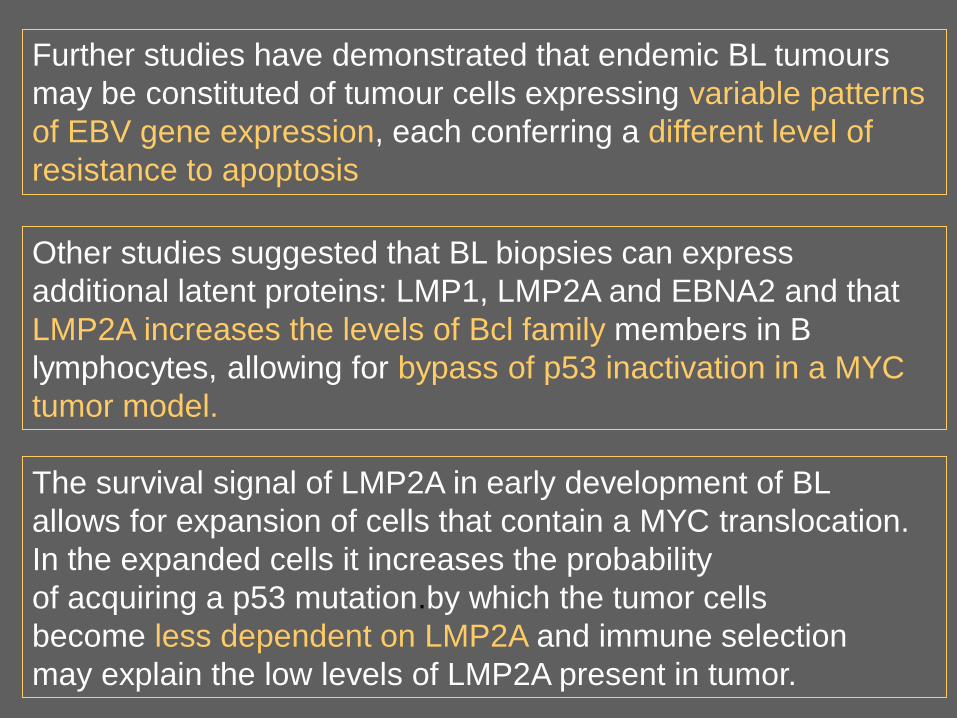
Further studies have demonstrated that endemic BL tumours
may be constituted of tumour cells expressing variable patterns
of EBV gene expression, each conferring a different level of
resistance to apoptosis
Other studies suggested that BL biopsies can express
additional latent proteins: LMP1, LMP2A and EBNA2 and that
LMP2A increases the levels of Bcl family members in B
lymphocytes, allowing for bypass of p53 inactivation in a MYC
tumor model.
The survival signal of LMP2A in early development of BL
allows for expansion of cells that contain a MYC translocation.
In the expanded cells it increases the probability
of acquiring a p53 mutation.by which the tumor cells
become less dependent on LMP2A and immune selection
may explain the low levels of LMP2A present in tumor.
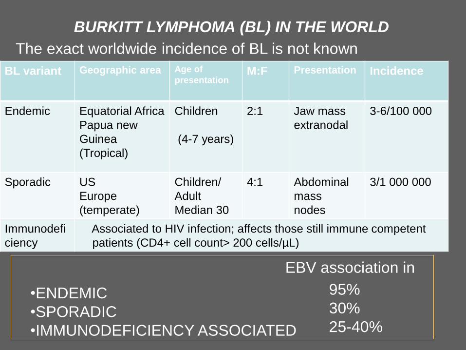
BURKITT LYMPHOMA (BL) IN THE WORLD
The exact worldwide incidence of BL is not known
BL variant Geographic area Age of
presentation M:F Presentation Incidence
Endemic Equatorial Africa
Papua new
Guinea
(Tropical)
Children
(4-7 years)
2:1 Jaw mass
extranodal
3-6/100 000
Sporadic US
Europe
(temperate)
Children/
Adult
Median 30
4:1 Abdominal
mass
nodes
3/1 000 000
Immunodefi
ciency
Associated to HIV infection; affects those still immune competent
patients (CD4+ cell count> 200 cells/µL)
•ENDEMIC
•SPORADIC
•IMMUNODEFICIENCY ASSOCIATED
95%
30%
25-40%
EBV association in
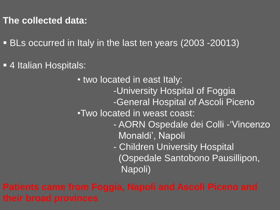
The collected data:
BLs occurred in Italy in the last ten years (2003 -20013)
4 Italian Hospitals:
• two located in east Italy:
-University Hospital of Foggia
-General Hospital of Ascoli Piceno
•Two located in weast coast:
- AORN Ospedale dei Colli -‘Vincenzo
Monaldi’, Napoli
- Children University Hospital
(Ospedale Santobono Pausillipon,
Napoli)
Patients came from Foggia, Napoli and Ascoli Piceno and
their broad provinces
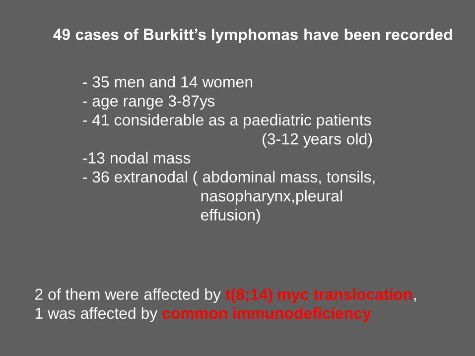
49 cases of Burkitt’s lymphomas have been recorded
- 35 men and 14 women
- age range 3-87ys
- 41 considerable as a paediatric patients
(3-12 years old)
-13 nodal mass
- 36 extranodal ( abdominal mass, tonsils,
nasopharynx,pleural
effusion)
2 of them were affected by t(8;14) myc translocation,
1 was affected by common immunodeficiency
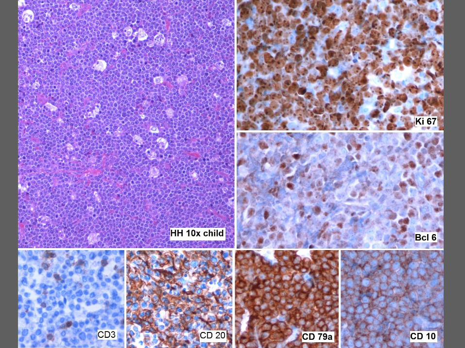
The final diagnosis of BL, obtained comparing -morphological features - immunohistochemical results (panel of antibodies including CD3, CD5, CD20, CD10, CD79a, bcl-2, bcl-6 and Ki-67 (MIB-1),
Immunohistochemical detection of LMP1 Expression
and EBER In Situ Hybridization Procedures have been
performed and evaluated according to standardized
guidelines
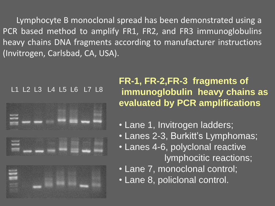
Lymphocyte B monoclonal spread has been demonstrated using a PCR based method to amplify FR1, FR2, and FR3 immunoglobulins heavy chains DNA fragments according to manufacturer instructions (Invitrogen, Carlsbad, CA, USA).
FR-1, FR-2,FR-3 fragments of
immunoglobulin heavy chains as
evaluated by PCR amplifications
• Lane 1, Invitrogen ladders;
• Lanes 2-3, Burkitt’s Lymphomas;
• Lanes 4-6, polyclonal reactive
lymphocitic reactions;
• Lane 7, monoclonal control;
• Lane 8, policlonal control.
L1 L2 L3 L4 L5 L6 L7 L8
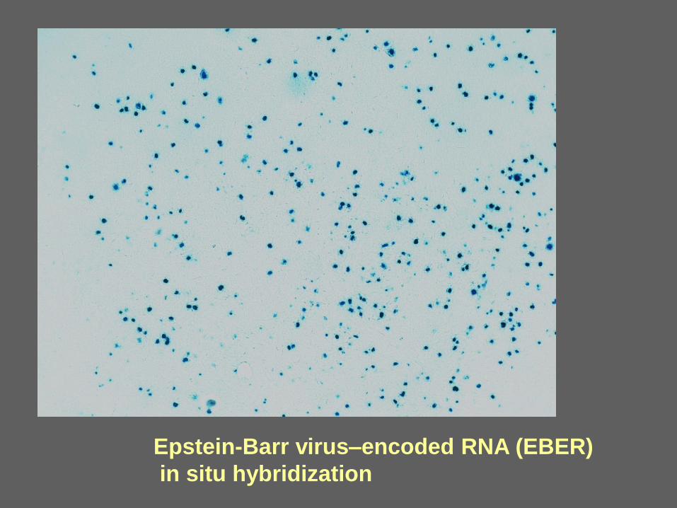
Epstein-Barr virus–encoded RNA (EBER)
in situ hybridization
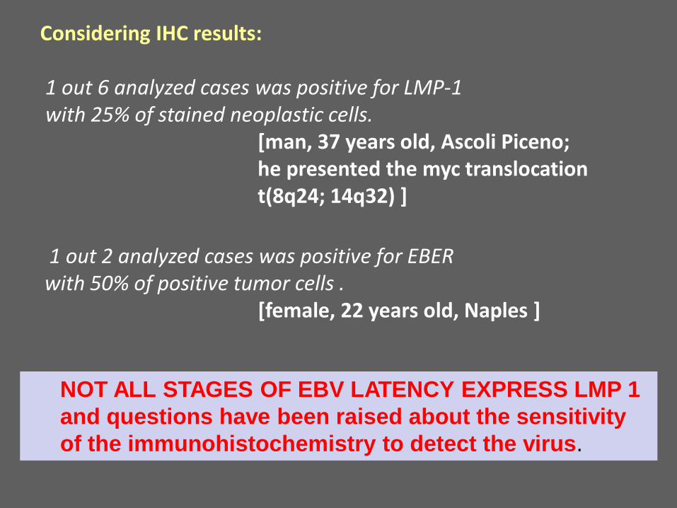
Considering IHC results: 1 out 6 analyzed cases was positive for LMP-1 with 25% of stained neoplastic cells. [man, 37 years old, Ascoli Piceno; he presented the myc translocation t(8q24; 14q32) ]
NOT ALL STAGES OF EBV LATENCY EXPRESS LMP 1
and questions have been raised about the sensitivity
of the immunohistochemistry to detect the virus.
1 out 2 analyzed cases was positive for EBER with 50% of positive tumor cells . [female, 22 years old, Naples ]
Overall, we can conclude that,
in our Italian study population,
the prevalence of EBV infection associated to
Burkitt’s Lymphoma was 25% (2 out 8 cases).
EBV has been detected only in adult patients,
one of them with typical myc translocation.