Embed Size (px)
Citation preview
The role of dendritic cells in the pathogenesis
of Staphylococcus aureus infection
Von der Fakultät für Lebenswissenschaften
der Technischen Universität Carolo-Wilhelmina
zu Braunschweig
zur Erlangung des Grades einer
Doktorin der Naturwissenschaften
(Dr. rer. nat.)
genehmigte
D i s s e r t a t i o n
von Daniela Schindler, geb. Bruhn
aus Gifhorn

1. Referentin: Privatdozentin Dr. Simone Bergmann
2. Referent: Professor Dr. Dieter Jahn
eingereicht am: 06.02.2012
mündliche Prüfung (Disputation) am: 02.04.2012
Druckjahr 2012

Vorveröffentlichungen der Dissertation
Vorveröffentlichungen der Dissertation
Teilergebnisse aus dieser Arbeit wurden mit Genehmigung der Fakultät für
Lebenswissenschaften, vertreten durch die Mentorin der Arbeit, in
folgenden Beiträgen vorab veröffentlicht:
Tagungsbeiträge
Bruhn D, Goldmann O, Medina E:
Role of dendritic cells in host defence against
Staphylococcus aureus (Poster).
40. Jahrestagung der Deutschen Gesellschaft für Immunologie (DGfI).
22. – 25. September 2010 in Leipzig
Bruhn D:
The role of dendritic cells in host defence against
Staphylococcus aureus (Vortrag).
61. Jahrestagung der Deutschen Gesellschaft für Hygiene und
Mikrobiologie e.V. (DGHM).
20. - 23 September 2009 in Göttingen

Table of contents
Table of contents
1. Abstract.............................................................................................. 1
2. Introduction ....................................................................................... 2
2.1 Staphylococcus aureus .................................................................... 2
2.1.1 Microbial characterization of Staphylococcus aureus ................. 2
2.1.2 S. aureus virulence factors ......................................................... 2
2.1.3 Adhesion and invasion of S. aureus ........................................... 4
2.1.4 S. aureus infections .................................................................... 5
2.1.5 Antibiotic resistance in S. aureus ............................................... 6
2.2 The host immune response to S. aureus .......................................... 6
2.3 Dendritic cells ................................................................................... 8
2.3.1 Origin and subdivision of dendritic cells ..................................... 8
2.3.2 Maturation of DCs ...................................................................... 9
2.3.3 Activation of T lymphocytes ...................................................... 10
2.3.4 DC subtypes ............................................................................. 10
2.3.5 DCs and their role in disease ................................................... 11
2.3.5 Pathogens strategies to evade DCs ......................................... 12
3. Aim of the work .................................................................................. 13
4. Material and Methods ........................................................................ 14
4.1 Bacterial strains .............................................................................. 14
4.2 Mice ................................................................................................ 14
4.3 S. aureus infection model ............................................................... 16
4.4 Assessment of pathology ............................................................... 16
4.5 Isolation of DCs from lungs and spleen .......................................... 16
4.6 Generation of bone marrow-derived DCs ....................................... 17
4.7 Adoptive Transfer of DCs ............................................................... 18
4.8 Collection of primary polymorph nuclear neutrophils ...................... 18

Table of contents
4.9 Partial depletion of PMNs in vivo .................................................... 18
4.10 Cytokine determination ................................................................. 18
4.11 Flow cytometry ............................................................................. 19
4.12 S. aureus infection of DCs ............................................................ 19
4.13 Live cell imaging ........................................................................... 20
4.14 Image analysis.............................................................................. 20
4.15 Intracellular bacterial viability assay ............................................. 20
4.16 Antigen-presentation assay .......................................................... 21
4.17 Statistical analysis ........................................................................ 21
5. Results ................................................................................................ 22
5.1 Phagocytic uptake of S. aureus by DCs ......................................... 22
5.2 S. aureus induces maturation of DCs ............................................. 24
5.3 The mouse model of S. aureus infection ........................................ 25
5.4 DCs are recruited into the organs of S. aureus-infected mice ........ 26
5.5 Depletion of DCs exacerbates the severity of S. aureus infection .. 28
5.6 Reconstitution of DCs-depleted mice by adoptive transfer of DCs
reverses the effect of DC depletion in the course of
S. aureus infection ............................................................................. 29
5.7 Adoptive transfer of DCs improves the course of infection in
BALB/c mice ...................................................................................... 32
5.8 DCs do not contribute to direct killing of S. aureus ......................... 32
5.9 Depletion of DCs leads to an enhanced recruitment of PMNs
into the lungs of infected mice ........................................................... 34
5.10 PMNs recruited within the lungs of DC-depleted mice
harbor higher numbers of viable intracellular bacteria than
PMNs within the lungs of non-depleted mice .................................... 35
5.11 Higher levels of CXC chemokines are produced in the
lungs of DC-depleted than in the lungs of non-depleted
S. aureus-infected mice .................................................................... 36

Table of contents
5.12 Partial depletion of PMNs does not reverse the detrimental
effect of DC depletion ....................................................................... 37
5.13 Influence of S. aureus in the capacity of DCs to stimulate
naïve antigen-specific CD4+ T cells .................................................. 37
6. Discussion.......................................................................................... 39
References ............................................................................................. 44
Acknowledgement ................................................................................. 54
Appendix ................................................................................................ 56
A. Antibiotics and chemicals ................................................................. 56
B. Buffer ............................................................................................... 57
C. Expendable materials and Instruments ............................................ 59
D. Media for bacteria culture ................................................................ 60
E. Media and supplements for cell culture ............................................ 61
F. Antibodies ........................................................................................ 61
G. Mice ................................................................................................. 62
H. Abbreviations ................................................................................... 62
I. Videos ............................................................................................... 64
J. List of figures .................................................................................... 64

Abstract
1
1. Abstract
Dendritic cells (DCs) play an essential role in orchestrating and regulating
the immune response against many pathogens. In this dissertation, the
importance of DCs in host defense against the clinical relevant bacterium
Staphylococcus aureus (S. aureus) was investigated.
Immunofluorescence live-cell imaging was used to demonstrate the high
efficiency of DCs to phagocyte S. aureus. Bacterial uptake induced a fast
maturation of DCs as shown by the up-regulation of the co-stimulatory
molecules CD40, CD80, CD86, and MHC class II, as well as by the pro-
duction of the inflammatory cytokines IL-6, IL-12, and TNFα. In addition,
DCs were rapidly mobilized and recruited into infected tissue after intrave-
nous inoculation of mice with S. aureus. CD11c-DTR transgenic mice
were then used to evaluate the relevance of DCs for host defense against
S. aureus. These transgenic mice express the diphtheria toxin (DT) recep-
tor on the CD11c promoter region, which allows for selective depletion of
DCs followings the intraperitoneal administration of DT. In vivo depletion of
DCs resulted in accelerated mortality and significantly increased bacterial
loads in the lungs and kidneys of infected mice. Reconstitution of DC-
depleted mice by adoptive transfer of DCs from BALB/c mice returned the
capacity of these animals to control S. aureus infection. Furthermore, in-
crease in DCs numbers in normal mice resulted in enhanced resistance to
infection.
The beneficial effect afforded by DCs was not attributed to a direct contri-
bution to bacterial killing. Interestingly, depletion of DCs resulted in a more
rapid influx of primary polymorph nuclear neutrophils (PMNs) to the site of
infection; a significant increase in the local production of CXC chemokines
and increased survival of S. aureus within the recruited PMNs.
The results of this study provide compelling evidence that DCs are im-
portant regulators of the host immune response to S. aureus and of benefit
for the host. These data may be of use in future studies for the develop-
ment of new therapeutic strategies for the treatment of S. aureus infec-
tions.

Introduction
2
2. Introduction
2.1 Staphylococcus aureus
2.1.1 Microbial characterization of Staphylococcus aureus
The species Staphylococcus aureus (S. aureus) is the most prominent of
the genus Staphylococcus in relation to human diseases [1, 2]. It was orig-
inally discovered by the surgeon Sir Alexander Ogston in pus from surgical
abscesses in the year 1880 and described as:
“Micrococcus, which, when limited in its extent and activity, causes acute
suppurative inflammation (phlegmon), produces, when more extensive
and intense in its action on the human system, the most virulent forms of
septicæmia and pyæmia” [3, 4].
S. aureus is a gram-positive, non motile, non spore-forming facultative an-
aerobic, catalase and coagulase positive coccus that is able to induce ß-
haemolysis. Traditionally, it was considered to be an extracellular patho-
gen [2, 5], but increasing evidence indicates that S. aureus might be a
facultative intracellular pathogen [6-10]. Staphylococci appear in grape-like
clusters. The species name “aureus”, which means “golden” in Latin is due
to the yellow-golden color of the colonies [11]. The color is imparted by
carotenoid pigments that supports the bacteria in the prevention of oxidant
killing by phagocytes [12]. Around 20% of the population is persistently
and around 30% intermittently colonized by S. aureus in the nose or in the
axilla region, groin areas, or gastrointestinal tract [13].
2.1.2 S. aureus virulence factors
The remarkable success of S. aureus as a pathogen is due to the expres-
sion of a wide array of virulence factors including a capsule as well as sur-
face-exposed and secreted proteins [14-16]. A schematic representation
of S. aureus virulence factors is depicted in Figure 1. In more detail, coag-
ulase is an extracellular protein that reacts with prothrombin in the blood to
form a complex called staphylothrombin. The protease activity of this
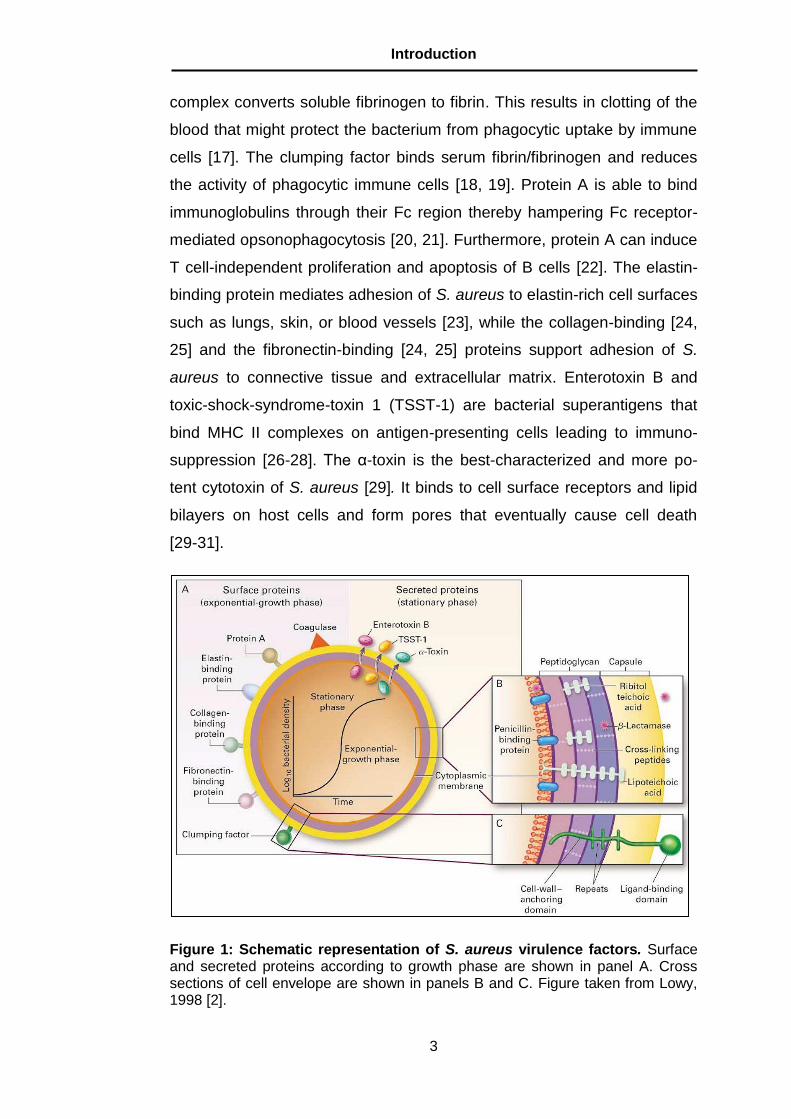
Introduction
3
complex converts soluble fibrinogen to fibrin. This results in clotting of the
blood that might protect the bacterium from phagocytic uptake by immune
cells [17]. The clumping factor binds serum fibrin/fibrinogen and reduces
the activity of phagocytic immune cells [18, 19]. Protein A is able to bind
immunoglobulins through their Fc region thereby hampering Fc receptor-
mediated opsonophagocytosis [20, 21]. Furthermore, protein A can induce
T cell-independent proliferation and apoptosis of B cells [22]. The elastin-
binding protein mediates adhesion of S. aureus to elastin-rich cell surfaces
such as lungs, skin, or blood vessels [23], while the collagen-binding [24,
25] and the fibronectin-binding [24, 25] proteins support adhesion of S.
aureus to connective tissue and extracellular matrix. Enterotoxin B and
toxic-shock-syndrome-toxin 1 (TSST-1) are bacterial superantigens that
bind MHC II complexes on antigen-presenting cells leading to immuno-
suppression [26-28]. The α-toxin is the best-characterized and more po-
tent cytotoxin of S. aureus [29]. It binds to cell surface receptors and lipid
bilayers on host cells and form pores that eventually cause cell death
[29-31].
Figure 1: Schematic representation of S. aureus virulence factors. Surface and secreted proteins according to growth phase are shown in panel A. Cross sections of cell envelope are shown in panels B and C. Figure taken from Lowy, 1998 [2].
Introduction
4
2.1.3 Adhesion and invasion of S. aureus
Adherence of S. aureus to the host extracellular matrix is the initial step in
the infection process and is mediated by bacterial surface adhesins,
known as MSCRAMMs (microbial surface components recognizing adhe-
sive matrix molecules) [32]. MSCRAMMs bind to extracellular matrix pro-
teins such as fibrinogen (ClfA, ClfB), fibronectin (FnbpA, FnbpB), and col-
lagen (Cna) [33]. Certain MSCRAMMs, such as ClfA and SdrC are found
in virtually all clinical S. aureus strains, whereas others, such as Cna and
SdrD, are present only in a subset of strains [34]. In addition to mediate
the bacterium attachment to the extracellular matrix, certain MSCRAMMs
are able to mediate the internalization of S. aureus within eukaryotic cells.
S. aureus is generally thought to be an extracellular pathogen, but it can
be internalized by a variety of cell types in vitro including fibroblasts, oste-
oblasts, keratinocytes, and endothelial cells [35]. This may be an important
process in systemic spread of infection, escape from antibiotic pressure
and evasion of the host immune system [36]. The dynamic process of S.
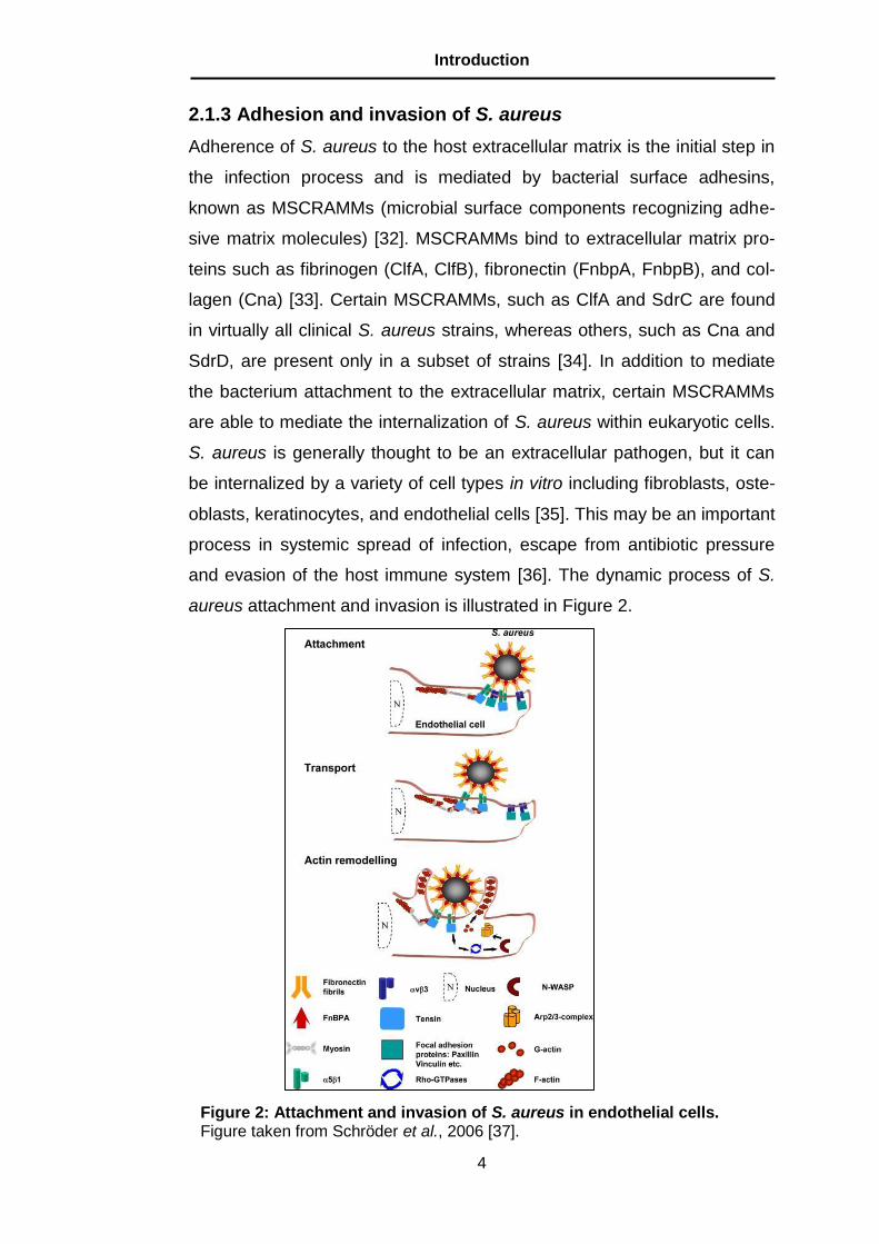
aureus attachment and invasion is illustrated in Figure 2.
Figure 2: Attachment and invasion of S. aureus in endothelial cells. Figure taken from Schröder et al., 2006 [37].
Introduction
5
The schematic representation in Figure 2 shows S. aureus attached to
α5β1-integrins on the surface of endothelial cells via fibronectin fibrils that
are bound to the fibronectin-binding protein FnBPA (attachment). The host
cell integrins are associated with myosin bound tensins, and by contrac-
tion of the myosin, the bacteria are transported towards the cell center
(transport). The uptake is performed by tensin-mediated actin reorganiza-
tion (actin remodeling).
2.1.4 S. aureus infections
S. aureus can cause a wide variety of infections including skin and soft-
tissue infections such as folliculitis, impetigo, furuncles, carbuncles, and
cellulites [2]. If the bacterium is able to enter the bloodstream, it can cause
live-threatening invasive infections such as bacteremia, infective endocar-
ditis, osteomyelitis, or sepsis [38, 39]. Other infections caused by S.
aureus include catheter-related infections, joint infections, pulmonary in-
fections, and central nervous system infections [39]. S. aureus is associat-
ed with an increasing number of both community-acquired and hospital-
acquired cases of pneumonia. Recent studies have reported that S.
aureus comprises more than 25% of community-acquired and more than
40% of hospital-acquired pneumonia cases [40]. A most severe outcome
of S. aureus bacteremia is the development of sepsis. Sepsis is described
as an extensive clinical syndrome that is due to an overwhelming re-
sponse of the immune system to an infection progress, followed by immu-
nosuppression [41, 42]. Whereas the most important mediator of gram
negative-associated sepsis is the lipopolysaccharide (LPS), in gram-
positive bacteria the cell wall components peptidoglycan and lipoteichoic
acid play a dominant role. In addition to these components, some strains
of S. aureus produce diverse exotoxins like TSST-1, which act as super-
antigens and are therefore able to induce T lymphocyte activation followed
by a massive release of pro-inflammatory cytokines [43].
Introduction
6
2.1.5 Antibiotic resistance in S. aureus
Treatment of S. aureus infections can be difficult since many strains have
developed resistance to several antibiotics. Penicillin was the first antibi-
otic introduced in the clinic to treat S. aureus infections. The mode of ac-
tion of penicillin consists in the inhibition of peptidoglycan synthesis on the
bacterial cell wall [44, 45]. Shortly after its introduction, S. aureus strains
developed resistance against beta-lactam antibiotics by the acquisition of
the enzyme beta-lactamase that hydrolytically destroys the beta-lactam
ring [46]. Methicillin, introduced in 1961, was the first of the semisynthetic
penicillinase-resistant penicillins due to a different steric orientation of the
beta-lactam ring. Unfortunately, S. aureus rapidly developed resistance
against methicillin and other semi-synthetic penicillins [47-49]. Resistance
to methicillin is mediated by the acquisition of the mecA gene, which
codes for the penicillin-binding protein PBP2a [50]. Methicillin-resistant
strains of S. aureus are commonly abbreviated as MRSA. In the USA, the
percentage of MRSA strains rose from 11% to 59.5% between the years
1998 – 2003 [51]. In Europe, the epidemiology of MRSA varies widely de-
pending on the country, ranging from 0.6% in Sweden to an average of
41% in Belgium, Greece, Ireland, and the United Kingdom [52].
Vancomycin has been the most reliable therapeutic agent against infec-
tions caused by MRSA strains. This glycopeptide antibiotic was introduced
in the year 1958 for the treatment of gram positive bacterial infections [53].
However, in 1996 the first MRSA resistant to vancomycin was isolated
from a Japanese patient [54, 55]. Subsequent isolation of several
vancomycin resistant S. aureus (VRSA) strains from different countries
has confirmed that the emergence of vancomycin resistance in S. aureus
is a global issue.
2.2 The host immune response to S. aureus
With the exception of diseases caused by specific toxins, the pathogenicity
of S. aureus is mediated by a variety of exoenzymes, adhesins, immuno-
modulating proteins, and the presence of cell wall compounds [56]. These
virulence factors in combination allow the bacterium to infect virtually any
Introduction
7
host tissue [57]. Recognition of S. aureus components by innate immune
cells is the first step of host defense. The toll-like receptors (TLRs) consti-
tute a family of receptors, which distinguish pathogen-associated molecu-
lar patterns such as LPS, peptidoglycan, and lipoprotein [44, 58]. Trigger-
ing of TLR signaling after pathogens recognition leads to the activation of
transcription factors that in turn induce transcription of genes coding for a
variety of proteins including cytokines and inflammatory mediators [59].
This results in massive invasion of immune cells to the site of infection.
Virulence studies using knockout mice have shown that TLRs play a criti-
cal role in the innate immune response to staphylococcal infections since
mice deficient in the expression of myeloid differentiation factor MyD88, a
cytoplasmic adaptor molecule essential for the signaling of IL-1R/TLR fam-
ily, are highly susceptible to this pathogen [60].
The staphylococcal protein A is a ligand for the TNF-α receptor 1, which is
largely expressed on the airway epithelium. The interaction between pro-
tein A and the TNF- -receptor 1 leads to an inflammatory response on
epithelial cells and the recruitment of primary polymorph nuclear neutro-
phils (PMNs) [61].
An effective immune response against S. aureus involves PMN recruit-
ment, which are required for bacterial clearance, and abscess formation
[62-64]. The importance of PMNs in host defense against S. aureus infec-
tion has been demonstrated by recurrent S. aureus infections in patients
with chronic granulomatous disease, who have a defect in the NADPH
oxidase and respiratory burst [65]. The resulting substantial invasion of
PMNs to the site of S. aureus infection is accompanied by central liquefac-
tion necrosis and formation of peripheral fibrin walls in an effort to prevent
microbial spread [66]. These structures are known as abscesses and they
generally have a defined fibrinous wall surrounding a purulent focus of
PMNs and bacteria.
The early events of the innate immune response culminate in the activa-
tion of the adaptive immune system, during which T and B cells capable of
specific antigen recognition are generated.
Introduction
8
2.3 Dendritic cells
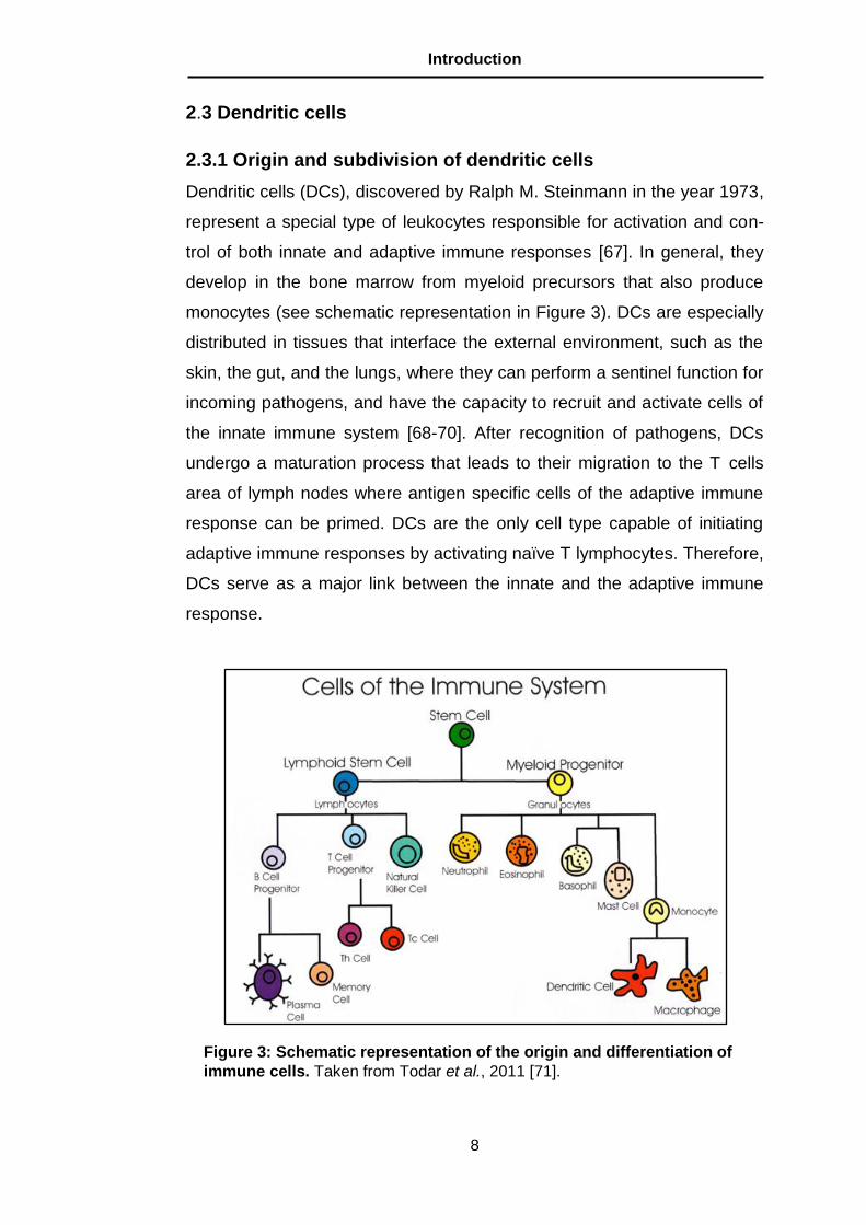
2.3.1 Origin and subdivision of dendritic cells
Dendritic cells (DCs), discovered by Ralph M. Steinmann in the year 1973,
represent a special type of leukocytes responsible for activation and con-
trol of both innate and adaptive immune responses [67]. In general, they
develop in the bone marrow from myeloid precursors that also produce
monocytes (see schematic representation in Figure 3). DCs are especially
distributed in tissues that interface the external environment, such as the
skin, the gut, and the lungs, where they can perform a sentinel function for
incoming pathogens, and have the capacity to recruit and activate cells of
the innate immune system [68-70]. After recognition of pathogens, DCs
undergo a maturation process that leads to their migration to the T cells
area of lymph nodes where antigen specific cells of the adaptive immune
response can be primed. DCs are the only cell type capable of initiating
adaptive immune responses by activating naïve T lymphocytes. Therefore,
DCs serve as a major link between the innate and the adaptive immune
response.
Figure 3: Schematic representation of the origin and differentiation of
immune cells. Taken from Todar et al., 2011 [71].

Introduction
9
2.3.2 Maturation of DCs
DCs reside in an immature state in several tissues such as skin, pharynx,
esophagus, or genitals. Furthermore, they are found in mucous mem-
branes like respiratory and gastrointestinal tracts [72, 73]. Immature DCs
have a radiating shape with their extensions (“dendrits”) outstretched
through the tight junctions of tissue epithelia cells, which increases their
ability to catch antigens [74]. After recognition of pathogens through sur-
face-exposed receptors, DCs increase their expression of the chemokine
receptor CCR7, which enables them to migrate from the site of infection
through the afferent lymphatic vessels to the T cell area of the draining
secondary lymphatic organs (lymph nodes, Peyer-Plaques in the gut, ton-
sils, spleen, and appendix) [75, 76]. During migration, DCs undergo a
maturation program that results in the up-regulation of co-stimulatory mo-
lecules such as CD40, CD80, and CD86, translocation of their MHC class
II to the cell surface and secretion of inflammatory cytokines (see sche-
matic representation in Figure 4) [77]. Furthermore, the morphology of the
DCs also changes with maturation. These changes involve reduction of
dendrits and development of membrane excrescences. The phagocytic
capacity of DCs is also lost during the maturation process [44].
Figure 4: Schematic representation of maturation and migration of dendritic cells. Taken from [78], modified.

Introduction
10
2.3.3 Activation of T lymphocytes
After reaching a lymphatic organ, DCs express a high amount of co-
stimulatory molecules to activate the different types of T cells. By presen-
ting the antigen via the MHC class I or II complexes and the interaction
between their B7 molecules with the CD28 receptor on the T cells, the T
cells become activated and leave the lymphatic organ via the efferent lym-
phatic vessels (see Figure 4 and Figure 5) [44].
2.3.4 DC subtypes
Different subtypes of DCs have been described according to the surface
expression of specific markers and tissue distribution [80, 81]. In mice,
immature conventional DCs are characterized by the expression of
CD11c, low levels of the co-stimulatory molecules CD80 and CD86, and
low levels of MHC class II. Interestingly, DCs can also express the T cell
markers CD4 and CD8 as well as CD11b. The CD8- DCs reside mostly in
the marginal zone of the lymph nodes while the CD8+ are mainly located in
the T cell area [81]. Another CD4- CD8- CD11b+ DC population also ex-
pressing moderate levels of the scavenger receptor CD205 has been iden-
tified in all lymph nodes [82]. Finally, DCs in the skin, also known as
Figure 5: Activation of T lymphocytes by DCs. Taken from Gilboa et al., 2004 [79].
Introduction
11
Langerhans cells, express high levels of Langerin [83].
In humans, DC subtypes are less characterized. Human DCs do not ex-
press CD8 and, in spleen and tonsils, DCs are positive for CD11b, CD11c,
and CD4 [81].
2.3.5 DCs and their role in disease
Due to their excellent ability to present antigens and thereby activate and
orchestrate the adaptive immune response, DCs play a pivotal role in the
host defense against pathogens [77]. In this regard, several studies have
demonstrated that DCs are required for the survival of mice suffering of
polymicrobial sepsis [84]. Decreased counts of circulating DCs have been
reported to correlate with disease severity and predicting a fatal outcome
in patients affected by the septic shock syndrome [85]. Furthermore, trans-
fer of bone-marrow derived DCs into the lungs of postseptic mice reversed
immunosuppression and conferred protection against Aspergillus infection
[86]. The importance of DCs has also been proved in non-septic situa-
tions. For example, DCs are an important inducer of T cell responses dur-
ing Cryptococcus neoformans infections [87] and contribute to the host
defense against Streptococcus pyogenes [88, 89]. Due to the importance
of DCs for host defense, certain pathogens have developed mechanisms
to avoid or even to use the DCs for their own benefit. Thus, DCs infected
with the measles virus undergo maturation but are unable to induce T cell
activation and show a high apoptosis rate [90, 91]. However, apoptotic
measles virus-infected DCs are strong inducers of maturation of uninfect-
ed DCs, what may finally contribute to viral clearance [91]. By replicating
and surviving within the DCs, Mycobacteria and Salmonella sp. can use
this eukaryotic cell to disseminate within the host [92-95].
Introduction
12
2.3.5 Pathogens strategies to evade DCs
As the DCs play a central role in the regulation of the immune response,
many pathogens have developed strategies to prevent DC recognition.
Salmonella enterica serovar Typhi is a motile bacterium that causes ty-
phoid fever in humans. Their flagellin subunit FliC is an attractive target for
DCs [96]. It has been shown, that the bacteria dramatically reduce the
production of FliC as soon as they invaded into the gut cells, as motility is
no longer required [97]. Therefore, by directing the DCs to induce a T cell
response against an antigen that is no longer present, Salmonella evades
the host immune response. Yersinia enterocolitica shows a more direct
strategy to evade DCs. During infection, the bacteria are able to inject ef-
fector proteins like Yersinia outer proteins (Yops) into the host cells, which
inhibits antigen presentation by the DCs [98]. Furthermore, Yops proteins
induce DC apoptosis, and therefore hindering the appropriate stimulation
of T cells due to the reduced number of DCs [99, 100]. Other pathogens
such as Helicobacter pylori are able to inhibit the cytokine release of DCs
by secreting a non-proteinaceous factor and thereby regulating the im-
mune response to its favor [101]. Streptococcus pneumoniae produces
haemolytic pneumolysin that inhibits the DC maturation, the induction of
proinflammatory cytokines, and the activation of the inflammasome. If the
bacteria are taken up by DCs, the intracellular production of pneumolysin
induces caspase-dependent apoptosis [102]. In summary, the strategies
developed by different pathogens to evade DCs are based upon their in-
terference with the generation and survival of DCs, the antigen-presenting
mechanisms, or the interference with T cell activation.
Aim of the work
13
3. Aim of the work
In the last decades, S. aureus has been recognized as one of the most
important leading cause of hospital- and community-acquired infections
worldwide. As the management of S. aureus infections is a growing clinical
challenge due to the widespread presence of multidrug-resistant S. aureus
strains, new therapeutic options are therefore needed for the treatment of
these infections in a more efficient way. This clinical challenge under-
scores the importance of a better understanding of the pathogenesis and
cellular mechanisms underlying S. aureus infections. As DCs play a cru-
cial role in protecting the host against pathogens, the aim of the current
study was to investigate the role of DCs in the complex process of
S. aureus invasive infection. For this purpose the interactions between
S. aureus and DCs has been examined using in vitro as well as in vivo
experimental systems. Live-cell imaging and immunofluorescence tech-
niques have been applied to visualize the phagocytic process of S. aureus
by DCs. In addition, CD11c-DTR transgenic mice, which allow the selec-
tive depletion of DCs in vivo, have been used to evaluate the relevance of
DCs for the host defense against S. aureus.
Material and Methods
14
4. Material and Methods
4.1 Bacterial strains
The S. aureus strain SH1000 was used in this study. SH1000 was devel-
oped in the year 2002 as a single-copy rsbU gene-complemented deriva-
tive of the wild-type strain 8325-4 [103]. rsbU encodes a positive regulator
for sigma factor b activity, that is responsible for a general stress response
[103]. Bacteria were grown to the Mid-Log phase at 37 ºC with shaking
(150 rpm) in brain heart infusion (BHI)-medium, collected by centrifugation
for 10 min at 4000 rpm, washed twice with sterile PBS and adjusted to 5 x
108 CFU/ml. For the inoculum preparation, the bacterial suspension was
diluted with PBS to the required concentration and the number of viable
bacteria was determined after serial diluting and plating onto agar contain-
ing 5% of sheep blood (Invitrogen, Karlsruhe, Germany). For live-cell im-
aging, a GFP-expressing SH1000 strain was used [104]. Bacteria were
grown to the Mid-Log phase at 37°C with shaking (150 rpm) in brain heart
infusion (BHI)-medium supplemented with 30 µg/ml of Chloramphenicol,
collected by centrifugation for 10 min at 4000 rpm, washed twice with ster-
ile PBS and adjusted to 5 x 10⁸ CFU/ml. To generate heat-killed
S. aureus, 109 bacteria were resuspended in 2 ml of PBS and incubated in
a water bath for 2 h at 95°C.
4.2 Mice
BALB/c mice were purchased from Harlan-Winkelmann (Borchen, Germa-
ny), B6.FVB-Tg [Itgax-DTR/GFP] 57Lan/J transgenic mice [105] (referred
in this study as CD11c-DTR) were obtained from Steffen Jung (The
Weizmann Institute of Science, Rehovot, Israel) and backcrossed against
a BALB/c background. These mice express a human diphtheria toxin (DT)
receptor in the promoter region of CD11c. Mice harboring this transgene
are transiently depleted of CD11c+ populations upon intraperitoneal injec-
tion of diphtheria toxin (DT) (see Figure 6). DC depletion persists for ap-
proximately 2 days after which the cell population gradually recovers. For

Material and Methods
15
systemic depletion of DCs, CD11c-DTR transgenic mice were injected
intraperiotoneally (i.p.) with 4 ng/g body weight of DT (Sigma,
Deisenhofen, Germany) in PBS 24 h prior to infection. CD11c-DTR inject-
ed i.p. with PBS were used as control. Ovalbumin specific mice B6.Cg-Tg
(TcraTcrb) 425Cbn/J (referred in this study as OT-II) were purchased from
Jackson Laboratory (Maine, USA). These mice express the mouse alpha-
chain and beta-chain T cell receptor that pairs with the CD4 co-receptor.
The T cell hybridoma clone is CD4+, MHC class II-restricted, and is specif-
ic for the ovalbumin residue 323-339 peptides in the context of the MHC
class II I-A2 molecule [106].
Mice were housed in a specific pathogen-free animal facility at the Helm-
holtz Centre for Infection Research. All experiments were approved by the
ethical board Niedersächsisches Landesamt für Verbraucherschutz und
Lebensmittelsicherheit, Oldenburg in Germany, reference number
33.11.42502-04-024/08.
Figure 6: Depletion of CD11c positive cells in CD11c-DTR mice. Figure taken from Bennett et al., 2007 [107].
Material and Methods
16
4.3 S. aureus infection model
Mice were inoculated with 4 x 10⁷ CFU of S. aureus in 150 µl of PBS via a
lateral tail vein. For determination of bacterial loads in organs, infected
mice were killed by CO2 asphyxiation at different times of infection and the
amount of bacteria was determined by preparing organ homogenates in 5
ml of PBS and plating 6-fold serial dilutions on blood agar (Invitrogen).
Colonies were counted after incubation for 24 h at 37°C. For collection of
serum, blood was collected at time of sacrifice, centrifuged at 4000 rpm for
10 min, and frozen at -80°C until use for further analysis.
4.4 Assessment of pathology
Lungs, kidneys, livers, and hearts were isolated from S. aureus-infected
DCs-depleted and non-depleted CD11c-DTR mice at 24 h after bacterial
inoculation, fixed in 10% neutral formalin, processed and embedded in
paraffin. Four- to five-micrometer sections were prepared and stained with
hematoxylin and eosin (H&E). The slides were examined by a pathologist
who was blinded to the study groups. Histopathological lesions were grad-
ed for severity on a scale of 0 to 3. The scores were as follows: 0, non-
pathological signs; 1, Multifocal PMNic interstitial pneumonia, focal puru-
lent necrotizing myocarditis, or focal nephritis; 2, focal purulent abscess
myocarditis, multifocal necrotizing hepatitis, focal purulent epicarditis, focal
PMNic myocarditis; and 3, multifocal purulent necrotizing
hepatitits.
4.5 Isolation of DCs from lungs and spleen
Spleens were meshed through a cell strainer and transformed in a single
cell suspension. Lungs were minced with a scalpel and enzymatically di-
gested with collagenase F and DNAse. Cell suspensions were incubated
at 37°C for 30 min and reactions were stopped with 10 mM EDTA solution
and red blood cells lysed after treatment with ammonium chloride. The
percentage of CD11c/CD11b+ DCs was assessed by flow cytometry. The
absolute numbers of DCs was calculated by multiplying the total cell

Material and Methods
17
counts by the percentage of CD11c/CD11b+ cells. In some experiments,
DCs were purified using anti-CD11c magnetic beads and positive selec-
tion MS columns (Miltenyi Biotec, Bergisch Gladbach, Germany). For this
purpose, cells were resuspended in 400 µl of RPMI without supplements
and 100 µl of CD11c micro beads (Miltenyi Biotec) were added to the cell
suspension. After gently mixing and incubation for 15 min at 4°C, 2 ml of
media was added and cells were centrifuged for 10 min at 1200 rpm. Dur-
ing this time, MS columns were placed in the magnetic field of a MACS
separator and rinsed with 500 µl of medium. After centrifugation, superna-
tant was thoroughly removed and cells resuspended in 500 µl of medium.
The cell suspension was applied to the MS columns and the flow-through
discarded. Columns were washed three times with medium and flushed
with 500 µl media to collect the retained cell population. The purity of the
positive fraction was generally >90% as confirmed by CD11c staining and
flow cytometry analysis. Purified splenic DCs were in vitro stimulated with
107 heat-killed S. aureus organisms or with 1 µM CpG for 16 h. The su-
pernatant was used to determine the concentrations of
cytokines.
4.6 Generation of bone marrow-derived DCs
Bone marrow cells were flushed from murine femurs and tibias. Progenitor
cells (106/ml) were resuspended in RPMI 1640 containing 5% FCS, 10
ng/ml of recombinant mouse GM-CSF (granulocyte-macrophage colony-
stimulating factor), and 2 ng/ml IL-4 (R&D Systems, Minneapolis, USA)
and cultured for 6 days at 37°C in 5% CO2. On days 2 and 4, DCs were
gently washed and fed with fresh medium and supplementary factors. On
day 6, the DC fraction was enriched using an OptiPrepTM (Axis-Shield,
Oslo, Norway) gradient. Purified DCs were collected from the gradient in-
terface and used for infection experiments. The purity of the resulting cell
population consisted of >80% of DCs as determined by flow cytometry
analysis using anti-mouse CD11c antibody (Becton Dickinson, San Jose,
California, USA) and a FACSCaliburTM flow cytometer (Becton Dickin-
son).
Material and Methods
18
4.7 Adoptive Transfer of DCs
Bone marrow-derived DCs from BALB/c mice were differentiated as de-
scribed above and 106 immature or LPS-matured DCs were intravenously
(i.v.) inoculated into S. aureus-infected BALB/c or DC-depleted CD11c-
DTR mice at 16 h of infection. For DCs tracking experiments, DCs were
labeled with 5(6)-Carboxyfluorescein diacetate N-succinimidyl ester
(CFSE) before adoptive transfer. For this purpose, up to 2x10⁷ DCs were
incubated with 1 µM CSFE (Sigma) in 1ml PBS for 15 min at room tem-
perature in the dark. The reaction was stopped by adding an equal amount
of FCS. Labeled DCs were collected by centrifugation, washed twice with
PBS and used for adoptive transfer experiments.
4.8 Collection of primary polymorph nuclear neutrophils
BALB/c mice were injected intraperitoneally with 1 mg of carrageenan 4
and 2 days prior experiment to deplete macrophages and to enrich the
primary polymorph nuclear neutrophils (PMNs). Mice were euthanized and
the abdominal cavity was flushed three times with 5 ml RPMI 1640 to col-
lect the PMNs. Cells were centrifuged and directly used for experiments.
4.9 Partial depletion of PMNs in vivo
To induce a partial depletion of PMNs in vivo, mice were injected intrave-
nously with 20 µg of anti-RB6 antibodies 24 h prior infection. This treat-
ment results in the depletion of about 30% of PMNs as determined by flow
cytometry.
4.10 Cytokine determination
Cytokine levels were determined in serum or lung homogenate by ELISA
(BD Pharmingen, San Diego, CA, USA), using matched antibody pairs and
recombinant cytokines as standards. 96-well flat-bottom microtitre plates
were coated overnight at 4°C with 100 µl/well of the corresponding primary
antibody diluted in coating buffer (0.05 M carbonate buffer, pH 9.6). The

Material and Methods
19
coated plates were washed six times with washing solution (0.1% Tween
20/PBS) using an ELISA washer. After the protein solution was removed,
plates were blocked with 200 µl per well of 10% FCS in PBS for 1 h at
37°C. The blocking solution was aspirated and serially diluted (1:2) serum
samples or undiluted lung homogenates were added (100 µl/well) and in-
cubated overnight at 4°C. After six washes with 0.1% Tween 20/PBS,
secondary biotinylated antibodies were added and plates were further in-
cubated for 2 h at 37°C. After several washing steps, 100 µl of HRP-
conjugated streptavidin diluted 1:1000 in PBS supplemented with 10%
FCS was added and plates were incubated for 30 min at room tempera-
ture. Plates were then washed 6 times and reactions were developed us-
ing 100 µl/well of Tetramethylbenzidine (TMB) solution. After appropriate
incubation times, the reaction was stopped with 50 µl/ well of 2N H2SO4
solution and the absorbance at 450 nm with wavelength correction at 570
nm was determined using a Tecan ELISA Reader.
4.11 Flow cytometry
Cells were incubated with purified rat anti–mouse CD16/CD32 antibodies
(Pharmingen) for 5 min to block Fc receptors and then stained with fluo-
rescent antibodies against CD11c, CD11b, Gr-1, CD40, CD80, CD86, or
MHC Class II for 30 min at 4°C. Labeled cells were analyzed by flow
cytometry in a FACSCaliburTM (Becton Dickinson).
4.12 S. aureus infection of DCs
Bone marrow-derived DCs were plated in 48-well plates at a density of 5 x
106/ml and infected with S. aureus at a multiplicity of infection (MOI) of 2
bacteria per DC. Infected DCs were incubated for 2 h in antibiotic-free
medium, washed twice with PBS to remove unbound bacteria and further
incubated in Dulbecco’s modified Eagle medium (DMEM) supplemented
with 10% FCS and 100 µg/ml of Gentamicin. DCs were collected by cen-
trifugation at the indicated times after infection, washed with PBS and
stained with antibodies for flow cytometry analysis.

Material and Methods
20
4.13 Live cell imaging
For live-cell imaging, 106 DCs were seeded on 35-mm glass-bottom dish-
es (MatTek Corporation, USA) in DMEM 10% FCS, washed with PBS and
subsequently maintained in imaging medium (DMEM without phenol red,
10% FCS, pH 7.4, 2 mM L-Glutamine). DCs were infected with GFP-
expressing S. aureus at a MOI of 1 bacterium per DC. In some experi-
ments, a solution of Lysotracker (Invitrogen, Germany) 1:20000 in DMEM
imaging medium was added to label the DC acidic compartments. Imaging
was started after 10 min of incubation using a Leica SP5 confocal micro-
scope (Leica Microsystems, Germany) with AOBS and AOTF and
equipped with environment control chamber (EMBLEM, Germany). During
live imaging, single focal plane was monitored in time (xyt scanning mode)
using 63x/1.4 HCX-PLAPO oil objective, Argon laser (488 nm) and DPSS
laser (561 nm), scanner frequency 400 Hz; line averaging 4.
4.14 Image analysis
Processing of the stacks and analysis of the fluorescence intensity was
performed using the ImageJ 1.38r software (Wayne Rasband, NIH USA).
All time-lapse series were acquired in xyt scanning mode at a rate of 1
frame every 20 seconds.
4.15 Intracellular bacterial viability assay
To determine the amount of viable S. aureus within PMNs, Gr-1+ cells
were purified from the lungs of infected mice using magnetic beads
(Miltenyi Biotec), treated with lysostaphin (5 µg/ml) for 15 min to eliminate
extracellular bacteria and extensively washed to eliminate antibiotic traces.
PMNs were collected by centrifugation, the cell pellet disrupted after
treatment with dH2O, and the amount of viable intracellular bacteria de-
termined after serial plating onto blood agar.
Material and Methods
21
4.16 Antigen-presentation assay
DCs were isolated from OT-II mice and differentiated as described in sec-
tion 4.6. After purification, DCs (105/well) were infected with S. aureus or
left uninfected. Some cells were furthermore co-incubated with 1mg/ml
ovalbumin peptide. After 1 h, cells were treated with 5 µg/ml Lysostaphin
for 15 min to eliminate non-phagocytosed bacteria. After washing, cells
were resuspended in fresh media containing 100 µg/ml Gentamicin and
further incubated for 6 h. DC were then harvested and seeded into a 96-
well cell culture plate (105/well). Purified CD4+ T cells isolated from spleen
and lymph nodes of OT-II mice were added (5 x 10⁵/well) and the cultures
were incubated for 3 days at 37°C, 5% CO2. For isolation and purification
of CD4+ T cells, spleen and lymph nodes were transformed in a single cell
suspension and incubated with anti-CD4-PE antibodies (dilution 1:100) for
15 min at 4°C. After washing, anti-PE-magnetic beads and labeled cells
were isolated following the procedure described in section 4.5. After 3
days of culture, 50 µl of fresh media supplemented with 1 µC [methyl-
3H]Thymidine was added and cells were incubated for further 18 h. Cells
were harvested on glass fibre filters by using the Inotech cell harvester.
Results were expressed as the arithmetic mean of [methyl-3H]Thymidine
uptake in counts per minute (cpm).
4.17 Statistical analysis
Data were analyzed using GraphPad Prism 4.0 (GraphPad software). Un-
less otherwise specified, all data are presented as mean standard devi-
ation (± SD). Comparison between groups was made by use of two-tailed
t-test. Comparison of survival time curves was performed by use of
Logrank test. P values <0.05 were considered as significant.
Results
22
5. Results
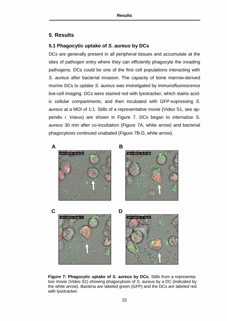
5.1 Phagocytic uptake of S. aureus by DCs
DCs are generally present in all peripheral tissues and accumulate at the
sites of pathogen entry where they can efficiently phagocyte the invading
pathogens. DCs could be one of the first cell populations interacting with
S. aureus after bacterial invasion. The capacity of bone marrow-derived
murine DCs to uptake S. aureus was investigated by immunofluorescence
live-cell imaging. DCs were stained red with lysotracker, which stains acid-
ic cellular compartments, and then incubated with GFP-expressing S.
aureus at a MOI of 1:1. Stills of a representative movie (Video S1, see ap-
pendix I. Videos) are shown in Figure 7. DCs began to internalize S.
aureus 30 min after co-incubation (Figure 7A, white arrow) and bacterial
phagocytosis continued unabated (Figure 7B-D, white arrow).
Figure 7: Phagocytic uptake of S. aureus by DCs. Stills from a representa-tive movie (Video S1) showing phagocytosis of S. aureus by a DC (indicated by the white arrow). Bacteria are labeled green (GFP) and the DCs are labeled red with lysotracker.
A
C D
B

Results
23
After 1 h of infection, a large proportion of the internalized bacteria appear
in yellow indicating co-localization within acidic phagocytic vacuoles. The
red and green fluorescence associated with DCs were then quantified us-
ing ImageJ software analysis, which plot the mean fluorescence of repre-
sentative cells in consecutive frames obtained from a live-cell imaging vid-
eo. Representative frames at time 0 and 64 minutes (min) of live imaging
are displayed in Figure 8A. As shown in Figure 8B, fluorescence associat-
ed with S. aureus significantly increased in sequential frames confirming
the continuous bacterial uptake by the DCs. Furthermore, the intensity of
red fluorescence (lysotracker) also increased in a chronological manner
indicating a progressive acidification of the phagosomes within the DCs
(Figure 8C).
Figure 8: Phagosome acidification in S. aureus-infected DCs. (A) Repre-sentative frames at time 0 (upper panels) and 64 (lower panels) min of a live-cell video showing lysotracker (red), S. aureus (green) and the overlaid image. Fluo-rescence profile of GFP (B) and lysotracker (C) were analyzed frame by frame using ImageJ software.
A
B C

Results
24
5.2 S. aureus induces maturation of DCs
Internalization of pathogens can subsequently trigger DC maturation and
migration from peripheral tissues to lymphoid organs. The process of DC
maturation, in general, involves a redistribution of MHC Class II from intra-
cellular endocytic compartments to the DC surface, an increase in the sur-
face expression of co-stimulatory molecules as well as secretion of cyto-
kines. Therefore, the ability of S. aureus to induce maturation of DCs was
determined by measuring the up-regulation of the surface maturation
markers CD40, CD80, CD86, and MHC class II at 6 h after bacterial infec-
tion. To this end, murine bone marrow-derived DCs were incubated with S.
aureus for 2 h, extracellular bacteria killed by addition of Gentamicin, and
DCs were further incubated for 6 h. Uninfected DCs were used as nega-
tive control and DCs stimulated with 2 µg/ml of LPS were used as positive
control. After 6 h of culture, DCs were labeled with FITC-conjugated mon-
oclonal antibodies to CD40, CD80, CD86, or MHC class II molecules and
PE-conjugated antibodies to CD11c, and analyzed by flow cytometry. The
CD11c+ cell population was gated and analyzed for the expression of
CD40, CD80, CD86, and MHC class II expression. A significant increase
in the mean fluorescence intensity (MFI) of the different maturation mark-
ers was detected in S. aureus-infected DCs when compared with DCs cul-
tured with medium alone (CTR) (Figure 9A). The average level of matura-
tion markers induced by S. aureus was significantly greater than that in-
duced by LPS after 6 h of stimulation (Figure 9A). In addition, the capacity
of S. aureus to induce IL-6, IL-12, and TNF- secretion by DCs was also
evaluated. For this purpose, the levels of these cytokines were determined
in the culture supernatant of S. aureus-infected DCs by ELISA. S. aureus
was found to induce the release of high levels of all these pro-
inflammatory cytokines with the mean values comparable to those induced
by LPS (Figure 9B).

Results
25
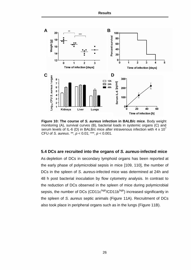
5.3 The mouse model of S. aureus infection
A murine model of S. aureus bacteremia was then used to determine the
relevance of DCs for host response against S. aureus. In this infection
model, BALB/c mice were intravenously inoculated with 4 x 10⁷ CFU of S.
aureus and the course of infection was monitored by determination of
weight loss, survival, and bacterial burdens in systemic organs at progres-
sive times after bacterial inoculation. Shortly after infection, all mice dis-
played overt clinical signs of morbidity as determined by a decrease in
their physical activity, a tendency to huddle, piloerection, and gradual
weight loss (Figure 10A). Infected mice showed a progressive mortality
starting at day 2 post-inoculation, with 100% mortality by day 4 of infection
(Figure 10B) and progressive bacterial growth in kidneys, liver and lungs
(Figure 10C). In addition, serum levels of IL-6, an early marker of gram-
positive sepsis [108], increased progressively in S. aureus-infected mice
(Figure 10D). Therefore, the inability of BALB/c mice to clear S. aureus
infection results in an overwhelming systemic inflammatory response that
culminates in the development of sepsis and death.
Figure 9: S. aureus induces maturation of DCs. (A) Mean fluorescence inten-sity (MFI) of CD40, CD80, CD86, or MHC class II molecules on DCs left untreat-ed (white bars), stimulated with LPS (hatched bars) or infected with S. aureus (black bars) determined by flow cytometry analysis. (B) Levels of IL-6, IL-12 and
TNF- determined in the culture supernatant by ELISA. Each bar represents the mean ± SD of triplicates from three independent experiments. *, p < 0.05; **, p < 0.01; ***, p < 0.001.
A B
Results
26
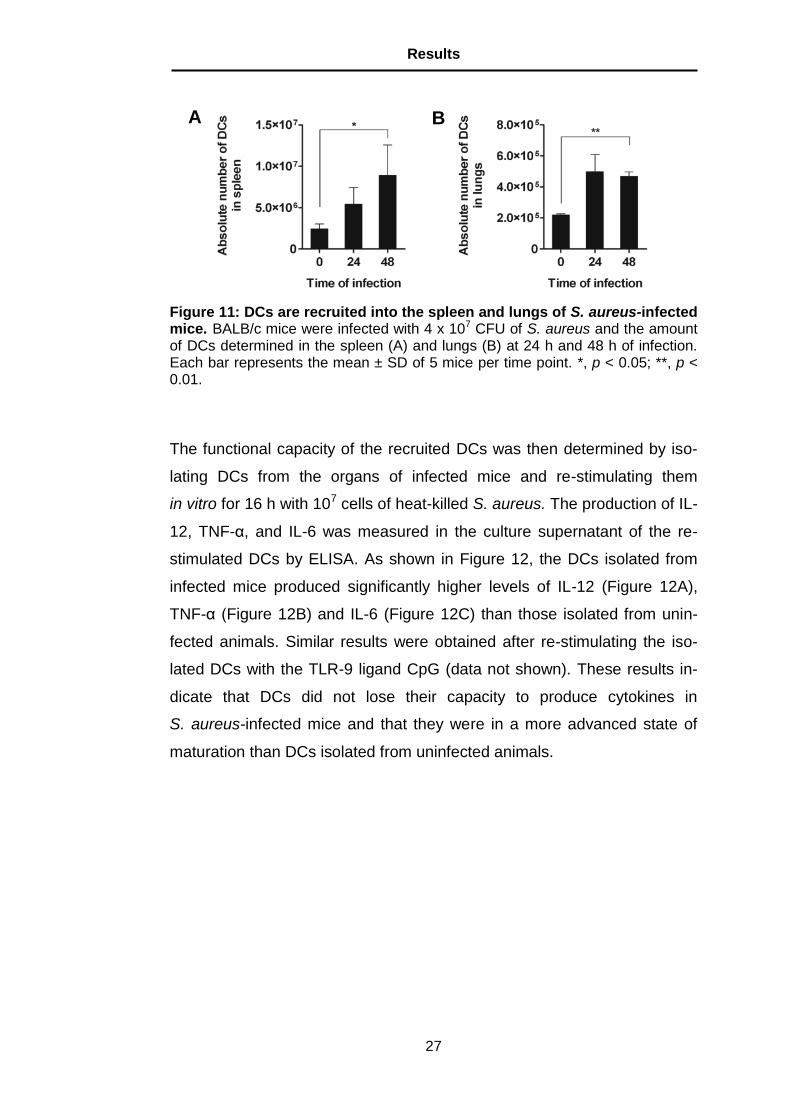
5.4 DCs are recruited into the organs of S. aureus-infected mice
As depletion of DCs in secondary lymphoid organs has been reported at
the early phase of polymicrobial sepsis in mice [109, 110], the number of
DCs in the spleen of S. aureus-infected mice was determined at 24h and
48 h post bacterial inoculation by flow cytometry analysis. In contrast to
the reduction of DCs observed in the spleen of mice during polymicrobial
sepsis, the number of DCs (CD11chigh/CD11bhigh) increased significantly in
the spleen of S. aureus septic animals (Figure 11A). Recruitment of DCs
also took place in peripheral organs such as in the lungs (Figure 11B).
Figure 10: The course of S. aureus infection in BALB/c mice. Body weight monitoring (A), survival curves (B), bacterial loads in systemic organs (C) and serum levels of IL-6 (D) in BALB/c mice after intravenous infection with 4 x 10⁷ CFU of S. aureus. **, p < 0.01; ***, p < 0.001.
A B
C D
Results
27
Figure 11: DCs are recruited into the spleen and lungs of S. aureus-infected mice. BALB/c mice were infected with 4 x 10⁷ CFU of S. aureus and the amount of DCs determined in the spleen (A) and lungs (B) at 24 h and 48 h of infection. Each bar represents the mean ± SD of 5 mice per time point. *, p < 0.05; **, p < 0.01.
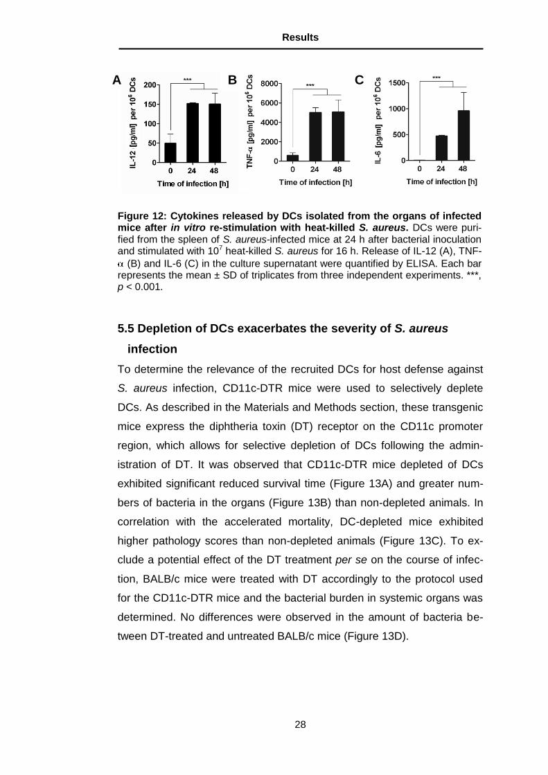
The functional capacity of the recruited DCs was then determined by iso-
lating DCs from the organs of infected mice and re-stimulating them
in vitro for 16 h with 107 cells of heat-killed S. aureus. The production of IL-
12, TNF-α, and IL-6 was measured in the culture supernatant of the re-
stimulated DCs by ELISA. As shown in Figure 12, the DCs isolated from
infected mice produced significantly higher levels of IL-12 (Figure 12A),
TNF-α (Figure 12B) and IL-6 (Figure 12C) than those isolated from unin-
fected animals. Similar results were obtained after re-stimulating the iso-
lated DCs with the TLR-9 ligand CpG (data not shown). These results in-
dicate that DCs did not lose their capacity to produce cytokines in
S. aureus-infected mice and that they were in a more advanced state of
maturation than DCs isolated from uninfected animals.
A B
Results
28
5.5 Depletion of DCs exacerbates the severity of S. aureus
infection
To determine the relevance of the recruited DCs for host defense against
S. aureus infection, CD11c-DTR mice were used to selectively deplete
DCs. As described in the Materials and Methods section, these transgenic
mice express the diphtheria toxin (DT) receptor on the CD11c promoter
region, which allows for selective depletion of DCs following the admin-
istration of DT. It was observed that CD11c-DTR mice depleted of DCs
exhibited significant reduced survival time (Figure 13A) and greater num-
bers of bacteria in the organs (Figure 13B) than non-depleted animals. In
correlation with the accelerated mortality, DC-depleted mice exhibited
higher pathology scores than non-depleted animals (Figure 13C). To ex-
clude a potential effect of the DT treatment per se on the course of infec-
tion, BALB/c mice were treated with DT accordingly to the protocol used
for the CD11c-DTR mice and the bacterial burden in systemic organs was
determined. No differences were observed in the amount of bacteria be-
tween DT-treated and untreated BALB/c mice (Figure 13D).
Figure 12: Cytokines released by DCs isolated from the organs of infected mice after in vitro re-stimulation with heat-killed S. aureus. DCs were puri-fied from the spleen of S. aureus-infected mice at 24 h after bacterial inoculation and stimulated with 107 heat-killed S. aureus for 16 h. Release of IL-12 (A), TNF-
(B) and IL-6 (C) in the culture supernatant were quantified by ELISA. Each bar represents the mean ± SD of triplicates from three independent experiments. ***, p < 0.001.
A
C A B
B C

Results
29
5.6 Reconstitution of DCs-depleted mice by adoptive transfer of
DCs reverses the effect of DC depletion in the course of
S. aureus infection
Van Rijt et al. reported that treatment of CD11c-DTR mice with DT also
induces the depletion of alveolar macrophages since this cell population
expresses high levels of CD11c [111]. Therefore, the effect of DT treat-
ment in the alveolar macrophage population of CD11c-DTR mice was in-
vestigated. CD11c and CD11b markers were used to differentiate
alveolar macrophages (CD11chigh/CD11blow) from lung DCs
(CD11cintermediate/CD11bhigh). The results show that treatment with DT in-
Figure 13: Depletion of DCs aggravates the severity of S. aureus infec-tion. (A) Survival curves of DCs-depleted (broken line) and non-depleted (continuous line) mice after intravenous inoculation with S. aureus. Bacteri-al loads (B) and pathology scores (C) in the organs of DCs-depleted (black bars) and non-depleted (white bars) mice at 24 h after bacterial inoculation. (D) Bacterial loads in the organs of DT-treated (black bars) or PBS-treated (white bars) BALB/c mice at 24 h after S. aureus inoculation. Data are rep-resentative of three independent experiments. **, p < 0.01; ***, p < 0.001.
A B
C D
Results
30
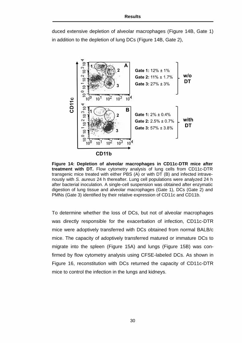
duced extensive depletion of alveolar macrophages (Figure 14B, Gate 1)
in addition to the depletion of lung DCs (Figure 14B, Gate 2),
To determine whether the loss of DCs, but not of alveolar macrophages
was directly responsible for the exacerbation of infection, CD11c-DTR
mice were adoptively transferred with DCs obtained from normal BALB/c
mice. The capacity of adoptively transferred matured or immature DCs to
migrate into the spleen (Figure 15A) and lungs (Figure 15B) was con-
firmed by flow cytometry analysis using CFSE-labeled DCs. As shown in
Figure 16, reconstitution with DCs returned the capacity of CD11c-DTR
mice to control the infection in the lungs and kidneys.
Figure 14: Depletion of alveolar macrophages in CD11c-DTR mice after treatment with DT. Flow cytometry analysis of lung cells from CD11c-DTR transgenic mice treated with either PBS (A) or with DT (B) and infected intrave-nously with S. aureus 24 h thereafter. Lung cell populations were analyzed 24 h after bacterial inoculation. A single-cell suspension was obtained after enzymatic digestion of lung tissue and alveolar macrophages (Gate 1), DCs (Gate 2) and PMNs (Gate 3) identified by their relative expression of CD11c and CD11b.

Results
31
Figure 15: Tracing of adoptively transferred CFSE-labeled DCs by flow cytometry. S. aureus-infected mice were intravenously injected with either ma-ture or immature CFSE-labeled DCs and single cells released from whole spleen (A) or lung (B) digestion were analyzed by flow cytometry. Each bar rep-resents the mean ± SD percentage of CFSE-stained DCs.
Figure 16: DC reconstitution improves the capacity of DCs-depleted CD11c-DTR mice to control S. aureus infection. Bacterial loads in the lungs, kidneys and livers of CD11c-DTR mice treated with DT 24 h prior inoculation with S. aureus and intravenously injected at 16 h of infection with 106 DCs obtained from BALB/c mice. CFU were determined in the different organs at 24 h after DCs transfer. Each bar represents the mean ± SD of three independent experi-ments. *, p < 0.05; ***, p < 0.001.
A B

Results
32
5.7 Adoptive transfer of DCs improves the course of infection in
BALB/c mice
As depletion of DCs resulted in disease exacerbation, it can be hypothe-
sized that increasing the amount of DCs could improve the course of infec-
tion in BALB/c mice and thus be used as a therapeutic intervention. To
test this assumption, BALB/c mice were injected with 106 immature or
LPS-matured DCs 16 h after bacterial inoculation and the bacterial loads
were determined in the systemic organs 24 h after the injection of DCs.
Results in Figure 17 show that both immature and mature DCs improved
the capacity of BALB/c mice to control S. aureus in the lungs.
5.8 DCs do not contribute to direct killing of S. aureus
The next step was to investigate the molecular mechanisms underlying the
beneficial effect afforded by DCs during experimental S. aureus infection.
As DCs are phagocytic cells, the ability of DCs to contribute to direct killing
of S. aureus was evaluated. For this purpose, DCs were infected with S.
aureus at a MOI of 1:10 and the amount of intracellular bacteria deter-
mined at progressive times of infection. The results in Figure 18 show that
S. aureus survived within the DCs.
Figure 17: Effect of adoptive transfer of DCs on the capacity of BALB/c mice to control S. aureus infection. Bacterial loads in the lungs, kidneys, and livers of BALB/c mice intravenously injected at 16 h after bacterial inoculation with either 106 mature DCs (white bars), 106 immature DCs (hatched bars), or PBS (black bars). CFU were determined in the different organs at 24 h after DCs transfer. Each bar represents the mean ± SD of three independent experiments. ***, p < 0.001.
Results
33
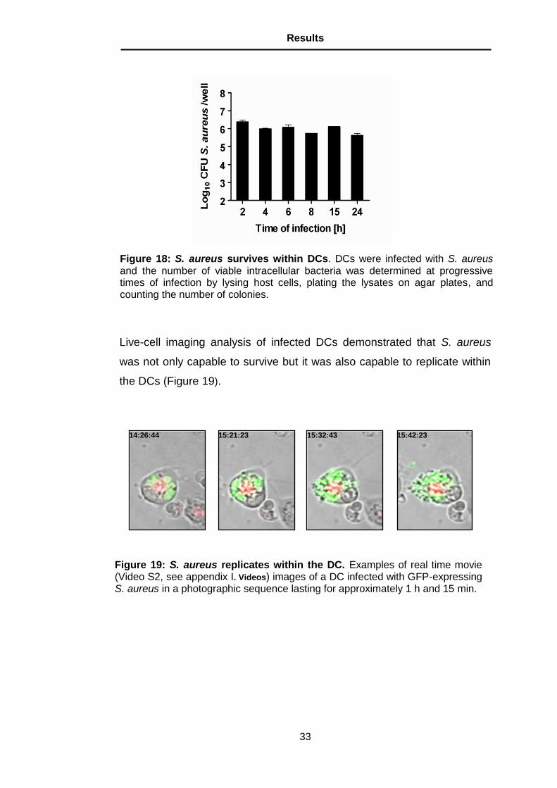
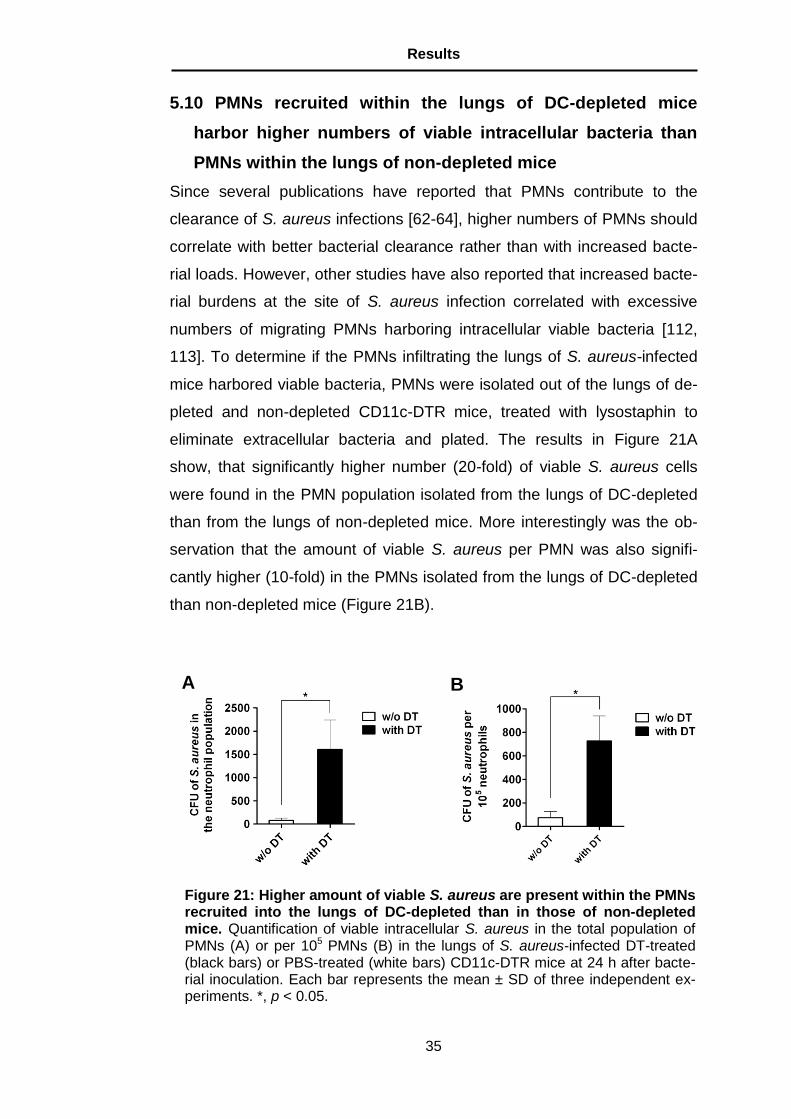
Live-cell imaging analysis of infected DCs demonstrated that S. aureus
was not only capable to survive but it was also capable to replicate within
the DCs (Figure 19).
Figure 18: S. aureus survives within DCs. DCs were infected with S. aureus and the number of viable intracellular bacteria was determined at progressive times of infection by lysing host cells, plating the lysates on agar plates, and counting the number of colonies.
Figure 19: S. aureus replicates within the DC. Examples of real time movie (Video S2, see appendix I. Videos) images of a DC infected with GFP-expressing S. aureus in a photographic sequence lasting for approximately 1 h and 15 min.
14:26:44 15:21:23 15:32:43 15:42:23

Results
34
5.9 Depletion of DCs leads to an enhanced recruitment of PMNs
into the lungs of infected mice
As a direct contribution of DCs to bacterial killing was excluded, the possi-
bility that the beneficial effect of DCs was due to their involvement in the
recruitment of inflammatory cells involved in bacterial clearance was in-
vestigated. For this purpose the lungs were removed from DCs-depleted
and non-depleted mice before and 24 h after S. aureus inoculation, di-
gested and the amount of PMNs was determined by flow cytometry analy-
sis. The results demonstrate an enhanced recruitment of PMNs into the
lungs of DC-depleted animals, not only in percentage (Figure 14, Gate 3),
but also in total numbers (Figure 20).
Figure 20: DC-depletion is associated with an increased recruitment of PMNs in the lungs of S. aureus-infected mice. (A) Representative histograms for expression of Gr-1 molecule (PMNs) in the lung cells of uninfected (upper panel), S. aureus-infected and PBS-treated (middle panel) or S. aureus-infected and DT-treated (lower panel) CD11c-DTR mice at 24 h after bacterial inoculation. (B) Absolute number of PMNs recruited into the lungs in the above-mentioned groups. Each bar represents the mean ± SD of three independent experiments. *, p < 0.05; **, p < 0.01; ***, p < 0.001.
A B
Results
35
5.10 PMNs recruited within the lungs of DC-depleted mice
harbor higher numbers of viable intracellular bacteria than
PMNs within the lungs of non-depleted mice
Since several publications have reported that PMNs contribute to the
clearance of S. aureus infections [62-64], higher numbers of PMNs should
correlate with better bacterial clearance rather than with increased bacte-
rial loads. However, other studies have also reported that increased bacte-
rial burdens at the site of S. aureus infection correlated with excessive
numbers of migrating PMNs harboring intracellular viable bacteria [112,
113]. To determine if the PMNs infiltrating the lungs of S. aureus-infected
mice harbored viable bacteria, PMNs were isolated out of the lungs of de-
pleted and non-depleted CD11c-DTR mice, treated with lysostaphin to
eliminate extracellular bacteria and plated. The results in Figure 21A
show, that significantly higher number (20-fold) of viable S. aureus cells
were found in the PMN population isolated from the lungs of DC-depleted
than from the lungs of non-depleted mice. More interestingly was the ob-
servation that the amount of viable S. aureus per PMN was also signifi-
cantly higher (10-fold) in the PMNs isolated from the lungs of DC-depleted
than non-depleted mice (Figure 21B).
Figure 21: Higher amount of viable S. aureus are present within the PMNs recruited into the lungs of DC-depleted than in those of non-depleted mice. Quantification of viable intracellular S. aureus in the total population of PMNs (A) or per 105 PMNs (B) in the lungs of S. aureus-infected DT-treated (black bars) or PBS-treated (white bars) CD11c-DTR mice at 24 h after bacte-rial inoculation. Each bar represents the mean ± SD of three independent ex-periments. *, p < 0.05.
A B
Results
36
5.11 Higher levels of CXC chemokines are produced in the
lungs of DC-depleted than in the lungs of non-depleted
S. aureus-infected mice
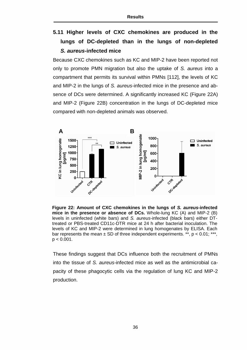
Because CXC chemokines such as KC and MIP-2 have been reported not
only to promote PMN migration but also the uptake of S. aureus into a
compartment that permits its survival within PMNs [112], the levels of KC
and MIP-2 in the lungs of S. aureus-infected mice in the presence and ab-
sence of DCs were determined. A significantly increased KC (Figure 22A)
and MIP-2 (Figure 22B) concentration in the lungs of DC-depleted mice
compared with non-depleted animals was observed.
These findings suggest that DCs influence both the recruitment of PMNs
into the tissue of S. aureus-infected mice as well as the antimicrobial ca-
pacity of these phagocytic cells via the regulation of lung KC and MIP-2
production.
Figure 22: Amount of CXC chemokines in the lungs of S. aureus-infected mice in the presence or absence of DCs. Whole-lung KC (A) and MIP-2 (B) levels in uninfected (white bars) and S. aureus-infected (black bars) either DT-treated or PBS-treated CD11c-DTR mice at 24 h after bacterial inoculation. The levels of KC and MIP-2 were determined in lung homogenates by ELISA. Each bar represents the mean ± SD of three independent experiments. **, p < 0.01; ***, p < 0.001.
A B

Results
37
5.12 Partial depletion of PMNs does not reverse the detrimental
effect of DC depletion
Gresham et al. [112] reported that partial depletion of PMNs in S. aureus-
infected mice resulted in lower amount of bacteria in organs and increased
survival rate. To determine if partial depletion of PMNs can reduce the
bacterial burdens in the experimental setting used in the study presented
here, DCs-depleted mice were treated with 5 µg of anti-RB6 antibodies to
achieve a partial depletion of circulating PMNs (30% of depletion) prior to
intravenous infection with S. aureus. As shown in Figure 23, a partial de-
pletion of PMNs did not influence the bacterial loads in the organs of DC-
depleted mice determined at 24 h after bacterial inoculation.
5.13 Influence of S. aureus in the capacity of DCs to stimulate
naïve antigen-specific CD4+ T cells
DCs are potent antigen-presenting cells (APCs) that possess the ability to
stimulate naïve T cells. To determine the influence of S. aureus in the ca-
pacity of DCs to stimulate naïve antigen-specific CD4+ T cells, DCs (either
infected or uninfected) were loaded with OVA323-339 or left untreated and
co-cultivated with CD4+ T cells from OT-II mice, which are specific for this
peptide. A significantly higher proliferation was observed when CD4+ T
Figure 23: Effect of partial depletion of PMNs on bacterial burdens in the organs of DC-depleted mice. Bacterial loads in the organs of DCs-depleted mice, which have been partially depleted of PMNs (black bars) or left untreated (white bars) at 24 h after intravenous inoculation with S. aureus. Each bar repre-sents the mean ± SD of two independent experiments.
Results
38
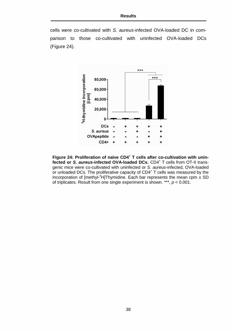
cells were co-cultivated with S. aureus-infected OVA-loaded DC in com-
parison to those co-cultivated with uninfected OVA-loaded DCs
(Figure 24).
Figure 24: Proliferation of naïve CD4+ T cells after co-cultivation with unin-fected or S. aureus-infected OVA-loaded DCs. CD4+ T cells from OT-II trans-genic mice were co-cultivated with uninfected or S. aureus-infected, OVA-loaded or unloaded DCs. The proliferative capacity of CD4+ T cells was measured by the incorporation of [methyl-3H]Thymidine. Each bar represents the mean cpm ± SD of triplicates. Result from one single experiment is shown. ***, p < 0.001.
Discussion
39
6. Discussion
During the last decades, an increasing number of infections caused by S.
aureus has been reported worldwide [1, 2]. The increasing prevalence of
staphylococcal infections is a direct consequence of the continuing evolu-
tion of antimicrobial resistance among S. aureus strains, added to the in-
creased use of implantable devices and the boosting in the number of pa-
tients with immunocompromised status because of HIV infection or immu-
nosuppression after transplantation or cancer treatment [114-116]. There-
fore, new therapeutic options with novel models of actions that may cir-
cumvent antibiotic resistance are required to tackle the problem posed by
S. aureus in a more effective way. In this regard, therapeutic approaches
aimed to enhance the efficiency of the host immune response to eliminate
S. aureus may represent the best option. However, a more precise
knowledge of the immune mechanisms involved in host defense against
S. aureus is required in order to understand how the immune system can
be manipulated to achieve a more efficient control of infection.
In this work, the role of DCs, a subset of cells unique in their capacity to
regulate the immune response, in the pathogenesis of S. aureus infections
was investigated.
First, the interactions between DCs and S. aureus were evaluated using
live-cell imaging microscopy (see section 5.1). The results of these exper-
iments demonstrate the remarkable capacity of DCs to trace and phago-
cyte S. aureus. After encounter S. aureus, DCs initiated a maturation pro-
cess as shown by the up-regulation of co-stimulatory molecules CD40,
CD80, CD86, and MHC class II as well as the release of proinflammatory
cytokines IL-6, IL-12, and TNFα.
A murine model of infection was used to investigate the behavior of DCs
during S. aureus infection under in vivo conditions. In this model, BALB/c
mice were intravenously infected with S. aureus strain SH1000 as previ-
ously described [64]. Examination of the amount of DCs in lungs and
spleen of infected mice at progressing times after bacterial inoculation
demonstrated that DCs were mobilized and actively recruited into infected

Discussion
40
tissue during the course of infection. Isolation and in vitro re-stimulation of
the recruited DCs with heat-killed S. aureus or with the TLR-9 ligand CpG
revealed that these cells were in a more advance stage of activation than
DCs from uninfected animals since they produced significantly higher lev-
els of inflammatory cytokines.
The next step was to determine the relevance of DCs for host defense
against S. aureus infection. For this purpose, CD11c-DTR transgenic
mice, which allows for selective depletion of DCs [105], were used. Deple-
tion of DCs resulted in substantial worsening of pathogen clearance, par-
ticularly in the lungs and kidneys, and in accelerated mortality of infected
animals. In accordance with this data, a recent study reported that deple-
tion of DCs in mice intranasally inoculated with S. aureus was associated
with an increased bacterial load in the lungs [117]. To exclude an influence
of the DT itself on the course of S. aureus infection, BALB/c mice were
treated with DT before infection in a similar way as CD11c-DTR mice were
treated and bacterial loads were monitored. Treatment with DT alone had
no effect on the course of infection confirming that the observed exacerba-
tion of infection was indeed caused by DC depletion and not by side ef-
fects of DT treatment.
As the lung was the most affected organ by DC depletion and it has been
shown that alveolar macrophages can also be depleted by DT treatment
[111], the potential depletion of alveolar macrophages by DT treatment in
the experimental settings used in this study was evaluated. The CD11c
and CD11b markers were used to differentiate alveolar macrophages
(CD11chigh/CD11blow) from lung DCs (CD11cintermediate/CD11bhigh) by FACS
analysis. The results show that, in addition to the depletion of lung DCs,
treatment with DT induced extensive depletion of alveolar macrophages
(Figure 14). To determine whether the loss of DCs but not of alveolar mac-
rophages was directly responsible for the exacerbation of infection, DC-
depleted CD11c-DTR mice were adoptively transferred with bone marrow-
derived DCs obtained from BALB/c mice. The obtained results demon-
strated that intravenous injection of DCs returned the capacity of DCs-
depleted CD11c-DTR mice to control the infection to the levels observed
Discussion
41
in non-depleted animals clearly confirming that the observed increased
severity of infection in DT-treated CD11c-DTR mice was specifically at-
tributable to the reduction of DCs (Figure 16).
As the above-presented data indicates that DCs played an important role
in host defense against S. aureus infection, it was then hypothesized that
increasing DC numbers in normal BALB/c mice could improve their re-
sistance to S. aureus infection. Indeed, adoptive transfer of either imma-
ture or LPS-matured DCs into normal BALB/ mice improved the capacity
of these animals to clear S. aureus in the lungs (Figure 17). These results
implied that DCs do not only play a protective role during S. aureus infec-
tion but may also be used as a therapeutic intervention.
The next step was to investigate the immunological mechanisms underly-
ing the beneficial effect afforded by DCs during S. aureus bloodstream
infection. As DCs are phagocytic cells, the ability of DCs to contribute to
direct killing of S. aureus was evaluated. For this purpose, DCs were in-
fected with S. aureus and the amount of intracellular bacteria was moni-
tored at progressive times of infection. Interestingly, the DCs were not ca-
pable to kill the intracellular S. aureus, as the number of viable bacteria
remained unaltered over time. Furthermore, live-imaging microscopy
demonstrated the ability of S. aureus to replicate within the infected DCs
(Video S2). Likewise, S. aureus has been shown to survive within other
professional phagocytes, such as human [118] and murine PMNs [112] as
well as human monocyte-derived macrophages [119]. Within these cells,
S. aureus can evade the lethal effect of the phagolysosome by producing
oxygen free radicals scavenger compounds such as catalases [120],
staphyloxanthin [121], and yellow carotenoid pigments [12].
After the demonstration that DCs do not play a major role in direct bacteri-
al clearance, it was then hypothesized that DCs may indirectly contribute
to bacterial killing through their participation in the recruitment of inflamma-
tory cells involved in bacterial eradication into the site of infection. As it
has long been known that PMNs play a key role in the host innate immune
response towards S. aureus [62-64], the amount of PMNs recruited in the
lungs of DC-depleted and non-depleted mice during the course of S.
Discussion
42
aureus infection was monitored. The obtained results revealed that the
recruitment of PMNs in infected tissue was increased rather than de-
creased in mice depleted of DCs (Figure 20). These findings demonstrate
that exacerbation of S. aureus infection in DC-depleted mice was not at-
tributable to a reduced recruitment of PMNs and also suggest that increas-
ing the number of PMNs in infected organs was rather detrimental to the
host. Despite their role in bacterial clearance, there is now substantial evi-
dence suggesting that through the release of cytotoxic compounds PMNs
themselves can contribute to the pathogenesis of tissue injury associated
with staphylococcal infection [122]. Furthermore, the results also indicate
that the increase neutrophil numbers in DC-depleted mice correlate with
increase in bacterial counts rather than with increased bacterial killing.
Similar observations have been reported in murine models of S. aureus
peritonitis [112] and hind paw infection [113] as well as in patients with S.
aureus bloodstream infections [123-125].
Even more interestingly was the observation that neutrophils isolated from
the lungs of DC-depleted mice harbored significantly higher numbers of
intracellular viable S. aureus than those isolated from non-depleted ani-
mals (Figure 21). This implies that the ability of the recruited neutrophils to
kill ingested S. aureus is even poorer in the absence of DCs. In this re-
gard, several studies have provided evidence that local release of CXC
chemokines promoted survival of S. aureus within neutrophils by facilitat-
ing bacterial internalization into an intracellular niche where they are pro-
tected from the host’s killing mechanisms [112, 113]. In accordance with
those studies, it was found that the levels of KC and MIP-2 in the lungs of
DC-depleted S. aureus-infected mice were significantly higher than in non-
depleted animals. Therefore, it is likely that DCs can regulate the produc-
tion of CXC chemokine by other cell populations such as epithelial cells in
the infected tissue.
In summary, the results of this study provide compelling evidence that
DCs are important for host defense against S. aureus infection. It can be
proposed that by orchestrating the local production of CXC chemokine and
the recruitment and functional activity of PMNs DCs serve as modulators

Discussion
43
of the local inflammatory responses elicited during S. aureus infection to
prevent tissue injury.
DCs are also very important players during the development of an adap-
tive immune response [44]. CD4+ T cells respond to peptide antigen dis-
played on MHC class II expressed on the surface of dendritic cells. Acces-
sory molecules such as CD80 and CD86 expressed by DCs are required
to ensure that T cells will divide and differentiate into effector cells. As DCs
were capable to up-regulate the expression of MHC class II and accessory
molecules in response to S. aureus infection, the capacity of infected DCs
to stimulate naïve antigen-specific CD4+ T cells was examined using OT-II
transgenic mice. The results show that OVA-loaded S. aureus-infected
DCs induced significantly higher levels of proliferation in CD4+ T cells from
OT-II mice than OVA-loaded uninfected DCs. These results indicate that
DCs may also play an important role driving the generation of the primary
antigen-specific immune response during S. aureus infection. More de-
tailed analysis of the role of DCs on the acquired immune response to S.
aureus should be performed in future studies.
References
44
References
1. Piper W, Innere Medizin. 2007, Heidelberg: Springer Medizin Verlag.
2. Lowy FD, Staphylococcus aureus infections. N Engl J Med, 1998. 339(8):
pp. 520 - 532.
3. Ogston A, Micrococcus poisoning. J Anat, 1882. 17: pp. 24 - 58.
4. Classics in infectious diseases: On abscesses: Alexander Ogston
(1844-1929). J Infect Dis, 1984. 6: pp. 122 - 128.
5. Boyce JM, Epidemiology and prevention of nosocomial infections: The
staphylococci in human diseases. 1997, New York: Churchill Livingstone.
6. Tuchscherr L, Medina E, Hussain M, et al., Staphylococcus aureus phe-
notype switching: an effective bacterial strategy to escape host immune
response and establish a chronic infection. EMBO Mol Med, 2011. 3(3):
pp. 129 - 141.
7. Lowy FD, Is Staphylococcus aureus an intracellular pathogen? Trends
Microbiol, 2000. 8(8): pp. 341 - 343.
8. Clement S, Vaudaux P, Francois P, et al., Evidence of an intracellular
reservoir in the nasal mucosa of patients with recurrent Staphylococcus
aureus rhinosinusitis. J Infect Dis, 2005. 192(6): pp. 1023 - 1028.
9. Sendi P, Proctor RA, Staphylococcus aureus as an intracellular pathogen:
the role of small colony variants. Trends Microbiol, 2009. 17(2):
pp. 54 - 58.
10. Zautner AE, Krause M, Stropahl G, et al., Intracellular Persisting Staphy-
lococcus aureus Is the Major Pathogen in Recurrent Tonsillitis. PLoS
ONE, 2010. 5(3): pp. e9452.
11. Oethinger M, Kurzlehrbuch Mikrobiologie und Immunologie. Vol. 11. 2004,
München / Jena: Urban & Fischer.
12. Liu GY, Essex A, Buchanan JT, et al., Staphylococcus aureus golden
pigment impairs neutrophil killing and promotes virulence through its anti-
oxidant activity. J Exp Med, 2005. 202: pp. 209 - 215.
13. Gordon RJ, Lowy FD, Pathogenesis of Methicillin-Resistant Staphylococ-
cus aureus Infection. Clin Infect Dis, 2008(46): pp. 350 - 359.
14. Lee CJ, The prospects for developing a vaccine against Staphylococcus
aureus. Trends Microbiol, 1996(4): pp. 162 - 166.
References
45
15. Foster TJ, McDevitt D, Surface-associated proteins of Staphylococcus
aureus : their possible roles in virulence. FEMS Microbiol Lett, 1994(118):
pp. 192 - 205.
16. Archer GL, Staphylococcus aureus: a well-armed pathogen. Clin Infect
Dis, 1998. 26(5): pp. 1179 - 1181.
17. Bodén MK, Flock JI, Fibrinogen-binding protein/clumping factor from
Staphylococcus aureus. Infect Immun, 1989. 57(8): pp. 2358–2363.
18. Palmqvist N , Patti JM , Tarkowski A, et al., Expression of staphylococcal
clumping factor A impedes macrophage phagocytosis. Microb Infect,
2004. 6(2): pp. 188 -195
19. Palmqvist N, Josefsson E, Tarkowski A, Clumping factor A-mediated viru-
lence during Staphylococcus aureus infection is retained despite fibrino-
gen depletion. Microb Infect, 2004. 6(2): pp. 196-201
20. Nardella FA, Teller DC, Barber CV, et al., IgG rheumatoid factors and
staphylococcal protein A bind to a common molecular site on IgG. JEM,
1985. 162(5): pp. 1811-1824.
21. Nilsson B, Moks T, Jansson B, et al., A synthetic IgG-binding domain
based on staphylococcal protein A. Protein Eng, 1987. 1(2): pp. 107-113.
22. Goodyear CS, Silverman GJ, Death by a B-cell superantigen: In vivo VH-
targeted apoptotic supraclonal B-cell deletion by a staphylococcal toxin.
J Exp Med, 2003. 197: pp. 1125-1139.
23. Downer R, Roche F, Park PW, et al., The Elastin-binding Protein of
Staphylococcus aureus(EbpS) Is Expressed at the Cell Surface as an In-
tegral Membrane Protein and Not as a Cell Wall-associated Protein. J Biol
Chem, 2004(277): pp. 243 - 250.
24. Hienz SA, Schennings T, Heimdahl A, et al., Collagen Binding of Staphy-
lococcus aureus Is a Virulence Factor in Experimental Endocarditis.
J Infect Dis, 1996. 147(1): pp. 83 - 88.
25. Höök M, Fibronection Binding Protein. 1997, Alfa-Laval Agri International
Aktiebolag: Sweden.
26. Bhakdi S, Tranum-Jensen J, Alpha-toxin of Staphylococcus aureus.
Microbiol Rev, 1991. 55(4): pp. 733 - 751.
27. Wahlsten JL, Ramakrishnan S, Separation of Function Between the Do-
mains of Toxic Shock Syndrome Toxin-1. J Immunol, 1998. 160:
pp. 854-859.
28. Muraille E, Smedt T DE, Andris F, et al., Staphylococcal enterotoxin B
induces an early and transient state of immunosuppression characterized
References
46
by V beta-unrestricted T cell unresponsiveness and defective antigen-
presenting cell functions J Immunol, 1997. 158(6): pp. 2638-2647.
29. Kluytmans J, van Belkum A, Verbrugh H, Nasal carriage of Staphylococ-
cus aureus: epidemiology, underlying mechanisms, and associated risks.
Clin Microbiol Rev, 1997. 10(3): pp. 505 - 520.
30. Speziale P, Pietrocola G, Rindi S, et al., Structural and functional role of
Staphylococcus aureus surface components recognizing adhesive matrix
molecules of the host. Future Microbiol, 2009. 4(10): pp. 1337 - 1352.
31. Bantel H, Sinha B, Domschke W, et al., α-Toxin is a mediator of Staphy-
lococcus aureus-induced cell death and activates caspases via the intrin-
sic death pathway independently of death receptor signaling. J Cell Biol,
2001. 155(4): pp. 637–648.
32. O'Connell DP, Nanavaty T, McDevitt D, et al., The fibrinogen-binding
MSCRAMM (clumping factor) of Staphylococcus aureus has a Ca2+-
dependent inhibitory site. J Biol Chem, 1998. 273(12): pp. 6821 - 6829.
33. Foster TJ, Höök M, Surface protein adhesins of Staphylococcus aureus.
Trends Microbiol, 1998. 6(12): pp. 484 - 488.
34. Peacock SJ, Moore CE, Justice A, et al., Virulent combinations of adhesin
and toxin genes in natural populations of Staphylococcus aureus. Infect
Immun, 2002. 70(9): pp. 4987 - 4996.
35. Hauck CR, Ohlsen K, Sticky connections: extracellular matrix protein
recognition and integrin-mediated cellular invasion by Staphylococcus
aureus. Current Opin Microbiol, 2006. 9(1): pp. 5 - 11.
36. Bartlett AH, Hulten KG, Staphylococcus aureus Pathogenesis - Secretion
Systems, Adhesins, and Invasins. PIDJ, 2010. 29(9): pp. 860 - 861.
37. Schröder A, Schröder B, Roppenser B, et al., Staphylococcus aureus
Fibronectin Binding Protein-A Induces Motile Attachment Sites and Com-
plex Actin Remodeling in Living Endothelial Cells. Mol Biol Cell, 2006.
17(12): pp. 5198 - 5210.
38. Chang F-Y, Stapylococcus aureus bacteremia and endocarditis.
J Microbiol Immunol Infect, 2000(33): pp. 63 - 68.
39. Bamberger DM, Boyd SE, Management of Staphylococcus aureus Infec-
tions. Am Fam Physician, 2005(12): pp. 2474-2481.
40. Kollef MK, Shorr A, Tabak YP, et al., Epidemiology and outcomes of
health-care associated pneumonia. Chest, 2005. 128: pp. 3854 - 3862.
References
47
41. Yocum RR, Rasmussen JR, Strominger JL, The mechanism of action of
penicillin. Penicillin acylates the active site of Bacillus stearothermophilus
D-alanine carboxypeptidase. J Biol Chem, 1980. 255(9): pp. 3977 - 3986.
42. Cohen J, The immunopathogenesis of sepsis. Nature, 2002. 420:
pp. 885 - 981.
43. Lavoie PM, Thibodeau J, Erard F, Sekaly R-P, Understanding the mecha-
nism of action of bacterial superantigens from a decade of research.
Immunol Rev, 2006. 168(1): pp. 257 - 269.
44. Janeway Jr., C.A., Travers, P., Walport, M., Shlomchik, M.J.,
Immunobiology. 6th ed. 2005, New York, London, parts 4 and 5: Garland
Science.
45. Citri N, Pollock MR, The biochemistry and function of beta-lactamase
(penicillinase). Adv Enzymol Relat Areas Mol Biol, 1966(28): pp. 237-323.
46. Fuda CC, Fisher JF, Mobashery S, Beta-lactam resistance in Staphylo-
coccus aureus: the adaptive resistance of a plastic genome. Cell Mol Life
Sci, 2005. 62(22): pp. 2617 - 2633.
47. Jevons, MP, Celbenin-resistant staphylococci. Brit Med J, 1961. 1:
pp. 124 - 125.
48. Barber, M, Methicillin-resistant staphylococci. J Clin Pathol, 1961. 14:
pp. 385 - 393.
49. Sutherland R, Rolinson GN, Characteristics of methicillin-resistant staphy-
lococci. J Bacteriol, 1964. 87(4): pp. 887-899.
50. Fuda C, Suvorov M, Vakulenko SB, et al., The Basis for Resistance to β-
Lactam Antibiotics by Penicillin-binding Protein 2a of Methicillin-resistant
Staphylococcus aureus. J Biol Chem, 2004. 279(39): pp. 40802-40806.
51. CDC NNIS System, National Nosocomial Infections Surveillance (NNIS)
System Report, data summary from January 1992 through June 2004. Am
J Infect Control, 2004: pp. 470 - 485.
52. Hiramatsu K, Vancomycin-resistant Staphylococcus aureus: a new model
of antibiotic resistance. Lancet Infect Dis, 2001. 1(3): pp. 147 - 155.
53. Chang S, Sievert DM, Hageman JC, et al., Vancomycin-Resistant Staphy-
lococcus aureus Investigative Team. Infection with vancomycin-resistant
Staphylococcus aureus containing the vanA resistance gene. N Engl J
Med, 2003. 348(14): pp. 1342 - 1347.
54. Siebert WT, Moreland N, Williams TW Jr, Synergy of vancomycin plus
cefazolin or cephalothin against methicillin-resistance Staphylococcus
epidermidis. J Infect Dis, 1979. 139(4): pp. 452 - 457.

References
48
55. Hiramatsu K, Hanaki H, Ino T, et al., Methicillin-resistant Staphylococcus
aureus clinical strain with reduced vancomycin susceptibility. J Antimicrob
Chemother, 1997. 40: pp. 135 - 136.
56. Fournier B, Philpott DJ, Recognition of Staphylococcus aureus by the
Innate Immune System. Clin Microbiol Rev, 2005. 18(3): pp. 521–540.
57. Romero-Pastrana F, Hernández-Jáuregui P, Baca BE, Characteristics of
Staphylococcus aureus infections to consider in designing an effective
vaccine. Nature Precedings, 2010. hdl:10101/npre.2010.4598.1.
58. Medzhitov R, Toll-like receptors and innate immunity. Nat Rev Immunol,
2001. 1(2): pp. 135 - 145.
59. Akira S, Uematsu S, Takeuchi O., Pathogen recognition and innate im-
munity. Cell Mol Life Sci, 2006. 124(4): pp. 783 - 801.
60. Takeuchi O, Hoshino K, Akira S, Cutting Edge: TLR2-Deficient and
MyD88-Deficient Mice Are Highly Susceptible to Staphylococcus aureus
Infection. J Immunol, 2000. 165: pp. 5392 - 5396.
61. Gómez MI, Lee A, Reddy B, et al., Staphylococcus aureus protein A in-
duces airway epithelial inflammatory responses by activating TNFR1. Nat
Med, 2004. 10(8): pp. 842 - 848.
62. Mölne L, Verdrengh M, Tarkowski A, Role of neutrophil leukocytes in cu-
taneous infection caused by Staphylococcus aureus. Infect Immun, 2000.
68: pp. 6162-6167.
63. Verdrengh M, Tarkowski A, Role of neutrophils in experimental septicemia
and septic arthritis induced by Staphylococcus aureus. Infect Immun,
1997. 67: pp. 2517-2521.
64. von Köckritz-Blickwede M, Rohde M, Oehmcke S, et al., Immunological
mechanisms underlying the genetic predisposition to severe Staphylococ-
cus aureus infection in the mouse model. Am J Pathol, 2008. 173:
pp. 1657-1668.
65. Heyworth PG, Cross AR, Curnutte JT, Chronic granulomatous disease.
Current Opin Immunol 2003. 15(5): pp. 578 - 584.
66. Cheng AG, DeDent AC, Schneewind O, et al., A play in four acts: Staphy-
lococcus aureus abscess formation. Trends Microbiol, 2011. 19(5):
pp. 225 - 232.
67. Steinman RM, Cohn ZA, Identification of a novel cell type in peripheral
lymphoid organs of mice. I. Morphology, quantitation, tissue distribution.
J Exp Med, 1973. 137(5): pp. 1142-1162.
References
49
68. Fernandez NC, Lozier A, Flament C, et al., Dendritic cells directly trigger
NK cell functions: cross-talk relevant in innate anti-tumor immune re-
sponses in vivo. Nat Med, 1999. 5(4): pp. 405 - 411.
69. Foti M, Granucci F, Aggujaro D, et al., Upon dendritic cell (DC) activation
chemokines and chemokine receptor expression are rapidly regulated for
recruitment and maintenance of DC at the inflammatory site. Int Immunol,
1999. 11(6): pp. 979 - 986.
70. Rescigno M, Martino M, Sutherland CL, et al., Dendritic cell survival and
maturation are regulated by different signaling pathways. J Exp Med,
1998. 188(11): pp. 2175 - 2180.
71. Todar, K. Immune Defense against Bacterial Pathogens: Adaptive or Ac-
quired Immunity (page 2). Todar´s Online Textbook of Bacteriology 2011
72. Niess JH, Brand S, Gu X, et al., CX3CR1-mediated dendritic cell access
to the intestinal lumen and bacterial clearance. Science, 2005. 307(5707):
pp. 254 - 258.
73. Randolph GJ, Angeli V, Swartz MA, Dendritic-cell trafficking to lymph
nodes through lymphatic vessels. Nat Rev Immunol, 2005. 5(8):
pp. 617 - 628.
74. Rescigno M, Urbano M, Valzasina B, et al., Dendritic cells express tight
junction proteins and penetrate gut epithelial monolayers to sample bacte-
ria. Nat Immunol, 2001. 2(4): pp. 361 - 367.
75. Riol-Blanco L, Sanchez-Sanchez N, Torres A, et al., The Chemokine Re-
ceptor CCR7 Activates in Dendritic Cells Two Signaling Modules That In-
dependently Regulate Chemotaxis and Migratory Speed. J Immunol,
2005. 174: pp. 4070-4080.
76. Lipscomb MF, Masten BJ, Dendritic Cells: Immune Regulators in Health
and Disease. Physiol Rev, 2002. 82(1): pp. 97 - 130.
77. Banchereau J, Steinman RM., Dendritic cells and the control of immunity.
Nature, 1998. 392(6673): pp. 245 - 252.
78. Molecular basis of T cell migration to effector sites [cited; Available from:
http://www.biken.osaka-u.ac.jp/COE/eng/project/pro14.html.
79. Gilboa, E, The promise of cancer vaccines. Nat Rev Cancer, 2004. 4:
pp. 401 - 411.
80. Ardavín C, Origin, precursors and differentiation of mouse dendritic cells.
Nat Rev Immunol, 2003. 3(7): pp. 582 - 590.

References
50
81. Romani N, Clausen BE, Stoitzner P, Langerhans cells and more: langerin-
expressing dendritic cell subsets in the skin. Immunol Rev, 2010. 234(1):
pp. 120 - 141.
82. Anjuère F, Martín P, Ferrero I, et al., Definition of dendritic cell subpopula-
tions present in the spleen, Peyer's patches, lymph nodes, and skin of the
mouse. Blood, 1999. 93(2): pp. 590 - 598.
83. Henri S, Vremec D, Kamath A, et al., The dendritic cell populations of
mouse lymph nodes. J Immunol, 2001. 167(2): pp. 741 - 748.
84. Scumpia PO, et al, CD11c+ Dendritic Cells Are Required for Survival in
Murine Polymicrobial Sepsis. Journal of immunology, 2005. 175:
pp. 3282-3286.
85. Guisset O, Dilhuydy MS, Thiébaut R, et al., Decrease in circulating den-
dritic cells predicts fatal outcome in septic shock. Intensive Care Med,
2007. 33(1): pp. 148 - 152.
86. Benjamim CF, et al, Reversal of long-therm sepsis-induces immunosup-
pression by dendritic cells. Blood, 2005. 105: pp. 3588 - 3595.
87. Wozniak KL, Vyas JM, Levitz SM, In vivo role of dendritic cells in a murine
model of pulmonary cryptococcosis. Infect Immun, 2006. 74(7):
pp. 3817 - 3824.
88. Loof TG, Rohde M, Chhatwal GS, et al., Role of Macrophages in Host
Resistance to Group A Streptococci. Infect Immun, 2004. 72(5).
89. Loof TG, Goldmann O, Medina E, Immune Recognition of Streptococcus
pyogenes by Dendritic Cells. Infect Immun, 2008. 76(7): pp. 2785 - 2792.
90. Servet-Delprat C, Vidalain PO, Bausinger H, et al., Measles virus induces
abnormal differentiation of CD40 ligand-activated human dendritic cells.
J Immunol, 2000. 164: pp. 1753 - 1760.
91. Marie JC, Kehren J, Trescol-Biemont MC, et al., Mechanism of measles
virus-induced suppression of inflammatory immune responses. Immunity,
2001. 14: pp. 69 - 79.
92. Lugton I, Mucosa-associated lymphoid tissues as sites for uptake, car-
riage and excretion of tubercle bacilli and other pathogenic mycobacteria.
Immunol Cell Biol, 1999. 77: pp. 364 - 372.
93. Marriott I, Hammond TG, Thomas EK, et al., Salmonella efficiently enter
and survive within cultured CD11c+ dendritic cells initiating cytokine ex-
pression. Eur J Immunol, 1999. 29(1107 - 1115).
References
51
94. Stockwin LH, McGonagle D, Martin IG, et al., Dendritic cells: immunologi-
cal sentinels with a central role in health and disease. Immunol Cell Biol,
2000. 78: pp. 91 - 102.
95. Dreher D, Kok M, Cochand L, et al., Genetic background of attenuated
Salmonella typhimurium has profound influence on infection and cytokine
patterns in human dendritic cells. J Leukoc Biol, 2001. 69: pp. 583 - 589.
96. Cookson BT, Bevan MJ, Identification of a natural T cell epitope present-
ed by Salmonella-infected macrophages and recognized by T cells from
orally immunized mice. J Immunol, 1997. 158: pp. 4310 - 4319.
97. Alaniz RC, Cummings LA, Bergman MA, et al., Salmonella typhimurium
coordinately regulates FliC location and reduces dendritic cell activation
and antigen presentation to CD4+ T cells. J Immunol, 2006. 177:
pp. 3983 - 3993.
98. Köberle M, Klein-Günther A, Schütz M, et al., Yersinia enterocolitica tar-
gets cells of the innate and adaptive immune system by injection of Yops
in a mouse infection model. PLoS Pathog, 2009. 5: pp. e1000551.
99. Trülzsch K, Geginat G, Sporleder T, et al., Yersinia outer protein P inhibits
CD8 T cell priming in the mouse infection model. J Immunol, 2005. 174:
pp. 4244 - 4251.
100. Erfurth SE, Gröbner S, Kramer U, et al. and Yersinia enterocolitica induc-
es apoptosis and inhibits surface molecule expression and cytokine pro-
duction in murine dendritic cells. Infect Immun, 2004. 72(7045 - 7054).
101. Kao JY, Rathinavelu S, Eaton KA, et al., Helicobacter pylori-secreted fac-
tors inhibit dendritic cell IL-12 secretion: a mechanism of ineffective host
defense. Am J Physiol Gastrointest Liver Physiol, 2006. 291:
pp. G73 - G81.
102. Littmann M, Albiger B, Frentzen A, et al., Streptococcus pneumoniae
evades human dendritic cell surveillance by pneumolysin expression.
EMBO Mol Med, 2009. 1(4): pp. 211 - 222.
103. Horsburgh MJ, Aish JL, White IJ, et al., σB Modulates Virulence Determi-
nant Expression and Stress Resistance: Characterization of a Functional
rsbU Strain Derived from Staphylococcus aureus 8325-4. J Bacteriol,
2002. 184(19): pp. 5457–5467.
104. Prajsnar TK, Cunliffe VT, Foster SJ, et al., A novel vertebrate model of
Staphylococcus aureus infection reveals phagocyte-dependent resistance
of zebrafish to non-host specialized pathogens. Cell Microbiol, 2008. 10:
pp. 2312 - 2325.
References
52
105. Jung S., Unutmaz D, Wong P, Sano G, De los Santos K, Sparwasser T,
Wu S, Vuthoori S, Ko K, Zavala F, Pamer EG, Littman DR, Lang RA, In
vivo depletion of CD11c+ dendritic cells abrogates priming of CD8+ T cells
by exogenous cell-associated antigens. Immunity, 2002. 17:
pp. 211-220.
106. Barnden MJ, Allison J, Heath WR, et al., Defective TCR expression in
transgenic mice constructed using cDNA-based alpha- and beta-chain
genes under the control of heterologous regulatory elements. Immunol
Cell Biol, 1998. 76(1): pp. 34 - 40.
107. Bennett CL, Clausen BJ, DC ablation in mice: promises, pitfalls, and chal-
lenges. Trends Immunol, 2007. 28(12): pp. 525-531
108. Feezor RJ, Oberholzer C, Baker HV, et al., Molecular characterization of
the acute inflammatory response to infections with gram-negative versus
gram-positive bacteria. Infect Immun, 2003. 71: pp. 5803-5813.
109. Efro PA, Martins A, Minnich D, et al, Characterization of the systemic loss
of dendritic cells in murine lymph nodes during polymicrobial sepsis. J
Immunol, 2004. 173: pp. 3035-3043.
110. Tinsley K, Grayson MH, Swanson PE, et al., Sepsis induces apoptosis
and profound depletion of splenic interdigitating and follicular dendritic
cells. J Immunol, 2003. 173: pp. 909-914.
111. van Rijt LS, Jung S, Kleinjan A, et al., In vivo depletion of lung CD11c+
dendritic cells during allergen challenge abrogates the characteristic fea-
tures of asthma. J Exp Med, 2005. 201: pp. 981-991.
112. Gresham HD, Lowrance JH, Caver TE, et al., Survival of Staphylococcus
aureus inside neutrophils contributes to infection. J Immunol, 2000. 164:
pp. 3713–3722.
113. McLoughlin RM, Solinga RM, Rich J, et al., CD4+ T cells and CXC
chemokines modulate the pathogenesis of Staphylococcus aureus wound
infections. Proc Natl Acad Sci, 2006. 103: pp. 10408-10413.
114. Steinberg JP, Clark CC, Hackman BO, Nosocomial and community-
acquired Staphylococcus aureus bacteremias from 1980 to 1993: impact
of intravascular devices and methicillin resistance. Clin Infect Dis, 1996.
23: pp. 255 - 259.
115. Fluit AC, Jones ME, Schmitz FJ, et al., Antimicrobial susceptibility and
frequency of occurrence of clinical blood isolates in Europe from the
SENTRY antimicrobial surveillance program, 1997 and 1998. Clin Infect
Dis, 2000. 30: pp. 454 - 460.
References
53
116. Furuno JP, Johnson JK, Schweizer ML, et al., Community-associated
methicillin-resistant Staphylococcus aureus bacteremia and endocarditis
among HIV patients: a cohort study. BMC Infect Dis, 2011. 11: pp. 298.
117. Martin FJ, Parker D, Harfenist BS, et al., Participation of CD11c+ Leuko-
cytes in Methicillin-Resistant Staphylococcus aureus Clearance from the
Lung. Infect Immun, 2001. 79(5): pp. 1898 - 1904.
118. Voyich JM, Braughton KR, Sturdevant DE, et al., Insights into mecha-
nisms used by Staphylococcus aureus to avoid destruction by human
neutrophils. J Immunol, 2005. 175: pp. 3907 - 3919.
119. Kubica M, Guzik K, Koziel J, et al. , A potential new pathway for Staphylo-
coccus aureus dissemination: the silent survival of S. aureus
phagocytosed by human monocyte-derived macrophages. PLoS ONE,
2008. 3:e1409.
120. Das D, Bishayi B., Staphylococcal catalase protects intracellularly sur-
vived bacteria by destroying H2O2 produced by the murine peritoneal
macrophages. Microb Pathog, 2009. 47: pp. 57 - 67.
121. Olivier AC, Lemaire S, Van Bambeke F, et al., Role of rsbU and
staphyloxanthin in phagocytosis and intracellular growth of Staphylococ-
cus aureus in human macrophages and endothelial cells. J Infect Dis,
2009. 200: pp. 1367 - 1370.
122. Diep BA, Chan L, Tattevin P, et al., Polymorphonuclear leukocytes medi-
ate Staphylococcus aureus Panton-Valentine leukocidin-induced lung in-
flammation and injury. Proc Natl Acad Sci, 2010. 107: pp. 5587 - 5592.
123. Velasco E, Byington R, Martins CA, et al., Comparative study of clinical
characteristics of neutropenic and non-neutropenic adult cancer patients
with bloodstream infections. Eur J Clin Microbiol Infect Dis, 2006. 25:
pp. 1 - 7.
124. Venditti M, Falcone M, Micozzi A, et al, Staphylococcus aureus bactere-
mia in patients with hematologic malignancies: a retrospective case-
control study. Haematologica, 2003. 88: pp. 923 - 930.
125. Thwaites GE, Gant V, Are bloodstream leukocytes Trojan Horses for the
metastasis of Staphylococcus aureus? Nat Rev Microbiol, 2011. 9:
pp. 215 - 222.
Acknowledgement
54
Acknowledgement
First of all, I want to sincerely thank my supervisor PD Dr. Eva Medina for
devising a very interesting project and supporting me in working within
this. I really appreciate her dedication, her advices, and support during
performing the experiments and writing the thesis.
Furthermore, I´m indebted to PD Dr. Simone Bergmann (TU
Braunschweig) for being a dedicated, supporting mentor and referee, and
for many motivating discussions. Prof. Dr. Dieter Jahn (TU Braunschweig)
agreed to act as a second referee on short notice. For this helpful attend-
ance I owe him special thanks.
As a member of my thesis committee, Prof. Dr. Dunja Bruder gave lots of
helpful advices and accompanied my thesis with friendly support.
My working group always afforded a very great working atmosphere with
nice scientific as well as not so scientific discussions. It was a pleasure to
spend my PhD time together with Dr. Oliver Goldmann, Dr. Sarah Horst,
Dr. Andrew Oxley, Dr. Jens Abel, Sabine Lehne, Claudia Höltje, Alva
Rosendahl, Christina Ziegler, and Anna Hetnar. Our technicians Sabine
Lehne and Claudia Höltje were always poised to share their wide experi-
ences. In particular, Dr. Oliver Goldmann was ever willing to answer many
questions and to start up “bullheaded” FACS machines…
Steffen Jung (The Weizmann Institute of Science, Rehovot, Israel), I like to
thank for providing the CD11-DTR transgenic mice. Prof. Dr. Ingo Schmitz
and Dr. Yvonne Rauter provided several fluorescent S. aureus SH1000
strains and thereby giving me the possibility to perform live-cell imaging
movies. Development of the movies would not have been possible without
the help and the knowledge of Dr. Maximiliano Gutierrez. Dr. Siegfried
Weiß offered a generous amount of anti-RB6 antibodies.
All other people from HZI that have accompanied me during my thesis,
sharing consumables and helpful advices I like to thank at this point as
well.

Acknowledgement
55
Meinen Eltern danke ich für ihre immerwährende Unterstützung, ihren
großartigen Optimismus, ihre Fröhlichkeit und dafür, dass sie immer an
mich glauben.
Meinem Mann Martin danke ich für all die liebevolle Unterstützung, die
Geduld und das Verständnis, die das Anfertigen einer Doktorarbeit
manchmal erforderlich machen. Danke, dass du dein Leben mit mir teilst!
Appendix
56
Appendix
A. Antibiotics and chemicals
[methyl-3H]Thymidine Amersham, Buchler, Germany
3,3´,5,5´-Tetramethylbenzidine
(TMB)
Sigma Aldrich, Steinheim, Germany
Agarose Sigma Aldrich, Steinheim, Germany
Ammonium chloride (NH4Cl) Roth, Karlsruhe, Germany
Aprotinin Sigma Aldrich, Steinheim, Germany
Bovine Serum Albumin fraction V
(BSA)
Applichem, Darmstadt, Germany
Carboxyfluorescein succinimidyl
ester (CFSE)
eBiosciences, Frankfurt, Germany
Chloramphenicol Sigma Aldrich, Steinheim, Germany
D-Glucose Applichem, Darmstadt, Germany
Disodium hydrogen phosphate
dihydrate (Na2HPO4·2H2O)
Roth, Karlsruhe, Germany
DNase I Quiagen, Hilden, Germany
Ethanol (96%) J.T. Baker, Griesheim, Germany
Ethylenediaminetetraacetic acid
(EDTA)
Sigma Aldrich, Steinheim, Germany
Fetal calf serum Biochrom, Berlin, Germany
Gentamicin Sigma Aldrich, Steinheim, Germany
Glycerol Sigma Aldrich, Steinheim, Germany
Hydrochloric acid (HCL), 37% J.T. Baker, Griesheim, Germany
Leupeptine Merck, Darmstadt, Germany
Lysozyme Sigma Aldrich, Steinheim, Germany
Ova323-339 peptide Genscript, Piscataway, NJ, USA
Appendix
57
Phenylmethyl sulfonyl fluoride
(PMSF)
Merck, Darmstadt, Germany
Polyoxyethylene Sorbitan
Monolaurate (Tween-20)
Sigma Aldrich, Steinheim, Germany
Potassium bicarbonate (KHCO3) Roth, Karlsruhe, Germany
Potassium chloride (KCl) Roth, Karlsruhe, Germany
Potassium dihydrogen phosphate
(KH2PO4)
Roth, Karlsruhe, Germany
Potassium dihydrogen phosphate
(KH2PO4)
Roth, Karlsruhe, Germany
Proteinase K Merck, Darmstadt, Germany
RPMI 1620 Gibco-Invitrogen, Darmstadt,
Germany
Sodium carbonate (Na2CO3) Roth, Karlsruhe, Germany
Sodium chloride (NaCl) Roth, Karlsruhe, Germany
Sodium hydroxide (NaOH) Sigma Aldrich, Steinheim, Germany
Sulfuric acid (H2SO4) Roth, Karlsruhe, Germany
Trizma Base Sigma Aldrich, Steinheim, Germany
B. Buffer
0.05% Tween/PBS (5 l) 250 ml 20x PBS
2.5 ml Tween 20
ad 5 l Aq. dest.
1 M HCl (100 ml) 8.4 ml 37% HCl
ad 100 ml Aq. dest.
1 M NaOH (500 ml) 20 g NaOH, ad 500 ml Aq. dest.
Appendix
58
1x PBS, pH 7.4 (1 l) 50 ml 20x PBS, pH 7.4
ad 1 l Aq. dest., autoclave
20x PBS, pH 7.4 (5 l) 800 g NaCl
20 g KCl
144 g Na2HPO4·2H2O
24 g KH2PO4
ad 5 l Aq. dest.
adjust pH 7.4 with NaOH
5 mM Tris, pH 8.8 (100 ml) 0.0606 g Trizma Base,
ad 100 ml Aq. dest.
adjust pH 8.8 with HCl
Ammonium-Chloride-Potassium
buffer (ACK)
0.01 M KHCO3
0.155 M NH4Cl
0.1 mM EDTA
ad 1 l Aq. dest.
adjust pH 7.4 with HCl
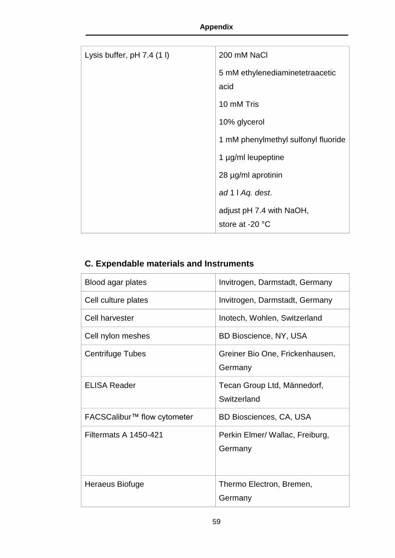
Appendix
59
Lysis buffer, pH 7.4 (1 l) 200 mM NaCl
5 mM ethylenediaminetetraacetic
acid
10 mM Tris
10% glycerol
1 mM phenylmethyl sulfonyl fluoride
1 µg/ml leupeptine
28 µg/ml aprotinin
ad 1 l Aq. dest.
adjust pH 7.4 with NaOH,
store at -20 °C
C. Expendable materials and Instruments
Blood agar plates Invitrogen, Darmstadt, Germany
Cell culture plates Invitrogen, Darmstadt, Germany
Cell harvester Inotech, Wohlen, Switzerland
Cell nylon meshes BD Bioscience, NY, USA
Centrifuge Tubes Greiner Bio One, Frickenhausen,
Germany
ELISA Reader Tecan Group Ltd, Männedorf,
Switzerland
FACSCalibur™ flow cytometer BD Biosciences, CA, USA
Filtermats A 1450-421 Perkin Elmer/ Wallac, Freiburg,
Germany
Heraeus Biofuge Thermo Electron, Bremen,
Germany
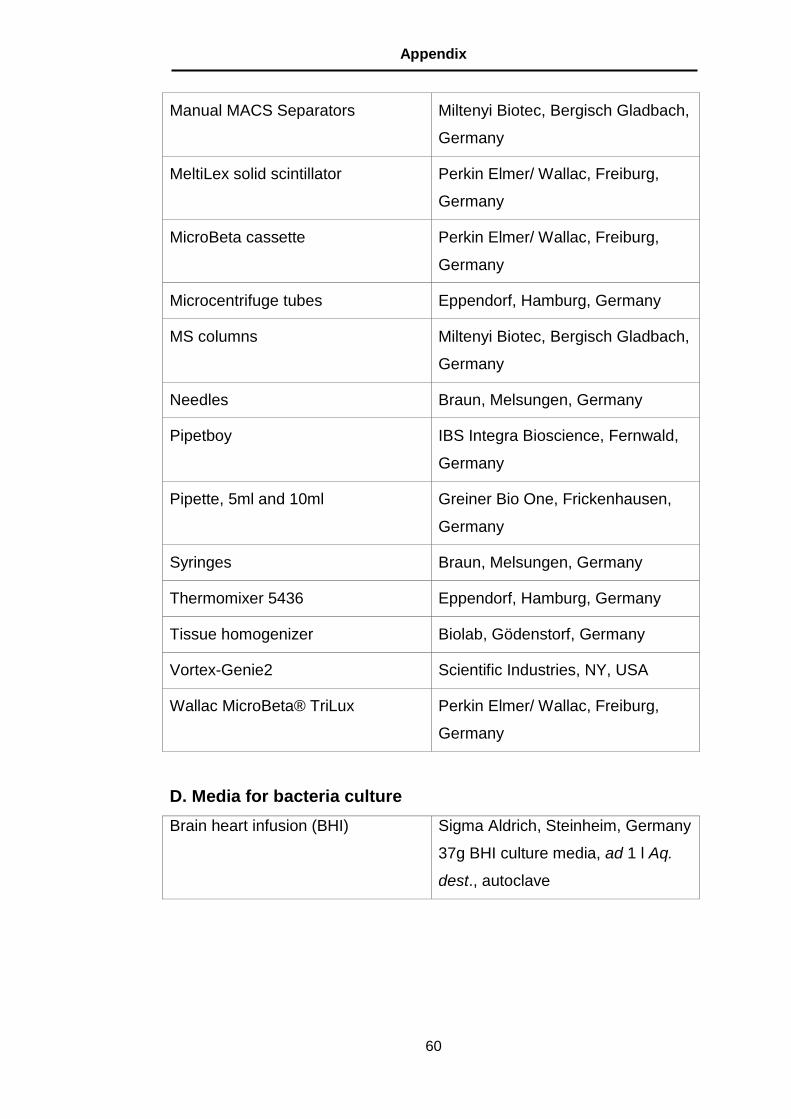
Appendix
60
Manual MACS Separators Miltenyi Biotec, Bergisch Gladbach,
Germany
MeltiLex solid scintillator Perkin Elmer/ Wallac, Freiburg,
Germany
MicroBeta cassette Perkin Elmer/ Wallac, Freiburg,
Germany
Microcentrifuge tubes Eppendorf, Hamburg, Germany
MS columns Miltenyi Biotec, Bergisch Gladbach,
Germany
Needles Braun, Melsungen, Germany
Pipetboy IBS Integra Bioscience, Fernwald,
Germany
Pipette, 5ml and 10ml Greiner Bio One, Frickenhausen,
Germany
Syringes Braun, Melsungen, Germany
Thermomixer 5436 Eppendorf, Hamburg, Germany
Tissue homogenizer Biolab, Gödenstorf, Germany
Vortex-Genie2 Scientific Industries, NY, USA
Wallac MicroBeta® TriLux Perkin Elmer/ Wallac, Freiburg,
Germany
D. Media for bacteria culture
Brain heart infusion (BHI) Sigma Aldrich, Steinheim, Germany
37g BHI culture media, ad 1 l Aq.
dest., autoclave
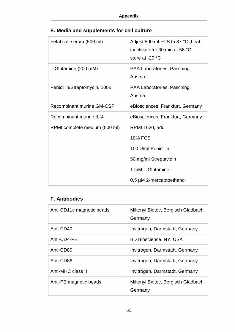
Appendix
61
E. Media and supplements for cell culture
Fetal calf serum (500 ml) Adjust 500 ml FCS to 37 °C ,heat-
inactivate for 30 min at 56 °C,
store at -20 °C
L-Glutamine (200 mM) PAA Laboratories, Pasching,
Austria
Penicillin/Streptomycin, 100x PAA Laboratories, Pasching,
Austria
Recombinant murine GM-CSF eBiosciences, Frankfurt, Germany
Recombinant murine IL-4 eBiosciences, Frankfurt, Germany
RPMI complete medium (500 ml) RPMI 1620, add
10% FCS
100 U/ml Penicillin
50 mg/ml Streptavidin
1 mM L-Glutamine
0.5 µM 2-mercaptoethanol
F. Antibodies
Anti-CD11c magnetic beads Miltenyi Biotec, Bergisch Gladbach,
Germany
Anti-CD40 Invitrogen, Darmstadt, Germany
Anti-CD4-PE BD Bioscience, NY, USA
Anti-CD80 Invitrogen, Darmstadt, Germany
Anti-CD86 Invitrogen, Darmstadt, Germany
Anti-MHC class II Invitrogen, Darmstadt, Germany
Anti-PE magnetic beads Miltenyi Biotec, Bergisch Gladbach,
Germany
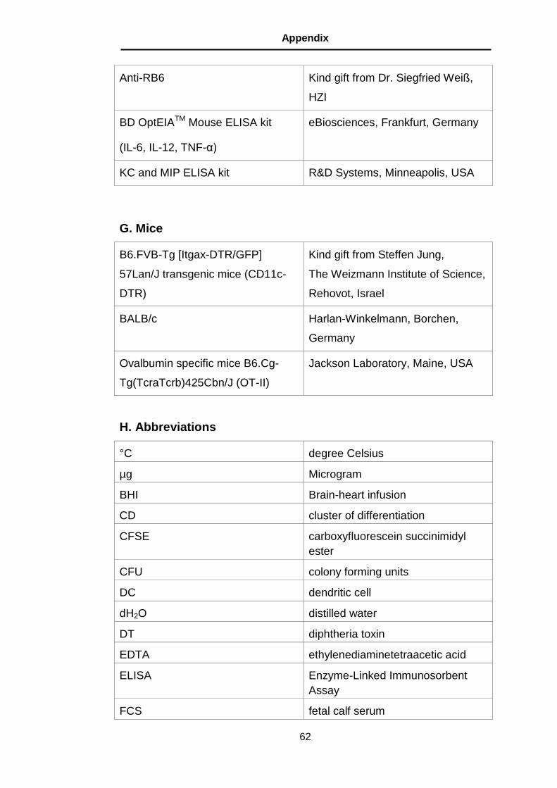
Appendix
62
Anti-RB6 Kind gift from Dr. Siegfried Weiß,
HZI
BD OptEIATM Mouse ELISA kit
(IL-6, IL-12, TNF-α)
eBiosciences, Frankfurt, Germany
KC and MIP ELISA kit R&D Systems, Minneapolis, USA
G. Mice
B6.FVB-Tg [Itgax-DTR/GFP]
57Lan/J transgenic mice (CD11c-
DTR)
Kind gift from Steffen Jung,
The Weizmann Institute of Science,
Rehovot, Israel
BALB/c Harlan-Winkelmann, Borchen,
Germany
Ovalbumin specific mice B6.Cg-
Tg(TcraTcrb)425Cbn/J (OT-II)
Jackson Laboratory, Maine, USA
H. Abbreviations
°C degree Celsius
µg Microgram
BHI Brain-heart infusion
CD cluster of differentiation
CFSE carboxyfluorescein succinimidyl
ester
CFU colony forming units
DC dendritic cell
dH2O distilled water
DT diphtheria toxin
EDTA ethylenediaminetetraacetic acid
ELISA Enzyme-Linked Immunosorbent
Assay
FCS fetal calf serum

Appendix
63
FnBPA fibronectin binding protein
GFP green fluorescencent protein
h Hour
H&E hematoxylin and eosin
i.p. Intraperitoneal
i.v. Intravenous
LPS lipopolysaccaride
MHC major histocompatibility complex
min Minute
ml Milliliter
mM milli molar
MOI multiplicity of infection
MRSA Methicillin-resistant Staphylococcus
aureus
MSCRAMMs microbial surface components
recognizing adhesive matrix mole-
cules
NADPH nicotinamide adenine dinucleotide
phosphate
nm Nanometer
PBS phosphate buffered saline
pH potentia Hydrogenii
PMN polymorphonuclear PMNs
Rpm rotates per minute
S. aureus Staphylococcus aureus
SD standard deviation
TCR T cell receptor
TMB 3,3',5,5'-Tetramethyl benzidine
TSST-1 toxic shock syndrome toxin 1
VRSA Vancomycin-resistant Staphylococ-
cus aureus
YOPs Yersinia outer proteins

Appendix
64
I. Videos
The Videos S1 (Phagocytic uptake of S. aureus by DCs) and S2
(S. aureus replicates within the DC) are available as avi files. Printed cop-
ies of this dissertation comprise the videos on the enclosed CD-R.
J. List of figures
Figure 1: Schematic representation of S. aureus virulence factors ............ 3
Figure 2: Attachment and invasion of S. aureus in endothelial cells .......... 4
Figure 3: Schematic representation of the origin and differentiation of
immune cells ............................................................................... 8
Figure 4: Schematic representation of maturation and migration of
dendritic cells .............................................................................. 9
Figure 5: Activation of T lymphocytes by DCs ......................................... 10
Figure 6: Depletion of CD11c positive cells in CD11c-DTR mice............. 15
Figure 7: Phagocytic uptake of S. aureus by DCs ................................... 22
Figure 8: Phagosome acidification in S. aureus-infected DCs ................. 23
Figure 9: S. aureus induces maturation of DCs ....................................... 25
Figure 10: The course of S. aureus infection in BALB/c mice .................. 26
Figure 11: DCs are recruited into the spleen and lungs of
S. aureus-infected mice.. ........................................................ 27
Figure 12: Cytokines released by DCs isolated from the organs of
infected mice after in vitro re-stimulation with heat-killed
S. aureus ................................................................................ 28
Figure 13: Depletion of DCs aggravates the severity of S. aureus
infection. ................................................................................. 29
Figure 14: Depletion of alveolar macrophages in CD11c-DTR mice
after treatment with DT. .......................................................... 30
Figure 15: Tracing of adoptively transferred CFSE-labeled DCs by
flow cytometry ......................................................................... 31
Figure 16: DC reconstitution improves the capacity of DCs-depleted
CD11c-DTR mice to control S. aureus infection. .................... 31
Figure 17: Effect of adoptive transfer of DCs on the capacity of
BALB/c mice to control S. aureus infection. ............................ 32

Appendix
65
Figure 18: S. aureus survives within DCs ................................................ 33
Figure 19: S. aureus replicates within the DC .......................................... 33
Figure 20: DC-depletion is associated with an increased recruitment of
PMNs in the lungs of S. aureus-infected mice ........................ 34
Figure 21: Higher amount of viable S. aureus are present within the
PMNs recruited into the lungs of DC-depleted than in those
of non-depleted mice.. ............................................................ 35
Figure 22: Amount of CXC chemokines in the lungs of
S. aureus-infected mice in the presence or absence of DCs .. 36
Figure 23: Effect of partial depletion of PMNs on bacterial burdens in
the organs of DC-depleted mice ............................................. 37
Figure 24: Proliferation of naïve CD4+ T cells after co-cultivation with
uninfected or S. aureus-infected OVA-loaded DCs ................. 38