Embed Size (px)
Citation preview

The role of cytosolic glutamine synthetases in abiotic stress
and development in Arabidopsis thaliana
A Thesis Submitted to the College of
Graduate Studies and Research
In Partial Fulfillment of the Requirements
For the Degree of Master of Science
In the Department of Biology
University of Saskatchewan
Saskatoon
By
YUANYUAN JI
© Copyright Yuanyuan Ji, April 2011. All rights reserved.

PERMISSION TO USE
In presenting this thesis in partial fulfillment of the requirements for a Postgraduate degree from
the University of Saskatchewan, I agree that the Libraries of this University may make it freely
available for inspection. I further agree that permission for copying of this thesis in any manner,
in whole or in part, for scholarly purposes may be granted by the professor who has supervised
my thesis work or, in their absence, by the Head of the Department or the Dean of the College in
which my thesis work was done. It is understood that any copying or publication or use of this
thesis or parts thereof for financial gain shall not be allowed without my written permission. It is
also understood that due recognition shall be given to me and to the University of Saskatchewan
in any scholarly use which may be made of any material in my thesis.
Requests for permission to copy or to make other use of material in this thesis in whole or part
should be addressed to:
Head of the Department of Biology
University of Saskatchewan
112 Science Place
Saskatoon, Saskatchewan S7N 5E2

1
ABSTRACT
Glutamine (Gln), a major nitrogen source in plants, is considered a central intermediate
that coordinates carbon-nitrogen assembly for plant growth and development. To maintain a
sufficient Gln supply, plant cells employ glutamine synthetases (GS), including cytosolic GS1
and plastidic GS2 for Gln production. Previous work has shown that the GS1 is responsive to
various environmental stresses. This study demonstrated the involvement of GS1s in Gln
homeostasis and the role of GS1 in abiotic stress tolerance in Arabidopsis. The GS1 family is
comprised of five isoforms in Arabidopsis thaliana. Gene expression profiling showed that
GLN1;1, GLN1;3 and GLN1;4 had similar expression patterns and were upregulated by abiotic
(salinity and cold) stresses, whereas GLN1;2 exhibited constitutive expression and no GLN1;5
transcript was detected under any of the conditions tested. Null T-DNA insertion mutants for the
five GS1 genes were obtained. Only the gln1;1 mutant displayed enhanced sensitivity to a GS
inhibitor, phosphinothricin, and to cold and salinity treatments, suggesting a nonredundant role
for GLN1;1. Increased stress sensitivity in gln1;1 was associated with accelerated accumulation
of reactive oxygen species (ROS), particularly in chloroplasts. To better understand the role of
cytosolic GS isoforms, we generated two different triple mutant combinations. Triple mutant
gln1;1/gln1;2/gln1;3 showed reduced growth at an early stage. The gln1;1/gln1;3/gln1;4 mutant
is pollen lethal, indicating an essential role of Gln in plant gametophyte development.
Collectively, our results establish a link between cytosolic Gln production, ROS accumulation,
plant stress tolerance and development.

2
ACKNOWLEDGMENTS
I am greatly indebted to my supervisor, Dr. Yangdou Wei, for his continuous support and
encouragement as well as excellent advice throughout my study. Without his help, this work
would not be possible. Dr. Wei was always there to listen and to give suggestions. He showed me
different ways to approach a research problem and taught me how to organize ideals based on the
readings. The way he works in the research really aspired me. I would also like to thank my
advisory committee members Drs. Peta Bonham-Smith and Pierre Fobert for their suggestions
and patience throughout my graduate program.
I would also like to thank Dr. Guosheng Liu, who acted as my minor advisor. He guided
me into this molecular world and taught me how to do the experiments in an efficient way. For
his guidance and advice, I really appreciate both professionally and personally.
I would like to thank the Department of Biology, University of Saskatchewan for providing
the financial support and the nice research atmosphere. An acknowledgment also goes to
graduate students in the Department of Biology, especially Xiaohui Bao, Lipu Wang and Qiang
Li.
I would like to deeply thank my parents and my family for their support, both financially
and mentally. Also, particular thanks to my husband, Chenlu Yang. He always be there with
patient and encourage.
3
TABLE OF CONTENTS
ABSTRACT .....................................................................................................................................1
ACKNOWLEDGMENTS ...............................................................................................................2
TABLE OF CONTENTS .................................................................................................................3
LIST OF FIGURES .........................................................................................................................5
LIST OF TABLES ...........................................................................................................................6
LIST OF ABBREVIATIONS ..........................................................................................................7
1. General Introduction ..................................................................................................................11
1.1 Nitrogen Assimilation and Transport in Plants ........................................................................11
1.1.1 Nitrate Uptake and Transport................................................................................................11
1.1.2 Ammonium Uptake and Transport .......................................................................................13
1.1.3 Amino Acid Uptake and Transport .......................................................................................15
1.2 Nitrogen Assimilatory Pathway ...............................................................................................18
1.2.1 Glutamine Synthetase (GS)...................................................................................................20
1.2.2 Glutamine Oxoglutarate Aminotransferase (GOGAT) .........................................................21
1.2.3 Glutamate Dehydrogenase (GDH) ........................................................................................22
1.3 Carbon and Nitrogen Partitioning ............................................................................................23
1.3.1 Interconnection between C Metabolism and N Metabolism during Photosynthesis ............24
1.3.2 Nitrogen Metabolism and Cellular Redox ............................................................................25
1.4 Stress Responses and Reactive Oxygen Species (ROS) ..........................................................27
1.4.1 Reactive Oxygen Species ......................................................................................................27
1.4.2 Generation of ROS during Stress Responses in Plants .........................................................27
1.4.3 ROS Toxicity and Signalling in Abiotic Stresses .................................................................29
2. Research Objectives ...................................................................................................................32
3. Materials and Methods ...............................................................................................................33
3.1 Plant Material and Growth Conditions ....................................................................................33
3.2 Characterization of Homozygous T-DNA Insertional Mutants ...............................................33
3.3 Stress Treatment.......................................................................................................................34
3.4 Pathogen Inoculation ...............................................................................................................35
3.5 RNA Extraction, cDNA Generation and Reverse Transcriptase-Polymerase Chain Reaction
(RT-PCR) .......................................................................................................................................35
3.6 Abiotic and Oxidative Stress Assays .......................................................................................35
4
3.7 Western Blot Analysis .............................................................................................................36
3.8 ROS Stimulation and Detection by Confocal Microscopy ......................................................37
4. Result .........................................................................................................................................38
4.1 Tissue Specific Expression of GLN1;1 and GLN1;4 ..............................................................38
4.2 Expression of GS1 Genes in Response to Abiotic Stresses .....................................................40
4.3 Expression of GS1s in Response to Pathogen Infection ..........................................................42
4.4 Generation of gln1;1~gln1;5 Mutants .....................................................................................45
4.5 GLN1;1 Is Required for Phosphinothricin (PPT) Tolerance ...................................................46
4.6 gln1;1 Mutant is Susceptible to Salt, Cold and Oxidative Stresses .........................................50
4.7 Isoforms GLN1;1, GLN1;2, and GLN1;3 Coordinate Primary Nitrogen Assimilation ..........57
4.8 GLN1;1, GLN1;3 and GLN1;4 Are Functionally Redundant for Pollen Development ..........59
5. Discussion ..................................................................................................................................63
5.1 GLN1;1 as a Major Cytosolic GS1 Isoform Contributes to Plant Tolerance against Abiotic
Stresses ...........................................................................................................................................63
5.2 The Roles of Cytosolic GS in Plant Growth and Development...............................................65
5.3 Involvement of Gln Metabolism in Regulation of Cellular Redox..........................................69
References ......................................................................................................................................73
5
LIST OF FIGURES
Figure 1.1 Uptake of NH4+ is completed by the spatial arrangement of AMTs in Arabidopsis
roots................................................................................................................................................14
Figure 1.2 Nitrogen assimilatory pathway .....................................................................................19
Figure 4.1 Tissue specific expression of GLN1;1 and GLN1;4 ....................................................41
Figure 4.2 RT-PCR analysis of expression patterns of GLN1 genes in response to different
abiotic stresses in Arabidopsis seedlings .......................................................................................43
Figure 4.3 RT-PCR analysis of GLN1 expression in response to Colletotrichum higginsianum
infection .........................................................................................................................................44
Figure 4.4 Schematic diagrams of GLN1 genes.............................................................................47
Figure 4.5 Characterization of null mutations for GLN1 isoforms ................................................48
Figure 4.6 gln1;1 mutants are more sensitive to PPT ....................................................................49
Figure 4.7 Response of gln1 mutants to abiotic stresses ...............................................................53
Figure 4.8 Response of gln1 mutants to different ROS generators ...............................................54
Figure 4.9 Organization of the electron transport chain and ATP synthesis in the thylakoid
membrane of plant chloroplast.......................................................................................................55
Figure 4.10 ROS production in Col-0 and gln1;1 mutant plants ...................................................56
Figure 4.11 GLN1 is involved in primary nitrogen assimilation ...................................................58
Figure 4.12 Impaired pollen development in gln1-1-/-
/gln1-3-/-
/gln1-4-/+
mutants ........................62

6
LIST OF TABLES
Table 1 Primer sequences for PCR amplification of GLN1 genes.................................................34
Table 2 Primer sequences for RT-PCR amplification of cDNA ....................................................36

7
LIST OF ABBREVIATIONS
2,4-D: 2,4-dichlorophenoxyacetic acid
2D: two dimension
2-OG: 2-oxoglutarate
3-PGA: 3-phosphoglycerate
AAP: amino acid permease
AMT: ammonium transporter
ANT1: aromatic and neutral amino acid transport 1
AOX: alternative oxidase
APC: amino acid-polyamine-choline
APX: ascorbate peroxidase
ATP: adenosine triphosphate
ATF: amino acid transport family
AUX1: auxin-resistance
C: carbon
CAT: catalase
CATs: cationic amino acid transporters
CCA1: circadian clock associated 1
CDPK: calcium-dependent protein kinase
cHATS: constitutive high affinity transport system
DCFDA: dichlorodihydrofluorescein diacetate
DHA: dehydroascorbate
DHAR: DHA reductase
8
ETC: electron transport chain
Fd: ferredoxin
Fd-GOGAT: ferredoxin-glutamate synthase
FNR: ferredoxin-NADP+ reductase
FTR: Fd:thioredoxin reductase
GABA: γ-aminobutyric acid
GDH: glutamate dehydrogenase
Gln: glutamine
Glu: glutamate
GOGAT: glutamate synthase
GPX: glutathione oxidase
GR: glutathione reductase
GS1/GLN1: cytosolic glutamine synthetase
GS2: plastidic glutamine synthetase
HATS: high affinity transport system
•HO: hydroxyl radical
HO2-: hydroperoxide
H2O2: hydrogen peroxide
HR: hypersensitive response
HSTFs: heat shock transcription factors
IAA: indole-3-acetic acid
iHATS: inducible high affinity transport system
KCN: potassium cyanide

9
LATS: low affinity transport system
LATs: L-type amino acid transporters
LHT: lysine/histidine transporter
LSU: large subunit of rubisco
MEP: methylammonium permease
MAPK: mitogen-activated protein kinase
MD: menadione
MSX: L-methionie-S-sulfoximine
MV: methyl viologen
N: nitrogen
NAD+: nicotinamide adenine dinucleotide
NADP+: nicotinamide adenine dinucleotide phosphate
NH4+: ammonium
NiR: nitrite reductase
NO3-: nitrate
NR: nitrate reductase
O2-: superoxide radical
1O2: singlet oxygen
P5CS1: pyrroline-5-carboxylate synthase 1
PETC: photosynthetic electron transport chain
PSI: photosystem I
PSII: photosystem II
PKA: protein kinase

10
PPT: phosphinothricin
ProTs: proline transporter
PUFAs: polyunsaturated fatty acids
RB: rose bengal
Rh: rhesus
ROS: reactive oxygen species
RuBP: ribulose 1,5-bisphosphate
SA: salicylic acid
SHAM: salicylhydroxamic acid
SOD: superoxide dismutase
Trx: thioredoxin
UQ: ubiquinone
UV: Ultraviolet

11
1. General Introduction
1.1 Nitrogen Assimilation and Transport in Plants
Nitrogen (N) is one of the most important elements for all organisms. Many biochemical
compounds required for cell development and reproduction contain N. For example, N is found
in the nucleoside phosphates and amino acids which comprise nucleic acids and proteins. In
plants, the availability and uptake of N is considered a major factor affecting plant productivity,
biomass, and crop yield (Coruzzi, 2003). In order to synthesize more protein for growth and
development, plants are able to assimilate N by taking up nitrate (NO3-), ammonium (NH4
+) and
amino acids from soil directly or through the symbiotic action of N-fixing bacteria. In this
introduction, I will focus on the three major N sources (NO3-, NH4
+ and amino acids) and
introduce how they are assimilated and transported in the plant.
1.1.1 Nitrate Uptake and Transport
Plants obtain NO3- from the soil by transporting it across the plasma membrane of epidermal and
cortical cells of the root using NO3- transporters (Forde, 2002). NO3
- concentration varies by
different types of soil depending on many environmental factors, such as rainfall, pH value,
temperature. There are large changes when soil is sampled within a small area (Miller et al.,
2007). Physiological investigations of NO3- uptake by roots have revealed that several NO3
-
transport systems are employed to cope with the variations in NO3- concentration in cultivated
soils (Crawford, 1995). These transport systems include the low affinity transport system (LATS)
(above 1 mM) and the high affinity transport system (HATS) (1μM to 1mM), which is comprised
of the constitutive (cHATS) and inducible systems (iHATS). The cHATS is present regardless of

12
NO3- supply, while iHATS is only stimulated by external application of NO3
- (Crawford, 1995).
In Arabidopsis thaliana, two genetically linked genes, AtNRT2.1 (NITRATE TRANSPORTER)
and AtNRT2.2, were cloned (Zhuo et al., 1999). A number of studies have reported that mutants
with impaired expression of AtNRT2.1 and AtNRT2.2 are defective in HATS activity (Filleur et
al., 2001; Orsel et al., 2004; Zhuo et al., 1999). Dramatic up-regulation of AtNRT2.1 and a
transient increase in AtNRT2.2 at the mRNA level are observed following reapplication of NO3-
to plants in NO3--deprived conditions, indicating these two nitrate transporters might belong to
iHATS (Okamoto et al., 2003). However, a recent study by Li et al., (2007) identified a new
allele of Arabidopsis mutant uniquely disrupted in AtNRT2.1, and this mutant Atnrt2.1 and
Atnrt2.2 were used to investigate the relative contribution of iHATS and cHATS. Their results
defined that AtNRT2.1 is the major contributor to iHATS and AtNRT2.2 makes a minor
contribution to iHATS, but AtNRT2.2 can partially compensate for a loss of AtNRT2.1. Unlike
HATS, there are more than 50 candidate genes involved in LATS in Arabidopsis. Six genes in the
AtNRT1 family were characterized in different tissues and found to be responsible for various
functions. For example, AtNRT1.2 and AtNRT1.4, two low-affinity transporters, are involved in
NO3- uptake and petiole NO3
- storage respectively. Transcripts of AtNRT1.2 were primarily
detected in roots (Huang et al., 1999), whereas AtNRT1.4 is expressed in leaf petioles (Chiu et al.,
2004). Recently, AtNRT1.6, a new member of LATS, was characterized by Almagro et al., (2008).
The expression of AtNRT1.6 is only detectable in reproductive tissues. The Atnrt1.6 mutant
showed reduced NO3- accumulation in mature seeds and an increased seed abortion rate
suggesting that AtNRT1.6 is involved in delivering NO3- to the developing embryo (Almagro et
al., 2008). AtNRT1.5 and AtNRT1.8 were identified in the same tissue of transport system
playing a role in long-distance transport from root to shoot (Li et al., 2010). Interestingly,

13
AtNRT1.1 was originally found to be a member of LATS, but it appears to possess dual affinity,
with a phosphorylation switch between high and low affinity ranges of NO3- uptake by cAMP-
dependent protein kinase (PKA) (Liu and Tsay, 2003).
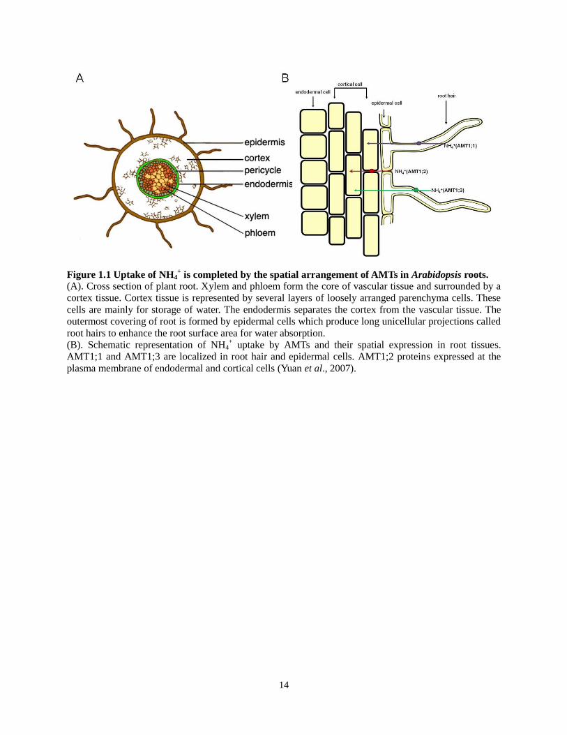
1.1.2 Ammonium Uptake and Transport
Plant roots are usually exposed to more NO3- than NH4
+ (Miller et al., 2007). High levels of
NH4+ are toxic to plants and result in growth inhibition (Britto and Kronzucker, 2001; Walch-Liu
et al., 2001). Thus, the uptake of NH4+ needs to be precisely regulated in order to adjust the
cellular NH4+ concentration. In most plant species, NH4
+ transporters belong to AMT1- and
AMT2/MEP-type (AMMONIUM TRANSPORTER/METHYLAMMONIUM PERMEASE)
classes of proteins and are encoded by gene families (Ludewig et al., 2001). Accumulating
evidence indicates that AMT-type NH4+ transporters represent the major entry pathways for NH4
+
assimilation in roots (Loque and von Wiren, 2004). In Arabidopsis, the AMT family is comprised
of five isoforms (AMT1;1~AMT1;5). A few studies have provided evidence that the tightly-
regulated high-affinity NH4+ uptake in Arabidopsis roots relies on the spatial arrangement of
AMT1;1, AMT1;2 and AMT1;3. AMT1;1 and AMT1;3 are epidermis-located proteins, that can
take up NH4+ directly from the soil. GFP fusion protein driven by the AMT1;2 promoter showed
that AMT1;2 is expressed in endodermal and cortical cells where the substrate (NH4+) must be
delivered by other transporters from the soil (See Figure 1.1) (Wang et al., 2007, Yuan et al.,
2007). Studies in single insertion mutants of amt1;1, amt1;2 and amt1;3 indicated that AMT1;1
and AMT1;3 contribute approximately two thirds of total NH4+ influx in N-deficient Arabidopsis
roots, while AMT1;2 is responsible for 18 to 26% of the overall NH4+ uptake capacity (Loqué
and von Wiren, 2004; Wang et al., 2007). In Arabidopsis, AMT2;1 is the only isoform in the
14
Figure 1.1 Uptake of NH4+ is completed by the spatial arrangement of AMTs in Arabidopsis roots.
(A). Cross section of plant root. Xylem and phloem form the core of vascular tissue and surrounded by a
cortex tissue. Cortex tissue is represented by several layers of loosely arranged parenchyma cells. These
cells are mainly for storage of water. The endodermis separates the cortex from the vascular tissue. The
outermost covering of root is formed by epidermal cells which produce long unicellular projections called
root hairs to enhance the root surface area for water absorption.
(B). Schematic representation of NH4+ uptake by AMTs and their spatial expression in root tissues.
AMT1;1 and AMT1;3 are localized in root hair and epidermal cells. AMT1;2 proteins expressed at the
plasma membrane of endodermal and cortical cells (Yuan et al., 2007).

15
MEP subfamily. Although AMT2;1 transcripts are upregulated under N deficiency, RNA
interference (RNAi) –mediated repression of AMT2;1 revealed that AMT2;1 was unable to
contribute to overall NH4+ influx indicating an additional role of AMT2;1 (Sohlenkamp et al.,
2002). While much of the research on molecular aspects of NH4+ transport has been performed
using Arabidopsis. Analysis for AMT-homologous sequences from tomato and rice showed three
LeAMT genes and ten putative OsAMT genes, respectively (Loqué and von Wiren, 2004).
1.1.3 Amino Acid Uptake and Transport
In all organisms, amino acids play fundamental roles in various processes, such as protein
synthesis, hormone metabolism, cell growth, nucleotide synthesis and N metabolism and serve as
principal, long distance transport forms of organic N (Wipf et al., 2002). So far, according to
different substrate specificities and affinities, as well as distinct subcellular localizations, amino
acid transporters have been divided into at least two major superfamilies: (1) The ATF (amino
acid transporter) family and (2) the APC (amino acid-polyamine-choline) family. In addition,
some amino acid transporters localized at the organelle membrane were also identified; for
example, DiT2.1, a chloroplast envelope membrane protein, functions in glutamate/malate
exchange in the photorespiratory pathway (Renne et al., 2003).
The ATF family was the first subfamily described in plants and constitute five sub-classes:
(i) AAP (amino acid permease), with eight members in Arabidopsis, mediate uptake of a
broad spectrum of amino acids. All AAP members in Arabidopsis except AAP7 have been
characterized. AAP1 was the first gene encoding an amino acid transporter to be cloned from
plants (Frommer et al., 1993; Hsu et al., 1993). With the capability to mediate transport of
neutral and acidic amino acids into root cells, AAP1 is localized to the plasma membrane (Lee et

16
al., 2007). AAP3, a member of AAPs with high expression in roots, was detected at the nuclear
membrane, ER (Endoplasmic Reticulum), Golgi bodies, endosomal vesicles and plasma
membrane by histochemical analysis of epitope-tagged AAP3 in Arabidopsis which indicates
that AAP3 might be involved in the trafficking pathway (Okumoto et al., 2004). Although AAPs
mediate uptake of a broad spectrum of amino acids, analyses from Fischer et al. (2002) indicated
that AAP1 and AAP5 were less efficient in delivering amino acids with aromatic side chains
(tryptophan, phenylalanine), β-methyl groups (threonine, valine and isoleucine) and cyclic amino
acids, whereas AAP3 and AAP6 mediated high levels of tryptophan transport. With the exception
of AAP6, AAPs fail to recognize aspartate (Fischer et al., 2002).
(ii) LHTs („lysine/histidine‟ transporters) are also high affinity transporters (μM level). In
Arabidopsis, there are 10 putative members AtLHT1-AtLHT10. Tissue specificity expression
revealed that AtLHT1, AtLHT2, and AtLHT3 are the predominant isoforms in Arabidopsis.
AtLHT2 shows a high expression in flower buds, AtLHT3 localizes specifically in stem and the
transcript of AtLHT1 is present in a broad range of tissues with a relative high level of expression
in root and leaf (Liu et al., 2010, Lee et al., 2004). Analysis of amino acid transport by the
expressed protein in yeast revealed that AtLHT1 (the first characterized member of LHT in
Arabidopsis) was a high-affinity transporter for both lysine and histidine (Chen and Bush, 1997).
More recently, AtLHT1 was shown to mediate the transport of a variety of amino acids with a
much higher affinity than AAPs (Hirner et al., 2006). Biochemical analysis of AtLHT2 in yeast
indicated that AtLHT2 plays an important role in the transport of uncharged and negatively
charged amino acids (Lee et al., 2004).
(iii) ProTs (proline transporters) serve to transport the amino acid proline and glycine betaine
as well as γ-aminobutyric acid (GABA). Unlike other amino acids, glycine betaine and GABA

17
are not able to incorporate into proteins. Originally, ProTs were described as specific transporters
for Pro (Fischer et al., 1995; Rentsch et al., 1996). However, competition studies using LeProT1
expressed in yeast indicated that other amino acids besides Pro might be potential substrates
(Schwacke et al., 1999). Indeed, direct transport assays demonstrated that LePro1 is able to
mediate the transport of glycine betaine, GABA and Pro (Schwacke et al., 1999). Moreover,
expression of AtProTs in a transport-deficient yeast mutant strain (22574d) which carries
mutations in the general amino acid (gap1), Pro (put4), and the GABA (uga4) permeases
demonstrated that, besides Pro, all three AtProTs have the highee affinity for glycine betaine and
a lower affinity for GABA (Grallath et al., 2005).
(iv) ANT1 (aromatic and neutral amino acid transporter 1) was the first amino acid
transporter identified in this subfamily and only characterized in Arabidopsis so far (Chen et al.,
2001). Sequence comparisons among the known amino acid transporters in plants suggest that
ANT1 is not a member of previously described amino acid transporter subfamilies due to the low
similarity with them (Chen et al., 2001). Analysis of ANT1 transport activity showed that ANT1
transports aromatic and neutral amino acids, as well as arginine, indole-3-acetic acid (IAA) and
2,4-dichlorophenoxyacetic acid (Chen et al., 2001).
(v) AUX1 (auxin-resistance) was named after the aux1 mutant, which displays an auxin-
resistant root growth phenotype and lacks root gravitropic curvature. The similarity of protein
sequence and structure between AUX1 and AAP1 suggests that AUX1 mediates the transport of
an amino acid-like signalling molecule (Bennett et al., 1996). Using Xenopus oocytes, Yang et al.
(2006) showed that IAA, which is structurally similar to tryptophan, is a substrate of AUX1
(Yang et al., 2006).
The APC transporter family is functionally, but not structurally, related to ATF transporters
18
and can be also classified into two subgroups including cationic amino acid transporters (CATs)
and L-type amino acid transporters (L-AATs) (Rentsch et al., 2007; Su et al., 2004). So far, L-
AATs have only been identified in animals, but nine putative CATs have been characterized in
Arabidopsis (Su et al., 2004). AtCAT5 is a high-affinity basic amino acid transporter and may
function in reassimilation of leaking amino acids at the leaf margin (Su et al., 2004). AtCAT6
mediates the transport of large, neutral and cationic amino acids with moderate affinity in
preference to other amino acids. Sink tissues, including developing seeds, lateral roots and
nematode-induced feeding structures, are specific localizations for AtCAT6 (Hammes et al.,
2006).
1.2 Nitrogen Assimilatory Pathway
NO3- is the common form of inorganic N in soil, but cannot directly be used as substrate for
protein synthesis in plants. To be used in plants, NO3- is first reduced to NO2
- by nitrate reductase
(NR) and NO2- is further catalyzed by nitrite reductase (NiR) to NH4
+. Because it is toxic to plant
cells, NH4+ needs to be efficiently assimilated into amino acids. NH4
+ is incorporated into
glutamine (Gln) as the primary assimilation pathway through the sequential action of glutamine
synthetase (GS) and glutamine:2-oxoglutarate amidotransferase (glutamate synthase, GOGAT),
called the GS/GOGAT cycle. Glutamate dehydrogenase (GDH) incorporates 2-oxoglutarate (2-
OG) and NH4+ to produce glutamate (Glu). Gln and Glu are the two entry points to further
amino-acid metabolism. Plants are able to employ both Gln and Glu as N donors for the
biosynthesis of other essential amino acids (Coruzzi, 2003). Since GS, GOGAT and GDH
constitute the checkpoints, partitioning organic and inorganic N metabolism, I provide an
overview of these key enzymes below (see Figure 1.2).

19
Figure 1.2 Nitrogen assimilatory pathway
A schematic figure to show the main N metabolic fluxes occurring in various organelles in leaf cell. NO3-
and NH4+ are two major inorganic N sources for plants. GS/GOGAT pathway is thought to be the
principal mechanism of primary and secondary NH4+ assimilation in cytosol and chloroplast. In
mitochondrion, GDH has a function in NH4+ assimilation under stress (from Forde and Lea, 2007). GS:
glutamine synthetase; Fd: ferredoxin; NADH: nicotinamide adenine dinucleotide (reduced); GOGAT:
glutamine oxoglutarate aminotransferase; GDH: glutamate dehydrogenase;

20
1.2.1 Glutamine Synthetase (GS)
GS takes charge of primary NH4+ assimilation in plants by catalyzing the ATP-dependent
conversion of Glu into Gln with scavenging NH4+. Two forms of GS are present in higher plants,
and are categorized according to their different cellular localization. GS1, the major cytosolic
form of GS in plant roots, plays a dominant role in NH4+ assimilation from the environment. GS2
is resident as a soluble protein in chloroplasts and mitochondria for NH4+ scavenging during
photorespiration (Taira et al., 2004).
Molecular biological studies identified multiple isoenzymes for GS1 in higher plants.
Different plant species have unequal numbers of cytosolic GS. For example, there are five
isoenzymes of GS1 (GLN1-1, GLN1-2, GLN1-3, GLN1-4 and GLN1-5) in maize (Martin et al.,
2006); rice plants possess three GS1 genes; OsGS1;1, OsGS1;2 and OsGS1;3 (Tabuchi et al.,
2005); and in Arabidopsis, five putative genes for GS1 have been identified, GLN1;1, GLN1;2,
GLN1;3, GLN1;4 and GLN1;5 (Ishiyama et al., 2004a). The presence of multiple GS1 genes has
made the analysis of contributions by each individual difficult. The rice OsGS1;1 knockout
mutant shows severely retarded growth throughout its lifespan and has reduced grain filling
(Tabuchi et al., 2005). The maize mutants gln1-3 and gln1-4 display reduced kernel number and
size, respectively, and the double mutant gln1-3/gln1-4 shows both phenotypes (Martin et al.,
2006). Although the functional importance of some individual GS1 isoforms has been
demonstrated, accumulating preliminary experiments revealed that the cytosolic GS1 genes were
differentially expressed in distinct tissues suggesting nonredundant roles of each isoform
(Ishiyama et al., 2004a; Peterman and Goodman, 1991; Sakakibara et al., 1996). Furthermore,
GS1 isoforms were identified that are responsive to abiotic stress. For instance, the activity of
GS1 in tomato increased after exposure to different NaCl concentrations (Debouba et al., 2006).

21
Two dimensional (2D) gel electrophoresis analysis of proteins revealed GS1 accumulation in
response to cold treatment (Kwon et al., 2007). Thus, characterization of the roles of individual
GS1 genes and their combined functions will greatly enhance our understanding of the
importance of the GS1 family in N assmiliation and abiotic stress response.
Most plants possess only one nuclear gene for GS2. GS2 is predominantly expressed in
leaves, where photorespiration occurs. Mutants defective in GS2 were originally identified in
barley by screening under photorespiratory conditions. These mutants were unable to
reassimilate photorepiratory NH4+ even in the presence of cytosolic GS (Wallsgrove et al., 1987).
A more recent study using transgenic GLN2:GFP plants of Arabidopsis found that the fusion
proteins was present not only in leaf chloroplasts, but also in mitochondria indicating a dual
trafficking property of GS2 (Taira et al., 2004).
1.2.2 Glutamine Oxoglutarate Aminotransferase (GOGAT)
Since the GS/GOGAT pathway ultimately controls the N flux in plant cells, GOGAT has been
studied intensively for decades and advances have been made in recent years (Forde and Lea,
2007). Plants possess two distinct GOGATs: the ferredoxin (Fd)- and NADH-dependent forms.
Fd-GOGAT is the major isoform in higher plants that contributes 95% to total GOGAT activity,
and is normally present in photosynthetic tissues, where it mediates primary N-assimilation or N-
reassimilation in photorespiration (Somerville and Ogren, 1980). Genes encoding Fd-GOGAT
have been characterized in many plants including maize, tobacco, barley and Arabidopsis (Avila
et al., 1993; Coschigano et al., 1998; Sakakibara et al., 1991; Zehnacker et al., 1992). GLU1
(formerly called GLS) was the first gene encoding Fd-GOGAT identified in Arabidopsis. Mutant
glu1 has less than 5% of wild-type GOGAT activity in leaves and shows a chlorotic and

22
conditional lethal phenotype (photorespiratory condition), suggesting an essential role for GLU1
in photorespiration. However, this conditional lethal phenotype could be rescued by suppressing
photorespiration (1% CO2) (Somerville and Ogren, 1980). GLU2 is expressed at low levels and
cannot compensate for the absence of GLU1 activity when photorespiration occurs (Coschigano
et al., 1998).
The other form of GOGAT relies on NADH as an electron donor. NADH-GOGAT has a
different expression pattern from Fd-GOGAT, and is predominantly in developing tissues. For
example, rice OsNADH-GOGAT1 is mainly expressed in root-tips and in the premature leaf
blade and spikelet at an early stage of ripening (Tabuchi et al., 2007). It has been proposed that
NADH-GOGAT and cytosolic GS play a major role in initial N assimilation and also N transport
in root because of the coincident expression of NADH-GOGAT and cytosolic GS (Tobin and
Yamaya, 2001). The glt1-T mutant, which lacks NADH-GOGAT mRNA and enzyme activity,
only exhibited growth inhibition under repressed photorespiration, whereas the Fd-GOGAT
mutant glu1 exhibited severe growth retardation, indicating nonredundant roles of NADH-
GOGAT and Fd-GOGAT in NH4+ assimilation (Lancien et al., 2002).
1.2.3 Glutamate Dehydrogenase (GDH)
GDH mediates a reversible reaction of either synthesis of Glu by amination or catabolism of Glu
by deamination. At an early stage, in order to fully develop the basal part of the shoot, plants
require a large amount of protein; in this case the GS/GOGAT cycle is much more active than
GDH. Later, during senescence, GS and GOGAT activities decrease, accompanied by GDH
protein accumulation and increased aminating activity (Loulakakis et al., 2002; Masclaux et al.,
2000). Due to the dual enzyme activity of GDH giving either 2-OG or Glu as products, GDH is
23
expected to function in the regulation of carbon/nitrogen (C/N) balance during the whole life
cycle of the plant (Miyashita et al., 2008). To date, there are at least two types of GDH genes
identified in plant species, each of them encoding distinct polypeptides α or β subunits of GDH.
Seven isoforms of GDH can be produced by a random assembling of the α and β subunits
(Reviewed by Purnell et al., 2005). Since the significance of the GS/GOGAT cycle in primary N
assimilation has been accepted, it is generally thought that GDH plays only a small or negligible
role in NH4+ assimilation instead of facilitating Glu catabolism (Magalhães et al., 1990). This is
evidenced by studies on the Arabidopsis mutant gdh1-1 defective in the α subunit of the enzyme
which revealed that GDH functions in the direction of Glu catabolism under C-limiting
conditions (Melo-Oliverira et al., 1996). However, the potential role of GDH in NH4+
assimilation under certain physiological conditions cannot be ignored. A recent study in tobacco
and grapevine showed that salinity-induced reactive oxygen species (ROS) and methionine
sulfoximine (GS inhibitor) inhibition result in an induction of α-GDH subunit expression, thus
initiating the NH4+ assimilatory direction of GDH (Skopelitis et al., 2006). These lines of
evidences demonstrate that GDH acts as a C-skeleton supplier when C is limiting or a stress-
responsive protein that offers an alternative route for ammonium assimilation in case of the
malfunction of the GS/GOGAT pathway.
1.3 Carbon and Nitrogen Partitioning
C and N are two major nutrient resources in plants. In all known lifeforms, C is the second most
abundant element by mass after oxygen. Particularly in plants, the biomass of crops is highly
affected by the composition of C, in terms of C-containing organic compounds such as sugar,
starch, and fat. N is found in various amino acids and proteins that are critically important for

24
plant growth. It should not be surprising that N assimilation is highly interconnected to C
metabolism in higher plants because the assimilation of N requires not only energy (eg: reduced
Fd, NADPH, ATP), but also C skeletons.
1.3.1 Interconnection between C Metabolism and N Metabolism during Photosynthesis
Photosynthesis produces organic compounds from inorganic C by coupling the energy of
absorbed light with endothermic metabolic processes. In eukaryotic plants, both light and dark
reactions of photosynthesis are carried out in chloroplasts. Light reactions are made up of
multiple reactions to convert light energy to chemical energy in the form of ATP and NADPH
while producing O2. The Calvin cycle, also called dark reaction because it does not require light,
incorporates C from inorganic CO2 into carbohydrates using the energy and reducing power
(ATP and NADPH) generated in the light reactions. These two phases take place in distinct
regions of the chloroplast; the thylakoid membrane for light reactions and the stroma for the dark
reactions. In most plants, a three-C compound, 3-phosphoglycerate (3-PGA) is found as the first
product in the Calvin cylce. In this case, plants that fix C into 3-PGA are termed C3 plants. In
addition, several plant species such as maize, sugarcane and some dicotyledonous plants initially
fix C into four-C oxaloacetate and are therefore called C4 plants. Both C3 and C4 plants employ a
key enzyme, ribulose bisphosphate carboxylase/oxygenase (rubisco), to catalyzes the
carboxylation of ribulose 1,5-bisphosphate (RuBP) to form C intermediate in photosynthesis. In
fact, rubisco is the main N-containing component accounting for close to 50% of the total protein
in photosynthetic plant cells (Krapp et al., 2005). Studies have shown that N deprivation can
reduce photosynthetic C fixation (Miller et al., 2010).
On the other side, in order to transfer N and build up reserves for subsequent use in growth,

25
plants assimilate inorganic N into N-transport amino acids which depends not only on energy but
also C skeletons. Since the assimilation and transport of N is reviewed in part 1.1 and 1.2, the
next paragraph will focus on the regulation of N assimilation by C metabolism.
As early as 20 years ago, it was showed that the uptake of NO3- by roots depends on the
efficiency of photosynthesis and carbohydrate supply (Pace et al., 1990). Considering nitrate
transport requires energy, it is logical to think that NO3- uptake may need photosynthesis as
power supply. In recent years, more studies have indicated that not only does N transport demand
energy, amino acid biosynthesis does as well. There are a number of C skeleton backbones that
are required for amino acid biosynthesis, such as 2-OG, oxaloacetate, 3-PGA and pyruvate. In
the GS/GOGAT cycle, 2-OG is the receptor for the amide group of Gln and the product for the
reaction is Glu. Since the Gln and Glu produced in GS/GOGAT cycle are donors for the
biosynthesis of all amino acids, 2-OG is of crucial importance for N metabolism. Mutants
lacking NAD-DEPENDENT ISOCITRATE DEHYDROGENASE (IDH), the gene which produces
2-OG, have reduced amounts of certain free amino acids in comparison to the wild type
(Lemaitre et al., 2007). Decreased rates of NO3- reduction was also observed in Arabidopsis
plants with antisense repression of DIT1, which encodes a translocator of 2-OG on the
chloroplast (Schneidereit et al., 2006). In this case, the supply of C skeletons is one of the
limiting factors for N assimilation. Therefore, it appears that C and N metabolisms are
interdependent on each other for substantial growth.
1.3.2 Nitrogen Metabolism and Cellular Redox
In oxygenic photosynthetic eukaryotes, many important biological processes involve
oxidation/reduction reactions, for instance, the process of photorespiration is highly dependent

26
on the oxidation/reduction between NAD+ and NADH in order to maintain the supply of energy.
The pathway of N assimilation in oxygenic photosynthetic plants also involves a series of redox
reactions. These are initiated with a two-electron reduction of NO3- to NO2
- by NR. This is
followed by a six-electron reduction of NO2- to NH4
+ by NiR. Finally, in the GS/GOGAT cycle, a
two-electron reductive reaction is catalyzed by GOGAT producing Glu from Gln and 2-OG
(Suzuki and Knaff, 2005). All three enzymes (NR, NiR and GOGAT) in cyanobacteria catalyzing
redox reactions involved in N metabolism are Fd dependent, while the higher plants NiR and
GOGAT utilize reduced Fd as the electron donor.
Plant Fds are small acidic proteins containing a single 2Fe-2S cluster acting as ubiquitous
redox carriers. Fds can interact with associated proteins, forming electrostatic complexes with
Fds providing negative charges and their partners contributing complementary positive charges
(Hase et al., 2006). Due to the very nature of its function, Fd interacts with many different
proteins, such as NADP reductase, sulfite reductase, thioredoxin (Trx) reductase, as well as NiR
and GOGAT (Schürmann et al., 2008). However, Fd does not function alone, a redox regulatory
system called Fdx/Trx system comprised of reduced Fd, a Trx, and the enzyme, Fd-thioredoxin
reductase (FTR) is more likely proposed playing an important role in N metabolism (Buchanan
et al., 2005). This regulatory system is light-dependent and only present in oxygenic
photosynthetic organisms. Light signal is perceived to initiate the redox cascades and reduced Fd
forms. The interaction between reduced Fd and enzyme FTR transmits the electron to Trxs thus
producing the reduced form of Trx. Once reduced, Trx interacts with specific disulfide sites on
target proteins thereby influencing enzyme activities (Schürmann and Buchanan, 2008).
Although this mechanism is not fully demonstrated in plants, the characterization of a mutant
lacking FTR, in Arabidopsis, exhibiting phenotypic perturbations and more sensitive to oxidative

27
stress indicates an important role of the Fdx/Trx system (Keryer et al., 2004). In addition, results
from spinach revealed that Fd-GOGAT is significantly activated by reduced Trx, further
implying that the N flow appears to be regulated by cellular redox via the Fdx/Trx system
(Lichter et al., 1998).
1.4 Stress Responses and Reactive Oxygen Species (ROS)
1.4.1 Reactive Oxygen Species
Reactive oxygen species (ROS) are highly reactive molecules containing oxygen. They form as a
natural byproduct of aerobic metabolism. There are four types of ROS, hydrogen peroxide
(H2O2), superoxide radical (O2-), hydroxyl radical (•HO), and singlet oxygen (
1O2). Different
ROS have very different properties: HO- reacts rapidly with all types of cellular components,
while the main target for O2- is Fe-S centers of proteins and
1O2 is particularly reactive with
conjugated double bonds such as those found in polyunsaturated fatty acids (PUFAs) (Moller et
al., 2007). Among all the ROS, H2O2 is the most stable and can exist in plant tissue at low
millimolar concentrations (Moller et al., 2007). Moreover, some of the ROS can transform into
different types of ROS in the presence of redox-active metals; •HO can be formed from H2O2
either through the fenton reaction or the Haber-Weiss reaction (Jaspers et al., 2010). Since ROS
are so active, influencing many cellular components, the mechanism of the generation and
removal of ROS and their roles have been studied extensively.
1.4.2 Generation of ROS during Stress Responses in Plants
Although the production of ROS is normally maintained at a low level, the equilibrium between

28
ROS production and scavenging systems can be disrupted by stresses such as high light, salt,
chilling, heat shock, drought, and pathogen attack. Most cellular compartments overproduce
ROS under such stress conditions. For example, in green parts of plants, chloroplasts, and
peroxisomes are major ROS producers, while in non-green parts and in darkness mitochondria
plays the major role in ROS production (Foyer and Noctor, 2003; Moller, 2001).
In photosynthetic cells, the photosynthetic electron transport chain (PETC) is the major
place of ROS production. When plants are exposed to environmental stresses, the Calvin cycle is
restricted and cannot consume NADPH rapidly enough, thus resulting in the accumulation of
excess NADPH in the thylakoid membrane. Meanwhile, an over reduction of the reaction centres
of photosystem I (PSI) and photosystem II (PSII) in PETC takes place under such environments,
with high levels of NADPH resulting from the accumulation of excess electrons. As a terminal
electron acceptor, O2 is reduced leading to the formation of O2- or hydroperoxide (HO2
-) (Apel
and Hirt, 2004, Miller et al., 2010). Although excessive production of ROS is detrimental to the
plant cell, the reduction of O2 is able to prevent overreduction of the electron transport chain
(Dat et al., 2000).
In addition to chloroplasts, peroxisomes that depend on the photorespiratory glycolate
oxidase reaction are the other major site of ROS production during photorespiration in green
parts of plants. Under conditions of restricted CO2 fixation, the oxygenase activity of rubisco
increases and produces more glycolate. The excess glycolate is translocated from chloroplasts to
peroxisomes and glycolate oxidase generates H2O2 as a by-product of glycolate oxidation
(Tsanko et al., 2006). H2O2 is also produced during fatty acid β-oxidation. In plants, β-oxidation
is the major pathway for degradation of fatty acids and some branched chain amino acids, and
only takes place in peroxisomes where H2O2, NADH, and acetyl-CoA are generated as major end

29
products (Baker et al., 2006; Nyathi and Baker, 2006).
Mitochondria are a major source of ROS in mammalian cells; however, in green plant
tissues, mitochondria do not make a large contribution to ROS production, but influence a
fraction of total ROS production in nonphotosynthetic cells (Foyer and Noctor, 2003). Although
in C3 plants, chloroplastic and peroxisomal H2O2 production may be up to 30-100 times faster
than H2O2 formation in the mitochondria, mitochondrial ROS are essentially associated with a
number of cellular processes like stress adaptation and programmed cell death (Foyer and Noctor,
2003). So far, the ROS production in plant mitochondria is still not well known because of the
few studies performed, but it is generally accepted that the rate of ROS production relies on the
reduction level of the electron transport chain (ETC). Complex I and Complex III in the ETC are
principal sites for ROS production. In addition, alternative oxidase (AOX) plays a role of
maintaining the reduction state of the ubiquinone (UQ) pool and reducing the production of ROS.
Inhibition of AOX by antimycin A or loss of the AOX1 gene in Arabidopsis causes an increase in
O2- (Giraud et al., 2008 and Rhoads et al., 2006).
1.4.3 ROS Toxicity and Signalling in Abiotic Stresses
In plants, ROS plays a multiplicity of roles such as suicide agents, antimicrobial compounds as
well as signaling molecules (Mehdy, 1994). The role of ROS can be viewed as a double-edged
molecular sword in plant physiology and pathology. Over-accumulation of ROS modifies
cellular components and causes damage to many signaling molecules (Moller et al., 2007). For
instance, the rapid increase in ROS concentration, termed the “oxidative burst”, stimulated by
environmental stress or pathogen invasion, leads to the death of plant cells due to the negative
modifications of proteins, PUFAs, DNA and carbohydrates (Moller et al., 2007).

30
Although ROS can result in cellular damage, ROS can also act as a signal to initiate a
series of responses under various abiotic stresses. Emerging evidence suggests that the signaling
role of ROS is very complex and subtle. Microarray analysis conducted on mutants defective in
important ROS-scavenging enzymes such as ascorbate peroxidase (APX1) and catalase (CAT)
enabled more extensive understanding of the regulatory pathways related to abiotic stress-
induced ROS (Davletova et al., 2005; Vandenabeele et al., 2004). Although a large number of
genes responsive to stress are still a functional mystery, at least one signal transduction pathway
was proposed to be affected by ROS. In this pathway ROS modify mitogen-activated protein
kinases (MAPK) cascade and ultimately alter gene expression by modulating transcription
factors (Jaspers et al., 2010).
MAPKs are serine/threonine kinases able to phosphorylate a wide range of substrates.
Phosphorylation of proteins is one of the most common post-translational modifications in all
organisms (Rodriguez et al., 2010). Molecular and genetic studies show that MAPKs are closely
involved in signaling of various biotic and abiotic responses (Colcombet and Hirt, 2008). Recent
studies have identified several MAPKs (MPK3, MPK4 and MPK6) activated in response to H2O2
(Desikan et al., 2001; Kovtun et al., 2000; Moon et al., 2003). However, the linkage between
MAPKs and ROS from upstream receptors to downstream targets is still unclear. H2O2-
dependent MPK3/MPK6 activation implies a signal sensor role for MAPK (Moon et al., 2003).
On the other hand, an Arabidopsis MAPKKK, MEKK1, functions in integrating ROS
homeostasis via MPK4, suggesting an additional role of MAPK as upstream regulator
(Nakagami et al., 2006).
In addition to MAPK pathways, ROS accumulation can also be sensed by redox-response
transcription factors. Heat shock transcription factors (Hstfs) are one class of stress-responsive

31
proteins. Hstfs are not only induced under heat conditions but also responsive to various stressful
environments such as strong light, drought, and salinity (Miller and Mittler, 2006). Hstfs are
involved in modulating ROS signalling networks during abiotic stress. For example, Arabidopsis
HstfA4a and HstfA8 showed a dramatic induction in the cytapx1 mutant (lacking cytosolic APX)
during light stress (Davletova et al., 2005). Suppression of LpHstfA1 in tomato plants resulted in
enhanced sensitivity to heat stress while overexpression of LpHstfA1 showed the opposite
phenotype (Baniwal et al., 2004; Mishra et al., 2002). The promoter of cytAPX1 contains an
Hstf-binding motif and this binding site is functional suggesting a close link between Hstf and
redox status (Panchuk et al., 2002; Rizhsky et al., 2004; Storozhenko et al., 1998).
32
2. Research Objectives
The main objective of my M.Sc. research is to determine the role of Arabidopsis cytosolic
glutamine synthetase 1 (GS1/GLN1) in the regulation of Gln homeostasis response to biotic and
abiotic stresses. Specific goals are:
a) To investigate the expression of Arabidopsis GS1 with respect to tissue specificity and in
response to various abiotic stresses and the pathogen infection.
b) To isolate and characterize T-DNA insertion knockout mutants for each GS1 isoform.
c) To determine the role of individual GS1 isoforms in abiotic stresses, and GS-inhibitor
(phosphinothricin) tolerance.
d) To create double and triple mutants of major GS1 isoforms for investigation of their
possible synergetic or additive function in plant growth, development and stress responses.

33
3. Materials and Methods
3.1 Plant Material and Growth Conditions
Arabidopsis Col-0 and their corresponding mutants, including gln1;1 (Salk_000459), gln1;2-1
(Salk_145235), gln1;2-2 (Salk_102291), gln1;3-1 (Salk_172283), gln1;3-2 (Salk_038156),
gln1;4-1 (Salk_042546), gln1;4-2 (Salk_147053), gln1;5-1 (Salk_086579), and gln1;5-2
(Salk_117504) single T-DNA insertion lines were obtained from the ABRC (Arabidopsis
Biological Resource Center).
Growth on soil: Seeds (Col-0 and mutants) were sown in soil and kept in darkness for 2 d
at 4oC before being transferred to the growth chamber. The growth chamber (22
oC) was
managed under controlled conditions (16 hrs light/8 hrs dark for seed production, 12 hrs light/12
hrs dark for experiments).
Growth on agar plates: Seeds (Col-0 and mutants) were surface sterilized by 33%
commercial bleach for 10 minutes and washed three times in sterile water. Sterilized seeds were
placed on (-N)-Murashige and Skoog (MS) media with different N sources including 5 mM Gln
or 20 mM NH4NO3, supplemented with 1% agar and 1% sucrose. Plates with seeds were then
kept in darkness at 4oC for 2 d and moved in incubator (22
oC) for plant growth (11 hrs light/13
hrs dark).
3.2 Characterization of Homozygous T-DNA Insertional Mutants
Confirmation of null mutant was carried out by RT-PCR. The gene-specific primers and T-DNA-
specific left-border primer LB are shown in Table 1. PCR conditions were 3 min at 94oC and 40
cycles of 50 sec at 94oC, 30 sec at 60
oC and 70 sec at 72
oC. After 40 cycles, a final extension at
34
72oC for 5 min was performed.
Table 1 Primer sequences for PCR amplification of GLN1 genes
Name Primer sequence
GLN1;1F 5‟-CAAAATTGTAACCTGGTGTGTCT-3‟
GLN1;1R 5‟-TCATGTCCATTCCAGAACCA-3‟
GLN1;2-1F 5‟-TGGTGGTTCTGGTATGGACA-3‟
GLN1;2-1R 5‟-CCAAGAACGGAGAATCGAAA-3‟
GLN1;2-2F 5‟-GGACACCATGAAACTGCTGA-3‟
GLN1;2-2R 5‟-CCCCATTGAGATCCACTTTG-3‟
GLN1;3-1F 5‟-TGTTTCGTTGTCCTGCTTTG-3‟
GLN1;3-1R 5‟-ACCAATTGGCCAGTTCACAT-3‟
GLN1;3-2F 5‟-AGGCACAACGCTGCTAAGAT-3‟
GLN1;3-2R 5‟-CAGCTGAAGCTTCCCTATCG-3‟
GLN1;4-1F 5‟-CGTTGAAAGATTTTGGGTTGA-3‟
GLN1;4-1R 5‟-CGTGCAAGTAGGGGAAACAT-3‟
GLN1;4-2F 5‟-ATATACACCGGCAGGAGAGC-3‟
GLN1;4-2R 5‟-CCCACACAAGCTCAGTTTCA-3‟
GLN1;5-1F 5‟-TCTTTCGGAACAAAAATGACG-3‟
GLN1;5-1R 5‟-AAGGCTCTTCAGCCTTCACA-3‟
GLN1;5-2F 5‟-CACTAGGTTGGCCTCTTGGT-3‟
GLN1;5-2R 5‟-TCTCGTTGCCTTCACCATAA-3‟
LB 5‟-GGCAATCAGCTGTTGCCCGTCTCACTGGT-3‟
3.3 Stress Treatment
Seeds of wild-type Col-0 and mutants were placed on MS medium following the procedure
described in section 3.1. After seed germination and seedling establishment (7 d after
stratification), seedlings were exposed to different stress treatments. In the salt stress treatment,
Arabidopsis seedlings were transferred from MS medium to filter papers saturated with 100 mM
NaCl (stressed) or H2O (control) in covered Petri dishes. The seedlings were collected at 0.5, 2,
and 4 hrs time points after treatment in both conditions. In the cold stress treatment, seedlings in
MS medium were exposed to 4 oC, materials were harvested at 0, 2, and 5 hrs time points after
treatment. All materials collected were flash frozen in liquid N2 and stored in -80oC freezer until
35
required.
3.4 Pathogen Inoculation
Four-week-old Arabidopsis plants in soil were sprayed with a conidial suspension of
Colletotrichum higginsianum (1×10³ spores μL
-1 in distilled water). After inoculation, each tray
was covered with a clear plastic lid. The plants inoculated with C. higginsianum were maintained
in 100% relative humidity by regular spraying with distilled water in a growth chamber
(Narusaka et al., 2004). Materials were collected at different intervals from 0 to 168 hrs post
inoculation (hpi), flash frozen in liquid N2 and stored at -80oC until required.
3.5 RNA Extraction, cDNA Generation and Reverse Transcriptase-Polymerase Chain
Reaction (RT-PCR)
Total RNA was extracted from 100 mg of materials using the TRIzol reagent (Invitrogen). cDNA
was generated using the SuperScriptTM
II Reverse Transcriptase kit (Invitrogen) following the
manufacturer‟s instructions. cDNA derived from 2µg total RNA was used as a template for each
PCR. Semi-quantitative RT-PCR analysis of transcripts of selected genes was performed by
using gene-specific primers shown in Table 2. PCR conditions were 3 min at 94oC and 29 cycles
of 50 sec at 94oC, 30 sec at 60
oC and 1 min at 72
oC. After 29 cycles, a final extension at 72
oC for
5 min was performed.
3.6 Abiotic and Oxidative Stress Assays
Sterilized seeds were plated on MS medium supplemented with one of the following chemicals:
0.5 mg/L Phosphinothricin (PPT), 100 mM NaCl, 0.1 μM methyl viologen, 100 μM menadione,

36
500 μM salicylhydroxamic acid, and 9 μM rose bengal. Every treatment included an MS plate
without any supplement grown under the same condition as the control. In the case of the cold
stress assay, sterilized seeds were plated on MS medium and kept in 22 oC in an incubator with
controlled condition (see section 3.1). Seven-day-old seedlings were then transferred to -18oC for
4hrs and recovered in 5 o
C for 16 d. All the Petri plates were vertically positioned.
Table 2 Primer sequences for RT-PCR amplification of cDNA
Name Primer sequence
cGLN1;1F 5‟-CCGAAACCGATTCCCGGTGA-3‟
cGLN1;1R 5‟-AAAAGCAGAATAAGCAGAGCAAA-3‟
cGLN1;2F 5‟-CAATGGGAGTTCCAAGTCGGC-3‟
cGLN1;2R 5‟-GATCATCCTTTCAAGGGTTCC-3‟
cGLN1;3F 5‟-CTGAGATCTCTGGTGTAATTG-3‟
cGLN1;3R 5‟-ACAACAATACGCCCATAATAA-3‟
cGLN1;4F 5‟-CGAACATGGATCCTTACACT-3‟
cGLN1;4R 5‟-AATCAAACATAATAAACAAA-3‟
cGLN1;5F 5‟-CCAATCCAGGGTGATTGGAA-3‟
cGLN1;5R 5‟-ACCGTAACCGGATCAAACAA-3‟
EF1α-F 5‟-CGTTGCCTCTAACTCCAAGG-3‟
EF1α-R 5‟-TCCTTCTTGTCCACGCTCTT-3‟
18S-F 5‟-ATGGCCGTTCTTAGTTGGTG-3‟
18S-R 5‟-GTACAAAGGGCAGGGACGTA-3‟
3.7 Western Blot Analysis
Arabidopsis seedlings were ground in liquid N and proteins were extracted with a solution
(50mM Tris, pH8.0, 2mM EDTA, 10% (v/v) glycerol and 0.01% 2-merceptoethonal). The
amount of total protein was determined by Bradford assay (Bradford, 1976) with bovine serum
albumin (BSA) as the standard. About 8 µg of protein were loaded on a 12% SDS
polyacrylamide gel and separated by electrophoresis. Protein profiles were analyzed according to
the procedure of Laemmli (1970) followed by Coomassie blue staining. Protein were also
electro-blotted onto PVDF membrane (Bio-Rad) and blocked by 4% skim milk TTBS (20mM

37
Tris, pH7.6, 137mM NaCl, 0.1% (v/v) Tween 20). The blocked PVDF membrane was then
incubated with 18 µg of lyophilised anti-GS serum (Bennett and Cullimore, 1989) in 10 ml of
TTBS at 37oC for 2 hrs following with 4 washes with TTBS (15 min each). The anti-GS blotted
membrane was incubated with horseradish peroxidase-conjugated anti-Rabbit IgG (1:15000)
(Amersham) at 37oC for 2 hrs followed by 3 washes with TTBS (15 min each), and detected by
enhanced chemiluminescence reagents.
3.8 ROS Stimulation and Detection by Confocal Microscopy
H2O2 production was triggered by spraying 10 μM methyl viologen on 3-week-old Arabidopsis
plants. Leaves were collected 4 hrs after treatment. Microscopic observations of ROS was
performed using a Zeiss META 510 confocal laser scanning microscope. Arabidopsis leaves
were stained with 50 μM 2‟7‟-dichlorohydrofluorescin diacetate (H2DCFDA) and washed twice
in K-phosphate buffer (20 mM, pH6.0). H2DCFDA signals were visualized with excitation at
488 nm (emission: 498 to 532nm). Chloroplast autofluorescence was visualized with
excitation/emission: 488/738-793 nm.

38
4. Result
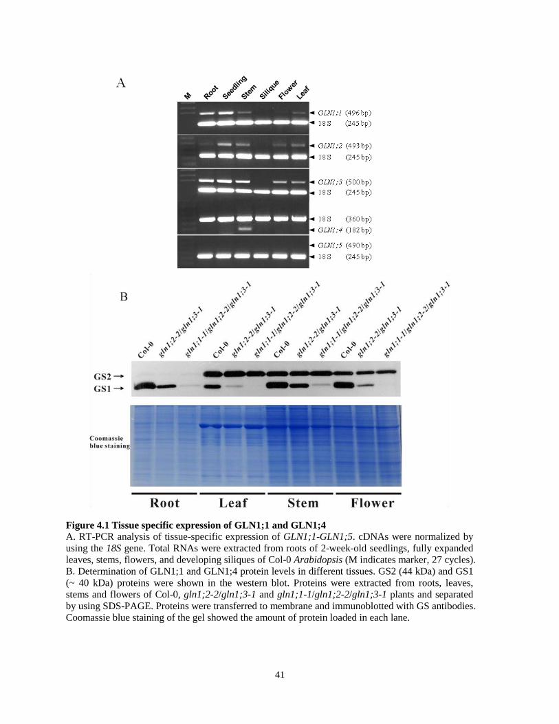
4.1 Tissue Specific Expression of GLN1;1 and GLN1;4
In Arabidopsis, GS2 is expressed specifically in the shoot and GS2 protein is restricted to
chloroplasts and mitochondria (Taira et al., 2004). The function of GS2 is for the assimilation of
ammonium released from photorespiration. Unlike GS2, GS1 has a number of isoforms
expressed in both shoots and roots (Ishiyama et al., 2004). Studies have shown that cytosolic GS
localizes mainly in the cytoplasm of mesophyll and vascular cells (Avila et al., 2001). Thus, it is
generally believed that GS1 is responsible for primary ammonium assimilation and transport in
plants. However, it is also important to note that although all the GS1 are expressed in the
cytosol of plant cells, they may have different roles due to distinct expression patterns in tissues.
Since previous work showed a tissue-specific pattern of GS accumulation and GS activity
in soybean (Fei et al., 2006), it may thus of interest to localize each of the isoforms with respect
to tissue-specificity in Arabidopsis in order to gain further insight into their individual
physiological roles. To this end, reverse transcriptase (RT)-PCR was used to examine the
expression of all five GS1 genes at the transcript level in specific tissues of Arabdiopsis. Because
the cDNA sequences of all 5 genes are highly similar, it is important to know the specificity of
each pair of primers. This issue is dealt more specifically in section 4.4, the gene specific primers
were able to amplify an expected band from wild-type plant, but not from the corresponding
mutants indicating the specificity of the primers (See Figure 4.5). These specific primers as
shown in Table 2 were used in RT-PCR analysis. As shown in Figure 4.1A, except GLN1;5,
transcript of all the other 4 isoforms can be detected. Interestingly, no expression of GS1 genes
was detected in siliques. GLN1;3 was one of the major isoforms among all of the GS1 proteins in
Arabidopsis seedlings (Figure 4.5B) and its transcripts appeared have the highest level of
39
expression among the GS1 genes (Figure 4.1A). Like GLN1;3, GLN1;2 transcripts could be
detected in different tissues. However, GLN1;1 and GLN1;4 showed more restricted pattern of
expression. A low level of GLN1;1 transcripts was detected in flowers, but high levels detected
in roots and seedlings. The only organ in which GLN1;4 transcripts were detected was the stem
(Figure 4.1A). Proteins were also extracted from different tissues including root, leaf, stem and
flower of 3 independent lines: wild-type, gln1;2-2/gln1;3-1, gln1;1-1/gln1;2-2/gln1;3-1 (the
mutants are described in section 4.7) and western blot was performed using GS antibody. Figure
4.1B shows the gel stained by coomassie blue indicating that similar amounts of total protein
were loaded. It is worthy to note that I have determined total protein content in each sample by
using the Bradford protein assay and loaded similar amounts of total protein in each well, even
though amounts of the large subunit of Rubisco (LSU) did not appear equivalent among different
tissue samples. Therefore, LSU is only good for protein normalization in the same tissues. The
expression level of GS2 protein varied in different tissues of Arabidopsis with a higher level of
expression in shoots than in roots. Because the transcript of GLN1;5 was never detected under
any condition, I assume that there is no expression of the GLN1;5 polypeptide in Arabidopsis
under the experimental conditions I examined. In leaves and flowers, western blot showed a band
in the gln1;2-2/gln1;3-1 double mutant but not in the gln1;1-1/gln1;2-2/gln1;3-1 triple mutant
indicating the band was GLN1;1. In roots and stems, the band in the gln1;2-2/gln1;3-1 double
mutant was stronger than in the triple mutant, which means GLN1;1 also expressed in roots and
stems. The broad expression of GLN1;1 in all tissues analyzed by western blot is consistent with
the RT-PCR results. The weak band in roots and stems of the triple mutant suggested that
GLN1;4 was present in these two types of tissue. When comparing GLN1 proteins in Col-0 and
gln1;2-2/gln1;3-1 double mutant among all the tissues, a dramatic decreased expression level of
40
GLN1 was observed in the gln1;2-2/gln1;3-1 double mutant which suggested the expression of
GLN1;2 and GLN1;3 were not restricted in any specific tissue. Interestingly, comparing the
expression of GLN1 protein in gln1;2-2/gln1;3-1 mutant to the wild-type plant, it appears that
GLN1;1 and GLN1;4 contribute major GLN1 expression in roots and stems suggesting a
possible role for them in primary N assimilation and transport.
4.2 Expression of GS1 Genes in Response to Abiotic Stresses
A number of studies have indicated an enhanced expression of GS1 protein is responsive to
various abiotic stresses, such as cold (Kwon et al., 2007), salt (Debouba et al., 2006), and H2O2
(Scarpeci et al., 2008). To determine which genes are specifically induced under stress
conditions, I treated Arabidopsis seedlings with salt and cold stresses and detected transcripts of
the 5 GS1 genes by RT-PCR. RD29A gene which is known to be induced by cold and salt stresses
was used to validate the treatments. As shown in Figure 4.2, no GLN1;5 expression was observed
in seedlings growing in normal conditions, neither was observed under the abiotic stresses. This
observation indicates that GLN1;5 is not expressed under normal and stress conditions. GLN1;1,
GLN1;3 and GLN1;4 were all induced by the 100 mM NaCl treatment at different times.
Transcript levels of GLN1;2 were maintained at a relatively steady level under salt stress, which
is higher than that in control plants treated with H2O. GLN1;1, GLN1;3 and GLN1;4 showed
similar expression patterns following the transfer of plants to 4 oC, while GLN1;2 exhibited a
constitutive expression with low temperature exposure.
41
Figure 4.1 Tissue specific expression of GLN1;1 and GLN1;4
A. RT-PCR analysis of tissue-specific expression of GLN1;1-GLN1;5. cDNAs were normalized by
using the 18S gene. Total RNAs were extracted from roots of 2-week-old seedlings, fully expanded
leaves, stems, flowers, and developing siliques of Col-0 Arabidopsis (M indicates marker, 27 cycles).
B. Determination of GLN1;1 and GLN1;4 protein levels in different tissues. GS2 (44 kDa) and GS1
(~ 40 kDa) proteins were shown in the western blot. Proteins were extracted from roots, leaves,
stems and flowers of Col-0, gln1;2-2/gln1;3-1 and gln1;1-1/gln1;2-2/gln1;3-1 plants and separated
by using SDS-PAGE. Proteins were transferred to membrane and immunoblotted with GS antibodies.
Coomassie blue staining of the gel showed the amount of protein loaded in each lane.

42
4.3 Expression of GS1s in Response to Pathogen Infection
In addition to responsiveness to abiotic stress, GS1 expression has been identified to be
associated with fungal and viral infection (Tavernier et al., 2007 and Pageau et al., 2006).
Moreover, Gln is a crucial nutrient for both plants and pathogens. Competition in acquisition of
Gln between them has a significant impact on disease development. Thus, investigation of the
response of GS1 genes is important for further characterizing the role of each GS1 gene involved
in defense against pathogens.
C. higginsianum is a hemibiotrophic fungus possessing a biotrophic phase and a
necrotrophic phase, and the invasion of fungus is a sophisticated process including spores
germination, appressorium formation, and primary and secondary hyphae generation. After plant
death, C. higginsianum employs a necrotrophic behavior for long-lasting survival (Liu et al.,
2007). Therefore the expression of each GS1 following C. higginsianum infection was analyzed
in a detailed time course investigation. The temporal expression pattern of the five GS1 genes was
examined over a 168 h post inoculation (hpi) period by RT-PCR. PR1 which is induced by
pathogen attacks was used as marker gene to show that the plants were responding to pathogen
infection. As shown in Figure 4.3, the transcripts of GLN1;1, GLN1;3 and GLN1;4 accumulate in
response to infection from 24 to 72 hpi. Whereas levels of GLN1;1 started to decrease after 72
hpi, GLN1;3 and GLN1;4 transcripts remained at a relatively high level until the plants died (144
hpi). These results suggest that GLN1;1, GLN1;3 and GLN1;4 might have roles in disease
development during C. higginsianum infection.

43
Figure 4.2 RT-PCR analysis of expression patterns of GLN1 genes in response to different
abiotic stresses in Arabidopsis seedlings.
(A). Time course of GLN1;1-GLN1;5 gene transcript accumulation in Arabidopsis seedlings
following salt treatment. RNA extracted from seedlings with H2O treatment at corresponding
time points were used as controls. cDNA were synthesized and RT-PCR performed. The amounts
of cDNA used were normalized based on the expression level of 18S. The expression level of
RD29A was shown to indicate that the seedlings were exposed in stress conditions (27 cycles).
(B). Time course of GLN1;1-GLN1;5 genes expression following cold treatment (27 cycles).

44
Figure 4.3 RT-PCR analysis of GLN1 expression in response to Colletotrichum higginsianum
infection.
Total RNA was isolated in a time course (0-168 hpi) from 4-week-old Arabidopsis leaves inoculated
with C. higginsianum by spraying (1×10³ spores μL
-1 in distilled water). RNAs from uninfected
leaves at 0 hpi were used as controls (Mock). cDNAs were normalized by using Actin 2 specific
primers. The transcript levels of the PR-1 gene indicated a successful C. higginsianum inoculation of
Arabidopsis plants (27 cycles).

45
4.4 Generation of gln1;1~gln1;5 Mutants
Nine independent T-DNA insertion lines were ordered from the SALK collection and named
gln1;1 (Salk_000459), gln1;2-1 (Salk_145235), gln1;2-2 (Salk_102291), gln1;3-1
(Salk_072283), gln1;3-2 (Salk_038156), gln1;4-1 (Salk_042546), gln1;4-2 (Salk_147053),
gln1;5-1 (Salk_086579), gln1;5-2 (Salk_117504). The putative T-DNA insertion sites associated
with individual GS1 genes are shown in Figure 4.4. In order to obtain homozygous mutants, T-
DNA insertions were identified at first by using combinations of gene- and T-DNA-specific
primer pairs (see materials and methods). In order to confirm the null mutation of each gene, RT-
PCR and western blotting were performed: Results show the absence of the full length transcript
of each gene and protein of each isoform in the perspective mutants, with the exception of
GLN1;5, for which neither the transcript nor the protein could be detected in Col-0 (Figure 4.5).
Moreover, the data demonstrate that in wild-type plants the total GS1 proteins could be separated
into 2 different bands by SDS-PAGE/western blot analysis by using the anti-GS antibody,
including GLN1;3 (top) and a mixture of GLN1;1, GLN1;2, and GLN1;4 (bottom). In the gln1;2
mutant, there was still a weak band at the bottom representing GLN1;1 and GLN1;4 which
means GLN1;1 and GLN1;4 could not be separated by the SDS-PAGE and overlapped with
GLN1;2. The results also showed that GS2 was equally expressed in all of the single mutants and
was not affected by the absence of any of the GS1 isoforms (Figure 4.5B). In addition, because I
had only one mutant line (Salk_000459) for isoform GLN1;1, confirmation of single T-DNA
insertion was conducted by using a cross between the gln1;1 homozygous and wild-type (Col-0)
plant. The F1 offsprings (GLN1;1/gln1;1 heterozygote) were then grown in soil to obtain the F2
generation. The seeds of the F2 generation were placed on the MS medium supplemented with
50 µg/mL kanamycin. After 2-week growth, the ratio between resistant and sensitive seedlings

46
was approximately 3:1 (93 resistant seedlings, 32 sensitive seedlings) indicating that it is highly
possible for gln1;1 mutant to have a single T-DNA insertion in the genome.
4.5 GLN1;1 Is Required for Phosphinothricin (PPT) Tolerance
PPT is a common herbicide used in agriculture. The main target of PPT is GS. For the purpose of
inhibiting the activity of GS, PPT binds to GS and occupies the position where glutamate is
supposed to be (Lea et al., 1984). In order to overcome the inactivation of GS, enhanced GS
expression is employed for improving N acquisition in plants. Studies on plants subjected to PPT
treatment have revealed that overexpression of GS enhances tolerance to PPT (Pascual et al.,
2008). Thus, in order to determine the tolerance of each mutant and the role of each gene, six
independent lines including wild type (Col-0) were grown on MS medium with 0.5 mg/L PPT.
After seeds were plated on the medium, a one-day delay of germination was observed among
some of the mutants. This also happened in the control plate and was probably due to seed
quality. After germination, as shown in Figure 4.6A, growth and development of leaf tissue and
root of gln1;1 seedlings were inhibited significantly. In contrast, the other 4 mutants
(gln1;2~gln1;5) had a similar growth rate and phenotype as wild-type plants. Because GS1
proteins are dominantly expressing in the root, the involvement of each isoenzyme in primary
root growth in response to PPT treatment was investigated by measuring the root length. Roots
of seedlings growing on plates with PPT were compared to control plates (without PPT). This
analysis showed that the degree of inhibition was much greater in gln1;1 and other mutants
including gln1;2, gln1;3,and gln1;4 also exhibited statistically shorter root length compared to
wild-type plants (Figure 4.6B). In conclusion, among all 5 homologs of GS1 family, GLN1;1 is
the most important member for PPT tolerance in both leaves and roots.

47
Figure 4.4 Schematic diagrams of GLN1 genes.
T-DNA insertion sites in GLN1;1, GLN1;2, GLN1;3, GLN1;4 and GLN1;5 and their
corresponding AGI codes are illustrated. T-DNA insertions are indicated with triangles, and exons
and introns are indicated as black boxes and lines, respectively.
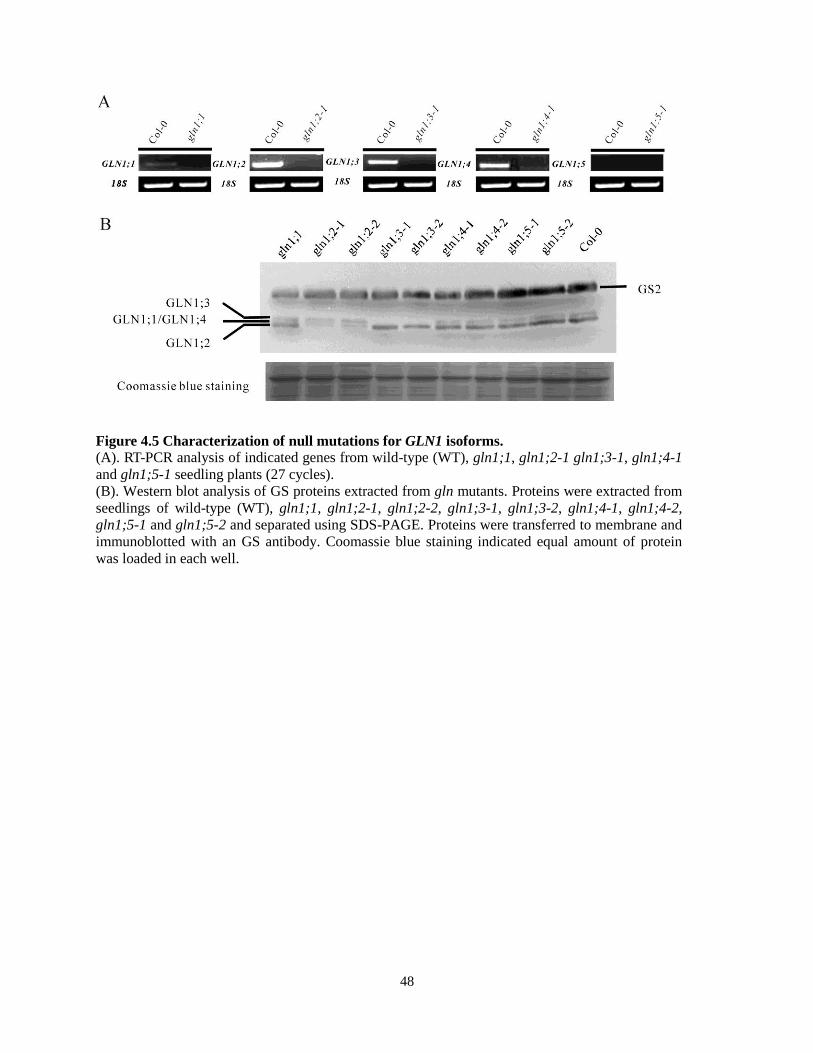
48
Figure 4.5 Characterization of null mutations for GLN1 isoforms.
(A). RT-PCR analysis of indicated genes from wild-type (WT), gln1;1, gln1;2-1 gln1;3-1, gln1;4-1
and gln1;5-1 seedling plants (27 cycles).
(B). Western blot analysis of GS proteins extracted from gln mutants. Proteins were extracted from
seedlings of wild-type (WT), gln1;1, gln1;2-1, gln1;2-2, gln1;3-1, gln1;3-2, gln1;4-1, gln1;4-2,
gln1;5-1 and gln1;5-2 and separated using SDS-PAGE. Proteins were transferred to membrane and
immunoblotted with an GS antibody. Coomassie blue staining indicated equal amount of protein
was loaded in each well.

49
Figure 4.6 gln1;1 mutants are more sensitive to PPT.
(A). 9-day-old seedlings of the wild-type, gln1;1, gln1;2, gln1;3, gln1;4 and gln1;5 mutant lines
grown on MS medium with (bottom) or without (top) 0.5 mg/L PPT.
(B). Quantification of root growth of WT and gln1 mutants on PPT. Asterisk indicates a P value (T-
test). *, P value < 0.05; **, P value < 0.01. Error bars represent standard error for three independent
experiments and twenty roots were measured respectively.

50
4.6 gln1;1 Mutant is Susceptible to Salt, Cold and Oxidative Stresses
The lack of GLN1;1 resulted in PPT sensitivity, suggesting that GLN1;1 might be important for
cell protection. Based on previous studies, GS1 is induced by salt (Debouba et al., 2005) and
cold (Kwon et al., 2007) stresses. Furthermore, our results also showed that the transcripts of
GLN1;1, GLN1;3 and GLN1;4 are up-regulated by abiotic stresses. Disruption in the expression
of GLN1;1, GLN1;3 or GLN1;4 may therefore affect salt or cold tolerance. Thus, it is of interest
to study the role of the GS1 genes in Arabidopsis seedlings against these abiotic stresses.
As shown in Figure 4.7A, the presence of 100 mM NaCl somewhat hindered the
greening of leaves and elongation of roots for all the genotypes tested. Compared with wild type,
the effect in the gln1;1 mutant was more severe, with growth and development of leaves being
significantly affected. The responses to low temperature and salinity stress in plant cells share
similar regulation pathway in which GS1 might be involved. With the expectation of similar
consequences of cold treatment as salt stress, seedlings were subjected to a cold shock stress
after 7 days of growth at room temperature on MS medium plate. Not surprisingly, the gln1;1
mutant developed more extensive necrosis and root growth inhibition resulting from cold stress
than wild-type plants and the other mutants (Figure 4.7B). These results indicate that the
enhanced expression of GS1 triggered by stresses plays an essential role in both salt and cold
stresses.
The cellular and molecular responses of plants to abiotic stress (including low
temperature, drought and salt) have been studied intensively (Xiong et al., 2002). ROS are
considered as a common signal involved in all of these stresses. Based on our results, the salt-
sensitive phenotype of gln1;1 suggested a possibility of enhanced ROS levels and prompted us to
further characterize the role of GLN1;1 in the ROS scavenging systems. Because external

51
application of different ROS generators is able to trigger a rapid accumulation of H2O2 or O2- in
different cellular locations (Apel and Hirt, 2004, Schwarzländer et al., 2009), I therefore
analyzed the effect of different ROS generators on the all the mutants. First, the response of
gln1;1 to methyl viologen (MV), an inducer of ROS that specifically produces O2- in
chloroplasts in a light-dependent fashion was tested. As Figure 4.8 shows, only the growth of the
gln1;1 mutant was significantly inhibited when compared to wild type in plates supplemented
with 0.1 μM MV. This inhibition was also reflected in root length. All seedlings, including wild
type and gln1;1, exhibited similar growth on MS control plates.
Since differences in root length between wild type and gln1;1 and dramatic variation of
leaf development by MV treatment was observed, there is a question of whether GLN1;1 is
specific for chloroplast protection. In order to answer this question, another two ROS inducers,
menadione (MD) and salicylhydroxamic acid (SHAM) (Schwarzländer et al., 2009), both
producing ROS in mitochondria were selected to apply to the mutants. I attempted a series of
concentrations of these two compounds, but did not observe any of them showing altered
inhibition of gln1 mutants compared to wild type (Figure 4.8). These results indicate that the
gln1;1 mutation enhances sensitivity specifically to methyl viologen, and that GLN1;1 might be
a key factor for protection of chloroplast under oxidative stress.
To confirm that GLN1;1 is only functional for defense against oxidative stress in
chloroplast, another ROS generator, rose bengal (RB), also targeting ROS in chloroplasts was
employed to treat all the lines for further investigation. Based on our prediction, I expected
similar growth behaviour as MV treatment; however, none of the mutants showed any more
serious inhibition than wild type in all of our plates supplemented with RB (Figure 4.8). It is
worth noting that even though MV and RB both contribute to ROS production in chloroplast, the

52
exact locations of ROS generation by these two inducer are different (see Figure 4.9). MV was
suggested to release the flavoprotein from the thylakoid membrane into the stroma, particularly
for flavoenzyme ferredoxin-NADP+ reductase (FNR), and results in O2
- over-accumulation
(Palatnik et al., 1997). In contrast, application of RB leads to high 1O2 formation in the stroma
(Fischer et al., 2007). The different results caused by these two ROS producers may due to the
different localizations of ROS production in the chloroplast.
The results above shows that the gln1;1 mutant has a prominent phenotype from MV
treatment. In order to confirm whether ROS are involved in growth inhibition of the gln1;1
mutant and where they are localized, I further examined the H2O2 generation in chloroplasts.
Leaves of wild type plant and the gln1;1 mutant were treated with 10 μM MV by spraying and
collected after 4 hrs of treatment. The non-fluorescence compound 2′, 7′-
dichlorodihydrofluorescein diacetate (H2DCFDA) producing a highly fluorescent compound
dichlorofluorescein (DCF) was used as a dye for intracellular H2O2 staining (Jakubowski and
Bartosz, 2000). As shown in Figure 4.10, H2O2 was not detected by confocal microscopy in the
control treatments for the wild type and gln1;1 mutant. However, 4 hrs treatment with 10 μM
MV, H2O2 signals were dramatically enhanced in the gln1;1 mutant leaves compared with wild
type, as shown by the bright green fluorescence resulting from staining with H2DCFDA. To
investigate the possible localization of H2O2, DCF signals were compared to chloroplast
autofluorescence derived from chlorophyll excitation by using a second channel of laser
scanning confocal microscopy. The fluorescence for H2O2 and chlorophyll showed a co-
localization pattern, suggesting that the H2O2 generation in gln1;1 mutants induced by MV took
place in the chloroplast.

53
Figure 4.7 Response of gln1 mutants to abiotic stresses.
(A). Left: 7-day-old seedlings of Col-0, gln1;1, gln1;2, gln1;3 and gln1;4 mutant lines grown in MS
medium with (bottom) or without (top) 100 mM NaCl. Right: Effect of NaCl on root length of the
Col-0 and gln1 mutants grown on MS medium plates.
(B). Left: 9-day-old seedlings of Col-0, gln1;1, gln1;2, gln1;3 and gln1;4 mutant lines grown in MS
medium at 22 ºC (top). 7-day-old seedlings were treated by cold shock at -18 ºC for 4 hrs and
recovered at 5 ºC for 16 d. Right: Effects of low temperature on root length of the Col-0 and gln1
mutants grown on MS medium plates.
Error bars represent SE for three independent experiments and twenty roots were measured
respectively. *, P value < 0.001 (T-test). Error bars represent SE for three independent experiments.

54
Figure 4.8 Response of gln1 mutants to different ROS generators.
Seedlings of the wild-type, gln1;1, gln1;2, gln1;3 and gln1;4 mutant lines grown in MS medium with
or without ROS generators (MV: methyl viologen; SHAM: salicylhydroxamic acid; RB: rose bengal;
MD: menadione). Plates with treatment were photographed at 21 days after incubation. Control plates
were photographed at 12 days after incubation.

55
Figure 4.9 Organization of the electron transport chain and ATP synthesis in the thylakoid
membrane of plant chloroplast The components of the chloroplast electron transport chain and the ATP-synthesizing apparatus are
shown. Electrons are transferred from H2O to NADP+. Accompanying the electron transfer, a proton
gradient is established across the membrane finally used for ATP synthesis. MV was suggested to
release the FNR from the thylakoid membrane into the soluble fraction resulting in an induction of
O2- (Palatnik et al., 1997). RB leads to high O2
- formation in the stroma (Fischer et al., 2007). The
diagram is based on Plant Physiology, Third edition by Lincoln Taiz and Eduardo Zeiger.

56
Figure 4.10 ROS production in Col-0 and gln1;1 mutant plants.
Signals were detected by laser scanning confocal microscopy. Leaves from 3-week-old wild type and
gln1;1 mutant plants were treated with or without 10 μM MV and stained with H2DCFDA (green).
DCF signals were visualized with excitation/emission at 488/505-530 nm. Chloroplast
autofluorescence (red) was visualized with excitation/emission: 488/>650 nm.

57
4.7 Isoforms GLN1;1, GLN1;2, and GLN1;3 Coordinate Primary Nitrogen Assimilation
Based on structural and biochemical studies, GS1 has been well-characterized as a key enzyme
for ammonium assimilation. For example, shoots from GS-inhibited plants which were treated
with MSX (L-methionine-S-sulfoximine) contained a higher NH4+ content than the control plants
(Skopelitis et al., 2006). Similar induction of NH4+ concentration was observed in PPT treated
tobacco plants (Purnell and Botella, 2007). In addition, biochemical assays also demonstrated
that GS had high Gln synthetic activity by quantifying L-Gln synthesized from NH4+ and L-Glu
(Ishiyama et al., 2004b). This evidence strongly suggests an essential role of GS in incorporating
NH4+ into the Gln organic form of N.
In order to further characterize the function of cytosolic GS, I used the genetic material
(gln1 mutants) to illustrate the role of this enzyme in incorporating NH4+ into organic N. Since
none of gln1 mutants showed any difference in NH4+ assimilation under both normal condition
(MS) and altered conditions with different forms of N (data not shown), the growth of double
and triple mutants was assessed on media containing inorganic N (NH4NO3) or organic N (Gln).
Because of functional redundancy of the five GS1 isoforms, the scarcity of mutants that are
defective in several genes of GS1 homologs somewhat impeded the characterization of GS1
functions. In order to comprehensively understand the principles underlying cooperative Gln
synthesis via numerous isoforms, the double mutants gln1;1-1/gln1;2-2, gln1;1-1/gln1;3-1,
gln1;1-1/gln1;4-1, gln1;2-2/gln1;3-1, gln1;3-1/gln1;4-1 and triple mutant gln1;1/gln1;2/gln1;3
were used in N assimilation assays. As I expected, all the single mutants (from gln1;1 to gln1;4)
exhibited similar growth as Col-0 wild type plants on MS medium with either NH4NO3 or Gln;
while a reduced growth of seedlings was observed clearly in the double mutant gln1;1/gln1;3
and more serious in the triple mutant

58
Figure 4.11 GLN1 is involved in primary nitrogen assimilation. (A). Arabidopsis seeds were germinated and grown on MS medium (top lane) containing 20 mM
NH4NO3 and MS medium supplemented with 5 mM Gln (bottom lane). The plates were
incubated in a growth chamber with 11/13 hrs light/dark period at 22°C and photographed at 5
days post incubation.
(B). Phenotype of 3-week-old Arabidopsis plant.
59
gln1;1/gln1;2/gln1;3 compared to other lines in MS with inorganic N which contains 20 mM
NH4NO3 (Figure 4.11A). In contrast, 5 mM Gln promoted the growth of gln1;1/gln1;3 and
gln1;1/gln1;2/gln1;3, partially restoring the phenotype of these two mutants (Figure 4.11A).
These results strongly suggest that Arabidopsis cytosolic GSs are crucial for the conversion of N
from inorganic (NH4+) to the organic (Gln) form in seedlings.
Cytosolic GS deficiency resulting in reduced growth and less grain were observed in wheat
and maize gln1 mutants (Martin et al., 2006; Tabuchi et al., 2005). Therefore, I proposed that
cytosolic GS may also regulate the growth behaviour of Arabidopsis at later stages of
development. In order to examine the effects of reduced GLN1s expression on plant growth, I
observed all these mutants throughout the vegetative stage. Interestingly, gln1;1/gln1;3 showed a
little bit smaller size than Col-0 until 3 weeks old. The gln1;1/gln1;2/gln1;3 mutant displayed
reduced growth until the 4th
week of growth (Figure 4.11B). These results further support the
hypothesis that GS1 positively regulates plant growth, especially GLN1;1, GLN1;2 and GLN1;3.
4.8 GLN1;1, GLN1;3 and GLN1;4 Are Functionally Redundant for Pollen Development
Based on previous studies that GS1 overexpressing tobacco plants exhibited enhanced growth
phenotype (Oliveira et al., 2002) and a GS1 deficient mutant showed decreased biomass in maize
(Martin et al., 2006) and male sterility in tobacco (Ribarits et al., 2007), I examined the
phenotype of each Arabidopsis mutant line including single mutants, double mutants and triple
mutants. Since reduced growth was observed in gln1;1/gln1;3 and gln1;1/gln1;2/gln1;3 mutants
before flowering (Figure 4.11B), I continued to generate triple mutants by crossing gln1;1-
1/gln1;3-1 with gln1;3-1/gln1;4-1 to obtain the gln1;1/gln1;3/gln1;4 (knockout of 3 stress-
induced isoforms) triple mutant. Interestingly, I was only able to only obtain gln1;1-/-/gln1;3
-/-/gln1;4
-/+

60
heterozygous mutants, but not the gln1;1/gln1;3/gln1;4 homozygous plants. According to
Mendel‟s law, the segregation ratio of self-pollination by gln1;1-/-
/gln1;3-/-
/gln1;4-/+
heterozygous mutant was supposed to be 1 gln1;1-/-
/gln1;3-/-
/gln1;4+/+
: 2 gln1;1-/-
/gln1;3-/-
/gln1;4-/+
: 1 gln1;1-/-
/gln1;3-/-
/gln1;4-/-
. Theoretically, I had 25% possibility to isolation
gln1;1/gln1;3/gln1;4 homozygous line. However, after several times of screening from the
offspring of gln1;1-/-
/gln1;3-/-
/gln1;4-/+
, I was still unable to obtain the homozygous plants
(numbers of plants screened each time was more than 100). Failure in obtaining a
gln1;1/gln1;3/gln1;4 homozygous line from these attempts suggests a possible situation that this
triple mutant might be lethal before the embryo stage, because the F2 generation of gln1;1-/-
/gln1;3-/-
/gln1;4-/+
mutant plant showed 98~100% germination rate and normal growth (data not
shown). In order to determine if the gln1;1/gln1;3/gln1;4 homozygote is lethal at embryo stage, I
examined the siliques from wild-type Col-0 and the gln1;1-/-
/gln1;3-/-
/gln1;4-/+
mutant. As Figure
4.12A shows, small siliques and undeveloped ovules were observed in plants of the gln1;1-/-
/gln1;3-/-
/gln1;4-/+
mutant while wild type showed long and full siliques without any unfertilized
ovules. This result strongly suggests that fertilization in flowers is disrupted in gln1;1-/-
/gln1;3-/-
/gln1;4-/+
mutant and results in unexpected segregation of genotypes.
Reduced fertility could be due to defects in either male or female gametes. Previous studies
in tobacco have shown that inactivation of cytosolic GS causing aborted pollen results in male
sterility (Ribarits et al., 2007). To test whether the male tissue is affected in the gln1;1-/-
/gln1;3-/-
/gln1;4-/+
mutant, I examined anthers in the opened flowers by Alexander‟s stain. As Figure
4.12B shows, compared to wild type, irregular size and shape of microspores in anthers from

61
gln1;1-/-
/gln1;3-/-
/gln1;4-/+
mutant were observed and most of the microspores were undeveloped,
with some even collapsed. When I released the pollen grains out of the anther, normal pollen
grains were stained with red color but the abnormal ones stained light blue (Figure 4.12B
bottom). Moreover, fertilization requires guidance of the pollen tube to deliver the sperm cells to
the ovules (Johnson and Preuss, 2002). Since significant aberrations of microspores in mature
anthers were observed in the mutant, I further analysed the germination of pollen tubes in wild-
type and mutant plants by using aniline blue staining. As Figure 4.12C shows, there were much
fewer pollen tubes visualized in the gln1;1-/-
/gln1;3-/-
/gln1;4-/+
mutant than in the wild type.
Collectively, these results indicate that full fertility requires at least one of GLN1;1, GLN1;3 and
GLN1;4 isoforms, otherwise the gln1;1/gln1;3/gln1;4 null mutant will be lethal due to the
impaired pollen development in gln1;1-/-
/gln1;3-/-
/gln1;4-/+
mutant.

62
Figure 4.12 Impaired pollen development in gln1-1-/-
/gln1-3-/-
/gln1-4-/+
mutants.
(A) Six siliques detached from the bottom of Arabidopsis stems of the Col-0 wild type and three
randomly selected gln1-1-/-
/gln1-3-/-
/gln1-4-/+
mutants were photographed (Left). Siliques from Col-0
and gln1-1-/-
/gln1-3-/-
/gln1-4-/+
mutant plants. Small, presumably unfertilized, ovules were visible in the
siliques of gln1-1-/-
/gln1-3-/-
/gln1-4-/+
mutant (Right). Asterisks designate unfertilized ovules in the
silique.
(B) Closed and opened anthers and pollen grains released from open anthers were stained with
Alexander‟s stain and photographed under a microscope. Normal grains were stained red while
abnormal ones were stained bright blue.
(C). Germinating tubes of pollens on stigma surface of open flowers of Col-0 and gln1-1-/-
/gln1-3-/-
/gln1-4-/+
stained by aniline blue were visualized under a light microscope (top) and florescence
microscopy (bottom). Bars=50 µM.

63
5. Discussion
5.1 GLN1;1 as a Major Cytosolic GS1 Isoform Contributes to Plant Tolerance against
Abiotic Stresses
An important aspect of current research in plant biology is using reverse genetic techniques to
identify key regulators related to abiotic stress tolerance. Although a large number of genes
respond to various abiotic stresses, only a fraction of them have been characterized to be
involved in stress tolerance while the physiological role for others is still speculative. Although
transcriptional regulation somehow varies between different stress stimuli, it is generally thought
that the response to cold, drought, and salt stress share a generic signalling pathway including the
following components: 1) Induction of Ca2+
influx which may reflect perception of the stress
signal, 2) Activation of phosphoprotein cascades, completed by protein kinases such as calcium-
dependent protein kinase (CDPK) and mitogen-activated protein kinase (MAPK), 3)
Transcription factors which aim to trigger or repress downstream genes to activate adaptive
responses (Sreenivasulu et al., 2007; Xiong et al., 2002). So far, many genes encoding CDPKs,
MAPKs, and transcription factors have been identified such as OsCDPK7, OsMAPK5, and
AtCBFs (CRT-binding factors) (Martin and Busconi, 2001; Saijo et al., 2000; Xiong and Yang,
2003). In the case of cytosolic GS, accumulating studies report an induction of GLN1 both at the
RNA and protein levels under various stress conditions (Debouba et al., 2006; Kwon et al.,
2007). It has also been observed that GS1 in leaves of Brassica napus can be post-translationally
regulated by reversible phosphorylation and that both protein kinase and protein phosphatase are
able to catalyze the reactions (Finnemann and Schjoerring, 2000). This type of post-translational
modification was also observed for Arabidopsis GLN1;1 by CDPK-related protein kinase (Li et
al., 2006). The phosphorylation status of GS1 is closely associated with its activity and stability,

64
while active GS1 always presents as the phosphorylated form (Finnemann and Schjoerring,
2000). All these results suggest that GS1 may be involved in the response to abiotic stress.
In my current work, I have employed molecular and genetic approaches to demonstrate the
physiological roles of cytosolic GS in abiotic stress and its involvement in ROS signalling in
Arabidopsis. With the co-operation of GOGAT, GS plays an important role in N assimilation in
plants. The product of GS, Gln, contributes to the biosynthesis of various organic N compounds
such as amino acids, nucleotides and chlorophyll. Under stress conditions, intracellular
hyperammonium would be produced and be toxic to the plant (Skopelitis et al., 2006). In order
to efficiently remove toxic ammonium and in defense to an extreme environment, plants require
a rearrangement of critical enzymes involved in N assimilation. The results from current work
show that GLN1;1, GLN1;3 and GLN1;4 of Arabidopsis are induced by both of salt and cold
treatments, which confirm the previous studies demonstrating that GS1 was responsive to salt
treatment and cold stress (Debouba et al., 2006; Kwon et al., 2007),.
Although the GLN1;1, GLN1;3 and GLN1;4 are likely to be involved in assimilation of
hyperammonium under stressful conditions, there is no genetic evidence in the current literature
to demonstrate the significance of each gene associated with stress responses. The project results
show that GLN1;1 is broadly expressed in different tissues, while western blot hybridization
showed a relative high expression in vascular-rich tissues (Figure 4.1B). The physiological role
of vascular-localized cytosolic GS was proposed to be related to N storage under stress
conditions in a previous study (Brugiere et al., 1999). Quantitative investigation of gene
expression of four GS1 genes in Arabidopsis roots revealed that GLN1;1 mRNA accumulated
abundantly compared to other genes in roots after a 3-day deprivation of N (Ishiyama et al.,
2004b). According to our results, unlike other mutants, gln1;1 mutant showed a serious growth
65
inhibition upon PPT treatment, as well as under abiotic stresses. This observation not only
confirms the functional importance of cytosolic GS in stress conditions, but emphasizes a major
role of GLN1;1 among the Arabidopsis GS1 members in tolerance of abiotic stresses.
It is important to note that there is a currently controversial conclusion made in rice. While
overexpressing plastidic GS in transgenic rice conferred tolerance to salt stress which is
consistent to our results (Hoshida et al., 2000), GS1;2-overexpressing lines of rice, produced by
an Agrobacterium tumefaciens-mediated transformation, exhibited higher sensitivity to salt and
cold stress conditions (Cai et al., 2009). These contrasting results might be explained by two
factors. First, rice is highly adapted to ammonium-based N nutrition. The uptake and transport of
NH4+ in rice primarily relies on GS/GOGAT cycle and OsAMTs. Previous studies found that
OsAMT1;2, which likely functions in NH4+ uptake from NH4
+-rich soils, was up-regulated by
Gln rather than NH4+ (Sonoda et al., 2003). The induction of OsGS1;2 expression therefore
produced more Gln and triggered an accumulation of OsAMT1;2, leading to hyperammonium in
plant cells. Second, the distinct expression pattern and tissue specificity of OsGS1;1, OsGS1;2
and OsGS1;3 indicates an different functional roles of the three genes in rice (Ishiyama et al.,
2004b; Tabuchi et al., 2005). The question as to why OsGS1;2 is important in sensitivity to
abiotic stress in rice still needs to be elucidated.
5.2 The Roles of Cytosolic GS in Plant Growth and Development
The effects of cytosolic GS on plant growth and yield have been fully investigated by analyzing
the phenotypes of GS1 deficiency mutants in rice and maize plants (Martin et al., 2006; Tabuchi
et al., 2005). Knocking out one key gene in both of these two species results in obvious growth
retardation or decreased biomass. In contrast, Arabidopsis mutants lacking any one GS1 gene
66
failed to show any significant growth inhibition under normal conditions and N-deficient
environment suggesting functional redundancy of these four detectable GS1 genes. Many key
enzymes like GS1 are encoded by duplicated genes, which are thought to perform redundant
roles in many aspects, preventing lethal auxotrophy caused by single gene mutation (Briggs et al.,
2006). Accordingly, I generated double and triple mutants by genetic crossing. I observed growth
retardation in the gln1;1/gln1;3 and gln1;1/gln1;2/gln1;3 mutants, and pollen lethality in
gln1;1/gln1;3/gln1;4, shedding light on the individual role of each gene.
GLN1;1, encoding a high affinity cytosolic GS, plays a major role in N assimilation among
the Arabidopsis GS1 members when plants are exposed to abiotic stress and the herbicide PPT.
The predominant function of GLN1;1 is likely interrelated with its unique enzymatic properties
or localization. Biochemical studies on the kinetic properties of each Arabidopsis GS1
isoenzyme revealed that GLN1;1 possesses the highest affinity for ammonium among the four
GS1 isoforms, suggesting a high efficiency of toxic ammonium attenuation (Ishiyama et al.,
2004b). With the regard of its localization, transgenic Arabidopsis plants (GLN1;1 promoter-GFP)
showed abundant accumulation of GFP at the root surface where the initial regulatory steps of
ammonium assimilation take place (Ishiyama et al., 2004b). Moreover, although our results
showed a broad expression pattern for GLN1;1 in the tissues I examined (including root, stem,
leaf, and flower), the relative protein expression of GLN1;1 varied a lot among different tissues,
with vascular-rich tissues such as root and stem possessing higher levels of expression (Figure
4.1). These results suggest that GLN1;1 might play a pivotal role in primary assimilation in roots
and N transport from source to sink tissue. Besides an essential role in vascular-rich organs,
GLN1;1 may also function in sink tissue. I have shown that, unlike other mutants, only gln1;1
displayed enhanced sensitivity specifically to methyl viologen, indicating that GLN1;1 might be

67
involved in reassimilation of ammonium produced by the photorespiratory pathway.
Although GLN1;3, encoding a low affinity isoform, did not show any significant role in
tolerance to abiotic stress in single mutants analysis, even though its expression is responsive to
such stresses, a synergic effect between GLN1;1 and GLN1;3, indicated by the obvious growth
retardation of the gln1;1/gln1;3 seedlings on MS medium, suggests an important role for
GLN1;3 as well as GLN1;1 (Figure 4.11A ). Unlike GLN1;1, which is expressed predominantly
in vascular-rich tissues, GLN1;3 mainly accumulated in sink tissues such as leaves and flowers.
In addition, recent studies by systems approaches showed that GLN1;3 is a light-responsive gene
and regulated by a master circadian clock control gene CCA1 (Gutierrez et al., 2008). This result
implies a sink-tissue specific role of GLN1;3. Moreover, the synergic phenotype of gln1;1/gln1;3
seedlings also suggests a partial function of GLN1;3 in seeds; because the growth inhibition of
gln1;1/gln1;3 was first observed in germinating seeds at a stage when there are no functional
chloroplast and leaf tissue. Therefore, I propose that GLN1;1 and GLN1;3 are the two major
cytosolic GS present in Arabidopsis.
GLN1;4, another isoform with high affinity for ammonium, appears not function in
seedlings, because no GLN1;4 transcripts could be detected at the seedling stage and the
gln1;1/gln1;4 double mutant grows as well as the wild-type plant (data not shown). Even in the
mature Arabidopsis plant, the mRNA of GLN1;4 is less abundant than other isoforms (Ishiyama
et al., 2004b). In fact, the pollen lethality of the gln1;1/gln1;3/gln1;4 triple mutant and the
reversion of lethality by applying any one of these 3 genes suggests that these 3 isoforms are
equally significant in the mature plant, particularly at the reproductive stage. However, GLN1;4
is not completely redundant to GLN1;1 and GLN1;3 due to its tissue specificity shown in our
western blot results (Figure 4.1B). GLN1;4 was identified as the only vascular-specific isoform
68
with an abundant expression and restricted localization in the root and stem, which is more
similar to GLN1;1. However, GLN1;4 was not detected in leaves and flowers, on the one hand,
implying its limited role in reproductive tissues, on the other hand, indicating that GLN1;4 might
be responsible for signal transferring, because in the gln1;1/gln1;2/gln1;3 mutant, the presence
of GLN1;4 in stem can restore the lethality even though there is no GS1 protein in leaves and
flowers.
GLN1;2 is classified as the lowest affinity isoform for ammonium among GS1 protein with
the Km values for GLN1;2 being almost twice as low as that of the other low affinity isoform
GLN1;3 (Ishiyama et al., 2004b). As the major form of cytosolic GS in Arabidopsis at seedling
stage (Figure 4.5B), GLN1;2 appears to contribute less to N assimilation than other isoforms.
Although the gln1;2 mutant did not show any difference in phenotype to abiotic stress and
oxidative stress compared to the wild type, transcripts of GLN1;2 were induced during leaf
senescence, implying a potential role of GLN1;2 in senescence (Diaz et al., 2008).
In addition, the function of GS for inorganic N assimilation in plants has been established
mainly based on biochemical studies in vitro. GS1 isoforms have been shown previously to
possess high affinity to ammonium in Arabidopsis by measuring enzyme activities (Ishiyama et
al., 2004b). In our work, the gln1;1/gln1;2/gln1;3 mutant showed a growth retardation in MS-N
media with NH4NO3 which implies that the cytosolic GS-deficiency mutants was not able to
utilize inorganic N to synthesize amino acids. However, this kind of growth inhibition could be
reversed by application of Gln to the MS-N medium indicating an essential role of GS1 in N
conversion from the inorganic to orgranic form. To the best of our knowledge, this is the first
genetic evidence showing that GS1 facilitates this conversion from inorganic N to organic N.
69
5.3 Involvement of Gln Metabolism in Regulation of Cellular Redox
Low temperature and high salinity are two different stresses stimulating distinct responses in
plant cells. For example, low temperature is known to modify membrane fluidity (Murata and
Los, 1997), while salt is known to change ionic and osmotic components (Cheeseman, 1988;
Munns and Tester, 2008; Tuteja, 2007). However, the generation of ROS including O2-, H2O2,
and HO-, is a general signal involved in most stresses due to the reduced rate of photosynthesis
(Apel and Hirt, 2004). Since GLN1;1, GLN1;3, and GLN1;4 showed similar responses to salt and
cold stress and enhanced sensitivity was observed in NaCl-treated gln1;1 mutants, we
hypothesize that the product of GLN1;1, Gln might play an important role in ROS scavenging.
Methyl viologen and rose bengal are two ROS elicitors both triggering ROS formation in
the chloroplast but in different locations (Fischer et al., 2007; Palatnik et al., 1997). Menadione
and salicylhydroxamic acid are also ROS propagators but targeting mitochondria (Shen et al.,
2006; Sweetlove et al., 2001). Our plate assays did not show any significant inhibition of the
gln1;1 mutant by elicitor challenge other than by methyl viologen. This finding uncovered the
involvement of Gln in attenuating ROS levels possible resulting from solubilization of FNR in
chloroplasts and confocal images further confirmed that Gln is specifically functional in the
chloroplast.
What is the mechanism underlying such an efficient ROS scavenging pathway? It is well
known that plants possess two systems for ROS detoxification in the chloroplast. One is an
enzymatic system, with superoxide dismutase (SOD), APX, and CAT being the major enzymes
involved in removing ROS. Most of these enzymes are located in many cellular compartments
(Myouga et al. 2008). The responsibility of these enzymes is to efficiently catalyze the reactions
for converting the toxic forms of ROS (O2- or H2O2) to low-toxic status. The other ROS

70
detoxification system is non-enzymatic, employing carotenes, tocopherol, ascorbic acid and
glutathione as antioxidants in most cellular compartments for defense against oxidative stress
(Mittler, 2002). Gln, one of the major amino acids present in the vascular tissue of plants, has
only been reported to influence the redox potential of the cell via enhancing the generation of
glutathione in animals but not in plants (Curi et al., 2007; Roth, 2007; Roth, 2008). Previous
studies in plants revealed that down-regulations were observed in both the protein and activity of
GS2 under different stress conditions, whereas GS1 protein was induced (Pageau et al., 2006;
Scarpeci et al., 2008; Simonovic and Anderson, 2007). The opposing behavior of GS1 and GS2
might be due to the fact that plants require Gln to reduce ROS production resulting from the
inhibition of photosynthesis in chloroplasts. Since Gln is consumed in chloroplasts under stress
conditions, unknown signals may somehow activate GS1 genes to compensate for Gln deficiency.
One possible purpose of Gln consumption is Pro accumulation; proline accumulation is a
common physiological response when plants are exposed to a wide range of environmental
stresses, such as salt, drought, high/low temperature, UV irradiation, and pathogen infection
(Hare and Cress, 1997; Saradhi et al., 1995; Verbruggen and Hermans, 2008). Pro is believed to
be an important osmolyte that functions as a molecular chaperone stabilizing the structure of
proteins, and to regulate the cellular redox potential by controlling free radical levels (Hare et al.,
1999). The biosynthesis of Pro primarily relies on the Glu pathway in plant cells (Ashton and
Desh Pal, 1993). Gln is the main source of Glu production through the GS/GOGAT cycle. A
previous study in tobacco has demonstrated that, instead of Glu, Gln is the limiting factor for Pro
production because 15
NH4+-labeling experiments showed that the accumulation rate of
15N-Pro
was similar to the rate of 15
N-Gln production while both of them were much lower in GS1-
deficient transgenic plants than in control plants. By contrast, the use of 15
N-Glu in GS1-
71
deficient transgenic plants was similar to that of the control plant (Brugiere et al., 1999). In
addition, a recent study indicated the transcript of P5CS1, encoding a enzyme catalyzing the rate-
limiting step from Glu to Pro, was strikingly enhanced in chloroplasts under high salinity
condition (Szekely et al., 2008). Based on these lines of evidence, glutamine might be involved
in ROS scavenging through the Pro pathway.
Moreover, FNR mediates the final step of photosynthetic electron transfer between
ferredoxin (Fd) and NADP+ and produces NADPH (Lintala et al., 2007). Balancing the
ATP/NADPH ratio in order to prevent excess accumulation of electrons is important for ROS
control, because excess electrons can be transferred to O2 thus generating ROS (Foyer and
Noctor, 2005; Mittler, 2002). ROS stimulation by methyl viologen is due to the release of FNR
from the thylakoid to the stroma thus resulting in the block of electron transport (Palatnik et al.,
1997). Indeed, malfunction of FNR and Fd both result in the enhanced accumulation of ROS in
plants (Hajirezaei et al., 2002; Palatnik et al., 2003; Voss et al., 2008). In this regard,
Arabidopsis fnr1 and fnr2 mutants with impaired function of FNR showed a transcriptional
upregulation of NR and GLN1;1, as well as an enhancement of N metabolism, suggesting that
Gln might be involved in alternative electron transfer pathways in the chloroplast (Lintala et al.,
2009; Lintala et al., 2007). Moreover, in vitro experiments demonstrated that the efficient
oxidation of NADPH relies on Gln and 2-oxoglutarate (Valadier et al., 2008). In this study, I
showed that the gln1;1 mutant was only sensitive to methyl viologen and displayed high levels
of ROS formation in chloroplast, further implying a significant role of Gln in the chloroplastic
redox system.
The function of GLN1;1 suggested to take place in chloroplast, is interesting to our
understanding of ROS scavenging systems. In addition to chloroplasts, mitochondria are also

72
major compartments for ROS production. Like GS2, Fd-GOGAT was reported to localize to
mitochondria and the glu1 mutant, which is defective in Fd-GOGAT, exhibited photorespiratory
chlorosis at ambient CO2 (Jamai et al., 2009). Thus, the chloroplast and mitochondrion both
necessitate the presence of the GS2/Fd-GOGAT cycle for reassimilation of photorespiratory
ammonium as well as for primary N assimilation. In this case, it is possible that GLN1;1 may be
required for ROS detoxification not only in the chloroplasts but also in mitochondria. However,
it is interesting that our observation is not in such prediction indicating the role of GS2/Fd-
GOGAT cycle may be distinct in these two compartments.

73
References
Almagro, A., Lin, S. H., and Tsay, Y. F. (2008). Characterization of the Arabidopsis nitrate
transporter NRT1.6 reveals a role of nitrate in early embryo development. Plant Cell 20, 3289-
3299.
Apel, K., and Hirt, H. (2004). Reactive oxygen species: metabolism, oxidative stress, and signal
transduction. Annu. Rev. Plant Biol. 55, 373-399.
Ashton, J. D., and Desh Pal, S. V. (1993). Proline biosynthesis and osmoregulation in plants.
Plant J. 4, 215-223.
Avila, C., Marquez, A. J., Pajuelo, P., Cannell, M. E., Wallsgrove, R. M., and Forde, B. G. (1993).
Cloning and sequence analysis of a cDNA for barley ferredoxin-dependent glutamate synthase
and molecular analysis of photorespiratory mutants deficient in the enzyme. Planta 189, 475-483.
Avila, C., Suárez, M. F., Gómez-Maldonado, J., and Cánovas, F. M. (2001). Spatial and temporal
expression of two cytosolic glutamine synthetase genes in Scots pine: functional implications on
N metabolism during early stages of conifer development. Plant J. 25, 93-102.
Baker, A., Graham, I. A., Holdsworth, M., Smith, S. M., and Theodoulou, F. L. (2006). Chewing
the fat: beta-oxidation in signalling and development. Trends Plant Sci. 11, 124-132.
Baniwal, S. K., Bharti, K., Chan, K. Y., Fauth, M., Ganguli, A., Kotak, S., Mishra, S. K., Nover,
L., Port, M., Scharf, K. D., Tripp, J., Weber, C., Zielinski, D., and Von, K. P. (2004). Heat stress
response in plants: a complex game with chaperones and more than twenty heat stress
transcription factors. J. Biosci. 29, 471-487.
Bennett, M. J. and Cullimore, J. V. (1989). Glutamine synthetase isoenzymes of Phaseolus
vulgaris L.: Subunits composition in developing root nodules and plumules. Planta 179: 433–440.
Bennett, M. J., Marchant, A., Green, H. G., May, S. T., Ward, S. P., Millner, P. A., Walker, A. R.,

74
Schulz, B., and Feldmann, K. A. (1996). Arabidopsis AUX1 gene: a permease-like regulator of
root gravitropism. Science 273, 948-950.
Briggs, G. C., Osmont, K. S., Shindo, C., Sibout, R., and Hardtke, C. S. (2006). Unequal genetic
redundancies in Arabidopsis--a neglected phenomenon? Trends Plant Sci. 11, 492-498.
Britto, D. T., and Kronzucker, H. J. (2001). Constancy of N turnover kinetics in the plant cell:
insights into the integration of subcellular N fluxes. Planta 213, 175-181.
Brugiere, N., Dubois, F., Limami, A. M., Lelandais, M., Roux, Y., Sangwan, R. S., and Hirel, B.
(1999). Glutamine synthetase in the phloem plays a major role in controlling proline production.
Plant Cell 11, 1995-2012.
Buchanan, B. B., and Balmer, Y. (2005). Redox regulation: a broadening horizon. Annu. Rev.
Plant Biol. 56, 187-220.
Cai, H., Zhou, Y., Xiao, J., Li, X., Zhang, Q., and Lian, X. (2009). Overexpressed glutamine
synthetase gene modifies N metabolism and abiotic stress responses in rice. Plant Cell Rep. 28,
527-537.
Cheeseman, J. M. (1988). Mechanisms of salinity tolerance in plants. Plant Physiol. 87, 547-550.
Chen, L., and Bush, D. R. (1997). LHT1, a lysine- and histidine-specific amino acid transporter
in Arabidopsis. Plant Physiol. 115, 1127-1134.
Chen, L., Ortiz-Lopez, A., Jung, A., and Bush, D. R. (2001). ANT1, an aromatic and neutral
amino acid transporter in Arabidopsis. Plant Physiol. 125, 1813-1820.
Chiu, C., Lin, C., Hsia, A., Su, R., Lin, H., and Tsay, Y. (2004). Mutation of a nitrate transporter,
AtNRT1:4, results in a reduced petiole nitrate content and altered leaf development. Plant Cell
Physiol. 45, 1139-1148.
Colcombet, J., and Hirt, H. (2008). Arabidopsis MAPKs: a complex signalling network involved
75
in multiple biological processes. Biochem. J. 413, 217-226.
Conklin, P. L., Williams, E. H., and Last, R. L. (1996). Environmental stress sensitivity of an
ascorbic acid-deficient Arabidopsis mutant. Proc. Natl. Acad. Sci. USA 93, 9970-9974.
Coruzzi, G., M. (2003). Primary N-assimilation into Amino Acids in Arabidopsis. (2003). The
Arabidopsis Book, American Society of Plant Biologists.
Coschigano, K. T., Melo-Oliveira, R., Lim, J., and Coruzzi, G. M. (1998). Arabidopsis gls
mutants and distinct Fd-GOGAT genes: Implications for photorespiration and primary N
assimilation. Plant Cell 10, 741-752.
Crawford, N. M. (1995). Nitrate: Nutrient and signal for plant growth. Plant Cell 7, 859-868.
Creissen, G., Firmin, J., Fryer, M., Kular, B., Leyland, N., Reynolds, H., Pastori, G., Wellburn, F.,
Baker, N., Wellburn, A., and Mullineaux, P. (1999). Elevated glutathione biosynthetic capacity in
the chloroplasts of transgenic tobacco plants paradoxically causes increased oxidative stress.
Plant Cell 11, 1277-1292.
Curi, R., Newsholme, P., Procopio, J., Lagranha, C., Gorjao, R., and Pithon-Curi, T. C. (2007).
Glutamine, gene expression, and cell function. Front Biosci. 12, 344-357.
Dat, J., Vandenabeele, S., Vranova, E., Van Montagu, M., Inze, D., and Van Breusegem, F.
(2000). Dual action of the active oxygen species during plant stress responses. Cell Mol. Life Sci.
57, 779-795.
Davletova, S., Rizhsky, L., Liang, H., Shengqiang, Z., Oliver, D. J., Coutu, J., Shulaev, V.,
Schlauch, K., and Mittler, R. (2005). Cytosolic ascorbate peroxidase 1 is a central component of
the reactive oxygen gene network of Arabidopsis. Plant Cell 17, 268-281.
Debouba, M., Gouia, H., Suzuki, A., and Ghorbel, M. H. (2006). NaCl stress effects on enzymes
involved in N assimilation pathway in tomato "Lycopersicon esculentum" seedlings. J. Plant

76
Physiol. 163, 1247-1258.
Desikan, R., Hancock, J. T., Ichimura, K., Shinozaki, K., and Neill, S. J. (2001). Harpin induces
activation of the Arabidopsis mitogen-activated protein kinases AtMPK4 and AtMPK6. Plant
Physiol. 126, 1579-1587.
Deslandes, L., Olivier, J., Theulieres, F., Hirsch, J., Feng, D. X., Bittner-Eddy, P., Beynon, J., and
Marco, Y. (2002). Resistance to Ralstonia solanacearum in Arabidopsis thaliana is conferred by
the recessive RRS1-R gene, a member of a novel family of resistance genes. Proc. Natl. Acad.
Sci. USA 99, 2404-2409.
Diaz, C., Lemaitre, T., Christ, A., Azzopardi, M., Kato, Y., Sato, F., Morot-Gaudry, J. F., Le Dily,
F., and Masclaux-Daubresse, C. (2008). N recycling and remobilization are differentially
controlled by leaf senescence and development stage in Arabidopsis under low N nutrition. Plant
Physiol. 147, 1437-1449.
Eulgem, T., Rushton, P. J., Robatzek, S., and Somssich, I. E. (2000). The WRKY superfamily of
plant transcription factors. Trends Plant Sci. 5, 199-206.
Fei, H., Chaillou, S., Hirel, B., Polowick, P., Mahon, J. D., and Vessey, J. K. (2006). Effects of
the overexpression of a soybean cytosolic glutamine synthetase gene (GS15) linked to organ-
specific promoters on growth and N accumulation of pea plants supplied with ammonium. Plant
Physiol. Biochem. 44, 543-550.
Filleur, S., Dorbe, M.-F., Cerezo, M., Orsel, M., Granier, F., Gojon, A., and Daniel-Vedele, F.
(2001). An Arabidopsis T-DNA mutant affected in Nrt2 genes is impaired in nitrate uptake.
FEBS Letters 489, 220-224.
Finnemann, J., and Schjoerring, J. K. (2000). Post-translational regulation of cytosolic glutamine
synthetase by reversible phosphorylation and 14-3-3 protein interaction. Plant J. 24, 171-181.
77
Fischer, B. B., Krieger-Liszkay, A., Hideg, E., Snyrychova, I., Wiesendanger, M., and Eggen, R.
I. (2007). Role of singlet oxygen in chloroplast to nucleus retrograde signaling in
Chlamydomonas reinhardtii. FEBS Lett 581, 5555-5560.
Fischer, W. N., Kwart, M., Hummel, S., and Frommer, W. B. (1995). Substrate specificity and
expression profile of amino acid transporters (AAPs) in Arabidopsis. J. Biol. Chem. 270, 16315-
16320.
Fischer, W. N., Loo, D. D., Koch, W., Ludewig, U., Boorer, K. J., Tegeder, M., Rentsch, D.,
Wright, E. M., and Frommer, W. B. (2002). Low and high affinity amino acid H+-cotransporters
for cellular import of neutral and charged amino acids. Plant J. 29, 717-731.
Forde, B. G. (2002). Local and long-range signaling pathways regulating plant responses to
nitrate. Annu. Rev. Plant Biol. 53, 203-224.
Forde, B. G., and Lea, P. J. (2007). Glutamate in plants: metabolism, regulation, and signalling. J.
Exp. Bot. 58, 2339-2358.
Foyer, C. H., and Noctor, G. (2003). Redox sensing and signalling associated with reactive
oxygen in chloroplasts, peroxisomes and mitochondria. Physiol. Plant. 119, 355-364.
Foyer, C. H., and Noctor, G. (2005). Redox homeostasis and antioxidant signaling: a metabolic
interface between stress perception and physiological responses. Plant Cell 17, 1866-1875.
Frommer, W. B., Hummel, S., and Riesmeier, J. W. (1993). Expression cloning in yeast of a
cDNA encoding a broad specificity amino acid permease from Arabidopsis thaliana. Proc. Natl.
Acad. Sci. USA 90, 5944-5948.
Giraud, E., Ho, L. H., Clifton, R., Carroll, A., Estavillo, G., Tan, Y. F., Howell, K. A., Ivanova, A.,
Pogson, B. J., Millar, A. H., and Whelan, J. (2008). The absence of alternative oxidase 1a in
Arabidopsis results in acute sensitivity to combined light and drought stress. Plant Physiol. 147,

78
595-610.
Grallath, S., Weimar, T., Meyer, A., Gumy, C., Suter-Grotemeyer, M., Neuhaus, J. M., and
Rentsch, D. (2005). The AtProT family. Compatible solute transporters with similar substrate
specificity but differential expression patterns. Plant Physiol. 137, 117-126.
Gutierrez, R. A., Stokes, T. L., Thum, K., Xu, X., Obertello, M., Katari, M. S., Tanurdzic, M.,
Dean, A., Nero, D. C., McClung, C. R., and Coruzzi, G. M. (2008). Systems approach identifies
an organic N-responsive gene network that is regulated by the master clock control gene CCA1.
Proc. Natl. Acad. Sci. USA 105, 4939-4944.
Hajirezaei, M. R., Peisker, M., Tschiersch, H., Palatnik, J. F., Valle, E. M., Carrillo, N., and
Sonnewald, U. (2002). Small changes in the activity of chloroplastic NADP+-dependent
ferredoxin oxidoreductase lead to impaired plant growth and restrict photosynthetic activity of
transgenic tobacco plants. Plant J. 29, 281-293.
Hammes, U. Z., Nielsen, E., Honaas, L. A., Taylor, C. G., and Schachtman, D. P. (2006).
AtCAT6, a sink-tissue-localized transporter for essential amino acids in Arabidopsis. Plant J. 48,
414-426.
Hare, P., Cress, W., and van Staden, J. (1999). Proline synthesis and degradation: a model system
for elucidating stress-related signal transduction. J. Exp. Bot. 50, 413-434.
Hare, P. D., and Cress, W. A. (1997). Metabolic implications of stress-induced proline
accumulation in plants. Plant Growth Regul. 21, 79-102.
Hase, T., Schürmann, P., and Knaff, D. B. (2006). The interaction of ferredoxin with ferredoxin-
dependent enzymes. Photosystem I. 24, 477-498.
Hirner, A., Ladwig, F., Stransky, H., Okumoto, S., Keinath, M., Harms, A., Frommer, W. B., and
Koch, W. (2006). Arabidopsis LHT1 is a high-affinity transporter for cellular amino acid uptake

79
in both root epidermis and leaf mesophyll. Plant Cell 18, 1931-1946.
Hoshida, H., Tanaka, Y., Hibino, T., Hayashi, Y., Tanaka, A., Takabe, T., and Takabe, T. (2000).
Enhanced tolerance to salt stress in transgenic rice that overexpresses chloroplast glutamine
synthetase. Plant Mol. Biol. 43, 103-111.
Hsu, L. C., Chiou, T. J., Chen, L., and Bush, D. R. (1993). Cloning a plant amino acid transporter
by functional complementation of a yeast amino acid transport mutant. Proc. Natl. Acad. Sci.
USA 90, 7441-7445.
Huang, N., Liu, K., Lo, H., and Tsay, Y. (1999). Cloning and functional characterization of an
Arabidopsis nitrate transporter gene that encodes a constitutive component of low-affinity uptake.
Plant Cell 11, 1381-1392.
Ishiyama, K., Inoue, E., Watanabe-Takahashi, A., Obara, M., Yamaya, T., and Takahashi, H.
(2004a). Kinetic properties and ammonium-dependent regulation of cytosolic isoenzymes of
glutamine synthetase in Arabidopsis. J. Biol. Chem. 279, 16598-16605.
Ishiyama, K., Inoue, E., Tabuchi, M., Yamaya, T., and Takahashi, H. (2004b). Biochemical
background and compartmentalized functions of cytosolic glutamine synthetase for active
ammonium assimilation in rice roots. Plant Cell Physiol. 45, 1640-1647.
Jakubowski, W. and Bartosz, G. (2000). 2,7-dichlorofluorescin oxidation and reactive oxygen
species: what does it measure? Cell Biol. Int. 24, 757-760.
Jamai, A., Salome, P. A., Schilling, S. H., Weber, A. P., and McClung, C. R. (2009). Arabidopsis
photorespiratory serine hydroxymethyltransferase activity requires the mitochondrial
accumulation of ferredoxin-dependent glutamate synthase. Plant Cell 21, 595-606.
Jaspers, P., and Kangasjärvi, J. (2010). Reactive oxygen species in abiotic stress signaling.
Physiol. Plant. 138, 405-413.

80
Johnson, M. A., and Preuss, D. (2002). Plotting a course: multiple signals guide pollen tubes to
their targets. Dev. Cell 2, 273-281.
Keryer, E., Collin, V., Lavergne, D., Lemaire, S., and Issakidis-Bourguet, E. (2004).
Characterization of Arabidopsis mutants for the variable subunit of ferredoxin:thioredoxin
reductase. Photosynth. Res. 79, 265-274.
Kliebenstein, D. J., Monde, R. A., and Last, R. L. (1998). Superoxide dismutase in Arabidopsis:
an eclectic enzyme family with disparate regulation and protein localization. Plant Physiol. 118,
637-650.
Knoth, C., Ringler, J., Dangl, J. L., and Eulgem, T. (2007). Arabidopsis WRKY70 is required for
full RPP4-mediated disease resistance and basal defense against Hyaloperonospora parasitica.
Mol. Plant Microbe Int. 20, 120-128.
Kovtun, Y., Chiu, W. L., Tena, G., and Sheen, J. (2000). Functional analysis of oxidative stress-
activated mitogen-activated protein kinase cascade in plants. Proc Natl Acad Sci USA 97, 2940-
2945.
Krapp, A., Saliba-Colombani, V., and Daniel-Vedele, F. (2005). Analysis of C and N metabolisms
and of C/N interactions using quantitative genetics. Photosynth. Res. 83, 251-263.
Kwon, S. J., Kwon, S. I., Bae, M. S., Cho, E. J., and Park, O. K. (2007). Role of the methionine
sulfoxide reductase MsrB3 in cold acclimation in Arabidopsis. Plant Cell Physiol. 48, 1713-1723.
Kyo, M., and Harada, H. (1986). Control of the developmental pathway of tobacco pollen in
vitro. Planta 168, 427-432.
Laemmli, U. K. (1970). Cleavage of structural proteins during the assembly of the head of
bacteriophage T4. Nature 227, 680-685.
Lancien, M., Martin, M., Hsieh, M. H., Leustek, T., Goodman, H., and Coruzzi, G. M. (2002).

81
Arabidopsis glt1-T mutant defines a role for NADH-GOGAT in the non-photorespiratory
ammonium assimilatory pathway. Plant J. 29, 347-358.
Lea, P. J., Joy, K. W., Ramos, J. L., and Guerrero, M. G. (1984). The action of 2-amino-4-
(methylphosphinyl)-butanoic acid (phosphinothricin) and its 2-oxo-derivative on the metabolism
of cyanobacteria and higher plants. Phytochem. 23, 1-6.
Lee, Y. H., and Tegeder, M. (2004). Selective expression of a novel high-affinity transport system
for acidic and neutral amino acids in the tapetum cells of Arabidopsis flowers. Plant J. 40, 60-74.
Lee, Y. H., Foster, J., Chen, J., Voll, L. M., Weber, A. P., and Tegeder, M. (2007). AAP1
transports uncharged amino acids into roots of Arabidopsis. Plant J. 50, 305-319.
Lemaitre, T., Urbanczyk-Wochniak, E., Flesch, V., Bismuth, E., Fernie, A. R., and Hodges, M.
(2007). NAD-dependent isocitrate dehydrogenase mutants of Arabidopsis suggest the enzyme is
not limiting for N assimilation. Plant Physiol. 144, 1546-1558.
Li, R. J., Hua, W., and Lu, Y. T. (2006). Arabidopsis cytosolic glutamine synthetase AtGLN1;1 is
a potential substrate of AtCRK3 involved in leaf senescence. Biochem. Biophys. Res. Commun.
342, 119-126.
Li, W., Wang, Y., Okamoto, M., Crawford, N. M., Siddiqi, M. Y., and Glass, A. D. M. (2007).
Dissection of the AtNRT2.1:AtNRT2.2 inducible high-affinity nitrate transporter gene cluster.
Plant Physiol. 143, 425-433.
Li, J., Fu. Y., Pike, S. M., Bao, J., Tian. W., Zhang. Y., Chen, C., Zhang, Y., Li, H., Huang, J., Li,
L., Schroeder, J. I., Gassmann, W., and Gong, J. (2010). The Arabidopsis nitrate transporter
NRT1.8 functions in nitrate removal from the xylem sap and mediates cadmium tolerance. Plant
Cell online publication
Lichter, A., and Hãberlein, I. (1998). A light-dependent redox-signal participates in the regulation
82
of ammonia fixation in chloroplast of higher plants-ferredoxin: glutamate synthase is a
thioredoxin-dependent enzyme. J. Plant Physiol. 153, 83-90.
Lintala, M., Allahverdiyeva, Y., Kangasjarvi, S., Lehtimaki, N., Keranen, M., Rintamaki, E., Aro,
E. M., and Mulo, P. (2009). Comparative analysis of leaf-type ferredoxin-NADP oxidoreductase
isoforms in Arabidopsis thaliana. Plant J. 57, 1103-1115.
Lintala, M., Allahverdiyeva, Y., Kidron, H., Piippo, M., Battchikova, N., Suorsa, M., Rintamaki,
E., Salminen, T. A., Aro, E. M., and Mulo, P. (2007). Structural and functional characterization of
ferredoxin-NADP+-oxidoreductase using knock-out mutants of Arabidopsis. Plant J. 49, 1041-
1052.
Liu, G., Kennedy, R., Greenshields, D. L., Peng, G., Forseille, L., Selvaraj, G., and Wei, Y.
(2007). Detached and attached Arabidopsis leaf assays reveal distinctive defense responses
against hemibiotrophic Colletotrichum spp. Mol. Plant Microbe Int. 20, 1308-1319.
Liu, K., and Tsay, Y. (2003). Switching between the two action modes of the dual-affinity nitrate
transporter CHL1 by phosphorylation. EMBO J. 22, 1005-1013.
Liu, G., Ji, Y., Bhuiyan, N. H., Pilot, G., Selvaraj, G., Zou, J., and Wei, Y. (2010). Amino acid
homeostasis modulates salicylic acid-associated redox status and defense responses in
Arabidopsis. Plant Cell online publication
Lombi, E., Hamon, R. E., Wieshammer, G., McLaughlin, M. J., and McGrath, S. P. (2004).
Assessment of the use of industrial by-products to remediate a copper- and arsenic-contaminated
soil. J. Environ. Qual. 33, 902-910.
Loque, D., and von Wiren, N. (2004). Regulatory levels for the transport of ammonium in plant
roots. J. Exp. Bot. 55, 1293-1305.
Loulakakis, K. A., Primikirios, N. I., Nikolantonakis, M. A., and Roubelakis-Angelakis, K. A.

83
(2002). Immunocharacterization of Vitis vinifera L. ferredoxin-dependent glutamate synthase,
and its spatial and temporal changes during leaf development. Planta 215, 630-638.
Ludewig, U., von Wiren, N., Rentsch, D., and Frommer, W. B. (2001). Rhesus factors and
ammonium: a function in efflux? Genome Biol. 2, REVIEWS1010.
Magalhães, J. R., Ju, G. C., Rich, P. J., and Rhodes, D. (1990). Kinetics of 15
NH4+ assimilation in
Zea mays: Preliminary studies with a glutamate dehydrogenase (GDH1) null mutant. Plant
Physiol. 94, 647-656.
Marmagne, A., Ferro, M., Meinnel, T., Bruley, C., Kuhn, L., Garin, J., Barbier-Brygoo, H., and
Ephritikhine, G. (2007). A high content in lipid-modified peripheral proteins and integral
receptor kinases features in the Arabidopsis plasma membrane proteome. Mol. Cell Proteomics 6,
1980-1996.
Martin, A., Lee, J., Kichey, T., Gerentes, D., Zivy, M., Tatout, C., Dubois, F., Balliau, T., Valot,
B., Davanture, M., Tercé-Laforgue, T., Quilleré, I., Coque, M., Gallais, A., Gonzalez-Moro, M.
B., Bethencourt, L., Habash, D. Z., Lea, P. J., Charcosset, A., Perez, P., Murigneux, A.,
Sakakibara, H., Edwards, K. J., and Hirel, B. (2006). Two cytosolic glutamine synthetase
isoforms of maize are specifically involved in the control of grain production. Plant Cell 18,
3252-3274.
Martin, M. L., and Busconi, L. (2001). A rice membrane-bound calcium-dependent protein
kinase is activated in response to low temperature. Plant Physiol. 125, 1442-1449.
Masclaux, C., Valadier, M. H., Brugiere, N., Morot-Gaudry, J. F., and Hirel, B. (2000).
Characterization of the sink/source transition in tobacco (Nicotiana tabacum L.) shoots in
relation to N management and leaf senescence. Planta 211, 510-518.
Mehdy, M. C. (1994). Active oxygen species in plant defense against pathogens. Plant Physiol.

84
105, 467-472.
Melo-Oliveira, R., Oliveira, I. C., and Coruzzi, G. M. (1996). Arabidopsis mutant analysis and
gene regulation define a nonredundant role for glutamate dehydrogenase in N assimilation. Proc.
Natl. Acad. Sci. USA 93, 4718-4723.
Miller, A., Fan, X., Orsel, M., Smith. S. J., and Wells, D. M. (2007). Nitrate transport and
signalling. J. Exp. Bot. 58, 2297-2306.
Miller, G., Suzuki, N., Rizhsky, L., Hegie, A., Koussevitzky, S., and Mittler, R. (2007). Double
mutants deficient in cytosolic and thylakoid ascorbate peroxidase reveal a complex mode of
interaction between reactive oxygen species, plant development, and response to abiotic stresses.
Plant Physiol. 144, 1777-1785.
Miller, G. A. D., and Mittler, R. O. N. (2006). Could heat shock transcription factors function as
hydrogen peroxide sensors in plants? Ann. Bot. 98, 279-288.
Miller, R., Wu, G., Deshpande, R. R., Vieler, A., Gärtner, K., Li, X., Moellering, E. R., Zäuner, S.,
Cornish, A. J., Liu, B., Bullard, B., Sears, B. B., Kuo, M., Hegg, E. L., Shachar-Hill, Y., Shiu, S.,
and Benning, C. (2010). Changes in transcript abundance in Chlamydomonas reinhardtii
following N deprivation predict diversion of metabolism. Plant Physiol. 154, 1737-1752.
Mishra, S. K., Tripp, J., Winkelhaus, S., Tschiersch, B., Theres, K., Nover, L., and Scharf, K. D.
(2002). In the complex family of heat stress transcription factors, HsfA1 has a unique role as
master regulator of thermotolerance in tomato. Genes Dev. 16, 1555-1567.
Mittler, R. (2002). Oxidative stress, antioxidants and stress tolerance. Trends Plant Sci. 7, 405-
410.
Miyashita, Y., and Good, A. G. (2008). NAD(H)-dependent glutamate dehydrogenase is essential
for the survival of Arabidopsis thaliana during dark-induced carbon starvation. J. Exp. Bot. 59,

85
667-680.
Moller, I. M. (2001). Plant mitochondria and oxidative stress: Electron transport, NADPH
turnover, and metabolism of reactive oxygen species. Annu. Rev. Plant Physiol. Plant Mol. Biol.
52, 561-591.
Moller, I. M., Jensen, P. E., and Hansson, A. (2007). Oxidative modifications to cellular
components in plants. Annu. Rev. Plant Biol. 58, 459-481.
Moon, H., Lee, B., Choi, G., Shin, D., Prasad, D. T., Lee, O., Kwak, S. S., Kim, D. H., Nam, J.,
Bahk, J., Hong, J. C., Lee, S. Y., Cho, M. J., Lim, C. O., and Yun, D. J. (2003). NDP kinase 2
interacts with two oxidative stress-activated MAPKs to regulate cellular redox state and
enhances multiple stress tolerance in transgenic plants. Proc. Natl. Acad. Sci. USA 100, 358-363.
Munns, R., and Tester, M. (2008). Mechanisms of salinity tolerance. Annu. Rev. Plant Biol. 59,
651-681.
Murata, N., and Los, D. A. (1997). Membrane fluidity and temperature perception. Plant Physiol.
115, 875-879.
Myouga, F., Hosoda, C., Umezawa, T., Iizumi, H., Kuromori, T., Motohashi, R., Shono, Y.,
Nagata, N., Ikeuchi, M., and Shinozaki, K. (2008). A heterocomplex of iron superoxide
dismutases defends chloroplast nucleoids against oxidative stress and is essential for chloroplast
development in Arabidopsis. Plant Cell 20, 3148-3162.
Nagae, M., Nakata, M., and Takahashi, Y. (2008). Identification of negative cis-acting elements
in response to copper in the chloroplastic iron superoxide dismutase gene of the moss Barbula
unguiculata. Plant Physiol. 146, 1687-1696.
Nakagami, H., Soukupova, H., Schikora, A., Zarsky, V., and Hirt, H. (2006). A mitogen-activated
protein kinase kinase kinase mediates reactive oxygen species homeostasis in Arabidopsis. J.

86
Biol. Chem. 281, 38697-38704.
Narendra, S., Venkataramani, S., Shen, G., Wang, J., Pasapula, V., Lin, Y., Kornyeyev, D.,
Holaday, A. S., and Zhang, H. (2006). The Arabidopsis ascorbate peroxidase 3 is a peroxisomal
membrane-bound antioxidant enzyme and is dispensable for Arabidopsis growth and
development. J. Exp. Bot. 57, 3033-3042.
Narusaka, Y., Narusaka, M., Park, P., Kubo, Y., Hirayama, T., Seki, M., Shiraishi, T., Ishida, J.,
Nakashima, M., Enju, A., Sakurai, T., Satou, M., Kobayashi, M., and Shinozaki, K. (2004).
RCH1, a locus in Arabidopsis that confers resistance to the hemibiotrophic fungal pathogen
Colletotrichum higginsianum. Mol. Plant Microbe Int. 17, 749-762.
Nyathi, Y., and Baker, A. (2006). Plant peroxisomes as a source of signalling molecules. Biochim.
Biophys. Acta 1763, 1478-1495.
Okamoto, M., Vidmar, J. J., and Glass, A. D. M. (2003). Regulation of NRT1 and NRT2 gene
families of Arabidopsis thaliana: responses to nitrate provision. Plant Cell Physiol. 44, 304-317.
Okumoto, S., Koch, W., Tegeder, M., Fischer, W. N., Biehl, A., Leister, D., Stierhof, Y. D., and
Frommer, W. B. (2004). Root phloem-specific expression of the plasma membrane amino acid
proton co-transporter AAP3. J. Exp. Bot. 55, 2155-2168.
Oliveira, I. C., Brears, T., Knight, T. J., Clark, A., and Coruzzi, G. M. (2002). Overexpression of
cytosolic glutamine synthetase. Relation to N, light, and photorespiration. Plant Physiol. 129,
1170-1180.
Orsel, M., Eulenburg, K., Krapp, A., and Daniel-Vedele, F. (2004). Disruption of the nitrate
transporter genes AtNRT2.1 and AtNRT2.2 restricts growth at low external nitrate concentration.
Planta 219, 714-721.
Pace, G. M., Volk, R. J., and Jackson, W. A. (1990). Nitrate reduction in response to CO2-limited

87
photosynthesis: relationship to carbohydrate supply and nitrate reductase activity in maize
seedlings. Plant Physiol. 92, 286-92.
Pageau, K., Reisdorf-Cren, M., Morot-Gaudry, J. F., and Masclaux-Daubresse, C. (2006). The
two senescence-related markers, GS1 (cytosolic glutamine synthetase) and GDH (glutamate
dehydrogenase), involved in N mobilization, are differentially regulated during pathogen attack
and by stress hormones and reactive oxygen species in Nicotiana tabacum L. leaves. J. Exp. Bot.
57, 547-557.
Palatnik, J. F., Tognetti, V. B., Poli, H. O., Rodriguez, R. E., Blanco, N., Gattuso, M., Hajirezaei,
M. R., Sonnewald, U., Valle, E. M., and Carrillo, N. (2003). Transgenic tobacco plants
expressing antisense ferredoxin-NADP(H) reductase transcripts display increased susceptibility
to photo-oxidative damage. Plant J. 35, 332-341.
Palatnik, J. F., Valle, E. M., and Carrillo, N. (1997). Oxidative stress causes ferredoxin-NADP+
reductase solubilization from the thylakoid membranes in methyl viologen-treated plants. Plant
Physiol. 115, 1721-1727.
Panchuk, II, Volkov, R. A., and Schoffl, F. (2002). Heat stress- and heat shock transcription
factor-dependent expression and activity of ascorbate peroxidase in Arabidopsis. Plant Physiol.
129, 838-853.
Pascual, M. B., Jing, Z. P., Kirby, E. G., Canovas, F. M., and Gallardo, F. (2008). Response of
transgenic poplar overexpressing cytosolic glutamine synthetase to phosphinothricin. Phytochem.
69, 382-389.
Perl-Treves, R., Nacmias, B., Aviv, D., Zeelon, E. P., and Galun, E. (1988). Isolation of two
cDNA clones from tomato containing two different superoxide dismutase sequences. Plant Mol.
Biol. 11, 609-623.

88
Peterman, T. K., and Goodman, H. M. (1991). The glutamine synthetase gene family of
Arabidopsis thaliana: light-regulation and differential expression in leaves, roots and seeds. Mol.
Gen. Genet. 230, 145-154.
Purnell, M. P., Skopelitis, D. S., Roubelakis-Angelakis, K. A., and Botella, J. R. (2005).
Modulation of higher-plant NAD(H)-dependent glutamate dehydrogenase activity in transgenic
tobacco via alteration of beta subunit levels. Planta 222, 167-180.
Purnell, M. P., and Botella, J. R. (2007). Tobacco isoenzyme 1 of NAD(H)-dependent glutamate
dehydrogenase catabolizes glutamate in vivo. Plant Physiol. 143, 530-539.
Renne, P., Dressen, U., Hebbeker, U., Hille, D., Flugge, U. I., Westhoff, P., and Weber, A. P.
(2003). The Arabidopsis mutant dct is deficient in the plastidic glutamate/malate translocator
DiT2. Plant J. 35, 316-331.
Rentsch, D., Hirner, B., Schmelzer, E., and Frommer, W. B. (1996). Salt stress-induced proline
transporters and salt stress-repressed broad specificity amino acid permeases identified by
suppression of a yeast amino acid permease-targeting mutant. Plant Cell 8, 1437-1446.
Rentsch, D., Schmidt, S., and Tegeder, M. (2007). Transporters for uptake and allocation of
organic N compounds in plants. FEBS Lett. 581, 2281-2289.
Rhoads, D. M., Umbach, A. L., Subbaiah, C. C., and Siedow, J. N. (2006). Mitochondrial
reactive oxygen species. Contribution to oxidative stress and interorganellar signaling. Plant
Physiol. 141, 357-366.
Ribarits, A., Mamun, A. N., Li, S., Resch, T., Fiers, M., Heberle-Bors, E., Liu, C. M., and
Touraev, A. (2007). Combination of reversible male sterility and doubled haploid production by
targeted inactivation of cytoplasmic glutamine synthetase in developing anthers and pollen. Plant
Biotechnol. J. 5, 483-494.
89
Rizhsky, L., Davletova, S., Liang, H., and Mittler, R. (2004). The zinc finger protein Zat12 is
required for cytosolic ascorbate peroxidase 1 expression during oxidative stress in Arabidopsis. J.
Biol. Chem. 279, 11736-11743.
Rizhsky, L., Liang, H., and Mittler, R. (2003). The water-water cycle is essential for chloroplast
protection in the absence of stress. J. Biol. Chem. 278, 38921-38925.
Rodriguez, M. Z., Petersen, M., and Mundy, J. (2010). Mitogen-activated protein kinase
signaling in plants. Annu. Rev. Plant Biol. 61, 621-649.
Roth, E. (2007). Immune and cell modulation by amino acids. Clin. Nutr. 26, 535-544.
Roth, E. (2008). Nonnutritive effects of glutamine. J. Nutr. 138, 2025S-2031S.
Rueda-Lopez, M., Crespillo, R., Canovas, F. M., and Avila, C. (2008). Differential regulation of
two glutamine synthetase genes by a single Dof transcription factor. Plant J. 56, 73-85.
Saijo, Y., Hata, S., Kyozuka, J., Shimamoto, K., and Izui, K. (2000). Over-expression of a single
Ca2+
-dependent protein kinase confers both cold and salt/drought tolerance on rice plants. Plant J.
23, 319-327.
Sakakibara, H., Shimizu, H., Hase, T., Yamazaki, Y., Takao, T., Shimonishi, Y., and Sugiyama, T.
(1996). Molecular identification and characterization of cytosolic isoforms of glutamine
synthetase in maize roots. J. Biol. Chem. 271, 29561-29568.
Sakakibara, H., Watanabe, M., Hase, T., and Sugiyama, T. (1991). Molecular cloning and
characterization of complementary DNA encoding for ferredoxin-dependent glutamate synthase
in maize leaf. J. Biol. Chem. 266, 2028-2035.
Saradhi, P. P., AliaArora, S., and Prasad, K. V. S. K. (1995). Proline accumulates in plants
exposed to UV radiation and protects them against UV-induced peroxidation. Biochem. Biophys.
Res. Commun. 209, 1-5.
90
Scarpeci, T. E., Zanor, M. I., Carrillo, N., Mueller-Roeber, B., and Valle, E. M. (2008).
Generation of superoxide anion in chloroplasts of Arabidopsis thaliana during active
photosynthesis: a focus on rapidly induced genes. Plant Mol. Biol. 66, 361-378.
Schneidereit, J., Häusler, R. E., Fiene, G., Kaiser, W. M., and Weber, A. P. (2006). Antisense
repression reveals a crucial role of the plastidic 2-oxoglutarate/malate translocator DiT1 at the
interface between carbon and N metabolism. Plant J. 45, 206-224.
Schürmann, P., and Buchanan, B. B. (2008). The ferredoxin/thioredoxin system of oxygenic
photosynthesis. Antioxid. Redox Signal. 10, 1235-1274.
Schwacke, R., Grallath, S., Breitkreuz, K. E., Stransky, E., Stransky, H., Frommer, W. B., and
Rentsch, D. (1999). LeProT1, a transporter for proline, glycine betaine, and gamma-amino
butyric acid in tomato pollen. Plant Cell 11, 377-392.
Schwarzländer, M., Fricker, M. D., and Sweetlove, L. J. (2009). Monitoring the in vivo redox
state of plant mitochondria: effect of respiratory inhibitors, abiotic stress and assessment of
recovery from oxidative challenge. Biochim. Biophys. Acta 1787, 468-475.
Shen, W., Wei, Y., Dauk, M., Tan, Y., Taylor, D. C., Selvaraj, G., and Zou, J. (2006). Involvement
of a glycerol-3-phosphate dehydrogenase in modulating the NADH/NAD+ ratio provides
evidence of a mitochondrial glycerol-3-phosphate shuttle in Arabidopsis. Plant Cell 18, 422-441.
Simonovic, A. D., and Anderson, M. D. (2007). Analysis of methionine oxides and N-
transporting amino acids in chilled and acclimated maize seedlings. Amino Acids 33, 607-613.
Skopelitis, D. S., Paranychianakis, N. V., Paschalidis, K. A., Pliakonis, E. D., Delis, I. D.,
Yakoumakis, D. I., Kouvarakis, A., Papadakis, A. K., Stephanou, E. G., and Roubelakis-
Angelakis, K. A. (2006). Abiotic stress generates ROS that signal expression of anionic
glutamate dehydrogenases to form glutamate for proline synthesis in tobacco and grapevine.

91
Plant Cell 18, 2767-2781.
Sohlenkamp, C., Wood, C. C., Roeb, G. W., and Udvardi, M. K. (2002). Characterization of
Arabidopsis AtAMT2, a high-affinity ammonium transporter of the plasma membrane. Plant
Physiol. 130, 1788-1796.
Somerville, C. R., and Ogren, W. L. (1980). Inhibition of photosynthesis in Arabidopsis mutants
lacking leaf glutamate synthase activity. Nature 286, 257-259.
Sonoda, Y., Ikeda, A., Saiki, S., Yamaya, T., and Yamaguchi, J. (2003). Feedback regulation of
the ammonium transporter gene family AMT1 by glutamine in rice. Plant Cell Physiol. 44, 1396-
1402.
Sreenivasulu, N., Sopory, S. K., and Kavi Kishor, P. B. (2007). Deciphering the regulatory
mechanisms of abiotic stress tolerance in plants by genomic approaches. Gene 388, 1-13.
Storozhenko, S., De Pauw, P., Van Montagu, M., Inze, D., and Kushnir, S. (1998). The heat-
shock element is a functional component of the Arabidopsis APX1 gene promoter. Plant Physiol.
118, 1005-1014.
Su, Y. H., Frommer, W. B., and Ludewig, U. (2004). Molecular and functional characterization of
a family of amino acid transporters from Arabidopsis. Plant Physiol. 136, 3104-3113.
Suzuki, A., and Knaff, D. B. (2005). Glutamate synthase: structure, mechanistic and regulatory
properties, and role in the amino acid metabolism. Photosynth. Res. 83, 191-217.
Sweetlove, L. J., Mowday, B., Hebestreit, H. F., Leaver, C. J., and Millar, A. H. (2001).
Nucleoside diphosphate kinase III is localized to the inter-membrane space in plant mitochondria.
FEBS Lett. 508, 272-276.
Szekely, G., Abraham, E., Cseplo, A., Rigo, G., Zsigmond, L., Csiszar, J., Ayaydin, F., Strizhov,
N., Jasik, J., Schmelzer, E., Kconcz, C., and Szabados, L. (2008). Duplicated P5CS genes of

92
Arabidopsis play distinct roles in stress regulation and developmental control of proline
biosynthesis. Plant J. 53, 11-28.
Tabuchi, M., Abiko, T., and Yamaya, T. (2007). Assimilation of ammonium ions and reutilization
of N in rice (Oryza sativa L.). J. Exp. Bot. 58, 2319-2327.
Tabuchi, M., Sugiyama, K., Ishiyama, K., Inoue, E., Sato, T., Takahashi, H., and Yamaya, T.
(2005). Severe reduction in growth rate and grain filling of rice mutants lacking OsGS1;1, a
cytosolic glutamine synthetase1;1. Plant J. 42, 641-651.
Taira, M., Valtersson, U., Burkhardt, B., and Ludwig, R. A. (2004). Arabidopsis thaliana GLN2-
encoded glutamine synthetase is dual targeted to leaf mitochondria and chloroplasts. Plant Cell
16, 2048-2058.
Tobin, A. K., and Yamaya, T. (2001). Cellular compartmentation of ammonium assimilation in
rice and barley. J. Exp. Bot. 52, 591-604.
Tsanko, S. G., Frank Van, B., Julie, M. S., Iliya, D., and Christophe, L. (2006). Reactive oxygen
species as signals that modulate plant stress responses and programmed cell death. BioEssays 28,
1091-1101.
Tuteja, N. (2007). Mechanisms of high salinity tolerance in plants. Methods Enzymol. 428, 419-
438.
Valadier, M. H., Yoshida, A., Grandjean, O., Morin, H., Kronenberger, J., Boutet, S., Raballand,
A., Hase, T., Yoneyama, T., and Suzuki, A. (2008). Implication of the glutamine
synthetase/glutamate synthase pathway in conditioning the amino acid metabolism in bundle
sheath and mesophyll cells of maize leaves. FEBS J. 275, 3193-3206.
Vandenabeele, S., Vanderauwera, S., Vuylsteke, M., Rombauts, S., Langebartels, C., Seidlitz, H.
K., Zabeau, M., Van Montagu, M., Inze, D., and Van Breusegem, F. (2004). Catalase deficiency

93
drastically affects gene expression induced by high light in Arabidopsis thaliana. Plant J. 39, 45-
58.
Verbruggen, N., and Hermans, C. (2008). Proline accumulation in plants: a review. Amino Acids
35, 753-759.
Voss, I., Koelmann, M., Wojtera, J., Holtgrefe, S., Kitzmann, C., Backhausen, J. E., and Scheibe,
R. (2008). Knockout of major leaf ferredoxin reveals new redox-regulatory adaptations in
Arabidopsis thaliana. Physiol. Plant 133, 584-598.
Walch-Liu, P., Neumann, G., and Engels, C. (2001). Response of shoot and root growth to supply
of different N forms is not related to carbohydrate and N status of tobacco plants. J. Plant Nutr.
Soil Sci. 164, 97-103.
Wallsgrove, R. M., Turner, J. C., Hall, N. P., Kendall, A. C., and Bright, S. W. (1987). Barley
mutants lacking chloroplast glutamine synthetase-biochemical and genetic analysis. Plant
Physiol. 83, 155-158.
Wang, J., Zhang, H., and Allen, R. D. (1999). Overexpression of an Arabidopsis peroxisomal
ascorbate peroxidase gene in tobacco increases protection against oxidative stress. Plant Cell
Physiol. 40, 725-732.
Wang, Z. Q., Yuan, Y. Z., Ou, J. Q., Lin, Q. H., and Zhang, C. F. (2007). Glutamine synthetase
and glutamate dehydrogenase contribute differentially to proline accumulation in leaves of wheat
(Triticum aestivum) seedlings exposed to different salinity. J. Plant Physiol. 164, 695-701.
Wipf, D., Ludewig, U., Tegeder, M., Rentsch, D., Koch, W., and Frommer, W. B. (2002).
Conservation of amino acid transporters in fungi, plants and animals. Trends Biochem. Sci. 27,
139-147.
Xiong, L., Schumaker, K. S., and Zhu, J. K. (2002). Cell signaling during cold, drought, and salt

94
stress. Plant Cell 14 Suppl, S165-183.
Xiong, L., and Yang, Y. (2003). Disease resistance and abiotic stress tolerance in rice are
inversely modulated by an abscisic acid-inducible mitogen-activated protein kinase. Plant Cell
15, 745-759.
Yang, Y., Hammes, U. Z., Taylor, C. G., Schachtman, D. P., and Nielsen, E. (2006). High-affinity
auxin transport by the AUX1 influx carrier protein. Curr. Biol. 16, 1123-1127.
Yuan, L., Loqué, D., Kojima, S., Rauch, S., Ishiyama, K., Inoue, E., Takahashi, H., and Wirén, N.
V. (2007). The organization of high-affinity ammonium uptake in Arabidopsis roots depends on
the spatial arrangement and biochemical properties of AMT1-type transporters. Plant Cell 19,
2636-2652
Zehnacker, C., Becker, T. W., Suzuki, A., Carrayol, E., Caboche, M., and Hirel, B. (1992).
Purification and properties of tobacco ferredoxin-dependent glutamate synthase, and isolation of
corresponding cDNA clones. Planta 187, 266-274.
Zhu, D., and Scandalios, J. G. (1992). Expression of the maize MnSod (Sod3) gene in MnSOD-
deficient yeast rescues the mutant yeast under oxidative stress. Genetics 131, 803-809.
Zhuo, D., Okamoto, M., Vidmar, J. J., and Glass, A. D. M. (1999). Regulation of a putative high-
affinity nitrate transporter (Nrt2.1At) in roots of Arabidopsis. Plant J. 17, 563-568.


![Cytosolic [Ca]](https://img.pdfslide.us/doc/110x75/56814e3f550346895dbbac79/cytosolic-ca.jpg)