Embed Size (px)
Citation preview
The role of CD81 in hepatoma biology and hepatitis C virus infection
By
Claire Brimacombe
A thesis submitted to the University of Birmingham for the degree of doctor of philosophy
College of Medical and Dental sciences
School of Immunity and Infection The University of Birmingham
Supervisors: Professor Jane McKeating and Dr. Peter Balfe January 2011

University of Birmingham Research Archive
e-theses repository This unpublished thesis/dissertation is copyright of the author and/or third parties. The intellectual property rights of the author or third parties in respect of this work are as defined by The Copyright Designs and Patents Act 1988 or as modified by any successor legislation. Any use made of information contained in this thesis/dissertation must be in accordance with that legislation and must be properly acknowledged. Further distribution or reproduction in any format is prohibited without the permission of the copyright holder.
ii
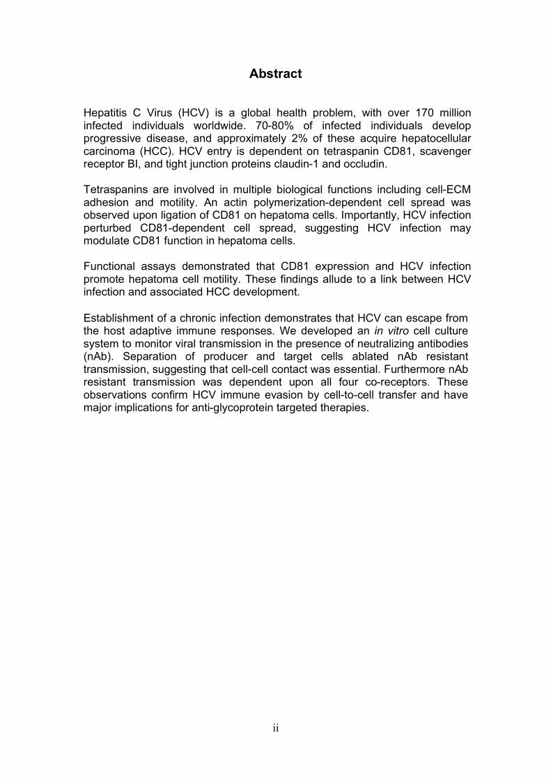
Abstract Hepatitis C Virus (HCV) is a global health problem, with over 170 million infected individuals worldwide. 70-80% of infected individuals develop progressive disease, and approximately 2% of these acquire hepatocellular carcinoma (HCC). HCV entry is dependent on tetraspanin CD81, scavenger receptor BI, and tight junction proteins claudin-1 and occludin. Tetraspanins are involved in multiple biological functions including cell-ECM adhesion and motility. An actin polymerization-dependent cell spread was observed upon ligation of CD81 on hepatoma cells. Importantly, HCV infection perturbed CD81-dependent cell spread, suggesting HCV infection may modulate CD81 function in hepatoma cells.
Functional assays demonstrated that CD81 expression and HCV infection promote hepatoma cell motility. These findings allude to a link between HCV infection and associated HCC development.
Establishment of a chronic infection demonstrates that HCV can escape from the host adaptive immune responses. We developed an in vitro cell culture system to monitor viral transmission in the presence of neutralizing antibodies (nAb). Separation of producer and target cells ablated nAb resistant transmission, suggesting that cell-cell contact was essential. Furthermore nAb resistant transmission was dependent upon all four co-receptors. These observations confirm HCV immune evasion by cell-to-cell transfer and have major implications for anti-glycoprotein targeted therapies.

iii
Dedication
I would like to dedicate this thesis to my parents John and Julia Brimacombe.
iv
Acknowledgements
I would like to thank my supervisors, Professor Jane Mckeating and Dr. Peter Balfe for their support, enthusiasm and guidance throughout this PhD project. In addition i would like to thank everyone in the Birmingham HCV Research group, both past and present, for technical assistance, encouragement and most importantly for making my time here so enjoyable. I would also like to thank, Dr Patricia Laylor for advice on adhesion assays, Dr. Victoria Heath and Sukhibir Johal for advising and helping with motility assays and Dr. Joshua Rappaport and Jenifer Thorley for technical support with live cell imaging. Lastly I would like to thank my family and friends, especially Christopher Bownes, for continued and unquestioning support.
v
Publications
Brimacombe CL., Grove J, Meredith LW, Hu K, Syder A, Flores V, Timpe J, Kreiger S, Baumert T, F, Tellinghuisen TL, Wong-Staal F, Balfe P, McKeating JA. Neutralizing antibody resistant hepatitis C virus cell-to-cell transmission. Journal of Virology 2011 Wagoner J, Negash A, Kane OJ Martinez LE, Nahimias Y, Bourne N, Owen DM, Grove J, Brimacombe C, McKeating JA, Pecheur EI, Graf TN, Oberlies NH, Lohmann V, Cao F, Tavis JE,, Polyak SJ. Multiple effects of silymarin on the hepatits C virus lifecycle. Hepatology 2010 Farquhar, MJ, Harris HJ, Diskar M, Jones S, Mee CJ, Nielsen SU, Brimacombe CL, Molina S, Toms GL, Maurel P, Howl J, Herberg FW, van Ijzendoon SC, Balfe P, McKearting JA. Protein kinase A-dependent step(s) in hepatitis C virus entry and infectivity. Journal of Virology 2008
vi
Table of contents
1. Introduction.................................................................................................. 1 1.1 History and Epidemiology of HCV.......................................................... 1 1.2 Disease progression and current treatment ........................................... 4
1.2.1 Disease progression........................................................................ 4 1.2.2 HCV immune escape....................................................................... 6 1.2.3 Treatments ...................................................................................... 9
1.3 Tools available to study HCV in vitro.................................................... 12 1.4 HCV lifecycle........................................................................................ 15
1.4.1 Genome and replication ................................................................ 15 1.4.2 Particle assembly and egress........................................................ 20 1.4.3 Attachment and entry .................................................................... 24 1.4.4 Viral transmission .......................................................................... 40
1.5 Project aims ......................................................................................... 42 2. Materials and Methods .............................................................................. 43
2.1 Basic techniques.................................................................................. 43 2.1.1 Tissue culture ................................................................................ 43 2.1.2 Antibodies...................................................................................... 44 2.1.3 Plasmids and proteins ................................................................... 45 2.1.4 HCVcc generation ......................................................................... 46 2.1.5 Retrovirus delivery......................................................................... 48 2.1.6: Transient transfection of Huh-7.5 cells. ........................................ 50 2.1.7: Cytotoxicity testing........................................................................ 52 2.1.8: Cholesterol quantification. ............................................................ 52 2.1.9: Flow cytometry ............................................................................. 53 2.1.10: Immuno-fluorescence................................................................. 55
2.2: Specific assays ................................................................................... 59 2.2.1: Antibody engagement cell spread assay...................................... 59 2.2.2: Recombinant sE2 engagement cell spread assay........................ 61 2.2.3: P-ERM western blot for anti-CD81 engagement time course assay................................................................................................................ 62 2.2.4: ECM adhesion assay.................................................................... 65 2.2.5: ECM ELISA .................................................................................. 67 2.2.6: Anti-CD81 engagement filopodia induction assay ........................ 68 2.2.7: Wound healing assay ................................................................... 69 2.2.8: Invasion assay.............................................................................. 70 2.2.9: Infectious co-culture assay........................................................... 73
3. Results: The role of CD81 in hepatoma cell spread. ................................. 76
3.0 Introduction .......................................................................................... 76 3.1 CD81 engagement promotes actin polymerisation dependent hepatoma cell spread.................................................................................................. 79 3.2 The CD81 C terminus links to the actin cytoskeleton through association with actin-associated proteins Ezrin Radoxin Moesin. ............................... 88 3.3 Investigation into the role of different signaling pathways involved in anti-CD81 induced cell spread................................................................... 93 3.4 HCV E2 glycoprotein does not induce hepatoma cell spread .............. 99 3.5 HCV infection perturbs anti-CD81 induced cell spread. ..................... 101
vii
3.6 Discussion.......................................................................................... 108 4. Results: The role of CD81 in hepatoma migration and effects of HCV infection. ...................................................................................................... 118
4.0 Introduction ........................................................................................ 118 4.1 Role of tetraspanin CD81 in hepatoma-ECM adhesion...................... 123 4.2 Role of CD81 in hepatoma invasion................................................... 133 4.3 HCV infection modulates hepatoma cell invasion .............................. 139 4.4 Discussion.......................................................................................... 150
5. Results: Neutralizing antibody resistant HCV transmission ..................... 157
5.0 Introduction ........................................................................................ 157 5.1 Establishment of an in vitro co-culture system to study neutralizing antibody resistant transmission................................................................ 159 5.2 Transmission of diverse HCV genotypes in co-culture....................... 164 5.3 nAb resistant transmission is dependent on cell contact and particle assembly.................................................................................................. 166 5.4 Receptor dependency of nAb resistant transmission of HCVcc......... 170
5.4.1 Role of tetraspanin CD81 in nAb resistant transmission.............. 170 5.4.2 Role of SR-BI in nAb resistant co-culture transmission ............... 177 5.4.3. Role of tight junction proteins CLDN-1 and Occludin in nAb resistant HCV transmission. ................................................................. 185 5.4.4 Receptor dependency of nAb resistant transmission of multiple HCV genotypes. ................................................................................... 189
5.5 Discussion.......................................................................................... 191 6.0 Final Remarks ....................................................................................... 199
viii
List of Figures Figure 1.1: HCV genome and gene products. ............................................... 15 Figure 1.2: Schematic drawing of HCV co-receptors..................................... 24 Figure 1.3: Liver organisation and hepatic polarity ........................................ 36 Figure 1.4: A possible pathway for HCV entry............................................... 40 Figure 2.1: Cholesterol standard curve.......................................................... 53 Figure 2.2: NS5A stain of HCVcc J6/JFH infected cells. ............................... 56 Figure 2.3: BCA protein assay standard curve. ............................................. 63 Figure 2.4:Fibronectin standard curve........................................................... 68 Figure 2.5: Schematic of wound healing assay. ............................................ 70 Figure 3.1: CD81 and F-actin expression in Huh-7.5 hepatoma cells............ 80 Figure 3.2: CD81 engagement promotes actin-polymerization dependent hepatoma cell spread. ................................................................................... 82 Figure 3.3: Diverse panel of anti-CD81 mAbs induce HepG2.CD81 cell spread. .......................................................................................................... 85 Figure 3.4: Antibody binding affinity is not indicative of anti-CD81 induced hepatoma cell spread. ................................................................................... 87 Figure 3.5: A role for CD81 C terminus in actin polymerization dependent hepatoma cell spread. ................................................................................... 89 Figure 3.6: Actin associated proteins Ezrin Radoxin Moesin (ERM) facilitate ant-CD81 induced hepatoma spread............................................................. 92 Figure 3.7: Effect of inhibitors to Rho family GTPases (Rho and Rac) and Ser/Thr kinases (ROCK and PKC) on anti-CD81 induced hepatoma cell spread. .......................................................................................................... 94 Figure 3-8: MAP Kinase independent cell spread.......................................... 96 Figure 3.9: Anti-CD81 induced hepatoma cell spread is dependent on large GTPase Dynamin. ......................................................................................... 98 Figure 3.10 HCV E2 glycoprotein engagement of CD81 did not induce hepatoma cell spread. ................................................................................. 100 Figure 3.11: HCV infection reduces anti-CD81 induced hepatoma spread. 103 Figure 3.12: Anti-CD81 induced spread of J6/JFH del B virus and JFH-1 CD81 mutant virus expressing hepatoma cells............................................ 105 Figure 3.13: CD81 dependent cell spread is sensitive to changes in cholesterol. .................................................................................................. 107 Figure 4.1: Development of hepatoma-ECM adhesion assay. .................... 124 Figure 4.2: CD81 is not involved in hepatoma - ECM adhesion. ................. 126 Figure 4.3 HCV infection does not alter cell-ECM adhesion........................ 128 Figure 4.4: HCV infection does not alter hepatoma ECM expression.......... 130 Figure 4.5: HCV infection does not alter hepatoma ECM expression.......... 132 Figure 4.6: Anti-CD81 mAbs do not induce hepatoma filopodia .................. 134 Figure 4.7: Anti-CD81 mAb has no effect on hepatoma migration or invasion..................................................................................................................... 136 Figure 4.8: CD81 expression increases hepatoma cell invasion. ................ 138 Figure 4.9: Effect of HCV infection on invasion. .......................................... 140 Figure 4.10: Huh-7.5 hepatoma cell invasion is independent of VEGF ....... 142 Figure 4-11: Hypoxia promotes Huh-7.5 cell invasion. ................................ 145
ix
Figure 4.12: HCV induced cell invasion is sensitive to HIF-1α inhibitor....... 147 Figure 4.13: HIF-1α dependent modulation of proliferation and infection. ... 149 Figure 5.1: Comparison of labeling method to enumerate HCV transmission..................................................................................................................... 160 Figure 5.2: The infectious co-culture assay. ................................................ 161 Figure 5.3: Effect of anti-glycoprotein antibodies on H77/JFH cell free infectivity and co-culture transmission. ........................................................ 163 Figure 5.4: Genotype transmission in co-culture. ........................................ 165 Figure 5.5: nAb resistant transmission requires cell contact........................ 167 Figure 5.6: Particle assembly is essential for co-culture transmission......... 169 Figure 5-7: Effect of anti-CD81 monoclonal antibodies on HCV transmission..................................................................................................................... 172 Figure 5.8: Transmission to a hepatoma cell line with low CD81 expression levels. .......................................................................................................... 174 Figure 5.9: Transmission of a CD81 negative mutant virus. ........................ 176 Figure 5.10: Effect of anti-SR-BI antibody on HCV transmission................. 178 Figure 5.11: Effect of SR-BI expression levels on HCV transmission.......... 180 Figure 5.12: Transmission of a cell culture adapted virus with reduced SR-BI dependency................................................................................................. 182 Figure 5.13: Effect of small molecular inhibitors of SR-BI on HCV transmission. ............................................................................................... 184 Figure 5.14: Claudin-1 is essential for nAb resistant transmission. ............. 187 Figure 5.15: Occludin is essential for nAb resistant transmission................ 188 Figure 6.1: Schematic diagram of CD81 as part of a TEM. ......................... 200 Figure 6.2: Possible mechanisms underlying HCV perturbation of CD81 function in hepatoma cell spread................................................................. 205 Figure 6.3: Possible mechanism(s) for HCV induced hepatoma migration and tumor progression through stabilization of HIF-1α....................................... 208 Figure 6.4: Possible routes of HCV cell-to-cell transmission in co-culture... 212
x
List of Tables
Table 2.1: Cell lines used .............................................................................. 43 Table 2.2: Antibodies used ............................................................................ 44 Table 2.3 Plasmids and proteins used........................................................... 45 Table 2.4: List of antibody concentrations. .................................................... 58 Table 2.5: Treatment summary for spread assays. ....................................... 60 Table 2.6: Treatment summary for invasion assays. ..................................... 72 Table 3-1: CD81 antibody epitope grouping. ................................................. 84 Table 5.1: Receptor dependency of multiple genotypes.............................. 190
xi
Abbreviations
AP-1: Activator protein -1 BSA: Bovine Serum Albumin CMFDA: 5-chloromethlfluorescein diacetate DAPI: 4’6-daimidino-2-phenylindole DMEM: Dulbecco’s modified medium DMSO: Dimethyl sulfoxide DNA: Deoxyribonucleic acid EC1: Extra-cellular loop 1 EC2: Extra-cellular loop 2 ECM: Extra-cellular matrix EGFR: Epidermal growth factor receptor ELISA: Enzyme linked immunosorbent assay EMT: Epithelial mesenchymal transition ER: Endoplasmic reticulum ERM: Ezrin Radoxin Moesin Ep: Electroporation ESCRT: Endosomal sorting complex required for transport F-actin: Filamentous actin FFU: Focus forming units FRET: Fluorescence resonance energy transfer GFP: Green fluorescent protein HBV: Hepatitis B virus HCC: Hepatocellular carcinoma HCV: Hepatitis C virus HCVcc: Hepatitis C virus propagated in vitro HCVpp: Hepatitis C virus pseudo particles HDL: High density lipoproteins HGF: Hepatocyte growth factor HIF: Hypoxic inducible factor HIV: Human Immunodeficiency virus Hr(s): Hour(s) HSV: Herpes Simplex Virus HTLV: Human T cell leukemia virus type 1 HVR: Hyper variable region IFN: Interferon IGF-1: Insulin like growth factor IU: Infectious Units IRF: Interferon regulatory factor ISGs: Interferon stimulated genes JFH: Virus isolated from a Japanese patient with fulminant hepatitis Jak-STAT: Janus Kinase-Signal transducer and activator of transcription KD: Knock down LDL: Low density lipoproteins mAbs: Monoclonal antibodies MβCD: Methyl-beta-cyclodextrin MFI: Median fluorescent intensity Min(s): Minute(s) MLV: Murine leukemia virus
xii
MMP: Matrix metalloproteinase mRNA: messenger RNA MTS: 3-(4,5-dimethylthiazol-2-yl)-5-(3-carboxymethoxyphenyl)-2-(4- sulfophenyl)-2H-tetrazolium (Cell proliferation assay) MTP: Microsomal transfer protein MVB: Multivesicular body nAb: Neutralizing antibody NASH: Non alcoholic steatohepatitis NK: Natural Killer cells OAS: 2’-5’ oligoadenylate synthetase ORF: Open reading frame PAMPs: Pathogen Associated Molecular Patterns PBS: Phosphate buffered saline PKC: Protein Kinase C PKR: Protein Kinase receptor PI4K: Phosphatidylinositol 4-kinase P-ERM: Phosphorylated Ezrin Radoxin Moesin PRR: Pathogen Recognition Receptors RIG-I: Retinoic acid inducible gene I RNA: Ribonucleic acid SGR: Sub-genomic replicon siRNA: small interfering RNA shRNA: small hairpin RNA SOCS: Suppressor of cytokine signaling SR-BI: Scavenger receptor BI TEM: Tetraspanin enriched microdomain TIRF: Total internal reflection fluorescence TLR: Toll-like receptor UTR: Un-translated region VEGF: Vascular endothelial growth factor WT: Wild type
1. Introduction
1.1 History and Epidemiology of HCV
During the 1970’s scientific advances were made enabling serological detection of
hepatitis A and B virus infection (35, 136), it quickly became apparent that at least
one other agent was responsible for hepatitis arising from blood transfusion, this was
commonly referred to as non-A non-B hepatitis (NANBH) (137). Despite a large
amount of research it wasn’t until over a decade later that Hepatitis C Virus (HCV)
was formerly identified. Choo and colleagues constructed a cDNA library from serum
containing the NANBH agent isolated from infected chimpanzee plasma and
successfully isolated a clone that specifically hybridized with RNA found only in
NANBH infected chimpanzees and encoded a protein that bound antibodies from
NANBH infected patients (206, 259). These findings enabled the establishment of
assays to screen for HCV in blood, and since introducing these tests in the early 90’s
the risk of transmission through blood transfusion in the developed world is now
extremely low (62).
HCV is endemic worldwide and according to figures from the WHO there are an
estimated 170 million people infected making up approximately 3% of the worlds
population. Prevalence varies greatly depending on geographical location. Areas
with the highest recorded prevalence are in Africa and Asia; Egypt for example has a
prevalence of 22% (148). Prevalence in North America, Japan and Western Europe
are lower ranging from 0.6% in Germany (363) to 2.2% in Italy (11, 430). It is likely
that these values under estimate the burden especially in the developing world where
2
there is less data available. In some areas the true burden may not yet be realized
due to the asymptomatic nature of the disease during both the acute and early
chronic phases of infection, preventing detection until late in disease progression (11,
430). Interestingly in different geographical locations the range in prevalence
determined by age varies considerably. In North America for example the highest
prevalence is among people between the age of 30 and 49 (12), this differs from
Japan and Italy where the highest prevalence is among the over 50’s (11). These
differences are consistent with young adults in North America being at greatest risk
around 20 years ago whilst in Japan the greatest risk was many years before this.
Like Japan, in Egypt the highest prevalence is in the over 50’s age group, however
unlike Japan the prevalence in the younger age groups is also extremely high (1, 11).
HCV is the sole member of the genus Hepacivirus within the Flaviridae family. This
family includes yellow fever and classical swine fever viruses, all of which are
enveloped viruses containing a single stranded positive sense RNA genome. HCV is
genetically very diverse with 7 major genotypes and many different subtypes (166,
442). Subtypes vary between 20 and 25% whilst genotypes can vary by 30% at the
nucleotide level (440, 443). The geographical prevalence and diversity of the
genotypes differ giving clues as to the origin of the virus (441). Genotype 1a, 1b and
3a are the most prevalent genotypes in the western world (440). Genotypes found in
Africa and South-East Asia associate with specific geographical areas and are much
more diverse. The genetic diversity of these viral strains suggest that the virus has
been present in human populations in these areas for a long time and that it is only in
recent history that the virus has transmitted to the western world (323). Genotypes 1,
2 and 4 are found specifically in sub Saharan Africa and genotypes 3 and 6 found in
3
South-East Asia (445). As for the origin of the 6 genotypes there is no evidence of
HCV or HCV-like virus in old world ape or monkey species as observed for HBV
(301). Interestingly though a distantly related virus named GB virus B has been
reported to infect tamarins and other new world primate species (441, 447). Further
studies are needed to understand the origin of HCV and its relation to GB virus B.
Risk factors of infection vary geographically and have altered over time. In the
developed world a major source of transmission was through increased use of blood
products in medical practices, as previously explained this is no longer a risk due to
routine screening of donated blood (62). Since the 1960’s injection drug abuse has
become the primary route of transmission (12). Other lower risk factors associated
with HCV transmission include perinatal, sexual and occupational transmission.
There is little evidence for risk associated with sexual transmission and the data from
occupational transmission suggests a low risk (11). Perinatal transmission occurs in
approximately 2.7-8.4% of cases, interestingly this value increases significantly in
mothers co-infected with HIV (139, 477). However in the developing world the picture
is very different with major risk associated with contaminated blood products a result
of financial constraints limiting screening (11). Another major risk factor in the
developing world is through vaccination(s) with contaminated needles. Two studies in
India demonstrated an association between HCV infection and visits to unlicensed
medical practitioners (86, 305). In Egypt the majority of HCV infections are
associated with a nationwide vaccination program against Schistosomiasis that was
carried out between 1960 and 1987 (148). Contaminated needles in vaccination
programs are likely to be the cause of infection in some developed countries where
prevalence is highest in older populations (430). As for transmission of the virus in
4
Africa and Southern East Asia before the use of modern medicine it is hypothesized
that transmission was and may still occur through tribal scarification practices or
through insect vectors such as mosquito’s or ticks. However, to date there is no
substantial evidence to support these theories (430).
1.2 Disease progression and current treatment
1.2.1 Disease progression
HCV is unusual compared to other flaviviruses due to its capability to persist in
infected individuals. Between 75 and 85% of individuals infected with HCV develop a
persistent infection (204).
Due to the asymptomatic nature of early infection there is little data on acute
infection, most of our current knowledge has been acquired from prospective studies
of transfusion patients, chimpanzee studies or though a number of acute cohorts
largely consisiting of injection drug users (IDUs) or health care workers (HCWs) (361,
392, 413, 492). Acute resolving infection typically lasts between 10 and 12 weeks.
RNA can be detected within the first two weeks post exposure and rises to a peak of
between 10^5 and 10^7 IU/ml at 6 to 10 weeks post infection, this often comes
shortly before a peak in serum alanine aminotransferase (ALT) levels, a marker for
liver injury (8, 135, 300, 476). The production of anti-HCV antibodies is more
variable, with most patients developing anti-HCV between 7 and 8 weeks post
infection (349, 369). The presence of neutralizing antibodies (nAbs) is critical in
ensuring successful clearance of many viruses (61). Although there are a few
examples where a nAb response during acute HCV infection has associated with

5
viral clearance (29, 268, 326, 377), the majority of studies show nAbs appearing after
acute infection indicating a more dominant role for nAbs in controlling chronic
infection (290). In contrast a cellular immune response is essential to ensure viral
clearance. HCV specific T-cell responses are detectable between 4 to 8 weeks post
infection. A correlation has been demonstrated on many occasions between a robust
CD8+ and CD4+ T-cell response and viral clearance (97, 242, 271, 437, 476). In
non-resolvers the response is often weak or not detected at all. Although most
patients do not exhibit any symptoms, evidence suggests that when symptoms do
occur there is an increased likelihood for successful viral clearance (9, 159).
The acute stage of a chronic infection with respect to antibody response, RNA level
and ALT levels cannot be distinguished from the acute stage of resolving patients
(204). If after 6 months HCV RNA levels are still detectable the patient is reported to
have developed a chronic infection (204). RNA levels in the blood remain stable
although they can vary greatly between donors and are not predictive of disease
outcome (544). ALT levels decrease after the initial boost during acute infection and
in the majority of cases levels remain elevated above normal and fluctuate during the
course of chronic infection, again ALT was found not to be a good predictor of liver
disease status (178). Some patients with a chronic infection have a mild non-
progressive disease whilst 20-30% develop complications including cirrhosis and end
stage liver disease, 2.5% of these patients develop hepatocellular carcinoma (HCC)
(6, 10, 71). HCV associated liver damage is explored in more depth in chapter 4.
Given the diversity of HCV it is surprising that large variations in clinical outcomes do
not occur, in fact all genotypes are capable of initiating a chronic infection that can
eventually lead to the development of liver disease and HCC. Although, as more

6
clinical data is collected differences between genotypes are becoming apparent. A
number of studies looking at European cohorts have revealed that genotype 1 is
more likely than genotypes 2 and 3 to firstly establish a persistent infection and
secondly once a persistent infection is established to cause a greater degree of liver
disease (311, 395, 542). A strong association between development of liver steatosis
and genotype 3 infection has also been demonstrated (2, 405). It is believed that a
block in lipoprotein secretion from hepatocytes causes this phenotype (426). More
work is needed to validate and determine the mechanism of these observed
differences.
Although the primary disease symptoms are liver related, chronic infection is also
associated with other extra-hepatic conditions suggesting the liver is not the only
reservoir for infection. Examples of which include auto-immune diseases such as
cryoglobulineimia and glomerulonephritis (129) as well as B-cell non-Hodgkin
lymphoma (562) and neurological conditions including cognitive disorders (193). Our
laboratory has previously demonstrated HCV association with B-lymphocytes aiding
infectivity in vitro (455) and we are currently investigating brain tissue as a further
reservoir for HCV (142)(Nicola Fletcher, submitted).
1.2.2 HCV immune escape HCV employs a number of mechanisms to hinder both the innate and adaptive
immune responses allowing a persistent chronic infection within the host (48, 182).
Briefly the intracellular innate immune response to viral infection is as follows; upon
viral entry into host cells Pathogen Recognition Receptors (PRR) present in the
cytoplasm identify signatures of viral infection such as single and double stranded
RNA, these are termed Pathogen Associated Molecular Patterns (PAMPs) (409). In
7
hepatocytes the predominant PRRs associated with HCV infection are Toll-like
receptor 3 (TLR-3) and RIG-I (276, 461, 548). Both of these PRR’s recognize HCV
double stranded RNA and induce a signaling cascade leading to the activation of
latent cellular transcription factors Interferon regulatory factor IRF (3) and nuclear
factor NFκB (280, 397). These transcription factors induce Interferon-beta (IFNβ)
gene transcription. IFNβ is then secreted from the infected cells and functions in an
autocrine and paracrine manner by engaging type I IFN receptors on the cell surface
eliciting the Jak-STAT pathway (412). This pathway results in the formation of
Interferon stimulated gene factor-3 (ISGF3) that enhances transcription of Interferon
stimulated genes (ISGs). ISGs are responsible for antiviral actions within the cell
(113, 153). Predominant anti-viral effecter genes include Protein kinase receptor
(PKR) that inhibits translation of viral RNA (512) and 2’-5’ oligoadenylate synthetase
(OAS) that cleaves HCV genomic RNA into non-functional products, inhibiting viral
replication (185). Other ISGs include p56, IRF7 and IRF3 (48). IRF7 and 3 induce
transcription of IFNα gene leading to a positive feedback loop increasing IFN
response (20). IFNα is also responsible for influencing the adaptive immune
response (412). For HCV to successfully persist in its host it needs to be able to
overcome these innate immune response. A number of HCV proteins including
Core, NS3/4A protease and NS5A, are implicated in hindering stages of this immune
response. NS3/4A for example blocks the RIG-I signaling pathway and completely
ablate TLR-3 signaling (147, 276). Core protein has been shown to induce SOCS1
and SOCS3 expression; that are negative regulators of the Jak-STAT pathway (49).
NS5A protein and glycoprotein E2 have both been implicated in inhibiting anti-viral
effecter genes. For example E2 can bind PKR inhibiting its function (367, 472) and
NS5A has been shown to inhibit PKR function (378) whilst also repressing OAS
8
functions (464). NS5A expression has also been shown to induce IL-8 expression; an
inhibitor of IFNα induced ISG expression (387).
HCV has also evolved to escape from cellular and adaptive immune responses. For
example HCV core protein has been implicated as an agent capable of inhibiting
immune cells directly. An in vitro study reported that HCV core protein inhibits T-cell
activation by interacting with complement factor gG1qR (244, 509). Of note core
protein from genotype1a virus was used in this study and the results could not be
repeated in a separate study with core from genotype 1b, indicating a possible
genotype specific function (182, 289). As yet this effect has not been confirmed in
vivo. As will be explained later HCV glycoprotein E2 binds co-receptor tetraspanin
CD81 (384). NK cell cytopathic function is inhibited upon engagement of cell surface
expressed CD81 with recombinant E2 protein, suggesting a possible mechanism for
HCV to perturb the host immune response (105, 486). Controversy over whether
these studies can be re-produced when E2 is expressed on a viral particle is
currently under debate (549). A further example is the induction of escape mutants to
nAb’s and T cell epitopes. A single infected host contains a quasispecies (closely
related species of virus subjected to genetic mutations, competition and selection)
population of HCV (117). A quasispecies population develops for two main reasons;
firstly HCV replicates rapidly allowing many opportunities for mutations to occur and
secondly HCV has a single strand positive sense genome that is replicated by an
RNA dependent RNA polymerase (RdRp). RdRp does not have proof reading ability
and permits rapid evolution of the virus (37). Neutralizing antibodies have been
shown to target the hypervariable region 1 (HVR-1) of glycoprotein E2 and mutations
have been detected in this region in human and chimpanzee studies that confer
9
escape from immune pressures (434, 450, 497). Mutations in regions inhibiting CTL
CD8+ T cell responses have also been detected in both chimpanzees and humans
suggesting another possible mechanism of viral escape (73, 131, 500, 505). As will
be discussed later HCV particles associate with lipoproteins (14, 351). This has been
suggested as an additional mechanism employed by HCV to disguise the virion from
the immune system reducing the effectiveness of nAb’s. Lastly recent work published
from our laboratory demonstrates that HCV can transmit in co-culture in the presence
of nAbs capable of neutralizing cell free virus, suggesting that HCV may transmit
from cell-to-cell via a novel route reducing exposure to the host immune system
(480). This is discussed in Chapter 5.
1.2.3 Treatments Standard treatment for HCV infection is a course of pegylated Interferon (IFN) and
Ribavarin. This can be extremely effective (up to 90%) if treatment is started within
the acute phase of infection (221, 510). After onset of chronic infection efficacy drops
and varies considerably depending on the infecting genotype. 70-80% of individuals
with genotypes 2 and 3 respond to Interferon based therapy compared to only 40-
50% of patients infected with genotype 1 (181, 267, 303). The mechanism of action
of IFN and Ribavarin based therapy is not fully understood. IFN is likely to reduce
infection at both the viral level by inducing Interferon stimulated genes (ISGs) that
have direct anti-viral functions as well as promoting the innate and adaptive immune
response (38, 425, 479). Ribavarin is a synthesized guanosine that has previously
been demonstrated to inhibit other RNA and DNA virus infections (138). It is thought
that its main mechanism of action against HCV may be through increasing
mutagenesis of viral RNA and thereby reducing specific infectivity (550). Limitations

10
in efficacy and severity of side effects associated with the current therapy make it
increasingly important to find new more effective therapies.
Many alternative therapies are being explored and some of which have/are currently
undergoing clinical trials with promising results (109, 428). Examples of these include
drugs targeting viral enzymes including the NS3-4A viral protease and NS5B RNA
polymerase (109, 261, 265, 394), these became promising targets after there
structures were defined in 1996 and 1999 respectively (4, 56, 243). NS3-4A is a
particularly attractive target not only because of its important role in the HCV lifecycle
but also because of its role as an inhibitor of the innate immune response as
previously described (147, 276). Our laboratory is currently working with a small
molecular inhibitor of SR-BI, a co-receptor for HCV entry, this compound is soon to
be trialed here in Birmingham. More information on this inhibitor is presented later in
chapter 5. Further immuno-modulating agents have also been considered as
therapeutics including TLR-7 and TLR-9 antagonists. Toll-like receptors are present
on a number of immune cells and recognize microbial agents inducing an immune
response primarily involving the induction of IFN whilst also priming an adaptive
immune response (465). TLR-7 and TLR-9 in particular recognize single stranded
RNA (50). Antagonists to TLR-7 have been trialed against HCV infection with
promising results (520). Complications have arisen with many of the more potent
anti-replicase agents including a rapid induction of viral escape mutants (258, 279,
329). It is therefore hypothesized that a combination of therapies targeting both virus
and host will be required to overcome this problem in the future (109).
11
As yet there is no effective vaccine available against HCV, this has been due
predominantly to the lack of knowledge on mechanisms of viral clearance, the
evidence for re-infection of both humans and chimpanzees as well as lack of
definitive evidence on the efficacy of neutralizing antibodies in vivo. Recent studies
have given more hope into the eventual production of an effective vaccine. Firstly
over time increasingly more information has become available on patients with
resolving infection increasing our understanding of what constitutes an effective
immune response (101, 271, 437). Secondly patients that are re-infected with HCV
are often protected against the development of a chronic infection (266, 322). Lastly
neutralizing antibodies have been detected that can cross-neutralize across different
genotypes suggesting that a broad spectrum vaccine may be possible and
furthermore a small number of studies have reported a correlation between the
induction of neutralizing antibodies in the early stages of infection and viral clearance
(29, 268, 326, 377). Supporting observations in humans, chimpanzee studies using
an E1E2 peptide vaccine, although unable to stop re-infection, were able to prevent
progression to a chronic infection in the majority of subjects tested (84). Chronic
infections create the greatest burden of HCV disease both financially and to the host
therefore the ability to reduce this occurrence would be extremely beneficial. More
recent studies are looking at methods to develop a vaccine that will elicit both the
production of cross neutralizing antibodies as well as inducing a broad cellular
response, responses that have previously been associated with viral clearance (65,
207, 282). Although trials in animal models have been relatively successful, human
trials are needed to determine their true efficacy. The use of vaccines as a
therapeutic treatment is also being investigated in the field, the theory behind this is
that a vaccine may be able to boost the immune system and therefore increase
12
efficacy of IFN based therapies (207). This theory is based on evidence that
increased immune response prior to treatment increases the likelihood of success
(34, 103, 347).
The absence of an effective prophylactic or vaccine for HCV together with the virus’s
propensity to cause considerable liver damage after long term infection makes it
absolutely crucial that more research is done to fully understand HCV infection.
1.3 Tools available to study HCV in vitro An in vitro system to study the full viral life cycle, from entry through to release of
infectious viral particles has only recently become available. Although a partial
sequence of HCV was identified in 1989 (85), it wasn’t until 1996 that the full HCV
genome sequence was completed using sequences isolated from patient H
(Huchinson 1977), a patient infected with a genotype 1a virus (249). With this
knowledge a model cDNA template was constructed (H77), and delivery of the
transcribed RNA into the liver of chimpanzee’s resulted in viral replication (248, 526).
This became a model system allowing the study of immune response and viral
evolution during infection (58). Unfortunately these clones were unable to replicate
and assemble infectious particles in cell culture.
Although a number of techniques have been used to study HCV entry the most
effective and widely used system is the retroviral pseudoparticle system (HCVpp).
Pseudoparticles comprise of an HIV capsid with HCV glycoproteins. HCVpp allow
glycoprotein dependent entry to be studied and have allowed considerable advances
to be made in determining host cell entry receptors (30, 121, 208). These

13
pseudoparticles can only undergo one round of replication and cannot transmit to
other cells.
A critical development in the race to find an in vitro culture system to study the full
viral lifecycle was made by Lohmann et al., in 1999. Lohman and colleagues
demonstrated replication of sub-genomic replicons in cell culture (292). The
development of the replicon system has been a critical tool for increasing our
understanding of HCV genome replication and a screening tool for the discovery of
new anti-viral agents (26, 47, 381). Although full-length replicons replicated efficiently
no infectious viral particles were produced and it was feared that Huh-7 cells may be
unable to assemble or release virus particles (382). Cell culture adapted mutations
were identified allowing increased replication efficacy in vitro but unfortunately the
mutations severely reduced infection in vivo (283, 291).
It wasn’t until 2005 that a full-length virus with no cell culture adaptive mutations was
identified and demonstrated to replicate and assemble virus particles in cell culture
(283, 499, 560). This was termed HCVcc, cc representing cell culture. JFH-1
(genotype 2a) was isolated from a Japanese patient with fulminant hepatitis and had
previously been shown to replicate in the form of a sub-genomic replicon without any
amino acid changes (237). Wakita and colleagues were the first to demonstrate that
the full length JFH-1 genome could replicate in Huh-7 hepatoma cells and importantly
also in the chimpanzee model (499). Chimera’s of JFH-1 consisting of core to NS2 of
J6 (genotype 2b) or H77 (genotype 1a) with the remaining non-structural proteins of
JFH-1 were developed and were reported to be infectious in chimpanzees and uPA-
SCID mice containing human liver grafts. Importantly the virus remained infectious in
14
cell culture after in vivo propagation (284, 545). The JFH-1 HCVcc system is the
basis for much of our research today and recent advances have led to the production
of numerous JFH-1 chimeric viruses representating the 7 major HCV genotypes
allowing genotype specific comparisons to be made (166).
HCVcc replicates most efficiently in the Huh-7 hepatoma cell line (47). Hepatocytes
in the liver are highly polarized and tight junction proteins claudin-1 and occludin
have been identified as critical co-receptors for HCV entry (133, 385). Huh-7 cells do
not polarize and therefore may not be the most appropriate representative model to
use. Our laboratory is currently working with a polarised hepatoblastoma cell line
HepG2, these cells exhibit hepatocyte polarity and support viral replication but have a
low permissivity to HCV infection making them challenging to use (319, 320). To
further in vitro studies a highly permissive cell line that exhibits hepatocyte polarity is
needed. A model system utilizing primary hepatocytes would be ideal but because of
difficulties in accessing and processing human liver tissue very little work has been
reported with primary cells. Recent developments in this area have involved the
establishment of new techniques to propagate primary hepatocytes for longer periods
of time before de-differentiation (229, 241). This is an area of research that is still in
its preliminary stages and needs to be developed in the future to allow greater
accessibility for more laboratories to use these techniques.
15
1.4 HCV lifecycle
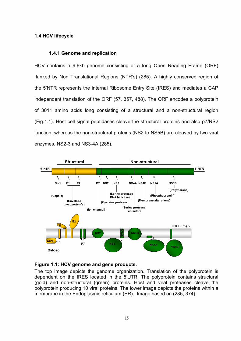
1.4.1 Genome and replication HCV contains a 9.6kb genome consisting of a long Open Reading Frame (ORF)
flanked by Non Translational Regions (NTR’s) (285). A highly conserved region of
the 5’NTR represents the internal Ribosome Entry Site (IRES) and mediates a CAP
independent translation of the ORF (57, 357, 488). The ORF encodes a polyprotein
of 3011 amino acids long consisting of a structural and a non-structural region
(Fig.1.1). Host cell signal peptidases cleave the structural proteins and also p7/NS2
junction, whereas the non-structural proteins (NS2 to NS5B) are cleaved by two viral
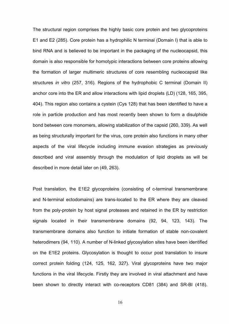
enzymes, NS2-3 and NS3-4A (285).
Figure 1.1: HCV genome and gene products. The top image depicts the genome organization. Translation of the polyprotein is dependent on the IRES located in the 5’UTR. The polyprotein contains structural (gold) and non-structural (green) proteins. Host and viral proteases cleave the polyprotein producing 10 viral proteins. The lower image depicts the proteins within a membrane in the Endoplasmic reticulum (ER). Image based on (285, 374).
16
The structural region comprises the highly basic core protein and two glycoproteins
E1 and E2 (285). Core protein has a hydrophilic N terminal (Domain I) that is able to
bind RNA and is believed to be important in the packaging of the nucleocapsid, this
domain is also responsible for homotypic interactions between core proteins allowing
the formation of larger multimeric structures of core resembling nucleocapsid like
structures in vitro (257, 316). Regions of the hydrophobic C terminal (Domain II)
anchor core into the ER and allow interactions with lipid droplets (LD) (128, 165, 395,
404). This region also contains a cystein (Cys 128) that has been identified to have a
role in particle production and has most recently been shown to form a disulphide
bond between core monomers, allowing stabilization of the capsid (260, 339). As well
as being structurally important for the virus, core protein also functions in many other
aspects of the viral lifecycle including immune evasion strategies as previously
described and viral assembly through the modulation of lipid droplets as will be
described in more detail later on (49, 263).
Post translation, the E1E2 glycoproteins (consisting of c-terminal transmembrane
and N-terminal ectodomains) are trans-located to the ER where they are cleaved
from the poly-protein by host signal proteases and retained in the ER by restriction
signals located in their transmembrane domains (92, 94, 123, 143). The
transmembrane domains also function to initiate formation of stable non-covalent
heterodimers (94, 110). A number of N-linked glycosylation sites have been identified
on the E1E2 proteins. Glycosylation is thought to occur post translation to insure
correct protein folding (124, 125, 162, 327). Viral glycoproteins have two major
functions in the viral lifecycle. Firstly they are involved in viral attachment and have
been shown to directly interact with co-receptors CD81 (384) and SR-BI (418).
17
Secondly they are thought to function as membrane fusion proteins coordinating pH
dependent fusion of the viral and cell membranes in the early endosome permitting
release of the viral genome into the cytoplasm of the host cells (208, 485, 496).
Sequence comparison with other flaviviruses revealed that HCV E2 glycoprotein was
likely to contain a class II fusion peptide (522) and a number of laboratories have
since identified the C-terminus of E2 as being important for cell fusion (144, 286).
Furthermore a number of regions in E1 have been discovered that are important for
cell fusion (156, 275). A recent study showed evidence for a functional role of E1 in
membrane fusion by measuring biophysical changes in membranes after interaction
with an E1 peptide representative of a previously identified fusion domain (375). It is
now widely believed that a number of regions are required for HCV-cell fusion events
and this is likely to involve both E1 and E2 proteins, although the mechanism is still
under debate (120, 130, 269, 371).
p7 is an integral membrane protein that is primarily located in the ER (67), it consists
of two hydrophobic transmembrane domains connected by a short basic loop that is
highly conserved between genotypes (173, 456). p7 forms oligomers and electron
microscopy studies have revealed the structure of p7 hexamers (172, 296) and
heptamers (91). These oligomeric structures form cation selective ion channels in
lipid bilayers (172, 368). As such p7 has been categorized as a member of a group of
viral permeability altering proteins called viroproteins; other examples include M2
from Influenza, vPu from HIV-1 and M from dengue virus (90). A number of in vitro
studies have demonstrated an essential role for p7 in virus assembly and release this
is supported by work showing that p7 is essential for infection in chimpanzees (230,
410, 456). Woznik and colleagues demonstrated that intracellular virus particles have
18
greater acid sensitivity compared to extra-cellular virions, suggesting a role for p7 in
protecting maturing virions from acidic conditions by removing H+ ions from
intracellular membranes (518). Inhibitors for p7 ion channel have been effective in in
vitro studies making p7 an attractive therapeutic target for the future (170, 171, 457).
NS2 has been shown to have multiple functions in the viral life cycle and like p7 has
been demonstrated to be critical for viral assembly (112, 225, 230). NS2 is a
hydrophobic protein with several transmembrane domains in the N-terminal region
(414, 525). NS2 is stimulated by cofactor NS3 and NS2-3 cysteine protease
(Comprising of the C-terminal of NS2 and the N terminal of NS3 (364, 475)) cleaves
the NS2/3 junction allowing NS3-4A serine protease to cleave all the downstream
sites (364, 475). The cleavage of the NS2/3 junction is required for replication of full-
length replicons (506) and also replication in chimpanzees (250). The N-terminal
transmembrane domain of NS2 is necessary for virus assembly (225) and the C
terminal protease domain but not its catalytic ability has also been demonstrated to
be necessary for virus assembly (230).
NS3 to NS5B proteins are all required for viral replication although due to the small
size of the viral genome it is likely that most viral proteins are multifunctional (338).
RNA viruses replicate their genome associated with altered cytoplasmic membranes
or membrane webs. Replication of HCV has been demonstrated to occur in
association with cytoplasmic membranous webs in a distinct subcellular replication
complex (RC) compartment (5, 165, 332). The positive sense RNA is translated to
make new viral proteins as well as acting as a template for an intermediate negative
19
RNA strand that forms the template for the production of multiple sense genomes
that are packaged into new virus particles (285).
NS4A is a cofactor for NS3, the presence of NS4A increases the stability of NS3 as
well as allowing the cleavage of NS4B-NS5A junction (516). The C terminal region of
NS4A is required for cleavage of the NS3-NS4A and NS5A-NS5B junctions (134).
Membrane anchorage of NS3-NS4A has been shown to occur through the N
terminus of NS4A (516). As previously discussed the NS3-NS4A protease also
functions in immune evasion by blocking the RIG-I and TLR-3 signaling pathways
(147, 276). Recent work has discovered that NS4A is also involved in recruiting
creatine kinase B (ATP generating enzyme) to the replication complex (186). The
initiation of the membrane web formation is down to NS4B protein (128). NS4B is an
integral membrane protein that is thought to contain four transmembrane domains
and interacts with other non-structural proteins as well as viral RNA (167). Recent
work implies that NS4B may also play a role in viral assembly (231).
NS5A is a three domain protein that is found in hypo and hyper phosphorylated forms
(469). This protein is involved in a number of stages of the viral lifecycle, the
multifunctional capacity of NS5A is most likely down to its ability to interact with
numerous host proteins (81, 82, 473, 474). NS5A is a critical member of the
replication complex and all three domains are able to bind viral RNA, it has been
suggested that part of its role in replication is to enhance NS5B activity (82, 146). As
previously discussed NS5A is also important for HCV immune evasion strategies by
effecting IFN sensitivity (378, 387, 464). Recently NS5A has also been shown to be
20
involved in viral particle assembly as discussed later on (473). The remaining
nonstructural domain, NS5B codes for the RNA dependent RNA polymerase (393).
1.4.2 Particle assembly and egress.
The exact mechanism by which viral particles are formed and released from the cell
is currently not well understood. Clues into the assembly process of HCV came from
observations that virus isolated from either in vitro studies or from serum of infected
patients contained particles of varying densities (74, 234, 285, 351). Andre et al.,
proposed that low density HCV particles associate with lipoproteins, he termed these
lipoviral particles (LVP) and likened them to very low density lipoprotein (VLDL) (14).
LVPs are enriched with high levels of triglycerides and both apolipoprotein B (Apo B)
and apolipoprotien E (Apo E) have been detected in the viral RNA containing low
density fractions (14, 215, 351). Recently Merz et al., established a high affinity
purifaction assay using flag tagged viral particles, permiting further investigation of
cell culture derived viral particle compostion. The lipid compositon of the viral
particles was found to be distinct from the host cell membrane, predominantly
consisting of Cholesteryl esters as well as high levels of Apo E and detectable levels
of cholesterol (324).
Particle assembly is believed to occur on/near lipid droplet associated membranes
that are derived from the ER (27, 331, 429). Nucleic acid association with core is
vital for nucleo-capsid formation and this association may initiate viral particle
formation (257). The C terminal region of HCV core protein is responsible for core
proteins association with lipid droplets and ER membranes (128, 165, 404).
Extensive work by McLauchlan and colleagues has demonstrated that core is
released from the ER and loaded onto lipid droplets during HCV infection and
21
importantly this correlates with virion production (53, 315, 317). Furthermore, core
protein induces the accumulation of lipid droplets and modulates their distribution in
infected cells (52). Lipid droplets move through the cytoplasm and interact with the
ER facilitating lipid and protein transport between organelles. Miyanari et al.,
demonstrated that core recruits non-structural proteins and replication complexes to
lipid droplets associated with the ER, facilitating particle assembly (331).
We have previously acknowledged that non-structural proteins p7, NS2 and NS5A
are involved in viral assembly. Work on p7 predominantly focused on protein
structure (91, 172, 296) however, recent research has demonstrated that p7
modulates the pH of membrane structures involved in viral production, most likely
providing protection for the immature virus from acidic conditions (518). As yet the
mechanism of NS2 function in assembly is not well understood (225, 230).
NS5A like core can be found on lipid droplets in infected cells. NS5A and core are
reported to interact with one another; importantly elimination of this association by
mutations in domain III of NS5A reduces particle assembly (16, 331). Furthermore
NS5A domain III also associates with Annexin A2 (phospholipid binding protein with
multiple functions including endosome trafficking), this interaction has been shown to
be important for viral assembly (21).
Proteins involved in the VLDL (Very low-density lipoproteins) secretion pathway have
been identified in membranous web compartments containing the HCV replication
complex, these include Apo E and ApoB (209). VLDLs are found only in the liver and
inhibition of VLDL secretion limits HCV release from Huh-7 cells suggesting this
22
process is closely linked to the VLDL assembly and secretion pathway (209).
Dependence on the VLDL secretion pathway for viral assembly and egress has been
proposed as an explanation for hepatic tropism (157, 209). There is controversy over
this data set as Jaing et al., were not able to repeat these observations and found
only the VLDL associated protein Apo-E and not Apo-B or microsomal transfer
protein (MTP) was important for viral assembly (223).
The important role of Apo E in viral assembly has been confirmed in a number of
studies. Recent studies have demonstrated that silencing Apo E reduces viral
assembly and egress, interestingly partial silencing was reported to permit viral
assembly but inhibit release resulting in an accumulation of vius particles in the
cytoplasm, indicating Apo E may be involved in two separate steps of viral assembly
and release (40). Furthermore Apo E associates with NS5A (40). Cun et al., has
recently confirmed that a specific alpha helix domain in the C terminal third of Apo E
is responsible for this association and most importantly blocking this specific
interaction using an ApoE deletion mutant protein inhibited viral production (108). It
has been hypothesized that the interaction of NS5A with Apo E may provide a link
between virus production and secretion (40).
In summary HCV assembly is likely to occur near lipid droplets that are associated
with membranous vesicles containing the replication complex, key viral proteins
involved in this process comprise NS5A and core protein and key host cell factors
include lipid droplets and Apo-E protein.
23
The area of research covering HCV egress is to date quite limiting. As previously
discussed the host cell protein Apo E is likely to be involved in egress as well as viral
assembly (40) and the specifics of the involvement of the VLDL secretion pathway in
viral egress is currently under debate (209, 223). Processing from high mannose to
complex-type sugars of N-linked glycans on envelope proteins of HCVpp indicates
post -translational modification in the Golgi a step that may support a link with VLDL
secretion pathway (355). Recent work has implicated the involvement of late
endosomes in viral release, a process completely separate from the endosomal
pathway utilsed by the virus for cell entry (263). Lai et al., demonstrated that viral
egress was dependent on the motility of early to late endosomes and hypothesized
that following assembly, virus particles are transported through early and late
endosomes to the plasma membrane where they are released (98, 263). This is
supported by recent findings that the endosomal sorting complex required for
transport (ESCRT) machinery in particular ESCRTIII and VSP4 permits HCV release
(98). Briefly ESCRT machinery is required by the cell to incorporate ubiquitinated
proteins into intra luminal vesicles within a multivesicular body (MVB) / late
endosome and to traffic these vesicles for degradation by the lysosome, it is also
used in cytokinesis and budding of enveloped viruses (517). ESCRT III and VSP4
are the most highly conserved of the ESCRT machinery; VSP4 is an AAA ATP that
enables recycling of ESCRT III protein (517). It is hoped that a greater understanding
of HCV viral egress will increase the availability of possible drug targets for the
future.
24
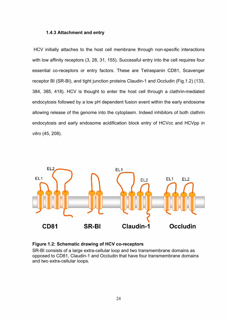
1.4.3 Attachment and entry
HCV initially attaches to the host cell membrane through non-specific interactions
with low affinity receptors (3, 28, 31, 155). Successful entry into the cell requires four
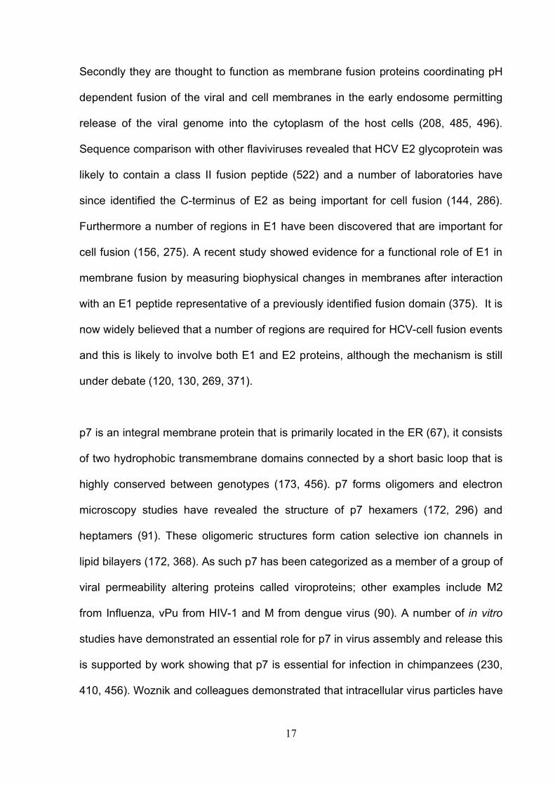
essential co-receptors or entry factors. These are Tetraspanin CD81, Scavenger
receptor BI (SR-BI), and tight junction proteins Claudin-1 and Occludin (Fig.1.2) (133,
384, 385, 418). HCV is thought to enter the host cell through a clathrin-mediated
endocytosis followed by a low pH dependent fusion event within the early endosome
allowing release of the genome into the cytoplasm. Indeed inhibitors of both clathrin
endocytosis and early endosome acidification block entry of HCVcc and HCVpp in
vitro (45, 208).
Figure 1.2: Schematic drawing of HCV co-receptors SR-BI consists of a large extra-cellular loop and two transmembrane domains as opposed to CD81, Claudin-1 and Occludin that have four transmembrane domains and two extra-cellular loops.
25
Attachment factors Low affinity receptors are thought to be important in the initial binding of the virion to
the host cell prior to interaction with high affinity receptors. Glycosaminoglycan’s
(GAG’s) are linear polysaccharides ubiquitously expressed on eukaryotic cell
membranes; highly sulfated GAG’s such as heparin sulphate (HS) are implicated in
the attachment and subsequent entry of a number of viruse for example other
flaviviridae including Dengue virus type 2 utlise HS as a viral receptor (80, 199). In
2003 Barth et al., demonstrated that sE2 binds HS suggesting it could be involved in
attachment of HCV (28). Furthermore treatment with heparinase an enzyme that
degrades heparin sulfate or heparin an analogue of HS, reduces HCVcc infection in
vitro indicating that HS is indeed important for initial attachment of HCV to the host
cell (31, 252). Interestingly upon formation of the E1E2 heterodimer E2 is no longer
able to bind to HS, indicating the binding site is no longer visible and that the
lipoproteins associated with HCV may be the factor mediating HCV attachment to HS
and not the glycoproteins (60, 63).
Other low affinity receptors implicated in HCV attachment include the C-type lectin
DC-SIGN (Dendritic cell-specific intercellular adhesion molecule-3-grabbing
nonintegrin) and the liver related molecule L-SIGN (Liver and Lymph node specific
DC-SIGN). DC-SIGN regulates the interaction between T-cells and dendritic cells
and is important for the binding, uptake and antigen processing of multiple pathogens
via the recognition of high mannose residues on the pathogens surface. L-SIGN has
also been identified as a capture receptor for pathogens and was proposed as a
capture receptor that could transmit virus to neighbouring cells (264, 293, 386). Both
DC-SIGN and L-SIGN have been demonstrated to interact with HCV envelope
26
glycoproteins (155). HCVpp and sE2 have been shown to bind to both molecules on
sinusoidal endothelial cells (264), and sE2 has also been found to bind DC-SIGN on
mature human monocyte-derived dendritic cells (386). This led to the proposition that
DC-SIGN and L-SIGN may capture and deliver HCV via dendritic cells to the liver, in
fact Lozach et al., demonstrated that HCVpp bound to L-SIGN and DC-SIGN positive
cells could be transmitted to Huh-7 cells in co-culture (293).
As previously described HCV has been isolated in low density fractions of plasma
and is believed to associate with low-density lipoproteins, indeed recent research has
identified Apo E, a component of low density lipoproteins, to be associated with
virions exhibiting peak infectivity (14, 358). It is therefore no surprise that low-density
lipoprotein receptor (LDL-R) was an obvious choice for an HCV receptor (3, 60).
LDL-R is the most important receptor for LDL in plasma. LDL binds to the receptor
and is endocytosed via clathrin dependent endocytosis. LDL-R is recycled to the cell
surface whilst LDL is released from the endosome via a pH dependent process
followed by degradation in lysosomes resulting in the release of cholesterol into the
cytoplasm (406). The first indirect evidence of HCV entry via LDL was shown by
Agnello et al., in 1999, where HCV uptake into cells correlated with low density
lipoprotein receptor activity and a reduction in uptake was observed in the presence
of LDL receptor antibodies (3). More recent work has demonstrated that antibodies
specific for LDL-R inhibit serum derived virus replication in primary hepatocyte
cultures, suggesting a role for LDL-R in HCV replication (333). Owen et al., silenced
LDL-R expression and demonstrated a reduction in HCV entry, these results were
substantiated by a rescue of infection following re-expression of LDL-R (358). Further
to this Owen et al., demonstrated the specific involvement of Apo E in LDL-R
27
mediated HCV entry by firstly inhibiting entry using Apo E specific antibodies and
secondly identifying that Apo E is associated with highly infectious virions that have a
high dependence on LDL-R (358). As yet there has been no direct evidence of an
association between HCVpp/HCVcc with LDL-Rs however this may reflect the
differential lipid association of particles propagated in vitro (60).
Receptors
CD81 is a member of the tetraspanin superfamily. It has two small intracellular
domains, four transmembrane domains and two extra cellular loops, a large extra
cellular loop (EC1) and a small extra cellular loop (EC2) (Fig.1.2). Tetraspanins form
networks at the cell surface and are reported to be involved in cell adhesion, motility,
cell activation, metastasis, and signal transduction (274). In the liver CD81 has been
demonstrated to regulate cell proliferation and more recently to regulate
hepatocellular carcinoma cell migration (42, 66, 312, 313). Chapter 3 and 4 of this
study further investigate the function of CD81 in hepatoma cells.
Pileri et al. demonstrated an interaction between HCV E2 glycoprotein and CD81 in
1999 (384). The large extra cellular loop of CD81 was demonstrated to bind soluble
E2 and subsequent experiments with both HCVpp and HCVcc validated the essential
role of CD81 in viral entry (99, 145, 314, 555). Zhang et al., 2004 reported that
monoclonal antibodies to CD81 and small interfering RNA’s to silence CD81 inhibited
HCVpp infection in vitro. McKeating et al., 2004 demonstrated the infection of diverse
HCV genotypes was dependent on CD81 expression. HCVcc and HCVpp only infect
the hepatoma cell line, HepG2 when engineered to express CD81, confirming the
critical role of CD81 in viral entry (555). Recently an in vivo study showed that
28
treatment with anti-CD81 antibodies prior to HCV infection of liver-uPA-SCID mice
provided complete protection from HCVcc infection but treatment after infection had
no effect (325). A number of studies have demonstrated that CD81 is a co-receptor
for HCV entry and functions post attachment of HCV to the cell membrane (5, 99,
335). Tan et al., 2003 showed that CD81 is involved in HCV internalisation and that
HCV particles can bind to the cell surface in the absence of CD81 but could only be
internalised when cell surface CD81 was present. Cormier et al., 2004 demonstrated
that an anti-CD81 monoclonal antibody inhibits HCV entry post attachment, further
supporting this conclusion.
Regions of E2 important for E2/CD81 binding have been identified by antibody
blocking experiments of E2 and structural modeling of E1E2 (360, 401). Owsianka et
al., 2006 demonstrated conserved regions of E2 that were critical for CD81 binding
for all genotypes. Specific residues on CD81 have been identified as critical for E2
binding. Four amino acid residues on the large extracellular loop of human CD81
were identified to differ from that of African green monkey CD81 that does not bind
E2. One of these residues 186 was subsequently identified as being critical for E2
binding by mutagenesis studies (197). Drummer et al., 2002 used random
mutagenesis to determine specific residues 182, 186, 184 and 162 on CD81 large
extracellular loop that were critical for E2 binding (122). The above studies used E2
to assess binding specific residues on CD81 important for virus entry. Since then
Flint et al., 2006 successfully infected HepG2 cells that expressed full length CD81
derived from a broad range of species and showed that sE2 binding and recombinant
CD81 blocking of HCVpp infection were not good predictors for successful HCVcc

29
infection (145). Implying that some of the previous work that identified residues
critical for E2 binding may not predict successful entry of HCV.
CD81 is closely associated with immunoglobulin like proteins EWI-2 and EWI-F.
These proteins are members of a novel family of immunoglobulin proteins that are
directly linked to actin linking ezrin-radixin-moesin (ERM) (411, 458). A cleavage
product of EWI-2, EWI-2 wint (without its N terminus) that is not present in
hepatocytes and was recently shown to inhibit HCV-CD81 interaction and reduce
HCV infection in Huh-7 cells (402), suggesting that inhibitor proteins may contribute
to the non-permissive nature of some cell types (194).
SR-BI is a lipoprotein receptor that binds HDL and oxidized LDL (oxLDL). It functions
by mediating cholesteryl ester uptake from HDL and controlling cholesterol efflux
(222). It is expressed highly in the liver although it can also be found in steroidogenic
tissue and macrophages (235). SR-BI is a cell membrane protein that has two
cytoplasmic terminal domains and a large extra cellular domain (Fig.1.2). The SR-BI
gene gives rise to at least three isoforms, SR-BI, SR-BII and SR-BIII (127) The
mechanism of SR-BI internalisation is unknown, however, SR-BII has been shown to
endocytose via a clathrin dependent pathway (126).
Scarselli et al., 2002 was the first to identify that SR-BI could interact with HCV E2.
Since this discovery further work has been carried out to characterize the role of SR-
BI as a HCV receptor (70, 174, 175). Initial work focused on the effect of lipoproteins
(SR-BI ligands) on HCV entry. Lavillette et al., 2005 found that human sera could
increase HCVpp infectivity and hypothesised that an agent in the serum was
30
responsible for the increased infectivity (268). As previously described lipoproteins in
serum have been found to associate with HCV in vivo (14, 351, 478) and to increase
HCV glycoprotein E2 cell binding (519). Treatment of cells with high density
lipoproteins (HDL) increased HCVpp infectivity, this was abrogated in the presence of
inhibitors of SR-BI selective cholesterol uptake (BLT-2, BLT-4) suggesting that HCV
entry may be dependent on the cholesterol uptake by SR-BI (495).
Grove et al., reported that both plasma and cell culture derived J6/JFH had increased
infectivity for Huh-7.5 cells transduced to over-express SR-BI and SR-BII (174).
Antibodies specific for SR-BI inhibited HCVcc infectivity, demonstrating a specific role
for this receptor in HCV entry and replication (70). Kinetic studies have shown that
SR-BI may have a role post viral attachment, as antibodies neutralized cell bound
virus (553). However, work by Catanese et al., 2009 contradicted these findings,
demonstrating that anti-SR-BI antibodies blocked vius attachment.
It is only very recently that the direct interaction of E2 with SR-BI has been
demonstrated to be important for HCV entry (69). Catanese et al., produced mutant
SR-BI proteins containing specific mutations required for sE2 binding and used them
to show that firstly they were unable to restore infectivity after SR-BI knockdown
treatment, suggesting that direct SR-BI E2 binding is important for virus infection.
Secondly they demonstrated that HDL binding and cholesterol efflux was maintained
in the mutant SR-BI expressing cells indicating that HDL and HCV E2 binding are
distinct (69).
31
Dreux et al., 2009 expressed SR-BI in SR-BI negative cell lines, SK-hep1 (human
liver endothelial cell) and BRL3A (rat hepatocarcinoma cell line), and demonstrated
permisivity to HCVpp and HCVcc infection. These results for the first time defined
SR-BI as an essential co-receptor. By expressing mutant SR-BI proteins in the SR-
BI negatie cell lines Dreux et al., concluded that intracellular domains of SR-BI were
important for HCV infection (118).
Kapadia et al., demonstrated the potential cooperation of CD81 and SR-BI in HCVcc
infection with monoclonal antibodies to CD81 and SR-B1 (235). Huh-7 cells were
incubated with both CD81 and SR-BI antibodies alone or in combination and
screened for their ability to support JFH-1 infection. Treatment of cells with antibodies
to both co-receptors simultaneously had a synergistic effect suggesting cooperation
between the two receptors. Grove et al., demonstrated that both CD81 and SR-BI
dependency could be altered by a single amino acid change in glycoprotein E2.
Mutation at amino acid 451 reduced SR-BI dependency and rendered the particle
more sensitive to neutralisation by glycoprotein antibodies and soluble CD81 as well
as altering particle density: infectivity relationship (175). These findings suggest a
co-coperation between HCV receptors.
Some cell lines that express CD81, SR-BI and LDL receptors are non-permissive
cells to HCV infection, implying that at least one other liver specific receptor was
needed. In 2007 Evans et al., screened a complementary DNA library from Huh-7.5
cells for genes that were able to confer HCVpp entry into non-permissive 293T cells.
These studies identified the tight junction protein claudin-1 (CLDN-1) as a critical
factor defining HCV entry (133). Claudin-1 is highly expressed within the liver and
32
recent evidence from our laboratory suggests that the protein localises to junctional
and non-junctional regions of the plasma membrane (396). Claudin-1 consists of four
transmembrane regions with two extra cellular loops (Fig. 1.2). The first extracellular
loop (EL1) of claudin-1 was found to be essential for HCV entry, however, there is so
far no evidence for a direct association between claudin-1 and HCV E1E2. Evans et
al., reported that claudin-1 plays a role downstream of CD81 and is required for HCV
glycoprotein mediated cell fusion (133). Furthermore Evans et al., identified two
residues in EL1 essential for virus entry, I32 and E48 (133). Cukierman et al.,
identified by mutational studies a further 7 residues (6 highly conserved resdues,
W30, GLW-51, C54 and C64 as well as a further residue D38) in EL1 that were
critical for HCV entry (107). Claudin-1 is part of a family of claudin proteins. It has
recently been shown that further family members, claudin-6 and claudin-9 can also
permit HCV entry into the claudin-1 negative hepatocellular carcinoma cell line
Bel7402 (321, 559).
A further tight junction protein occludin was recently implicated as an important entry
factor(288, 385). Ploss et al., showed that expression of human occludin in murine
cells rendered the cells permissive for infection with HCVpp and HCVcc. It is hoped
that this knowledge may help to provide a mouse model for HCV infection by
expressing human occludin. Occludin like claudin-1 has two extra-cellular loops EL1
and EL2 (Fig.1.2). Work by Benedicto et al., using shRNA to silence occludin
expression confirmed a role for occludin in virus entry and eliminated the possibility
of a role in replication. Furthermore Benedicto et al., established that occludin was
not involved in attachment of the virion and demonstrated a possible role in
glycoprotein dependent cell fusion, a later step in entry (39). Since then occludin EL2
33
has been identified by a number of studies to be important for mediating HCV entry
(286, 328). Liu et al., expressed deletion mutant occludin proteins in 786-0 cells,
(human renal carcinoma cells that are naturally occludin deficient) and demonstrated
that deletion of EL2 abolished HCVpp and HCVcc infection whilst still exhibiting
comparable cell surface expression to wild type (287). Mitcha et al., further confirmed
the importance of EL2 by expressing occludin from different species in 786-0 cells
and comparing permissivity to HCVpp, they were able to further define the area of
importance to the second half of EL2 (328). A number of different splice variants of
occludin have been identified (302). Kohaar et al., investigated the diversity of
occludin present in normal human liver and compared activity when expressed in
786-0 cells (245). Some of the splice variants were not expressed at the cell surface
and were unable to support infection, suggesting that the variability in splice variants
identified across different human livers may contribute towards partly explaining the
differences in disease outcome between patients and may also help to explain tissue
tropism (245). Importantly expression of CD81, SR-BI, claudin-1 and occludin in non-
liver derived cells (786-0(renal carcinoma cell line), TZM (HeLa cell-derived cervical
carinoma cell line) and NIH3T3 (Mouse embroyo fibroblast cell line)) renders the cells
permissive to HCV infection indicating that these four entry factors are the minimal
requirement for viral entry (385).
34
Receptor complex and endocytosis
The involvement of many receptors in HCV entry suggests a complex process
that is only now beginning to be investigated. Preliminary studies addressed receptor
localization in liver tissue and investigated protein co-localisation by confocal
imaging. To fully interpret this data it is important to appreciate the liver architecture
(Figure 1.4A). The liver is a complex organ that is fed by both the portal vein and the
hepatic artery. Blood is discharged from the portal vein and hepatic artery into
sinusoids. Blood then travels through the sinusoids that weave around the
hepatocytes until it reaches the central vein where it leaves the liver (433). Bile ducts
are integrated throughout the structure and act to remove bile from hepatocytes and
take it to the duodenum via the common hepatic bile duct (490). The liver is made up
primarily of polarised parenchyma hepatocytes (493, 502) and other non
parenchymal cells, examples of which include; Kupffer cells (resident macrophages),
stellate / fat storing cells, and liver sinusoidal endothelial cells (LSEC) that form a
permeated barrier between hepatocytes and the sinusoidal blood flow (493)
(Fig.1.4B). The gap between the sinusoidal endothelium and the hepatocytes is
termed the space of Disse and is where tissue fluid flows outward towards the
lymphatics and is also where stellate cells can be found (433). Hepatocytes are
highly polarised and their membrane can be split into three different surfaces: the
sinusoidal / basolateral membrane faces the sinusoids and space of Disse and has
irregular sized and spaced microvilli; the apical / canalicular membrane faces the bile
and is separated from the other surfaces by the presence of tight junctions and
desmosomes; finally, the third side faces other hepatocytes (433).
35
Reynolds et al., demonstrated the co-localisation of CD81 and claudin-1 at the
sinusoidal (basolateral) and canalicular (apical) domains of polarized hepatocytes
and the co-localisation of SR-BI and claudin-1 only at the sinusoidal domain (396). It
is the sinusoidal domain of the hepatocytes that will be exposed to the sinusoidal
blood and the site that we presume will come into contact with the virus. Evidence as
to occludin localization in the liver is limited; investigations in our laboratory suggest
localization predominantly at the bile canalicular, this is significantly different to
expression in diseased livers where it is expressed at both the sinusoidal and
canalicular membranes (Wilson, unpublished data).
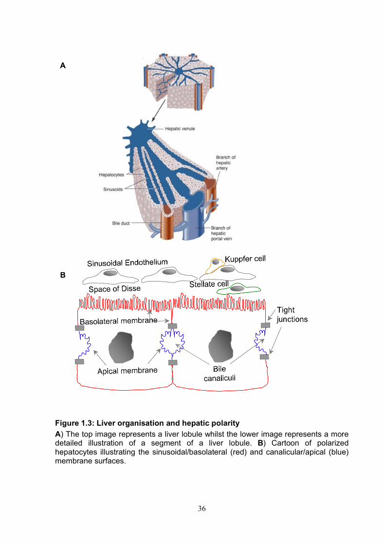
36
Figure 1.3: Liver organisation and hepatic polarity A) The top image represents a liver lobule whilst the lower image represents a more detailed illustration of a segment of a liver lobule. B) Cartoon of polarized hepatocytes illustrating the sinusoidal/basolateral (red) and canalicular/apical (blue) membrane surfaces.
37
To understand whether the receptors form a complex, specific interactions between
the co-receptors need to be determined. To date interactions between CD81, CLDN
and occludin have been investigated (188, 189). Work from our laboratory has
investigated these associations using FRET (Fluorescence resonance energy
transfer), a technique that allows distances of very closely associated fluorescently
tagged proteins to be measured (less than 10nm). FRET occurred between
fluorescent tagged CD81 and claudin-1 in permissive and non-permissive cells
suggesting co-localization and formation of a co-receptor complex (189). Further
work by Harris & Davis et al., demonstrated that receptor active claudins (Claudin-1,
6 and 9) exhibit a 1 to 1 stoichiometry with co-receptor CD81. Furthermore disruption
of claudin-1-CD81 association ablated HCV entry demonstrating that this complex
defines HCV entry. Of equal importance claudin-1 - occludin interactions were not
found to define HCV entry (188).
Since the identification of tight junction proteins claudin-1 and occludin as receptors
for HCV entry there has been much speculation as to whether HCV may utilize a
cellular receptor to traffic to tight junctions where claudin-1 and occludin reside to
permit entry. A similar mechanism has previously been observed for group B
coxsackievirus (CVB) infection that utilzes a cellular receptor, decay accelerating
factor (DAF) to co-ordinate the movement of virus particles from the apical surface of
polarized epithelial cells to reach a further receptor, cosackievirus and adenovirus
receptor (CAR) that is a component of the tight junction (102). These speculations
were hightened by research carried out by Brazzoli et al., showing a re-organisation
of CD81 to tight junctions following sE2 engagement (54). Importantly our laboratory
has been unable to repeat these results (Michelle Farquar, unpublished data).
38
Benedicto et al., 2009 suggested that occludin may be responsible for re-localisation
of the virus to sites of entry, as yet there is no evidence for a direct association
between extra-cellular occludin and HCV glycoprotein’s making this hypothesis
unlikely (39, 287). Work in our laboratory would argue against the need for re-
localization of the virion to tight junctions as the CD81-claudin-1 complexes are
predominantly at the basolateral membrane of polarised HepG2 cells (Cell line that
exhibits hepatic polarity (319) and not tight junctions (188). Furthermore Coller et al.,
recently published data showing the movement of fluorescent labeled HCV particles
along filopodia and across the cell body followed by internalization at sites that were
predominantly not cell junctions (96).
Liu et al., recently demonstrated that dynamin is important for HCV entry, this is no
surprise as dynamin is a large GTPase that functions in the scission of vesicles from
the cell membrane including clathrin mediated endocytosis, which has previously
been shown to be involved in HCV entry in vitro (45, 208). Indeed our laboratory has
recently shown that dynamin is important for HCV entry (Farquar, submitted). Liu et
al., demonstrated that occludin association with dynamin is important for entry,
suggesting that this association may be involved in coordinating HCV endocytosis
HCV (287). This is particularly interesting as recent work in our laboratory has shown
that CD81 engagement by either anti-CD81 or sE2 promotes a clathrin and dynamin
dependent co-endocytosis of CD81 and claudin-1 to early endosomes, arguing that
HCV glycoprotein engagement with CD81 may mediate endocytosis of the virus-
receptor complex. Our data supports previous observations by Coller et al., that
visualized internalization of fluorescently labeled virions after association with
claudin-1 and CD81 (96). The lack of an endocytic motif on CD81 and the knowledge
39
that CD81 lacking the N or C terminus is still endocytosed suggests that this process
may be coordinated by an associated protein and that the previously identified CD81-
claudin-1 interaction may be important in facilitating this process (Farquar,
submitted), (188, 189).
The final stages of virus entry into the cell involve viral-cell fusion inside the early
endosome allowing the release of the viral genome into the cytoplasm. Our
laboratory has shown that antibodies to CD81 are able to block entry at late time
points post infection suggesting that CD81 might be involved in these later stages of
entry as well as attachment (Farquar, submitted). Claudin-1 (133) and occludin (39,
287) have also been implicated to function in these later stages of entry. Taken
together these studies indicate that CD81, claudin-1 and occludin will need to be
present inside the early endosome for the final stages of viral entry, whether they get
there in association with one another and / or in association with the virus remains
unknown. Evidently further work is needed to understand the complex nature of HCV
entry. A cartoon depicting a possible route for HCV entry is shown in Figure 1.4.
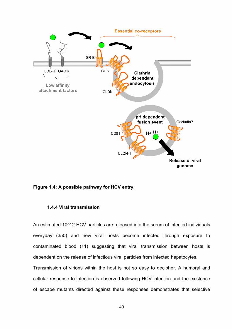
40
Figure 1.4: A possible pathway for HCV entry.
1.4.4 Viral transmission An estimated 10^12 HCV particles are released into the serum of infected individuals
everyday (350) and new viral hosts become infected through exposure to
contaminated blood (11) suggesting that viral transmission between hosts is
dependent on the release of infectious viral particles from infected hepatocytes.
Transmission of virions within the host is not so easy to decipher. A humoral and
cellular response to infection is observed following HCV infection and the existence
of escape mutants directed against these responses demonstrates that selective
41
pressure occurs in vivo (73, 131, 434, 450, 497, 500, 505). However, HCV infection
is able to persist in the presence of neutralizing antibodies suggesting a direct cell-to-
cell route of transmission may also be occurring (290). The ability to transmit directly
from cell-to-cell without the release of infectious viral particles is often an advantage
for the virus, it enables a more efficient mode of transmission and furthermore helps
to protect the virion from the host immune system. Our laboratory has recently
developed an assay to determine in vitro transmission. Timpe et al., 2008 used this
assay to demonstrate that HCV can transmit efficiently in the presence of nAbs
suggesting the presence of a novel direct cell-to-cell route of HCV transmission
(480). This is investigated further in chapter 5 of this study.
42
1.5 Project aims This project focuses on three areas. HCV entry requires glycoprotein engagement of
tetraspanin CD81 (384), and a number of studies using immune cells have
demonstrated that engagement of CD81 by antibody or recombinant sE2 protein
induces a rearrangement of the actin cytoskeleton and alters cell function(s) (95, 104,
486). Our first goal focuses on the role of CD81 engagement on hepatoma cell
morphology using a panel of anti-CD81 antibodies that recognize novel CD81
epitopes.
In a small but significant proportion of patients, HCV infection leads to the
development of hepatocellular carcinoma (6, 10, 71). Tetraspanins have been
reported to alter the metastatic potential of a number of different cancers (561).
Therefore our second goal investigates a role for tetraspanin CD81 in hepatoma-
ECM adhesion and migration and whether HCV infection modulates CD81
dependent function and/or CD81 independent hepatoma adhesion and migration
potential.
Recent work in the HCV field suggests that HCV infection may transmit within a host
in a neutralizing antibody resistant manner involving a direct cell-to-cell route of
transmission (480). In the final chapter we characterized the receptor dependency of
neutralizing antibody resistant transmission of HCV in vitro.
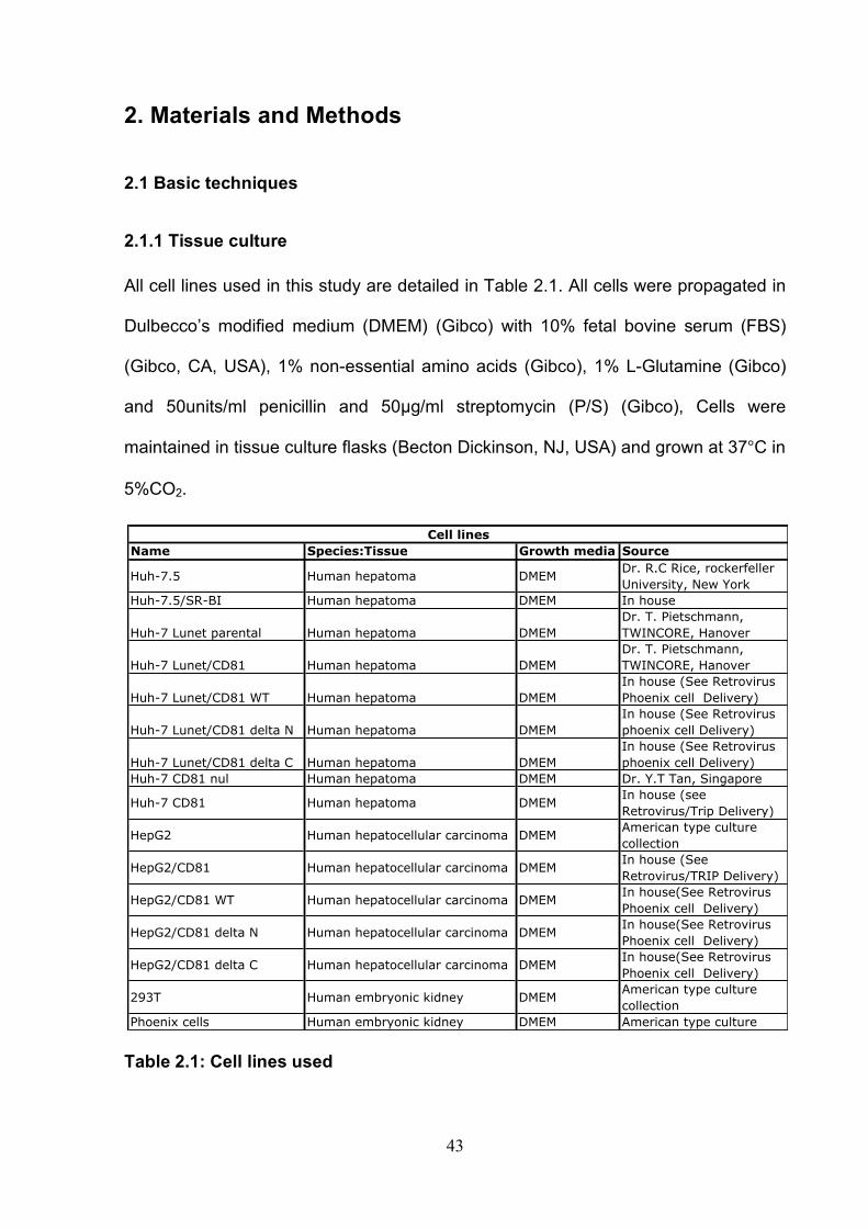
43
2. Materials and Methods
2.1 Basic techniques
2.1.1 Tissue culture
All cell lines used in this study are detailed in Table 2.1. All cells were propagated in
Dulbecco’s modified medium (DMEM) (Gibco) with 10% fetal bovine serum (FBS)
(Gibco, CA, USA), 1% non-essential amino acids (Gibco), 1% L-Glutamine (Gibco)
and 50units/ml penicillin and 50µg/ml streptomycin (P/S) (Gibco), Cells were
maintained in tissue culture flasks (Becton Dickinson, NJ, USA) and grown at 37°C in
5%CO2.
Table 2.1: Cell lines used
Name Species:Tissue Growth media Source
Huh-7.5 Human hepatoma DMEMDr. R.C Rice, rockerfeller
University, New York
Huh-7.5/SR-BI Human hepatoma DMEM In house
Huh-7 Lunet parental Human hepatoma DMEM
Dr. T. Pietschmann,
TWINCORE, Hanover
Huh-7 Lunet/CD81 Human hepatoma DMEM
Dr. T. Pietschmann,
TWINCORE, Hanover
Huh-7 Lunet/CD81 WT Human hepatoma DMEM
In house (See Retrovirus
Phoenix cell Delivery)
Huh-7 Lunet/CD81 delta N Human hepatoma DMEM
In house (See Retrovirus
phoenix cell Delivery)
Huh-7 Lunet/CD81 delta C Human hepatoma DMEM
In house (See Retrovirus
phoenix cell Delivery)
Huh-7 CD81 nul Human hepatoma DMEM Dr. Y.T Tan, Singapore
Huh-7 CD81 Human hepatoma DMEMIn house (see
Retrovirus/Trip Delivery)
HepG2 Human hepatocellular carcinoma DMEMAmerican type culture
collection
HepG2/CD81 Human hepatocellular carcinoma DMEMIn house (See
Retrovirus/TRIP Delivery)
HepG2/CD81 WT Human hepatocellular carcinoma DMEMIn house(See Retrovirus
Phoenix cell Delivery)
HepG2/CD81 delta N Human hepatocellular carcinoma DMEMIn house(See Retrovirus
Phoenix cell Delivery)
HepG2/CD81 delta C Human hepatocellular carcinoma DMEMIn house(See Retrovirus
Phoenix cell Delivery)
293T Human embryonic kidney DMEMAmerican type culture
collection
Phoenix cells Human embryonic kidney DMEM American type culture
Cell lines
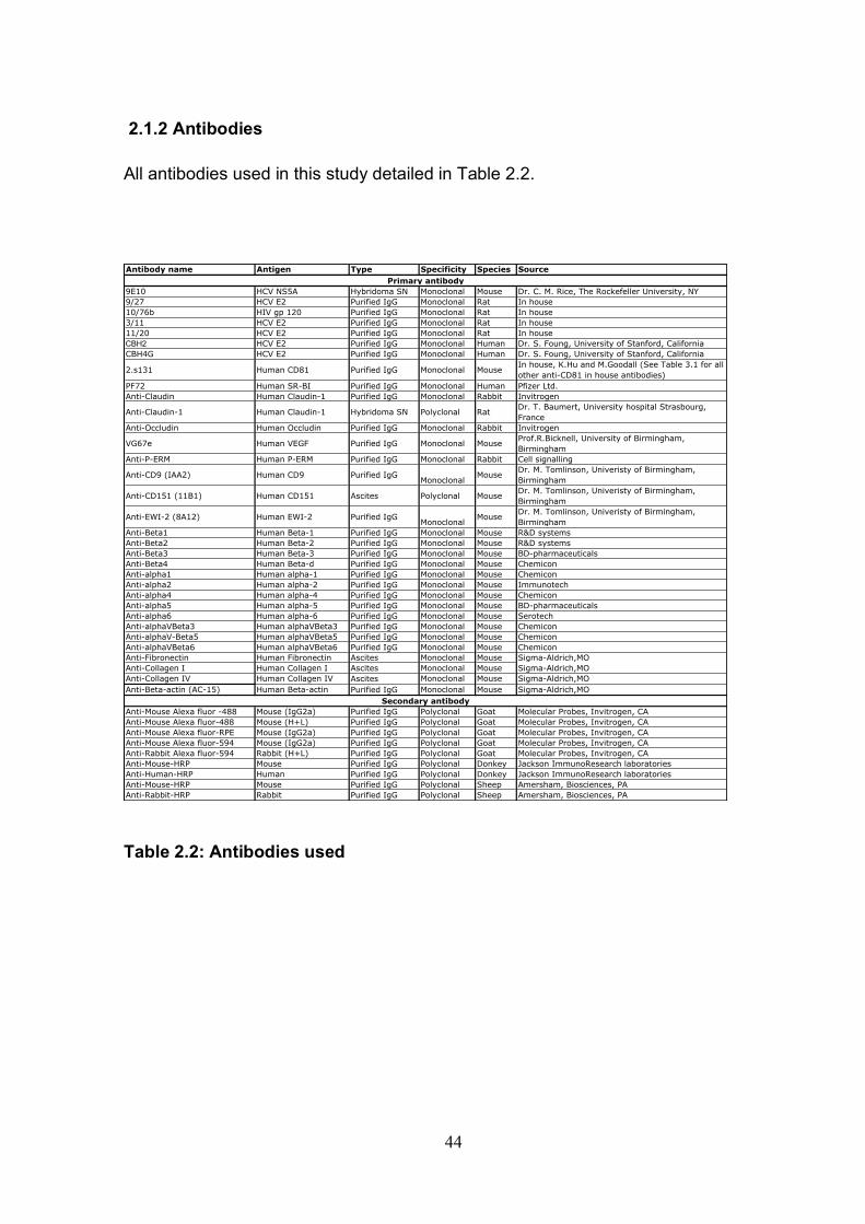
44
2.1.2 Antibodies All antibodies used in this study detailed in Table 2.2.
Table 2.2: Antibodies used
Antibody name Antigen Type Specificity Species Source
9E10 HCV NS5A Hybridoma SN Monoclonal Mouse Dr. C. M. Rice, The Rockefeller University, NY
9/27 HCV E2 Purified IgG Monoclonal Rat In house
10/76b HIV gp 120 Purified IgG Monoclonal Rat In house
3/11 HCV E2 Purified IgG Monoclonal Rat In house
11/20 HCV E2 Purified IgG Monoclonal Rat In house
CBH2 HCV E2 Purified IgG Monoclonal Human Dr. S. Foung, University of Stanford, California
CBH4G HCV E2 Purified IgG Monoclonal Human Dr. S. Foung, University of Stanford, California
2.s131 Human CD81 Purified IgG Monoclonal MouseIn house, K.Hu and M.Goodall (See Table 3.1 for all
other anti-CD81 in house antibodies)
PF72 Human SR-BI Purified IgG Monoclonal Human Pfizer Ltd.
Anti-Claudin Human Claudin-1 Purified IgG Monoclonal Rabbit Invitrogen
Anti-Claudin-1 Human Claudin-1 Hybridoma SN Polyclonal RatDr. T. Baumert, University hospital Strasbourg,
France
Anti-Occludin Human Occludin Purified IgG Monoclonal Rabbit Invitrogen
VG67e Human VEGF Purified IgG Monoclonal MouseProf.R.Bicknell, University of Birmingham,
Birmingham
Anti-P-ERM Human P-ERM Purified IgG Monoclonal Rabbit Cell signalling
Anti-CD9 (IAA2) Human CD9 Purified IgGMonoclonal
MouseDr. M. Tomlinson, Univeristy of Birmingham,
Birmingham
Anti-CD151 (11B1) Human CD151 Ascites Polyclonal MouseDr. M. Tomlinson, Univeristy of Birmingham,
Birmingham
Anti-EWI-2 (8A12) Human EWI-2 Purified IgGMonoclonal
MouseDr. M. Tomlinson, Univeristy of Birmingham,
Birmingham
Anti-Beta1 Human Beta-1 Purified IgG Monoclonal Mouse R&D systems
Anti-Beta2 Human Beta-2 Purified IgG Monoclonal Mouse R&D systems
Anti-Beta3 Human Beta-3 Purified IgG Monoclonal Mouse BD-pharmaceuticals
Anti-Beta4 Human Beta-d Purified IgG Monoclonal Mouse Chemicon
Anti-alpha1 Human alpha-1 Purified IgG Monoclonal Mouse Chemicon
Anti-alpha2 Human alpha-2 Purified IgG Monoclonal Mouse Immunotech
Anti-alpha4 Human alpha-4 Purified IgG Monoclonal Mouse Chemicon
Anti-alpha5 Human alpha-5 Purified IgG Monoclonal Mouse BD-pharmaceuticals
Anti-alpha6 Human alpha-6 Purified IgG Monoclonal Mouse Serotech
Anti-alphaVBeta3 Human alphaVBeta3 Purified IgG Monoclonal Mouse Chemicon
Anti-alphaV-Beta5 Human alphaVBeta5 Purified IgG Monoclonal Mouse Chemicon
Anti-alphaVBeta6 Human alphaVBeta6 Purified IgG Monoclonal Mouse Chemicon
Anti-Fibronectin Human Fibronectin Ascites Monoclonal Mouse Sigma-Aldrich,MO
Anti-Collagen I Human Collagen I Ascites Monoclonal Mouse Sigma-Aldrich,MO
Anti-Collagen IV Human Collagen IV Ascites Monoclonal Mouse Sigma-Aldrich,MO
Anti-Beta-actin (AC-15) Human Beta-actin Purified IgG Monoclonal Mouse Sigma-Aldrich,MO
Anti-Mouse Alexa fluor -488 Mouse (IgG2a) Purified IgG Polyclonal Goat Molecular Probes, Invitrogen, CA
Anti-Mouse Alexa fluor-488 Mouse (H+L) Purified IgG Polyclonal Goat Molecular Probes, Invitrogen, CA
Anti-Mouse Alexa fluor-RPE Mouse (IgG2a) Purified IgG Polyclonal Goat Molecular Probes, Invitrogen, CA
Anti-Mouse Alexa fluor-594 Mouse (IgG2a) Purified IgG Polyclonal Goat Molecular Probes, Invitrogen, CA
Anti-Rabbit Alexa fluor-594 Rabbit (H+L) Purified IgG Polyclonal Goat Molecular Probes, Invitrogen, CA
Anti-Mouse-HRP Mouse Purified IgG Polyclonal Donkey Jackson ImmunoResearch laboratories
Anti-Human-HRP Human Purified IgG Polyclonal Donkey Jackson ImmunoResearch laboratories
Anti-Mouse-HRP Mouse Purified IgG Polyclonal Sheep Amersham, Biosciences, PA
Anti-Rabbit-HRP Rabbit Purified IgG Polyclonal Sheep Amersham, Biosciences, PA
Primary antibody
Secondary antibody
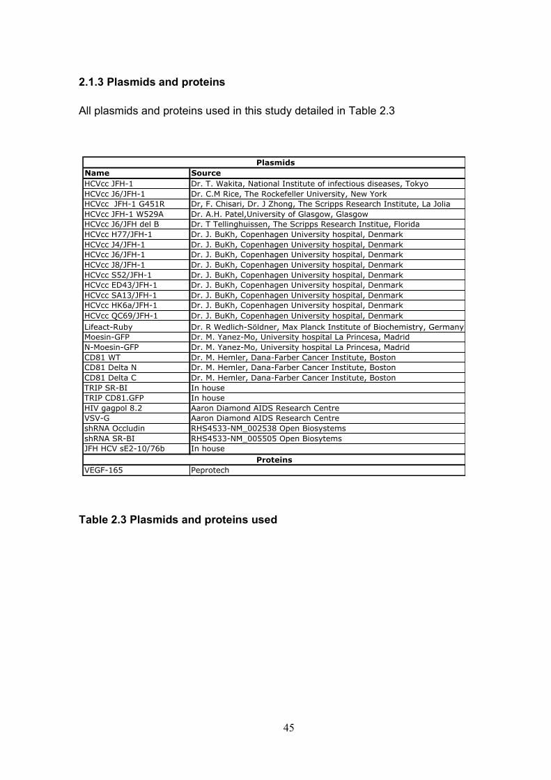
45
2.1.3 Plasmids and proteins All plasmids and proteins used in this study detailed in Table 2.3
Table 2.3 Plasmids and proteins used
Name Source
HCVcc JFH-1 Dr. T. Wakita, National Institute of infectious diseases, Tokyo
HCVcc J6/JFH-1 Dr. C.M Rice, The Rockefeller University, New York
HCVcc JFH-1 G451R Dr, F. Chisari, Dr. J Zhong, The Scripps Research Institute, La Jolia
HCVcc JFH-1 W529A Dr. A.H. Patel,University of Glasgow, Glasgow
HCVcc J6/JFH del B Dr. T Tellinghuissen, The Scripps Research Institue, Florida
HCVcc H77/JFH-1 Dr. J. BuKh, Copenhagen University hospital, Denmark
HCVcc J4/JFH-1 Dr. J. BuKh, Copenhagen University hospital, Denmark
HCVcc J6/JFH-1 Dr. J. BuKh, Copenhagen University hospital, Denmark
HCVcc J8/JFH-1 Dr. J. BuKh, Copenhagen University hospital, Denmark
HCVcc S52/JFH-1 Dr. J. BuKh, Copenhagen University hospital, Denmark
HCVcc ED43/JFH-1 Dr. J. BuKh, Copenhagen University hospital, Denmark
HCVcc SA13/JFH-1 Dr. J. BuKh, Copenhagen University hospital, Denmark
HCVcc HK6a/JFH-1 Dr. J. BuKh, Copenhagen University hospital, Denmark
HCVcc QC69/JFH-1 Dr. J. BuKh, Copenhagen University hospital, Denmark
Lifeact-Ruby Dr. R Wedlich-Söldner, Max Planck Institute of Biochemistry, Germany
Moesin-GFP Dr. M. Yanez-Mo, University hospital La Princesa, Madrid
N-Moesin-GFP Dr. M. Yanez-Mo, University hospital La Princesa, Madrid
CD81 WT Dr. M. Hemler, Dana-Farber Cancer Institute, Boston
CD81 Delta N Dr. M. Hemler, Dana-Farber Cancer Institute, Boston
CD81 Delta C Dr. M. Hemler, Dana-Farber Cancer Institute, Boston
TRIP SR-BI In house
TRIP CD81.GFP In house
HIV gagpol 8.2 Aaron Diamond AIDS Research Centre
VSV-G Aaron Diamond AIDS Research Centre
shRNA Occludin RHS4533-NM_002538 Open Biosystems
shRNA SR-BI RHS4533-NM_005505 Open Biosytems
JFH HCV sE2-10/76b In house
VEGF-165 Peprotech
Plasmids
Proteins
46
2.1.4 HCVcc generation
All HCVcc viruses used in this study were constructed around the non-structural
regions of JFH-1, a unique genome that was identified to assemble HCV particles in
cell culture and is predominantly used throughout the HCV field of research (166,
283, 499, 560). RNA was transcribed from a plasmid containing the RNA genome
(Table 2.3) and introduced into Huh-7.5 cells by electroporation.
RNA synthesis
• 5µg of the plasmid containing a cDNA clone of the HCV genome was linearized
using Xba-1 digest (Promega)
• 1µg of the RNA was used as a template for RNA transcription using the
megascript T7 kit (Ambion, Austin, TX), the reaction mixture was incubated at
37°C for 2hrs after which the RNA was purified using the RNeasy MinElute Kit
(Qiagen, NL) according to manufacturer’s instructions.
• RNA quality was assessed by gel electrophoresis on a 1% agarose gel (Bioline,
UK). Typical yields as measured by UV spectrophotometry (Amersham), were
250-1500ng/µl.
Electroporation
• Huh-7.5 cells were grown to 60-80% confluence. Trypsin was used to remove the
cells from the tissue culture plastic.
• Cells were washed twice by centrifugation at 1200rpm for 3min at 1°C and re-
suspension in 50ml ice-cold phosphate buffered saline (PBS).
• 1.5x10⁷ cells were re-suspended in 1ml of ice-cold PBS and placed on ice.
• 400µl of cell suspension was then mixed with 4ng of transcribed RNA in a cuvette
and electroporated under the following conditions; 5x100uS(820V) pulses with 1.1
second delay between pulses.
47
• Electroporated cells were then left for 5min at room temperature to rest prior to
suspension in 10ml Iscove’s modified dulbecco’s medium (IMDM) (plus10%
human serum, 1% non essential amino acids, 1% pen strep). 8ml of re-
suspended cells were placed into a T75 flask, and the remainder put into 2 wells
of a 24 well tissue culture plate to allow the monitoring of HCV protein expression.
Cells incubated at 37°C at category three containment level. Media was replaced
with DMEM (3% FBS) the following day.
• 72hrs post electroporation cells in the 24 well plates were fixed with ice-cold
methanol and stained for HCV non-structural protein, NS5A (As described in
section 2.1.7).
• Electroporated cells were used in assays at 72hrs or 96hrs post electroporation.
In this scenario no viral particles were harvested.
• Where HCV particles were harvested to use in cell free infection assays the
following procedure was followed; HCVcc particles were harvested between 4
and 14 days post EP, after which the cells were discarded. To harvest, infected
cells were cultured in a minimal volume of DMEM (3% FBS) and media containing
secreted virions collected every 8-14hrs. Harvested virus was frozen prior to
titration using the standard infection assay.
Standard infection assay
All infection assays in this study were performed as follows.
• 0.75x10⁴ cells were seeded per well into a 96 well tissue culture plate.
• The following day cells were infected with virus diluted in DMEM (3% FBS) in a
total volume of 100µl per well.
• The virus was left on overnight and media replaced the following day.
48
• 48hrs post infection cells were fixed with ice-cold methanol and stained for HCV
non-structural protein, NS5A (As described in section 2.1.7).
• Infected cells were counted as individual cells or foci of cells using a fluorescent
microscope, Nikon TE2000. Infectious units (IU) or foci forming units (FFU) per
ml were then determined.
2.1.5 Retrovirus delivery
TRIP retrovirus gene delivery
The TRIP system is a retrovirus gene expression vector developed by Zennou et. al.
(554). It produces virus vector particles, formed around a replication deficient HIV
gag-pol core, that bear the envelope glycoprotein of vesicular stomatitis virus
(VSVG). These particles can package a gene of interest as an RNA transcript,
subsequent transduction of a cell line with the TRIP system results in reverse
transcription of the target gene and its insertion into the genomic DNA. In this study
transduced cells were not under selection, however they maintained exogenous gene
expression for around 1 month, after which they were discarded. The following
plasmids were delivered by TRIP retrovirus delivery CD81.GFP / SR-BI / shRNA
Occludin and shRNA SR-BI.
• TRIP particles were produced by Fugene (Roche, Switzerland) transfection of
293T cells.
• Fugene/plasmid complex was made up as follows;
o Following amounts of plasmids (400ng VSVG envelope, 600ng TRIP gag-
pol and 600ng target gene) added to 6µl Fugene dissolved in 100µl
Optimem (Gibco).
o This was incubated at room temperature for 20min to allow formation of
complex.
49
• Cells were transfected in 6 well tissue culture plates for 8hrs, after which the
culture media was changed to DMEM (3% FBS + P/S).
• Transfection efficiency was monitored by the inclusion of a TRIP plasmid
encoding enhanced green fluorescent protein as a target gene (TRIP EGFP).
• Culture media containing TRIP particles was harvested at 48 and 72hrs post
transfection and passed through a 0.2µM filter to remove any contaminating 293T
cells. Transduction of target cells was performed immediately.
• Target cells were seeded at 4x10⁵cells/well in a 6 well tissue culture plate 24hrs
prior to transduction. To transduce, cells were incubated overnight with harvested
TRIP culture media diluted 1:1 in DMEM (3% FBS + P/S). After which the media
was changed to DMEM (10% FBS + P/S).
• Transduction efficiency was assessed after 48hrs by monitoring expression of
TRIP EGFP and flow cytometric detection of target gene(s).
Phoenix cell retrovirus gene delivery
Pheonix cells are 293T cells that have been engineered to express gag-pol and
envelope protein. Upon transient transfection with a plasmid containing LTR’s (Long
terminal repeats), cells produce retrovirus containing the required DNA. This system
was used in this study in the production of CD81 mutant expressing Huh-7 Lunet and
HepG2 cells. Like the TRIP retroviral system, subsequent transduction of a cell line
with the retrovirus results in reverse transcription of the target gene and its insertion
into the genomic DNA. Transduced cells were maintained under Zeosin selection
(Invitrogen). HepG2 cells under 400µg/ml and Huh-7 Lunet cells under 600µg/ml.
• Retroviral particles were produced by Fugene transfection (Roche, Switzerland)
of phoenix cells.
• Briefly, phoenix cells were seeded at 7x10⁵ cells per well into a 6 well culture dish
50
coated with poly-L-lysine hydro bromide (Sigma, Ca, USA) in DMEM (10% FBS)
24hrs prior to transfection. 1hr prior to transfection media refreshed with P/S free
DMEM (3%FBS), 1.5ml per well.
• Fugene/plasmid complex was made up as follows;
o 1µg DNA added to 6µl Fugene dissolved in 100µl Optimem (Gibco).
o This was incubated at room temperature for 20min to allow formation of
complex.
• Cells were transfected in 6 well tissue culture plates overnight, after which the
culture media was changed to DMEM (3% FBS + P/S).
• Culture media containing retroviral particles was harvested at 48 and 72hrs post
transfection and passed through a 0.2µM filter to remove any contaminating
phoenix cells. Transduction of target cells was performed immediately.
• Target cells were seeded at 1.5x10⁵cells/well in a 6 well tissue culture
plate 24hrs prior to transduction. To transduce, cells were incubated overnight
with harvested retrovirus culture media diluted 1:1 in DMEM (3% FBS + P/S).
After which the media was changed to DMEM (10% FBS + P/S).
• 72hrs post transduction cells were put under selection, control non-transduced
cells were used to gauge how effective the transduction was and to verify
selection efficacy.
2.1.6: Transient transfection of Huh-7.5 cells. Plasmid transfection
Moesin-GFP, N-Moesin-GFP and LifeAct-Ruby DNA plasmids were all transefected
into Huh-7.5 cells using Lipofectamine transfection.
• Transfections were carried out using the Lipofectamine (Invitrogen, CA, USA) kits
according to the manufacturer’s guidelines.
51
• Briefly, Huh-7.5 cells were seeded at 6x10⁵ cells per well in a 6 well culture dish
in P/S free media 24hrs prior to transfection.
• Plasmid/lipofectamine complex made up as follows;
o 4µg DNA added to 200µl of Optimem (Gibco) and mixed gently.
o 8µl lipofectamine reagent added to 200µl of Optimen and mixed gently.
o Diluted DNA and diluted lipofectamine reagent combined and incubated at
room temperature for 15min to allow formation of complex.
• Complex added to well containing 1000µl of P/S free DMEM.
• Transfections were carried out at 37°C for 8hrs.
• After which the transfection mixture was replaced with DMEM (3% FBS + P/S).
• Expression efficiency was typically monitored 48 hrs post transfection.
siRNA transfection
Claudin-1 and CD81 siRNA (Invitrogen) were transfected into Huh-7.5 cells using
Lipofectamine transfection.
• Transfections were carried out using the Lipofectamine RNAiMAX (Invitrogen,
CA, USA) kits according to the manufacturer’s guidelines.
• Briefly, Huh-7.5 cells were seeded at 6x10⁵ cells per well into a 6 well tissue
culture dish in P/S free media 24hrs prior to transfection.
• siRNA-RNAiMax complex was made up as follows;
o 100pmol siRNA added to 200µl of Optimem (Gibco) and mixed gently.
o 4µl RNAiMAX added to 200µl of Optimen and mixed gently.
o Diluted siRNA and diluted RNAiMAX combined and incubated at room
temperature for 15min to allow formation of complex.
• Complex added to well containing 1000µl of P/S free DMEM.
• Transfections were carried out at 37°C for 8hrs.
52
• After which the transfection mixture was replaced with DMEM (3% FBS + P/S).
• Expression efficiency was typically monitored 48hrs post transfection.
2.1.7: Cytotoxicity testing
The cytotoxicity of compounds used in this study was tested using the Cell Titer One
Solution Cell Proliferation (MTS) Assay (Promega) according to the manufacturer’s
guidelines.
• Briefly, cells were seeded at 1.5x10⁴ cells per well of a 48 well tissue culture
plate.
• Cells then treated with compound at a defined concentration and cultured for a
defined period of time, (24 or 48hrs) as stated in figure legends.
• Cells then washed with PBS followed by incubation with 200µl MTS (tetrazolium
compound) at working concentrations and incubated for 2hrs at 37°C
• 100µl of supernatant transferred to a 96 well plate and absorbance read at
490nm. Final absorbance was determined by deleting absorbance recorded for
an empty control well.
2.1.8: Cholesterol quantification. The Amplex Red Cholesterol Assay Kit (Invitrogen, CA, USA) was used to quantify
total cellular cholesterol according to the manufacturer’s guidelines.
• Briefly cells were enumerated using a haemocytometer and 25x104 cells were
pelleted by centrifugation (12,000rpm for 5min) and lysed in 250µls of 1x
Reaction buffer.
• 50µl of cholesterol samples diluted in reaction buffer plus cholesterol
reference controls were added to separate wells of a 96 well plate.
• 50µl of Amplex Red Reagent was then added to samples.

53
• Wells then incubated at 37°C for 30min.
• Fluorescence was read in a Multi detector microplate reader (Biotech Synergy
HT), excitation 530 and emission detection at 590.
• A no cholesterol control was used as a background. Final fluorescence
reading deduced by subtracting the background.
• In each assay a standard curve was produced using the cholesterol reference
standard (Fig.2.1) allowing cholesterol levels to be expressed as µM
cholesterol per 1x10⁴ cells.
Figure 2.1: Cholesterol standard curve
2.1.9: Flow cytometry
HCV infection level
Throughout this study HCV infection level was determined using a primary antibody
directed at the non-structural region 5A (NS5A) of HCV (9E10).
54
• Cells were removed from tissue culture plastic with trypsin.
• 25x104 cells were seeded per well into a 96 well round bottom plate and fixed with
1% PFA for 5min at 4°C.
• Fixed cells were incubated at room temperature in PBS plus 1% BSA (blocking)
and 0.1% saponin (permeabilisation) for 20min.
Cells were now removed from containment level 3
• Cells centrifuged at 1200rpm and the pellet re-suspended in primary antibody
9E10 (diluted in PBS + 1% BSA, 0.1% saponin) for 30min followed by 2x PBS
washes.
• This was followed by incubation with secondary antibody Alexa-fluor 488 or RPE
conjugated anti-mouse IgG 2a (Invitrogen) (diluted in PBS + 1% BSA, 0.1%
saponin) for 30min at room temperature and 2x PBS washes.
• Percentage NS5A positive cells were determined by running the cells through a
FACScalibur flow cytometer (BD Biosciences) and analysis on FLowJo software
(Tree Star, San Carlos, CA).
Cell surface protein expression
• Cell surface expression was determined on live cells. Trypsin or cell dissociation
buffer (as stated in figure legend) was used to remove the cells from tissue
culture plastic.
• Cells were blocked with FACS buffer (PBS + 2% BSA, 0.01% Azide) for 20min at
room temperature.
• 25x104 cells were then seeded per well into a 96 well round bottom plate and
centrifuged at 12,000rpm to pellet the cells.
• Cells were then re-suspended in 100µl of primary antibody diluted in FACS buffer,
(concentrations in Table 2.4), and incubated for 30min at room temperature.
55
• This was followed by 2x PBS washes and then incubation with 100µl of
appropriate secondary antibody (concentrations in Table 2.2) for 30min at room
temperature.
• Cells were then washed again (2xPBS) and fixed with 1% Para formaldehyde
(PFA) for 5min at 4°C.
• Protein surface expression was determined by running cells through a
FACScalibur flow cytometer (BD Biosciences) and analysis using FlowJo
software (Tree Star, San Carlos, CA).
2.1.10: Immuno-fluorescence
HCV infection level
Throughout this study to visualize HCV infectivity we stained the cells using a primary
antibody directed at HCV non-structural region NS5A (9E10).
• Cells were fixed with ice-cold methanol for 5min.
• Fixed cells were then incubated with PBS plus 1% BSA (blocking) and 0.1%
saponin (permeabilisation) for 20min.
• Primary antibody (9E10) was added to the cells at 1:200 dilution in PBS + 1%BSA
+ 0.1% saponin and incubated at room temperature for 30min followed by 2x PBS
washes.
• Alexa fluor 488 or 594 conjugated anti-mouse IgG2a secondary antibody was
then added at 1:1000 dilution in PBS + 1%BSA + 0.1% saponin for a further
30min at room temperature, followed by 2x PBS washes
• NS5A positive cells visualized on Nikon TE2000 fluorescence microscope on a
Zeiss META head confocal microscope.

56
Figure 2.2: NS5A stain of HCVcc J6/JFH infected cells. Huh-7.5 cells 72hrs post EP with J6/JFH RNA. NS5A stained with 9E10 primary antibody and Alexa-flour 488 conjugated secondary antibody (Green), cell nuclei counterstained with DAPI (Blue), visualized on Nikon TE2000 fluorescence microscope.
Cellular protein expression
To visualize cellular protein expression by confocal microscopy cells were seeded
onto 24 well plates containing 13µm glass cover slips. For fluorescent microscopy
analysis only, glass cover slips were not used.
• Cells were fixed with appropriate fixative (See Table 2.4) or stained live (SR-BI
expression only).
• Fixed cells were then incubated with PBS/1% BSA (blocking) and 0.1% saponin
(permeabilisation) or Triton-X-100 (permeabilisation) (P-ERM only) for 20min.
This step not included for SR-BI expression.
• Primary antibody was added to the cells at appropriate dilution in PBS + 1%BSA
+ 0.1% saponin, PBS/1%BSA (P-ERM only) or serum free DMEM (SR-BI only)
and incubated at room temperature for 30min followed by 2x PBS washes. See
table 2.4 for antibody concentrations.
• Appropriate Alexa-fluor 488 or 594 conjugated secondary antibody was then
added at 1:1000 dilution in PBS/1%BSA/0.1%saponin, PBS/1%BSA (P-ERM
only) or serum free DMEM (SR-BI only) for a further 30min at room temperature,
57
followed by 2x PBS washes. Cells stained for SR-BI expression then fixed with
3% EM grade formaldehyde.
• Cells counter stained with DAPI (sigma) diluted in PBS.
• Stained cells visualized and imaged on Nikon TE2000 fluorescence microscope
or cover slips mounted onto glass slides using ProLong Antifade mounting agent
(Invitrogen, CA, USA) and imaged on a Zeiss META head confocal microscope
with a 63x water immersion objective. Background fluorescence of samples was
corrected based on control samples stained with species-matched IgG and
secondary antibody only.
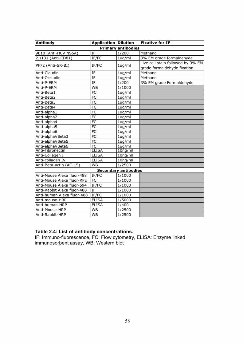
58
Table 2.4: List of antibody concentrations. IF: Immuno-fluorescence, FC: Flow cytometry, ELISA: Enzyme linked immunosorbent assay, WB: Western blot
Antibody Application Dilution Fixative for IF
9E10 (Anti-HCV NS5A) IF 1/200 Methanol
2.s131 (Anti-CD81) IF/FC 1ug/ml 3% EM grade formaldehyde
PF72 (Anti-SR-BI) IF/FC 1ug/mlLive cell stain followed by 3% EM
grade formaldehyde fixation
Anti-Claudin IF 1ug/ml Methanol
Anti-Occludin IF 1ug/ml Methanol
Anti-P-ERM IF 1/200 3% EM grade Formaldehyde
Anti-P-ERM WB 1/1000
Anti-Beta1 FC 1ug/ml
Anti-Beta2 FC 1ug/ml
Anti-Beta3 FC 1ug/ml
Anti-Beta4 FC 1ug/ml
Anti-alpha1 FC 1ug/ml
Anti-alpha2 FC 1ug/ml
Anti-alpha4 FC 1ug/ml
Anti-alpha5 FC 1ug/ml
Anti-alpha6 FC 1ug/ml
Anti-alphaVBeta3 FC 1ug/ml
Anti-alphaVBeta5 FC 1ug/ml
Anti-alphaVBeta6 FC 1ug/mlAnti-Fibronectin ELISA 10ng/ml
Anti-Collagen I ELISA 10ng/ml
Anti-collagen IV ELISA 10ng/ml
Anti-Beta-actin (AC-15) WB 1/2500
Anti-Mouse Alexa fluor-488 IF/FC 1/1000
Anti-Mouse Alexa fluor-RPE FC 1/1000
Anti-Mouse Alexa fluor-594 IF/FC 1/1000
Anti-Rabbit Alexa fluor-488 IF 1/1000
Anti-human Alexa fluor-488 IF/FC 1/1000
Anti-mouse-HRP ELISA 1/5000
Anti-human-HRP ELISA 1/400
Anti-Mouse-HRP WB 1/2500
Anti-Rabbit-HRP WB 1/2500
Primary antibodies
Secondary antibodies
59
2.2: Specific assays Chapter 3
2.2.1: Antibody engagement cell spread assay For all cell spread assays cells were serum starved overnight prior to assay. Specific
treatments were carried out on the morning of the assay as specified in table 2.5. For
the cholesterol modulation assays the cells were removed from tissue culture plastic
with trypsin and treated at 37°C in solution in universals (2x10⁶ cells in 1ml of
DMEM). For the remaining spread assays adherent cells were treated in a T25 flask
or well of a 6 well tissue culture dish.
• 96 well ELISA plates (Thermo electron corporation) were prepared by coating
with specified antibody at 5µg/ml diluted in 100µl of PBS and incubated at 4°C
overnight.
• Antibody coated plates were then blocked with PBS/1%BSA for 30min and
washed 2x with PBS.
• With the exception of the cholesterol modulation assay where cells are already in
solution, the cells were removed from tissue culture plastic using trypsin. 3x10⁴
cells were then seeded per well in 100µl of serum free DMEM.
• Phase images were taken of cells at specified time points post seeding. For
assays carried out in category three containment level, cells were fixed by adding
EM grade formaldehyde directly to the wells (final concentration 3.4%) prior to
removing cells for imaging.
• A Nikon TE2000 fluorescence microscope was used to image the cells. The total
number of spreading and non-spread cells were enumerated in a field of view and
percentage cell spread determined (See Fig.3.2 for assay set up).
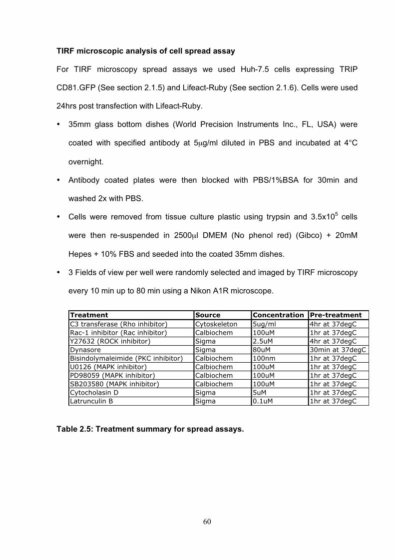
60
TIRF microscopic analysis of cell spread assay
For TIRF microscopy spread assays we used Huh-7.5 cells expressing TRIP
CD81.GFP (See section 2.1.5) and Lifeact-Ruby (See section 2.1.6). Cells were used
24hrs post transfection with Lifeact-Ruby.
• 35mm glass bottom dishes (World Precision Instruments Inc., FL, USA) were
coated with specified antibody at 5µg/ml diluted in PBS and incubated at 4°C
overnight.
• Antibody coated plates were then blocked with PBS/1%BSA for 30min and
washed 2x with PBS.
• Cells were removed from tissue culture plastic using trypsin and 3.5x10⁵ cells
were then re-suspended in 2500µl DMEM (No phenol red) (Gibco) + 20mM
Hepes + 10% FBS and seeded into the coated 35mm dishes.
• 3 Fields of view per well were randomly selected and imaged by TIRF microscopy
every 10 min up to 80 min using a Nikon A1R microscope.
Table 2.5: Treatment summary for spread assays.
Treatment Source Concentration Pre-treatment
C3 transferase (Rho inhibitor) Cytoskeleton 5ug/ml 4hr at 37degC
Rac-1 inhibitor (Rac inhibitor) Calbiochem 100uM 1hr at 37degC
Y27632 (ROCK inhibitor) Sigma 2.5uM 4hr at 37degC
Dynasore Sigma 80uM 30min at 37degC
Bisindolymaleimide (PKC inhibitor) Calbiochem 100nm 1hr at 37degC
U0126 (MAPK inhibitor) Calbiochem 100uM 1hr at 37degC
PD98059 (MAPK inhibitor) Calbiochem 100uM 1hr at 37degC
SB203580 (MAPK inhibitor) Calbiochem 100uM 1hr at 37degC
Cytocholasin D Sigma 5uM 1hr at 37degC
Latrunculin B Sigma 0.1uM 1hr at 37degC

61
2.2.2: Recombinant sE2 engagement cell spread assay
Plate preparation
• 96 well ELISA plates (Thermo electron corporation) were prepared by coating
with 10/76b capture antibody at 1µg/ml diluted in 100µl of PBS and incubated at
4°C overnight.
• Well then blocked with PBS/1%BSA for 1hr.
• Recombinant soluble E2 (JFH-10/76b) was then added to the wells at 1 in 3
dilution in PBS and incubated at 37°C for 2hrs.
• sE2 coated plates were then blocked with PBS/1%BSA for 30min and washed 2x
with PBS.
• Control wells contained 10/76b only or empty wells blocked with PBS/1%BSA.
Spread assay
• Huh-7.5 cells were serum starved overnight and then removed from tissue culture
plastic using trypsin. 3x10⁴ cells were then seeded per well in 100µl of serum free
DMEM.
• Phase images were taken of cells at 1hr post seeding using a Nikon TE2000
fluorescence microscope.
ELISA for detection of bound sE2.
• A cocktail of human anti-E2 antibodies (CBH2 and CBH4B) at 4ug/ml were added
to each well and incubated at room temperature for 45min followed by 3x washes
with PBS.
• Wells then incubated with appropriate HRP conjugated secondary antibody
(Jackson ImmunoResearch laboratories) for 45min.
62
• Bound antibodies were visualized using TMB substrate (BioFX laboratories) and
reaction was terminated after 5min incubation with stop reagent (BioFX
laboratories).
• Absorbance was read on plate reader at 490nm (Multiskin ascent, Thermo
Electron Corporation).
2.2.3: P-ERM western blot for anti-CD81 engagement time course assay. Lysate preparation • lysates were prepared by seeding 1x10⁶ Huh-7.5 cells per well into wells of a 6
well tissue culture dish. Cells were then serum starved overnight.
• Cells stimulated with anti-CD81 mAb (1.s262) diluted at 5µg/ml in serum free
DMEM for 0,1,5,10,15 and 30min.
• Tissue culture media was removed and the cell monolayer rinsed with PBS at
room temperature. All subsequent steps were carried out on ice using fresh, ice-
cold buffers to prevent protein degradation.
• 2ml of ice cold PBS were added per well and adherent cells removed using a cell
scraper. The cell suspension was transferred to universal and pelleted by
centrifugation at 1200 rpm for 5 min at 4°C.
The cell pellet was re-suspended in 500µl of NP40 lysis buffer (10mM Tris-HCL;
pH 7.5, 2mM CaCl2, 2mM MgCl2, 1%NP40 plus1x Complete Mini Protease
Inhibitor Cocktail Tablet + 1x Complete PhosStop Phosphatase Inhibitor Cocktail
Tablet (Roche)) and incubated on ice for 30 min.
• The lysate was centrifuged at 15 000 rpm in a Biofuge primo R centrifuge
(Heraeus) for 15 min at 4°C to separate nuclei and unsolubilized cell membranes
from protein, after which the supernatant was collected and
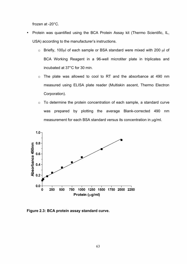
63
frozen at -20°C.
• Protein was quantified using the BCA Protein Assay kit (Thermo Scientific, IL,
USA) according to the manufacturer’s instructions.
o Briefly, 100µl of each sample or BSA standard were mixed with 200 µl of
BCA Working Reagent in a 96-well microtiter plate in triplicates and
incubated at 37°C for 30 min.
o The plate was allowed to cool to RT and the absorbance at 490 nm
measured using ELISA plate reader (Multiskin ascent, Thermo Electron
Corporation).
o To determine the protein concentration of each sample, a standard curve
was prepared by plotting the average Blank-corrected 490 nm
measurement for each BSA standard versus its concentration in µg/ml.
Figure 2.3: BCA protein assay standard curve.
64
SDS Polyacrylmide Gel Electrophoresis (SDS-PAGE).
Proteins were separated on 12% SDS gel.
• To prepare samples, defined amounts of protein were mixed with 3x Laemmli
loading dye (H2O + 30% v/v Glycerol + 6% w/v SDS + 0.02% v/v Bromophenol
Blue + 0.2M Tris-HCl; pH 6.8 plus 10% 2-mercaptoethanol) (Reducing conditions)
and the total volume adjusted to 25µl with H2O. Samples were heat-denatured at
95°C for 5min and allowed to cool to room temperature prior to loading.
• Proteins were separated by electrophoresis using the Mini Protean 3 System
(Bio-Rad Laboratories, CA, USA) according to the manufacturer’s instructions.
o Briefly, 20µl of protein sample were loaded per lane and gels run at 200
volts for 30-45 min.
o Proteins were transferred onto PVDF membranes (Millipore, MA, USA)
using a Mini Trans-Blot Electrophoretic Transfer Cell System (Bio-Rad)
according to the manufacturer’s recommendations.
o Briefly, PVDF membranes were cut to the appropriate size and pre-treated
with methanol for 1-2 min, rinsed with ultra pure water, and incubated in
transfer buffer (25mM Trizma base + 0.2M Glycine + 200 ml MeOH + 0.5
ml 10% SDS) at room temperature for 5-20 min. Gels were equilibrated in
transfer buffer for 5min to prevent shrinking and incomplete transfer.
o Transfer was carried out at 350 A for 60min at room temperature.
Immunoprobing and chemiluminescent detection of proteins.
• All subsequent steps were carried out in 50 ml Falcon tubes with gentle agitation
on a tube roller (Barloworld Scientific, UK) at RT.
• To block unspecific binding of antibodies, membranes were incubated in 2.5 ml of
TBST (10mMTrizma base + 0.1M Sodium Chloride + 10% v/v Tween 20) + 5%
65
dry milk for 1hr.
• After which the blocking buffer was removed and the membranes were incubated
with primary antibody, Anti-P-ERM, diluted in TBST + 5% BSA overnight followed
by 5 x 5min washes, then Anti-β-actin diluted in TBST + 5% dry milk for 1hr
followed by 5 x 5min washes with excess TBST.
• Membrane then Incubated with appropriate HRP conjugated secondary antibody
(Amersham, Biosciences, PA) diluted in 2ml TBST + 5% dry milk followed by 5 x
5min washes with excess TBST.
• Chemiluminescent detection of HRP-conjugated antibodies was performed using
the ECL Western Blotting Detection System (Amersham, UK) according to the
manufacturer’s instructions. Briefly, membranes were immersed in ECL detection
reagent for 1 min, wrapped in cling film, and exposed to CL- XPosure Blue X-Ray
Film (Thermo Scientific) for 1-5 min.
Antibody concentrations detailed in Table 2.4
Chapter 4
2.2.4: ECM adhesion assay. Plate preparation
• 96 well plates (Falcon, Becton Dickinson) were coated with human extra-cellular
matrix (ECM) proteins; Fibronectin (Sigma), Collagen type I (Sigma), Collagen
type VI (BD Biosciences), and laminin (Sigma) at 10µg/ml (50µl/well) and
incubated at room temperature for 1hr.
• Wells were then blocked with PBS/1% BSA for 30min followed by 3x PBS
washes.
66
Adhesion assay
• Hepatoma cells were serum starved overnight and then removed from tissue
culture plastic with trypsin.
• Cells were counted using a haemocytometer and 4x10⁴ cells were seeded per
well in 50µl serum free DMEM. For crystal violet calibration curve 0.5, 1, 2, 3, 4
and 5x10⁴ cells were seeded per well.
• Cells were incubated at 37°C for a defined period of time.
• Non-adhered cells were removed with PBS wash.
• Adhered cells were then fixed with ice-cold methanol for 5min.
• Adhered cells were then stained with crystal violet to determine adhesion or
stained for HCV non-structural protein NS5A to determine percentage infectivity
of adhered cells.
Crystal violet stain
• Fixed cells were incubated with 0.1% crystal violet for 1hr
• Cells were then washed 3x with PBS and crystal violet solubalised in 36% acetic
acid.
• Absorbance was read at 600nm on plate reader (Multiskin ascent, Thermo
Electron Corporation).
• Control wells with no cells were used to determine background. Specific adhesion
was determined by subtracting the absorbance read on PBS/1% BSA only coated
plates from ECM coated plates that had been blocked with PBS/1% BSA.
Quantification of HCV infected cells
• Fixed cells were stained for HCV non-structural protein NS5A as described in
section 2.1.10 and then cell nuclei counterstained with DAPI (Sigma).

67
• To determine the proportion of infected cells, three fields of view were imaged
using the Nikon TE2000 fluorescence microscope. The number of NS5A positive
cells and total nuclei per field of view were enumerated allowing calculation of
percentage-infected cells.
2.2.5: ECM ELISA Plate preparations
• For control assays 96 well plates were coated with gelatin for 1hr followed by
specific concentrations of human fibronectin, collagen I and collagen IV for a
further 1hr incubation (100µl/well).
• To determine ECM production of Huh-7.5 cells, 0.5x10⁴ cells in 200µl DMEM (3 or
10% FBS) were seeded per well into gelatin coated 96 well plates. The cells were
then incubated at 37°C for a specified time.
• Cells lysed with cell lysis buffer (20mM NH4OH, 0.5% Triton X-100). The lysis
step allowed the removal of plates from the level three containment laboratory.
• A titration of human fibronectin was carried out alongside Huh-7.5 fibronectin
production assays enabling the concentration of fibronectin produced in µg/ml to
be determined (See Fig.2.4)
• Plates were analyzed immediately by ELISA or stored at 4°C prior to analysis.
ELISA
• Wells were blocked with 1% gelofusine in 0.5% Tween20/PBS for 1hr, followed by
3x washes with wash buffer (0.5%Tween20/PBS).
• Wells were incubated with ECM specific primary antibodies at a concentration of
10ng/ml for 45min followed by 3x washes with wash buffer.
• Wells then incubated with appropriate HRP conjugated secondary antibody
(Jackson ImmunoResearch laboratories) for 45min.
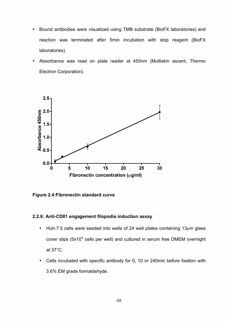
68
• Bound antibodies were visualized using TMB substrate (BioFX laboratories) and
reaction was terminated after 5min incubation with stop reagent (BioFX
laboratories).
• Absorbance was read on plate reader at 450nm (Multiskin ascent, Thermo
Electron Corporation).
Figure 2.4:Fibronectin standard curve
2.2.6: Anti-CD81 engagement filopodia induction assay
• Huh-7.5 cells were seeded into wells of 24 well plates containing 13µm glass
cover slips (5x10⁴ cells per well) and cultured in serum free DMEM overnight
at 37°C.
• Cells incubated with specific antibody for 0, 10 or 240min before fixation with
3.6% EM grade formaldehyde.
69
• For cells fixed at zero hours (prior to addition of anti-CD81 mAb), Anti-CD81
mAb diluted in 0.5%BSA/PBS was added to the wells for 1hr at 37°C
• All cells were then incubated with Alexa fluor 488 conjugated secondary
antibody diluted in 0.5%BSA/PBS for 30min at room temperature followed by
2xPBS washes.
• Phalloidin-594 (Sigma) diluted 1 in 200 in 0.5%BSA/PBS was then added and
incubated at room temperature for 30min followed by 2xPBS washes.
• Cells then counterstained with DAPI (Sigma) for 1min at room temperature.
• Stained cells visualized mounted onto glass slides using ProLong Antifade
mounting agent (Invitrogen, CA, USA) and imaged on a Zeiss META head
confocal microscope with a 63x water immersion objective. Background
fluorescence of samples was corrected based on control samples stained with
species-matched IgG and secondary antibody only.
2.2.7: Wound healing assay Huh-7.5 cells (7x10⁵ cells per well) were seeded into wells of a 6 well dish 24hrs prior
to the start of the assay and serum starved overnight. For assays using CD81 knock
down cells (See section 2.1.6), the cells were seeded 24hrs prior to transfection and
48hrs prior to the start of the assay. In this scenario cells were serum starved for 3hrs
on the morning of the assay.
• A P200 tip was used to form a wound in the cell monolayer.
• Media was then refreshed with serum free DMEM containing 15µg/ml mitomycin
C (SIGMA-ALDRICH, MO USA) and images of wound taken immediately (0hrs)
• Wells then incubated at 37°C for 24hrs and then further images of the wound
were then taken. A black marker pen was used to mark the area of the wound

70
being imaged; this ensured the same area was imaged at each time point (See
Fig.2.5).
• Images taken by phase on a Nikon TE2000 fluorescence microscope and area of
wound determined using IPLab 4.0 (BD Biosciences) software.
Figure 2.5: Schematic of wound healing assay.
2.2.8: Invasion assay Mock vs. HCV infected cell invasion assays
Huh-7.5 cells were electroporated with in vitro transcribed full length HCV RNA or
mock RNA (see section 2.1.4). Cells were cultured in DMEM (3% FBS) and then
serum starved overnight prior to assay. Invasion assays were set up 96hr post
electroporation.
• Cells were labeled with CMFDA (Invitrogen, CA USA) by incubating the cells at
37°C with 5µM CMFDA (DMEM + 3% FBS) for 30min.
• Labeled cells were then washed with PBS and removed from tissue culture plastic
with trypsin.
71
• Cells were pelleted by centrifugation at 12,000rpm for 5min and the pellet re-
suspended in serum free DMEM.
• A viable cell count was performed using trypan blue (Invitogen, CA USA) and a
heamocytometer. 4x10⁴ cells (200µl) were seeded into the top well of a collagen
coated (calf collagen, Sigma) 8µm pore transwell (BD Falcon, CA USA) in a 24
well tissue culture plate. Cells were also seeded onto a 24 well plate as a control.
• The bottom chamber was filled with 400µl serum free DMEM. Cells were cultured
for 24hr at 37°C.
• Non-migratory cells were mechanically removed with a cotton bud and confirmed
under light microscope.
• Migratory and control cells were fixed with ice cold methanol for 5min and then
stained for HCV non-structural protein NS5A (See sectin 2.1.4) using 9E10
primary antibody and Alexa-fluor 594 conjugated secondary antibody.
• Three fields of view per well were captured on a Nikon TE2000 fluorescence
microscope. The number of invaded cells was enumerated per field of view. The
proportion of infected invaded cells and those in the control wells were also
determined.
All other invasion assays
For the majority of assays hepatoma cells were serum starved overnight prior to
assay set up and any specified pretreatment steps were carried out in serum free
conditions (See Table 2.6). For assays using CD81 knock down cells, the cells were
used 24hr post transfection and serum starved for 3hr prior to the assay.
Cells were removed from tissue culture plastic with trypsin. Cells were pelleted by
centrifugation at 12,000rpm for 5min. The pellet was then re-suspended in serum
free DMEM.

72
• A viable cell count was performed using trypan blue and a heamocytometer.
4x10⁴ cells (200µl) were seeded into the top well of a collagen coated (calf
collagen, Sigma) transwell (Falcon 8µM pore, 24 well plate).
• The bottom chamber was filled with 400µl serum free DMEM. Where compounds
were used cells were seeded in the presence of the compound and the
compound was also present in the bottom chamber of the transwell.
• Cells were cultured for 24hr at 37°C 5% 02. Where specified cells were cultured in
a hypoxic incubator (New Brunswick Galaxy 48R, Eppendorf) at 1% 02.
• Non-migratory cells were mechanically removed with a cotton bud and confirmed
under light microscope.
• Migrated cells were fixed with ice-cold methanol for 5min and then stained with
0.1% crystal violet for 1hr followed by 3x PBS washes.
• Three fields of view per well were captured on a Nikon TE2000 fluorescence
microscope. The numbers of invaded cells were enumerated for each field of
view.
Table 2.6: Treatment summary for invasion assays.
Antibody concentrations detailed in Table 2.4
Name Source Concentration Pre-treatment
HIF NSC-134754 Dr. M. Ashcroft, University College London,
London 1uM 3hrs at 37degC
Anti-VEGF (VG67e)Prof.R.Bicknell, University of Birmingham,
Birmingham400ng/ml
VEGF-165 Peprotech 10ng/ml
Anti-CD81 (1.s262) In house, K.Hu and M.Goodall 0.1,1,10ug/ml

73
Chapter 5
2.2.9: Infectious co-culture assay
Standard assay for flow cytometry analysis
• Huh-7.5 cells were electroporated with in vitro transcribed full length HCV RNA
(See section 2.1.4) 72hrs prior to use in assay.
• Naïve unlabelled target cells were seeded into collagen coated (calf collagen,
Sigma) 12 well plates at a seeding density of 12.5x10⁴ cells per well. To
determine the role of receptors in co-culture transmission target cells were
seeded in the presence of control or receptor antagonists (Antibody and
concentration specified in the figure legend) and allowed to rest for 1hr at 37°C
prior to addition of producer cells.
• Infected producer cells were labeled with CMFDA (Invitrogen, CA USA) by
incubating the cells at 37°C with 5µM CMFDA (DMEM + 3% FBS) for 30min.
• Labeled producer cells were then washed with PBS and removed from the tissue
culture plastic with trypsin. 12.5x10⁴ producer cells were then seeded into co-
culture with the naïve target cells. To enable distinction between nAb resistant
and nAb sensitive transmission producer cells were seeded in the presence or
absence of control or anti-glycoprotein antibody (Antibody and concentration
specified in the figure legend). Final co-culture contained a 1:1 ratio of producer:
target cells, each well totaling 25x104 cells in 1ml of DMEM (3% FBS).
• 48hr post co-culture supernatants were collected and used in a standard
infectious assay to allow quantification of cell-free virus and to determine efficacy
of anti-glycoprotein antibody (See section 2.1.4). Trypsin was used to harvest the
cells from each well.
74
• Harvested cells were seeded into a 96 well round bottom dish where they were
centrifuged at 12,000rpm and fixed with 1%PFA for 5min at 4°C.
• Cells were then stained for HCV non-structural protein NS5A and analyzed by
flow cytometry as described in section 2.1.9. This allowed determination of de
novo transmission events (See Fig.5.2).
Co-culture assays to determine requirement for cell contact
All assays were performed in collagen coated 6 well dishes and in the presence or
absence of control or anti-glycoprotein antibody.
• For standard seeding density: A total of 50x104 cells were seeded per well at a
1:1 ratio of producer: target cells and at a final volume of 2ml per well.
• For 0.25x seeding density: A total of 12.5x10⁴ cells were seeded per well, again at
a ratio of 1:1 producer: target cells and at a final volume of 2ml per well.
• For indirect co-culture: 25x104 target cells were seeded into a collagen coated 6
well dish and 25x104 infected producer cells were seeded onto the underside of
an inverted 0.1µM transwell insert (BD Falcon, CA USA) and incubated at 37°C
for 30min. The transwell inserts were then carefully added to the 6 well dishes
containing the target cells.
• 48hrs post co-culture supernatants were collected. For in-direct co-culture
supernatant was collected from the top and bottom chambers. Supernatants were
then used in a standard infectious assay (See section 2.1.4).
• For standard and 0.25x seeding density cells were harvested, fixed, stained and
analyzed as previously described for standard assay. For in-direct co-culture
producer and target cells were analyzed independently.
75
Optimized co-culture assay for confocal microscopy analysis
To enable optimum visualization of foci of infection a number of preliminary
experiments were carried whereby different producer: target ratios and cell seeding
densities were explored. The optimized assay was as follows:
• The co-culture was performed on collagen coated 13µm glass cover slips in 24
well plates.
• Producer and target cells were seeded at a 1:20 ratio at 0.75x standard seeding
density (9.4x10⁴ cells per well) in a total volume of 500µl.
• 48hr post co-culture the cells were fixed with ice-cold methanol and stained for
non-structural protein NS5A (See section 2.1.10) using 9E10 primary antibody
and anti-mouse IgG2a Alexa-fluor 594 secondary antibody.
• Cells nuclei were counterstained with (DAPI) (Sigma) and mounted onto glass
slides using ProLong Antifade mounting agent (Invitrogen, CA, USA).
• Stained cells were imaged on a Zeiss META head confocal microscope with a
40x water immersion objective. Background fluorescence of samples was
corrected based on control samples stained with species-matched IgG and
secondary antibody only.
76
3. Results: The role of CD81 in hepatoma cell spread.
3.0 Introduction
Tetraspanins are widely expressed small membrane proteins that share common
structural features: four transmembrane domains, a small intracellular loop, two short
intracytoplasmic termini, and two extra-cellular loops (EC1 and EC2). They associate
with each other and non-tetraspanin partner proteins, CD81 is reported to interact
with tetraspanin CD9, immunoglobulin like proteins EWI-2 and EWI-F as well as
integrins α3β1, α4β1 and αvβ5, and signaling proteins including protein kinase C
(PKC) (42, 74, 411, 427, 458, 481, 528, 556). Posttranslational palmitoylation of
juxta-membrane cysteine residues are essential to stabilize protein-protein
interactions and to permit a link to cholesterol and gangliosides, allowing the
formation of higher ordered networks at the cell surface termed tetraspanin enriched
microdomains (TEMS) (43, 76, 77, 536). However, the role of palmitoylation may
vary depending on the tetraspanin in question (77). TEMS are distinct from lipid rafts
since they do not contain glycosylphosphatidylinositol-anchored proteins or caveolin
(196, 270).
Tetraspanins are described to function as ‘molecular organizers’ by regulating
associated signaling proteins via lateral protein-protein interactions. At the present
time there is no data to support the existence of tetraspanin-specific agonist ligands
(299, 561). Their role(s) can vary depending on both the cell type and tetraspanin in
question and include cell adhesion, proliferation, motility, activation and signal
transduction, processes often associated with cancer progression (561).
77
All anti-tetraspanin antibodies published to date bind EC2 that is implicated in
tetraspanin-tetraspanin and tetraspanin-partner protein interactions (459). Anti-
tetraspanin antibodies are thought to function by blocking tetraspanin – partner
protein interactions (177, 352, 427) or by activating tetraspanin intracellular signaling
through cross-linking and aggregation of cell surface proteins (449). Tetraspanin
CD81 was originally identified as the target protein for an antibody that inhibited the
proliferation of a number of lymphoid cell lines (356). Since then studies with anti-
CD81 antibodies have suggested a role for CD81 in: Human T cell leukemia virus
type I (HTLV) (218) and Human immunodeficiency virus (HIV) (164) induced
syncytium formation; rat astrocyte morphology (158), T-cell maturation (51), and
hepatic stellate cell migration. These studies have highlighted the diversity of role(s)
for CD81 in different cell types and yet the mechanisms underlying these cellular
processes are poorly defined.
Several studies have reported that recombinant forms of HCV E2 glycoprotein and
anti-CD81 engagement of immune cells promote actin cytoskeletal rearrangement
and altered cellular functions (104, 105, 253, 486, 498) including: T cell stimulation
via the activation of Lck a Src family protein kinase (104, 498); inhibition of NK
cytolytic activity (105, 486) and B-cell activation and proliferation (93, 403). These
findings suggest that HCV engagement of CD81 on immune cells may promote liver
inflammation and implicate a role for HCV in B-lymphocyte disorders including
cryoglobulinemia and B cell non-Hodgkin lymphoma (93, 104, 403). However a
recent study by Yoon and colleagues demonstrated that HCVcc had no effect on NK
cell activity, suggesting that earlier reports may be an artefact of using recombinant
HCV E2 (549).

78
Coffey et al., reported ezrin phosphorylation following CD81 engagement of B
lymphocytes and hypothesized that ezrin and F-actin rearrangement(s) following
CD81 engagement may be an underlying mechanism of CD81 function in other cell
types (95). Anti-CD81 induced phosphorylation of Ezrin Radoxin Moesin (ERM)
proteins on NK cells promotes their migration (253). Ligation of CD81 expressed on
hepatoma cells (Huh-7) has been reported to activate MAPK-ERK and Rho-family
GTPase signaling pathways, promoting cellular proliferation, and facilitating HCV
entry (54, 66, 558).
The aim of this chapter is to ascertain the effect(s) of CD81 engagement using a
panel of antibodies recognizing novel epitopes for their effect(s) on hepatoma
morphology and to ascertain whether HCV infection perturbs CD81-dependent
cellular processes.
79
3.1 CD81 engagement promotes actin polymerisation dependent hepatoma cell spread. To ascertain the distribution of CD81 and actin in the hepatoma cell line Huh-7.5
(47) the cells were fixed and stained with anti-CD81 mAb 2.s131 and phaloidin (F-
actin stain) and imaged by confocal microscopy. CD81 localized at the cell
membrane and in intracellular punctate regions, and co-localized with F-actin
specifically at areas of cell contacts (Pearson’s co-localization co-efficient Rr =
0.4210 (+/- 0.1121)) (Fig.3.1A). To determine whether anti-CD81 engagement of
hepatoma cells promotes actin polymerization and subsequent changes in cellular
morphology the cells were engineered to express GFP tagged CD81 and Ruby
tagged Lifeact. The latter is a fluorophore tagged 17aa peptide that binds to
filamentous actin (F-actin) without inhibiting its function allowing live cell imaging of
the actin cytoskeleton (399). AcGFP.CD81 and Lifeact-Ruby expressing Huh-7.5
cells were seeded onto glass bottom 35mm dishes that had been coated with anti-
CD81mAb 1.s262, anti-Beta-1 Integrin or irrelevant IgG control and blocked with 1%
BSA to prevent non-specific binding. The cells were imaged by total internal
reflection microscopy (TIRF). TIRF microscopy is an imaging technique that utilizes
an evanescent wave to excite only the first 100nm of sample enabling highly
sensitive images to be taken of the cell membrane (310). Images were collected
every 10 mins over the course of 80 mins. Both anti-CD81 and anti-Beta-1 integrin
promoted hepatoma cell spread, whereas the cells failed to attach and spread on
control irrelevant IgG coated plates and could not be imaged (Fig.3.2B&C). Specific
areas of actin - CD81 co-localization were observed in both anti-CD81 and anti-Beta-
1 treated cells, indicating that their co-localization is not dependent on anti-CD81
engagement.
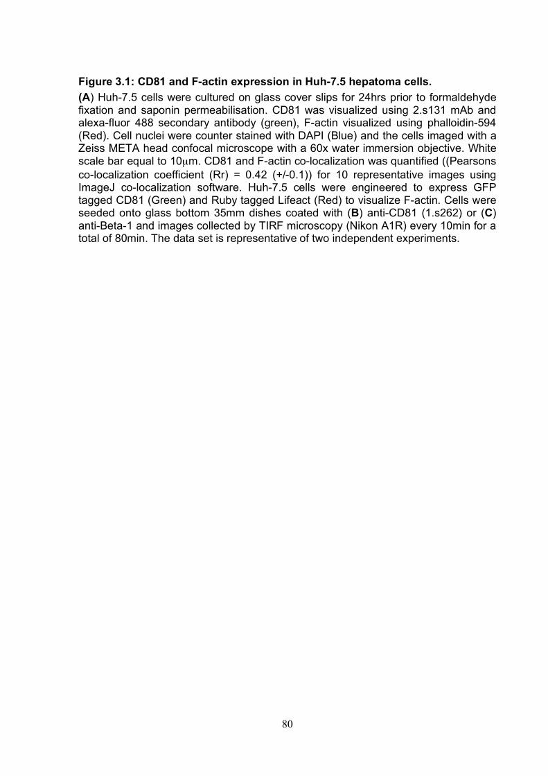
80
Figure 3.1: CD81 and F-actin expression in Huh-7.5 hepatoma cells. (A) Huh-7.5 cells were cultured on glass cover slips for 24hrs prior to formaldehyde fixation and saponin permeabilisation. CD81 was visualized using 2.s131 mAb and alexa-fluor 488 secondary antibody (green), F-actin visualized using phalloidin-594 (Red). Cell nuclei were counter stained with DAPI (Blue) and the cells imaged with a Zeiss META head confocal microscope with a 60x water immersion objective. White scale bar equal to 10µm. CD81 and F-actin co-localization was quantified ((Pearsons co-localization coefficient (Rr) = 0.42 (+/-0.1)) for 10 representative images using ImageJ co-localization software. Huh-7.5 cells were engineered to express GFP tagged CD81 (Green) and Ruby tagged Lifeact (Red) to visualize F-actin. Cells were seeded onto glass bottom 35mm dishes coated with (B) anti-CD81 (1.s262) or (C) anti-Beta-1 and images collected by TIRF microscopy (Nikon A1R) every 10min for a total of 80min. The data set is representative of two independent experiments.
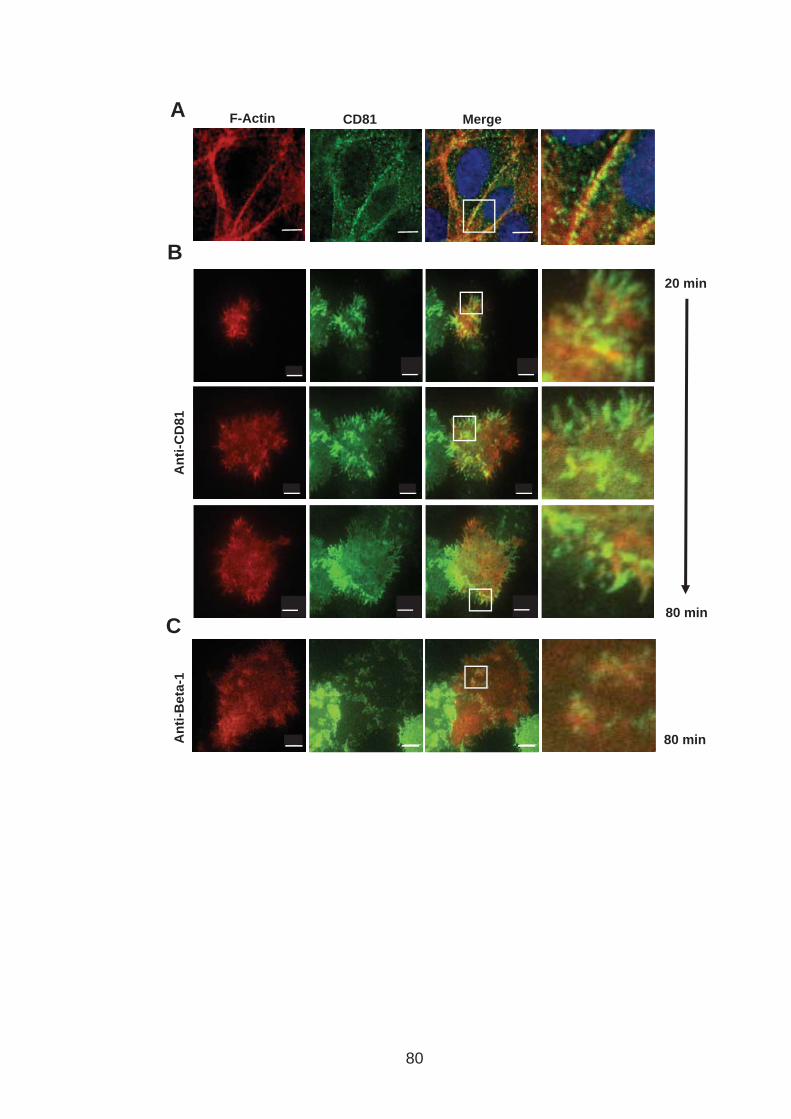
A
20 min
80 min
An
ti-C
D81
An
ti-B
eta
-1
B
80 min
C
F-Actin CD81 Merge
80
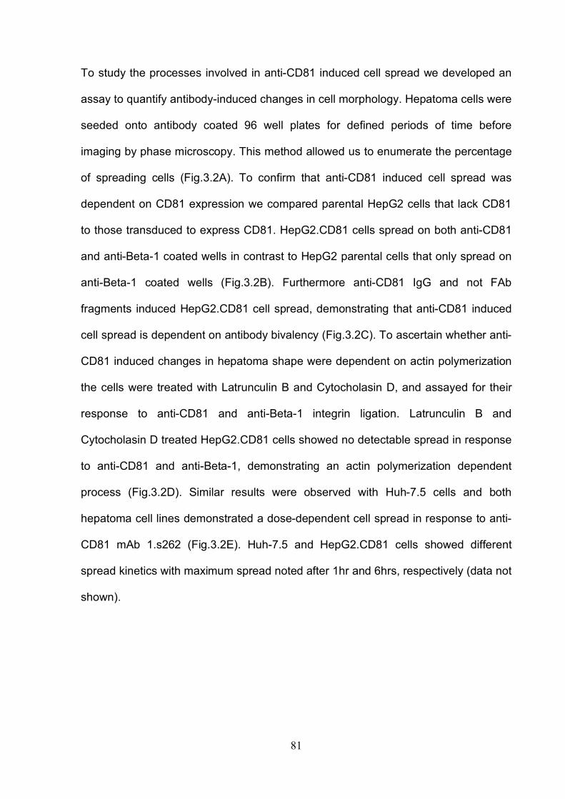
81
To study the processes involved in anti-CD81 induced cell spread we developed an
assay to quantify antibody-induced changes in cell morphology. Hepatoma cells were
seeded onto antibody coated 96 well plates for defined periods of time before
imaging by phase microscopy. This method allowed us to enumerate the percentage
of spreading cells (Fig.3.2A). To confirm that anti-CD81 induced cell spread was
dependent on CD81 expression we compared parental HepG2 cells that lack CD81
to those transduced to express CD81. HepG2.CD81 cells spread on both anti-CD81
and anti-Beta-1 coated wells in contrast to HepG2 parental cells that only spread on
anti-Beta-1 coated wells (Fig.3.2B). Furthermore anti-CD81 IgG and not FAb
fragments induced HepG2.CD81 cell spread, demonstrating that anti-CD81 induced
cell spread is dependent on antibody bivalency (Fig.3.2C). To ascertain whether anti-
CD81 induced changes in hepatoma shape were dependent on actin polymerization
the cells were treated with Latrunculin B and Cytocholasin D, and assayed for their
response to anti-CD81 and anti-Beta-1 integrin ligation. Latrunculin B and
Cytocholasin D treated HepG2.CD81 cells showed no detectable spread in response
to anti-CD81 and anti-Beta-1, demonstrating an actin polymerization dependent
process (Fig.3.2D). Similar results were observed with Huh-7.5 cells and both
hepatoma cell lines demonstrated a dose-dependent cell spread in response to anti-
CD81 mAb 1.s262 (Fig.3.2E). Huh-7.5 and HepG2.CD81 cells showed different
spread kinetics with maximum spread noted after 1hr and 6hrs, respectively (data not
shown).
82
Figure 3.2: CD81 engagement promotes actin-polymerization dependent hepatoma cell spread. (A) Cartoon of antibody induced cell spread assay. mAbs are immobilized onto ELISA plates (5µg/ml, 4°C overnight incubation). Wells blocked with PBS/1%BSA for 30min at room temperature. Cells were seeded at 3x10⁴ cells/well and incubated at 37°C for a defined period of time and imaged by phase microscopy. (B) Phase images of HepG2 and HepG2.CD81 spread on anti-CD81 1.s262, anti-Beta-1 integrin and irrelevant IgG control coated plates at 6hrs post seeding. (C) Phase images of HepG2.CD81 spread on anti-CD81 2.s66 IgG and 2.s66 Fab and irrelevant IgG control coated plates at 6hrs post seeding (D) Phase images of HepG2.CD81 spread on anti-CD81 1.s262 and anti-Beta-1 integrin coated plates after pre-treating cells with Cytocholasin D, Latrunculin B or DMSO control. (E) Dose dependent spread of HepG2.CD81 (6hrs post seeding) and Huh-7.5 cells (1hr post seeding) in response to CD81 ligation with mAb1.s262 at 0.1, 0.3, 1, 3 and 10µg/ml. Results are representative of three independent experiments.
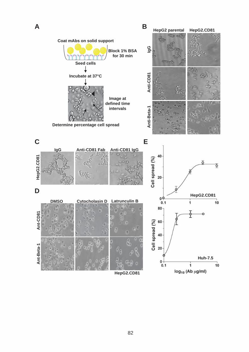
HepG2 parental HepG2.CD81
HepG2.CD81
Anti-CD81 Fab
Hep
G2.C
D81
IgG Anti-CD81 IgG
A B
C
D
E
Block 1% BSA
for 30 min
Determine percentage cell spread
Coat mAbs on solid support
Seed cells
Incubate at 37°C
Image at
defined time
intervals
IgG
A
nti
-CD
81
An
ti-B
eta
-1
Cytocholasin D DMSO
An
t-C
D81
An
t-B
eta
-1
Latrunculin B
HepG2.CD81
Huh-7.5
82
83
Our laboratory developed a panel of anti-CD81 mAbs that have been mapped to a
number of EC2 epitopes. The antibodies were screened for their reactivity with
mutant CD81 EC2 proteins described by Drummer et al., 2002 & 2005 (Screen
carried out by Ke Hu), enabling the mAbs to be classified into 6 epitope groups
(Table 3.1). To evaluate the epitope specificity of antibody induced cell spread a
selection of antibodies were screened for their ability induce HepG2.CD81 cell
spread (Fig.3.3). All antibodies induced between 30% and 55% of HepG2.CD81 cells
to spread. The high frequency of cell spread induced by anti-CD81 mAbs from
different epitope groups suggest that specific EC2 epitope binding is not required to
induce cell spread.

84
Table 3.1: CD81 antibody epitope grouping.
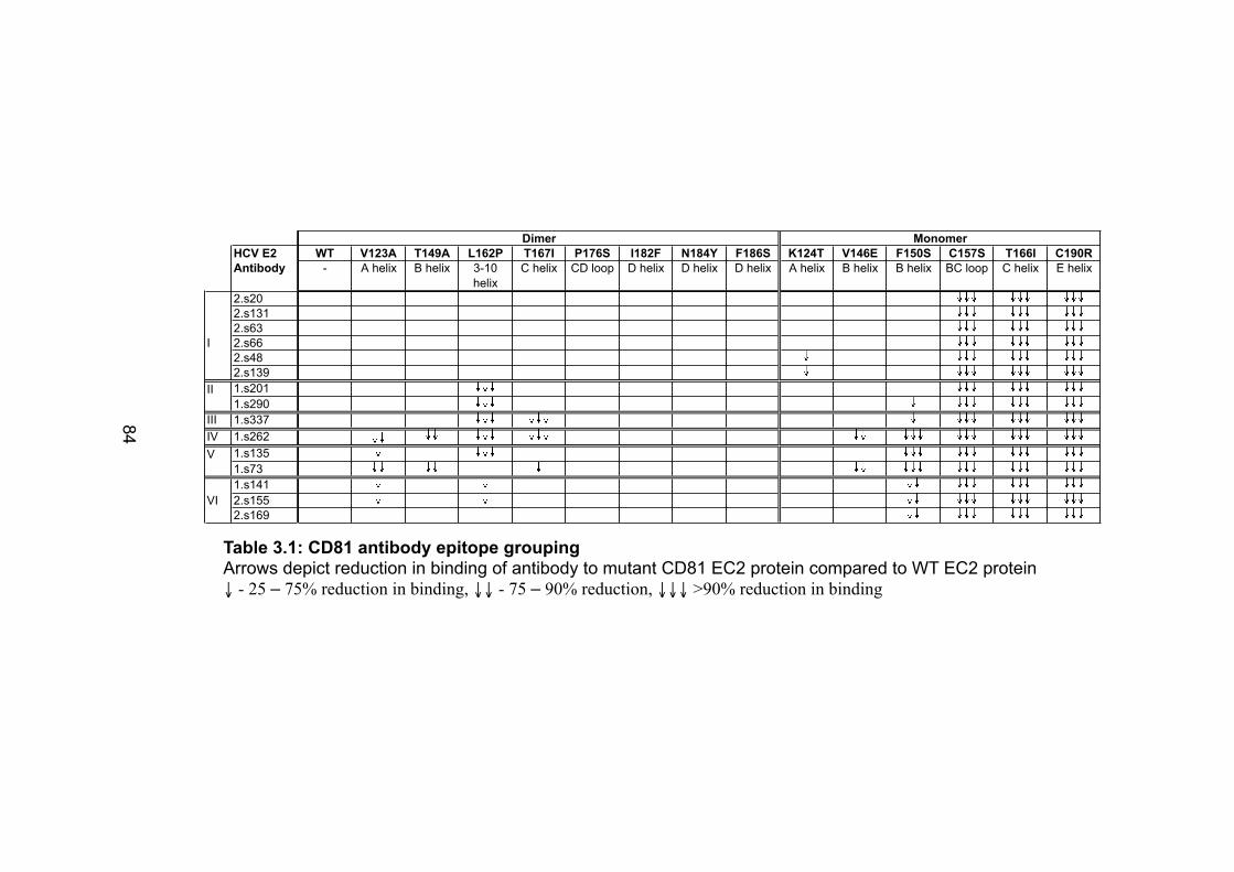
HCV E2 WT V123A T149A L162P T167I P176S I182F N184Y F186S K124T V146E F150S C157S T166I C190R
Antibody - A helix B helix 3-10
helix
C helix CD loop D helix D helix D helix A helix B helix B helix BC loop C helix E helix
2.s20
2.s131
2.s63
I 2.s66
2.s48
2.s139
1.s201
1.s290
III 1.s337
IV 1.s262
1.s135
1.s73
1.s141
VI 2.s155
2.s169
Dimer Monomer
II
V
Table 3.1: CD81 antibody epitope grouping Arrows depict reduction in binding of antibody to mutant CD81 EC2 protein compared to WT EC2 protein ↓ - 25 – 75% reduction in binding, ↓↓ - 75 – 90% reduction, ↓↓↓ >90% reduction in binding
84
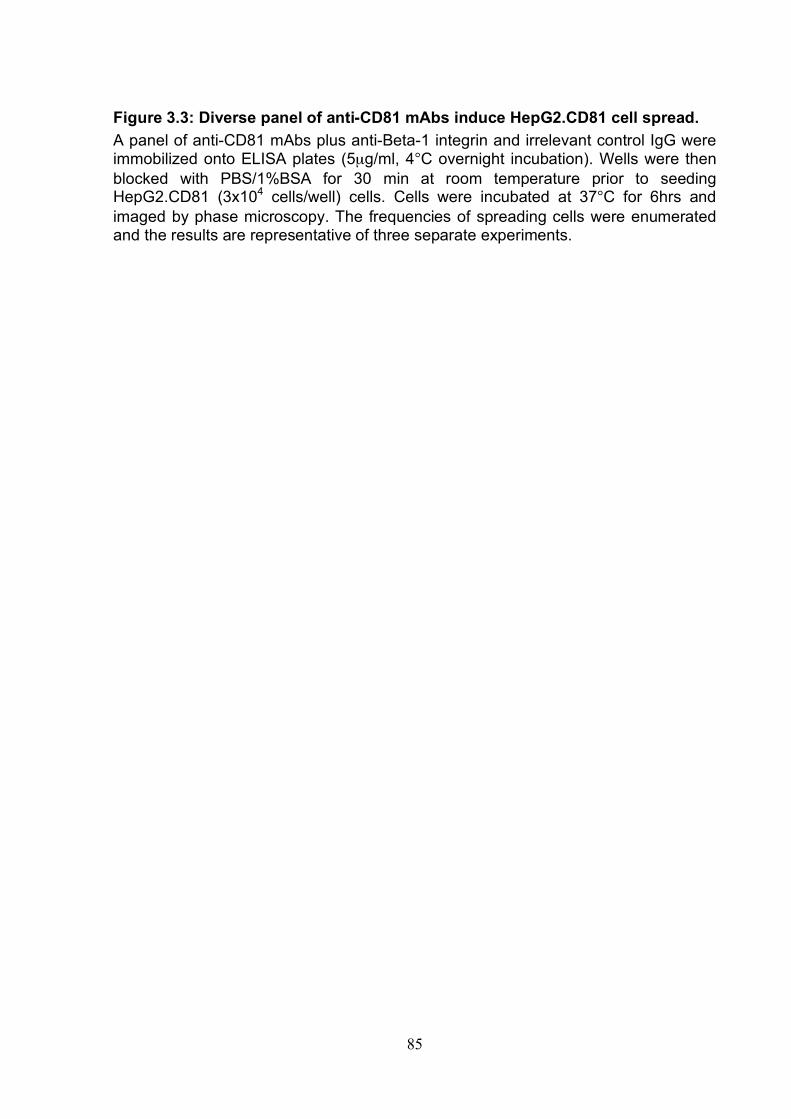
85
Figure 3.3: Diverse panel of anti-CD81 mAbs induce HepG2.CD81 cell spread. A panel of anti-CD81 mAbs plus anti-Beta-1 integrin and irrelevant control IgG were immobilized onto ELISA plates (5µg/ml, 4°C overnight incubation). Wells were then blocked with PBS/1%BSA for 30 min at room temperature prior to seeding HepG2.CD81 (3x10⁴ cells/well) cells. Cells were incubated at 37°C for 6hrs and imaged by phase microscopy. The frequencies of spreading cells were enumerated and the results are representative of three separate experiments.
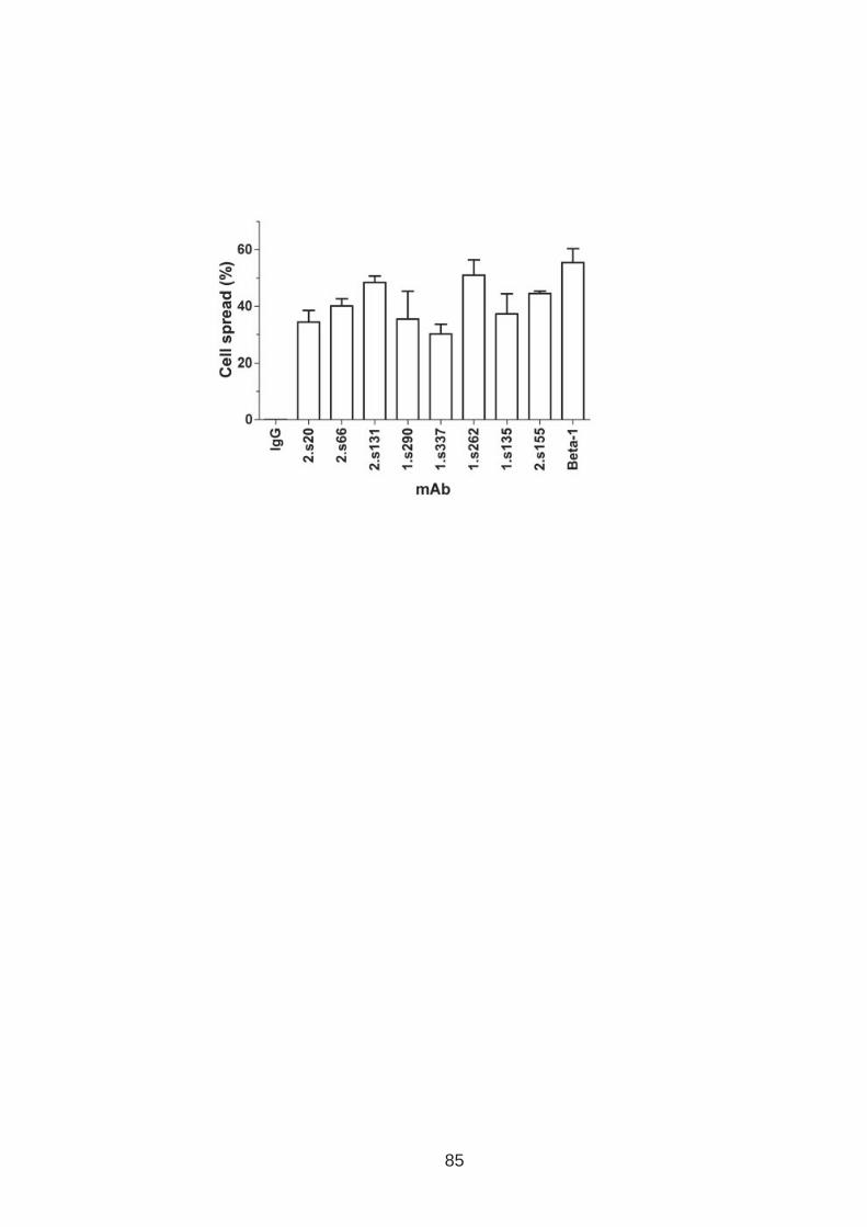
85

86
To explore whether antibody affinity for CD81 associates with cell spread the
percentage of cell spread was compared to binding affinity (Kd) (Fig.3.4A). Kd was
determined for a selected number of mAbs using a technique called surface plasmon
resonance. Briefly soluble CD81 EC2 was immobilized onto the surface of a
biosensor chip and anti-CD81 binding was determined by flowing over increasing
concentrations of antibody (Christopher Davis performed the surface plasmon
resonance assays and determined the Kd value for each antibody). No association
was observed between the Kd and the frequency of cell spread. Since the Kd values
were determined using a recombinant protein that may not be representative of
cellular expressed forms of CD81 we also compared cell spread to cell surface
bound antibody. Cell surface bound antibody was determined by flow cytometry
analysis at antibody saturating concentrations and expressed as median
fluorescence intensity (MFI) (Fig.3.4B) (flow cytometry analysis performed by Ke Hu).
Again no association was observed suggesting that antibody binding affinity is not
indicative of anti-CD81 induced cell spread.
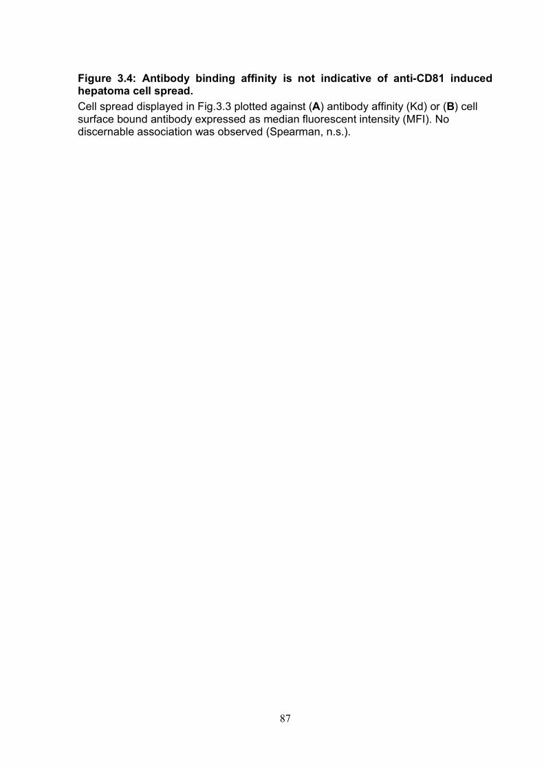
87
Figure 3.4: Antibody binding affinity is not indicative of anti-CD81 induced hepatoma cell spread. Cell spread displayed in Fig.3.3 plotted against (A) antibody affinity (Kd) or (B) cell surface bound antibody expressed as median fluorescent intensity (MFI). No discernable association was observed (Spearman, n.s.).
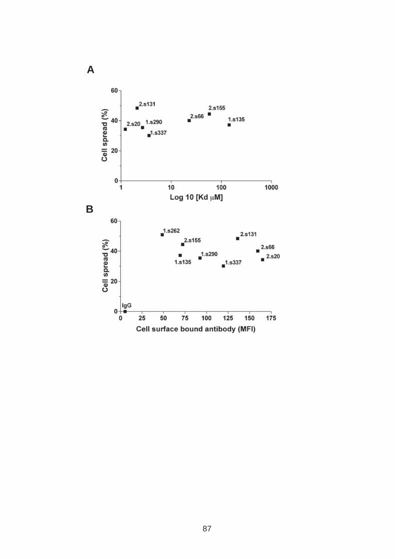
A
B
87
88
3.2 The CD81 C terminus links to the actin cytoskeleton through association with actin-associated proteins Ezrin Radoxin Moesin.
Recent reports have identified the C terminus of CD81 as an essential regulator of
CD81 induced effects (95, 411). To investigate whether anti-CD81 induced hepatoma
cell spread is dependent on either the N or C termini, two independent cell lines that
express low or negligible endogenous CD81 (HepG2 cells and Huh-7 Lunet cells)
were transduced to express wild type (WT) or mutant CD81 proteins lacking the N-
terminal (CD81ΔN) or C-terminal (CD81ΔC) regions. Cell surface CD81 expression was
determined by flow cytometry and the Median fluorescence intensity (MFI) recorded
(Fig.3.5A). HepG2 and Huh-7 Lunet cells expressing CD81ΔC demonstrated a
significantly reduced level of anti-CD81 induced cell spread compared to cells
expressing wild type CD81, whilst no difference was observed in anti-Beta-1 induced
cell spread. In contrast deletion of the N terminus had no detectable effect on anti-
CD81 or ant-Beta-1 induced cell spread compared to wild type control in either cell
line (Fig.3.5B).
89
Figure 3.5: A role for CD81 C terminus in actin polymerization dependent hepatoma cell spread. HepG2 and Huh-7 Lunet cells were transduced to express wild type CD81 (WT), CD81ΔN and CD81ΔC proteins. (A) Cell surface expression of CD81 was analyzed by flow cytometry (anti-CD81 2.s131mAb used). CD81 expression (Black line) IgG control (Solid grey). Median fluorescent intensity (MFI) was determined and is stated in the top right corner of each graph. (B) Parental and transduced cells were seeded onto anti-CD81 1.s262, anti-Beta-1 integrin, and irrelevant control IgG coated wells (All coated at 5µg/ml). Phase images were taken and the frequency of spreading cells determined. Results are representative of three independent experiments. Error bars indicate standard deviation from the mean. A significant difference between percentage cell spread determined for CD81ΔC compared to WT expressing cells in response to anti-CD81 mAb ligation was observed in both a HepG2 and Huh-7 Lunet cell background (p= 0.0020 and <0.0001 respectively, Two tailed unpaired T-test).
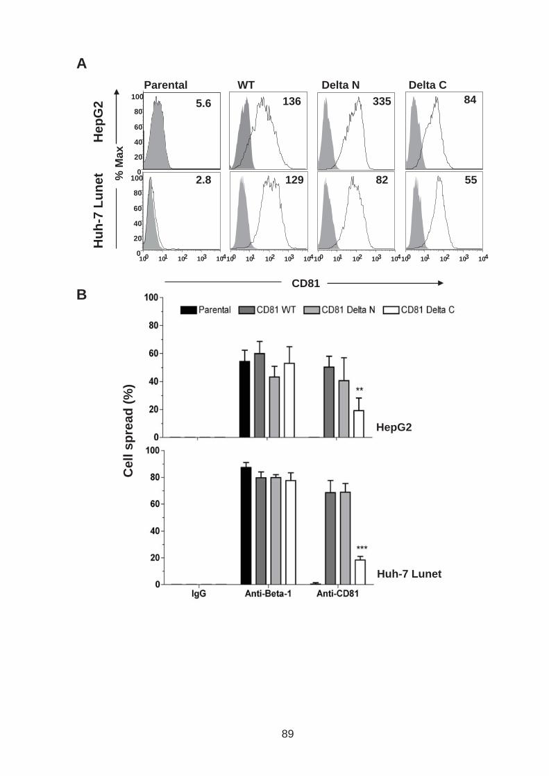
A
B
Cell s
pre
ad
(%
)
HepG2
Huh-7 Lunet
Hep
G2
Hu
h-7
Lu
net %
Max
CD81
WT Delta N Delta C
10 0 10 1 10 2 10 3 10 4
129 82 55
136 335 84
0 20 40 60 80
100
10 0 10 1 10 2 10 3 10 4 10 0 10 1 10 2 10 3 10 4 10 0 10 1 10 2 10 3 10 4
0 20 40 60 80
100 Parental
5.6
2.8
89

90
Ezrin Radoxin Moesin (ERM) proteins bind actin and recent studies have highlighted
a role for CD81-ERM association in a number of different cell types including Retinal
pigment epithelium (RPE), Human umbilical vein endothelial cells (HUVEC), HeLa
cells and lymphocytes (74, 365). Intramolecular associations between the N and C
termini of ERM proteins hold the proteins in an inactive (closed) conformation.
Phosphorylation of threonine residues in the actin binding domain(s) (ezrin Thr567,
radixin Thr564, moesin Thr558) promote an active (open) conformation that allows
the C terminus to interact with actin and the N terminal (FERM) domain to bind
membrane proteins (140, 308, 448). Recent reports have demonstrated an essential
role for the CD81 C terminus to associate with ERM in a number of cell lines (95,
411).
Given our earlier observation showing a role for the CD81 C terminus in hepatoma
cell spread we were interested to investigate the role of ERM proteins in CD81
dependent hepatoma spread. To determine whether CD81 engagement promotes
ERM phosphorylation, Huh-7.5 hepatoma cells were incubated with anti-CD81 mAb
1.s262 for defined periods of time before lysis and western blot analysis with
antibodies specific for phosphorylated ERM (P-ERM) and actin. P-ERM was detected
in the untreated cell lysates and showed no detectable increase following antibody
treatment (Fig.3.6A). Additional experiments confirmed P-ERM expression in Huh-7.5
cells that co-localized with CD81 at the cell surface (Pearson’s co-localization
coefficient, Rr = 0.662 +/- 0.047) (Fig.3.6B). Finally, to determine whether ERM
proteins are functionally relevant in anti-CD81 induced cell spread Huh-7.5 cells were
transduced to express a GFP tagged dominant negative Moesin, N-Moesin-GFP, and
control, Moesin-GFP (13) (Fig.3.6C). N-moesin-GFP was previously reported to alter
91
cell morphology by interfering with ERM dependent processes (13). Huh-7.5 cells
expressing the dominant negative N-Moesin-GFP showed significantly reduced anti-
CD81 induced spread compared to control Moesin-GFP expressing cells,
demonstrating a role for ERM proteins in anti-CD81 induced cell spread (Fig.3.6D).
Deletion of CD81 C terminus or use of a dominant negative ERM protein significantly
reduced but did not eradicate anti-CD81 induced cell spread, suggesting that CD81
may be linked to the actin cytoskeleton through a number of indirect interactions
consistent with CD81 being part of a TEM.
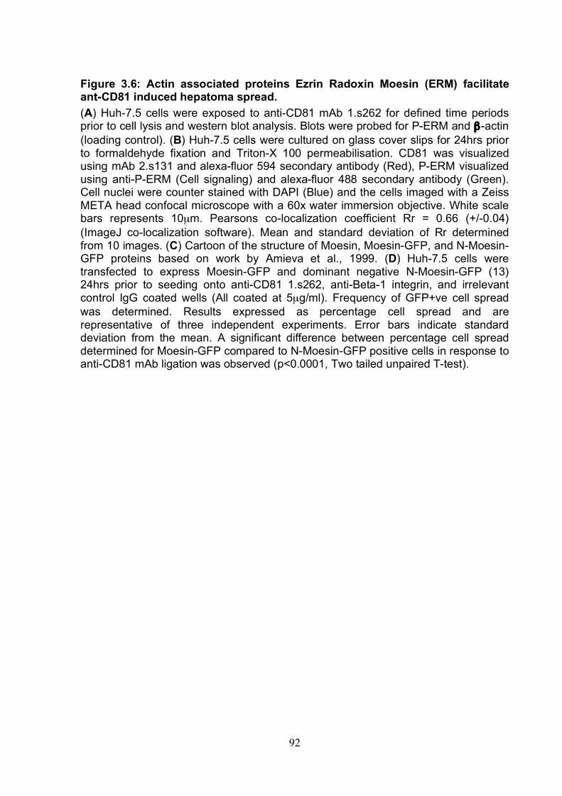
92
Figure 3.6: Actin associated proteins Ezrin Radoxin Moesin (ERM) facilitate ant-CD81 induced hepatoma spread. (A) Huh-7.5 cells were exposed to anti-CD81 mAb 1.s262 for defined time periods prior to cell lysis and western blot analysis. Blots were probed for P-ERM and β-actin (loading control). (B) Huh-7.5 cells were cultured on glass cover slips for 24hrs prior to formaldehyde fixation and Triton-X 100 permeabilisation. CD81 was visualized using mAb 2.s131 and alexa-fluor 594 secondary antibody (Red), P-ERM visualized using anti-P-ERM (Cell signaling) and alexa-fluor 488 secondary antibody (Green). Cell nuclei were counter stained with DAPI (Blue) and the cells imaged with a Zeiss META head confocal microscope with a 60x water immersion objective. White scale bars represents 10µm. Pearsons co-localization coefficient Rr = 0.66 (+/-0.04) (ImageJ co-localization software). Mean and standard deviation of Rr determined from 10 images. (C) Cartoon of the structure of Moesin, Moesin-GFP, and N-Moesin-GFP proteins based on work by Amieva et al., 1999. (D) Huh-7.5 cells were transfected to express Moesin-GFP and dominant negative N-Moesin-GFP (13) 24hrs prior to seeding onto anti-CD81 1.s262, anti-Beta-1 integrin, and irrelevant control IgG coated wells (All coated at 5µg/ml). Frequency of GFP+ve cell spread was determined. Results expressed as percentage cell spread and are representative of three independent experiments. Error bars indicate standard deviation from the mean. A significant difference between percentage cell spread determined for Moesin-GFP compared to N-Moesin-GFP positive cells in response to anti-CD81 mAb ligation was observed (p<0.0001, Two tailed unpaired T-test).
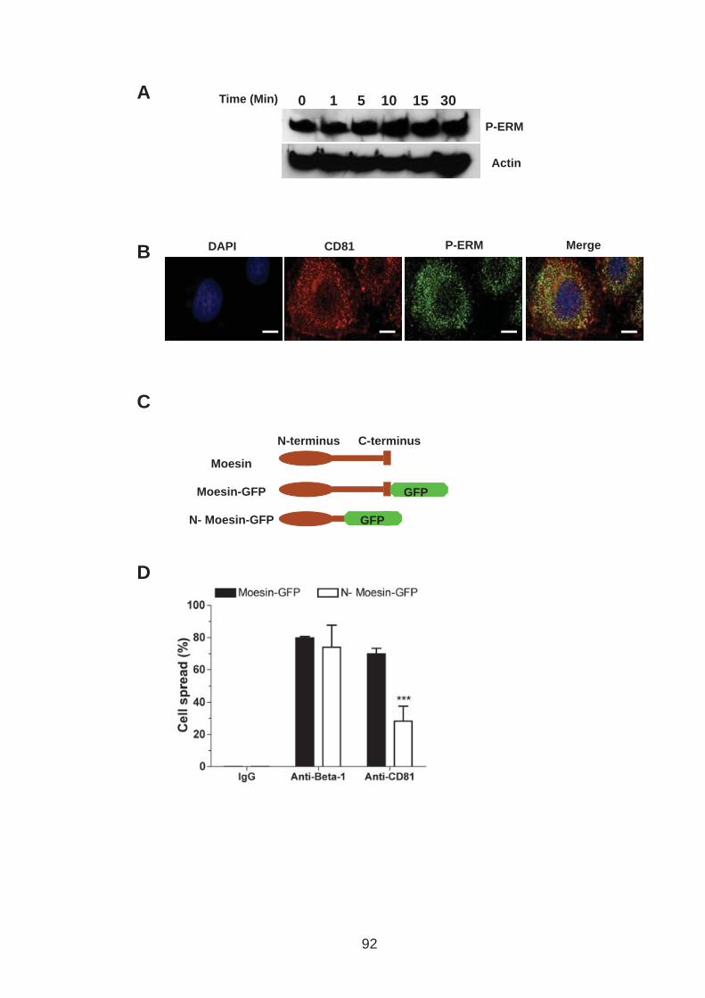
CD81 DAPI P-ERM Merge
P-ERM
Actin
0 1 5 10 15 30 Time (Min) A
B
D
C
Moesin
N-terminus C-terminus
Moesin-GFP
N- Moesin-GFP
GFP
GFP
92
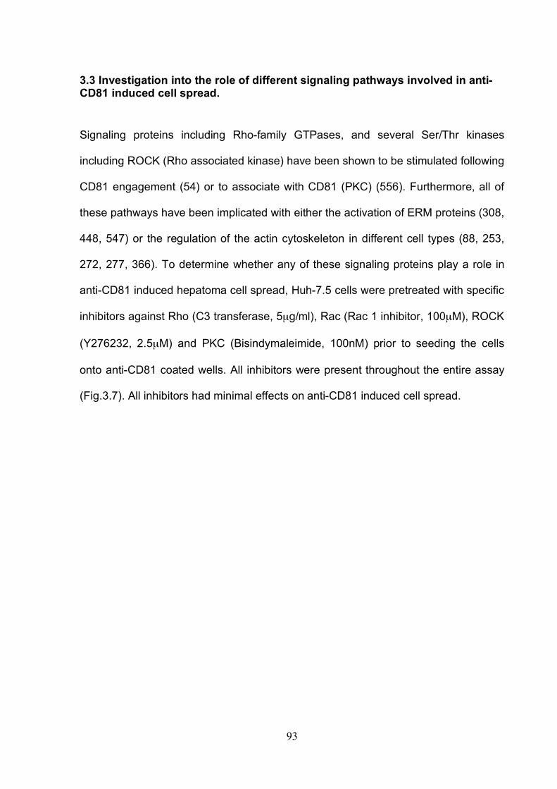
93
3.3 Investigation into the role of different signaling pathways involved in anti-CD81 induced cell spread.
Signaling proteins including Rho-family GTPases, and several Ser/Thr kinases
including ROCK (Rho associated kinase) have been shown to be stimulated following
CD81 engagement (54) or to associate with CD81 (PKC) (556). Furthermore, all of
these pathways have been implicated with either the activation of ERM proteins (308,
448, 547) or the regulation of the actin cytoskeleton in different cell types (88, 253,
272, 277, 366). To determine whether any of these signaling proteins play a role in
anti-CD81 induced hepatoma cell spread, Huh-7.5 cells were pretreated with specific
inhibitors against Rho (C3 transferase, 5µg/ml), Rac (Rac 1 inhibitor, 100µM), ROCK
(Y276232, 2.5µM) and PKC (Bisindymaleimide, 100nM) prior to seeding the cells
onto anti-CD81 coated wells. All inhibitors were present throughout the entire assay
(Fig.3.7). All inhibitors had minimal effects on anti-CD81 induced cell spread.
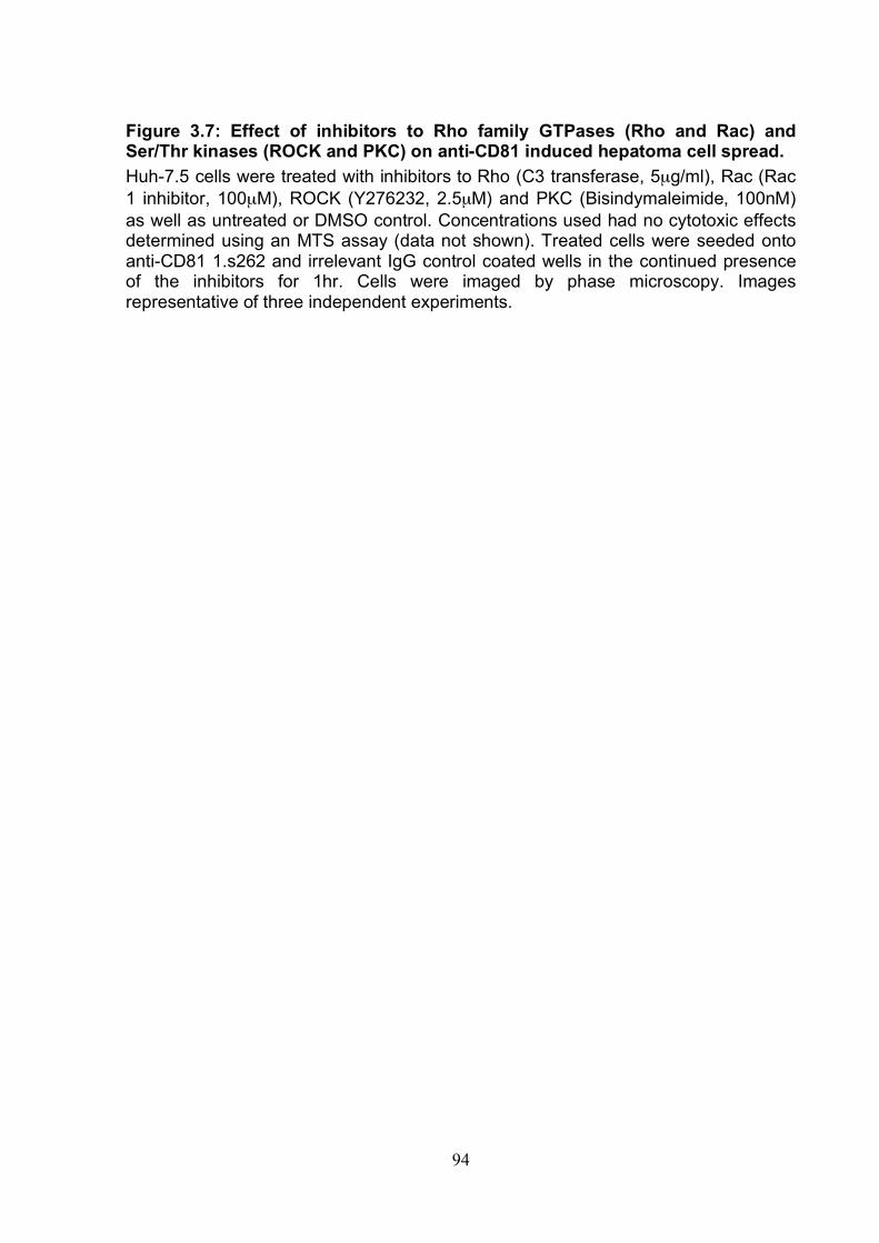
94
Figure 3.7: Effect of inhibitors to Rho family GTPases (Rho and Rac) and Ser/Thr kinases (ROCK and PKC) on anti-CD81 induced hepatoma cell spread. Huh-7.5 cells were treated with inhibitors to Rho (C3 transferase, 5µg/ml), Rac (Rac 1 inhibitor, 100µM), ROCK (Y276232, 2.5µM) and PKC (Bisindymaleimide, 100nM) as well as untreated or DMSO control. Concentrations used had no cytotoxic effects determined using an MTS assay (data not shown). Treated cells were seeded onto anti-CD81 1.s262 and irrelevant IgG control coated wells in the continued presence of the inhibitors for 1hr. Cells were imaged by phase microscopy. Images representative of three independent experiments.
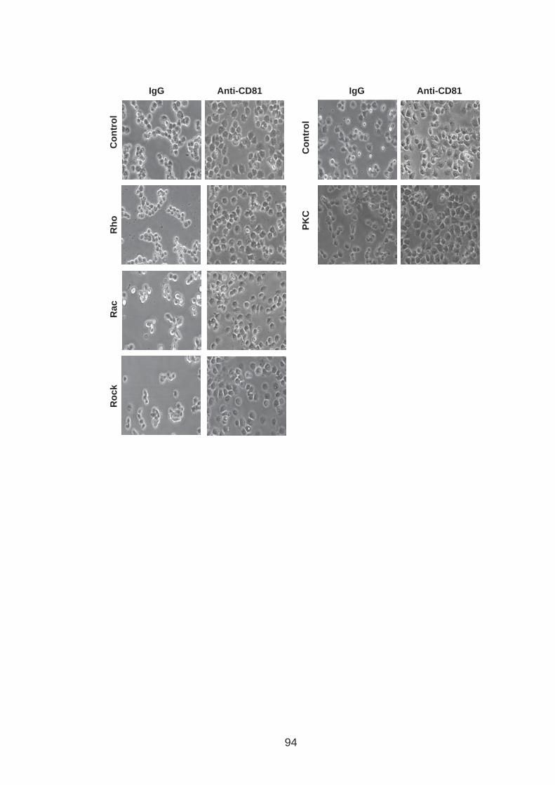
PK
C
Co
ntr
ol
IgG Anti-CD81
Rh
o
Rac
Ro
ck
Co
ntr
ol
IgG Anti-CD81
94
95
Ab engagement of CD81 has been reported to activate mitogen activated protein
kinase (MAPK) in a number of cell types resulting in different cellular responses,
including the activation of T cells (104) and increased proliferation of hepatoma cell
lines (54, 66, 498). MAPK signaling has been associated with the modulation of cell
spread, including the regulation of macrophage, fibroblast and endothelial cell spread
(115, 451, 491). AP-1 (Activator protein-1) is a transcription factor regulated by
MAPK (489, 514). Our laboratory has previously used an AP-1-luciferase reporter
assay to monitor MAPK signaling in Huh-7.5 cells. Huh-7.5 cells have a high basal
MAPK activity that is reduced following treatment with MAPK inhibitors U0126,
PD98059, and SB203580 (All AP-1 reporter work was carried out by Michelle
Farqhuar). To ascertain whether MAPK signaling is involved in anti-CD81 induced
hepatoma spread, Huh-7.5 cells were treated with the inhibitors and seeded onto
anti-CD81 and monitored for spread after 1hr. None of the inhibitors modulated anti-
CD81 induced cell spread implying that this process is MAPK independent (Fig.3.8).

96
Figure 3-8: MAP Kinase independent cell spread Huh-7.5 cells were treated with MAPK inhibitors (SB203580, PD98059, and U0126) at 100µM and DMSO control. Treated cells were then seeded onto anti-CD81 1.s262 and irrelevant IgG control coated wells in the continued presence of the inhibitors for 1hr. Concentrations used had no cytotoxic effects determined using an MTS assay (data not shown). Cells were imaged by phase microscopy and representative images presented.

Anti-CD81 IgG
Co
ntr
ol
U0
12
6
PD
98
05
9
SB
203580
96
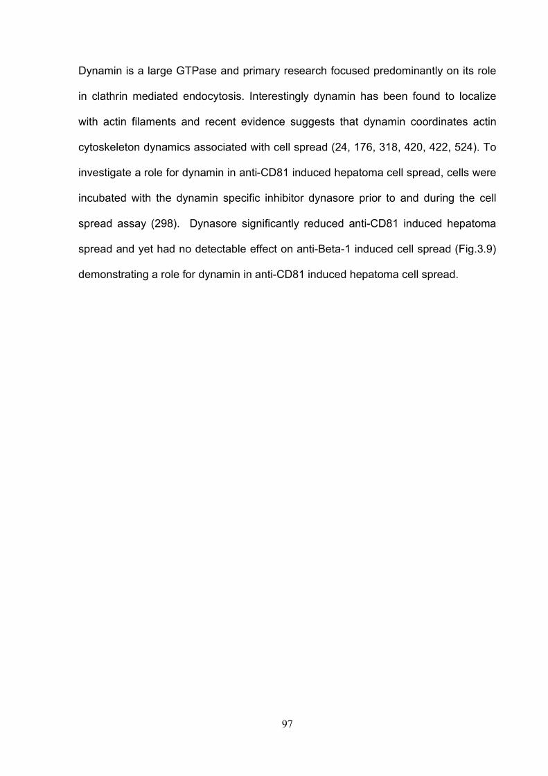
97
Dynamin is a large GTPase and primary research focused predominantly on its role
in clathrin mediated endocytosis. Interestingly dynamin has been found to localize
with actin filaments and recent evidence suggests that dynamin coordinates actin
cytoskeleton dynamics associated with cell spread (24, 176, 318, 420, 422, 524). To
investigate a role for dynamin in anti-CD81 induced hepatoma cell spread, cells were
incubated with the dynamin specific inhibitor dynasore prior to and during the cell
spread assay (298). Dynasore significantly reduced anti-CD81 induced hepatoma
spread and yet had no detectable effect on anti-Beta-1 induced cell spread (Fig.3.9)
demonstrating a role for dynamin in anti-CD81 induced hepatoma cell spread.
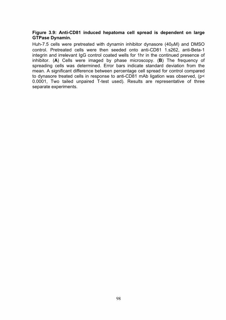
98
Figure 3.9: Anti-CD81 induced hepatoma cell spread is dependent on large GTPase Dynamin. Huh-7.5 cells were pretreated with dynamin inhibitor dynasore (40µM) and DMSO control. Pretreated cells were then seeded onto anti-CD81 1.s262, anti-Beta-1 integrin and irrelevant IgG control coated wells for 1hr in the continued presence of inhibitor. (A) Cells were imaged by phase microscopy. (B) The frequency of spreading cells was determined. Error bars indicate standard deviation from the mean. A significant difference between percentage cell spread for control compared to dynasore treated cells in response to anti-CD81 mAb ligation was observed, (p< 0.0001, Two tailed unpaired T-test used). Results are representative of three separate experiments.
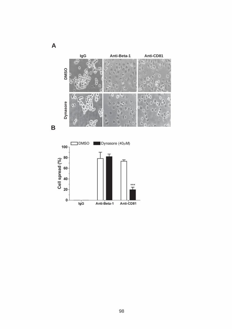
Dyn
aso
re
DM
SO
IgG Anti-CD81 Anti-Beta-1
A
B
98
99
3.4 HCV E2 glycoprotein does not induce hepatoma cell spread HCV E2 glycoproteins have been reported to bind to CD81 (384) and we were
interested to know whether sE2 would induce hepatoma spread. Given our earlier
observation that bivalent anti-CD81 was required to promote cell spread, we
captured sE2 (JFH-1) at 1 in 3 dilution onto 96 well plates via an antibody specific for
an epitope tag expressed at the C-terminus of E2. sE2 was bound by the specific
antibody and yet failed to promote hepatoma spread (Fig.3.10). Previous research
has reported modulation of intracellular signaling upon sE2-CD81 binding and HCV
infection (54, 66) making it important to determine the effects of HCV infection on
anti-CD81 induced cell spread.
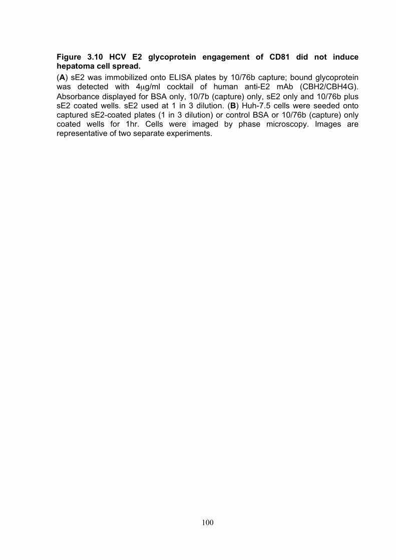
100
Figure 3.10 HCV E2 glycoprotein engagement of CD81 did not induce hepatoma cell spread. (A) sE2 was immobilized onto ELISA plates by 10/76b capture; bound glycoprotein was detected with 4µg/ml cocktail of human anti-E2 mAb (CBH2/CBH4G). Absorbance displayed for BSA only, 10/7b (capture) only, sE2 only and 10/76b plus sE2 coated wells. sE2 used at 1 in 3 dilution. (B) Huh-7.5 cells were seeded onto captured sE2-coated plates (1 in 3 dilution) or control BSA or 10/76b (capture) only coated wells for 1hr. Cells were imaged by phase microscopy. Images are representative of two separate experiments.
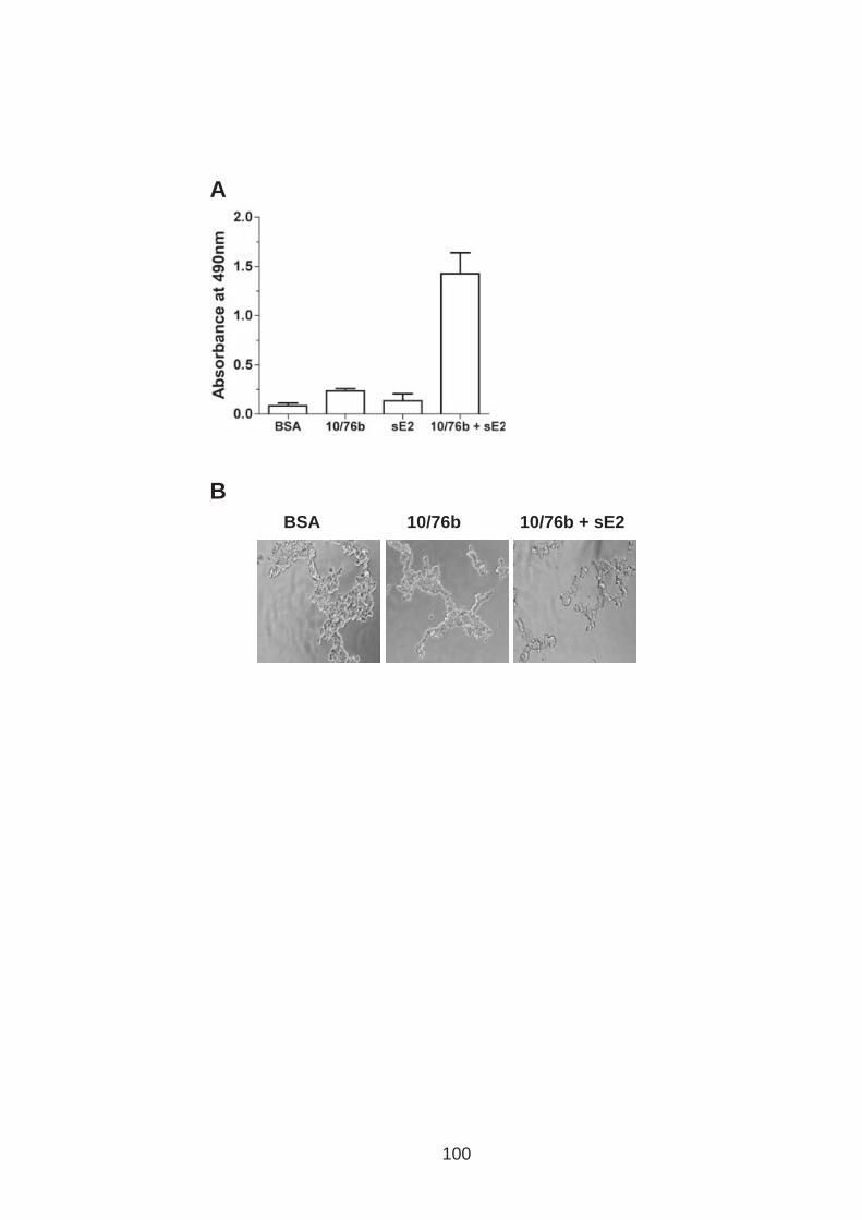
BSA 10/76b + sE2 10/76b
B
A
100
101
3.5 HCV infection perturbs anti-CD81 induced cell spread. Huh-7.5 cells were electroporated with genomic HCV RNA of different JFH-1
chimeric strains (J6/JFH genotype 1b and SA13/JFH genotype 5a) and 96hr post
electroporation mock and HCV infected cells were assessed for their ability to spread
in response to anti-CD81 ligation. The frequency of cells expressing NS5A was
determined by flow cytometry. HCV infected cells showed a reduced spread in
response to anti-CD81 stimulation (Fig.3.11A). It is important to note that the
frequency of infected cells varied between electroporations and this in turn reflected
the defect in anti-CD81 induced hepatoma cell spread observed, despite this HCV
infection repeatedly perturbed anti-CD81 induced cell spread.
To determine whether this phenomenon was common for other anti-tetraspanin and
associated proteins, Huh-7.5 cells supporting full-length HCVcc (J6/JFH) or control
mock infected Huh-7.5 cells were seeded onto plates coated with antibodies specific
for tetraspanins and tetraspanin associated proteins. Furthermore to ascertain
whether this perturbation was due to HCV replication the assay was performed with
Huh-7.5 cells supporting JFH-1 sub genomic replicon (SGR). J6/JFH infected cells
showed reduced spread compared to mock cells in response to anti-CD81 and anti-
CD9 induced spread. In contrast J6/JFH infected cells spread in a comparable
manner to mock cells in response to other anti-tetraspanin and associated protein
specific antibodies. Hepatoma cells supporting SGR RNA spread in a comparable
manner to mock infected cells in response to all antibodies tested (Fig.3.11B),
suggesting that virus particle genesis or the viral structural proteins perturb anti-
CD81 induced changes in hepatoma morphology.

102
Many viruses down regulate their receptors to prevent super infection (55, 348, 511).
To investigate whether virus infection modulated CD81 expression, uninfected and
infected cells were stained for CD81 and Beta-1 integrin expression (Fig.11C). No
differences were noted in cell surface CD81 or Beta-1 integrin expression,
demonstrating that reduced cell surface CD81 expression does not account for the
observed changes in anti-CD81 induced cell spread.
103
Figure 3.11: HCV infection reduces anti-CD81 induced hepatoma spread. (A) HCV strain J6/JFH and SA13/JFH RNA were delivered into Huh-7.5 cells by electroporation (ep) and 96hr later seeded onto anti-CD81mAb 1.s262 coated wells for 1hr. Cells were imaged by phase microscopy and the frequency of spreading cells determined. A significant difference between percentage cell spread determined for J6/JFH and SA13/JFH infected cells compared to mock cells in response to anti-CD81 mAb ligation was observed, (p = 0.0195 and 0.0053 respectively, Two tailed unpaired T-test). HCV NS5A expression was determined for each cell population by flow cytometry. (B) J6/JFH full length and JFH-1 sub-genomic (SGR) RNA were delivered into Huh-7.5 cells by ep and 96hrs later seeded onto anti-CD81 1.s262, anti-CD9, anti-CD51, anti-EWI-2, anti-Beta-1 integrin and irrelevant IgG control coated plates. Phase images were taken at 1hr post seeding and the frequency of spread cells determined. HCV NS5A protein expression was determined for each cell population by flow cytometry. A significant difference between percentage cell spread determined for J6/JFH compared to mock cells in response to anti-CD81 and anti-CD9 mAb ligation was observed, (p=0.0005 and 0.0046 respectively, Two tailed unpaired T-test). (C) Cell surface CD81 and Beta-1 integrin expression were determined for J6/JFH, SGR and mock infected Huh-7.5 cells using flow cytometry. Error bars indicate standard deviation from the mean. Results are representative of three indepent experiments.
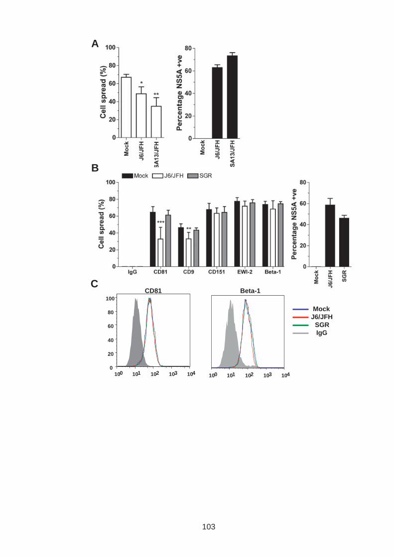
A
10 0 10 1 10 2 10 3 10 4 0
20
40
60
80
100
10 0 10 1 10 2 10 3 10 4
CD81 Beta-1
Mock
J6/JFH
SGR
IgG
B
C
103
104
To investigate whether HCV particles or virus binding to CD81 are required to perturb
anti-CD81 induced cell spread two mutant viruses were employed: J6/JFH del B
contained a mutation in domain III of NS5A that prevents a phosphorylation event
that is critical for particle assembly whilst permitting genome replication and protein
translation (473); JFH1W529A contains a mutation that prevents E2-CD81 binding
(515). Both viruses had no significant effect on anti-CD81 induced cell spread
suggesting that HCV particle genesis and/or CD81 engagement is essential to
modulate anti-CD81 induced cell spread (Fig.3.12A). The frequencies of NS5A
positive cells were determined by flow cytometry (Fig.3.12B). For the mutant viruses,
J6/JFH del B and JFH1W529A, the frequency of NS5A positive cells were low, 42 and
48 % respectively, compared to full length J6/JFH, 85%, suggesting a possible
alternative explanation for theminimal effect(s) on anti-CD81 induced hepatoma
spread.
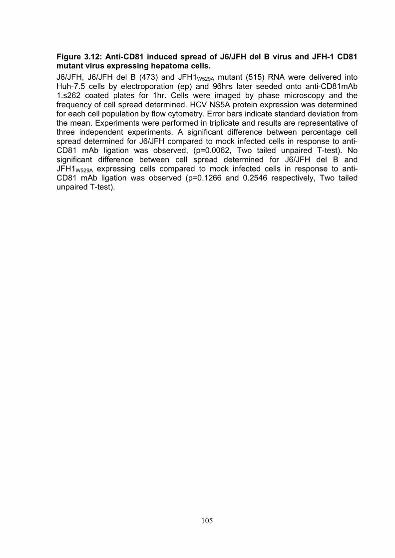
105
Figure 3.12: Anti-CD81 induced spread of J6/JFH del B virus and JFH-1 CD81 mutant virus expressing hepatoma cells. J6/JFH, J6/JFH del B (473) and JFH1W529A mutant (515) RNA were delivered into Huh-7.5 cells by electroporation (ep) and 96hrs later seeded onto anti-CD81mAb 1.s262 coated plates for 1hr. Cells were imaged by phase microscopy and the frequency of cell spread determined. HCV NS5A protein expression was determined for each cell population by flow cytometry. Error bars indicate standard deviation from the mean. Experiments were performed in triplicate and results are representative of three independent experiments. A significant difference between percentage cell spread determined for J6/JFH compared to mock infected cells in response to anti-CD81 mAb ligation was observed, (p=0.0062, Two tailed unpaired T-test). No significant difference between cell spread determined for J6/JFH del B and JFH1W529A expressing cells compared to mock infected cells in response to anti-CD81 mAb ligation was observed (p=0.1266 and 0.2546 respectively, Two tailed unpaired T-test).
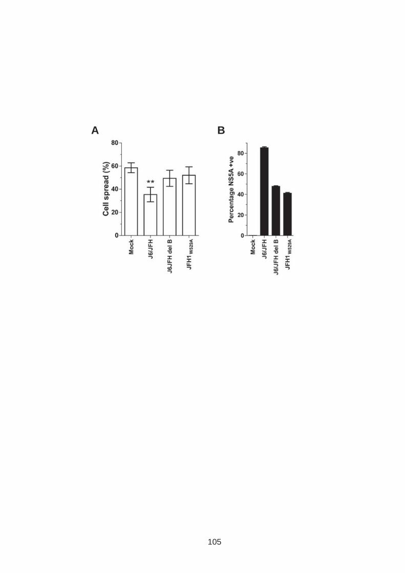
A B
105
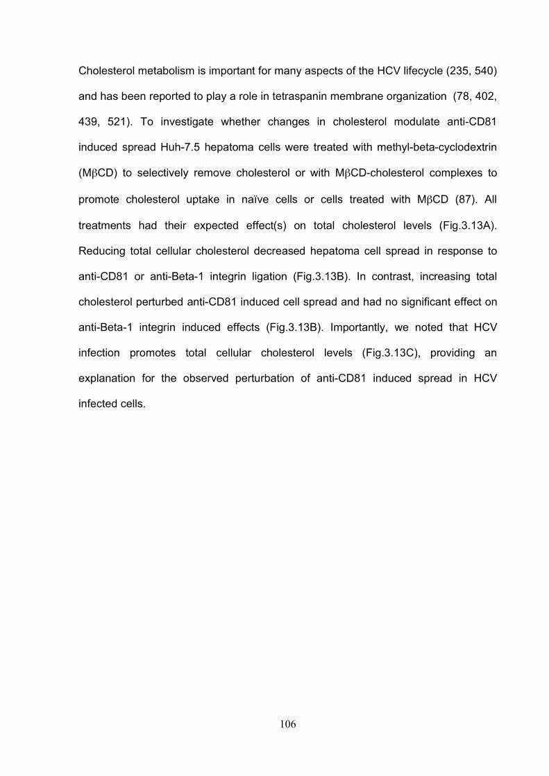
106
Cholesterol metabolism is important for many aspects of the HCV lifecycle (235, 540)
and has been reported to play a role in tetraspanin membrane organization (78, 402,
439, 521). To investigate whether changes in cholesterol modulate anti-CD81
induced spread Huh-7.5 hepatoma cells were treated with methyl-beta-cyclodextrin
(MβCD) to selectively remove cholesterol or with MβCD-cholesterol complexes to
promote cholesterol uptake in naïve cells or cells treated with MβCD (87). All
treatments had their expected effect(s) on total cholesterol levels (Fig.3.13A).
Reducing total cellular cholesterol decreased hepatoma cell spread in response to
anti-CD81 or anti-Beta-1 integrin ligation (Fig.3.13B). In contrast, increasing total
cholesterol perturbed anti-CD81 induced cell spread and had no significant effect on
anti-Beta-1 integrin induced effects (Fig.3.13B). Importantly, we noted that HCV
infection promotes total cellular cholesterol levels (Fig.3.13C), providing an
explanation for the observed perturbation of anti-CD81 induced spread in HCV
infected cells.
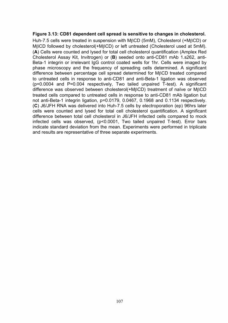
107
Figure 3.13: CD81 dependent cell spread is sensitive to changes in cholesterol. Huh-7.5 cells were treated in suspension with MβCD (5mM), Cholesterol (+MβCD) or MβCD followed by cholesterol(+MβCD) or left untreated (Cholesterol used at 5mM). (A) Cells were counted and lysed for total cell cholesterol quantification (Amplex Red Cholesterol Assay Kit, Invitrogen) or (B) seeded onto anti-CD81 mAb 1.s262, anti-Beta-1 integrin or irrelevant IgG control coated wells for 1hr. Cells were imaged by phase microscopy and the frequency of spreading cells determined. A significant difference between percentage cell spread determined for MβCD treated compared to untreated cells in response to anti-CD81 and anti-Beta-1 ligation was observed (p=0.0004 and P=0.004 respectively, Two tailed unpaired T-test). A significant difference was observed between cholesterol(+MβCD) treatment of naïve or MβCD treated cells compared to untreated cells in response to anti-CD81 mAb ligation but not anti-Beta-1 integrin ligation, p=0.0179, 0.0467, 0.1968 and 0.1134 respectively. (C) J6/JFH RNA was delivered into Huh-7.5 cells by electroporation (ep) 96hrs later cells were counted and lysed for total cell cholesterol quantification. A significant difference between total cell cholesterol in J6/JFH infected cells compared to mock infected cells was observed, (p<0.0001, Two tailed unpaired T-test). Error bars indicate standard deviation from the mean. Experiments were performed in triplicate and results are representative of three separate experiments.
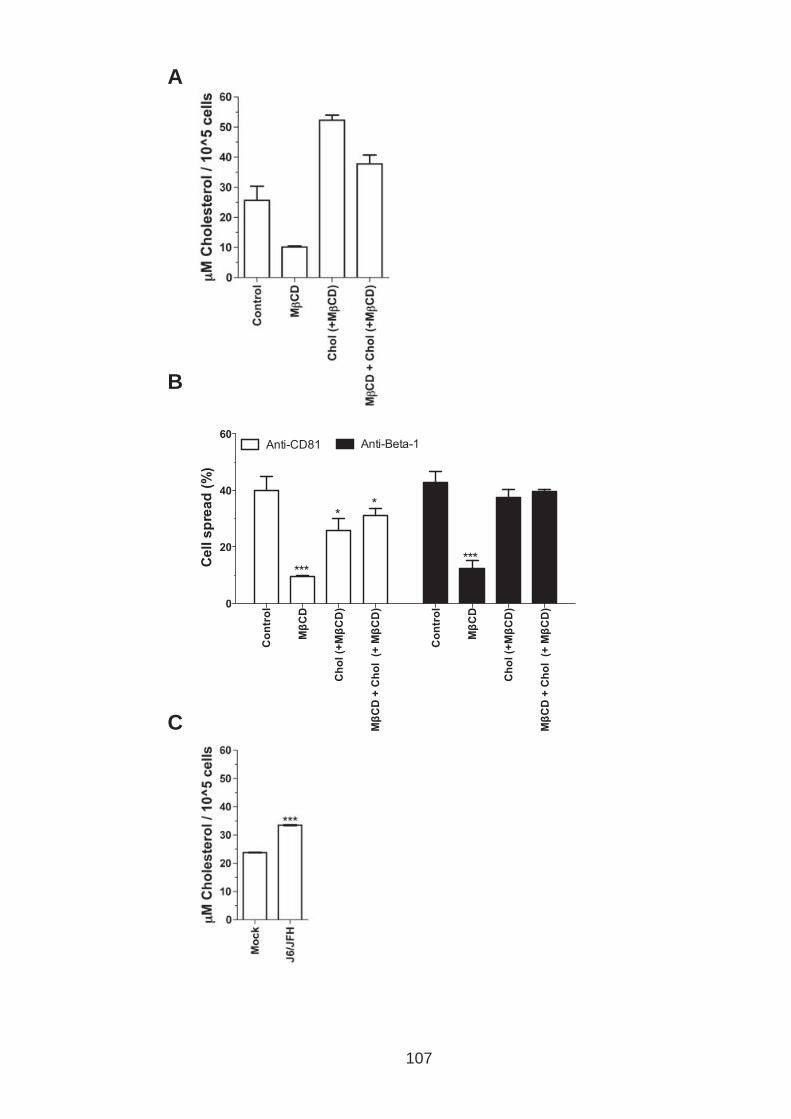
A
B
C
107

108
3.6 Discussion
The function of tetraspanin CD81 in hepatoma cell biology is largely unknown. In this
study we used a diverse panel of novel CD81 EC2 binding antibodies to investigate
the effect(s) of ligation on hepatoma cell morphology. Antibodies can be useful tools
for investigating tetraspanin function(s), they are reported to disrupt partner protein
interactions or promote intracellular signaling through protein aggregation at the cell
surface (177, 352, 427, 449). Importantly anti-CD81 mAbs may also mimic ligation of
CD81 by HCV glycoproteins. Ligation of CD81 on immune cells by anti-CD81 mAb or
HCV sE2 glycoprotein activates numerous signaling cascades and induces actin
dependent morphological changes (95, 104, 253, 342, 486, 498). We observed
hepatoma spread upon engagement of a diverse panel of immobilized anti-CD81
mAbs but not recombinant protein sE2. Importantly HCV infection was found to
perturb this response. Our observations point to an indirect mechanism of inhibition
through perturbation of intracellular events possibly the result of increased total
cellular cholesterol levels. Tetraspanins are often associated with cancer progression
and hence these observations may have important consequences in HCV associated
liver disease.
To investigate the effect(s) of CD81 ligation on hepatoma cells, the cells were
seeded onto anti-CD81 mAb immobilized onto ELISA plates and cultured for defined
periods of time before imaging by phase microscopy (Fig.3.2A). Huh-7.5 and
HepG2.CD81 cells were observed to spread in a dose dependent manner in
response to IgG whilst they did not spread after binding corresponding Fab
fragments, indicating this process is dependent on antibody bivalency and cross-
109
linking CD81 is most likely required (Fig.3.2C). Furthermore actin polymerization
inhibitors Latrunculin B (Lat B) and Cytochalasin D (Cyt D) successfully blocked cell
spread revealing the process to be dependent on actin polymerization (Fig.3.2.D).
The actin polymerization inhibitors have different modes of action; Lat B binds actin
monomers preventing polymerization (100, 336) whilst Cyt D binds F-actin high
affinity binding sites inhibiting filament elongation (141).
CD81 and F-Actin were observed to co-localize at hepatoma cell contacts and further
analysis using TIRF microscopy demonstrated that during anti-CD81 and anti-Beta-1
integrin induced hepatoma cell spread, actin and CD81 co-localized in specific
regions on the lower surface of the cell (Fig.3.1). These observations demonstrate
that CD81-F-actin co-localization is not dependent on CD81 engagement as
previously observed on B cells (95). An explanation for this could be due to
differences in cell type, resting lymphocytes used in the previous studies are cultured
in suspension as opposed to hepatoma cells that adhere to cell culture plastic and
grow in monolayers. They are also quiescent, held in the G0 stage of the cellular
lifecycle, differing again from hepatoma cells that are continually dividing (151).
The regions of high CD81 expression observed on the cell surface of Huh-7.5 cells
by TIRF microscopy may represent tetraspanin-enriched microdomains (TEMS).
Tetraspanins are reported to continually interchange between confined (TEM) and
randomly diffused states (132), whether tetraspanins interact with partner proteins
and function outside of TEMS is relatively unknown and currently under debate (530).
Interestingly CD81 is reported to function independently of TEMS for HCV entry

110
(402). New live cell imaging techniques, for example single particle tracking are only
now becoming available to properly address these questions (132, 530).
A high frequency of cell spread in response to CD81 ligation by antibodies targeting
different epitopes of CD81 (Fig.3.3), suggesting that antibody-induced cell spread is
independent of EC2 epitope. Cell spread did not correlate with antibody affinity (Kd)
determined using recombinant CD81 EC2 (Fig.3.4A). Since recombinant EC2 may
not be truly representative of in vitro CD81, cell spread was also compared to cell
surface bound antibody (MFI), again no correlation was observed (Fig.3.4B). These
results suggest that cell spread is independent of antibody binding affinity. To
determine whether ligation of other cell surface proteins could induce hepatoma cell
spread we compared cell spread to antibodies specific for different cell surface
proteins (data not shown). All tetraspanin and integrin antibodies tested induced cell
spread to varying degrees, whereas antibodies to HCV co-receptor claudin-1 induced
cell spread to a much lower degree than anti-CD81. Antibodies targeting a further
HCV co-receptor, SR-BI, did not induce cell spread. As well as differences in the
frequency of cell spread observed, the morphology of the spreading cells varied
depending on the cell surface protein engaged.
CD81 induced hepatoma cell spread was repeatedly perturbed by HCV infection. It is
not uncommon for viruses to down regulate cell surface receptors to prevent super-
infection (55, 348, 511), we therefore compared CD81 expression of naïve and
infected Huh-7.5 cells (Fig.3.11C). No change in CD81 expression was observed
eliminating this as a possible mechanism. Infection only perturbed anti-CD9 induced
cell spread and had no effect on antibodies to other other tetraspanin (CD63, CD82)
111
or associated proteins (EWI-2, Beta-1 integrin) (Fig.3.11B). CD81 and CD9 have
been reported to work in conjunction with one another, in vitro studies have shown a
strong phenotype only upon silencing both CD81 and CD9 (72, 262, 466) and single
tetraspanin knock out mice are reported to have a mild phenotype compared to
double knock outs (466, 467). The observed inhibition of both CD81 and CD9
induced spread suggests these tetraspanins may be working together in this process.
sE2 ligation of CD81 did not induce hepatoma cell spread (Fig.310), this differs from
previous observations where antibody and sE2 engagement of CD81 have induced
similar effects (54, 95, 104, 105, 253). Yoon et al., 2009 reported that HCV infection
had no effect on NK cell function suggesting that earlier results with sE2 may be
incorrect, demonstrating that recombinant E2 is not always representative of E2 on
viral particles most likely due to configuration and concentration differences.
To ascertain whether HCV infection inhibits anti-CD81 induced spread directly
through HCV envelope binding to CD81 or indirectly through HCV replication and/or
protein expression, experiments were performed using Huh-7.5 cells expressing
mutant viruses and a JFH-1 sub genomic replicon (Fig.3.11&12). Mutant virus
J6/JFH del B has a mutation in domain III of NS5A that prevents assembly of viral
particles (473) and JFH1W529A mutant virus was previously demonstrated to be
deficient in CD81 binding (515). No reduction in anti-CD81 induced spread of cells
expressing these mutant viruses or JFH-1 sub-genomic replicon was observed
compared to controls. These results suggest firstly that HCV genome replication is
not enough to drive this perturbation and secondly that engagement of CD81 by HCV
is essential in this process. In all assays cells were cultured for 96hrs post delivery of
viral RNA to ensure a high level of infection, unfortunately due to variations in

112
electroporation efficiency and the fact that sub-genomic RNA and mutant viruses are
unable to transmit infection (See chapter 5) we were unable to achieve a comparable
level of infection compared to the full length viruses. Less than 50% of J6/JFH del B,
JFH1W529A and sub-genomic replicon expressing cells were NS5A positive compared
to greater than 60% of the full-length virus infected cells. Therefore the lower NS5A
expression levels may be an alternative explanation for these results, thus it is still
un-resolved as to whether CD81 engagement is needed for this phenotype.
The C terminus of CD81 has previously been reported to be important for eliciting
anti-CD81 induced affects on immune cells (95, 411). Hepatoma cells expressing a
CD81 protein lacking its C terminus (CD81ΔC) had a significantly reduced spread
compared to cells expressing CD81 WT and CD81ΔN protein upon CD81 ligation,
implying that the C terminus is also important for CD81 function in hepatoma cells
(Fig.3.5). A PDZ domain located on the C terminus of CD81 is reported to bind actin-
associated proteins Ezrin, Radoxin, Moesin (ERM) (411). Threonine phosphorylation
of all ERM proteins (ezrin Thr567, radixin Thr564, and moesin Thr558) induces a
conformational change leading to activation (140, 308, 448). We failed to observe
any change in P-ERM levels following antibody ligation of CD81 on hepatoma cells
(Fig.3.6A) as previously reported for NK and B cells respectively (95, 253). A high
basal level of P-ERM was observed in our hepatoma cells that may have masked
any small changes in P-ERM levels. Despite this CD81 co-localized with P-ERM at
the surface of hepatoma cells, and cells expressing a dominant negative N-moesin
protein had significantly reduced anti-CD81 induced cell spread (Fig.3.6). These
observations suggest that ERM proteins do indeed play a role in anti-CD81 induced
hepatoma cell spread and we hypothesize this is through interactions with the C
113
terminus of CD81. The CD81ΔC and N-moesin-GFP expressing cells only exhibited
partial inhibition of anti-CD81 induced cell spread suggesting that this is not the only
means by which CD81 can link to the actin cytoskeleton consistent with CD81
functioning as part of a TEM in this process.
Of note HCV infection was not inhibited in cells expressing the dominant negative N-
moesin-GFP protein compared to control (data not shown) suggesting firstly that
HCV infection is independent of ERM proteins and secondly that the mechanism
underlying anti-CD81 induced spread is independent of the mechanism involved in
HCV entry. Coffey et al., 2009 observed a Syk dependent Tyrosine phosphorylation
of Ezrin upon CD81 ligation in B cells. Syk is a non-receptor protein kinase and was
originally thought to only be present in heamatopoetic cells but has since been
identified in a number of non-haematopoietic cell lineages including endothelium,
epithelium and importantly hepatocytes (487, 527). Interestingly HCV NS5A N-
terminus interacts directly with Syk and NS5A expression has been reported to inhibit
Syk kinase activity (220), this could be a possible explanation for HCV perturbation of
anti-CD81 induced cell spread. Further work is necessary to determine whether Syk
dependent tyrosine phosphorylation of Ezrin upon CD81 ligation occurs in
hepatocytes.
CD81 ligation has been associated with regulating a number of signaling cascades.
MAPK signaling for example was previously demonstrated to increase upon CD81
ligation on hepatoma cells (54, 66, 558). Work in our laboratory using an AP-1
reporter system demonstrated that MAPK inhibitors (U0126, PD98059, and
SB203580) inhibit MAPK signaling in Huh-7.5 cells (Michelle Farhquar, unpublished
114
data). However none of these inhibitors were found to have any observable effect on
hepatoma cell spread (Fig.3.8) suggesting CD81 induced hepatoma cell spread is
MAPK independent. A further kinase Protein Kinase C (PKC) is also associated with
CD81 (556), and inhibion of PKC again had no effect on anti-CD81 induced
hepatoma cell spread. Rho GTPases are primarily involved in modulation of actin
dynamics and have also been associated with CD81 function (54, 272, 366, 398). We
investigated the effect(s) of a number of Rho GTPase family inhibitors; again none of
the inhibitors had any observable effect(s) on cell spread (Fig.3.7). The inhibitors
used all have limitations in specificity that may explain their inability to inhibit spread.
(438, 494) (154, 438). Another Rho family GTPase member that is not tested in this
study is cdc42 again this GTPase has been implicated in modulating cell spread
(389). The involvement of Rho GTPases in cancer progression is fueling ongoing
research to identify more specific inhibitors (295), these may be useful for the future
work needed to dismiss the involvement of Rho GTPase family members in anti-
CD81 induced hepatoma cell spread.
Dynasore, an inhibitor of the large GTPase dynamin, specifically perturbed anti-CD81
induced cell spread and not anti-Beta-1 integrin induced cell spread (Fig.3.9). These
results suggest that dynamin is either directly or indirectly involved in CD81 function.
Further experiments are necessary to determine whether dynamin specifically inhibits
CD81 function or whether this perturbation is common to other tetraspanins. To date
no literature exists on a link between tetraspanins and dynamin although dynamin is
reported to associate with actin (176, 318, 419, 420). Dynamin has also been
reported to regulate the localization of Rac a member of the Rho family GTPases
(422). Although our data does not implicate Rac GTPase involvement in anti-CD81
115
induced hepatoma cell spread our analysis is not conclusive and reports have
previously implicated Rho GTPases in anti-CD81 induced effects (54). It is therefore
plausible that inhibition of dynamin may indirectly inhibit anti-CD81 induced cell
spread through inhibition of Rac. Furthermore, a direct association between HCV co-
receptor tight junction protein Occludin and dynamin has recently been reported
(287) and research carried out by our laboratory has demonstrated that Occludin can
be found associated with Claudin-1 and CD81 on the surface of hepatoma cells (188)
linking CD81 indirectly with dynamin.
Cholesterol is important for correct tetraspanin localization and function (78, 402,
439, 521). In fact levels of membrane cholesterol can alter the function of numerous
cell surface proteins effecting, cell signaling, adhesion and migration (390).
Modulation of cholesterol levels have previously been shown to reduce CD81-
Claudin-1 and CD81-CD81 association in hepatoma cells (188). Further to this a
number of studies have demonstrated that cholesterol depletion inhibits HCV entry
(188, 235, 402). To determine whether cholesterol levels modulate anti-CD81
induced hepatoma cell spread, Huh-7.5 cells were pre-treated with MβCD to deplete
cholesterol or with MβCD-cholesterol complexes to increase cholesterol uptake. Anti-
CD81 and anti-Beta-1 integrin induced hepatoma cell spread were reduced upon
depletion of cellular cholesterol. Furthermore a significant decrease in anti-CD81 but
not anti-Beta-1 integrin induced cell spread upon addition of cholesterol was
observed (Fig.3.13). These findings suggest that cholesterol levels are important for
CD81 function in hepatoma cells.
116
Numerous viruses rely on and modulate cholesterol metabolism for effective viral
production and replication and HCV is no exception (111). Hepatocytes acquire
cholesterol through two major processes, firstly through the mevalonate pathway that
synthesizes cholesterol and other lipids from acetyl-CoA (163) and secondly by
acquisition of cholesterol from low and high-density lipoproteins (LDL and HDL) in the
plasma through LDL receptor and Scavenger receptor BI (SR-BI) respectively (254,
406). A number of studies have reported that inhibitors of steps in the mevalonate
pathway can inhibit HCV replication demonstrating HCV reliance on lipid and
cholesterol metabolism (86, 217, 236). HCV infection in vivo is often associated with
reduced serum cholesterol levels (201, 344, 426). A number of mechanisms are
thought to be responsible for this for example HCV infection induces oxidative stress
through mitochondrial dysfunction resulting in increased levels of reactive oxygen
species (ROS) that in turn reduce fatty acid oxidation and export (470). Peroxisome
proliferator activated receptor alpha (PPRPα) expression is reduced (114), PPRPα is
responsible for up-regulating genes involved in transport and β-oxidation of fatty
acids (453). Microsomal triglyceride transfer protein (MTP) activity is also reported to
be reduced upon HCV infection (376), MTP is involved in the assembly and secretion
of VLDL (213). Furthermore HCV infection up regulates numerous genes associated
with cholesterol and fatty acid metabolism by activating ER associated transcription
factors SREBPs (sterol regulatory element binding protein) (504). The increase in
production together with reduced release ultimately results in higher cholesterol/lipid
levels inside the cell thought to provide an ideal environment for HCV replication and
production (463). In support of the literature we observed a significant increase in
cellular cholesterol levels of J6/JFH infected Huh-7.5 cells compared to mock
infected cells (Fig.3.13C). The observed increase in cellular cholesterol levels
117
provides an explanation for the perturbation of anti-CD81 induced hepatoma cell
spread by HCV infection.
The main function of the liver is to control cholesterol homeostasis and therefore
HCV induced alteration(s) in cholesterol metabolism may have many pathological
consequences. An example of which is Liver steatosis, caused by accumulation of
lipid droplets, it occurs in approximately 73% of patients infected with genotype 3a
and 50% of patients infected with other genotypes (343, 344). Until now changes in
cholesterol levels induced by HCV infection have not been studied with respect to
effect(s) on cell surface protein function(s), this is an area of research that will need
further attention in the future.
118
4. Results: The role of CD81 in hepatoma migration and effects of HCV infection. 4.0 Introduction
The major causes of liver disease are viral hepatitis B and C, alcoholic liver disease
and non alcoholic steatohepatitis (NASH). Fibrosis, a wound healing response to
damage is a characteristic feature of liver disease and is associated with increased
extra-cellular matrix (ECM) production (32). ECM comprises approximately 3% of the
relative mass of a normal liver and has an important role in structure and cell
proliferation, gene expression, migration and differentiation (36). In normal liver ECM
is found in the portal tracts, sinusoidal walls and central veins (281) and comprises
predominantly of collagens I, III, IV and V. Collagens I, III, and V are found in the
portal tract and central vein whereas collagen IV is found with laminin in a low density
basement-like membrane along the sinusoids. The other major ECM components of
the liver include fibronectin, tenascin and nidogen. Increased ECM restricts intra-
hepatic blood flow and promotes fibrotic scars that can promote the development of
regenerating hepatocyte nodules, defined as liver cirrhosis (150). Intra-hepatic blood
flow is restricted and hepatocyte function diminishes. Cirrhosis is a major clinical risk
factor in the development of hepatocellular carcinoma (HCC) (273).
Unlike HBV, HCV does not integrate into the host genome and the role of HCV in
HCC pathogenesis is unclear. The observation that HCC develops in HCV infected
patients predominantly after liver cirrhosis (68, 200, 246, 273) supports an indirect
mechanism for HCV induced pathology. In rare cases HCC has been reported to
develop prior to cirrhosis and recent research has reported a link between HCV
protein expression and oncogenic cellular pathways, including core (17, 33, 340),
119
NS3 (294) and NS5A (89, 330). In patients suffering from HCC, recurrence of a
tumour following treatment is high leading to a poor long-term survival prognosis
(537). In a study by Yang et al. 64% of patients (n=348) developed intrahepatic
recurrences and 14% had extrahepatic metastasis after tumor resection. Recurrence
after treatment is thought to occur because of multicentric HCC or local intrahepatic
metastasis (537).
Metastasis is the process by which a tumor cell moves from the primary tumor to a
secondary site, this process is complex and involves a number of steps including;
detachment from the primary tumor, migration/invasion through surrounding tissue,
and transport through blood or lymph vessels and growth at the secondary site (546).
These steps involve transition from a non-motile, polarized epithelial cell to a non-
polarized, motile, mesenchymal cell state, a reversible process termed epithelial
mesenchymal transition (EMT) (546). EMT is not only associated with cancer
progression but is a fundamental process during development and tissue
regeneration (83, 233). Numerous factors are involved in the induction of EMT
associated with cancer progression, including transforming growth factor beta (TGFβ)
(551), hepatocyte growth factor (HGF) (417) and insulin like growth factor (IGF-1)
(168). Furthermore, the ECM composition has been demonstrated to induce EMT
(435). Interestingly, the stabilization of hypoxic inducible factor (HIF-1α) has also
been implicated in the induction of EMT (198, 532, 534, 535) and recent research
demonstrating that HCV infection stabilizes HIF-1α under normoxic conditions,
highlights a novel mechanism for HCV associated HCC pathogenesis (191, 341,
400).
120
Changes in cell-cell and cell-ECM adhesion are mediated by an altered expression of
adhesion proteins such as E-Cadherin (370), N-Cadherin (211) and specific integrins
(187). Integrins are a large family of heterodimeric cell membrane proteins containing
an alpha and beta subunit that interact with different ECM ligands and coordinate
inside out and outside in signaling (212). Not surprisingly they have been implicated
in regulating hepatocellular adhesion and migration (345, 346, 531, 538). The
transcription factors Snail, Slug and TWIST are all implicated in controlling E-
cadherin expression during EMT (64, 183, 532) and they can all be regulated by HIF-
1α (534, 535). The modulation of actin cytoskeleton dynamics is mediated through
the induction of complex signaling cascades. For example, TGFβ induced EMT
engages the cytoskeletol modeling Rho GTPases (408) and different kinases
including mitogen activated protein kinase MAPK (22) and phosphoinositol-3 kinase
PI3K (23). It Is important to note that not all metastatic cancers possess the
molecular signatures of EMT (471, 508), however current evidence demonstrates
that hepatocellular carcinoma involves EMT of hepatocytes (33, 160, 169, 552).
As previously mentioned tetraspanins have been implicated in tumor progression
(561). Tetraspanins are commonly expressed on filopodia and lamellipodia, a prime
location to co-ordinate cell adhesion and invasion (372). A number of tetraspanins
are directly or indirectly linked to integrins and have been demonstrated to modulate
integrin signaling and localization (41, 42, 195, 427). For example, CD151
redistributes integrins α3β1 and α6β4 to the leading edge of a migrating cell (513). In
addition, tetraspanins can recruit signaling proteins such as protein kinase C (PKC)
and phosphatidylinositol 4-kinase (PI4K) to be in close proximity with integrins (539,
556).
121
Expression levels of certain tetraspanins have been reported to associate with
patient outcome, for example high CD151 expression levels in lung, colon and
prostate cancer predict poor patient outcome (15, 190, 482). In contrast, high CD82
expression levels are associated with good prognosis (484). CD9 expression has
been associated with both tumor suppression and progression, depending on the cell
type (203, 416, 501). Tetraspanin expression levels are hypothesized to modulate
cell migration and adhesion by regulating the function or enzymatic activity of
associated partner proteins (530). For example tetraspanin CD151 activates
proMMP-7, a matrix metalloproteinase involved in ECM degradation, and increased
CD151 expression promotes MMP-7 function and thus results in increased
metastatic potential of the cell in question (436). A further example is the association
of tetraspanin CD82 with integrin α6 and epidermal growth factor receptor (EGFR),
where CD82 induces the internalization of these surface proteins and concomitant
changes in cell-ECM adhesion and invasive potential (192, 353). In addition to
investigating the effect(s) of tetraspanin expression on cellular processes in vitro
studies have used anti-tetraspanin antibodies to elucidate the role of tetraspanin
function as discussed in the previous chapter.
CD81 has been reported to interact with a number of integrins namely α3β1, α4β1
and αvβ5 (42, 75, 427, 481, 528) and in some instances these associations have
been implicated in altering cell-ECM adhesion (481, 528). The role of CD81 as a
coordinator of metastasis is not straightforward, with some studies reporting CD81 as
a tumor suppressor (42, 481) and others suggesting a tumor promoter role (262, 312,
528). Mazzocca and colleagues reported that activation of CD81 with anti-CD81
122
mAbs in liver tumor cells increased their proliferation and CD81 expression
decreased cell motility (66, 313).
In the previous chapter we established that engagement of CD81 on hepatoma cells
induced an actin polymerization dependent cell spread that was perturbed by HCV
infection. In this chapter we aim to expand on these findings by investigating a
possible role for CD81 in hepatoma migration and whether HCV infection perturbs
these cellular processes.
123
4.1 Role of tetraspanin CD81 in hepatoma-ECM adhesion To investigate whether CD81 is involved in hepatoma cell-ECM adhesion we
established an assay to quantify cell adhesion. Cells were seeded in 96 well tissue
culture plate and incubated for 1hr at 37°C. Non-adhered cells were removed by
washing and the remaining cells fixed with methanol and stained with crystal violet.
Adhered cells were quantified by measuring absorbance at 600nm. A linear
relationship between the number of cells seeded and absorbance was observed
(Fig.4.1A). A maximal number of hepatoma cells successfully adhered to the plate
following 15min incubation (Fig.4.1B). To investigate the relative adhesion of Huh-7.5
cells to different ECM proteins, their association to laminin, fibronectin, collagen I and
collagen IV coated wells was determined. ECM coated wells were incubated for
30min with 1% BSA to block non-specific binding prior to seeding the cells. Results
are presented as specific adhesion to ECM by subtracting the optical density
obtained for BSA only coated wells (Fig.4.1C).
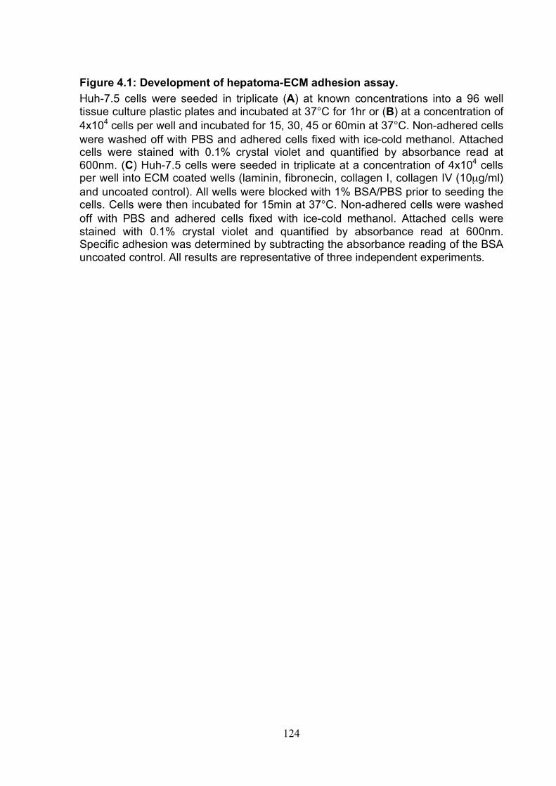
124
Figure 4.1: Development of hepatoma-ECM adhesion assay. Huh-7.5 cells were seeded in triplicate (A) at known concentrations into a 96 well tissue culture plastic plates and incubated at 37°C for 1hr or (B) at a concentration of 4x10⁴ cells per well and incubated for 15, 30, 45 or 60min at 37°C. Non-adhered cells were washed off with PBS and adhered cells fixed with ice-cold methanol. Attached cells were stained with 0.1% crystal violet and quantified by absorbance read at 600nm. (C) Huh-7.5 cells were seeded in triplicate at a concentration of 4x10⁴ cells per well into ECM coated wells (laminin, fibronecin, collagen I, collagen IV (10µg/ml) and uncoated control). All wells were blocked with 1% BSA/PBS prior to seeding the cells. Cells were then incubated for 15min at 37°C. Non-adhered cells were washed off with PBS and adhered cells fixed with ice-cold methanol. Attached cells were stained with 0.1% crystal violet and quantified by absorbance read at 600nm. Specific adhesion was determined by subtracting the absorbance reading of the BSA uncoated control. All results are representative of three independent experiments.
Ad
he
red
ce
lls
(O
D 6
00
nm
) A
dh
ere
d c
ell
s (
OD
60
0n
m)
Sp
ec
ific
ad
he
sio
n (
OD
60
0n
m)
A
B
C
124
125
Initial experiments investigated whether CD81 engagement would alter Huh-7.5
adhesion to fibronectin. Huh-7.5 cell monolayers were removed from tissue culture
plastic using trypsin and cell suspensions treated with a panel of anti-CD81mAbs
(Described in section 3.1) prior to seeding onto fibronectin-coated wells. All mAbs
were used at 1µg/ml, a concentration previously demonstrated to saturate the cell
surface by flow cytometry (Flow cytometry analysis carried out by Ke Hu, data not
shown). As a positive control we used an antibody against integrin β1 subunit,
previously shown to inhibit cell-ECM adhesion (46). All of the anti-CD81 mAbs had a
minimal effect on Huh-7.5 cell adhesion to fibronectin whilst the anti-Beta-1 (integrin)
antibody significantly reduced cell adhesion (Fig.4.2A).
CD81 expression levels were previously reported to reduce epithelial cell adhesion to
fibronectin (481). We therefore investigated the role of CD81 expression on
hepatoma-ECM adhesion. To do this we engineered two hepatoma cell lines (CD81
negative), HepG2 and Huh-7 CD81null cells to express CD81 and compared their
adhesion to different ECM proteins, laminin, collagen I, collagen IV and fibronectin.
CD81 cell surface expression was confirmed by flow cytometry. No differences were
noted in hepatoma-ECM adhesion between parental and CD81 expressing cells
(Fig.4.2B&C).
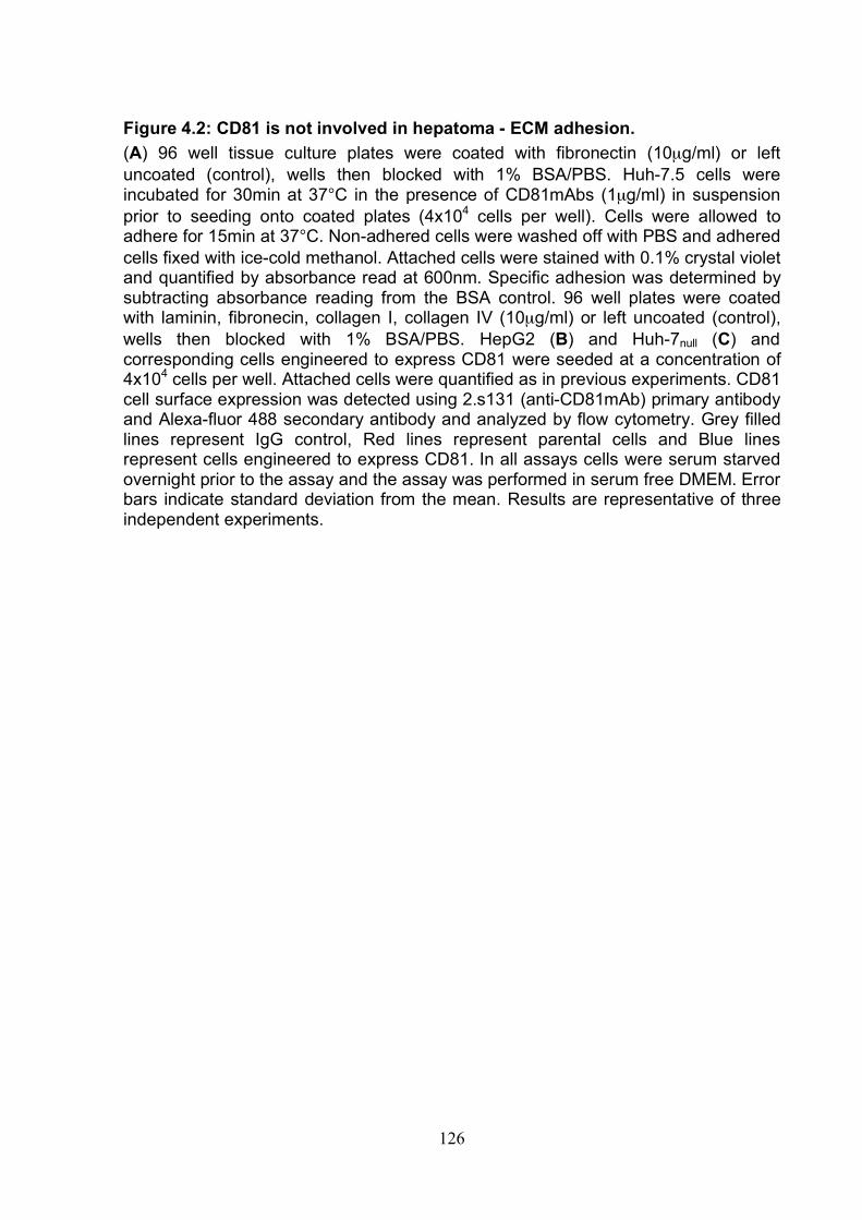
126
Figure 4.2: CD81 is not involved in hepatoma - ECM adhesion. (A) 96 well tissue culture plates were coated with fibronectin (10µg/ml) or left uncoated (control), wells then blocked with 1% BSA/PBS. Huh-7.5 cells were incubated for 30min at 37°C in the presence of CD81mAbs (1µg/ml) in suspension prior to seeding onto coated plates (4x10⁴ cells per well). Cells were allowed to adhere for 15min at 37°C. Non-adhered cells were washed off with PBS and adhered cells fixed with ice-cold methanol. Attached cells were stained with 0.1% crystal violet and quantified by absorbance read at 600nm. Specific adhesion was determined by subtracting absorbance reading from the BSA control. 96 well plates were coated with laminin, fibronecin, collagen I, collagen IV (10µg/ml) or left uncoated (control), wells then blocked with 1% BSA/PBS. HepG2 (B) and Huh-7null (C) and corresponding cells engineered to express CD81 were seeded at a concentration of 4x10⁴ cells per well. Attached cells were quantified as in previous experiments. CD81 cell surface expression was detected using 2.s131 (anti-CD81mAb) primary antibody and Alexa-fluor 488 secondary antibody and analyzed by flow cytometry. Grey filled lines represent IgG control, Red lines represent parental cells and Blue lines represent cells engineered to express CD81. In all assays cells were serum starved overnight prior to the assay and the assay was performed in serum free DMEM. Error bars indicate standard deviation from the mean. Results are representative of three independent experiments.
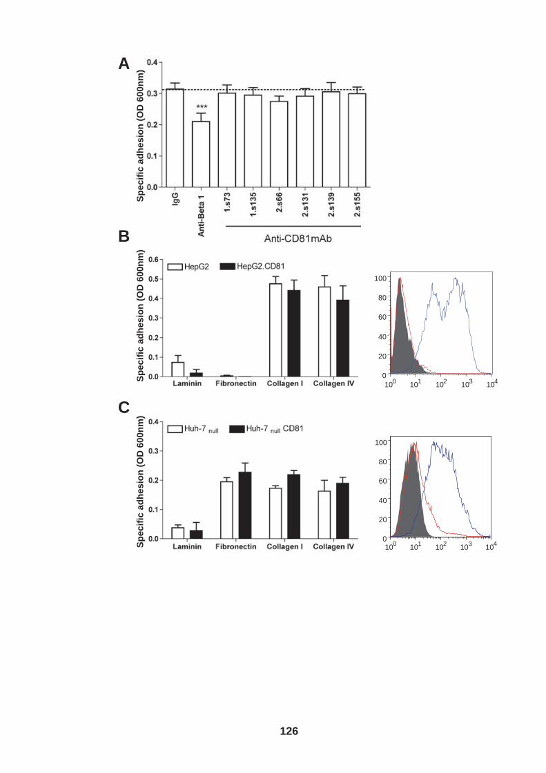
A
B
C
10 0 10 1 10 2 10 3 10 4 0
20 40 60 80
100
10 0 10 1 10 2 10 3 10 4 0
20 40 60 80
100
Sp
ec
ific
ad
he
sio
n (
OD
60
0n
m)
Sp
ec
ific
ad
he
sio
n (
OD
60
0n
m)
Sp
ec
ific
ad
he
sio
n (
OD
60
0n
m)
126
127
Several viruses have been reported to modulate cell surface expression of adhesion
molecules including integrins resulting in altered cell-ECM adhesion. Examples
include: Ebola virus infection down-regulates β1 integrin expression and reduces the
adhesion of infected cells to ECM (446); Rotavirus infection increases integrin
expression of intestinal cells (184) and Epstein-Barr virus up-regulates α5 integrins in
transformed B lymphocytes and promotes their invasion (210). To investigate
whether HCV infection alters hepatoma-ECM adhesion, mock and HCV strain J6/JFH
infected Huh-7.5 cells (3 days post infection) were seeded onto different ECM protein
coated wells. Adhered cells were fixed with methanol, and stained with crystal violet
to determine adhesion or with anti-NS5A antibody to determine the frequency of
ECM-bound infected cells. No significant differences were noted between the
adhesion of mock or J6/JFH infected cells to fibronectin (Fig.4.3A-B), demonstrating
that HCV infection does not modulate hepatoma-ECM adhesion. In support of this
conclusion HCV infection did not alter integrin expression (Fig.4.3C).
128
Figure 4.3 HCV infection does not alter cell-ECM adhesion 96 well plates were coated with fibronectin, laminin, collagen I, collagen IV (10µg/ml ) or left uncoated, wells then blocked with 1% BSA/PBS. (A) Huh-7.5 cells were infected with J6/JFH HCVcc. At day 3 post infection J6/JFH infected and naïve Huh-7.5 cells were seeded at 4x10⁴ cells per well into the different ECM coated wells or (B) at days 3, 4 and 5 post infection cells were seeded onto fibronectin coated wells. Cells were allowed to adhere for 15min at 37°C and attached cells were then either stained with crystal violet to determine specific adhesion as in previous assays or anti-NS5A and DAPI to determine HCV infection level of adhered cells. For (B) a proportion of cells were PFA fixed each day, stained for NS5A and analyzed by flow cytometry to quantify infection level of cells prior to seeding (Approximately 65, 84 and 93% NS5A +ve at days 3,4 and 5 respectively). (C) Integrin cell surface expression of J6/JFH HCVcc infected or naïve Huh-7.5 cells was determined by flow cytometry using specific anti-integrin antibodies as detailed in Table 2.4. Cell surface expression expressed as median fluorescence intensity (MFI). Error bars indicate standard deviation from the mean. Experiments were performed in triplicate and results are representative of three independent experiments.
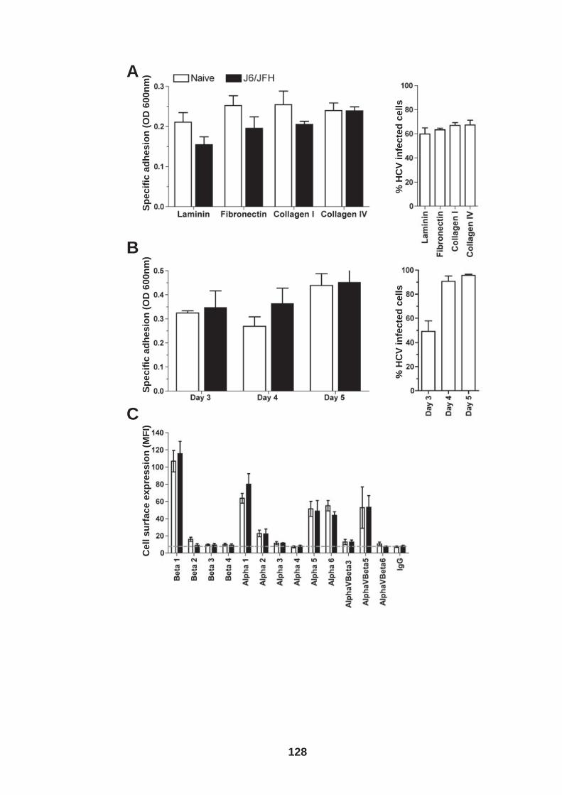
A
B
C
Ce
ll s
urf
ac
e e
xp
res
sio
n (
MF
I)
% H
CV
in
fec
ted
ce
lls
Sp
ec
ific
ad
he
sio
n (
OD
60
0n
m)
Sp
ec
ific
ad
he
sio
n (
OD
60
0n
m)
% H
CV
in
fec
ted
ce
lls
128
129
As previously mentioned ECM plays an essential role in liver structure and function
(36) and increased ECM production is frequently associated with liver disease (32).
Hepatic stelate cells are thought to be the most important cells in coordinating liver
fibrosis and express collagens and basement membrane (149). Hepatocytes are the
dominant source of plasma fibronectin in the body and although the role of plasma
fibronectin in fibrosis is not known, mRNA for cellular fibronectin has also been
reported in hepatocytes. Thus, the role of hepatocyte ECM expression in fibrosis is
still under debate (161, 307, 334, 507). Recent reports demonstrating hepatocyte
EMT argue for a more dominant role of hepatocytes in fibrosis than originally thought
(33, 160, 552).
To ascertain whether HCV infection alters hepatoma ECM production we developed
a quantitative ELISA to measure collagen I, collagen IV and fibronectin ECM proteins
(Fig.4.4A). Huh-7.5 cells were seeded onto gelatin-coated plates and infected with
mock or J6/JFH HCVcc and 96hr post infection cells were lysed and ECM expression
quantified. A replicate plate was methanol fixed and stained for NS5A to determine
the level of infected cells (Fig. 4.4B). Naïve and J6/JFH infected Huh-7.5 cells
expressed comparable levels of fibronectin and collagen I, no detectable collagen IV.
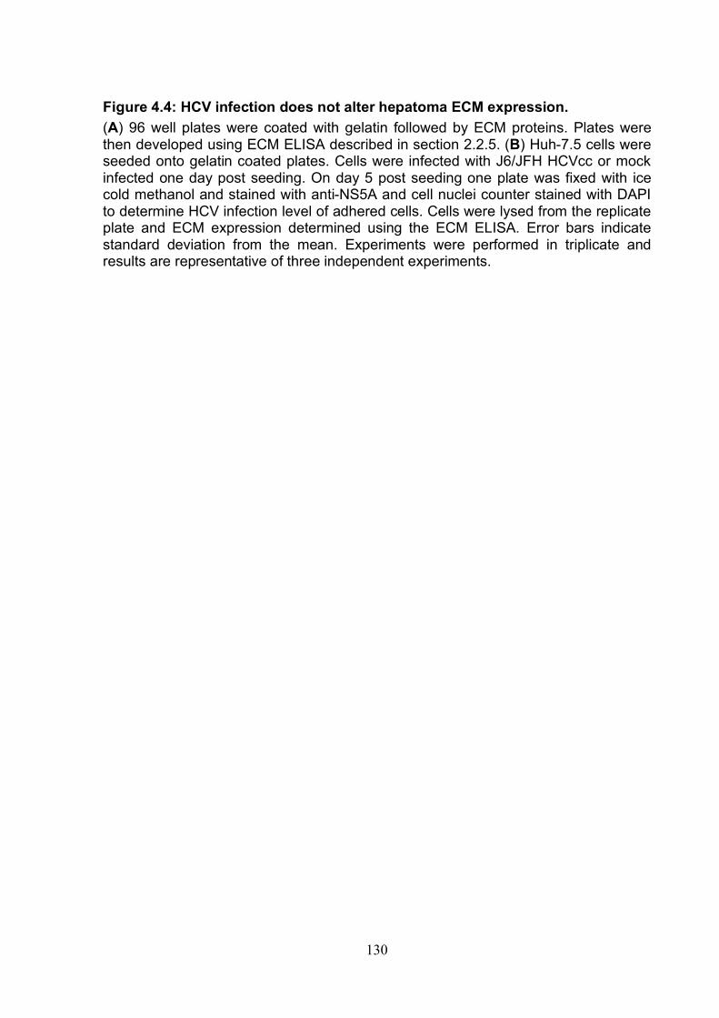
130
Figure 4.4: HCV infection does not alter hepatoma ECM expression. (A) 96 well plates were coated with gelatin followed by ECM proteins. Plates were then developed using ECM ELISA described in section 2.2.5. (B) Huh-7.5 cells were seeded onto gelatin coated plates. Cells were infected with J6/JFH HCVcc or mock infected one day post seeding. On day 5 post seeding one plate was fixed with ice cold methanol and stained with anti-NS5A and cell nuclei counter stained with DAPI to determine HCV infection level of adhered cells. Cells were lysed from the replicate plate and ECM expression determined using the ECM ELISA. Error bars indicate standard deviation from the mean. Experiments were performed in triplicate and results are representative of three independent experiments.
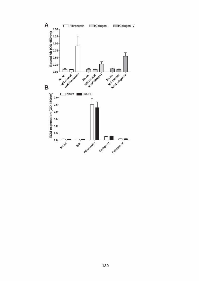
A
B
EC
M e
xp
res
sio
n (
OD
45
0n
m)
Bo
un
d A
b (
OD
45
0n
m)
130
131
To investigate this further the production of fibronectin was determined over 5 days,
and no significant differences were observed (Fig.4.5A). Finally we compared
fibronectin expression during the first 12hrs post seeding. Mock and J6/JFH infected
cells were seeded onto gelatin-coated plates and cells lysed 4, 8 and 12hrs later
(Fig.4.5B). Both mock and J6/JFH infected cells expressed comparable levels of
fibronectin over the first 12hrs post seeding.
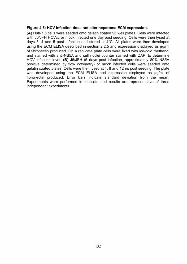
132
Figure 4.5: HCV infection does not alter hepatoma ECM expression. (A) Huh-7.5 cells were seeded onto gelatin coated 96 well plates. Cells were infected with J6/JFH HCVcc or mock infected one day post seeding. Cells were then lysed at days 3, 4 and 5 post infection and stored at 4°C. All plates were then developed using the ECM ELISA described in section 2.2.5 and expression displayed as µg/ml of fibronectin produced. On a replicate plate cells were fixed with ice-cold methanol and stained with anti-NS5A and cell nuclei counter stained with DAPI to determine HCV infection level. (B) J6/JFH (5 days post infection, approximately 80% NS5A positive determined by flow cytometry) or mock infected cells were seeded onto gelatin coated plates. Cells were then lysed at 4, 8 and 12hrs post seeding. The plate was developed using the ECM ELISA and expression displayed as µg/ml of fibronectin produced. Error bars indicate standard deviation from the mean. Experiments were performed in triplicate and results are representative of three independent experiments.
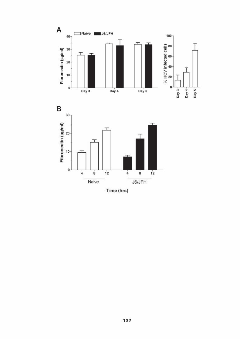
A
B
Fib
ron
ec
tin
(μμ
g/m
l)
% H
CV
in
fec
ted
ce
lls
132
133
4.2 Role of CD81 in hepatoma invasion The ability of a cell to migrate or invade though ECM is an important phenotypic
feature that defines a cells metastatic potential. Invading cells induce membrane
ruffles at their leading edge in a process involving the reduction of rigid actin stress
fibers leaving only fine cortical actin (25, 256). This leads to the formation of flat or
sheet like projections containing a branched actin network termed lamellipodia.
Filopodia emerge from lamellipodia as rod like protrusions with tight bundled actin.
Invadopodia is the term for a further cellular membrane protrusion, these are actin
rich protrusions containing high expression levels of integrins and cell MT1-MMPs
and enable remodeling and cell penetration into ECM (18, 546).
Tetraspanin-integrin complexes localize to microvilli like protrusions, including CD81-
α3β1 complexes on mammary carcinoma cells. Interestingly anti-CD81 mAbs have
been reported to induce cellular protrusions and matrix metalloproteinase MMP-2
production and increased cell invasion (460). We previously observed that anti-CD81
ligation promoted hepatoma cell spread. To determine whether antibody engagement
induced microvilli or filopodia like protrusions, Huh-7.5 cells were seeded onto glass
cover slips and exposed to anti-CD81 mAbs or IgG control (2µg/ml). Wells were fixed
with EM grade formaldehyde at 0, 10 and 240min post exposure to antibody. CD81
was visualized using a 488 conjugated secondary antibody and F-actin visualized
with phalloidin-594, cells were imaged by confocal microscopy. Huh-7.5 cells had
numerous microvilli or filopodia protruding from their cell body with CD81 expressed
at the cell surface, however we failed to observe any increase in filopodia following
anti-CD81 stimulation (Fig.4.6).
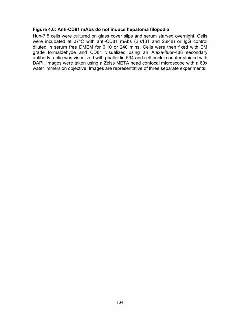
134
Figure 4.6: Anti-CD81 mAbs do not induce hepatoma filopodia Huh-7.5 cells were cultured on glass cover slips and serum starved overnight. Cells were incubated at 37°C with anti-CD81 mAbs (2.s131 and 2.s48) or IgG control diluted in serum free DMEM for 0,10 or 240 mins. Cells were then fixed with EM grade formaldehyde and CD81 visualized using an Alexa-fluor-488 secondary antibody, actin was visualized with phalloidin-594 and cell nuclei counter stained with DAPI. Images were taken using a Zeiss META head confocal microscope with a 60x water immersion objective. Images are representative of three separate experiments.
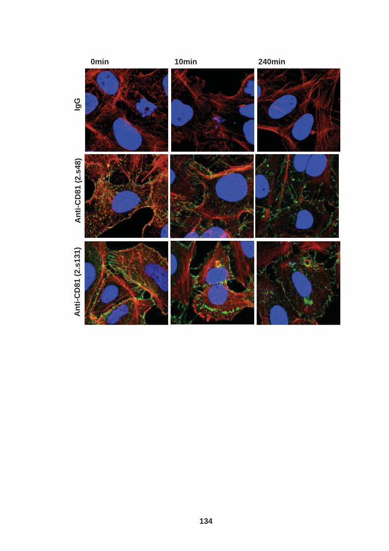
An
ti-C
D8
1 (
2.s
48
) A
nti
-CD
81
(2
.s1
31
) Ig
G
0min 10min 240min
134

135
To investigate whether CD81 engagement has a role in hepatoma migration a
confluent monolayer of Huh-7.5 cells were wounded using a p200 tip and wound
closure measured over 24hrs in the presence of anti-CD81 mAb (1.s262) or
irrelevant control IgG (Fig.4.7A). No differences were observed in wound closure in
antibody treated or untreated cells.
As previously described the processes involved in cell migration and cell invasion
differ and we therefore investigated the effect(s) of CD81 ligation on hepatoma
invasion through a collagen coated 8µm pore-size transwell insert (Fig.4.7B). Cells
were incubated for 24hrs at 37°C followed by mechanical removal of non-invaded
cells and methanol fixation of invaded cells. The invaded cells were stained with
crystal violet and counted. Anti-CD81 mAb had no effect on hepatoma invasion.
136
Figure 4.7: Anti-CD81 mAb has no effect on hepatoma migration or invasion. (A) Huh-7.5 cells (1x10⁶) were seeded into 6 well dishes and serum starved overnight. A wound was performed using a P200 tip and media replaced with serum free DMEM ( plus mitomycin C at 15µg/ml) containing anti-CD81 mAb (1.s262) or IgG at 10µg/ml. Cells were incubated at 37°C and phase images taken at 0 and 24hrs. Percentage wound closure determined using IPLab 4.0 (BD Biosciences). (B) Huh-7.5 cells were serum starved overnight. Trypsin was used to remove the cells from the tissue culture plastic prior to re-suspension in serum free DMEM containing anti-CD81 mAb (1.s262) at 0.1,1, and 10µg/ml or irrelevant control IgG at 10µg/ml. Cells were seeded (4x10⁴ cells/200ul/cell) into the top chamber of an 8µm pore collagen coated transwell insert. The lower chamber contained 300µl of DMEM containing the corresponding treatment. Cells were incubated at 37°C for 24hrs. Non-invaded cells were mechanically removed and invaded cells methanol fixed and stained with 0.1% crystal violet. 3x fields of view were imaged per transwell at 40x magnifcation on a fluorescent microscope (NikonTE2000, Japan). The number of invaded cells was determined and invasion expressed relative to control. Two tailed unpaired T-test’s were performed to determine whether there was a significant difference in wound closure or invasion between untreated and anti-CD81 treated cells. No significant differences were observed P>0.05. Error bars indicate standard deviation from the mean. Assays were performed in duplicate and the results are representative of three separate experiments.

A B IgG Anti-CD81
C
0hr
24hr
136
137
To determine whether CD81 expression modulates hepatoma cell migration, CD81
expression was silenced using CD81 siRNA treatment and wound closure compared
to irrelevant control siRNA treated cells (Fig.4.8A). The assay was performed in the
presence of mitomycin C, mitomycin C prevents cell proliferation and thus enables
discrimination between cell proliferation and cell migration. Cells were fixed and
stained for CD81 at the end of the assay to confirm reduction in CD81 expression.
We observed a small but significant reduction in cell migration of siRNA CD81
treated cells compared to control.
Two approaches were taken to investigate the effects of CD81 expression on
hepatoma cell invasion through a collagen coated transwell insert. Firstly invasion of
HepG2 parental cells and those engineered to express CD81 (HepG2.CD81) were
compared. Secondly CD81 expression was silenced in Huh-7.5 cells using CD81
siRNA treatment and invasion compared to irrelevant control siRNA treated cells. A
control well was stained for CD81 to confirm CD81 silencing. We observed increased
invasion of HepG2 cells expressing CD81 compared to parental cells demonstrating
that CD81 expression increases hepatoma invasion potential (Fig.4.8B). Confirming
this phenomenon we observed a decrease in the invasion of the CD81 silenced Huh-
7.5 cells compared to control (Fig.4.8C). These results demonsrate in two
independent hepatoma cell lines that CD81 expression increases hepatoma cell
invasion and implicates CD81 as a tumour promoter.

138
Figure 4.8: CD81 expression increases hepatoma cell invasion. (A) Huh-7.5 cells were seeded into 6 well dishes and transfected with si CD81 RNA (CD81 KD) or irrelevant control siRNA. 24hrs post transfection a wound was performed using a P200 tip and media replaced with serum free DMEM, cells were incubated at 37°C and images taken at 0 and 24hrs post wound. Images display expression of CD81 KD cells and control cells taken 24hrs post wound on. CD81 expression visualized using anit-CD81 (2.s131) primary antibody and alexa-fluor 488 secondary antibody (green). A two tailed unpaired T-test was performed to determine whether there was a significant difference in wound closure between control and CD81 KD cells. A significant difference was observed P=0.0387. (B) HepG2 and HepG2 cells engineered to express CD81 (HepG2.CD81) or (C) Huh-7.5 control and Huh-7.5 CD81 KD cells were cultured in a tissue culture flask in serum free conditions overnight, cells were trypsinised and re-suspended in serum free DMEM. Cells then seeded (4x10⁴ cells/200µl/well) in the top chamber of an 8µm pore collagen coated transwell insert. The lower chamber contained 300µl of serum free DMEM. Cells were incubated at 37°C for 24hrs. Non-invaded cells were mechanically removed and invaded cells methanol fixed and stained with 0.1% crystal violet. 3x fields of view were imaged per transwell at 40x magnification on a on a fluorescent microscope (NikonTE2000, Japan). The number of invaded cells was determined and invasion expressed relative to control. Images display CD81 expression of CD81 KD cells and control cells taken at the end of the assay in replicate wells. CD81 expression was visualized using anti-CD81 specific antibody (2.s131) and alexa-fluor 488 secondary antibody. Two tailed unpaired T-tests were performed to determine whether there was a significant difference in invasion between HepG2 and HepG2.CD81 cells and control and CD81 KD cells. A significant difference was observed, P=< 0.0001 and 0.004 respectively. Error bars indicate standard deviation from the mean. Assays were performed in duplicate and the results are representative of three separate experiments.
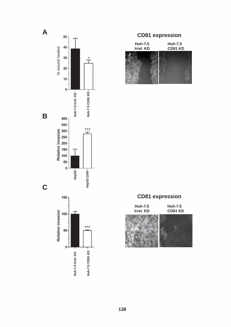
Huh-7.5
Irrel. KD
Huh-7.5
CD81 KD
A
C
B
% w
ound h
eale
d
Re
lati
ve
in
va
sio
n
Re
lati
ve
in
va
sio
n
Hu
h-7
.5 I
rre
l. K
D
Hu
h-7
.5 C
D81 K
D
Hu
h-7
.5 I
rre
l. K
D
Hu
h-7
.5 C
D81 K
D
Huh-7.5
Irrel. KD
Huh-7.5
CD81 KD
CD81 expression
CD81 expression
138
139
4.3 HCV infection modulates hepatoma cell invasion We previously demonstrated that HCV infection modulates CD81 dependent
hepatoma cell spread (Chapter 3) and were interested to assess the effect(s) of HCV
infection on hepatoma invasion and migration. Unfortunately due to category three
restrictions of imaging we were unable to study the effects of infection on hepatoma
migration in a scratch wound assay.
Huh-7.5 cells were electroporated with J6/JFH 96hrs prior to the assay. The invasion
assay was carried out as previously described with the following exceptions; (i) cells
were labeled with CMFDA (cell tracker green) prior to the assay instead of a crystal
violet stain post invasion, (ii) cells were seeded in replicate (control) 24 well plates
not containing transwell inserts, the control and invaded cells were then stained for
NS5A at the end of the assay to determine the frequency of infected invaded cells
compared to non-invaded cells. A greater number of cells in the J6/JFH infected
culture invaded compared to the uninfected cells (Fig.4.9A). However, no difference
was observed in the frequency of NS5A expressing cells in the control and invaded
cell population (Fig.4.9B) and thus we hypothesize that a soluble factor may be
responsible for the increased invasive phenotype of the infected cell population.
140
Figure 4.9: Effect of HCV infection on invasion. (A) HCV J6/JFH RNA was delivered into Huh-7.5 cells by electroporation (ep). 72hrs post ep cells were cultured in serum free conditions overnight. Cells labeled with CMFDA, removed from tissue culture plastic with trypsin and re-suspension in serum free DMEM. Cells seeded (4x10⁴ cells/200µl/well) into the top chamber of an 8µm pore collagen coated transwell insert or control 24 well plates not containing a transwell insert. The lower chamber contained 300µl of serum free DMEM. Cells were incubated at 37°C for 24hrs. Non-invaded cells were mechanically removed and the invaded and control cells methanol fixed. 3x fields of view were imaged per transwell at 40x magnification on a fluorescent microscope (NikonTE2000, Japan). The number of invaded cells was determined and invasion expressed relative to control. A Two tailed unpaired T-test was performed to determine whether there was a significant difference in the invasion of naive and J6/JFH infected cells. A significant difference was observed P=0.0010. (B) Fixed cells were stained for NS5A and infection level determined for control and invaded cells. Error bars indicate standard deviation from the mean. Assays were performed in duplicate and the results are representative of three separate experiments.
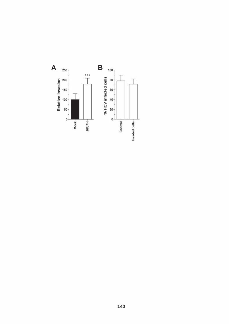
B A
% H
CV
in
fec
ted
ce
lls
140
141
Previous research reported that HCV infection promotes Vascular endothelial growth
factor (VEGF) expression (191, 341, 400). Chris Mee has since confirmed this in our
laboratory (Fig.4.10A). VEGF promotes angiogenesis and elevated VEGF levels in
patients with HCC are considered to be one of the most important indicators of
increased tumor angiogenesis (214). Angiogenesis is the process by which new
blood vessels are formed and is vital in tumor development. Without a blood supply
tumors cannot grow above a certain size due to hypoxia and lack of essential
nutrients. Blood supply is also essential for tumor cells to metastasize to other areas
of the liver or to other organs, a common complication of HCC (537). Apart from its
important role in angiogenesis VEGF has been demonstrated to reduce HepG2
hepatoma cell polarity (319, 423). VEGF has also been reported to increase the
migration of HepG2 cells (531). We therefore investigated whether exogenous VEGF
stimulation (Fig.4.10B) or neutralization of endogenous VEGF with an anti-VEGF
mAb (VG67e) (Fig.4.10C) would modulate Huh-7.5 cell invasion. We observed no
effect of exogenous VEGF or VG67e on Huh-7.5 cell invasion suggesting that VEGF
does not promote Huh-7.5 cell invasion.
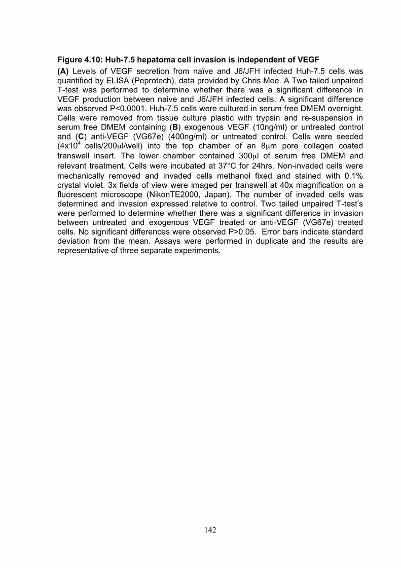
142
Figure 4.10: Huh-7.5 hepatoma cell invasion is independent of VEGF (A) Levels of VEGF secretion from naïve and J6/JFH infected Huh-7.5 cells was quantified by ELISA (Peprotech), data provided by Chris Mee. A Two tailed unpaired T-test was performed to determine whether there was a significant difference in VEGF production between naive and J6/JFH infected cells. A significant difference was observed P<0.0001. Huh-7.5 cells were cultured in serum free DMEM overnight. Cells were removed from tissue culture plastic with trypsin and re-suspension in serum free DMEM containing (B) exogenous VEGF (10ng/ml) or untreated control and (C) anti-VEGF (VG67e) (400ng/ml) or untreated control. Cells were seeded (4x10⁴ cells/200µl/well) into the top chamber of an 8µm pore collagen coated transwell insert. The lower chamber contained 300µl of serum free DMEM and relevant treatment. Cells were incubated at 37°C for 24hrs. Non-invaded cells were mechanically removed and invaded cells methanol fixed and stained with 0.1% crystal violet. 3x fields of view were imaged per transwell at 40x magnification on a fluorescent microscope (NikonTE2000, Japan). The number of invaded cells was determined and invasion expressed relative to control. Two tailed unpaired T-test’s were performed to determine whether there was a significant difference in invasion between untreated and exogenous VEGF treated or anti-VEGF (VG67e) treated cells. No significant differences were observed P>0.05. Error bars indicate standard deviation from the mean. Assays were performed in duplicate and the results are representative of three separate experiments.
A B C
142
143
VEGF production is increased in hepatoma cells following HCV infection through
stabilization of hypoxia inducible facor -1alpha (HIF-1α) (191, 341, 400). HIF-1α is a
regulatory sub unit of the HIF transcription complex. Under normal cellular oxygen
levels HIF-1α is continually expressed and degraded by the following oxygen
dependent process; HIF-1α is hydroxylated by HIF prolylhydroxylases enabling it to
be recognized by von Hippel-Lindau tumor suppressor protein (pVHL). pVHL is a
recognition component of the E3 ubiquitin ligase complex and targets HIF-1α for
proteasomal degradation (238). If HIF-1α becomes stably expressed it is
translocated to the nucleus where it binds to ubiquitously expressed HIf-1β and
recruits other transcriptional co-factors making up the HIF transcription complex
(238). As well as VEGF the HIF transcription complex is responsible for the up-
regulation of numerous genes responsible for proliferation, cell survival, metastasis
and glycolysis (238). HIF-1α can be stabilized through a number of mechanisms
including growth factor induction, genetic mutations of oxygen sensing genes, loss of
tumor suppressor function, mitochondrial dysfunction as well as hypoxia (388). To
determine whether hepatoma cell invasion increases upon stabilization of HIF-1α we
compared invasion of Huh-7.5 cells under hypoxic (1%O2) or normoxic (22% O2)
conditions. We observed a significant increase in cell invasion under hypoxic
conditions compared to the normoxic control (Fig.4.12A&B). To establish whether the
increased invasive potential is dependent on HIF-1α, hepatoma cells were treated
with the HIF-1 translation inhibitor, NSC-134754 (1µM) (79, 388). We observed a
significant decrease in cell invasion under hypoxic conditions in the presence of
inhibitor whilst only a moderate inhibition was observed under normoxic conditions.
We propose that the increased invasion of HCV infected hepatoma cells may be
144
mediated through a VEGF independent pathway induced by the stablization of HIF-
1α.
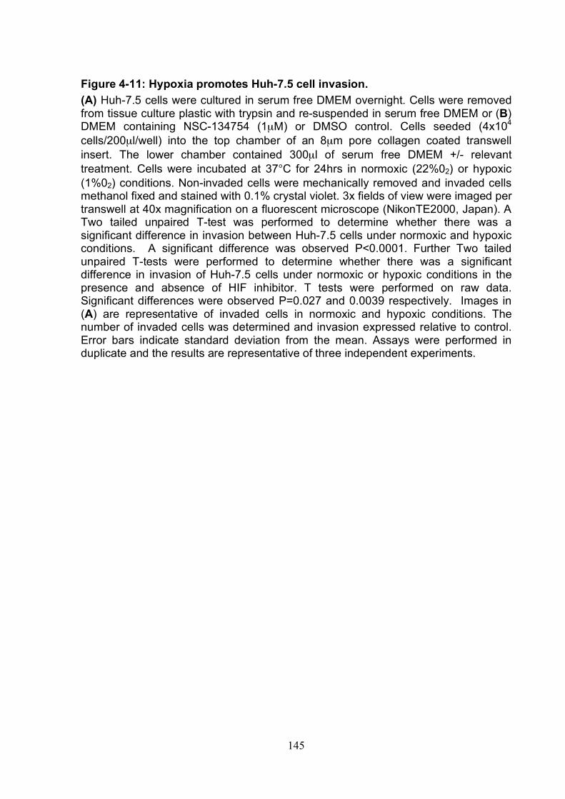
145
Figure 4-11: Hypoxia promotes Huh-7.5 cell invasion. (A) Huh-7.5 cells were cultured in serum free DMEM overnight. Cells were removed from tissue culture plastic with trypsin and re-suspended in serum free DMEM or (B) DMEM containing NSC-134754 (1µM) or DMSO control. Cells seeded (4x10⁴
cells/200µl/well) into the top chamber of an 8µm pore collagen coated transwell insert. The lower chamber contained 300µl of serum free DMEM +/- relevant treatment. Cells were incubated at 37°C for 24hrs in normoxic (22%02) or hypoxic (1%02) conditions. Non-invaded cells were mechanically removed and invaded cells methanol fixed and stained with 0.1% crystal violet. 3x fields of view were imaged per transwell at 40x magnification on a fluorescent microscope (NikonTE2000, Japan). A Two tailed unpaired T-test was performed to determine whether there was a significant difference in invasion between Huh-7.5 cells under normoxic and hypoxic conditions. A significant difference was observed P<0.0001. Further Two tailed unpaired T-tests were performed to determine whether there was a significant difference in invasion of Huh-7.5 cells under normoxic or hypoxic conditions in the presence and absence of HIF inhibitor. T tests were performed on raw data. Significant differences were observed P=0.027 and 0.0039 respectively. Images in (A) are representative of invaded cells in normoxic and hypoxic conditions. The number of invaded cells was determined and invasion expressed relative to control. Error bars indicate standard deviation from the mean. Assays were performed in duplicate and the results are representative of three independent experiments.
Normoxia Hypoxia
A B
C
145
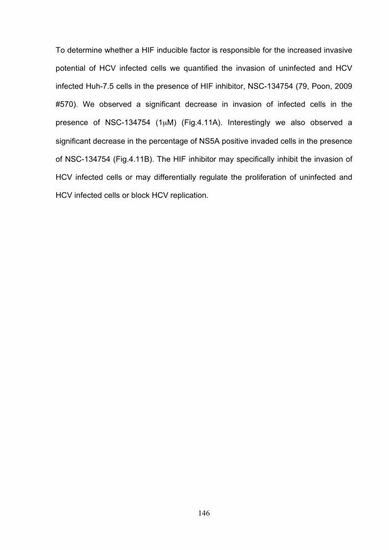
146
To determine whether a HIF inducible factor is responsible for the increased invasive
potential of HCV infected cells we quantified the invasion of uninfected and HCV
infected Huh-7.5 cells in the presence of HIF inhibitor, NSC-134754 (79, Poon, 2009
#570). We observed a significant decrease in invasion of infected cells in the
presence of NSC-134754 (1µM) (Fig.4.11A). Interestingly we also observed a
significant decrease in the percentage of NS5A positive invaded cells in the presence
of NSC-134754 (Fig.4.11B). The HIF inhibitor may specifically inhibit the invasion of
HCV infected cells or may differentially regulate the proliferation of uninfected and
HCV infected cells or block HCV replication.

147
Figure 4.12: HCV induced cell invasion is sensitive to HIF-1α inhibitor. HCV J6/JFH RNA or control no RNA was delivered into Huh-7.5 cells by electroporation (ep). 72hrs post ep cells cultured in serum free conditions overnight. HCV infected and control cells were labeled with CMFDA, removed from tissue culture plastic with trypsin and re-suspended in serum free DMEM containing NSC-134754 (1µM) or DMSO control. Cells were seeded (4x10⁴ cells/200µl/well) into the top chamber of an 8µm pore collagen coated transwell insert. The lower chamber contained 300µl of serum free DMEM plus relevant treatment. Cells were then incubated at 37°C for 24hrs. Non-invaded cells were mechanically removed and invaded cells methanol fixed. (A) 3x fields of view were imaged per transwell at 40x magnification on a fluorescent microscope (NikonTE2000, Japan). The number of invaded cells was determined and invasion expressed relative to control. No significant difference in invasion of naïve cells in the presence and absence of HIF inhibitor was observed (P=0.1726, two tailed unpaired T-test). A significant difference was observed in invasion of J6/JFH infected Huh-7.5 cells in the presence and absence of HIF inhibitor (P=0.0270, two tailed unpaired T-test). (B) Fixed cells were stained for NS5A and infection level determined for control and NSC-134754 treated cells. A significant difference was observed in infection level of invaded cells in the presence and absence of HIF inhibitor (P=0.0013, two tailed unpaired T-test). Error bars indicate standard deviation from the mean. Assays were performed in duplicate and the results are representative of three separate experiments.
A
B
% H
CV
in
fec
ted
ce
lls
147
148
To investigate this further we assessed the effects of the HIF inhibitor on HCV
replication and hepatoma proliferation. At 1µM the HIF inhibitor successfully inhibited
HCV infection (Fig.4.12A). To determine whether the HIF inhibitor affects HCV
transmission J6/JFH infected cells were treated with NSC-134754 and viral spread
quantified over a 48hr period by monitoring NS5A expression by flow cytometry
(Fig.4.12B). A decrease in the percentage of infected cells in the presence of NSC-
134754 compared to control was observed, demonstrating that NSC-134754 inhibits
new rounds of infection. To determine whether the HIF inhibitor has a cytotoxic or
anti-proliferative effect on Huh-7.5 cells we performed an MTS assay (colorimetric
test that measures metabolic function). We observed a small decrease in MTS signal
after a 24hr treatment with NSC-134754 compared to control (Fig.4.12C), this can be
more clearly seen in the corresponding phase images (Fig.4.12D). Results suggest
that HCV infection may be dependent on HIF-1α stabilization.

149
Figure 4.13: HIF-1α dependent modulation of proliferation and infection. (A) Huh-7.5 cells were treated with NSC-134754 and control at 5,1,and 0.1 µM plus DMSO control. 24hrs post treatment an MTS assay was performed to determine metabolic function results presented as absorbance at 490nm (minus background). (B) Representative phase images of treated cells prior to MTS assay. (C) Huh-7.5 cells were seeded into 96 well tissue culture plate (0.75x10⁴ cells/well), the following day cells were pre-treated for 1hr with DMSO control or NSC-134754 (1µM) containing DMEM (3% FBS). Cells were infected with J6/JFH HCVcc in the presence or absence of inhibitor. 48hrs post infection cells were fixed with ice-cold methanol and stained for NS5A. Infected cells enumerated and infection level represented as Infectious units (IU) per ml. (D) HCV J6/JFH RNA was delivered into Huh-7.5 cells by electroporation (ep). 48hrs post ep cells seeded into 12 well dishes (25x104cells/well) in the presence of DMSO control or NSC-134754 (1µM) containing DMEM (3% FBS). 0, 24 and 48hrs post treatment cells were harvested, fixed, stained for NS5A and quantified using flow cytometry. Results presented as frequency of NS5A positive cells. Error bars indicate standard deviation from the mean. Assays were performed in duplicate and the results are representative of three independent experiments.
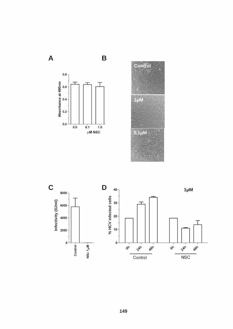
A B
0.1 M
Control
1.0 M 1 M
C D
Infe
cti
vit
y (
IU/m
l)
1 M
% H
CV
in
fec
ted
ce
lls
149
150
4.4 Discussion HCV infection is one of the leading causes of hepatocellular carcinoma and recent
research suggests HCV protein expression may be directly responsible (17, 33, 89,
294, 330, 340). Liver cancer is very difficult to treat and is associated with a high
frequency of relapse and metastasis (537). As such a greater understanding of the
processes involved is desperately needed for the development of future anti-cancer
drugs. In this chapter we demonstrate a role for HCV receptor tetraspanin CD81 in
hepatoma invasion and furthermore report that HCVcc infection increases the
invasive potential of hepatoma cells.
Tetraspanins are relatively small membrane proteins and are not believed to directly
interact with extra-cellular matrix (ECM) proteins (530), however a number of
tetraspanins namely CD151, CD82, CD9 and importantly CD81 are associated with
integrins and may therefore be able to indirectly alter cell-ECM interactions (427, 42,
43, 195) . Major integrin-ligand binding combinations for the ECM proteins we used
are as follows; Fibronectin binds αIIβ3, αVβ3, αVβ6, αVβ1, α5β1, α8β1, collagen
binds αI0β1, α2β1, α1β1, α11β1, and finally laminin binds α10β1, α2β1, α1β1, same
as collagen, and additionally α7β1, α6β4, α6β1 and α3β1 (212). A number of integrin
subunits were detected on the cell surface of Huh-7.5 hepatoma cells (Fig.4.3.B).
This included high expression levels of the most predominant beta subunit, β1, as
well as detection of a number of alpha subunits that form dimers with the β1 subunit
to bind fibronectin (α5), collagen, (α1, α2) and laminin (α1, α2, α6). We observed no
effect of CD81 expression on hepatoma cell (Huh-7 and HepG2 cells) adhesion to
different ECM proteins (Fig.4.2B&C). In addition to studying the effect(s) of
tetraspanin expression on cell adhesion, tetraspanin targeting antibodies have also
151
been reported to alter cell-ECM adhesion by regulating tetraspanin – partner protein
interactions (177, 352, 427). Adhesion of hepatoma cells to ECM protein, fibronectin,
was not affected by ligation of cell surface CD81 with a diverse panel of anti-CD81
mAbs (previously described in chapter 3) (Fig.4.2A). In conclusion our findings
demonstrate that CD81 is not involved in hepatoma cell-ECM adhesion.
Previous research reported the induction of microtubule like projections from
mammary carcinoma cells embedded in matrigel after exposure to CD81 mAbs,
thought to be coordinated through an association of CD81 with laminin binding
integrin α3β1 (460). We were unable to detect α3 integrin cell surface expression on
our hepatoma cells using flow cytometry analysis. Numerous filopodia like
protrusions were observed extending from the cell body of Huh-7.5 cells and CD81
was highly expressed along the cell membrane of these protrusions, ideally located
to function in the modulation of filopodia and cell migration. However, CD81
engagement did not promote filopodia/microtubule like protrusions (Fig.4.6).
Furthermore ligation of CD81 had no effect on hepatoma cell migration and invasion
(Fig.4.7A&B).
However, CD81 expression was observed to increase hepatoma migration in two
independent cell lines and in both the scratch wound and collagen invasion transwell
assays (Fig.4.8). This is in contrast to an earlier publication by Mazzoca et al., 2008
(313) who reported decreased invasion of CD81 expressing HepG2 cells in response
to Insulin-like growth factor (IGF-1) stimulation. CD81 was reported to coordinate this
process through its association with type II phosphoinositide 4-kinase (PI4KII)
leading to the re-localization of actinin-4 into CD81 enriched intracellular vesicles
152
preventing actin cytoskeleton remodeling. These findings were supported with an in
vivo mouse model experiment whereby Huh-7 cells with un-detectable levels of
CD81 were observed to metastasize more than those expressing CD81, of note the
differences between the two cell types were small and the error bars large. The
discrepancies between the two data sets are difficult to explain. IGF-1 is an inducer
of epithelial mesenchymal transition (EMT) (168), a complex process often involved
in tumor progression, it is therefore important to study cell invasion/migration in the
absence of exogenous factors. In the absence of IGF-1 Mazzoca et al., (2008) (313)
observed no differences in the invasion of HepG2 and HepG2 cells expressing
CD81. Interestingly CD81 was observed to be highly expressed in intracellular
vesicles in the HepG2 cells featured in Mazzoca et al., 2008 (313) this differs from
our cells where CD81 is predominantly expressed at the plasma membrane. In vivo
studies of hepatocytes in liver tissue also report that CD81 is expressed
predominantly around the plasma membrane (396). This comparison suggests that
CD81 may not be localized correctly in the HepG2 cells in Mazzoca’s paper, and may
explain the discrepancy between our results. In support of Mazzoca’s findings a
previous publication reported that a loss of CD81 expression was associated with
differentiation and metastasis in HCC patients, however this report is limited having
only a small sample number (219). Observations in our laboratory would suggest that
CD81 expression is not altered in the diseased liver (396). It is clear that further in
vivo research is needed to clarify whether CD81 expression is associated with liver
cancer progression.
In support of our findings that CD81 promotes hepatoma invasion and migration
CD81 expression in other cell types has been reported to increase their invasive
153
potential (72, 262, 312, 528). Recent reports are beginning to unravel the highly
dynamic and complex relationship between CD81 and its partner proteins (72, 247,
262). CD81 is reported to associate with MT1-MMP (247, 262) along with other
tetraspanins including CD151, CD9 and TSPN12 (262, 529). MT1-MMP is a
membrane bound matrix metalloproteinase that enables cell penetration into ECM,
high expression levels of MT1-MMP have so far been reported to associate with poor
prognosis in breast cancer patients, no research to date has looked at its role in HCC
(205, 224). Lafleur et al., 2009 (262) reported that CD81 expression increases MT1-
MMP function by preventing lysosomal degradation, this may explain the increased
migration observed in our hepatoma cells. Interestingly in the study by Lafeur et al.,
2009 a double knock out of CD81 and CD9 was needed to observe this phenotype,
in future studies it may be of interest to investigate whether a stronger phenotype is
observed following knock down of both CD81 and CD9 in hepatoma cells. Other
CD81 associated membrane proteins have also been reported to modulate the
migratory phenotype of cells, including immunoglobulin-like proteins EWI-2 and EWI-
F (72, 247, 411). Chambrion et al., 2010 (72) reported that CD9 and CD81
expression in HEK-293 cells negatively regulated EWI-F(CD9-P1) induced effects on
cell migration. A further publication this time investigating EWI-2 function reported
that expression of EWI-2 to glioblastoma cell lines reduced CD9 and CD81
association with MMP-2 and MT1-MMP leading to a decrease in invasion potential
(247). These observations suggest that EWI-2 and EWI-F expression levels may
regulate CD81 localization and function or vice versa and are worth considering in
future experiments. Previous research into tetraspanin partner-protein interactions
and function has predominantly centered on biochemical-immunoprecipitation
studies. Current research utilizing new live cell imaging techniques in particular total
154
internal reflection microscopy (TIRF) and single particle tracking technologies
suggest that tetraspanins are more dynamic than originally thought and diffuse
between tetraspanin enriched microdomains as a single or a group of molecule(s)
(132). Our laboratory has now started to use these techniques for in vitro study of
CD81 dynamics in hepatoma cells (Helen Harris, unpublished data).
In the previous chapter we reported that HCV infection reduced CD81 induced cell
spread, we therefore wanted to investigate the effect(s) of infection on CD81 function
in hepatoma-ECM adhesion and invasion through collagen. We observed no effect(s)
of infection on hepatoma integrin expression (Fig.4.3C), hepatoma–ECM adhesion
(Fig 4.3A&B), or hepatoma ECM expression (Fig. 4.4 & 4.5). However infection
promoted hepatoma invasion through collagen (Fig. 4.9).
Since antibody engagement of CD81 had no effect on hepatoma cell-ECM adhesion,
migration or invasion we hypothesized that it would be unlikely for HCV particle
association with cell surface expressed CD81 to account for the increased invasion
of infected cells. Given our results that siRNA silencing of CD81 expression reduced
hepatoma invasion, we hypothesize that CD81 expression levels may predict the
invasive capacity of hepatocytes. Earlier results showing that HCV infection does not
alter CD81 expression levels (Fig.3.11C) eliminate this as a possible explanation for
their increased invasive capacity.
The complex nature of cellular migration and invasion means that HCV infection
could increase hepatoma invasion through numerous mechanisms independent of
CD81. If HCV infected cells have an increased invasive potential perse one might
155
expect the frequency of infected invaded cells to be greater than the control
population. This was not the case and thus we postulated that a soluble factor
secreted from the infected cells might have a paracrine effect on the entire infected
cell population (Fig.4.9). Previous research reported that HCV infection causes
mitochondrial deregulation resulting in stabilization of HIF-1α (191, 341, 380, 400).
Nasimuzzaman et al., 2007 (341) identified that HIF-1α stabilization in hepatoma
cells induced by HCV protein expression resulted in an up-regulation of VEGF
expression. Increased expression of VEGF upon HCV infection was confirmed by
work performed in our laboratory (Fig.4.10). We hypothesized that VEGF may
regulate hepatoma migration as previously described (531). However, exogenous
VEGF or neutralization of endogenous VEGF had no effect on Huh-7.5 cell invasion
(Fig.4.10). These results suggest HCV infection induced increase in invasion
potential is VEGF independent.
To investigate whether a HIF-1α inducible factor may be responsible for the
increased invasion observed for HCV infected cells, uninfected Huh-7.5 cells were
subjected to hypoxic conditions and their invasion compared to cells in replicate wells
in normoxic conditions. A dramatic increase in hepatoma cell invasion was observed
under hypoxic conditions compared to control, this increased invasion was inhibited
by HIF-1α inhibitor NSC-134754 (Fig.4.12), demonstrating that HIF-1α stabilization
promotes hepatoma invasion. The HIF-1α inhibitor inhibited the invasion of infected
cells, and interestingly the frequency of infected invaded cells was reduced
compared to control (Fig.4.13). There are several interpretations of this data; HIF-1α
inhibitor (i) specifically reduced the invasion of HCV infected cells or (ii) differentially
regulated the proliferation of uninfected and HCV infected cells or (iii) blocked HCV
156
replication. To ascertain whether the HIF-1α is essential for HCV replication we
investigated cell free infection and transmission of HCV infection in the presence and
absence of the HIF-1α inhibitor. The HIF-1α inhibitor completely blocked HCV
infection and furthermore transmission of HCV infection during cell culture was
perturbed (Fig.4.14). These results suggest that HCV infection is dependent on HIF-
1α and explain why there was a reduced frequency of infected invaded cells in the
presence of inhibitor. A modest reduction in Huh-7.5 cell proliferation was observed
in the presence of the HIF-1α inhibitor at 1µM concentration, this was most clearly
observed by phase microscopy (Fig.4.14). It is reported that Huh-7.5 cells under
normal conditions do not stably express HIF-1α (341, 400), suggesting NSC-134754
exerts a degree of non-specific inhibition of hepatoma cell proliferation.
We can conclude that HCV infection increases hepatoma cell invasion through a
VEGF independent pathway and postulate that this could be through an alternative
protein induced by HIF-1α stabilization. We can also speculate that induction of HIF-
1α may be an advantage to HCV infection whilst having a possible pathological
consequence for the host.
157
5. Results: Neutralizing antibody resistant HCV transmission
5.0 Introduction
It is accepted that HCV persists in vivo predominantly through rapid sequence
evolution. HCV replicates using an RNA dependent polymerase (RdRp) that has no
proof reading ability, thus resulting in a highly varied viral population in an individual
patient termed a quasispecies (closely related species of virus subjected to genetic
mutations, competition and selection) (37, 117). In general the humoral immune
response is one of the first lines of defense against microbial infections. Neutralizing
antibodies (nAbs) are readily detected in chronically infected patients (29, 290).
Although these antibodies have been shown to effectively neutralize infection in
standard in vitro assays they are un-able to clear the virus in vivo (454). Studies in
both humans and chimpanzees have shown that HCV evolves to escape from nAbs
(290, 497). In particular sequence evolution in a highly variable region of E2 (HVR-1)
is reported to occur in response to nAbs (434, 450, 497). HCV has also been shown
to escape nAbs targeting functional domains of E2 for example the CD81 binding
domain (7, 152, 180, 226, 240, 359). Escaping from these antibodies though comes
at a great price to viral particle infectivity (239). It is therefore likely that HCV utilizes
other mechanisms to escape the humoral immune response.
Many viruses have developed mechanisms to transmit directly from cell-to-cell within
an infected host. These routes of transmission are less reliant on particle diffusion
and are generally considered to be more efficient. In some cases cell-to-cell
transmission can protect virions from nabs and are therefore often defined as
neutralizing antibody resistant (337, 415).

158
The HCV retroviral pseudo-particle system (HCVpp) has been used to determine the
efficacy of patient nAbs against native E1 and E2 glycoprotein’s (30, 179, 208, 391).
HCVpp has been invaluable to understand the humoral immune response to HCV
although as previously explained this system has its limitations. HCVpp can only be
used to measure viral cell entry and not secondary infection events and therefore
cannot be used to understand cell-to-cell transmission of the virus. With the recent
development of the cell culture system allowing efficient propagation of full length
HCV virus genomes in vitro (HCVcc) it is now possible to investigate cell-to-cell
transmission of HCV (283, 499, 560).
A recent study from our laboratory reported that HCVcc infection can spread in the
presence of an agarose overlay, traditionally used to minimize cell free viral
dissemination, and in the presence of nAbs, suggesting that HCV can transmit via
cell free and cell-to-cell routes (480). A new technique using two-photon microscopy
detected clusters of virus antigen expressing hepatocytes within infected liver tissue,
again indicative of HCV cell-to-cell transmission (278).
In this study we aimed to establish an in vitro co-culture assay to investigate nAb
resistant cell-to-cell HCV transmission.
159
5.1 Establishment of an in vitro co-culture system to study neutralizing antibody resistant transmission.
To quantify viral transmission in vitro it is essential to distinguish naïve uninfected
target cells from infected producer cells. Timpe et al., 2008 previously published a
co-culture assay in which target cells were labeled with a fluorescent dye, CMFDA
,and co-cultured with unlabelled producer cells. Following a 48hr incubation the cells
were harvested, stained for viral antigen NS5A and analysed by flow cytometry.
NS5A+ / CMFDA+ cells were identified as infected target cells. It has since become
apparent that this method yields false positives, by the aggregation of unlabeled
infected producer cells and labeled target cells. In contrast labeling infected producer
cells instead of target cells and quantification of CMFDA-/NS5A+ cells eliminates
false positives as demonstrated in Figure 5.1.
The revised co-culture assay used throughout this study is depicted in Figure 5.2.
Briefly Huh-7.5 cells were electroporated with full length HCV RNA and incubated at
37°C for 72hrs. These infected producer cells were labeled with CMFDA and co-
cultured at a 1:1 ratio with naïve unlabelled “target cells” in the presence of control or
nAb for a defined period of time. Upon culmination of the assay both the supernatant
and cells were harvested. The levels of infectious extra-cellular virus were measured
by inoculating Huh-7.5 cells as listed in the materials and methods. Harvested cells
were fixed, stained for NS5A, and analyzed by flow cytometry to determine the
frequency of newly infected target cells. Non-structural protein NS5A is only
expressed once replication has occurred making it a good marker for infectivity (383).
Despite the elimination of all cell free virus, 14.6% of target cells still became infected
(Fig.5.2), demonstrating the presence of nAb resistant HCV transmission.
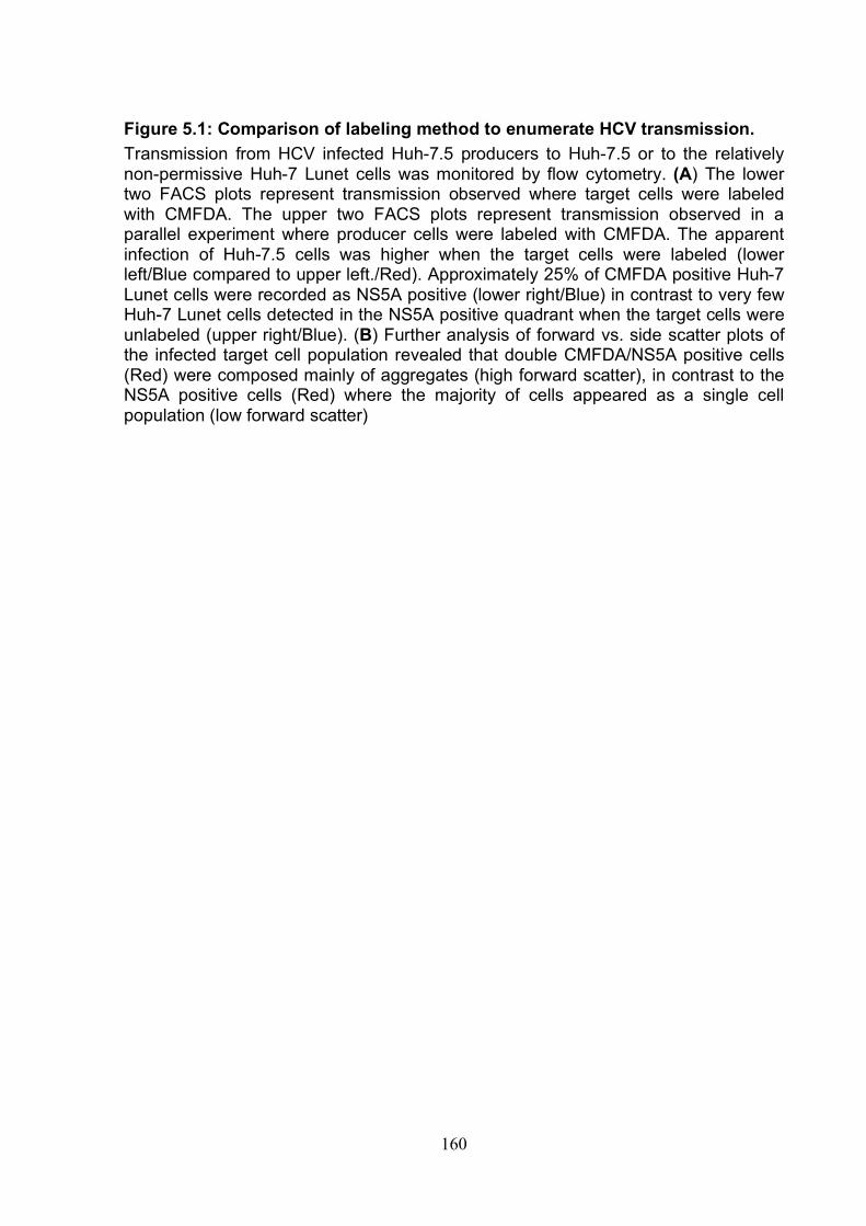
160
Figure 5.1: Comparison of labeling method to enumerate HCV transmission. Transmission from HCV infected Huh-7.5 producers to Huh-7.5 or to the relatively non-permissive Huh-7 Lunet cells was monitored by flow cytometry. (A) The lower two FACS plots represent transmission observed where target cells were labeled with CMFDA. The upper two FACS plots represent transmission observed in a parallel experiment where producer cells were labeled with CMFDA. The apparent infection of Huh-7.5 cells was higher when the target cells were labeled (lower left/Blue compared to upper left./Red). Approximately 25% of CMFDA positive Huh-7 Lunet cells were recorded as NS5A positive (lower right/Blue) in contrast to very few Huh-7 Lunet cells detected in the NS5A positive quadrant when the target cells were unlabeled (upper right/Blue). (B) Further analysis of forward vs. side scatter plots of the infected target cell population revealed that double CMFDA/NS5A positive cells (Red) were composed mainly of aggregates (high forward scatter), in contrast to the NS5A positive cells (Red) where the majority of cells appeared as a single cell population (low forward scatter)
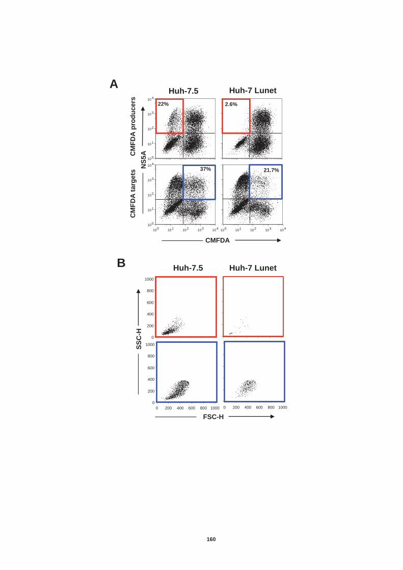
CMFDA
NS
5A
Huh-7.5 Huh-7 Lunet
CM
FD
A p
rod
uc
ers
C
MF
DA
targ
ets
B
A
10 0 10 1 10 2 10 3 10 4
10 0 10 1 10 2 10 3 10 4 10 0 10 1 10 2 10 3 10 4
10 0 10 1 10 2 10 3 10 4
21.7%
2.6%
37%
22%
0 200 400 600 800
1000
0 200 400 600 800 1000 0 200 400 600 800
1000
0 200 400 600 800 1000 FSC-H
SS
C-H
Huh-7.5 Huh-7 Lunet
160
161
Figure 5.2: The infectious co-culture assay. (A) HCV strain H77/JFH RNA was delivered into producer cells by electroporation (ep). 72h post ep producer cells were labeled with CMFDA and co-cultured with unlabeled naïve target cells at a 1:1 ratio in the presence of 10µg/ml irrelevant control IgG or anti-E2 nAb 9/27. (B) i) Supernatant was collected upon termination of the assay and used to infect naïve Huh-7.5 cells in a standard infectious assay, infectivity was readily detectable in the presence of control IgG, none was observed in culture media containing nAb, data represented as foci forming units per ml (FFU/ml). ii) Cells were harvested, fixed, stained for NS5A and quantified by flow cytometry. The cartoon is typical of the data obtained; events appearing within the top left hand quadrant represent (CMFDA - / NS5A +) infected target cells, representative dot plots are shown where the frequency of infected target cells is depicted.
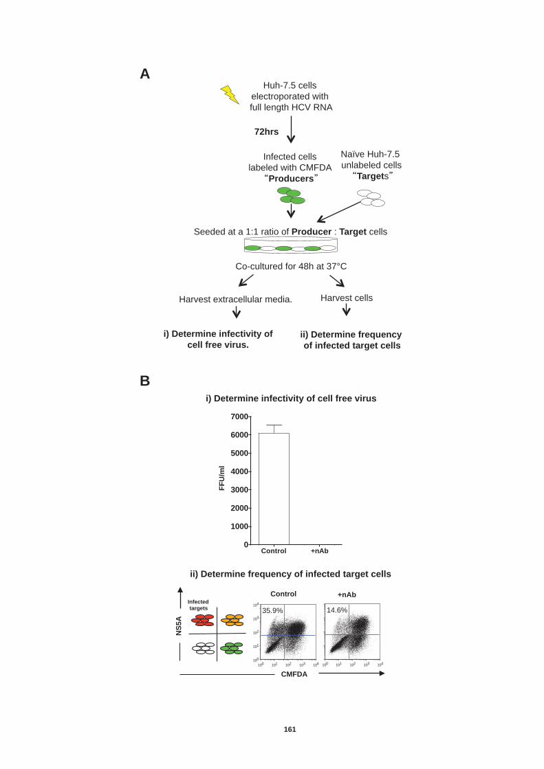
i) Determine infectivity of
cell free virus. ii) Determine frequency
of infected target cells
Huh-7.5 cells
electroporated with
full length HCV RNA
Infected cells
labeled with CMFDA
Producers
Co-cultured for 48h at 37°C
Seeded at a 1:1 ratio of Producer : Target cells
72hrs
Harvest cells Harvest extracellular media.
Naïve Huh-7.5
unlabeled cells
Targets
i) Determine infectivity of cell free virus
CMFDA
NS
5A
+nAb Control Infected targets
ii) Determine frequency of infected target cells
Control +nAb0
1000
2000
3000
4000
5000
6000
7000
10 0 10 1 10 2 10 3 10 4 10 0 10 1 10 2 10 3 10 4
10 0 10 1 10 2 10 3 10 4
35.9% 14.6%
FF
U/m
l
A
B
161
162
We next wanted to establish whether antibodies to different epitopes of E2 also fail to
neutralize cell culture transmission. Three epitope specific antibodies were used
9/27, 3/11 and 11/20 (208). These antibodies target specific regions of H77 E2
glycoprotein. 9/27 targets the HVR-1 region (396-407) and 11/20 and 3/11 target
different regions of the CD81 binding domain (412-423 and 436-447 respectively).
Pooled patient IgG isolated from 6 HCV chronically infected patients was also used.
All antibodies were evaluated for their effects on HCV strain H77/JFH chimera,
transmission (Fig.5.3).
Patient IgG and mAbs 9/27 and 11/20 neutralized all detectable cell-free virus at the
following concentrations; 3µg/ml for 9/27, 10µg/ml for 11/20 and 300µg/ml for patient
sera. In the absence of detectable extra-cellular virus HCV transmission still
occurred, interestingly the percentage of newly infected target cells varied depending
on the antibody under study. Both 9/27 and 11/20 were the most effective Abs at
reducing transmission and continued to neutralize infection once all cell-free virus
had been eliminated, however they were unable to completely ablate transmission at
a maximum concentration of 30µgml.
From these assays we were able to define optimal conditions to measure cell-to-cell
transmission. For H77/JFH chimera we used mAb 9/27 at 4µg/ml. This was the most
effective neutralizing antibody and at this concentration we could be confident that all
cell free virus was neuralized. Where other JFH-1 chimeric viruses were used, cell
free infectivity was neutralized using pooled patient IgG at 300µg/ml.
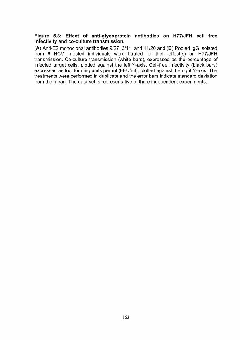
163
Figure 5.3: Effect of anti-glycoprotein antibodies on H77/JFH cell free infectivity and co-culture transmission. (A) Anti-E2 monoclonal antibodies 9/27, 3/11, and 11/20 and (B) Pooled IgG isolated from 6 HCV infected individuals were titrated for their effect(s) on H77/JFH transmission. Co-culture transmission (white bars), expressed as the percentage of infected target cells, plotted against the left Y-axis. Cell-free infectivity (black bars) expressed as foci forming units per ml (FFU/ml), plotted against the right Y-axis. The treatments were performed in duplicate and the error bars indicate standard deviation from the mean. The data set is representative of three independent experiments.
A
9/27 (HVR-1)
B
3/11 (412-423)
11/20 (436-447)
Human HCV +ve IgG
% I
nfe
cte
d t
arg
ets
FF
U/m
l
IgG (μg/ml)
Co-culture transmission Cell-free infectivity
163
164
5.2 Transmission of diverse HCV genotypes in co-culture.
HCV can be grouped into 7 major genotypes and numerous sub-types (166, 444). To
investigate whether all genotypes can transmit in co-culture in the presence of nAbs
Huh-7.5 cells were electroporated with RNA from 11 chimeric JFH viruses,
representative of all major genotypes and subtypes (166). Co-culture transmission
was determined in the presence of pooled patient IgG at 300µg/ml and control IgG.
The levels of infectious cell free virus varied between the different genotypes and
generally predicted efficiency of co-culture transmission (Fig.5.4A), for example
genotype 5a virus (SA13/JFH) produced the highest level of cell-free virus and
transmitted the most efficiently (Fig.5.4C). Cell-free virus of genotypes 1a, 1b, 3, 4, 5
and 6 were completely neutralized by pooled patient IgG (Fig.5.4B). We determined
the proportion of nAb resistant (co-culture transmission in the presence of nAb) and
nAb sensitive transmission (Total co-culture transmission minus nAb resistant
transmission) for all viruses (Fig.5.4C). nAb resistant transmission constituted greater
than 50% of the transmission events with the exception of genotype 6a virus
(HK61/JFH) indicating that this is a preferred route of transmission in co-culture
under the conditions used in this assay.

165
Figure 5.4: Genotype transmission in co-culture. A panel of chimeric JFH-1 RNA was delivered into Huh-7.5 cells by ep. 72hrs post ep cells were labeled with CMFDA and co-cultured with unlabelled target cells in the presence of pooled patient IgG or control IgG (300µg/ml) for 48hrs. Supernatants were harvested at the end of the assay and evaluated for their levels of infectious virus, (A) cell free infectivity displayed as foci forming units per ml (FFU/ml) and (B) neutralization of cell-free infectivity presented as percentage neutralization. (C) Cells were harvested, stained for NS5A and analysed by flow cytometry to determine the frequency of newly infected target cells. Results displayed as a stacked histogram, the white bars represent nAb resistant (Percentage infected target cells in the presence of nAb) and black bars represent nAb sensitive (Total transmission – nAb resistant transmission) transmission. The treatments were performed in duplicate and the error bars indicate standard deviation from the mean. The data set is representative of two independent experiments.
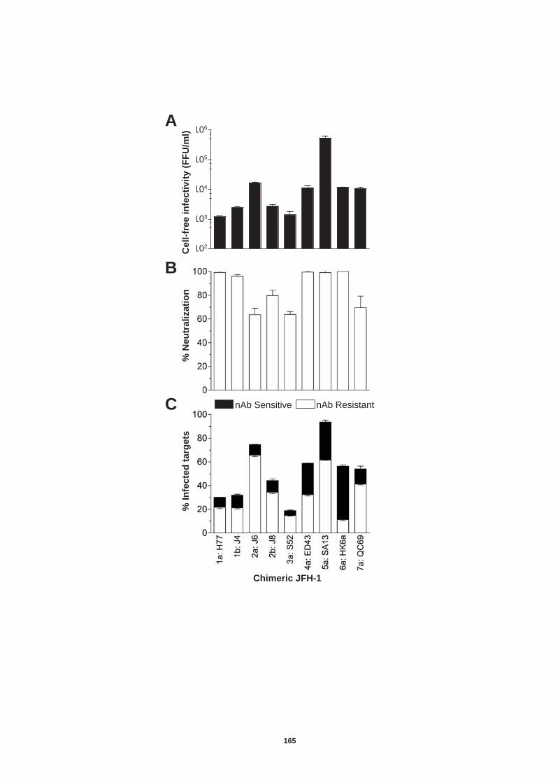
A
B
C
102
103
104
105
106
% N
eu
tra
liza
tio
n
Ce
ll-f
ree
in
fec
tiv
ity
(F
FU
/ml)
%
In
fecte
d t
arg
ets
nAb Sensitive nAb Resistant
Chimeric JFH-1
165
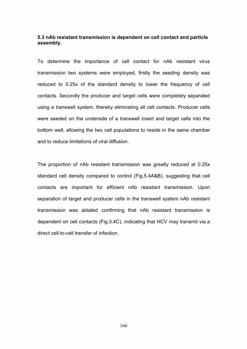
166
5.3 nAb resistant transmission is dependent on cell contact and particle assembly.
To determine the importance of cell contact for nAb resistant virus
transmission two systems were employed, firstly the seeding density was
reduced to 0.25x of the standard density to lower the frequency of cell
contacts. Secondly the producer and target cells were completely separated
using a transwell system, thereby eliminating all cell contacts. Producer cells
were seeded on the underside of a transwell insert and target cells into the
bottom well, allowing the two cell populations to reside in the same chamber
and to reduce limitations of viral diffusion.
The proportion of nAb resistant transmission was greatly reduced at 0.25x
standard cell density compared to control (Fig.5.4A&B), suggesting that cell
contacts are important for efficient nAb resistant transmission. Upon
separation of target and producer cells in the transwell system nAb resistant
transmission was ablated confirming that nAb resistant transmission is
dependent on cell contacts (Fig.5.4C), indicating that HCV may transmit via a
direct cell-to-cell transfer of infection.
167
Figure 5.5: nAb resistant transmission requires cell contact. H77/JFH RNA was delivered into Huh-7.5 cells by ep and used in the following assays set up in parallel (A) Standard assay. (B) 0.25x standard density. (C) Transwell assay; Producer cells were seeded on the underside of a transwell insert and target cells seeded into the bottom chamber. For each assay an irrelevant control IgG or nAb 9/27 was used at 4µg/ml. For (A) and (B) cells were harvest as normal and for (C) the target cells were harvested separately. All cells were stained for NS5A and analysed by flow cytometry. Results are represented as stacked histograms displaying nAb resistant (white bars) and nAb sensitive (black bars) transmission. For all assays cell free infection was determined in a standard infectious assay and >95% of cell free infection was neutralized in the presence of nAb 9/27. The treatments were performed in duplicate and the error bars indicate standard deviation from the mean. The data set is representative of three independent experiments.
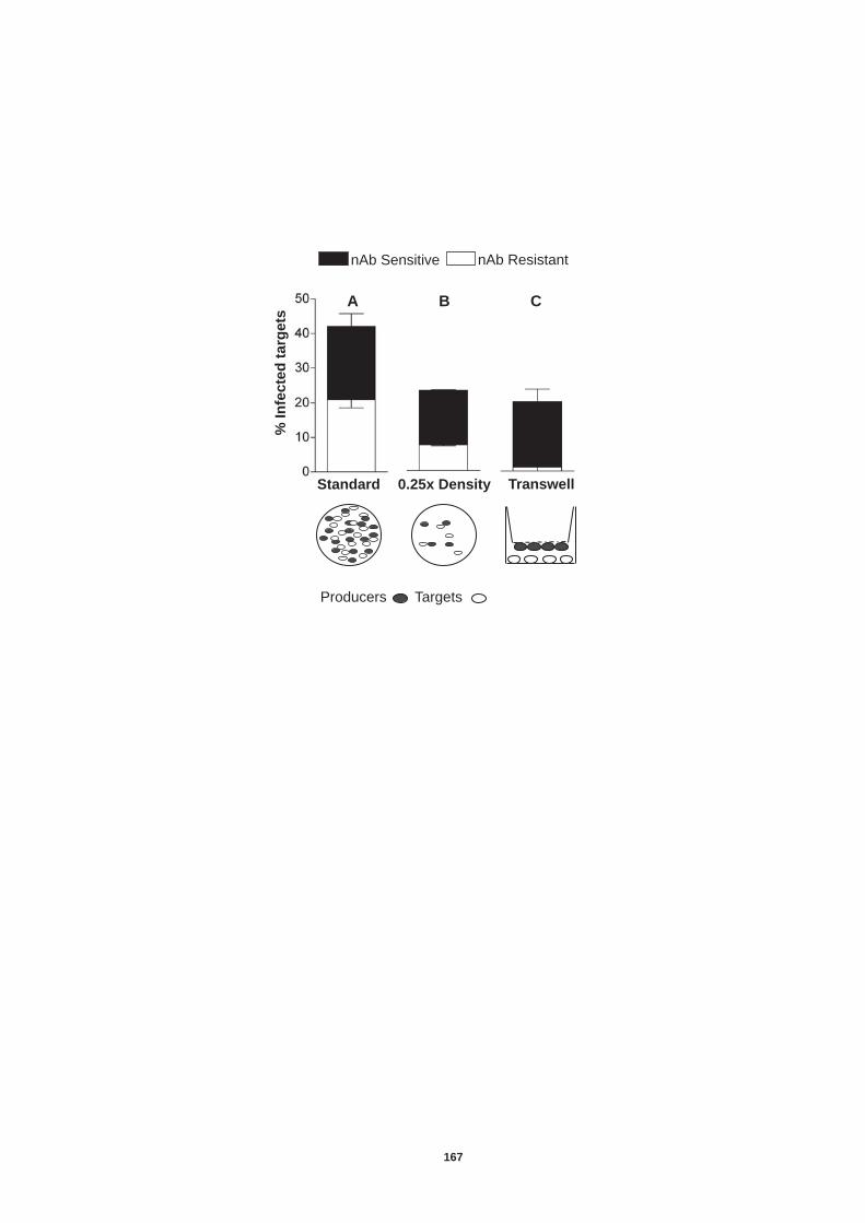
Standard 0.25x Density Transwell
nAb Sensitive nAb Resistant
% I
nfe
cte
d t
arg
ets
Producers Targets
A C B
167
168
It is possible that HCV infection can transmit in a nAb resistant manner via
direct transfer of RNA or incomplete viral particles through a cell fusion event
similar to that reported for herpes viruses (483). Further to this it has been
suggested that HCV RNA can be incorporated into exosomes and may
transfer HCV infectivity (306, 424).
To investigate whether viral particles are required for nAb resistant
transmission a mutant virus J6/JFH del B was employed that contains a
mutation in the third domain of NS5A preventing a phosphorylation event that
is critical for particle assembly whilst permitting wild type levels of replication
and protein translation (473). To ensure that control (J6/JFH) and mutant
(J6/JFH del B) producer cells supported comparable levels of replicating RNA
at the start of the assay NS5A expression of the cells was monitored by flow
cytometry prior to co-culture T=0hr (Fig.5.6). No nAbs were used in this
assay. The mutant virus showed minimal evidence of transmission indicating
that particle assembly is essential for viral transmission in co-culture.
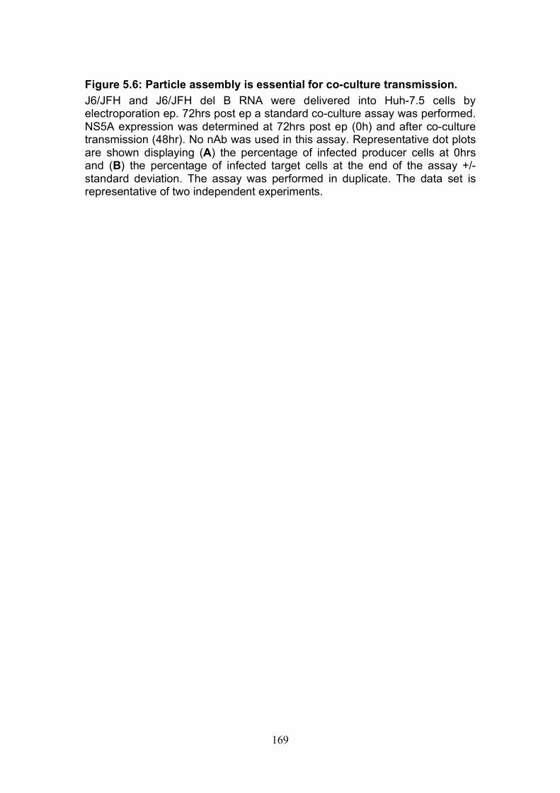
169
Figure 5.6: Particle assembly is essential for co-culture transmission. J6/JFH and J6/JFH del B RNA were delivered into Huh-7.5 cells by electroporation ep. 72hrs post ep a standard co-culture assay was performed. NS5A expression was determined at 72hrs post ep (0h) and after co-culture transmission (48hr). No nAb was used in this assay. Representative dot plots are shown displaying (A) the percentage of infected producer cells at 0hrs and (B) the percentage of infected target cells at the end of the assay +/- standard deviation. The assay was performed in duplicate. The data set is representative of two independent experiments.
CMFDA
NS
5A
J6/J
FH
J6/J
FH
del
B
21%
10 0 10 1 10 2 10 3 10 4
0.03 %
10 0 10 1 10 2 10 3 10 4
20.6 %
10 0 10 1 10 2 10 3 10 4 10 0 10 1 10 2 10 3 10 4
24.6 %
T=0hr T=48hr
169
170
5.4 Receptor dependency of nAb resistant transmission of HCVcc
Results so far demonstrate that HCV can transmit efficiently in co-culture in a
nAb resistant manner, that is dependent on cell contact and particle
assembly. These results suggest that HCV transmits via a direct cell-to-cell
manner as well as through release of cell free viral particles. We next wished
to determine whether direct cell-to-cell transmission is dependent on all four
entry factors, CD81, SR-BI, Claudin-1 and Occludin, previously demonstrated
to be essential for cell-free viral entry (39, 133, 288, 384, 385, 418).
5.4.1 Role of tetraspanin CD81 in nAb resistant transmission.
HCV glycoprotein E2 binds to a defined domain on the large extra cellular
loop of tetraspanin CD81, an essential co-receptor reported to be involved in
the early steps of HCV entry (119, 384, 468). To date there has been
contradicting evidence for the dependency of CD81 in cell-to-cell
transmission. Timpe et al., 2008 and Witteveldt et al., 2009 have shown that
HCV can transmit in co-culture in a CD81 independent manner, however both
of these studies labelled the target cells, and we believe this may have
resulted in false positives (Fig.5.1). A further study reported that CD81 was
essential for HCV transmission (407). To confirm whether CD81 is indeed
essential for nAb resistant co-culture transmission the effects of a panel of
anti-CD81 monoclonal antibodies on HCV transmission were investigated. All
of the anti-receptor antibodies were incubated with the target cells for 1hr at a
concentration of 5µg/ml prior to culturing with H77/JFH infected producer cells
and nAb 9/27. All anti-CD81 antibodies inhibited greater than 95% of nAb
171
resistant and nAb sensitive transmission, demonstrating that both routes of
transmission are CD81 dependent (Fig.5.7A). To investigate this further a
titration of one of the anti-CD81 monoclonal antibodies, 2.s131 was performed
(Fig.5.7B). The antibody inhibited nAb resistant and nAb sensitive
transmission to comparable levels.
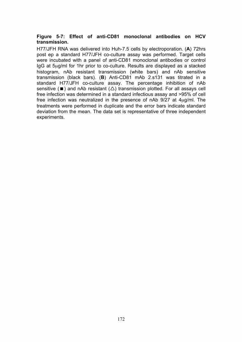
172
Figure 5-7: Effect of anti-CD81 monoclonal antibodies on HCV transmission. H77/JFH RNA was delivered into Huh-7.5 cells by electroporation. (A) 72hrs post ep a standard H77/JFH co-culture assay was performed. Target cells were incubated with a panel of anti-CD81 monoclonal antibodies or control IgG at 5µg/ml for 1hr prior to co-culture. Results are displayed as a stacked histogram, nAb resistant transmission (white bars) and nAb sensitive transmission (black bars). (B) Anti-CD81 mAb 2.s131 was titrated in a standard H77/JFH co-culture assay. The percentage inhibition of nAb sensitive () and nAb resistant () transmission plotted. For all assays cell free infection was determined in a standard infectious assay and >95% of cell free infection was neutralized in the presence of nAb 9/27 at 4µg/ml. The treatments were performed in duplicate and the error bars indicate standard deviation from the mean. The data set is representative of three independent experiments.
nAb Sensitive nAb Resistant
Anti-CD81 mAb (5μg/ml)
% I
nfe
cte
d t
arg
ets
Log10 [anti-CD81] (μg/ml)
% I
nh
ibit
ion
A
B
172
173
To confirm whether CD81 is essential for co-culture transmission we used a
Huh-7 derived cell line (Lunet) that was reported to express low to
undetectable levels of CD81 (251). CD81 cell surface expression was
determined by flow cytometry and a small frequency of Lunet parental cells
expressed low levels of CD81. Lunet parental cells transduced to stably
express CD81 expressed similar levels to Huh-7.5 cells (Fig.5.8A). H77/JFH
and SA13/JFH transmission to parental Lunet cells and those transduced to
express CD81 was monitored (Fig.5.8B). No nAb was used during co-culture.
A small amount of SA13/JFH NS5A expression could be detected in the Lunet
parental cells after co-culture, suggesting a potential CD81 independent
transmission route. Alternatively, this transmission could be attributed to the
small population of cells expressing CD81. To investigate this anti-CD81
(2.s131) was added to the co-culture, the low level of transmission previously
observed was eliminated, suggesting this was not the result of CD81
independent transmission (Fig.5.8B). These findings were confirmed in a
further assay that was analysed instead by confocal microscopy (Fig.5.8C).
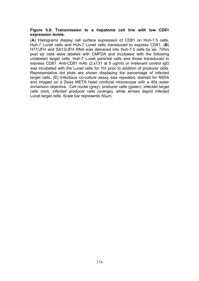
174
Figure 5.8: Transmission to a hepatoma cell line with low CD81 expression levels. (A) Histograms display cell surface expression of CD81 on Huh-7.5 cells, Huh-7 Lunet cells and Huh-7 Lunet cells transduced to express CD81. (B) H77/JFH and SA13/JFH RNA was delivered into Huh-7.5 cells by ep. 72hrs post ep cells were labeled with CMFDA and incubated with the following unlabeled target cells; Huh-7 Lunet parental cells and those transduced to express CD81. Anti-CD81 mAb (2.s131 at 5 µg/ml) or irrelevant control IgG was incubated with the Lunet cells for 1hr prior to addition of producer cells. Representative dot plots are shown displaying the percentage of infected target cells. (C) Infectious co-culture assay was repeated, stained for NS5A and imaged on a Zeiss META head confocal microscope with a 40x water immersion objective. Cell nuclei (grey), producer cells (green), infected target cells (red), infected producer cells (orange), white arrows depict infected Lunet target cells. Scale bar represents 50µm.

B
10 0 10 1 10 2 10 3 10 4 10 0 10 1 10 2 10 3 10 4 10 0 10 1 10 2 10 3 10 4 0 20 40 60 80
100 %
of
Ma
x
CD81
Huh-7.5 Lunet Lunet CD81 A
Lunet CD81 Lunet Lunet (+ anti-CD81)
10 0 10 1 10 2 10 3 10 4 10 0 10 1 10 2 10 3 10 4 10 0 10 1 10 2 10 3 10 4 10 0 10 1 10 2 10 3 10 4
H77/J
FH
S
A13
NS
5A
14.1%
81.3%
0.9%
2.3%
0.4%
10 0 10 1 10 2 10 3 10 4
0.1%
C
SA
13
H77
Lunet Lunet CD81 Lunet (+anti-CD81)
CMFDA
174
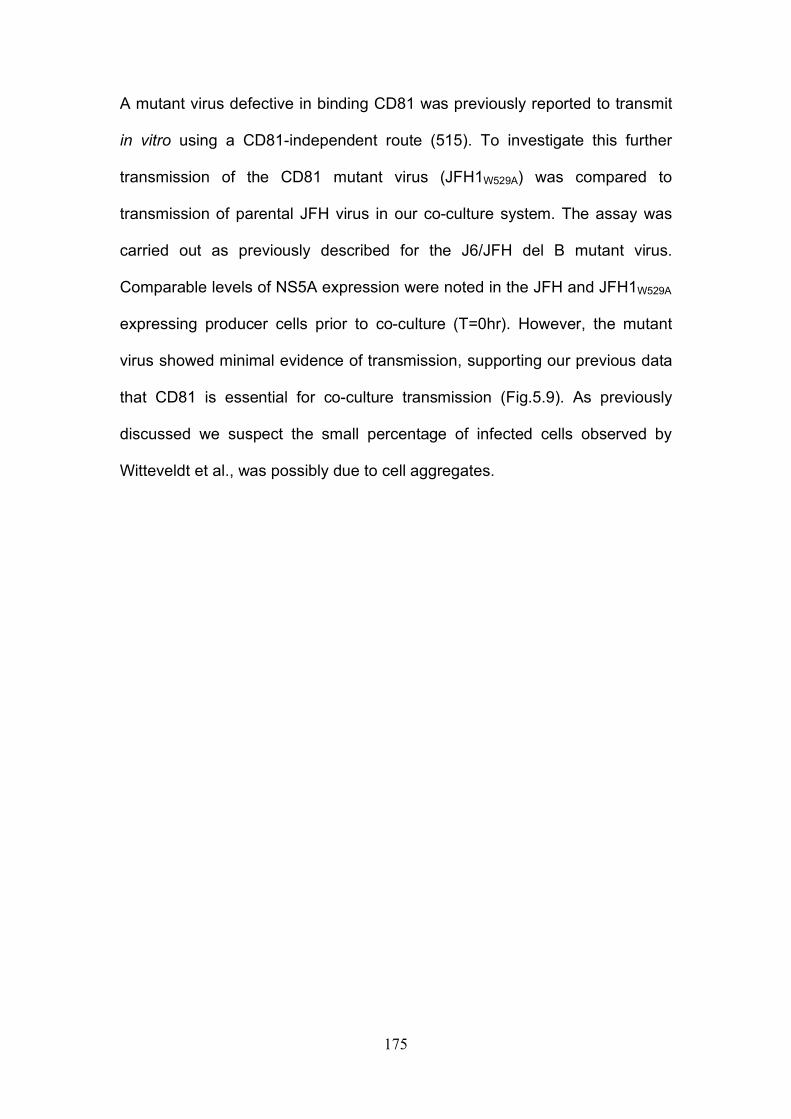
175
A mutant virus defective in binding CD81 was previously reported to transmit
in vitro using a CD81-independent route (515). To investigate this further
transmission of the CD81 mutant virus (JFH1W529A) was compared to
transmission of parental JFH virus in our co-culture system. The assay was
carried out as previously described for the J6/JFH del B mutant virus.
Comparable levels of NS5A expression were noted in the JFH and JFH1W529A
expressing producer cells prior to co-culture (T=0hr). However, the mutant
virus showed minimal evidence of transmission, supporting our previous data
that CD81 is essential for co-culture transmission (Fig.5.9). As previously
discussed we suspect the small percentage of infected cells observed by
Witteveldt et al., was possibly due to cell aggregates.
176
Figure 5.9: Transmission of a CD81 negative mutant virus. JFH and JFH1W529A RNA was delivered into Huh-7.5 cells by ep. 72hrs post ep a standard co-culture assay was performed. NS5A expression was determined at 72hr post ep (0hr) and after co-culture transmission (48hr). No nAb was used in this assay. Representative dot plots are shown displaying (A) the percentage of infected producer cells at 0hr and (B) the percentage of infected target cells at the end of the assay. The assay was performed in duplicate. The data set is representative of two independent experiments.
10 0 10 1 10 2 10 3 10 4
10 0
10 1
10 2
10 3
10 4
38%
10 0
10 1
10 2
10 3
10 4
10 0 10 1 10 2 10 3 10 4
33%
CMFDA
NS
5A
JF
H-1
JF
H-1
CD
81 -
ve m
uta
nt
T=0hr T=48hr
3 %
0.04%
176
177
5.4.2 Role of SR-BI in nAb resistant co-culture transmission SR-BI is a cell membrane protein involved in cholesterol metabolism; it
functions as a receptor for high-density lipoproteins. It has also been shown to
bind directly to HCV E2 glycoprotein and is believed to be important in the
early stages of HCV attachment and entry (174, 418). To investigate the role
of SR-BI in nAb resistant co-culture transmission the effect of anti-SR-BI
antibody on transmission was investigated. As previously described the target
cells were cultured in the presence of the anti-receptor antibody for 1hr prior
to addition of H77/JFH infected producer cells and nAb 9/27. Titration of the
anti-SR-BI antibody revealed that nAb resistant transmission is preferentially
inhibited at all concentrations (Fig.5.10A). To investigate whether this
phenomenon was due to a potential synergy between 9/27, a neutralizing
antibody specific for E2 HVR-1, and the anti-SR-BI antibody transmission in
the presence of another epitope specific antibody 11/20 was determined
(Fig.5.10B). 11/20 targets part of the E2 CD81 binding domain (208). A
significant difference between anti-SR-BI inhibition of nAb resistant and
sensitive transmission was observed in the presence of both epitope specific
nAbs indicating the synergy is not specific to the HVR-1 targeting antibody.
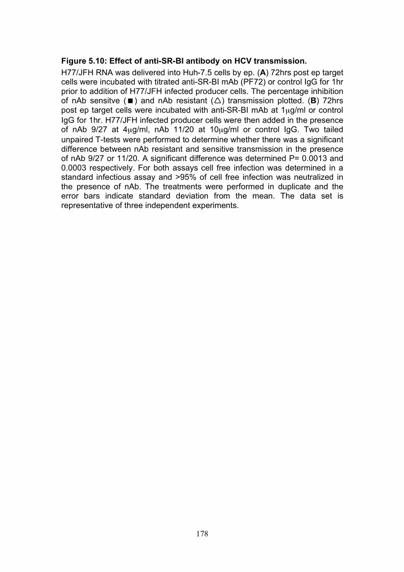
178
Figure 5.10: Effect of anti-SR-BI antibody on HCV transmission. H77/JFH RNA was delivered into Huh-7.5 cells by ep. (A) 72hrs post ep target cells were incubated with titrated anti-SR-BI mAb (PF72) or control IgG for 1hr prior to addition of H77/JFH infected producer cells. The percentage inhibition of nAb sensitve () and nAb resistant () transmission plotted. (B) 72hrs post ep target cells were incubated with anti-SR-BI mAb at 1µg/ml or control IgG for 1hr. H77/JFH infected producer cells were then added in the presence of nAb 9/27 at 4µg/ml, nAb 11/20 at 10µg/ml or control IgG. Two tailed unpaired T-tests were performed to determine whether there was a significant difference between nAb resistant and sensitive transmission in the presence of nAb 9/27 or 11/20. A significant difference was determined P= 0.0013 and 0.0003 respectively. For both assays cell free infection was determined in a standard infectious assay and >95% of cell free infection was neutralized in the presence of nAb. The treatments were performed in duplicate and the error bars indicate standard deviation from the mean. The data set is representative of three independent experiments.
A
B nAb Sensitive nAb Resistant
% I
nh
ibit
ion
178
179
To further examine the role of SR-BI in nAb resistant transmission, virus
dissemination was determined to Huh-7.5 cells and those transduced to over
express SR-BI. Our laboratory previously published that cell-free virus
infection of these cells induced greater foci size indicative of increased cell-to-
cell transmission (175). nAb resistant transmission of virus to target cells over
expressing SR-BI significantly increased however, SR-BI overexpression had
no effect on nAb sensitive transmission (Fig.5.11A). This data indicates that
SR-BI expression may be a limiting factor for efficient nAb resistant
transmission.
To further investigate the role of SR-BI in nAb resistant infection, SR-BI was
silenced in Huh-7.5 cells and transmission compared to parental Huh-7.5
cells. Receptor staining of parental and SR-BI silenced Huh-7.5 cells was
carried out to determine efficacy of SR-BI silencing and to verify any off target
effects (Fig.5.11B). SR-BI expression level was reduced and expression level
of the other receptors remained with the exception of tetraspanin CD81 that
was reduced; this was confirmed by flow cytometry (Fig.5.11C). Silencing SR-
BI significantly inhibited both routes of viral transmission and did not
preferentially inhibit nAb resistant transmission as expected from previous
results (Fig.5.11D). It has recently been published that silencing SR-BI
reduced cholesterol levels and inhibited plasmodium infection of hepatocytes
(523). A sample of SR-BI silenced cells were lysed at the start of the assay to
determined cholesterol levels. This confirmed that indeed SR-BI silenced cells
had reduced levels of total cholesterol (Fig.5.11E).

180
Figure 5.11: Effect of SR-BI expression levels on HCV transmission. H77/JFH RNA was delivered into Huh-7.5 cells by ep. (A) A standard co-culture assay was performed with control target cells and those transduced to over express SR-BI. A significant difference in nAb resistant transmission to control and SR-BI over expressing cells was observed, (P=0.0007, two tailed unpaired T-test). No significant difference was observed in nAb sensitive transmission, (P=0.2430, two tailed unpaired T-test). Huh-7.5 target cells were transduced to express sh SR-BI RNA (SR-BI KD). (B) Receptor expression of SR-BI KD and control cells. Images taken on a Zeiss META head confocal microscope with a 60x water immersion objective, receptor expression visualized using alexa-fluor 488 secondary antibody (green) and DAPI (blue) show cell nuclei. Scale bar represents 10µm. (C) SR-BI and CD81 expression also determined by flow cytometry (Anti-SR-BI (PF72) and anti-CD81 (2.s131mAb) used). (D) A standard H77/JFH co-culture assay was performed with control and SR-BI KD cells as targets. Two tailed unpaired T-tests were performed to determine whether there was a significant difference in nAb sensitive and resistant transmission between control and SR-BI KD cells. A significant difference was determined P= 0.0127 and 0.0045 respectively. For both co-culture assays supernatants were collected to determine cell free infection and >95% of cell free infection was shown to be neutralized in the presence of nAb 9/27 at 4µg/ml. (E) Total cholesterol levels were determined in control and SR-BI KD cells using Invitrogen Amplex Red Cholesterol Assay Kit. Assays were performed in duplicate and the error bars indicate standard deviation from the mean. The data sets are representative of two independent experiments.
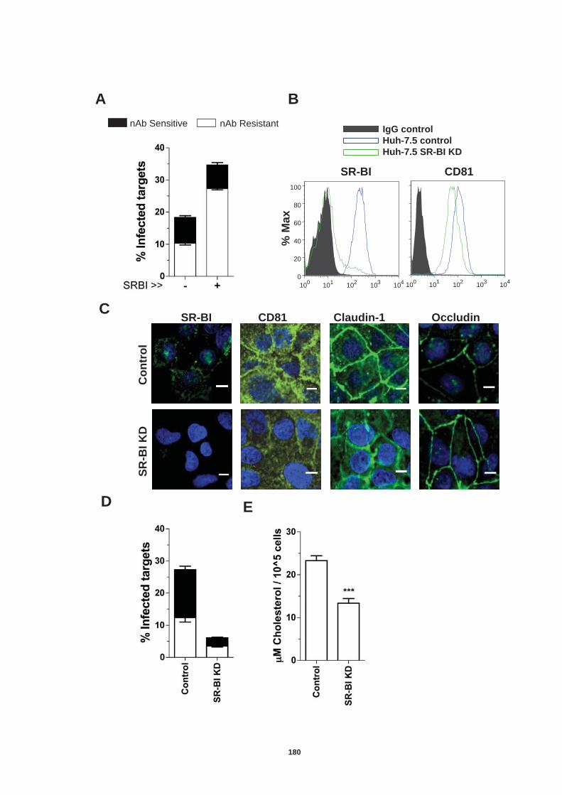
A B
C
D
CD81 SR-BI Claudin-1 Occludin
SR
-BI
KD
C
on
tro
l
10 0 10 1 10 2 10 3 10 4 0
20 40 60 80
100
10 0 10 1 10 2 10 3 10 4
CD81 SR-BI
% M
ax
IgG control
Huh-7.5 control
Huh-7.5 SR-BI KD
E
nAb Sensitive nAb Resistant
180
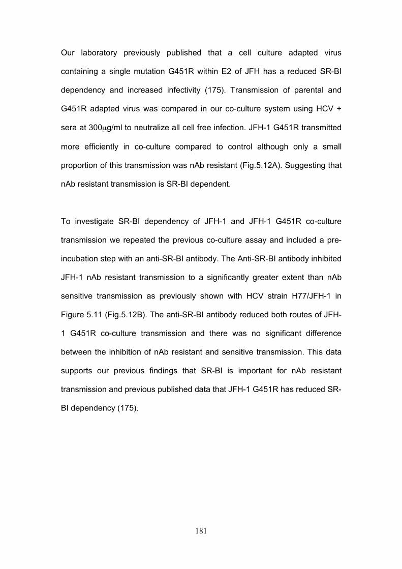
181
Our laboratory previously published that a cell culture adapted virus
containing a single mutation G451R within E2 of JFH has a reduced SR-BI
dependency and increased infectivity (175). Transmission of parental and
G451R adapted virus was compared in our co-culture system using HCV +
sera at 300µg/ml to neutralize all cell free infection. JFH-1 G451R transmitted
more efficiently in co-culture compared to control although only a small
proportion of this transmission was nAb resistant (Fig.5.12A). Suggesting that
nAb resistant transmission is SR-BI dependent.
To investigate SR-BI dependency of JFH-1 and JFH-1 G451R co-culture
transmission we repeated the previous co-culture assay and included a pre-
incubation step with an anti-SR-BI antibody. The Anti-SR-BI antibody inhibited
JFH-1 nAb resistant transmission to a significantly greater extent than nAb
sensitive transmission as previously shown with HCV strain H77/JFH-1 in
Figure 5.11 (Fig.5.12B). The anti-SR-BI antibody reduced both routes of JFH-
1 G451R co-culture transmission and there was no significant difference
between the inhibition of nAb resistant and sensitive transmission. This data
supports our previous findings that SR-BI is important for nAb resistant
transmission and previous published data that JFH-1 G451R has reduced SR-
BI dependency (175).
182
Figure 5.12: Transmission of a cell culture adapted virus with reduced SR-BI dependency. JFH-1 and JFH-1 G451R RNA were delivered into Huh-7.5 cells by ep. (A) 72hrs post ep a standard co-culture assay was performed. The stacked histogram displays nAb resistant (white bars) and nAb sensitive transmission (black bars). (B) Target cells were incubated with anti-SR-BI mAb (PF72) or IgG control at 1µg/ml for 1hr prior to addition of JFH-1 and JFH-1 G451R infected producer cells. The mAb was present throughout the duration of the assay. Results are represented as percentage inhibition of nAb resistant (white bars) and nAb sensitive (black bars) transmission. A significant difference between percentage inhibition of nAb resistant and sensitive transmission of JFH-1 virus was determined, (P=0.0092, two tailed unpaired T-test). No significant difference for JFH-1 G451R was observed, (P=0.2096, two tailed unpaired T-test). For all assays cell free infection was determined in a standard infectious assay and >95% of cell free infection was neutralized in the presence of pooled patient IgG (300µg/ml). Assays were performed in duplicate and the error bars indicate standard deviation from the mean. The data set is representative of three independent experiments.
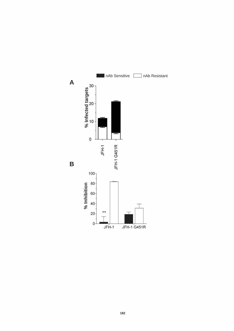
nAb Sensitive nAb Resistant
% I
nh
ibit
ion
A
B
% I
nfe
cte
d t
arg
ets
182
183
Syder and colleagues recently reported a number of small molecular inhibitors
that target SR-BI as effective inhibitors of HCV entry (462). ITX5061 and
ITX7650 have been reported to function by inhibiting HCV E2 glycoprotein-
SR-BI interaction and not by affecting overall SR-BI expression levels. We
wanted to investigate their efficacy to inhibit nAb resistant and nAb sensitive
transmission. An MTS assay (colorimetric test that measures metabolic
function) was performed to ascertain the effect(s) of ITX compounds on
hepatoma cell proliferation (Fig.5.13A). The inhibitors were then used in our
standard co-culture assay (Fig.5.13B). These inhibitors were relatively
ineffective at targeting cell free infection but as with antibodies targeting SR-BI
they were able to efficiently inhibit nAb resistant transmission.
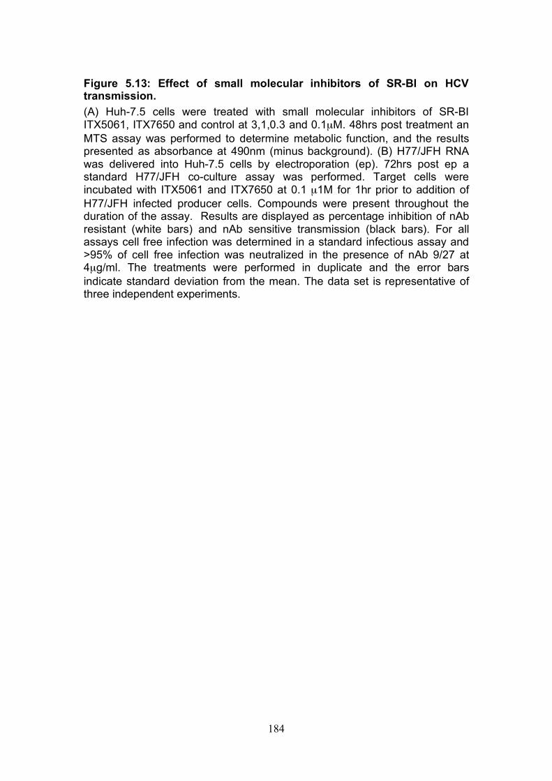
184
Figure 5.13: Effect of small molecular inhibitors of SR-BI on HCV transmission. (A) Huh-7.5 cells were treated with small molecular inhibitors of SR-BI ITX5061, ITX7650 and control at 3,1,0.3 and 0.1µM. 48hrs post treatment an MTS assay was performed to determine metabolic function, and the results presented as absorbance at 490nm (minus background). (B) H77/JFH RNA was delivered into Huh-7.5 cells by electroporation (ep). 72hrs post ep a standard H77/JFH co-culture assay was performed. Target cells were incubated with ITX5061 and ITX7650 at 0.1 µ1M for 1hr prior to addition of H77/JFH infected producer cells. Compounds were present throughout the duration of the assay. Results are displayed as percentage inhibition of nAb resistant (white bars) and nAb sensitive transmission (black bars). For all assays cell free infection was determined in a standard infectious assay and >95% of cell free infection was neutralized in the presence of nAb 9/27 at 4µg/ml. The treatments were performed in duplicate and the error bars indicate standard deviation from the mean. The data set is representative of three independent experiments.
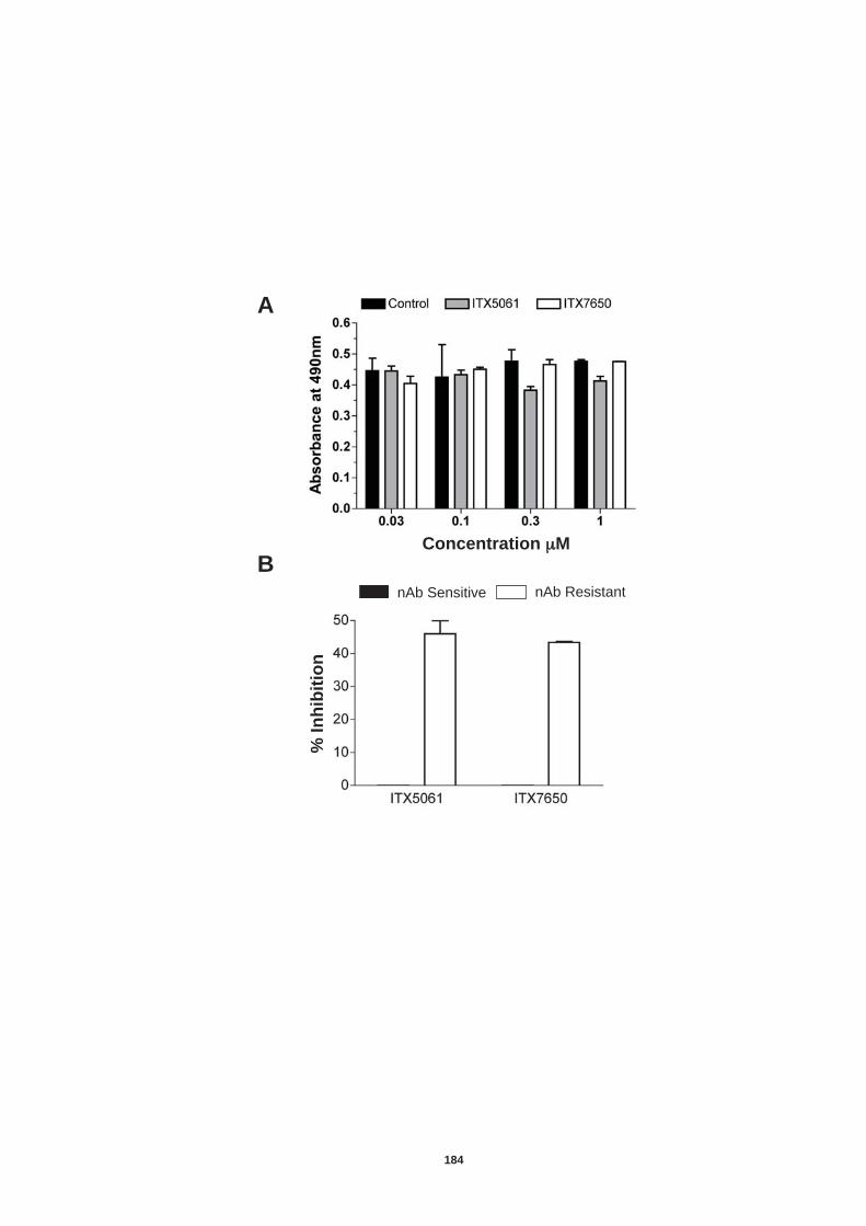
nAb Sensitive nAb Resistant
% I
nh
ibit
ion
A
B Concentration μM
184
185
5.4.3. Role of tight junction proteins CLDN-1 and Occludin in nAb resistant HCV transmission.
The tight junction proteins Claudin-1 and Occludin have only recently been
identified as co-receptors for HCV, as such there are limited tools available to
study these receptors. Recent studies indicate that they are both important in
the latter stages of HCV entry and as yet there is no evidence for a direct
interaction of these proteins with the HCV glycoproteins (39, 133, 288, 385).
To study the role of Claudin-1 in nAb resistant transmission a recently
described polyclonal antiserum raised to Claudin-1 was used (255). Target
cells were incubated with a pre-immune and a post-immune serum for 1hr
prior to the addition of H77/JFH infected producer cells. A standard co-culture
assay was then performed using nAb 9/27 at 4µg/ml to neutralize cell free
infectivity. The polyclonal anti-Claudin-1 antibody inhibited approximately 50%
of nAb sensitive transmission and approximately 30% of nAb resistant
transmission, indicating both routes of transmission require Claudin-1
(Fig.5.14A).
Further to this we carried out a standard co-culture assay using Claudin-1
silenced cells as targets. Receptor staining of control and silenced cells were
carried out to determine the efficacy of silencing and to confirm expression of
other HCV co-receptors. Immuno-fluorescence results revealed that Occludin
expression was also reduced upon Claudin-1 silencing (Fig.5.14B). Claudin-1
silencing reduced nAb sensitive and resistant transmission by 47 and 32 %
186
respectively supporting our previous data that both routes of transmission
require Claudin-1 (Fig.5.14C).
No antibodies to extra-cellular Occludin epitopes are available; we therefore
silenced Occludin in the target cells and performed a standard co-culture
assay as previously carried out for Claudin-1. Receptor staining of control and
Occludin silenced cells was carried out to determine efficacy of silencing and
to confirm expression of other HCV co-receptors. Occludin was efficiently
silenced and no differences in co-receptor expression were observed
(Fig.5.14A). Occludin silencing reduced nAb sensitive and nAb resistant
transmission by 80% and 60% respectively (Fig.5.14B). Results indicate that
both tight junction proteins Claudin-1 and Occludin are essential for both
routes of transmission.
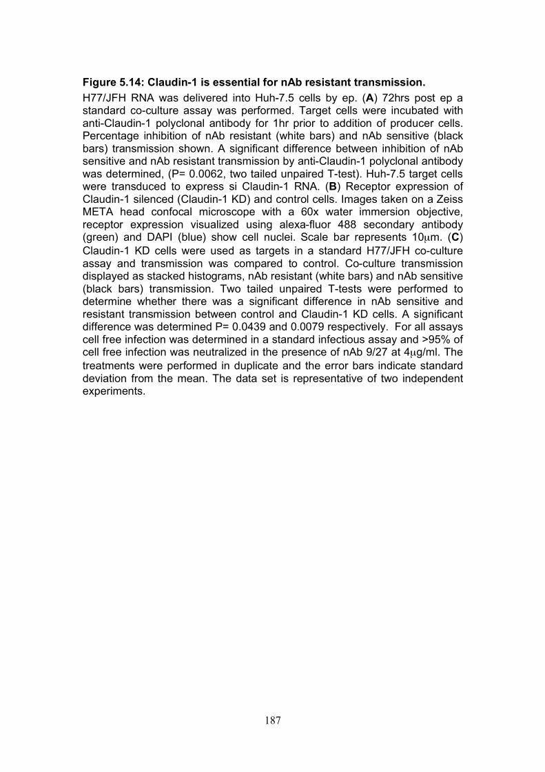
187
Figure 5.14: Claudin-1 is essential for nAb resistant transmission. H77/JFH RNA was delivered into Huh-7.5 cells by ep. (A) 72hrs post ep a standard co-culture assay was performed. Target cells were incubated with anti-Claudin-1 polyclonal antibody for 1hr prior to addition of producer cells. Percentage inhibition of nAb resistant (white bars) and nAb sensitive (black bars) transmission shown. A significant difference between inhibition of nAb sensitive and nAb resistant transmission by anti-Claudin-1 polyclonal antibody was determined, (P= 0.0062, two tailed unpaired T-test). Huh-7.5 target cells were transduced to express si Claudin-1 RNA. (B) Receptor expression of Claudin-1 silenced (Claudin-1 KD) and control cells. Images taken on a Zeiss META head confocal microscope with a 60x water immersion objective, receptor expression visualized using alexa-fluor 488 secondary antibody (green) and DAPI (blue) show cell nuclei. Scale bar represents 10µm. (C) Claudin-1 KD cells were used as targets in a standard H77/JFH co-culture assay and transmission was compared to control. Co-culture transmission displayed as stacked histograms, nAb resistant (white bars) and nAb sensitive (black bars) transmission. Two tailed unpaired T-tests were performed to determine whether there was a significant difference in nAb sensitive and resistant transmission between control and Claudin-1 KD cells. A significant difference was determined P= 0.0439 and 0.0079 respectively. For all assays cell free infection was determined in a standard infectious assay and >95% of cell free infection was neutralized in the presence of nAb 9/27 at 4µg/ml. The treatments were performed in duplicate and the error bars indicate standard deviation from the mean. The data set is representative of two independent experiments.
A
B
C
Cla
ud
in-1
KD
C
on
tro
l
CD81 SR-BI Claudin-1 Occludin
nAb Sensitive nAb Resistant
187
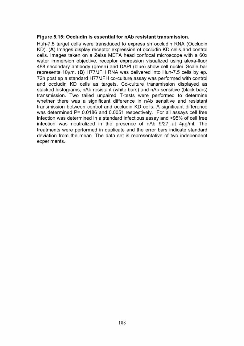
188
Figure 5.15: Occludin is essential for nAb resistant transmission. Huh-7.5 target cells were transduced to express sh occludin RNA (Occludin KD). (A) Images display receptor expression of occludin KD cells and control cells. Images taken on a Zeiss META head confocal microscope with a 60x water immersion objective, receptor expression visualized using alexa-fluor 488 secondary antibody (green) and DAPI (blue) show cell nuclei. Scale bar represents 10µm. (B) H77/JFH RNA was delivered into Huh-7.5 cells by ep. 72h post ep a standard H77/JFH co-culture assay was performed with control and occludin KD cells as targets. Co-culture transmission displayed as stacked histograms, nAb resistant (white bars) and nAb sensitive (black bars) transmission. Two tailed unpaired T-tests were performed to determine whether there was a significant difference in nAb sensitive and resistant transmission between control and occludin KD cells. A significant difference was determined P= 0.0186 and 0.0051 respectively. For all assays cell free infection was determined in a standard infectious assay and >95% of cell free infection was neutralized in the presence of nAb 9/27 at 4µg/ml. The treatments were performed in duplicate and the error bars indicate standard deviation from the mean. The data set is representative of two independent experiments.
A
B
Occlu
din
KD
C
on
tro
l
CD81 SR-BI Claudin-1 Occludin
nAb Sensitive nAb Resistant
188
189
5.4.4 Receptor dependency of nAb resistant transmission of multiple HCV genotypes.
In this project so far, we have only studied the effect(s) of antibody
antagonists on genotype 1a chimeric virus H77/JFH co-culture transmission.
To strengthen our findings the effect(s) of these antagonists were determined
for multiple HCV genotypes. A pooled patient sera at 300µg/ml was able to
neutralize cell free infectivity of JFH chimera’s representative of genotypes 1a,
1b, 3, 4, 5 and 6 in co-culture (Fig.5.4B). Therefore a standard co-culture
assay was performed using patient IgG to neutralize cell free infectivity for all
of the above genotypes. Target cells were incubated for 1hr with the following
inhibitors prior to co-culture; anti-CD81 (2.s131 at 5µg/ml), anti-CLDN-1
(polyclonal antibody at 1in 100), anti-SR-BI (PF72 at 5µg/ml) and small
molecular SR-BI inhibitors (ITX5061 and ITX7650 at 1µM) (Table 5.1).
Sensitivity to anti-CD81 antibodies were similarly high for all genotypes and
inhibited both nAb resistant and sensitive transmission equally with the
exception of SA13/JFH. The anti-CLDN-1 polyclonal antibody was not
efficient at blocking co-culture transmission and in the majority of cases
blocked nAb sensitive to a greater extent than nAb resistant transmission.
Treatment with the anti-SR-BI antibody and SR-BI small molecule inhibitor
ITX5061 had comparable effects on transmission and both antagonists
preferentially blocked the nAb resistant route.
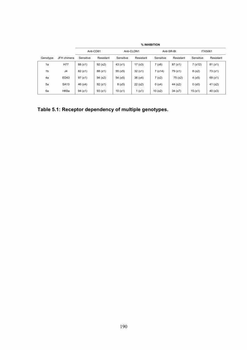
190
Table 5.1: Receptor dependency of multiple genotypes.
Genotype JFH chimera Sensitive Resistant Sensitive Resistant Sensitive Resistant Sensitive Resistant
1a H77 88 (±1) 92 (±2) 43 (±1) 17 (±3) 7 (±6) 87 (±1) 7 (±12) 81 (±1)
1b J4 82 (±1) 88 (±1) 55 (±5) 32 (±1) 7 (±14) 79 (±1) 8 (±2) 73 (±1)
4a ED43 97 (±1) 94 (±2) 54 (±5) 36 (±4) 7 (±2) 75 (±2) 4 (±5) 69 (±1)
5a SA13 46 (±4) 92 (±1) 8 (±5) 22 (±2) 0 (±4) 44 (±2) 0 (±0) 41 (±2)
6a HK6a 94 (±1) 93 (±1) 10 (±1) 1 (±1) 10 (±2) 34 (±7) 15 (±1) 40 (±3)
% INHIBITION
Anti-CD81 Anti-CLDN1 Anti-SR-BI ITX5061
191
5.5 Discussion
In this study we developed a co-culture assay that allowed us to examine
HCV transmission in vitro (Fig.5.2). Our results support previous observations
made using standard infectious assays; cell free HCV particles are relatively
sensitive to neutralizing antibodies in vitro (29, 359, 560). Importantly our
results demonstrate that a proportion of co-culture transmission is highly
insensitive to neutralization by antibodies targeting the viral glycoproteins. We
envisage that the co-culture system used in this study demonstrates
neutralizing antibody sensitivities that are more akin to the in vivo situation
than standard infection assays.
To investigate the sensitivity of HCV transmission to different antibodies
targeting the viral glycoproteins, H77/JFH chimera (genotype 1a virus) was
studied. Patient IgG isolated from HCV infected patients and eptiope specifc
antibodies 11/20 and 9/27 targeting the CD81 binding domain and the HVR-1
of E2 respectively neutralized > 95% of the infectivity of cell-free virus whilst
still allowing new rounds of virus transmission to occur (Fig.5.3). Interestingly
different levels of co-culture transmission were observed depending on the
antibody studied. Importantly, once all cell free virus infectivity was
neutralized, mAbs 11/20 and 9/27 were able to limit co-culture transmission
although they were unable to completely abrogate all transmission at a final
concentration of 30µg/ml. These results suggest that nAb resistant
transmission does not occur through a sealed junction, that is impermeable to
HCV glycoprotein targeting antibodies. Rather susceptibility to neutralization
192
is variable and dependent on the antibody in question, this maybe due to
differences in antibody affinities or target epitope availability. The majority of
HCV RNA is reported to associate with lipoproteins in vitro and in vivo, these
are termed lipo-viro-particles (LVP) (14). It has previously been reported that
lipoprotein association increases specific infectivity and decreases
neutralization efficacy (175, 215). It is therefore interesting to postulate
whether the associated lipoproteins may aid nAb resistant viral transmission.
To further characterize co-culture transmission we defined nAb resistant cell-
to-cell transmission as the proportion of transmission that occurred in the
presence of a anti-glycoprotein specific Ab capable of inhibiting between 90
and 100% of all cell-free virus infectivity. For the majority of experiments we
investigated H77/JFH transmission and used the most proficient epitope
specific antibody 9/27 at 4µg/ml, a concentration able to effectively neutralize
cell free infectivity. When it was not possible to use H77/JFH we used patient
IgG isolated from HCV infected patients that was able to neutralize cell free
infectivity at 300µg/ml.
The proportion of nAb resistant transmission decreased when target and
producer cell contacts were reduced (Fig.5.5A) and was ablated upon
separation of the cells (Fig.5.5B). These findings suggest that cell contacts
are essential for nAb resistant transmission and HCV can transmit via direct
cell-to-cell route(s) as well as release of cell-free virus. A mutant virus (J6/JFH
del B) that is un-able to assemble viral particles did not transmit infection in
co-culture (Fig.5.6), demonstrating that viral particles are required for co-
193
culture transmission. Furthermore, comparison of the levels of infectious virus
released from the chimeric JFH viruses and the percentage of target cells
infected in co-culture revealed an association between infectious cell-free
virus and transmission, suggesting that particles are required to initiate both
nAb resistant and sensitive forms of transmission (Fig 5.6). These findings do
not support a recent hypothesis that HCV RNA directly transmits through cell-
to-cell fusion events via the exosomal excretory pathway (424).
Importantly we demonstrated that diverse HCV genotypes can transmit via
nAb resistant cell-to-cell route(s) using a panel of chimeric JFH viruses
expressing the structural proteins that represent the major genotypes (166)
(Fig.5.4). For 7 out of the 11 strains studied > 95% of cell-free infectivity was
neutralized by IgG purified from the sera of HCV infected subjects (Fig.5.4B).
We can be confident that these genotypes transmit in a nAb resistant manner.
However the remaining genotypes were only partially neutralized by the
patient IgG, therefore we cannot conclusively determine whether these
viruses transmit in a nAb resistant manner.
Four co-receptors are known to be essential for entry of cell-free virus, we
were interested to investigate the receptor dependency nAb resistant cell-to-
cell transmission. Contradicting evidence exists as to whether the tetraspanin
CD81 is essential for HCV co-culture transmission (229, 407, 480, 515).
Timpe et al., 2008 reported that HCV could transmit to a CD81 negative
HepG2 cell line. Witteveldt et al., 2009 supported these findings by showing
that a mutant virus (JFH1W529A) that was un-able to interact with CD81 was
194
able to transmit in vitro. We believe these findings were a result of
misinterpretation of flow cytometry data due to the formation of aggregates
between labeled target cells and unlabelled infected producer cells (Fig. 5.1).
We failed to observe any detectable transmission of infectivity of the
JFH1W529A virus in our co-culture system (Fig.5.9), suggesting that HCV
interaction with CD81 is indeed essential, as previously reported by Russel et
al., 2008. A panel of anti-CD81 monoclonal antibodies were able to block
greater than 85% of both routes of transmission for all HCV genotypes with
the exception of genotype 5a virus SA13/JFH (Table 5.1). Suggesting that for
the majority of genotypes CD81 is equally important for all routes of co-culture
transmission. Transmission of H77/JFH and SA13/JFH was monitored to
Lunet cells that express very low levels of CD81 (Fig.5.8B&C). Negligible
transmission of H77/JFH was recorded to Lunet cells and this was rescued by
ectopic CD81 expression, supporting our previous data that CD81 is essential
for co-culture transmission. Notably a small population of SA13/JFH infected
Lunet cells was observed, importantly both flow cytometry analysis and
confocal imaging detected this transmission. To investigate whether this was
evidence supporting CD81 independent transmission an anti-CD81 mAb was
added to the co-culture. Anti-CD81 mAb completely abrogated SA13/JFH
transmission to lunet cells demonstrating that the previous transmission
observed was not evidence of CD81 independent transmission rather it was
showing the efficiency of SA13/JFH transmission. Recent data from our
laboratory revealed that SA13/JFH RNA level per infected cell is comparable
to other genotypes indicating the difference in infectivity is unlikely to be due
to an increase in replication efficacy (Un published data by Dr. P. Balfe and

195
Dr. N. Fletcher). These results suggest that SA13/JFH is able to utilize a
lower threshold of CD81 compared to other viruses. Koutsoudakis et al.,
reported that HCVcc needs a threshold level of CD81 to infect hepatoma cells
(251). It would be interesting to see if the same threshold holds true for
SA13/JFH. In summary we have demonstrated that CD81 is indispensable for
HCV infection and transmission in co-culture.
Our observations demonstrate that both tight junction proteins Claudin-1 and
Occludin are required for HCV co-culture transmission. Previous studies using
parental 293T cells and those transduced to express Claudin-1 reported that
claudin-1 is essential for co-culture transmission of HCVcc but did not
discriminate between nAb resistant and nAb sensitive transmission (480). We
observed that nAb sensitive transmission was blocked to a greater extent than
nAb resistant transmission by an anti-Claudin-1 polyclonal antibody for all
genotypes tested (Table 5.1). Supporting this, nAb sensitive transmission was
more sensitive to Claudin-1 expression than nAb resistant transmission
(Fig.5.14C). Of note silencing Claudin-1 expression also reduced Occludin
expression (Fig.5.14B). Therefore the observed reduction in transmission may
have resulted from a reduction in both Claudin-1 and Occludin expression. To
date no research has previously looked at the role of Occludin in HCV co-
culture transmission. No antibodies are available to block Occludin function in
HCV entry; therefore to investigate the role of Occludin in co-culture
transmission Occludin expression was silenced in the target cells. nAb
resistant and nAb sensitive transmission to occludin silenced target cells
196
compared to parental cells was equally reduced, suggesting that Occludin is
important for both routes of transmission (Fig.5.15).
Our data suggests that SR-BI expression may be a limiting factor for nAb
resistant cell-to-cell HCV transmission. A mutant virus, JFH-1 G451R, that
was previously reported to be relatively SR-BI independent (175),
demonstrated minimal nAb resistant compared to nAb sensitive transmission
(Fig.5.12). Furthermore small molecular inhibitors targeting SR-BI and an anti-
SR-BI antibody blocked significantly greater levels of nAb resistant compared
to nAb sensitive transmission (Table 5.1). This was demonstrated with a large
panel of HCV genotypes and occurred in the presence of antibodies targeting
different epitopes and polyclonal IgG purified from HCV infected patient sera.
Suggesting these observations were not a result of a synergy between a
specific anti-E2 antibody and a specific SR-BI targeting antibody or molecule.
Further to this over expressing SR-BI in target cells promoted viral infection
and specifically nAb resistant transmission (Fig.5.11A). Our laboratory
previously published that over expression of SR-BI increased foci size of
infection compared to control, further supporting our findings that SR-BI
expression may be particularly important for nAb resistant cell-to-cell
transmission (175). We hypothesized that silencing SR-BI would specifically
reduce nAb resistant transmission compared to nAb sensitive transmission.
However silencing SR-BI expression in the target cell reduced both routes of
transmission equally (Fig.5.11D). Silencing SR-BI has previously been shown
to modulate membrane cholesterol levels and CD81 localisation reducing
Plasmodium sporozite infection of hepatocytes (523). It is also reported that
197
cholesterol depletion reduces CD81 expression, perturbs CD81-Claudin-1
association and reduces HCVpp and HCVcc entry (188, 235, 402). CD81
directly associates with membrane cholesterol (78) it is therefore not
surprising that cholesterol levels may modulate CD81 and its function in HCV
entry. Cholesterol has also been shown to be important in other stages of the
HCV life cycle including replication and particle assembly and release (209,
236, 541). We found that total cholesterol levels were significantly reduced
compared to control hepatoma cells following SR-BI silencing and CD81 cell
surface expression was reduced (Fig.11.B,C&E). The cholesterol depletion
may have altered CD81 localization in the plasma membrane, reducing
epitope availability for the antibody and in turn lowering the detection level of
cell surface CD81. Reduced cellular cholesterol may also increase CD81
internalization as reported for tetraspanin CD82 (521). It is therefore likely
that silencing SR-BI may have numerous effects that reduce HCV
transmission. Our results emphasize the important role SR-BI plays in Huh-
7.5 cells and that it is always important to consider indirect effects of silencing.
We hypothesize that SR-BI may have an essential role in cell-to-cell spread of
HCV in vivo and may be a good target for future therapeutics. Unlike other co-
receptors SR-BI is predominantly expressed in the liver, therefore reducing
the opportunity for off-target effects.
In conclusion we have observed in vitro the efficient transfer of HCV in the
presence of neutralizing antibodies, indicating a possible mechanism by which
HCV infection can persist in vivo. This has major consequences for the use of
E2 specific antibodies as therapeutics and highlights receptors in particular
198
SR-BI as possible targets to inhibit nAb resistant transmission. Furthermore
the co-culture system we have developed and used throughout this study may
be a preferred system to use when determining efficacy of future therapeutics
due to its high stringency compared to standard infectious assays. However it
is important to remember that although HCV may transmit efficiently by a cell
associated route within the host the release of cell free viral particles is
essential for HCV to transmit from host to host.
199
6.0 Final Remarks Tetraspanin CD81 function in hepatoma biology:
Actin polymerization dependent morphological changes were observed upon
ligation of CD81 on the surface of hepatoma cells with immobilized IgG but
not HCV sE2. This process was found to be highly dependent on cellular
cholesterol levels, where both a decrease and an increase in total cholesterol
perturbed anti-CD81 induced cell spread. Cholesterol interacts directly with
CD81 and is reported to coordinate the localization of tetraspanins in TEMs
(78, 402, 439, 521), suggesting that anti-CD81 induced spread is dependent
on CD81 interactions with partner proteins. In chapter 3 we utilized the
phenomenon of cell spread to investigate the involvement of different partner
proteins and signaling pathways in CD81 function in hepatoma cells. Figure
6.1 illustrates reported partner proteins of CD81. In chapter 4 functional
assays were performed determining that CD81 does not play a role in
hepatoma-ECM adhesion, however CD81 expression increases hepatoma
cell migration and invasion. We postulate that our findings from chapter 3 may
be useful for predicting possible mechanisms involved in CD81 induced
hepatoma cell migration.
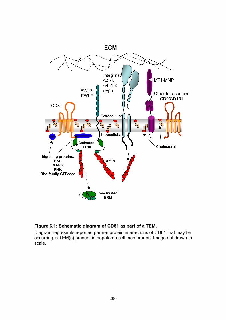
200
Figure 6.1: Schematic diagram of CD81 as part of a TEM. Diagram represents reported partner protein interactions of CD81 that may be occurring in TEM(s) present in hepatoma cell membranes. Image not drawn to scale.
201
Notably anti-CD81 induced hepatoma cell spread was dependent on the C
terminus of CD81 (Fig.3.5), previously reported to contain a PDZ domain
believed to permit association with actin-associated proteins Ezrin Radoxin
Moesin (ERM) (95, 411). Further to this CD81 co-localized with activated
ERM proteins in hepatoma cells and anti-CD81 induced cell spread was
dependent on ERM function (Fig.3.6). Our observations suggest the presence
of a functional link between the CD81 C terminus and ERM proteins in
hepatoma cells as illustrated in Figure 6.1. Importantly this association was
not important for CD81 to function as an HCV co-receptor (data not shown).
However we can speculate that the link between CD81 and the actin
cytoskeleton through ERM proteins may be important for CD81 function in
tumor cell migration. Indeed a number of reports have identified ERM protein
Ezrin as an indicator of hepatoma cell metastasis potential (354, 543, 557).
This has also been confirmed by in vivo studies of Ezrin expression in tumors
of hepatitis B virus (HBV) infected HCC patients, where higher levels of Ezrin
expression associate with a smaller tumour size and a higher frequency of
metastasis (543).
Immunoglobulin like proteins EWI-2 and EWI-F associate with CD81 and are
reported to associate with ERM proteins. Interestingly their association with
ERM proteins has been reported to negatively regulate cell migration (411).
Our observations together with these reports suggest that the relationship
between CD81, EWI-2, EWI-F and ERM is important in determining cellular-
migration potential.
202
Although previous reports have identified a number of different signaling
cascades to be involved in CD81 function, inhibitors to these pathways had no
observable effect on anti-CD81 induced cell spread (Fig.3.7-8). This may be
explained by compensatory actions of different pathways. Interestingly
inhibition of the large GTPase, dynamin, perturbed hepatoma cell spread
suggesting an important role for dynamin in CD81 function (Fig.3.9). Dynamin
is reported to play a role in HCV entry through its role in clathrin mediated
endocytosis (287)(Farquar, submitted), however until now it has not been
associated with tetraspanin function.
A number of different integrin sub-units were detected on the surface of
hepatoma cells, some of which have previously been identified to associate
either directly or indirectly with CD81 (42, 75, 427, 481, 528). CD81
expression or cell surface ligation did not modulate hepatma–ECM adhesion
suggesting that CD81 is not involved in coordinating integrin function in
hepatoma–ECM interactions (Fig.4.2). However, CD81 expression was
observed to increase invasion of hepatoma cells (Fig.4.8), a process
potentially coordinated through CD81’s association with integrins. Additional
partner proteins not investigated in this study that could be involved in
hepatoma invasion include the matrix metalloproteinase MT1-MMP. Lafleur et
al., reported MT1-MMP to be stabilized by CD81 expression, promoting
invasive potential (262).
In summary, we have identified that CD81 expression increases hepatoma
cell migration and invasion in vitro. We hypothesize that this may have
203
consequences for hepatoma carcinoma development in vivo, CD81
expression levels could prove to be a useful marker for tumor progression.
This is an area of research that has been paid little attention to in the past, our
findings help justify this as an important area of research in the future. CD81
is ubiquitously expressed making it an unattractive target for therapeutics for
fear of off target effects, and importantly we observed no inhibition of
migration in response to ligation with soluble IgG (Fig.4.7). The later
observation also makes it unlikely that HCV E2 binding to CD81 will modulate
CD81 function in migration and invasion. Cholesterol levels, dynamin, ERM
and CD81 C terminal function were all observed to be important in
coordinating anti-CD81 induced hepatoma cell spread (Chapter 3) and may
also be involved in CD81 function in hepatoma migration and invasion.
HCV perturbation of anti-CD81 induced cell spread:
Recombinant forms of truncated HCV E2 glycoprotein directly bind CD81
(384), and HCV has been shown to bind sCD81 (30, 208) therefore we
speculated as to whether HCV infection may modulate CD81 function in
hepatoma cells. Interestingly HCV infection perturbed anti-CD81 induced
hepatoma cell spread (Fig.3.11). CD81 expression levels were comparable for
naïve and infected cells, eliminating this as a possible explanation for the
attenuated spread (Fig.3.11). We hypothesized a number of possible
mechanisms that could be responsible for this observation, illustrated in
Figure 6.2. Firstly HCV engagement of CD81 may directly perturb CD81
partner protein interactions (Fig.6.2A). Supporting this hypothesis viruses
unable to associate with CD81 had no effect on anti-CD81 induced spread
204
(Fig.3.12). However the infection levels recorded for the cell populations
expressing these viruses were relatively low, providing an alternative
explanation for these results. Secondly we hypothesized that modulation of
CD81-ERM association/activation, or dynamin function including inhibition of
down stream signaling pathways may be responsible (Fig.6.2B-D). For
example HCV NS5A protein is reported to inhibit Syk kinase activity (220) and
tyrosine phosphorylation of Ezrin upon CD81 ligation on B cells is dependent
on Syk (95). Thus if this is also true for hepatoma cells, NS5A expression may
be responsible for the observed perturbation. Lastly, we hypothesized that
HCV modulation of cellular cholesterol levels may perturb anti-CD81 induced
hepatoma cell spread (Fig.6.2E). Supporting this hypothesis an increase in
total cellular cholesterol levels in HCV infected cells was recorded and
elevated levels of cholesterol perturbed anti-CD81 induced cell spread
(Fig.3.13).
Given our earlier results implicating CD81 as a promoter for hepatoma cell
migration and invasion we postulate that these findings might have
consequences for HCV associated hepatoma carcinoma.
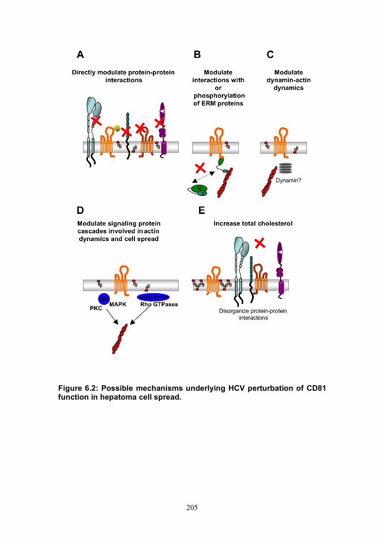
205
Figure 6.2: Possible mechanisms underlying HCV perturbation of CD81 function in hepatoma cell spread.

206
HCV as a tumor promoter:
An increase in hepatoma cell invasion was observed upon HCV infection in
vitro, suggesting that HCV infection may directly increase the risk of
developing HCC in vivo. As previously suggested this may be due to an
alteration of CD81 function by HCV infection. However the processes involved
in cell migration are extremely complex and are likely to be controlled through
numerous mechanisms. Previous research in the HCV field has reported that
expression of certain HCV proteins may be responsible for the development
of HCC (17, 33, 89, 294, 330, 340), expression of HCV core protein for
example was reported to increase HepG2 cell migration through modulation of
E-cadherin levels (17). Until now no one has previously reported an increase
in invasion potential of hepatoma cells upon expression of full-length
replicating HCV virus.
Stabilization of HIF-1α following HCV infection has been reported by a
number of laboratories (191, 341, 400). The HIF transcription complex formed
upon stabilization of HIF-1α is responsible for the transcription of numerous
proteins involved in tumor cell progression and has recently been linked with
the development of epithelial mesenchymal transition (EMT) (198, 534, 535).
We postulated that stabilization of HIF-lα might be responsible for the
observed increase in hepatoma cell invasion (Fig. 6.3). This was supported by
our observations that HCV induced migration as well as hypoxia induced
migration of Huh-7.5 cells was perturbed by HIF-1α inhibitor NSC-134754
(Fig.4.11-12). A commonly known growth factor transcribed by the HIF
transcription factor is VEGF. VEGF has previously been reported to be
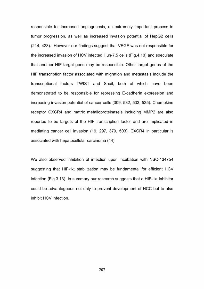
207
responsible for increased angiogenesis, an extremely important process in
tumor progression, as well as increased invasion potential of HepG2 cells
(214, 423). However our findings suggest that VEGF was not responsible for
the increased invasion of HCV infected Huh-7.5 cells (Fig.4.10) and speculate
that another HIF target gene may be responsible. Other target genes of the
HIF transcription factor associated with migration and metastasis include the
transcriptional factors TWIST and Snail, both of which have been
demonstrated to be responsible for repressing E-cadherin expression and
increasing invasion potential of cancer cells (309, 532, 533, 535). Chemokine
receptor CXCR4 and matrix metalloproteinase’s including MMP2 are also
reported to be targets of the HIF transcription factor and are implicated in
mediating cancer cell invasion (19, 297, 379, 503). CXCR4 in particular is
associated with hepatocellular carcinoma (44).
We also observed inhibition of infection upon incubation with NSC-134754
suggesting that HIF-1α stabilization may be fundamental for efficient HCV
infection (Fig.3.13). In summary our research suggests that a HIF-1α inhibitor
could be advantageous not only to prevent development of HCC but to also
inhibit HCV infection.
208
Figure 6.3: Possible mechanism(s) for HCV induced hepatoma migration and tumor progression through stabilization of HIF-1α .
209
Neutralizing antibody resistant transmission of HCV: Finally we investigated nAb resistant transmission of HCV in vitro (Chapter 5).
The results we obtained suggest that HCV transmits efficiently in co-culture
via a cell contact and particle-dependent means of transmission that is highly
insensitive to nAbs. This nAb resistant transmission is dependent on all co-
receptors previously shown to be essential for cell-free entry indicating the
mechanism of entry is likely to be the same. Here we postulate possible
mechanisms for this highly efficient neutralizing antibody resistant
transmission.
It is unlikely that HCV transmits through cell fusion events or formation of
synctia, as demonstrated by many herpes viruses since this would negate the
need for correct viral assembly and usage of viral co-receptors (415, 483).
Huh-7.5 cells use in this study do not polarize (319). Consequently we were
unable to determine whether tight junctions may be used to transmit virus as
observed for other viruses such as Herpes Simplex Virus (HSV). HSV has
been demonstrated to target virions to cell junctions of epithelial cells to
promote transmission of infection (227). The liver is highly ordered and made
up predominantly of hepatocytes that are highly polarized. HepG2 cells
provide a model system to study hepatocyte polarity (319, 320). Unfortunately
HepG2 cells have reduced permissivity to support HCV replication and are
unsuitable for use in the co-culture assay (320). Future work is needed to
investigate cell-to-cell transmission in a polarized cell line. In the current
system with Huh-7.5 cells it is unlikely that HCV exploits cell junctions that are
210
impermeable to HCV glycoprotein targeting antibodies since we found
differences in efficacy of antibodies able to inhibit co-culture HCV
transmission. It is more likely that HCV transmits either through a virological
synapse similar to those induced by HIV between T cells or through utilization
of cellular protrusions for example filopodia, microvillus or nanotubes (337,
415). See Figure 6.4 for illustrations of plausible routes of transmission.
HIV and HTLV induce virological synapes between T-cells (216, 228, 362).
These allow efficient transfer of virus but are still permeable to neutralizing
antibodies (304). New techniques allowing visualization of virions have
resulted in an increase in the number of viruses known to utilize cellular
protrusions for intercellular transmission. Examples include HIV that has
recently been shown to travel in nanotubes connecting T-cells allowing
efficient transmission (452). This differs from murine leukemia virus (MLV)
that attaches to receptors on filopodia of target cells where it then proceeds to
travel along the surface using F-actin retrograde flow to reach the target cell
body (432). A number of viruses including MLV remain associated with the
cell surface once released and are directed to adjacent cells in a process
called cell “surfing”, MLV has recently been shown to be assembled at sites of
cell contact further increasing the efficiency of transmission (59, 304, 421,
431). African Swine Fever virus and vaccinia viruses utilize cellular
protrusions in a completely different way, actin tails propel the virions to
neighbouring cells (106, 232). Interestingly vaccinia viruses use this system to
propel themselves across infected cells until they reach naïve cells further
increasing efficacy of transmission (116).
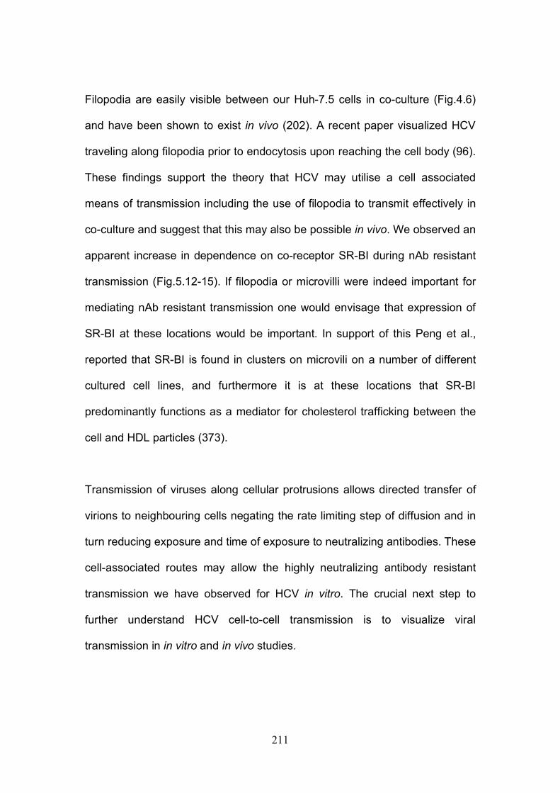
211
Filopodia are easily visible between our Huh-7.5 cells in co-culture (Fig.4.6)
and have been shown to exist in vivo (202). A recent paper visualized HCV
traveling along filopodia prior to endocytosis upon reaching the cell body (96).
These findings support the theory that HCV may utilise a cell associated
means of transmission including the use of filopodia to transmit effectively in
co-culture and suggest that this may also be possible in vivo. We observed an
apparent increase in dependence on co-receptor SR-BI during nAb resistant
transmission (Fig.5.12-15). If filopodia or microvilli were indeed important for
mediating nAb resistant transmission one would envisage that expression of
SR-BI at these locations would be important. In support of this Peng et al.,
reported that SR-BI is found in clusters on microvili on a number of different
cultured cell lines, and furthermore it is at these locations that SR-BI
predominantly functions as a mediator for cholesterol trafficking between the
cell and HDL particles (373).
Transmission of viruses along cellular protrusions allows directed transfer of
virions to neighbouring cells negating the rate limiting step of diffusion and in
turn reducing exposure and time of exposure to neutralizing antibodies. These
cell-associated routes may allow the highly neutralizing antibody resistant
transmission we have observed for HCV in vitro. The crucial next step to
further understand HCV cell-to-cell transmission is to visualize viral
transmission in in vitro and in vivo studies.
212
Figure 6.4: Possible routes of HCV cell-to-cell transmission in co-culture.
213
1. Abdel-Aziz, F., M. Habib, M. K. Mohamed, M. Abdel-Hamid, F. Gamil, S. Madkour, N. N. Mikhail, D. Thomas, A. D. Fix, G. T. Strickland, W. Anwar, and I. Sallam. 2000. Hepatitis C virus (HCV) infection in a community in the Nile Delta: population description and HCV prevalence. Hepatology 32:111-5.
2. Adinolfi, L. E., M. Gambardella, A. Andreana, M. F. Tripodi, R. Utili, and G. Ruggiero. 2001. Steatosis accelerates the progression of liver damage of chronic hepatitis C patients and correlates with specific HCV genotype and visceral obesity. Hepatology 33:1358-64.
3. Agnello, V., G. Abel, M. Elfahal, G. B. Knight, and Q. X. Zhang. 1999. Hepatitis C virus and other flaviviridae viruses enter cells via low density lipoprotein receptor. Proc Natl Acad Sci U S A 96:12766-71.
4. Ago, H., T. Adachi, A. Yoshida, M. Yamamoto, N. Habuka, K. Yatsunami, and M. Miyano. 1999. Crystal structure of the RNA-dependent RNA polymerase of hepatitis C virus. Structure 7:1417-26.
5. Aizaki, H., K. J. Lee, V. M. Sung, H. Ishiko, and M. M. Lai. 2004. Characterization of the hepatitis C virus RNA replication complex associated with lipid rafts. Virology 324:450-61.
6. Alberti, A., L. Chemello, and L. Benvegnu. 1999. Natural history of hepatitis C. J Hepatol 31 Suppl 1:17-24.
7. Allander, T., K. Drakenberg, A. Beyene, D. Rosa, S. Abrignani, M. Houghton, A. Widell, L. Grillner, and M. A. Persson. 2000. Recombinant human monoclonal antibodies against different conformational epitopes of the E2 envelope glycoprotein of hepatitis C virus that inhibit its interaction with CD81. J Gen Virol 81:2451-9.
8. Alter, H. J., R. Sanchez-Pescador, M. S. Urdea, J. C. Wilber, R. J. Lagier, A. M. Di Bisceglie, J. W. Shih, and P. D. Neuwald. 1995. Evaluation of branched DNA signal amplification for the detection of hepatitis C virus RNA. J Viral Hepat 2:121-32.
9. Alter, H. J., and L. B. Seeff. 2000. Recovery, persistence, and sequelae in hepatitis C virus infection: a perspective on long-term outcome. Semin Liver Dis 20:17-35.
10. Alter, M. J. 1997. Epidemiology of hepatitis C. Hepatology 26:62S-65S.
11. Alter, M. J. 2007. Epidemiology of hepatitis C virus infection. World J Gastroenterol 13:2436-41.
12. Alter, M. J., D. Kruszon-Moran, O. V. Nainan, G. M. McQuillan, F. Gao, L. A. Moyer, R. A. Kaslow, and H. S. Margolis. 1999. The prevalence of hepatitis C virus infection in the United States, 1988 through 1994. N Engl J Med 341:556-62.
13. Amieva, M. R., P. Litman, L. Huang, E. Ichimaru, and H. Furthmayr. 1999. Disruption of dynamic cell surface architecture of NIH3T3 fibroblasts by the N-terminal domains of moesin and ezrin: in vivo imaging with GFP fusion proteins. J Cell Sci 112 ( Pt 1):111-25.
14. Andre, P., F. Komurian-Pradel, S. Deforges, M. Perret, J. L. Berland, M. Sodoyer, S. Pol, C. Brechot, G. Paranhos-Baccala, and V. Lotteau. 2002. Characterization of low- and very-low-density hepatitis C virus RNA-containing particles. J Virol 76:6919-28.
15. Ang, J., M. Lijovic, L. K. Ashman, K. Kan, and A. G. Frauman. 2004. CD151 protein expression predicts the clinical outcome of low-
214
grade primary prostate cancer better than histologic grading: a new prognostic indicator? Cancer Epidemiol Biomarkers Prev 13:1717-21.
16. Appel, N., M. Zayas, S. Miller, J. Krijnse-Locker, T. Schaller, P. Friebe, S. Kallis, U. Engel, and R. Bartenschlager. 2008. Essential role of domain III of nonstructural protein 5A for hepatitis C virus infectious particle assembly. PLoS Pathog 4:e1000035.
17. Arora, P., E. O. Kim, J. K. Jung, and K. L. Jang. 2008. Hepatitis C virus core protein downregulates E-cadherin expression via activation of DNA methyltransferase 1 and 3b. Cancer Lett 261:244-52.
18. Artym, V. V., Y. Zhang, F. Seillier-Moiseiwitsch, K. M. Yamada, and S. C. Mueller. 2006. Dynamic interactions of cortactin and membrane type 1 matrix metalloproteinase at invadopodia: defining the stages of invadopodia formation and function. Cancer Res 66:3034-43.
19. Arvidsson, Y., A. Bergstrom, L. Arvidsson, E. Kristiansson, H. Ahlman, and O. Nilsson. Hypoxia stimulates CXCR4 signalling in ileal carcinoids. Endocr Relat Cancer 17:303-16.
20. Au, W. C., P. A. Moore, D. W. LaFleur, B. Tombal, and P. M. Pitha. 1998. Characterization of the interferon regulatory factor-7 and its potential role in the transcription activation of interferon A genes. J Biol Chem 273:29210-7.
21. Backes, P., D. Quinkert, S. Reiss, M. Binder, M. Zayas, U. Rescher, V. Gerke, R. Bartenschlager, and V. Lohmann. Role of annexin A2 in the production of infectious hepatitis C virus particles. J Virol 84:5775-89.
22. Bakin, A. V., C. Rinehart, A. K. Tomlinson, and C. L. Arteaga. 2002. p38 mitogen-activated protein kinase is required for TGFbeta-mediated fibroblastic transdifferentiation and cell migration. J Cell Sci 115:3193-206.
23. Bakin, A. V., A. K. Tomlinson, N. A. Bhowmick, H. L. Moses, and C. L. Arteaga. 2000. Phosphatidylinositol 3-kinase function is required for transforming growth factor beta-mediated epithelial to mesenchymal transition and cell migration. J Biol Chem 275:36803-10.
24. Baldassarre, M., A. Pompeo, G. Beznoussenko, C. Castaldi, S. Cortellino, M. A. McNiven, A. Luini, and R. Buccione. 2003. Dynamin participates in focal extracellular matrix degradation by invasive cells. Mol Biol Cell 14:1074-84.
25. Ballestrem, C., B. Wehrle-Haller, and B. A. Imhof. 1998. Actin dynamics in living mammalian cells. J Cell Sci 111 ( Pt 12):1649-58.
26. Bartenschlager, R., and V. Lohmann. 2001. Novel cell culture systems for the hepatitis C virus. Antiviral Res 52:1-17.
27. Bartenschlager, R., F. Penin, V. Lohmann, and P. Andre. Assembly of infectious hepatitis C virus particles. Trends Microbiol.
28. Barth, H., C. Schafer, M. I. Adah, F. Zhang, R. J. Linhardt, H. Toyoda, A. Kinoshita-Toyoda, T. Toida, T. H. Van Kuppevelt, E. Depla, F. Von Weizsacker, H. E. Blum, and T. F. Baumert. 2003. Cellular binding of hepatitis C virus envelope glycoprotein E2 requires cell surface heparan sulfate. J Biol Chem 278:41003-12.
29. Bartosch, B., J. Bukh, J. C. Meunier, C. Granier, R. E. Engle, W. C. Blackwelder, S. U. Emerson, F. L. Cosset, and R. H. Purcell. 2003. In vitro assay for neutralizing antibody to hepatitis C virus: evidence for
215
broadly conserved neutralization epitopes. Proc Natl Acad Sci U S A 100:14199-204.
30. Bartosch, B., J. Dubuisson, and F. L. Cosset. 2003. Infectious hepatitis C virus pseudo-particles containing functional E1-E2 envelope protein complexes. J Exp Med 197:633-42.
31. Basu, A., T. Kanda, A. Beyene, K. Saito, K. Meyer, and R. Ray. 2007. Sulfated homologues of heparin inhibit hepatitis C virus entry into mammalian cells. J Virol 81:3933-41.
32. Bataller, R., and D. A. Brenner. 2005. Liver fibrosis. J Clin Invest 115:209-18.
33. Battaglia, S., N. Benzoubir, S. Nobilet, P. Charneau, D. Samuel, A. L. Zignego, A. Atfi, C. Brechot, and M. F. Bourgeade. 2009. Liver cancer-derived hepatitis C virus core proteins shift TGF-beta responses from tumor suppression to epithelial-mesenchymal transition. PLoS One 4:e4355.
34. Baumert, T. F., S. Wellnitz, S. Aono, J. Satoi, D. Herion, J. Tilman Gerlach, G. R. Pape, J. Y. Lau, J. H. Hoofnagle, H. E. Blum, and T. J. Liang. 2000. Antibodies against hepatitis C virus-like particles and viral clearance in acute and chronic hepatitis C. Hepatology 32:610-7.
35. Bayer, M. E., B. S. Blumberg, and B. Werner. 1968. Particles associated with Australia antigen in the sera of patients with leukaemia, Down's Syndrome and hepatitis. Nature 218:1057-9.
36. Bedossa, P., and V. Paradis. 2003. Liver extracellular matrix in health and disease. J Pathol 200:504-15.
37. Behrens, S. E., L. Tomei, and R. De Francesco. 1996. Identification and properties of the RNA-dependent RNA polymerase of hepatitis C virus. Embo J 15:12-22.
38. Bekisz, J., H. Schmeisser, J. Hernandez, N. D. Goldman, and K. C. Zoon. 2004. Human interferons alpha, beta and omega. Growth Factors 22:243-51.
39. Benedicto, I., F. Molina-Jimenez, B. Bartosch, F. L. Cosset, D. Lavillette, J. Prieto, R. Moreno-Otero, A. Valenzuela-Fernandez, R. Aldabe, M. Lopez-Cabrera, and P. L. Majano. 2009. The tight junction-associated protein occludin is required for a postbinding step in hepatitis C virus entry and infection. J Virol 83:8012-20.
40. Benga, W. J., S. E. Krieger, M. Dimitrova, M. B. Zeisel, M. Parnot, J. Lupberger, E. Hildt, G. Luo, J. McLauchlan, T. F. Baumert, and C. Schuster. Apolipoprotein E interacts with hepatitis C virus nonstructural protein 5A and determines assembly of infectious particles. Hepatology 51:43-53.
41. Berditchevski, F. 2001. Complexes of tetraspanins with integrins: more than meets the eye. J Cell Sci 114:4143-51.
42. Berditchevski, F., and E. Odintsova. 1999. Characterization of integrin-tetraspanin adhesion complexes: role of tetraspanins in integrin signaling. J Cell Biol 146:477-92.
43. Berditchevski, F., E. Odintsova, S. Sawada, and E. Gilbert. 2002. Expression of the palmitoylation-deficient CD151 weakens the association of alpha 3 beta 1 integrin with the tetraspanin-enriched microdomains and affects integrin-dependent signaling. J Biol Chem 277:36991-7000.
216
44. Bertran, E., L. Caja, E. Navarro, P. Sancho, J. Mainez, M. M. Murillo, A. Vinyals, A. Fabra, and I. Fabregat. 2009. Role of CXCR4/SDF-1 alpha in the migratory phenotype of hepatoma cells that have undergone epithelial-mesenchymal transition in response to the transforming growth factor-beta. Cell Signal 21:1595-606.
45. Blanchard, E., S. Belouzard, L. Goueslain, T. Wakita, J. Dubuisson, C. Wychowski, and Y. Rouille. 2006. Hepatitis C virus entry depends on clathrin-mediated endocytosis. J Virol 80:6964-72.
46. Blaschke, F., P. Stawowy, S. Goetze, O. Hintz, M. Grafe, U. Kintscher, E. Fleck, and K. Graf. 2002. Hypoxia activates beta(1)-integrin via ERK 1/2 and p38 MAP kinase in human vascular smooth muscle cells. Biochem Biophys Res Commun 296:890-6.
47. Blight, K. J., J. A. McKeating, J. Marcotrigiano, and C. M. Rice. 2003. Efficient replication of hepatitis C virus genotype 1a RNAs in cell culture. J Virol 77:3181-90.
48. Bode, J. G., E. D. Brenndorfer, and D. Haussinger. 2007. Subversion of innate host antiviral strategies by the hepatitis C virus. Arch Biochem Biophys 462:254-65.
49. Bode, J. G., S. Ludwig, C. Ehrhardt, U. Albrecht, A. Erhardt, F. Schaper, P. C. Heinrich, and D. Haussinger. 2003. IFN-alpha antagonistic activity of HCV core protein involves induction of suppressor of cytokine signaling-3. Faseb J 17:488-90.
50. Boehme, K. W., and T. Compton. 2004. Innate sensing of viruses by toll-like receptors. J Virol 78:7867-73.
51. Boismenu, R., M. Rhein, W. H. Fischer, and W. L. Havran. 1996. A role for CD81 in early T cell development. Science 271:198-200.
52. Boulant, S., M. W. Douglas, L. Moody, A. Budkowska, P. Targett-Adams, and J. McLauchlan. 2008. Hepatitis C virus core protein induces lipid droplet redistribution in a microtubule- and dynein-dependent manner. Traffic 9:1268-82.
53. Boulant, S., P. Targett-Adams, and J. McLauchlan. 2007. Disrupting the association of hepatitis C virus core protein with lipid droplets correlates with a loss in production of infectious virus. J Gen Virol 88:2204-13.
54. Brazzoli, M., A. Bianchi, S. Filippini, A. Weiner, Q. Zhu, M. Pizza, and S. Crotta. 2008. CD81 is a central regulator of cellular events required for hepatitis C virus infection of human hepatocytes. J Virol 82:8316-29.
55. Breiner, K. M., S. Urban, B. Glass, and H. Schaller. 2001. Envelope protein-mediated down-regulation of hepatitis B virus receptor in infected hepatocytes. J Virol 75:143-50.
56. Bressanelli, S., L. Tomei, A. Roussel, I. Incitti, R. L. Vitale, M. Mathieu, R. De Francesco, and F. A. Rey. 1999. Crystal structure of the RNA-dependent RNA polymerase of hepatitis C virus. Proc Natl Acad Sci U S A 96:13034-9.
57. Brown, E. A., H. Zhang, L. H. Ping, and S. M. Lemon. 1992. Secondary structure of the 5' nontranslated regions of hepatitis C virus and pestivirus genomic RNAs. Nucleic Acids Res 20:5041-5.
58. Bukh, J. 2004. A critical role for the chimpanzee model in the study of hepatitis C. Hepatology 39:1469-75.
217
59. Burckhardt, C. J., and U. F. Greber. 2009. Virus movements on the plasma membrane support infection and transmission between cells. PLoS Pathog 5:e1000621.
60. Burlone, M. E., and A. Budkowska. 2009. Hepatitis C virus cell entry: role of lipoproteins and cellular receptors. J Gen Virol 90:1055-70.
61. Burton, D. R. 2002. Antibodies, viruses and vaccines. Nat Rev Immunol 2:706-13.
62. Busch, M. P., S. A. Glynn, S. L. Stramer, D. M. Strong, S. Caglioti, D. J. Wright, B. Pappalardo, and S. H. Kleinman. 2005. A new strategy for estimating risks of transfusion-transmitted viral infections based on rates of detection of recently infected donors. Transfusion 45:254-64.
63. Callens, N., Y. Ciczora, B. Bartosch, N. Vu-Dac, F. L. Cosset, J. M. Pawlotsky, F. Penin, and J. Dubuisson. 2005. Basic residues in hypervariable region 1 of hepatitis C virus envelope glycoprotein e2 contribute to virus entry. J Virol 79:15331-41.
64. Cano, A., M. A. Perez-Moreno, I. Rodrigo, A. Locascio, M. J. Blanco, M. G. del Barrio, F. Portillo, and M. A. Nieto. 2000. The transcription factor snail controls epithelial-mesenchymal transitions by repressing E-cadherin expression. Nat Cell Biol 2:76-83.
65. Capone, S., A. Meola, B. B. Ercole, A. Vitelli, M. Pezzanera, L. Ruggeri, M. E. Davies, R. Tafi, C. Santini, A. Luzzago, T. M. Fu, A. Bett, S. Colloca, R. Cortese, A. Nicosia, and A. Folgori. 2006. A novel adenovirus type 6 (Ad6)-based hepatitis C virus vector that overcomes preexisting anti-ad5 immunity and induces potent and broad cellular immune responses in rhesus macaques. J Virol 80:1688-99.
66. Carloni, V., A. Mazzocca, and K. S. Ravichandran. 2004. Tetraspanin CD81 is linked to ERK/MAPKinase signaling by Shc in liver tumor cells. Oncogene 23:1566-74.
67. Carrere-Kremer, S., C. Montpellier-Pala, L. Cocquerel, C. Wychowski, F. Penin, and J. Dubuisson. 2002. Subcellular localization and topology of the p7 polypeptide of hepatitis C virus. J Virol 76:3720-30.
68. Castello, G., S. Scala, G. Palmieri, S. A. Curley, and F. Izzo. HCV-related hepatocellular carcinoma: From chronic inflammation to cancer. Clin Immunol 134:237-50.
69. Catanese, M. T., H. Ansuini, R. Graziani, T. Huby, M. Moreau, J. K. Ball, G. Paonessa, C. M. Rice, R. Cortese, A. Vitelli, and A. Nicosia. Role of scavenger receptor class B type I in hepatitis C virus entry: kinetics and molecular determinants. J Virol 84:34-43.
70. Catanese, M. T., R. Graziani, T. von Hahn, M. Moreau, T. Huby, G. Paonessa, C. Santini, A. Luzzago, C. M. Rice, R. Cortese, A. Vitelli, and A. Nicosia. 2007. High-avidity monoclonal antibodies against the human scavenger class B type I receptor efficiently block hepatitis C virus infection in the presence of high-density lipoprotein. J Virol 81:8063-71.
71. Cerny, A., and F. V. Chisari. 1999. Pathogenesis of chronic hepatitis C: immunological features of hepatic injury and viral persistence. Hepatology 30:595-601.
218
72. Chambrion, C., and F. Le Naour. The tetraspanins CD9 and CD81 regulate CD9P1-induced effects on cell migration. PLoS One 5:e11219.
73. Chang, K. M., B. Rehermann, J. G. McHutchison, C. Pasquinelli, S. Southwood, A. Sette, and F. V. Chisari. 1997. Immunological significance of cytotoxic T lymphocyte epitope variants in patients chronically infected by the hepatitis C virus. J Clin Invest 100:2376-85.
74. Chang, K. S., J. Jiang, Z. Cai, and G. Luo. 2007. Human apolipoprotein e is required for infectivity and production of hepatitis C virus in cell culture. J Virol 81:13783-93.
75. Chang, Y., and S. C. Finnemann. 2007. Tetraspanin CD81 is required for the alpha v beta5-integrin-dependent particle-binding step of RPE phagocytosis. J Cell Sci 120:3053-63.
76. Charrin, S., S. Manie, M. Billard, L. Ashman, D. Gerlier, C. Boucheix, and E. Rubinstein. 2003. Multiple levels of interactions within the tetraspanin web. Biochem Biophys Res Commun 304:107-12.
77. Charrin, S., S. Manie, M. Oualid, M. Billard, C. Boucheix, and E. Rubinstein. 2002. Differential stability of tetraspanin/tetraspanin interactions: role of palmitoylation. FEBS Lett 516:139-44.
78. Charrin, S., S. Manie, C. Thiele, M. Billard, D. Gerlier, C. Boucheix, and E. Rubinstein. 2003. A physical and functional link between cholesterol and tetraspanins. Eur J Immunol 33:2479-89.
79. Chau, N. M., P. Rogers, W. Aherne, V. Carroll, I. Collins, E. McDonald, P. Workman, and M. Ashcroft. 2005. Identification of novel small molecule inhibitors of hypoxia-inducible factor-1 that differentially block hypoxia-inducible factor-1 activity and hypoxia-inducible factor-1alpha induction in response to hypoxic stress and growth factors. Cancer Res 65:4918-28.
80. Chen, Y., T. Maguire, R. E. Hileman, J. R. Fromm, J. D. Esko, R. J. Linhardt, and R. M. Marks. 1997. Dengue virus infectivity depends on envelope protein binding to target cell heparan sulfate. Nat Med 3:866-71.
81. Chen, Y. C., W. C. Su, J. Y. Huang, T. C. Chao, K. S. Jeng, K. Machida, and M. M. Lai. Polo-like kinase 1 is involved in hepatitis C virus replication by hyperphosphorylating NS5A. J Virol 84:7983-93.
82. Chen, Y. J., Y. H. Chen, L. P. Chow, Y. H. Tsai, P. H. Chen, C. Y. Huang, W. T. Chen, and L. H. Hwang. Heat shock protein 72 is associated with the hepatitis C virus replicase complex and enhances viral RNA replication. J Biol Chem 285:28183-90.
83. Choi, S. S., and A. M. Diehl. 2009. Epithelial-to-mesenchymal transitions in the liver. Hepatology 50:2007-13.
84. Choo, Q. L., G. Kuo, R. Ralston, A. Weiner, D. Chien, G. Van Nest, J. Han, K. Berger, K. Thudium, C. Kuo, and et al. 1994. Vaccination of chimpanzees against infection by the hepatitis C virus. Proc Natl Acad Sci U S A 91:1294-8.
85. Choo, Q. L., G. Kuo, A. J. Weiner, L. R. Overby, D. W. Bradley, and M. Houghton. 1989. Isolation of a cDNA clone derived from a blood-borne non-A, non-B viral hepatitis genome. Science 244:359-62.
219
86. Chowdhury, A., A. Santra, S. Chaudhuri, G. K. Dhali, S. Chaudhuri, S. G. Maity, T. N. Naik, S. K. Bhattacharya, and D. N. Mazumder. 2003. Hepatitis C virus infection in the general population: a community-based study in West Bengal, India. Hepatology 37:802-9.
87. Christian, A. E., M. P. Haynes, M. C. Phillips, and G. H. Rothblat. 1997. Use of cyclodextrins for manipulating cellular cholesterol content. J Lipid Res 38:2264-72.
88. Chun, J. S., and B. S. Jacobson. 1993. Requirement for diacylglycerol and protein kinase C in HeLa cell-substratum adhesion and their feedback amplification of arachidonic acid production for optimum cell spreading. Mol Biol Cell 4:271-81.
89. Chung, Y. L., M. L. Sheu, and S. H. Yen. 2003. Hepatitis C virus NS5A as a potential viral Bcl-2 homologue interacts with Bax and inhibits apoptosis in hepatocellular carcinoma. Int J Cancer 107:65-73.
90. Ciampor, F. 2003. The ion channels coded by viruses. Acta Microbiol Immunol Hung 50:433-42.
91. Clarke, D., S. Griffin, L. Beales, C. S. Gelais, S. Burgess, M. Harris, and D. Rowlands. 2006. Evidence for the formation of a heptameric ion channel complex by the hepatitis C virus p7 protein in vitro. J Biol Chem 281:37057-68.
92. Cocquerel, L., S. Duvet, J. C. Meunier, A. Pillez, R. Cacan, C. Wychowski, and J. Dubuisson. 1999. The transmembrane domain of hepatitis C virus glycoprotein E1 is a signal for static retention in the endoplasmic reticulum. J Virol 73:2641-9.
93. Cocquerel, L., C. C. Kuo, J. Dubuisson, and S. Levy. 2003. CD81-dependent binding of hepatitis C virus E1E2 heterodimers. J Virol 77:10677-83.
94. Cocquerel, L., A. Op de Beeck, M. Lambot, J. Roussel, D. Delgrange, A. Pillez, C. Wychowski, F. Penin, and J. Dubuisson. 2002. Topological changes in the transmembrane domains of hepatitis C virus envelope glycoproteins. Embo J 21:2893-902.
95. Coffey, G. P., R. Rajapaksa, R. Liu, O. Sharpe, C. C. Kuo, S. W. Krauss, Y. Sagi, R. E. Davis, L. M. Staudt, J. P. Sharman, W. H. Robinson, and S. Levy. 2009. Engagement of CD81 induces ezrin tyrosine phosphorylation and its cellular redistribution with filamentous actin. J Cell Sci 122:3137-44.
96. Coller, K. E., K. L. Berger, N. S. Heaton, J. D. Cooper, R. Yoon, and G. Randall. 2009. RNA interference and single particle tracking analysis of hepatitis C virus endocytosis. PLoS Pathog 5:e1000702.
97. Cooper, S., A. L. Erickson, E. J. Adams, J. Kansopon, A. J. Weiner, D. Y. Chien, M. Houghton, P. Parham, and C. M. Walker. 1999. Analysis of a successful immune response against hepatitis C virus. Immunity 10:439-49.
98. Corless, L., C. M. Crump, S. D. Griffin, and M. Harris. Vps4 and the ESCRT-III complex are required for the release of infectious hepatitis C virus particles. J Gen Virol 91:362-72.
99. Cormier, E. G., F. Tsamis, F. Kajumo, R. J. Durso, J. P. Gardner, and T. Dragic. 2004. CD81 is an entry coreceptor for hepatitis C virus. Proc Natl Acad Sci U S A 101:7270-4.

220
100. Coue, M., S. L. Brenner, I. Spector, and E. D. Korn. 1987. Inhibition of actin polymerization by latrunculin A. FEBS Lett 213:316-8.
101. Cox, A. L., T. Mosbruger, G. M. Lauer, D. Pardoll, D. L. Thomas, and S. C. Ray. 2005. Comprehensive analyses of CD8+ T cell responses during longitudinal study of acute human hepatitis C. Hepatology 42:104-12.
102. Coyne, C. B., L. Shen, J. R. Turner, and J. M. Bergelson. 2007. Coxsackievirus entry across epithelial tight junctions requires occludin and the small GTPases Rab34 and Rab5. Cell Host Microbe 2:181-92.
103. Cramp, M. E., S. Rossol, S. Chokshi, P. Carucci, R. Williams, and N. V. Naoumov. 2000. Hepatitis C virus-specific T-cell reactivity during interferon and ribavirin treatment in chronic hepatitis C. Gastroenterology 118:346-55.
104. Crotta, S., V. Ronconi, C. Ulivieri, C. T. Baldari, N. M. Valiante, S. Abrignani, and A. Wack. 2006. Cytoskeleton rearrangement induced by tetraspanin engagement modulates the activation of T and NK cells. Eur J Immunol 36:919-29.
105. Crotta, S., A. Stilla, A. Wack, A. D'Andrea, S. Nuti, U. D'Oro, M. Mosca, F. Filliponi, R. M. Brunetto, F. Bonino, S. Abrignani, and N. M. Valiante. 2002. Inhibition of natural killer cells through engagement of CD81 by the major hepatitis C virus envelope protein. J Exp Med 195:35-41.
106. Cudmore, S., P. Cossart, G. Griffiths, and M. Way. 1995. Actin-based motility of vaccinia virus. Nature 378:636-8.
107. Cukierman, L., L. Meertens, C. Bertaux, F. Kajumo, and T. Dragic. 2009. Residues in a Highly Conserved Claudin-1 Motif Are Required for Hepatitis C Virus Entry and Mediate the Formation of Cell-Cell Contacts. J Virol.
108. Cun, W., J. Jiang, and G. Luo. The C-terminal {alpha}-helix domain of apolipoprotein E is required for interaction with nonstructural protein 5A and assembly of hepatitis C virus. J Virol.
109. De Francesco, R., and G. Migliaccio. 2005. Challenges and successes in developing new therapies for hepatitis C. Nature 436:953-60.
110. Deleersnyder, V., A. Pillez, C. Wychowski, K. Blight, J. Xu, Y. S. Hahn, C. M. Rice, and J. Dubuisson. 1997. Formation of native hepatitis C virus glycoprotein complexes. J Virol 71:697-704.
111. Deng, Y., Z. A. Almsherqi, M. M. Ng, and S. D. Kohlwein. Do viruses subvert cholesterol homeostasis to induce host cubic membranes? Trends Cell Biol 20:371-9.
112. Dentzer, T. G., I. C. Lorenz, M. J. Evans, and C. M. Rice. 2009. Determinants of the hepatitis C virus nonstructural protein 2 protease domain required for production of infectious virus. J Virol 83:12702-13.
113. Der, S. D., A. Zhou, B. R. Williams, and R. H. Silverman. 1998. Identification of genes differentially regulated by interferon alpha, beta, or gamma using oligonucleotide arrays. Proc Natl Acad Sci U S A 95:15623-8.
114. Dharancy, S., M. Malapel, G. Perlemuter, T. Roskams, Y. Cheng, L. Dubuquoy, P. Podevin, F. Conti, V. Canva, D. Philippe, L. Gambiez, P. Mathurin, J. C. Paris, K. Schoonjans, Y. Calmus, S. Pol, J.

221
Auwerx, and P. Desreumaux. 2005. Impaired expression of the peroxisome proliferator-activated receptor alpha during hepatitis C virus infection. Gastroenterology 128:334-42.
115. Dobreva, I., G. Waeber, R. W. James, and C. Widmann. 2006. Interleukin-8 secretion by fibroblasts induced by low density lipoproteins is p38 MAPK-dependent and leads to cell spreading and wound closure. J Biol Chem 281:199-205.
116. Doceul, V., M. Hollinshead, L. van der Linden, and G. L. Smith. Repulsion of superinfecting virions: a mechanism for rapid virus spread. Science 327:873-6.
117. Domingo, E., and J. Gomez. 2007. Quasispecies and its impact on viral hepatitis. Virus Res 127:131-50.
118. Dreux, M., V. L. Dao Thi, J. Fresquet, M. Guerin, Z. Julia, G. Verney, D. Durantel, F. Zoulim, D. Lavillette, F. L. Cosset, and B. Bartosch. 2009. Receptor complementation and mutagenesis reveal SR-BI as an essential HCV entry factor and functionally imply its intra- and extra-cellular domains. PLoS Pathog 5:e1000310.
119. Drummer, H. E., I. Boo, A. L. Maerz, and P. Poumbourios. 2006. A conserved Gly436-Trp-Leu-Ala-Gly-Leu-Phe-Tyr motif in hepatitis C virus glycoprotein E2 is a determinant of CD81 binding and viral entry. J Virol 80:7844-53.
120. Drummer, H. E., I. Boo, and P. Poumbourios. 2007. Mutagenesis of a conserved fusion peptide-like motif and membrane-proximal heptad-repeat region of hepatitis C virus glycoprotein E1. J Gen Virol 88:1144-8.
121. Drummer, H. E., A. Maerz, and P. Poumbourios. 2003. Cell surface expression of functional hepatitis C virus E1 and E2 glycoproteins. FEBS Lett 546:385-90.
122. Drummer, H. E., K. A. Wilson, and P. Poumbourios. 2002. Identification of the hepatitis C virus E2 glycoprotein binding site on the large extracellular loop of CD81. J Virol 76:11143-7.
123. Dubuisson, J., F. Penin, and D. Moradpour. 2002. Interaction of hepatitis C virus proteins with host cell membranes and lipids. Trends Cell Biol 12:517-23.
124. Dubuisson, J., and C. M. Rice. 1996. Hepatitis C virus glycoprotein folding: disulfide bond formation and association with calnexin. J Virol 70:778-86.
125. Duvet, S., A. Op De Beeck, L. Cocquerel, C. Wychowski, R. Cacan, and J. Dubuisson. 2002. Glycosylation of the hepatitis C virus envelope protein E1 occurs posttranslationally in a mannosylphosphoryldolichol-deficient CHO mutant cell line. Glycobiology 12:95-101.
126. Eckhardt, E. R., L. Cai, S. Shetty, Z. Zhao, A. Szanto, N. R. Webb, and D. R. Van der Westhuyzen. 2006. High density lipoprotein endocytosis by scavenger receptor SR-BII is clathrin-dependent and requires a carboxyl-terminal dileucine motif. J Biol Chem 281:4348-53.
127. Eckhardt, E. R., L. Cai, B. Sun, N. R. Webb, and D. R. van der Westhuyzen. 2004. High density lipoprotein uptake by scavenger receptor SR-BII. J Biol Chem 279:14372-81.
222
128. Egger, D., B. Wolk, R. Gosert, L. Bianchi, H. E. Blum, D. Moradpour, and K. Bienz. 2002. Expression of hepatitis C virus proteins induces distinct membrane alterations including a candidate viral replication complex. J Virol 76:5974-84.
129. El-Serag, H. B., H. Hampel, C. Yeh, and L. Rabeneck. 2002. Extrahepatic manifestations of hepatitis C among United States male veterans. Hepatology 36:1439-45.
130. Epand, R. M. 2003. Fusion peptides and the mechanism of viral fusion. Biochim Biophys Acta 1614:116-21.
131. Erickson, A. L., Y. Kimura, S. Igarashi, J. Eichelberger, M. Houghton, J. Sidney, D. McKinney, A. Sette, A. L. Hughes, and C. M. Walker. 2001. The outcome of hepatitis C virus infection is predicted by escape mutations in epitopes targeted by cytotoxic T lymphocytes. Immunity 15:883-95.
132. Espenel, C., E. Margeat, P. Dosset, C. Arduise, C. Le Grimellec, C. A. Royer, C. Boucheix, E. Rubinstein, and P. E. Milhiet. 2008. Single-molecule analysis of CD9 dynamics and partitioning reveals multiple modes of interaction in the tetraspanin web. J Cell Biol 182:765-76.
133. Evans, M. J., T. von Hahn, D. M. Tscherne, A. J. Syder, M. Panis, B. Wolk, T. Hatziioannou, J. A. McKeating, P. D. Bieniasz, and C. M. Rice. 2007. Claudin-1 is a hepatitis C virus co-receptor required for a late step in entry. Nature 446:801-5.
134. Failla, C., L. Tomei, and R. De Francesco. 1994. Both NS3 and NS4A are required for proteolytic processing of hepatitis C virus nonstructural proteins. J Virol 68:3753-60.
135. Farci, P., H. J. Alter, D. Wong, R. H. Miller, J. W. Shih, B. Jett, and R. H. Purcell. 1991. A long-term study of hepatitis C virus replication in non-A, non-B hepatitis. N Engl J Med 325:98-104.
136. Feinstone, S. M., A. Z. Kapikian, and R. H. Purceli. 1973. Hepatitis A: detection by immune electron microscopy of a viruslike antigen associated with acute illness. Science 182:1026-8.
137. Feinstone, S. M., A. Z. Kapikian, R. H. Purcell, H. J. Alter, and P. V. Holland. 1975. Transfusion-associated hepatitis not due to viral hepatitis type A or B. N Engl J Med 292:767-70.
138. Feld, J. J., and J. H. Hoofnagle. 2005. Mechanism of action of interferon and ribavirin in treatment of hepatitis C. Nature 436:967-72.
139. Ferrero, S., P. Lungaro, B. M. Bruzzone, C. Gotta, G. Bentivoglio, and N. Ragni. 2003. Prospective study of mother-to-infant transmission of hepatitis C virus: a 10-year survey (1990-2000). Acta Obstet Gynecol Scand 82:229-34.
140. Fievet, B., D. Louvard, and M. Arpin. 2007. ERM proteins in epithelial cell organization and functions. Biochim Biophys Acta 1773:653-60.
141. Flanagan, M. D., and S. Lin. 1980. Cytochalasins block actin filament elongation by binding to high affinity sites associated with F-actin. J Biol Chem 255:835-8.
142. Fletcher, N. F., J. P. Yang, M. J. Farquhar, K. Hu, C. Davis, Q. He, K. Dowd, S. C. Ray, S. E. Krieger, J. Neyts, T. F. Baumert, P. Balfe, J. A. McKeating, and F. Wong-Staal. Hepatitis C virus infection of neuroepithelioma cell lines. Gastroenterology 139:1365-74.
223
143. Flint, M., and J. A. McKeating. 1999. The C-terminal region of the hepatitis C virus E1 glycoprotein confers localization within the endoplasmic reticulum. J Gen Virol 80 ( Pt 8):1943-7.
144. Flint, M., J. M. Thomas, C. M. Maidens, C. Shotton, S. Levy, W. S. Barclay, and J. A. McKeating. 1999. Functional analysis of cell surface-expressed hepatitis C virus E2 glycoprotein. J Virol 73:6782-90.
145. Flint, M., T. von Hahn, J. Zhang, M. Farquhar, C. T. Jones, P. Balfe, C. M. Rice, and J. A. McKeating. 2006. Diverse CD81 proteins support hepatitis C virus infection. J Virol 80:11331-42.
146. Foster, T. L., T. Belyaeva, N. J. Stonehouse, A. R. Pearson, and M. Harris. All three domains of the hepatitis C virus nonstructural NS5A protein contribute to RNA binding. J Virol 84:9267-77.
147. Foy, E., K. Li, R. Sumpter, Jr., Y. M. Loo, C. L. Johnson, C. Wang, P. M. Fish, M. Yoneyama, T. Fujita, S. M. Lemon, and M. Gale, Jr. 2005. Control of antiviral defenses through hepatitis C virus disruption of retinoic acid-inducible gene-I signaling. Proc Natl Acad Sci U S A 102:2986-91.
148. Frank, C., M. K. Mohamed, G. T. Strickland, D. Lavanchy, R. R. Arthur, L. S. Magder, T. El Khoby, Y. Abdel-Wahab, E. S. Aly Ohn, W. Anwar, and I. Sallam. 2000. The role of parenteral antischistosomal therapy in the spread of hepatitis C virus in Egypt. Lancet 355:887-91.
149. Friedman, S. L. 2008. Hepatic stellate cells: protean, multifunctional, and enigmatic cells of the liver. Physiol Rev 88:125-72.
150. Friedman, S. L. 2003. Liver fibrosis -- from bench to bedside. J Hepatol 38 Suppl 1:S38-53.
151. Furukawa, Y., J. A. DeCaprio, A. Freedman, Y. Kanakura, M. Nakamura, T. J. Ernst, D. M. Livingston, and J. D. Griffin. 1990. Expression and state of phosphorylation of the retinoblastoma susceptibility gene product in cycling and noncycling human hematopoietic cells. Proc Natl Acad Sci U S A 87:2770-4.
152. Gal-Tanamy, M., Z. Y. Keck, M. Yi, J. A. McKeating, A. H. Patel, S. K. Foung, and S. M. Lemon. 2008. In vitro selection of a neutralization-resistant hepatitis C virus escape mutant. Proc Natl Acad Sci U S A 105:19450-5.
153. Gale, M., Jr., and E. M. Foy. 2005. Evasion of intracellular host defence by hepatitis C virus. Nature 436:939-45.
154. Gao, Y., J. B. Dickerson, F. Guo, J. Zheng, and Y. Zheng. 2004. Rational design and characterization of a Rac GTPase-specific small molecule inhibitor. Proc Natl Acad Sci U S A 101:7618-23.
155. Gardner, J. P., R. J. Durso, R. R. Arrigale, G. P. Donovan, P. J. Maddon, T. Dragic, and W. C. Olson. 2003. L-SIGN (CD 209L) is a liver-specific capture receptor for hepatitis C virus. Proc Natl Acad Sci U S A 100:4498-503.
156. Garry, R. F., and S. Dash. 2003. Proteomics computational analyses suggest that hepatitis C virus E1 and pestivirus E2 envelope glycoproteins are truncated class II fusion proteins. Virology 307:255-65.
224
157. Gastaminza, P., G. Cheng, S. Wieland, J. Zhong, W. Liao, and F. V. Chisari. 2008. Cellular determinants of hepatitis C virus assembly, maturation, degradation, and secretion. J Virol 82:2120-9.
158. Geisert, E. E., Jr., L. Yang, and M. H. Irwin. 1996. Astrocyte growth, reactivity, and the target of the antiproliferative antibody, TAPA. J Neurosci 16:5478-87.
159. Gerlach, J. T., H. M. Diepolder, M. C. Jung, N. H. Gruener, W. W. Schraut, R. Zachoval, R. Hoffmann, C. A. Schirren, T. Santantonio, and G. R. Pape. 1999. Recurrence of hepatitis C virus after loss of virus-specific CD4(+) T-cell response in acute hepatitis C. Gastroenterology 117:933-41.
160. Giannelli, G., C. Bergamini, E. Fransvea, C. Sgarra, and S. Antonaci. 2005. Laminin-5 with transforming growth factor-beta1 induces epithelial to mesenchymal transition in hepatocellular carcinoma. Gastroenterology 129:1375-83.
161. Glasgow, J. E., and R. W. Colman. 1984. Fibronectin synthesized by a human hepatoma cell line. Cancer Res 44:3022-8.
162. Goffard, A., N. Callens, B. Bartosch, C. Wychowski, F. L. Cosset, C. Montpellier, and J. Dubuisson. 2005. Role of N-linked glycans in the functions of hepatitis C virus envelope glycoproteins. J Virol 79:8400-9.
163. Goldstein, J. L., and M. S. Brown. 1990. Regulation of the mevalonate pathway. Nature 343:425-30.
164. Gordon-Alonso, M., M. Yanez-Mo, O. Barreiro, S. Alvarez, M. A. Munoz-Fernandez, A. Valenzuela-Fernandez, and F. Sanchez-Madrid. 2006. Tetraspanins CD9 and CD81 modulate HIV-1-induced membrane fusion. J Immunol 177:5129-37.
165. Gosert, R., D. Egger, V. Lohmann, R. Bartenschlager, H. E. Blum, K. Bienz, and D. Moradpour. 2003. Identification of the hepatitis C virus RNA replication complex in Huh-7 cells harboring subgenomic replicons. J Virol 77:5487-92.
166. Gottwein, J. M., T. K. Scheel, T. B. Jensen, J. B. Lademann, J. C. Prentoe, M. L. Knudsen, A. M. Hoegh, and J. Bukh. 2009. Development and characterization of hepatitis C virus genotype 1-7 cell culture systems: role of CD81 and scavenger receptor class B type I and effect of antiviral drugs. Hepatology 49:364-77.
167. Gouttenoire, J., F. Penin, and D. Moradpour. Hepatitis C virus nonstructural protein 4B: a journey into unexplored territory. Rev Med Virol 20:117-29.
168. Graham, T. R., H. E. Zhau, V. A. Odero-Marah, A. O. Osunkoya, K. S. Kimbro, M. Tighiouart, T. Liu, J. W. Simons, and R. M. O'Regan. 2008. Insulin-like growth factor-I-dependent up-regulation of ZEB1 drives epithelial-to-mesenchymal transition in human prostate cancer cells. Cancer Res 68:2479-88.
169. Gressner, O. A., M. S. Rizk, E. Kovalenko, R. Weiskirchen, and A. M. Gressner. 2008. Changing the pathogenetic roadmap of liver fibrosis? Where did it start; where will it go? J Gastroenterol Hepatol 23:1024-35.
170. Griffin, S. Inhibition of HCV p7 as a therapeutic target. Curr Opin Investig Drugs 11:175-81.
225
171. Griffin, S. D. 2009. Plugging the holes in hepatitis C virus antiviral therapy. Proc Natl Acad Sci U S A 106:12567-8.
172. Griffin, S. D., L. P. Beales, D. S. Clarke, O. Worsfold, S. D. Evans, J. Jaeger, M. P. Harris, and D. J. Rowlands. 2003. The p7 protein of hepatitis C virus forms an ion channel that is blocked by the antiviral drug, Amantadine. FEBS Lett 535:34-8.
173. Griffin, S. D., R. Harvey, D. S. Clarke, W. S. Barclay, M. Harris, and D. J. Rowlands. 2004. A conserved basic loop in hepatitis C virus p7 protein is required for amantadine-sensitive ion channel activity in mammalian cells but is dispensable for localization to mitochondria. J Gen Virol 85:451-61.
174. Grove, J., T. Huby, Z. Stamataki, T. Vanwolleghem, P. Meuleman, M. Farquhar, A. Schwarz, M. Moreau, J. S. Owen, G. Leroux-Roels, P. Balfe, and J. A. McKeating. 2007. Scavenger receptor BI and BII expression levels modulate hepatitis C virus infectivity. J Virol 81:3162-9.
175. Grove, J., S. Nielsen, J. Zhong, M. F. Bassendine, H. E. Drummer, P. Balfe, and J. A. McKeating. 2008. Identification of a residue in hepatitis C virus E2 glycoprotein that determines scavenger receptor BI and CD81 receptor dependency and sensitivity to neutralizing antibodies. J Virol 82:12020-9.
176. Gu, C., S. Yaddanapudi, A. Weins, T. Osborn, J. Reiser, M. Pollak, J. Hartwig, and S. Sever. Direct dynamin-actin interactions regulate the actin cytoskeleton. Embo J.
177. Gutierrez-Lopez, M. D., S. Ovalle, M. Yanez-Mo, N. Sanchez-Sanchez, E. Rubinstein, N. Olmo, M. A. Lizarbe, F. Sanchez-Madrid, and C. Cabanas. 2003. A functionally relevant conformational epitope on the CD9 tetraspanin depends on the association with activated beta1 integrin. J Biol Chem 278:208-18.
178. Haber, M. M., A. B. West, A. D. Haber, and A. Reuben. 1995. Relationship of aminotransferases to liver histological status in chronic hepatitis C. Am J Gastroenterol 90:1250-7.
179. Haberstroh, A., E. K. Schnober, M. B. Zeisel, P. Carolla, H. Barth, H. E. Blum, F. L. Cosset, G. Koutsoudakis, R. Bartenschlager, A. Union, E. Depla, A. Owsianka, A. H. Patel, C. Schuster, F. Stoll-Keller, M. Doffoel, M. Dreux, and T. F. Baumert. 2008. Neutralizing host responses in hepatitis C virus infection target viral entry at postbinding steps and membrane fusion. Gastroenterology 135:1719-1728 e1.
180. Hadlock, K. G., R. E. Lanford, S. Perkins, J. Rowe, Q. Yang, S. Levy, P. Pileri, S. Abrignani, and S. K. Foung. 2000. Human monoclonal antibodies that inhibit binding of hepatitis C virus E2 protein to CD81 and recognize conserved conformational epitopes. J Virol 74:10407-16.
181. Hadziyannis, S. J., H. Sette, Jr., T. R. Morgan, V. Balan, M. Diago, P. Marcellin, G. Ramadori, H. Bodenheimer, Jr., D. Bernstein, M. Rizzetto, S. Zeuzem, P. J. Pockros, A. Lin, and A. M. Ackrill. 2004. Peginterferon-alpha2a and ribavirin combination therapy in chronic hepatitis C: a randomized study of treatment duration and ribavirin dose. Ann Intern Med 140:346-55.
226
182. Hahn, Y. S. 2003. Subversion of immune responses by hepatitis C virus: immunomodulatory strategies beyond evasion? Curr Opin Immunol 15:443-9.
183. Hajra, K. M., D. Y. Chen, and E. R. Fearon. 2002. The SLUG zinc-finger protein represses E-cadherin in breast cancer. Cancer Res 62:1613-8.
184. Halasz, P., G. Holloway, S. J. Turner, and B. S. Coulson. 2008. Rotavirus replication in intestinal cells differentially regulates integrin expression by a phosphatidylinositol 3-kinase-dependent pathway, resulting in increased cell adhesion and virus yield. J Virol 82:148-60.
185. Han, J. Q., and D. J. Barton. 2002. Activation and evasion of the antiviral 2'-5' oligoadenylate synthetase/ribonuclease L pathway by hepatitis C virus mRNA. Rna 8:512-25.
186. Hara, H., H. Aizaki, M. Matsuda, F. Shinkai-Ouchi, Y. Inoue, K. Murakami, I. Shoji, H. Kawakami, Y. Matsuura, M. M. Lai, T. Miyamura, T. Wakita, and T. Suzuki. 2009. Involvement of creatine kinase B in hepatitis C virus genome replication through interaction with the viral NS4A protein. J Virol.
187. Haraguchi, M., T. Okubo, Y. Miyashita, Y. Miyamoto, M. Hayashi, T. N. Crotti, K. P. McHugh, and M. Ozawa. 2008. Snail regulates cell-matrix adhesion by regulation of the expression of integrins and basement membrane proteins. J Biol Chem 283:23514-23.
188. Harris, H. J., C. Davis, J. G. Mullins, K. Hu, M. Goodall, M. J. Farquhar, C. J. Mee, K. McCaffrey, S. Young, H. Drummer, P. Balfe, and J. A. McKeating. Claudin association with CD81 defines hepatitis C virus entry. J Biol Chem 285:21092-102.
189. Harris, H. J., M. J. Farquhar, C. J. Mee, C. Davis, G. M. Reynolds, A. Jennings, K. Hu, F. Yuan, H. Deng, S. G. Hubscher, J. H. Han, P. Balfe, and J. A. McKeating. 2008. CD81 and claudin 1 coreceptor association: role in hepatitis C virus entry. J Virol 82:5007-20.
190. Hashida, H., A. Takabayashi, T. Tokuhara, N. Hattori, T. Taki, H. Hasegawa, S. Satoh, N. Kobayashi, Y. Yamaoka, and M. Miyake. 2003. Clinical significance of transmembrane 4 superfamily in colon cancer. Br J Cancer 89:158-67.
191. Hassan, M., D. Selimovic, H. Ghozlan, and O. Abdel-Kader. 2009. Hepatitis C virus core protein triggers hepatic angiogenesis by a mechanism including multiple pathways. Hepatology.
192. He, B., L. Liu, G. A. Cook, S. Grgurevich, L. K. Jennings, and X. A. Zhang. 2005. Tetraspanin CD82 attenuates cellular morphogenesis through down-regulating integrin alpha6-mediated cell adhesion. J Biol Chem 280:3346-54.
193. Heckmann, J. G., C. Kayser, D. Heuss, B. Manger, H. E. Blum, and B. Neundorfer. 1999. Neurological manifestations of chronic hepatitis C. J Neurol 246:486-91.
194. Helle, F., and J. Dubuisson. 2008. Hepatitis C virus entry into host cells. Cell Mol Life Sci 65:100-12.
195. Hemler, M. E. 2001. Specific tetraspanin functions. J Cell Biol 155:1103-7.
196. Hemler, M. E. 2005. Tetraspanin functions and associated microdomains. Nat Rev Mol Cell Biol 6:801-11.
227
197. Higginbottom, A., E. R. Quinn, C. C. Kuo, M. Flint, L. H. Wilson, E. Bianchi, A. Nicosia, P. N. Monk, J. A. McKeating, and S. Levy. 2000. Identification of amino acid residues in CD81 critical for interaction with hepatitis C virus envelope glycoprotein E2. J Virol 74:3642-9.
198. Higgins, D. F., K. Kimura, W. M. Bernhardt, N. Shrimanker, Y. Akai, B. Hohenstein, Y. Saito, R. S. Johnson, M. Kretzler, C. D. Cohen, K. U. Eckardt, M. Iwano, and V. H. Haase. 2007. Hypoxia promotes fibrogenesis in vivo via HIF-1 stimulation of epithelial-to-mesenchymal transition. J Clin Invest 117:3810-20.
199. Hilgard, P., and R. Stockert. 2000. Heparan sulfate proteoglycans initiate dengue virus infection of hepatocytes. Hepatology 32:1069-77.
200. Hirohashi, S., and Y. Kanai. 2003. Cell adhesion system and human cancer morphogenesis. Cancer Sci 94:575-81.
201. Hofer, H., H. C. Bankl, F. Wrba, P. Steindl-Munda, M. Peck-Radosavljevic, C. Osterreicher, C. Mueller, A. Gangl, and P. Ferenci. 2002. Hepatocellular fat accumulation and low serum cholesterol in patients infected with HCV-3a. Am J Gastroenterol 97:2880-5.
202. Holz, L. E., A. Warren, D. G. Le Couteur, D. G. Bowen, and P. Bertolino. CD8+ T cell tolerance following antigen recognition on hepatocytes. J Autoimmun 34:15-22.
203. Hong, I. K., Y. M. Kim, D. I. Jeoung, K. C. Kim, and H. Lee. 2005. Tetraspanin CD9 induces MMP-2 expression by activating p38 MAPK, JNK and c-Jun pathways in human melanoma cells. Exp Mol Med 37:230-9.
204. Hoofnagle, J. H. 2002. Course and outcome of hepatitis C. Hepatology 36:S21-9.
205. Hotary, K., E. Allen, A. Punturieri, I. Yana, and S. J. Weiss. 2000. Regulation of cell invasion and morphogenesis in a three-dimensional type I collagen matrix by membrane-type matrix metalloproteinases 1, 2, and 3. J Cell Biol 149:1309-23.
206. Houghton, M. 2009. The long and winding road leading to the identification of the hepatitis C virus. J Hepatol 51:939-48.
207. Houghton, M., and S. Abrignani. 2005. Prospects for a vaccine against the hepatitis C virus. Nature 436:961-6.
208. Hsu, M., J. Zhang, M. Flint, C. Logvinoff, C. Cheng-Mayer, C. M. Rice, and J. A. McKeating. 2003. Hepatitis C virus glycoproteins mediate pH-dependent cell entry of pseudotyped retroviral particles. Proc Natl Acad Sci U S A 100:7271-6.
209. Huang, H., F. Sun, D. M. Owen, W. Li, Y. Chen, M. Gale, Jr., and J. Ye. 2007. Hepatitis C virus production by human hepatocytes dependent on assembly and secretion of very low-density lipoproteins. Proc Natl Acad Sci U S A 104:5848-53.
210. Huang, S., D. Stupack, A. Liu, D. Cheresh, and G. R. Nemerow. 2000. Cell growth and matrix invasion of EBV-immortalized human B lymphocytes is regulated by expression of alpha(v) integrins. Oncogene 19:1915-23.
211. Hulit, J., K. Suyama, S. Chung, R. Keren, G. Agiostratidou, W. Shan, X. Dong, T. M. Williams, M. P. Lisanti, K. Knudsen, and R. B.
228
Hazan. 2007. N-cadherin signaling potentiates mammary tumor metastasis via enhanced extracellular signal-regulated kinase activation. Cancer Res 67:3106-16.
212. Humphries, J. D., A. Byron, and M. J. Humphries. 2006. Integrin ligands at a glance. J Cell Sci 119:3901-3.
213. Hussain, M. M., J. Shi, and P. Dreizen. 2003. Microsomal triglyceride transfer protein and its role in apoB-lipoprotein assembly. J Lipid Res 44:22-32.
214. Huynh, H., V. C. Ngo, J. Fargnoli, M. Ayers, K. C. Soo, H. N. Koong, C. H. Thng, H. S. Ong, A. Chung, P. Chow, P. Pollock, S. Byron, and E. Tran. 2008. Brivanib alaninate, a dual inhibitor of vascular endothelial growth factor receptor and fibroblast growth factor receptor tyrosine kinases, induces growth inhibition in mouse models of human hepatocellular carcinoma. Clin Cancer Res 14:6146-53.
215. Icard, V., O. Diaz, C. Scholtes, L. Perrin-Cocon, C. Ramiere, R. Bartenschlager, F. Penin, V. Lotteau, and P. Andre. 2009. Secretion of hepatitis C virus envelope glycoproteins depends on assembly of apolipoprotein B positive lipoproteins. PLoS One 4:e4233.
216. Igakura, T., J. C. Stinchcombe, P. K. Goon, G. P. Taylor, J. N. Weber, G. M. Griffiths, Y. Tanaka, M. Osame, and C. R. Bangham. 2003. Spread of HTLV-I between lymphocytes by virus-induced polarization of the cytoskeleton. Science 299:1713-6.
217. Ikeda, M., K. Abe, M. Yamada, H. Dansako, K. Naka, and N. Kato. 2006. Different anti-HCV profiles of statins and their potential for combination therapy with interferon. Hepatology 44:117-25.
218. Imai, T., and O. Yoshie. 1993. C33 antigen and M38 antigen recognized by monoclonal antibodies inhibitory to syncytium formation by human T cell leukemia virus type 1 are both members of the transmembrane 4 superfamily and associate with each other and with CD4 or CD8 in T cells. J Immunol 151:6470-81.
219. Inoue, G., N. Horiike, and M. Onji. 2001. The CD81 expression in liver in hepatocellular carcinoma. Int J Mol Med 7:67-71.
220. Inubushi, S., M. Nagano-Fujii, K. Kitayama, M. Tanaka, C. An, H. Yokozaki, H. Yamamura, H. Nuriya, M. Kohara, K. Sada, and H. Hotta. 2008. Hepatitis C virus NS5A protein interacts with and negatively regulates the non-receptor protein tyrosine kinase Syk. J Gen Virol 89:1231-42.
221. Jaeckel, E., M. Cornberg, H. Wedemeyer, T. Santantonio, J. Mayer, M. Zankel, G. Pastore, M. Dietrich, C. Trautwein, and M. P. Manns. 2001. Treatment of acute hepatitis C with interferon alfa-2b. N Engl J Med 345:1452-7.
222. Ji, H., C. S. Fraser, Y. Yu, J. Leary, and J. A. Doudna. 2004. Coordinated assembly of human translation initiation complexes by the hepatitis C virus internal ribosome entry site RNA. Proc Natl Acad Sci U S A 101:16990-5.
223. Jiang, J., and G. Luo. 2009. Apolipoprotein E but not B is required for the formation of infectious hepatitis C virus particles. J Virol 83:12680-91.
224. Jiang, W. G., G. Davies, T. A. Martin, C. Parr, G. Watkins, M. D. Mason, and R. E. Mansel. 2006. Expression of membrane type-1
229
matrix metalloproteinase, MT1-MMP in human breast cancer and its impact on invasiveness of breast cancer cells. Int J Mol Med 17:583-90.
225. Jirasko, V., R. Montserret, N. Appel, A. Janvier, L. Eustachi, C. Brohm, E. Steinmann, T. Pietschmann, F. Penin, and R. Bartenschlager. 2008. Structural and functional characterization of nonstructural protein 2 for its role in hepatitis C virus assembly. J Biol Chem 283:28546-62.
226. Johansson, D. X., C. Voisset, A. W. Tarr, M. Aung, J. K. Ball, J. Dubuisson, and M. A. Persson. 2007. Human combinatorial libraries yield rare antibodies that broadly neutralize hepatitis C virus. Proc Natl Acad Sci U S A 104:16269-74.
227. Johnson, D. C., M. Webb, T. W. Wisner, and C. Brunetti. 2001. Herpes simplex virus gE/gI sorts nascent virions to epithelial cell junctions, promoting virus spread. J Virol 75:821-33.
228. Jolly, C., K. Kashefi, M. Hollinshead, and Q. J. Sattentau. 2004. HIV-1 cell to cell transfer across an Env-induced, actin-dependent synapse. J Exp Med 199:283-93.
229. Jones, C. T., M. T. Catanese, L. M. Law, S. R. Khetani, A. J. Syder, A. Ploss, T. S. Oh, J. W. Schoggins, M. R. MacDonald, S. N. Bhatia, and C. M. Rice. Real-time imaging of hepatitis C virus infection using a fluorescent cell-based reporter system. Nat Biotechnol 28:167-71.
230. Jones, C. T., C. L. Murray, D. K. Eastman, J. Tassello, and C. M. Rice. 2007. Hepatitis C virus p7 and NS2 proteins are essential for production of infectious virus. J Virol 81:8374-83.
231. Jones, D. M., A. H. Patel, P. Targett-Adams, and J. McLauchlan. 2009. The hepatitis C virus NS4B protein can trans-complement viral RNA replication and modulates production of infectious virus. J Virol 83:2163-77.
232. Jouvenet, N., M. Windsor, J. Rietdorf, P. Hawes, P. Monaghan, M. Way, and T. Wileman. 2006. African swine fever virus induces filopodia-like projections at the plasma membrane. Cell Microbiol 8:1803-11.
233. Kalluri, R., and R. A. Weinberg. 2009. The basics of epithelial-mesenchymal transition. J Clin Invest 119:1420-8.
234. Kanto, T., N. Hayashi, T. Takehara, H. Hagiwara, E. Mita, M. Naito, A. Kasahara, H. Fusamoto, and T. Kamada. 1995. Density analysis of hepatitis C virus particle population in the circulation of infected hosts: implications for virus neutralization or persistence. J Hepatol 22:440-8.
235. Kapadia, S. B., H. Barth, T. Baumert, J. A. McKeating, and F. V. Chisari. 2007. Initiation of hepatitis C virus infection is dependent on cholesterol and cooperativity between CD81 and scavenger receptor B type I. J Virol 81:374-83.
236. Kapadia, S. B., and F. V. Chisari. 2005. Hepatitis C virus RNA replication is regulated by host geranylgeranylation and fatty acids. Proc Natl Acad Sci U S A 102:2561-6.
237. Kato, T., T. Date, M. Miyamoto, A. Furusaka, K. Tokushige, M. Mizokami, and T. Wakita. 2003. Efficient replication of the genotype
230
2a hepatitis C virus subgenomic replicon. Gastroenterology 125:1808-17.
238. Ke, Q., and M. Costa. 2006. Hypoxia-inducible factor-1 (HIF-1). Mol Pharmacol 70:1469-80.
239. Keck, Z. Y., S. H. Li, J. Xia, T. von Hahn, P. Balfe, J. A. McKeating, J. Witteveldt, A. H. Patel, H. Alter, C. M. Rice, and S. K. Foung. 2009. Mutations in hepatitis C virus E2 located outside the CD81 binding sites lead to escape from broadly neutralizing antibodies but compromise virus infectivity. J Virol 83:6149-60.
240. Keck, Z. Y., T. K. Li, J. Xia, M. Gal-Tanamy, O. Olson, S. H. Li, A. H. Patel, J. K. Ball, S. M. Lemon, and S. K. Foung. 2008. Definition of a conserved immunodominant domain on hepatitis C virus E2 glycoprotein by neutralizing human monoclonal antibodies. J Virol 82:6061-6.
241. Khetani, S. R., and S. N. Bhatia. 2008. Microscale culture of human liver cells for drug development. Nat Biotechnol 26:120-6.
242. Kim, A. Y., T. Kuntzen, J. Timm, B. E. Nolan, M. A. Baca, L. L. Reyor, A. C. Berical, A. J. Feller, K. L. Johnson, J. S. Wiesch, G. K. Robbins, R. T. Chung, B. D. Walker, M. Carrington, T. M. Allen, and G. M. Lauer. Spontaneous Control of HCV Is Associated With Expression of HLA-B*57 and Preservation of Targeted Epitopes. Gastroenterology.
243. Kim, J. L., K. A. Morgenstern, C. Lin, T. Fox, M. D. Dwyer, J. A. Landro, S. P. Chambers, W. Markland, C. A. Lepre, E. T. O'Malley, S. L. Harbeson, C. M. Rice, M. A. Murcko, P. R. Caron, and J. A. Thomson. 1996. Crystal structure of the hepatitis C virus NS3 protease domain complexed with a synthetic NS4A cofactor peptide. Cell 87:343-55.
244. Kittlesen, D. J., K. A. Chianese-Bullock, Z. Q. Yao, T. J. Braciale, and Y. S. Hahn. 2000. Interaction between complement receptor gC1qR and hepatitis C virus core protein inhibits T-lymphocyte proliferation. J Clin Invest 106:1239-49.
245. Kohaar, I., A. Ploss, E. Korol, K. Mu, J. W. Schoggins, T. R. O'Brien, C. M. Rice, and L. Prokunina-Olsson. Splicing diversity of the human OCLN gene and its biological significance for hepatitis C virus entry. J Virol 84:6987-94.
246. Koike, K. 2007. Hepatitis C virus contributes to hepatocarcinogenesis by modulating metabolic and intracellular signaling pathways. J Gastroenterol Hepatol 22 Suppl 1:S108-11.
247. Kolesnikova, T. V., A. R. Kazarov, M. E. Lemieux, M. A. Lafleur, S. Kesari, A. L. Kung, and M. E. Hemler. 2009. Glioblastoma inhibition by cell surface immunoglobulin protein EWI-2, in vitro and in vivo. Neoplasia 11:77-86, 4p following 86.
248. Kolykhalov, A. A., E. V. Agapov, K. J. Blight, K. Mihalik, S. M. Feinstone, and C. M. Rice. 1997. Transmission of hepatitis C by intrahepatic inoculation with transcribed RNA. Science 277:570-4.
249. Kolykhalov, A. A., S. M. Feinstone, and C. M. Rice. 1996. Identification of a highly conserved sequence element at the 3' terminus of hepatitis C virus genome RNA. J Virol 70:3363-71.
231
250. Kolykhalov, A. A., K. Mihalik, S. M. Feinstone, and C. M. Rice. 2000. Hepatitis C virus-encoded enzymatic activities and conserved RNA elements in the 3' nontranslated region are essential for virus replication in vivo. J Virol 74:2046-51.
251. Koutsoudakis, G., E. Herrmann, S. Kallis, R. Bartenschlager, and T. Pietschmann. 2007. The level of CD81 cell surface expression is a key determinant for productive entry of hepatitis C virus into host cells. J Virol 81:588-98.
252. Koutsoudakis, G., A. Kaul, E. Steinmann, S. Kallis, V. Lohmann, T. Pietschmann, and R. Bartenschlager. 2006. Characterization of the early steps of hepatitis C virus infection by using luciferase reporter viruses. J Virol 80:5308-20.
253. Kramer, B., D. Schulte, C. Korner, C. Zwank, A. Hartmann, M. Michalk, J. Sohne, B. Langhans, H. D. Nischalke, M. Coenen, C. Mohl, A. Vogt, M. Hennenberg, T. Sauerbruch, U. Spengler, and J. Nattermann. 2009. Regulation of NK cell trafficking by CD81. Eur J Immunol 39:3447-58.
254. Krieger, M. 1999. Charting the fate of the "good cholesterol": identification and characterization of the high-density lipoprotein receptor SR-BI. Annu Rev Biochem 68:523-58.
255. Krieger, S. E., M. B. Zeisel, C. Davis, C. Thumann, H. J. Harris, E. K. Schnober, C. Mee, E. Soulier, C. Royer, M. Lambotin, F. Grunert, V. L. Dao Thi, M. Dreux, F. L. Cosset, J. A. McKeating, C. Schuster, and T. F. Baumert. Inhibition of hepatitis C virus infection by anti-claudin-1 antibodies is mediated by neutralization of E2-CD81-claudin-1 associations. Hepatology 51:1144-57.
256. Krueger, E. W., J. D. Orth, H. Cao, and M. A. McNiven. 2003. A dynamin-cortactin-Arp2/3 complex mediates actin reorganization in growth factor-stimulated cells. Mol Biol Cell 14:1085-96.
257. Kunkel, M., M. Lorinczi, R. Rijnbrand, S. M. Lemon, and S. J. Watowich. 2001. Self-assembly of nucleocapsid-like particles from recombinant hepatitis C virus core protein. J Virol 75:2119-29.
258. Kuntzen, T., J. Timm, A. Berical, N. Lennon, A. M. Berlin, S. K. Young, B. Lee, D. Heckerman, J. Carlson, L. L. Reyor, M. Kleyman, C. M. McMahon, C. Birch, J. Schulze Zur Wiesch, T. Ledlie, M. Koehrsen, C. Kodira, A. D. Roberts, G. M. Lauer, H. R. Rosen, F. Bihl, A. Cerny, U. Spengler, Z. Liu, A. Y. Kim, Y. Xing, A. Schneidewind, M. A. Madey, J. F. Fleckenstein, V. M. Park, J. E. Galagan, C. Nusbaum, B. D. Walker, G. V. Lake-Bakaar, E. S. Daar, I. M. Jacobson, E. D. Gomperts, B. R. Edlin, S. M. Donfield, R. T. Chung, A. H. Talal, T. Marion, B. W. Birren, M. R. Henn, and T. M. Allen. 2008. Naturally occurring dominant resistance mutations to hepatitis C virus protease and polymerase inhibitors in treatment-naive patients. Hepatology 48:1769-78.
259. Kuo, G., Q. L. Choo, H. J. Alter, G. L. Gitnick, A. G. Redeker, R. H. Purcell, T. Miyamura, J. L. Dienstag, M. J. Alter, C. E. Stevens, and et al. 1989. An assay for circulating antibodies to a major etiologic virus of human non-A, non-B hepatitis. Science 244:362-4.
232
260. Kushima, Y., T. Wakita, and M. Hijikata. A disulfide-bonded dimer of the core protein of hepatitis C virus is important for virus-like particle production. J Virol 84:9118-27.
261. Kwong, A. D., L. McNair, I. Jacobson, and S. George. 2008. Recent progress in the development of selected hepatitis C virus NS3.4A protease and NS5B polymerase inhibitors. Curr Opin Pharmacol 8:522-31.
262. Lafleur, M. A., D. Xu, and M. E. Hemler. 2009. Tetraspanin proteins regulate membrane type-1 matrix metalloproteinase-dependent pericellular proteolysis. Mol Biol Cell 20:2030-40.
263. Lai, C. K., K. S. Jeng, K. Machida, and M. M. Lai. Hepatitis C virus egress and release depends on endosomal trafficking of core protein. J Virol.
264. Lai, W. K., P. J. Sun, J. Zhang, A. Jennings, P. F. Lalor, S. Hubscher, J. A. McKeating, and D. H. Adams. 2006. Expression of DC-SIGN and DC-SIGNR on human sinusoidal endothelium: a role for capturing hepatitis C virus particles. Am J Pathol 169:200-8.
265. Lamarre, D., P. C. Anderson, M. Bailey, P. Beaulieu, G. Bolger, P. Bonneau, M. Bos, D. R. Cameron, M. Cartier, M. G. Cordingley, A. M. Faucher, N. Goudreau, S. H. Kawai, G. Kukolj, L. Lagace, S. R. LaPlante, H. Narjes, M. A. Poupart, J. Rancourt, R. E. Sentjens, R. St George, B. Simoneau, G. Steinmann, D. Thibeault, Y. S. Tsantrizos, S. M. Weldon, C. L. Yong, and M. Llinas-Brunet. 2003. An NS3 protease inhibitor with antiviral effects in humans infected with hepatitis C virus. Nature 426:186-9.
266. Lanford, R. E., B. Guerra, D. Chavez, C. Bigger, K. M. Brasky, X. H. Wang, S. C. Ray, and D. L. Thomas. 2004. Cross-genotype immunity to hepatitis C virus. J Virol 78:1575-81.
267. Lau, J. Y., G. L. Davis, L. E. Prescott, G. Maertens, K. L. Lindsay, K. Qian, M. Mizokami, and P. Simmonds. 1996. Distribution of hepatitis C virus genotypes determined by line probe assay in patients with chronic hepatitis C seen at tertiary referral centers in the United States. Hepatitis Interventional Therapy Group. Ann Intern Med 124:868-76.
268. Lavillette, D., Y. Morice, G. Germanidis, P. Donot, A. Soulier, E. Pagkalos, G. Sakellariou, L. Intrator, B. Bartosch, J. M. Pawlotsky, and F. L. Cosset. 2005. Human serum facilitates hepatitis C virus infection, and neutralizing responses inversely correlate with viral replication kinetics at the acute phase of hepatitis C virus infection. J Virol 79:6023-34.
269. Lavillette, D., E. I. Pecheur, P. Donot, J. Fresquet, J. Molle, R. Corbau, M. Dreux, F. Penin, and F. L. Cosset. 2007. Characterization of fusion determinants points to the involvement of three discrete regions of both E1 and E2 glycoproteins in the membrane fusion process of hepatitis C virus. J Virol 81:8752-65.
270. Le Naour, F., M. Andre, C. Boucheix, and E. Rubinstein. 2006. Membrane microdomains and proteomics: lessons from tetraspanin microdomains and comparison with lipid rafts. Proteomics 6:6447-54.
271. Lechner, F., D. K. Wong, P. R. Dunbar, R. Chapman, R. T. Chung, P. Dohrenwend, G. Robbins, R. Phillips, P. Klenerman, and B. D.

233
Walker. 2000. Analysis of successful immune responses in persons infected with hepatitis C virus. J Exp Med 191:1499-512.
272. Lee, J. H., T. Katakai, T. Hara, H. Gonda, M. Sugai, and A. Shimizu. 2004. Roles of p-ERM and Rho-ROCK signaling in lymphocyte polarity and uropod formation. J Cell Biol 167:327-37.
273. Levrero, M. 2006. Viral hepatitis and liver cancer: the case of hepatitis C. Oncogene 25:3834-47.
274. Levy, S., S. C. Todd, and H. T. Maecker. 1998. CD81 (TAPA-1): a molecule involved in signal transduction and cell adhesion in the immune system. Annu Rev Immunol 16:89-109.
275. Li, H. F., C. H. Huang, L. S. Ai, C. K. Chuang, and S. S. Chen. 2009. Mutagenesis of the fusion peptide-like domain of hepatitis C virus E1 glycoprotein: involvement in cell fusion and virus entry. J Biomed Sci 16:89.
276. Li, K., E. Foy, J. C. Ferreon, M. Nakamura, A. C. Ferreon, M. Ikeda, S. C. Ray, M. Gale, Jr., and S. M. Lemon. 2005. Immune evasion by hepatitis C virus NS3/4A protease-mediated cleavage of the Toll-like receptor 3 adaptor protein TRIF. Proc Natl Acad Sci U S A 102:2992-7.
277. Li, Y., T. Harada, Y. T. Juang, V. C. Kyttaris, Y. Wang, M. Zidanic, K. Tung, and G. C. Tsokos. 2007. Phosphorylated ERM is responsible for increased T cell polarization, adhesion, and migration in patients with systemic lupus erythematosus. J Immunol 178:1938-47.
278. Liang, Y., T. Shilagard, S. Y. Xiao, N. Snyder, D. Lau, L. Cicalese, H. Weiss, G. Vargas, and S. M. Lemon. 2009. Visualizing hepatitis C virus infections in human liver by two-photon microscopy. Gastroenterology 137:1448-58.
279. Lin, C., K. Lin, Y. P. Luong, B. G. Rao, Y. Y. Wei, D. L. Brennan, J. R. Fulghum, H. M. Hsiao, S. Ma, J. P. Maxwell, K. M. Cottrell, R. B. Perni, C. A. Gates, and A. D. Kwong. 2004. In vitro resistance studies of hepatitis C virus serine protease inhibitors, VX-950 and BILN 2061: structural analysis indicates different resistance mechanisms. J Biol Chem 279:17508-14.
280. Lin, R., C. Heylbroeck, P. Genin, P. M. Pitha, and J. Hiscott. 1999. Essential role of interferon regulatory factor 3 in direct activation of RANTES chemokine transcription. Mol Cell Biol 19:959-66.
281. Lin, X. Z., M. H. Horng, Y. N. Sun, S. C. Shiesh, N. H. Chow, and X. Z. Guo. 1998. Computer morphometry for quantitative measurement of liver fibrosis: comparison with Knodell's score, colorimetry and conventional description reports. J Gastroenterol Hepatol 13:75-80.
282. Lin, Y., T. Kwon, J. Polo, Y. F. Zhu, S. Coates, K. Crawford, C. Dong, M. Wininger, J. Hall, M. Selby, D. Coit, A. Medina-Selby, C. McCoin, P. Ng, D. Drane, D. Chien, J. Han, M. Vajdy, and M. Houghton. 2008. Induction of broad CD4+ and CD8+ T-cell responses and cross-neutralizing antibodies against hepatitis C virus by vaccination with Th1-adjuvanted polypeptides followed by defective alphaviral particles expressing envelope glycoproteins gpE1 and gpE2 and nonstructural proteins 3, 4, and 5. J Virol 82:7492-503.
283. Lindenbach, B. D., M. J. Evans, A. J. Syder, B. Wolk, T. L. Tellinghuisen, C. C. Liu, T. Maruyama, R. O. Hynes, D. R. Burton,
234
J. A. McKeating, and C. M. Rice. 2005. Complete replication of hepatitis C virus in cell culture. Science 309:623-6.
284. Lindenbach, B. D., P. Meuleman, A. Ploss, T. Vanwolleghem, A. J. Syder, J. A. McKeating, R. E. Lanford, S. M. Feinstone, M. E. Major, G. Leroux-Roels, and C. M. Rice. 2006. Cell culture-grown hepatitis C virus is infectious in vivo and can be recultured in vitro. Proc Natl Acad Sci U S A 103:3805-9.
285. Lindenbach, B. D., and C. M. Rice. 2005. Unravelling hepatitis C virus replication from genome to function. Nature 436:933-8.
286. Liu, R., M. Tewari, R. Kong, R. Zhang, P. Ingravallo, and R. Ralston. A peptide derived from hepatitis C virus E2 envelope protein inhibits a post-binding step in HCV entry. Antiviral Res 86:172-9.
287. Liu, S., W. Kuo, W. Yang, W. Liu, G. A. Gibson, K. Dorko, S. C. Watkins, S. C. Strom, and T. Wang. The second extracellular loop dictates Occludin-mediated HCV entry. Virology 407:160-70.
288. Liu, S., W. Yang, L. Shen, J. R. Turner, C. B. Coyne, and T. Wang. 2009. Tight junction proteins claudin-1 and occludin control hepatitis C virus entry and are downregulated during infection to prevent superinfection. J Virol 83:2011-4.
289. Liu, Z. X., H. Nishida, J. W. He, M. M. Lai, N. Feng, and G. Dennert. 2002. Hepatitis C virus genotype 1b core protein does not exert immunomodulatory effects on virus-induced cellular immunity. J Virol 76:990-7.
290. Logvinoff, C., M. E. Major, D. Oldach, S. Heyward, A. Talal, P. Balfe, S. M. Feinstone, H. Alter, C. M. Rice, and J. A. McKeating. 2004. Neutralizing antibody response during acute and chronic hepatitis C virus infection. Proc Natl Acad Sci U S A 101:10149-54.
291. Lohmann, V., F. Korner, A. Dobierzewska, and R. Bartenschlager. 2001. Mutations in hepatitis C virus RNAs conferring cell culture adaptation. J Virol 75:1437-49.
292. Lohmann, V., F. Korner, J. Koch, U. Herian, L. Theilmann, and R. Bartenschlager. 1999. Replication of subgenomic hepatitis C virus RNAs in a hepatoma cell line. Science 285:110-3.
293. Lozach, P. Y., H. Lortat-Jacob, A. de Lacroix de Lavalette, I. Staropoli, S. Foung, A. Amara, C. Houles, F. Fieschi, O. Schwartz, J. L. Virelizier, F. Arenzana-Seisdedos, and R. Altmeyer. 2003. DC-SIGN and L-SIGN are high affinity binding receptors for hepatitis C virus glycoprotein E2. J Biol Chem 278:20358-66.
294. Lu, L., L. Wei, G. Peng, Y. Mu, K. Wu, L. Kang, X. Yan, Y. Zhu, and J. Wu. 2008. NS3 protein of hepatitis C virus regulates cyclooxygenase-2 expression through multiple signaling pathways. Virology 371:61-70.
295. Lu, Q., F. M. Longo, H. Zhou, S. M. Massa, and Y. H. Chen. 2009. Signaling through Rho GTPase pathway as viable drug target. Curr Med Chem 16:1355-65.
296. Luik, P., C. Chew, J. Aittoniemi, J. Chang, P. Wentworth, Jr., R. A. Dwek, P. C. Biggin, C. Venien-Bryan, and N. Zitzmann. 2009. The 3-dimensional structure of a hepatitis C virus p7 ion channel by electron microscopy. Proc Natl Acad Sci U S A 106:12712-6.
235
297. Luo, Y., D. L. He, L. Ning, S. L. Shen, L. Li, X. Li, H. E. Zhau, and L. W. Chung. 2006. Over-expression of hypoxia-inducible factor-1alpha increases the invasive potency of LNCaP cells in vitro. BJU Int 98:1315-9.
298. Macia, E., M. Ehrlich, R. Massol, E. Boucrot, C. Brunner, and T. Kirchhausen. 2006. Dynasore, a cell-permeable inhibitor of dynamin. Dev Cell 10:839-50.
299. Maecker, H. T., S. C. Todd, and S. Levy. 1997. The tetraspanin superfamily: molecular facilitators. Faseb J 11:428-42.
300. Major, M. E., K. Mihalik, J. Fernandez, J. Seidman, D. Kleiner, A. A. Kolykhalov, C. M. Rice, and S. M. Feinstone. 1999. Long-term follow-up of chimpanzees inoculated with the first infectious clone for hepatitis C virus. J Virol 73:3317-25.
301. Makuwa, M., S. Souquiere, P. Telfer, E. Leroy, O. Bourry, P. Rouquet, S. Clifford, E. J. Wickings, P. Roques, and F. Simon. 2003. Occurrence of hepatitis viruses in wild-born non-human primates: a 3 year (1998-2001) epidemiological survey in Gabon. J Med Primatol 32:307-14.
302. Mankertz, J., J. S. Waller, B. Hillenbrand, S. Tavalali, P. Florian, T. Schoneberg, M. Fromm, and J. D. Schulzke. 2002. Gene expression of the tight junction protein occludin includes differential splicing and alternative promoter usage. Biochem Biophys Res Commun 298:657-66.
303. Manns, M. P., J. G. McHutchison, S. C. Gordon, V. K. Rustgi, M. Shiffman, R. Reindollar, Z. D. Goodman, K. Koury, M. Ling, and J. K. Albrecht. 2001. Peginterferon alfa-2b plus ribavirin compared with interferon alfa-2b plus ribavirin for initial treatment of chronic hepatitis C: a randomised trial. Lancet 358:958-65.
304. Martin, N., S. Welsch, C. Jolly, J. A. Briggs, D. Vaux, and Q. J. Sattentau. Virological synapse-mediated spread of human immunodeficiency virus type 1 between T cells is sensitive to entry inhibition. J Virol 84:3516-27.
305. Marx, M. A., K. G. Murugavel, S. Sivaram, P. Balakrishnan, M. Steinhoff, S. Anand, D. L. Thomas, S. Solomon, and D. D. Celentano. 2003. The association of health-care use and hepatitis C virus infection in a random sample of urban slum community residents in southern India. Am J Trop Med Hyg 68:258-62.
306. Masciopinto, F., C. Giovani, S. Campagnoli, L. Galli-Stampino, P. Colombatto, M. Brunetto, T. S. Yen, M. Houghton, P. Pileri, and S. Abrignani. 2004. Association of hepatitis C virus envelope proteins with exosomes. Eur J Immunol 34:2834-42.
307. Matsui, S., T. Takahashi, Y. Oyanagi, S. Takahashi, S. Boku, K. Takahashi, K. Furukawa, F. Arai, and H. Asakura. 1997. Expression, localization and alternative splicing pattern of fibronectin messenger RNA in fibrotic human liver and hepatocellular carcinoma. J Hepatol 27:843-53.
308. Matsui, T., M. Maeda, Y. Doi, S. Yonemura, M. Amano, K. Kaibuchi, S. Tsukita, and S. Tsukita. 1998. Rho-kinase phosphorylates COOH-terminal threonines of ezrin/radixin/moesin (ERM) proteins and regulates their head-to-tail association. J Cell Biol 140:647-57.
236
309. Matsuo, N., H. Shiraha, T. Fujikawa, N. Takaoka, N. Ueda, S. Tanaka, S. Nishina, Y. Nakanishi, M. Uemura, A. Takaki, S. Nakamura, Y. Kobayashi, K. Nouso, T. Yagi, and K. Yamamoto. 2009. Twist expression promotes migration and invasion in hepatocellular carcinoma. BMC Cancer 9:240.
310. Mattheyses, A. L., S. M. Simon, and J. Z. Rappoport. Imaging with total internal reflection fluorescence microscopy for the cell biologist. J Cell Sci 123:3621-8.
311. Mazzeo, C., F. Azzaroli, S. Giovanelli, A. Dormi, D. Festi, A. Colecchia, A. Miracolo, P. Natale, G. Nigro, A. Alberti, E. Roda, and G. Mazzella. 2003. Ten year incidence of HCV infection in northern Italy and frequency of spontaneous viral clearance. Gut 52:1030-4.
312. Mazzocca, A., V. Carloni, S. Sciammetta, C. Cordella, P. Pantaleo, A. Caldini, P. Gentilini, and M. Pinzani. 2002. Expression of transmembrane 4 superfamily (TM4SF) proteins and their role in hepatic stellate cell motility and wound healing migration. J Hepatol 37:322-30.
313. Mazzocca, A., F. Liotta, and V. Carloni. 2008. Tetraspanin CD81-Regulated Cell Motility Plays a Critical Role in Intrahepatic Metastasis of Hepatocellular Carcinoma. Gastroenterology.
314. McKeating, J. A., L. Q. Zhang, C. Logvinoff, M. Flint, J. Zhang, J. Yu, D. Butera, D. D. Ho, L. B. Dustin, C. M. Rice, and P. Balfe. 2004. Diverse hepatitis C virus glycoproteins mediate viral infection in a CD81-dependent manner. J Virol 78:8496-505.
315. McLauchlan, J. 2009. Lipid droplets and hepatitis C virus infection. Biochim Biophys Acta 1791:552-9.
316. McLauchlan, J. 2000. Properties of the hepatitis C virus core protein: a structural protein that modulates cellular processes. J Viral Hepat 7:2-14.
317. McLauchlan, J., M. K. Lemberg, G. Hope, and B. Martoglio. 2002. Intramembrane proteolysis promotes trafficking of hepatitis C virus core protein to lipid droplets. Embo J 21:3980-8.
318. McNiven, M. A., L. Kim, E. W. Krueger, J. D. Orth, H. Cao, and T. W. Wong. 2000. Regulated interactions between dynamin and the actin-binding protein cortactin modulate cell shape. J Cell Biol 151:187-98.
319. Mee, C. J., M. J. Farquhar, H. J. Harris, K. Hu, W. Ramma, A. Ahmed, P. Maurel, R. Bicknell, P. Balfe, and J. A. McKeating. Hepatitis C virus infection reduces hepatocellular polarity in a vascular endothelial growth factor-dependent manner. Gastroenterology 138:1134-42.
320. Mee, C. J., J. Grove, H. J. Harris, K. Hu, P. Balfe, and J. A. McKeating. 2008. Effect of cell polarization on hepatitis C virus entry. J Virol 82:461-70.
321. Meertens, L., C. Bertaux, L. Cukierman, E. Cormier, D. Lavillette, F. L. Cosset, and T. Dragic. 2008. The tight junction proteins claudin-1, -6, and -9 are entry cofactors for hepatitis C virus. J Virol 82:3555-60.
322. Mehta, S. H., A. Cox, D. R. Hoover, X. H. Wang, Q. Mao, S. Ray, S. A. Strathdee, D. Vlahov, and D. L. Thomas. 2002. Protection against persistence of hepatitis C. Lancet 359:1478-83.
237
323. Mellor, J., E. C. Holmes, L. M. Jarvis, P. L. Yap, and P. Simmonds. 1995. Investigation of the pattern of hepatitis C virus sequence diversity in different geographical regions: implications for virus classification. The International HCV Collaborative Study Group. J Gen Virol 76 ( Pt 10):2493-507.
324. Merz, A., G. Long, M. S. Hiet, B. Bruegger, P. Chlanda, P. Andre, F. Wieland, J. Krijnse-Locker, and R. Bartenschlager. Biochemical and morphological properties of hepatitis C virus particles and determination of their lipidome. J Biol Chem.
325. Meuleman, P., J. Hesselgesser, M. Paulson, T. Vanwolleghem, I. Desombere, H. Reiser, and G. Leroux-Roels. 2008. Anti-CD81 antibodies can prevent a hepatitis C virus infection in vivo. Hepatology 48:1761-8.
326. Meunier, J. C., R. E. Engle, K. Faulk, M. Zhao, B. Bartosch, H. Alter, S. U. Emerson, F. L. Cosset, R. H. Purcell, and J. Bukh. 2005. Evidence for cross-genotype neutralization of hepatitis C virus pseudo-particles and enhancement of infectivity by apolipoprotein C1. Proc Natl Acad Sci U S A 102:4560-5.
327. Meunier, J. C., A. Fournillier, A. Choukhi, A. Cahour, L. Cocquerel, J. Dubuisson, and C. Wychowski. 1999. Analysis of the glycosylation sites of hepatitis C virus (HCV) glycoprotein E1 and the influence of E1 glycans on the formation of the HCV glycoprotein complex. J Gen Virol 80 ( Pt 4):887-96.
328. Michta, M. L., S. E. Hopcraft, C. M. Narbus, Z. Kratovac, B. Israelow, M. Sourisseau, and M. J. Evans. Species-specific regions of occludin required for hepatitis C virus cell entry. J Virol.
329. Migliaccio, G., J. E. Tomassini, S. S. Carroll, L. Tomei, S. Altamura, B. Bhat, L. Bartholomew, M. R. Bosserman, A. Ceccacci, L. F. Colwell, R. Cortese, R. De Francesco, A. B. Eldrup, K. L. Getty, X. S. Hou, R. L. LaFemina, S. W. Ludmerer, M. MacCoss, D. R. McMasters, M. W. Stahlhut, D. B. Olsen, D. J. Hazuda, and O. A. Flores. 2003. Characterization of resistance to non-obligate chain-terminating ribonucleoside analogs that inhibit hepatitis C virus replication in vitro. J Biol Chem 278:49164-70.
330. Milward, A., J. Mankouri, and M. Harris. Hepatitis C virus NS5A protein interacts with beta-catenin and stimulates its transcriptional activity in a phosphoinositide-3 kinase-dependent fashion. J Gen Virol 91:373-81.
331. Miyanari, Y., K. Atsuzawa, N. Usuda, K. Watashi, T. Hishiki, M. Zayas, R. Bartenschlager, T. Wakita, M. Hijikata, and K. Shimotohno. 2007. The lipid droplet is an important organelle for hepatitis C virus production. Nat Cell Biol 9:1089-97.
332. Miyanari, Y., M. Hijikata, M. Yamaji, M. Hosaka, H. Takahashi, and K. Shimotohno. 2003. Hepatitis C virus non-structural proteins in the probable membranous compartment function in viral genome replication. J Biol Chem 278:50301-8.
333. Molina, S., V. Castet, C. Fournier-Wirth, L. Pichard-Garcia, R. Avner, D. Harats, J. Roitelman, R. Barbaras, P. Graber, P. Ghersa, M. Smolarsky, A. Funaro, F. Malavasi, D. Larrey, J. Coste, J. M. Fabre, A. Sa-Cunha, and P. Maurel. 2007. The low-density lipoprotein
238
receptor plays a role in the infection of primary human hepatocytes by hepatitis C virus. J Hepatol 46:411-9.
334. Moretti, F. A., A. K. Chauhan, A. Iaconcig, F. Porro, F. E. Baralle, and A. F. Muro. 2007. A major fraction of fibronectin present in the extracellular matrix of tissues is plasma-derived. J Biol Chem 282:28057-62.
335. Morikawa, K., Z. Zhao, T. Date, M. Miyamoto, A. Murayama, D. Akazawa, J. Tanabe, S. Sone, and T. Wakita. 2007. The roles of CD81 and glycosaminoglycans in the adsorption and uptake of infectious HCV particles. J Med Virol 79:714-23.
336. Morton, W. M., K. R. Ayscough, and P. J. McLaughlin. 2000. Latrunculin alters the actin-monomer subunit interface to prevent polymerization. Nat Cell Biol 2:376-8.
337. Mothes, W., N. M. Sherer, J. Jin, and P. Zhong. Virus cell-to-cell transmission. J Virol.
338. Murray, C. L., C. T. Jones, and C. M. Rice. 2008. Architects of assembly: roles of Flaviviridae non-structural proteins in virion morphogenesis. Nat Rev Microbiol 6:699-708.
339. Murray, C. L., C. T. Jones, J. Tassello, and C. M. Rice. 2007. Alanine scanning of the hepatitis C virus core protein reveals numerous residues essential for production of infectious virus. J Virol 81:10220-31.
340. Nakamoto, S., F. Imazeki, K. Fukai, K. Fujiwara, M. Arai, T. Kanda, Y. Yonemitsu, and O. Yokosuka. Association between mutations in the core region of hepatitis C virus genotype 1 and hepatocellular carcinoma development. J Hepatol 52:72-8.
341. Nasimuzzaman, M., G. Waris, D. Mikolon, D. G. Stupack, and A. Siddiqui. 2007. Hepatitis C virus stabilizes hypoxia-inducible factor 1alpha and stimulates the synthesis of vascular endothelial growth factor. J Virol 81:10249-57.
342. Nattermann, J., H. Zimmermann, A. Iwan, M. von Lilienfeld-Toal, L. Leifeld, H. D. Nischalke, B. Langhans, T. Sauerbruch, and U. Spengler. 2006. Hepatitis C virus E2 and CD81 interaction may be associated with altered trafficking of dendritic cells in chronic hepatitis C. Hepatology 44:945-54.
343. Negro, F. 2004. Hepatitis C virus and liver steatosis: when fat is not beautiful. J Hepatol 40:533-5.
344. Negro, F., and A. J. Sanyal. 2009. Hepatitis C virus, steatosis and lipid abnormalities: clinical and pathogenic data. Liver Int 29 Suppl 2:26-37.
345. Nejjari, M., Z. Hafdi, J. Dumortier, A. F. Bringuier, G. Feldmann, and J. Y. Scoazec. 1999. alpha6beta1 integrin expression in hepatocarcinoma cells: regulation and role in cell adhesion and migration. Int J Cancer 83:518-25.
346. Nejjari, M., Z. Hafdi, G. Gouysse, M. Fiorentino, O. Beatrix, J. Dumortier, C. Pourreyron, C. Barozzi, A. D'Errico, W. F. Grigioni, and J. Y. Scoazec. 2002. Expression, regulation, and function of alpha V integrins in hepatocellular carcinoma: an in vivo and in vitro study. Hepatology 36:418-26.

239
347. Nelson, D. R., C. G. Marousis, T. Ohno, G. L. Davis, and J. Y. Lau. 1998. Intrahepatic hepatitis C virus-specific cytotoxic T lymphocyte activity and response to interferon alfa therapy in chronic hepatitis C. Hepatology 28:225-30.
348. Nethe, M., B. Berkhout, and A. C. van der Kuyl. 2005. Retroviral superinfection resistance. Retrovirology 2:52.
349. Netski, D. M., T. Mosbruger, E. Depla, G. Maertens, S. C. Ray, R. G. Hamilton, S. Roundtree, D. L. Thomas, J. McKeating, and A. Cox. 2005. Humoral immune response in acute hepatitis C virus infection. Clin Infect Dis 41:667-75.
350. Neumann, A. U., N. P. Lam, H. Dahari, D. R. Gretch, T. E. Wiley, T. J. Layden, and A. S. Perelson. 1998. Hepatitis C viral dynamics in vivo and the antiviral efficacy of interferon-alpha therapy. Science 282:103-7.
351. Nielsen, S. U., M. F. Bassendine, A. D. Burt, C. Martin, W. Pumeechockchai, and G. L. Toms. 2006. Association between hepatitis C virus and very-low-density lipoprotein (VLDL)/LDL analyzed in iodixanol density gradients. J Virol 80:2418-28.
352. Nishiuchi, R., N. Sanzen, S. Nada, Y. Sumida, Y. Wada, M. Okada, J. Takagi, H. Hasegawa, and K. Sekiguchi. 2005. Potentiation of the ligand-binding activity of integrin alpha3beta1 via association with tetraspanin CD151. Proc Natl Acad Sci U S A 102:1939-44.
353. Odintsova, E., T. Sugiura, and F. Berditchevski. 2000. Attenuation of EGF receptor signaling by a metastasis suppressor, the tetraspanin CD82/KAI-1. Curr Biol 10:1009-12.
354. Okamura, D., M. Ohtsuka, F. Kimura, H. Shimizu, H. Yoshidome, A. Kato, and M. Miyazaki. 2008. Ezrin expression is associated with hepatocellular carcinoma possibly derived from progenitor cells and early recurrence after surgical resection. Mod Pathol 21:847-55.
355. Op De Beeck, A., C. Voisset, B. Bartosch, Y. Ciczora, L. Cocquerel, Z. Keck, S. Foung, F. L. Cosset, and J. Dubuisson. 2004. Characterization of functional hepatitis C virus envelope glycoproteins. J Virol 78:2994-3002.
356. Oren, R., S. Takahashi, C. Doss, R. Levy, and S. Levy. 1990. TAPA-1, the target of an antiproliferative antibody, defines a new family of transmembrane proteins. Mol Cell Biol 10:4007-15.
357. Otto, G. A., and J. D. Puglisi. 2004. The pathway of HCV IRES-mediated translation initiation. Cell 119:369-80.
358. Owen, D. M., H. Huang, J. Ye, and M. Gale, Jr. 2009. Apolipoprotein E on hepatitis C virion facilitates infection through interaction with low-density lipoprotein receptor. Virology 394:99-108.
359. Owsianka, A. M., A. W. Tarr, Z. Y. Keck, T. K. Li, J. Witteveldt, R. Adair, S. K. Foung, J. K. Ball, and A. H. Patel. 2008. Broadly neutralizing human monoclonal antibodies to the hepatitis C virus E2 glycoprotein. J Gen Virol 89:653-9.
360. Owsianka, A. M., J. M. Timms, A. W. Tarr, R. J. Brown, T. P. Hickling, A. Szwejk, K. Bienkowska-Szewczyk, B. J. Thomson, A. H. Patel, and J. K. Ball. 2006. Identification of conserved residues in the E2 envelope glycoprotein of the hepatitis C virus that are critical for CD81 binding. J Virol 80:8695-704.
240
361. Page-Shafer, K., B. L. Pappalardo, L. H. Tobler, B. H. Phelps, B. R. Edlin, A. R. Moss, T. L. Wright, D. J. Wright, T. R. O'Brien, S. Caglioti, and M. P. Busch. 2008. Testing strategy to identify cases of acute hepatitis C virus (HCV) infection and to project HCV incidence rates. J Clin Microbiol 46:499-506.
362. Pais-Correia, A. M., M. Sachse, S. Guadagnini, V. Robbiati, R. Lasserre, A. Gessain, O. Gout, A. Alcover, and M. I. Thoulouze. Biofilm-like extracellular viral assemblies mediate HTLV-1 cell-to-cell transmission at virological synapses. Nat Med 16:83-9.
363. Palitzsch, K. D., B. Hottentrager, K. Schlottmann, E. Frick, A. Holstege, J. Scholmerich, and W. Jilg. 1999. Prevalence of antibodies against hepatitis C virus in the adult German population. Eur J Gastroenterol Hepatol 11:1215-20.
364. Pallaoro, M., A. Lahm, G. Biasiol, M. Brunetti, C. Nardella, L. Orsatti, F. Bonelli, S. Orru, F. Narjes, and C. Steinkuhler. 2001. Characterization of the hepatitis C virus NS2/3 processing reaction by using a purified precursor protein. J Virol 75:9939-46.
365. Pan, Y., C. Brown, X. Wang, and E. E. Geisert. 2007. The developmental regulation of CD81 in the rat retina. Mol Vis 13:181-9.
366. Parri, M., and P. Chiarugi. Rac and Rho GTPases in cancer cell motility control. Cell Commun Signal 8:23.
367. Pavio, N., D. R. Taylor, and M. M. Lai. 2002. Detection of a novel unglycosylated form of hepatitis C virus E2 envelope protein that is located in the cytosol and interacts with PKR. J Virol 76:1265-72.
368. Pavlovic, D., D. C. Neville, O. Argaud, B. Blumberg, R. A. Dwek, W. B. Fischer, and N. Zitzmann. 2003. The hepatitis C virus p7 protein forms an ion channel that is inhibited by long-alkyl-chain iminosugar derivatives. Proc Natl Acad Sci U S A 100:6104-8.
369. Pawlotsky, J. M. 1999. Diagnostic tests for hepatitis C. J Hepatol 31 Suppl 1:71-9.
370. Peinado, H., D. Olmeda, and A. Cano. 2007. Snail, Zeb and bHLH factors in tumour progression: an alliance against the epithelial phenotype? Nat Rev Cancer 7:415-28.
371. Peisajovich, S. G., and Y. Shai. 2003. Viral fusion proteins: multiple regions contribute to membrane fusion. Biochim Biophys Acta 1614:122-9.
372. Penas, P. F., A. Garcia-Diez, F. Sanchez-Madrid, and M. Yanez-Mo. 2000. Tetraspanins are localized at motility-related structures and involved in normal human keratinocyte wound healing migration. J Invest Dermatol 114:1126-35.
373. Peng, Y., W. Akmentin, M. A. Connelly, S. Lund-Katz, M. C. Phillips, and D. L. Williams. 2004. Scavenger receptor BI (SR-BI) clustered on microvillar extensions suggests that this plasma membrane domain is a way station for cholesterol trafficking between cells and high-density lipoprotein. Mol Biol Cell 15:384-96.
374. Penin, F., J. Dubuisson, F. A. Rey, D. Moradpour, and J. M. Pawlotsky. 2004. Structural biology of hepatitis C virus. Hepatology 39:5-19.
241
375. Perez-Berna, A. J., G. Pabst, P. Laggner, and J. Villalain. 2009. Biophysical characterization of the fusogenic region of HCV envelope glycoprotein E1. Biochim Biophys Acta 1788:2183-93.
376. Perlemuter, G., A. Sabile, P. Letteron, G. Vona, A. Topilco, Y. Chretien, K. Koike, D. Pessayre, J. Chapman, G. Barba, and C. Brechot. 2002. Hepatitis C virus core protein inhibits microsomal triglyceride transfer protein activity and very low density lipoprotein secretion: a model of viral-related steatosis. Faseb J 16:185-94.
377. Pestka, J. M., M. B. Zeisel, E. Blaser, P. Schurmann, B. Bartosch, F. L. Cosset, A. H. Patel, H. Meisel, J. Baumert, S. Viazov, K. Rispeter, H. E. Blum, M. Roggendorf, and T. F. Baumert. 2007. Rapid induction of virus-neutralizing antibodies and viral clearance in a single-source outbreak of hepatitis C. Proc Natl Acad Sci U S A 104:6025-30.
378. Pflugheber, J., B. Fredericksen, R. Sumpter, Jr., C. Wang, F. Ware, D. L. Sodora, and M. Gale, Jr. 2002. Regulation of PKR and IRF-1 during hepatitis C virus RNA replication. Proc Natl Acad Sci U S A 99:4650-5.
379. Phillips, R. J., J. Mestas, M. Gharaee-Kermani, M. D. Burdick, A. Sica, J. A. Belperio, M. P. Keane, and R. M. Strieter. 2005. Epidermal growth factor and hypoxia-induced expression of CXC chemokine receptor 4 on non-small cell lung cancer cells is regulated by the phosphatidylinositol 3-kinase/PTEN/AKT/mammalian target of rapamycin signaling pathway and activation of hypoxia inducible factor-1alpha. J Biol Chem 280:22473-81.
380. Piccoli, C., R. Scrima, G. Quarato, A. D'Aprile, M. Ripoli, L. Lecce, D. Boffoli, D. Moradpour, and N. Capitanio. 2007. Hepatitis C virus protein expression causes calcium-mediated mitochondrial bioenergetic dysfunction and nitro-oxidative stress. Hepatology 46:58-65.
381. Pietschmann, T., and R. Bartenschlager. 2001. The hepatitis C virus replicon system and its application to molecular studies. Curr Opin Drug Discov Devel 4:657-64.
382. Pietschmann, T., V. Lohmann, A. Kaul, N. Krieger, G. Rinck, G. Rutter, D. Strand, and R. Bartenschlager. 2002. Persistent and transient replication of full-length hepatitis C virus genomes in cell culture. J Virol 76:4008-21.
383. Pietschmann, T., V. Lohmann, G. Rutter, K. Kurpanek, and R. Bartenschlager. 2001. Characterization of cell lines carrying self-replicating hepatitis C virus RNAs. J Virol 75:1252-64.
384. Pileri, P., Y. Uematsu, S. Campagnoli, G. Galli, F. Falugi, R. Petracca, A. J. Weiner, M. Houghton, D. Rosa, G. Grandi, and S. Abrignani. 1998. Binding of hepatitis C virus to CD81. Science 282:938-41.
385. Ploss, A., M. J. Evans, V. A. Gaysinskaya, M. Panis, H. You, Y. P. de Jong, and C. M. Rice. 2009. Human occludin is a hepatitis C virus entry factor required for infection of mouse cells. Nature 457:882-6.
386. Pohlmann, S., J. Zhang, F. Baribaud, Z. Chen, G. J. Leslie, G. Lin, A. Granelli-Piperno, R. W. Doms, C. M. Rice, and J. A. McKeating.
242
2003. Hepatitis C virus glycoproteins interact with DC-SIGN and DC-SIGNR. J Virol 77:4070-80.
387. Polyak, S. J., K. S. Khabar, D. M. Paschal, H. J. Ezelle, G. Duverlie, G. N. Barber, D. E. Levy, N. Mukaida, and D. R. Gretch. 2001. Hepatitis C virus nonstructural 5A protein induces interleukin-8, leading to partial inhibition of the interferon-induced antiviral response. J Virol 75:6095-106.
388. Poon, E., A. L. Harris, and M. Ashcroft. 2009. Targeting the hypoxia-inducible factor (HIF) pathway in cancer. Expert Rev Mol Med 11:e26.
389. Price, L. S., J. Leng, M. A. Schwartz, and G. M. Bokoch. 1998. Activation of Rac and Cdc42 by integrins mediates cell spreading. Mol Biol Cell 9:1863-71.
390. Ramprasad, O. G., G. Srinivas, K. S. Rao, P. Joshi, J. P. Thiery, S. Dufour, and G. Pande. 2007. Changes in cholesterol levels in the plasma membrane modulate cell signaling and regulate cell adhesion and migration on fibronectin. Cell Motil Cytoskeleton 64:199-216.
391. Ray, R., K. Meyer, A. Banerjee, A. Basu, S. Coates, S. Abrignani, M. Houghton, S. E. Frey, and R. B. Belshe. Characterization of antibodies induced by vaccination with hepatitis C virus envelope glycoproteins. J Infect Dis 202:862-6.
392. Ray, S. C., Y. M. Wang, O. Laeyendecker, J. R. Ticehurst, S. A. Villano, and D. L. Thomas. 1999. Acute hepatitis C virus structural gene sequences as predictors of persistent viremia: hypervariable region 1 as a decoy. J Virol 73:2938-46.
393. Reich, S., R. P. Golbik, R. Geissler, H. Lilie, and S. E. Behrens. Mechanisms of activity and inhibition of the hepatitis C virus RNA-dependent RNA polymerase. J Biol Chem 285:13685-93.
394. Reiser, M., H. Hinrichsen, Y. Benhamou, H. W. Reesink, H. Wedemeyer, C. Avendano, N. Riba, C. L. Yong, G. Nehmiz, and G. G. Steinmann. 2005. Antiviral efficacy of NS3-serine protease inhibitor BILN-2061 in patients with chronic genotype 2 and 3 hepatitis C. Hepatology 41:832-5.
395. Resti, M., P. Jara, L. Hierro, C. Azzari, R. Giacchino, G. Zuin, L. Zancan, S. Pedditzi, and F. Bortolotti. 2003. Clinical features and progression of perinatally acquired hepatitis C virus infection. J Med Virol 70:373-7.
396. Reynolds, G. M., H. J. Harris, A. Jennings, K. Hu, J. Grove, P. F. Lalor, D. H. Adams, P. Balfe, S. G. Hubscher, and J. A. McKeating. 2008. Hepatitis C virus receptor expression in normal and diseased liver tissue. Hepatology 47:418-27.
397. Richmond, A. 2002. Nf-kappa B, chemokine gene transcription and tumour growth. Nat Rev Immunol 2:664-74.
398. Ridley, A. J., H. F. Paterson, C. L. Johnston, D. Diekmann, and A. Hall. 1992. The small GTP-binding protein rac regulates growth factor-induced membrane ruffling. Cell 70:401-10.
399. Riedl, J., A. H. Crevenna, K. Kessenbrock, J. H. Yu, D. Neukirchen, M. Bista, F. Bradke, D. Jenne, T. A. Holak, Z. Werb, M. Sixt, and R. Wedlich-Soldner. 2008. Lifeact: a versatile marker to visualize F-actin. Nat Methods 5:605-7.
243
400. Ripoli, M., A. D'Aprile, G. Quarato, M. Sarasin-Filipowicz, J. Gouttenoire, R. Scrima, O. Cela, D. Boffoli, M. H. Heim, D. Moradpour, N. Capitanio, and C. Piccoli. Hepatitis C virus-linked mitochondrial dysfunction promotes hypoxia-inducible factor 1 alpha-mediated glycolytic adaptation. J Virol 84:647-60.
401. Roccasecca, R., H. Ansuini, A. Vitelli, A. Meola, E. Scarselli, S. Acali, M. Pezzanera, B. B. Ercole, J. McKeating, A. Yagnik, A. Lahm, A. Tramontano, R. Cortese, and A. Nicosia. 2003. Binding of the hepatitis C virus E2 glycoprotein to CD81 is strain specific and is modulated by a complex interplay between hypervariable regions 1 and 2. J Virol 77:1856-67.
402. Rocha-Perugini, V., M. Lavie, D. Delgrange, J. Canton, A. Pillez, J. Potel, C. Lecoeur, E. Rubinstein, J. Dubuisson, C. Wychowski, and L. Cocquerel. 2009. The association of CD81 with tetraspanin-enriched microdomains is not essential for Hepatitis C virus entry. BMC Microbiol 9:111.
403. Rosa, D., G. Saletti, E. De Gregorio, F. Zorat, C. Comar, U. D'Oro, S. Nuti, M. Houghton, V. Barnaba, G. Pozzato, and S. Abrignani. 2005. Activation of naive B lymphocytes via CD81, a pathogenetic mechanism for hepatitis C virus-associated B lymphocyte disorders. Proc Natl Acad Sci U S A 102:18544-9.
404. Rouille, Y., F. Helle, D. Delgrange, P. Roingeard, C. Voisset, E. Blanchard, S. Belouzard, J. McKeating, A. H. Patel, G. Maertens, T. Wakita, C. Wychowski, and J. Dubuisson. 2006. Subcellular localization of hepatitis C virus structural proteins in a cell culture system that efficiently replicates the virus. J Virol 80:2832-41.
405. Rubbia-Brandt, L., R. Quadri, K. Abid, E. Giostra, P. J. Male, G. Mentha, L. Spahr, J. P. Zarski, B. Borisch, A. Hadengue, and F. Negro. 2000. Hepatocyte steatosis is a cytopathic effect of hepatitis C virus genotype 3. J Hepatol 33:106-15.
406. Rudenko, G., and J. Deisenhofer. 2003. The low-density lipoprotein receptor: ligands, debates and lore. Curr Opin Struct Biol 13:683-9.
407. Russell, R. S., J. C. Meunier, S. Takikawa, K. Faulk, R. E. Engle, J. Bukh, R. H. Purcell, and S. U. Emerson. 2008. Advantages of a single-cycle production assay to study cell culture-adaptive mutations of hepatitis C virus. Proc Natl Acad Sci U S A 105:4370-5.
408. Sahai, E., and C. J. Marshall. 2002. RHO-GTPases and cancer. Nat Rev Cancer 2:133-42.
409. Saito, T., and M. Gale, Jr. 2007. Principles of intracellular viral recognition. Curr Opin Immunol 19:17-23.
410. Sakai, A., M. S. Claire, K. Faulk, S. Govindarajan, S. U. Emerson, R. H. Purcell, and J. Bukh. 2003. The p7 polypeptide of hepatitis C virus is critical for infectivity and contains functionally important genotype-specific sequences. Proc Natl Acad Sci U S A 100:11646-51.
411. Sala-Valdes, M., A. Ursa, S. Charrin, E. Rubinstein, M. E. Hemler, F. Sanchez-Madrid, and M. Yanez-Mo. 2006. EWI-2 and EWI-F link the tetraspanin web to the actin cytoskeleton through their direct association with ezrin-radixin-moesin proteins. J Biol Chem 281:19665-75.
244
412. Samuel, C. E. 2001. Antiviral actions of interferons. Clin Microbiol Rev 14:778-809, table of contents.
413. Santantonio, T., E. Medda, C. Ferrari, P. Fabris, G. Cariti, M. Massari, S. Babudieri, M. Toti, R. Francavilla, F. Ancarani, G. Antonucci, G. Scotto, V. Di Marco, G. Pastore, and T. Stroffolini. 2006. Risk factors and outcome among a large patient cohort with community-acquired acute hepatitis C in Italy. Clin Infect Dis 43:1154-9.
414. Santolini, E., L. Pacini, C. Fipaldini, G. Migliaccio, and N. Monica. 1995. The NS2 protein of hepatitis C virus is a transmembrane polypeptide. J Virol 69:7461-71.
415. Sattentau, Q. 2008. Avoiding the void: cell-to-cell spread of human viruses. Nat Rev Microbiol 6:815-26.
416. Sauer, G., J. Windisch, C. Kurzeder, V. Heilmann, R. Kreienberg, and H. Deissler. 2003. Progression of cervical carcinomas is associated with down-regulation of CD9 but strong local re-expression at sites of transendothelial invasion. Clin Cancer Res 9:6426-31.
417. Savagner, P., K. M. Yamada, and J. P. Thiery. 1997. The zinc-finger protein slug causes desmosome dissociation, an initial and necessary step for growth factor-induced epithelial-mesenchymal transition. J Cell Biol 137:1403-19.
418. Scarselli, E., H. Ansuini, R. Cerino, R. M. Roccasecca, S. Acali, G. Filocamo, C. Traboni, A. Nicosia, R. Cortese, and A. Vitelli. 2002. The human scavenger receptor class B type I is a novel candidate receptor for the hepatitis C virus. Embo J 21:5017-25.
419. Schafer, D. A. 2004. Regulating actin dynamics at membranes: a focus on dynamin. Traffic 5:463-9.
420. Schafer, D. A., S. A. Weed, D. Binns, A. V. Karginov, J. T. Parsons, and J. A. Cooper. 2002. Dynamin2 and cortactin regulate actin assembly and filament organization. Curr Biol 12:1852-7.
421. Schelhaas, M., H. Ewers, M. L. Rajamaki, P. M. Day, J. T. Schiller, and A. Helenius. 2008. Human papillomavirus type 16 entry: retrograde cell surface transport along actin-rich protrusions. PLoS Pathog 4:e1000148.
422. Schlunck, G., H. Damke, W. B. Kiosses, N. Rusk, M. H. Symons, C. M. Waterman-Storer, S. L. Schmid, and M. A. Schwartz. 2004. Modulation of Rac localization and function by dynamin. Mol Biol Cell 15:256-67.
423. Schmitt, M., A. Horbach, R. Kubitz, A. Frilling, and D. Haussinger. 2004. Disruption of hepatocellular tight junctions by vascular endothelial growth factor (VEGF): a novel mechanism for tumor invasion. J Hepatol 41:274-83.
424. Schorey, J. S., and S. Bhatnagar. 2008. Exosome function: from tumor immunology to pathogen biology. Traffic 9:871-81.
425. Sen, G. C. 2001. Viruses and interferons. Annu Rev Microbiol 55:255-81.
426. Serfaty, L., T. Andreani, P. Giral, N. Carbonell, O. Chazouilleres, and R. Poupon. 2001. Hepatitis C virus induced hypobetalipoproteinemia: a possible mechanism for steatosis in chronic hepatitis C. J Hepatol 34:428-34.
245
427. Serru, V., F. Le Naour, M. Billard, D. O. Azorsa, F. Lanza, C. Boucheix, and E. Rubinstein. 1999. Selective tetraspan-integrin complexes (CD81/alpha4beta1, CD151/alpha3beta1, CD151/alpha6beta1) under conditions disrupting tetraspan interactions. Biochem J 340 ( Pt 1):103-11.
428. Sharma, S. D. Hepatitis C virus: molecular biology & current therapeutic options. Indian J Med Res 131:17-34.
429. Shavinskaya, A., S. Boulant, F. Penin, J. McLauchlan, and R. Bartenschlager. 2007. The lipid droplet binding domain of hepatitis C virus core protein is a major determinant for efficient virus assembly. J Biol Chem 282:37158-69.
430. Shepard, C. W., L. Finelli, and M. J. Alter. 2005. Global epidemiology of hepatitis C virus infection. Lancet Infect Dis 5:558-67.
431. Sherer, N. M., J. Jin, and W. Mothes. Directional spread of surface-associated retroviruses regulated by differential virus-cell interactions. J Virol 84:3248-58.
432. Sherer, N. M., M. J. Lehmann, L. F. Jimenez-Soto, C. Horensavitz, M. Pypaert, and W. Mothes. 2007. Retroviruses can establish filopodial bridges for efficient cell-to-cell transmission. Nat Cell Biol 9:310-5.
433. Sherlock, S., Dooley, J. 2002. Disease of the Liver and Biliary system. Blackwell Publishing 7th Edition.
434. Shimizu, Y. K., M. Hijikata, A. Iwamoto, H. J. Alter, R. H. Purcell, and H. Yoshikura. 1994. Neutralizing antibodies against hepatitis C virus and the emergence of neutralization escape mutant viruses. J Virol 68:1494-500.
435. Shintani, Y., M. Maeda, N. Chaika, K. R. Johnson, and M. J. Wheelock. 2008. Collagen I promotes epithelial-to-mesenchymal transition in lung cancer cells via transforming growth factor-beta signaling. Am J Respir Cell Mol Biol 38:95-104.
436. Shiomi, T., I. Inoki, F. Kataoka, T. Ohtsuka, G. Hashimoto, R. Nemori, and Y. Okada. 2005. Pericellular activation of proMMP-7 (promatrilysin-1) through interaction with CD151. Lab Invest 85:1489-506.
437. Shoukry, N. H., A. Grakoui, M. Houghton, D. Y. Chien, J. Ghrayeb, K. A. Reimann, and C. M. Walker. 2003. Memory CD8+ T cells are required for protection from persistent hepatitis C virus infection. J Exp Med 197:1645-55.
438. Shutes, A., C. Onesto, V. Picard, B. Leblond, F. Schweighoffer, and C. J. Der. 2007. Specificity and mechanism of action of EHT 1864, a novel small molecule inhibitor of Rac family small GTPases. J Biol Chem 282:35666-78.
439. Silvie, O., S. Charrin, M. Billard, J. F. Franetich, K. L. Clark, G. J. van Gemert, R. W. Sauerwein, F. Dautry, C. Boucheix, D. Mazier, and E. Rubinstein. 2006. Cholesterol contributes to the organization of tetraspanin-enriched microdomains and to CD81-dependent infection by malaria sporozoites. J Cell Sci 119:1992-2002.
440. Simmonds, P. 2004. Genetic diversity and evolution of hepatitis C virus--15 years on. J Gen Virol 85:3173-88.
246
441. Simmonds, P. 2001. The origin and evolution of hepatitis viruses in humans. J Gen Virol 82:693-712.
442. Simmonds, P. 1995. Variability of hepatitis C virus. Hepatology 21:570-83.
443. Simmonds, P., E. C. Holmes, T. A. Cha, S. W. Chan, F. McOmish, B. Irvine, E. Beall, P. L. Yap, J. Kolberg, and M. S. Urdea. 1993. Classification of hepatitis C virus into six major genotypes and a series of subtypes by phylogenetic analysis of the NS-5 region. J Gen Virol 74 ( Pt 11):2391-9.
444. Simmonds, P., D. B. Smith, F. McOmish, P. L. Yap, J. Kolberg, M. S. Urdea, and E. C. Holmes. 1994. Identification of genotypes of hepatitis C virus by sequence comparisons in the core, E1 and NS-5 regions. J Gen Virol 75 ( Pt 5):1053-61.
445. Simmonds, P., A. Tuplin, and D. J. Evans. 2004. Detection of genome-scale ordered RNA structure (GORS) in genomes of positive-stranded RNA viruses: Implications for virus evolution and host persistence. Rna 10:1337-51.
446. Simmons, G., R. J. Wool-Lewis, F. Baribaud, R. C. Netter, and P. Bates. 2002. Ebola virus glycoproteins induce global surface protein down-modulation and loss of cell adherence. J Virol 76:2518-28.
447. Simons, J. N., T. J. Pilot-Matias, T. P. Leary, G. J. Dawson, S. M. Desai, G. G. Schlauder, A. S. Muerhoff, J. C. Erker, S. L. Buijk, M. L. Chalmers, and et al. 1995. Identification of two flavivirus-like genomes in the GB hepatitis agent. Proc Natl Acad Sci U S A 92:3401-5.
448. Simons, P. C., S. F. Pietromonaco, D. Reczek, A. Bretscher, and L. Elias. 1998. C-terminal threonine phosphorylation activates ERM proteins to link the cell's cortical lipid bilayer to the cytoskeleton. Biochem Biophys Res Commun 253:561-5.
449. Singethan, K., N. Muller, S. Schubert, D. Luttge, D. N. Krementsov, S. R. Khurana, G. Krohne, S. Schneider-Schaulies, M. Thali, and J. Schneider-Schaulies. 2008. CD9 clustering and formation of microvilli zippers between contacting cells regulates virus-induced cell fusion. Traffic 9:924-35.
450. Smith, D. B. 1999. Evolution of the hypervariable region of hepatitis C virus. J Viral Hepat 6 Suppl 1:41-6.
451. Smith, S. D., Z. M. Jaffer, J. Chernoff, and A. J. Ridley. 2008. PAK1-mediated activation of ERK1/2 regulates lamellipodial dynamics. J Cell Sci 121:3729-36.
452. Sowinski, S., C. Jolly, O. Berninghausen, M. A. Purbhoo, A. Chauveau, K. Kohler, S. Oddos, P. Eissmann, F. M. Brodsky, C. Hopkins, B. Onfelt, Q. Sattentau, and D. M. Davis. 2008. Membrane nanotubes physically connect T cells over long distances presenting a novel route for HIV-1 transmission. Nat Cell Biol 10:211-9.
453. Staels, B., J. Dallongeville, J. Auwerx, K. Schoonjans, E. Leitersdorf, and J. C. Fruchart. 1998. Mechanism of action of fibrates on lipid and lipoprotein metabolism. Circulation 98:2088-93.
454. Stamataki, Z., J. Grove, P. Balfe, and J. A. McKeating. 2008. Hepatitis C virus entry and neutralization. Clin Liver Dis 12:693-712, x.
247
455. Stamataki, Z., C. Shannon-Lowe, J. Shaw, D. Mutimer, A. B. Rickinson, J. Gordon, D. H. Adams, P. Balfe, and J. A. McKeating. 2009. Hepatitis C virus association with peripheral blood B lymphocytes potentiates viral infection of liver-derived hepatoma cells. Blood 113:585-93.
456. Steinmann, E., F. Penin, S. Kallis, A. H. Patel, R. Bartenschlager, and T. Pietschmann. 2007. Hepatitis C virus p7 protein is crucial for assembly and release of infectious virions. PLoS Pathog 3:e103.
457. StGelais, C., T. L. Foster, M. Verow, E. Atkins, C. W. Fishwick, D. Rowlands, M. Harris, and S. Griffin. 2009. Determinants of hepatitis C virus p7 ion channel function and drug sensitivity identified in vitro. J Virol 83:7970-81.
458. Stipp, C. S., T. V. Kolesnikova, and M. E. Hemler. 2001. EWI-2 is a major CD9 and CD81 partner and member of a novel Ig protein subfamily. J Biol Chem 276:40545-54.
459. Stipp, C. S., T. V. Kolesnikova, and M. E. Hemler. 2003. Functional domains in tetraspanin proteins. Trends Biochem Sci 28:106-12.
460. Sugiura, T., and F. Berditchevski. 1999. Function of alpha3beta1-tetraspanin protein complexes in tumor cell invasion. Evidence for the role of the complexes in production of matrix metalloproteinase 2 (MMP-2). J Cell Biol 146:1375-89.
461. Sumpter, R., Jr., Y. M. Loo, E. Foy, K. Li, M. Yoneyama, T. Fujita, S. M. Lemon, and M. Gale, Jr. 2005. Regulating intracellular antiviral defense and permissiveness to hepatitis C virus RNA replication through a cellular RNA helicase, RIG-I. J Virol 79:2689-99.
462. Syder, A. J., H. Lee, M. B. Zeisel, J. Grove, E. Soulier, J. Macdonald, S. Chow, J. Chang, T. F. Baumert, J. A. McKeating, J. McKelvy, and F. Wong-Staal. Small molecule scavenger receptor BI antagonists are potent HCV entry inhibitors. J Hepatol.
463. Syed, G. H., Y. Amako, and A. Siddiqui. Hepatitis C virus hijacks host lipid metabolism. Trends Endocrinol Metab 21:33-40.
464. Taguchi, T., M. Nagano-Fujii, M. Akutsu, H. Kadoya, S. Ohgimoto, S. Ishido, and H. Hotta. 2004. Hepatitis C virus NS5A protein interacts with 2',5'-oligoadenylate synthetase and inhibits antiviral activity of IFN in an IFN sensitivity-determining region-independent manner. J Gen Virol 85:959-69.
465. Takeda, K., T. Kaisho, and S. Akira. 2003. Toll-like receptors. Annu Rev Immunol 21:335-76.
466. Takeda, Y., P. He, I. Tachibana, B. Zhou, K. Miyado, H. Kaneko, M. Suzuki, S. Minami, T. Iwasaki, S. Goya, T. Kijima, T. Kumagai, M. Yoshida, T. Osaki, T. Komori, E. Mekada, and I. Kawase. 2008. Double deficiency of tetraspanins CD9 and CD81 alters cell motility and protease production of macrophages and causes chronic obstructive pulmonary disease-like phenotype in mice. J Biol Chem 283:26089-97.
467. Takeda, Y., I. Tachibana, K. Miyado, M. Kobayashi, T. Miyazaki, T. Funakoshi, H. Kimura, H. Yamane, Y. Saito, H. Goto, T. Yoneda, M. Yoshida, T. Kumagai, T. Osaki, S. Hayashi, I. Kawase, and E. Mekada. 2003. Tetraspanins CD9 and CD81 function to prevent the fusion of mononuclear phagocytes. J Cell Biol 161:945-56.
248
468. Tan, Y. J., S. P. Lim, P. Ng, P. Y. Goh, S. G. Lim, Y. H. Tan, and W. Hong. 2003. CD81 engineered with endocytotic signals mediates HCV cell entry: implications for receptor usage by HCV in vivo. Virology 308:250-69.
469. Tanji, Y., T. Kaneko, S. Satoh, and K. Shimotohno. 1995. Phosphorylation of hepatitis C virus-encoded nonstructural protein NS5A. J Virol 69:3980-6.
470. Tardif, K. D., G. Waris, and A. Siddiqui. 2005. Hepatitis C virus, ER stress, and oxidative stress. Trends Microbiol 13:159-63.
471. Tarin, D., E. W. Thompson, and D. F. Newgreen. 2005. The fallacy of epithelial mesenchymal transition in neoplasia. Cancer Res 65:5996-6000; discussion 6000-1.
472. Taylor, D. R., S. T. Shi, P. R. Romano, G. N. Barber, and M. M. Lai. 1999. Inhibition of the interferon-inducible protein kinase PKR by HCV E2 protein. Science 285:107-10.
473. Tellinghuisen, T. L., K. L. Foss, and J. Treadaway. 2008. Regulation of hepatitis C virion production via phosphorylation of the NS5A protein. PLoS Pathog 4:e1000032.
474. Tellinghuisen, T. L., K. L. Foss, J. C. Treadaway, and C. M. Rice. 2008. Identification of residues required for RNA replication in domains II and III of the hepatitis C virus NS5A protein. J Virol 82:1073-83.
475. Thibeault, D., R. Maurice, L. Pilote, D. Lamarre, and A. Pause. 2001. In vitro characterization of a purified NS2/3 protease variant of hepatitis C virus. J Biol Chem 276:46678-84.
476. Thimme, R., D. Oldach, K. M. Chang, C. Steiger, S. C. Ray, and F. V. Chisari. 2001. Determinants of viral clearance and persistence during acute hepatitis C virus infection. J Exp Med 194:1395-406.
477. Thomas, D. L., S. A. Villano, K. A. Riester, R. Hershow, L. M. Mofenson, S. H. Landesman, F. B. Hollinger, K. Davenny, L. Riley, C. Diaz, H. B. Tang, and T. C. Quinn. 1998. Perinatal transmission of hepatitis C virus from human immunodeficiency virus type 1-infected mothers. Women and Infants Transmission Study. J Infect Dis 177:1480-8.
478. Thomssen, R., S. Bonk, C. Propfe, K. H. Heermann, H. G. Kochel, and A. Uy. 1992. Association of hepatitis C virus in human sera with beta-lipoprotein. Med Microbiol Immunol 181:293-300.
479. Tilg, H. 1997. New insights into the mechanisms of interferon alfa: an immunoregulatory and anti-inflammatory cytokine. Gastroenterology 112:1017-21.
480. Timpe, J. M., Z. Stamataki, A. Jennings, K. Hu, M. J. Farquhar, H. J. Harris, A. Schwarz, I. Desombere, G. L. Roels, P. Balfe, and J. A. McKeating. 2008. Hepatitis C virus cell-cell transmission in hepatoma cells in the presence of neutralizing antibodies. Hepatology 47:17-24.
481. Tohami, T., L. Drucker, H. Shapiro, J. Radnay, and M. Lishner. 2007. Overexpression of tetraspanins affects multiple myeloma cell survival and invasive potential. Faseb J 21:691-9.
482. Tokuhara, T., H. Hasegawa, N. Hattori, H. Ishida, T. Taki, S. Tachibana, S. Sasaki, and M. Miyake. 2001. Clinical significance of CD151 gene expression in non-small cell lung cancer. Clin Cancer Res 7:4109-14.
249
483. Tomishima, M. J., G. A. Smith, and L. W. Enquist. 2001. Sorting and transport of alpha herpesviruses in axons. Traffic 2:429-36.
484. Tonoli, H., and J. C. Barrett. 2005. CD82 metastasis suppressor gene: a potential target for new therapeutics? Trends Mol Med 11:563-70.
485. Tscherne, D. M., C. T. Jones, M. J. Evans, B. D. Lindenbach, J. A. McKeating, and C. M. Rice. 2006. Time- and temperature-dependent activation of hepatitis C virus for low-pH-triggered entry. J Virol 80:1734-41.
486. Tseng, C. T., and G. R. Klimpel. 2002. Binding of the hepatitis C virus envelope protein E2 to CD81 inhibits natural killer cell functions. J Exp Med 195:43-9.
487. Tsuchida, S., S. Yanagi, R. Inatome, J. Ding, P. Hermann, T. Tsujimura, N. Matsui, and H. Yamamura. 2000. Purification of a 72-kDa protein-tyrosine kinase from rat liver and its identification as Syk: involvement of Syk in signaling events of hepatocytes. J Biochem 127:321-7.
488. Tsukiyama-Kohara, K., N. Iizuka, M. Kohara, and A. Nomoto. 1992. Internal ribosome entry site within hepatitis C virus RNA. J Virol 66:1476-83.
489. Turpaev, K. T. 2006. [Role of transcription factor AP-1 in integration of cellular signalling systems]. Mol Biol (Mosk) 40:945-61.
490. Vander, S., Lucianos 2004. Human phsiology. Chapter 15. 491. Varon, C., P. Rottiers, J. Ezan, E. Reuzeau, C. Basoni, I. Kramer,
and E. Genot. 2008. TGFbeta1 regulates endothelial cell spreading and hypertrophy through a Rac-p38-mediated pathway. Biol Cell 100:537-50.
492. Villano, S. A., D. Vlahov, K. E. Nelson, S. Cohn, and D. L. Thomas. 1999. Persistence of viremia and the importance of long-term follow-up after acute hepatitis C infection. Hepatology 29:908-14.
493. Vinken, M., P. Papeleu, S. Snykers, E. De Rop, T. Henkens, J. K. Chipman, V. Rogiers, and T. Vanhaecke. 2006. Involvement of cell junctions in hepatocyte culture functionality. Crit Rev Toxicol 36:299-318.
494. Vogelsgesang, M., A. Pautsch, and K. Aktories. 2007. C3 exoenzymes, novel insights into structure and action of Rho-ADP-ribosylating toxins. Naunyn Schmiedebergs Arch Pharmacol 374:347-60.
495. Voisset, C., N. Callens, E. Blanchard, A. Op De Beeck, J. Dubuisson, and N. Vu-Dac. 2005. High density lipoproteins facilitate hepatitis C virus entry through the scavenger receptor class B type I. J Biol Chem 280:7793-9.
496. Voisset, C., and J. Dubuisson. 2004. Functional hepatitis C virus envelope glycoproteins. Biol Cell 96:413-20.
497. von Hahn, T., J. C. Yoon, H. Alter, C. M. Rice, B. Rehermann, P. Balfe, and J. A. McKeating. 2007. Hepatitis C virus continuously escapes from neutralizing antibody and T-cell responses during chronic infection in vivo. Gastroenterology 132:667-78.
498. Wack, A., E. Soldaini, C. Tseng, S. Nuti, G. Klimpel, and S. Abrignani. 2001. Binding of the hepatitis C virus envelope protein E2
250
to CD81 provides a co-stimulatory signal for human T cells. Eur J Immunol 31:166-75.
499. Wakita, T., T. Pietschmann, T. Kato, T. Date, M. Miyamoto, Z. Zhao, K. Murthy, A. Habermann, H. G. Krausslich, M. Mizokami, R. Bartenschlager, and T. J. Liang. 2005. Production of infectious hepatitis C virus in tissue culture from a cloned viral genome. Nat Med 11:791-6.
500. Wang, H., and D. D. Eckels. 1999. Mutations in immunodominant T cell epitopes derived from the nonstructural 3 protein of hepatitis C virus have the potential for generating escape variants that may have important consequences for T cell recognition. J Immunol 162:4177-83.
501. Wang, J. C., L. R. Begin, N. G. Berube, S. Chevalier, A. G. Aprikian, H. Gourdeau, and M. Chevrette. 2007. Down-regulation of CD9 expression during prostate carcinoma progression is associated with CD9 mRNA modifications. Clin Cancer Res 13:2354-61.
502. Wang, L., and J. L. Boyer. 2004. The maintenance and generation of membrane polarity in hepatocytes. Hepatology 39:892-9.
503. Wang, X., C. Li, Y. Chen, Y. Hao, W. Zhou, C. Chen, and Z. Yu. 2008. Hypoxia enhances CXCR4 expression favoring microglia migration via HIF-1alpha activation. Biochem Biophys Res Commun 371:283-8.
504. Waris, G., D. J. Felmlee, F. Negro, and A. Siddiqui. 2007. Hepatitis C virus induces proteolytic cleavage of sterol regulatory element binding proteins and stimulates their phosphorylation via oxidative stress. J Virol 81:8122-30.
505. Weiner, A., A. L. Erickson, J. Kansopon, K. Crawford, E. Muchmore, A. L. Hughes, M. Houghton, and C. M. Walker. 1995. Persistent hepatitis C virus infection in a chimpanzee is associated with emergence of a cytotoxic T lymphocyte escape variant. Proc Natl Acad Sci U S A 92:2755-9.
506. Welbourn, S., R. Green, I. Gamache, S. Dandache, V. Lohmann, R. Bartenschlager, K. Meerovitch, and A. Pause. 2005. Hepatitis C virus NS2/3 processing is required for NS3 stability and viral RNA replication. J Biol Chem 280:29604-11.
507. Wells, R. G. 2008. Cellular sources of extracellular matrix in hepatic fibrosis. Clin Liver Dis 12:759-68, viii.
508. Wicki, A., F. Lehembre, N. Wick, B. Hantusch, D. Kerjaschki, and G. Christofori. 2006. Tumor invasion in the absence of epithelial-mesenchymal transition: podoplanin-mediated remodeling of the actin cytoskeleton. Cancer Cell 9:261-72.
509. Widell, A., V. Molnegren, F. Pieksma, M. Calmann, J. Peterson, and S. R. Lee. 2002. Detection of hepatitis C core antigen in serum or plasma as a marker of hepatitis C viraemia in the serological window-phase. Transfus Med 12:107-13.
510. Wiegand, J., P. Buggisch, W. Boecher, S. Zeuzem, C. M. Gelbmann, T. Berg, W. Kauffmann, B. Kallinowski, M. Cornberg, E. Jaeckel, H. Wedemeyer, and M. P. Manns. 2006. Early monotherapy with pegylated interferon alpha-2b for acute hepatitis C infection: the HEP-NET acute-HCV-II study. Hepatology 43:250-6.
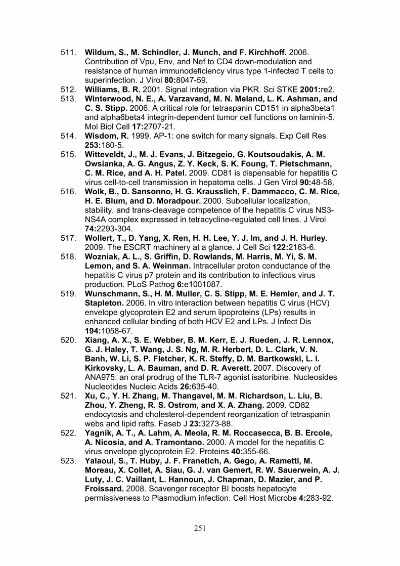
251
511. Wildum, S., M. Schindler, J. Munch, and F. Kirchhoff. 2006. Contribution of Vpu, Env, and Nef to CD4 down-modulation and resistance of human immunodeficiency virus type 1-infected T cells to superinfection. J Virol 80:8047-59.
512. Williams, B. R. 2001. Signal integration via PKR. Sci STKE 2001:re2. 513. Winterwood, N. E., A. Varzavand, M. N. Meland, L. K. Ashman, and
C. S. Stipp. 2006. A critical role for tetraspanin CD151 in alpha3beta1 and alpha6beta4 integrin-dependent tumor cell functions on laminin-5. Mol Biol Cell 17:2707-21.
514. Wisdom, R. 1999. AP-1: one switch for many signals. Exp Cell Res 253:180-5.
515. Witteveldt, J., M. J. Evans, J. Bitzegeio, G. Koutsoudakis, A. M. Owsianka, A. G. Angus, Z. Y. Keck, S. K. Foung, T. Pietschmann, C. M. Rice, and A. H. Patel. 2009. CD81 is dispensable for hepatitis C virus cell-to-cell transmission in hepatoma cells. J Gen Virol 90:48-58.
516. Wolk, B., D. Sansonno, H. G. Krausslich, F. Dammacco, C. M. Rice, H. E. Blum, and D. Moradpour. 2000. Subcellular localization, stability, and trans-cleavage competence of the hepatitis C virus NS3-NS4A complex expressed in tetracycline-regulated cell lines. J Virol 74:2293-304.
517. Wollert, T., D. Yang, X. Ren, H. H. Lee, Y. J. Im, and J. H. Hurley. 2009. The ESCRT machinery at a glance. J Cell Sci 122:2163-6.
518. Wozniak, A. L., S. Griffin, D. Rowlands, M. Harris, M. Yi, S. M. Lemon, and S. A. Weinman. Intracellular proton conductance of the hepatitis C virus p7 protein and its contribution to infectious virus production. PLoS Pathog 6:e1001087.
519. Wunschmann, S., H. M. Muller, C. S. Stipp, M. E. Hemler, and J. T. Stapleton. 2006. In vitro interaction between hepatitis C virus (HCV) envelope glycoprotein E2 and serum lipoproteins (LPs) results in enhanced cellular binding of both HCV E2 and LPs. J Infect Dis 194:1058-67.
520. Xiang, A. X., S. E. Webber, B. M. Kerr, E. J. Rueden, J. R. Lennox, G. J. Haley, T. Wang, J. S. Ng, M. R. Herbert, D. L. Clark, V. N. Banh, W. Li, S. P. Fletcher, K. R. Steffy, D. M. Bartkowski, L. I. Kirkovsky, L. A. Bauman, and D. R. Averett. 2007. Discovery of ANA975: an oral prodrug of the TLR-7 agonist isatoribine. Nucleosides Nucleotides Nucleic Acids 26:635-40.
521. Xu, C., Y. H. Zhang, M. Thangavel, M. M. Richardson, L. Liu, B. Zhou, Y. Zheng, R. S. Ostrom, and X. A. Zhang. 2009. CD82 endocytosis and cholesterol-dependent reorganization of tetraspanin webs and lipid rafts. Faseb J 23:3273-88.
522. Yagnik, A. T., A. Lahm, A. Meola, R. M. Roccasecca, B. B. Ercole, A. Nicosia, and A. Tramontano. 2000. A model for the hepatitis C virus envelope glycoprotein E2. Proteins 40:355-66.
523. Yalaoui, S., T. Huby, J. F. Franetich, A. Gego, A. Rametti, M. Moreau, X. Collet, A. Siau, G. J. van Gemert, R. W. Sauerwein, A. J. Luty, J. C. Vaillant, L. Hannoun, J. Chapman, D. Mazier, and P. Froissard. 2008. Scavenger receptor BI boosts hepatocyte permissiveness to Plasmodium infection. Cell Host Microbe 4:283-92.
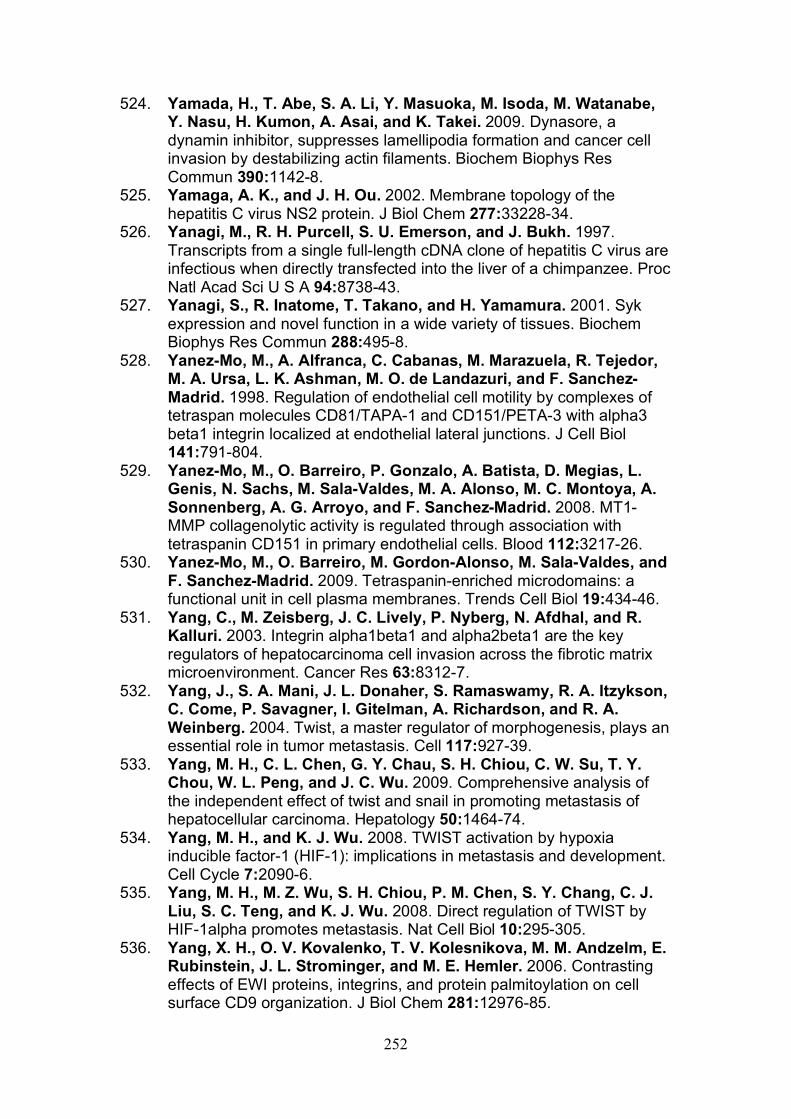
252
524. Yamada, H., T. Abe, S. A. Li, Y. Masuoka, M. Isoda, M. Watanabe, Y. Nasu, H. Kumon, A. Asai, and K. Takei. 2009. Dynasore, a dynamin inhibitor, suppresses lamellipodia formation and cancer cell invasion by destabilizing actin filaments. Biochem Biophys Res Commun 390:1142-8.
525. Yamaga, A. K., and J. H. Ou. 2002. Membrane topology of the hepatitis C virus NS2 protein. J Biol Chem 277:33228-34.
526. Yanagi, M., R. H. Purcell, S. U. Emerson, and J. Bukh. 1997. Transcripts from a single full-length cDNA clone of hepatitis C virus are infectious when directly transfected into the liver of a chimpanzee. Proc Natl Acad Sci U S A 94:8738-43.
527. Yanagi, S., R. Inatome, T. Takano, and H. Yamamura. 2001. Syk expression and novel function in a wide variety of tissues. Biochem Biophys Res Commun 288:495-8.
528. Yanez-Mo, M., A. Alfranca, C. Cabanas, M. Marazuela, R. Tejedor, M. A. Ursa, L. K. Ashman, M. O. de Landazuri, and F. Sanchez-Madrid. 1998. Regulation of endothelial cell motility by complexes of tetraspan molecules CD81/TAPA-1 and CD151/PETA-3 with alpha3 beta1 integrin localized at endothelial lateral junctions. J Cell Biol 141:791-804.
529. Yanez-Mo, M., O. Barreiro, P. Gonzalo, A. Batista, D. Megias, L. Genis, N. Sachs, M. Sala-Valdes, M. A. Alonso, M. C. Montoya, A. Sonnenberg, A. G. Arroyo, and F. Sanchez-Madrid. 2008. MT1-MMP collagenolytic activity is regulated through association with tetraspanin CD151 in primary endothelial cells. Blood 112:3217-26.
530. Yanez-Mo, M., O. Barreiro, M. Gordon-Alonso, M. Sala-Valdes, and F. Sanchez-Madrid. 2009. Tetraspanin-enriched microdomains: a functional unit in cell plasma membranes. Trends Cell Biol 19:434-46.
531. Yang, C., M. Zeisberg, J. C. Lively, P. Nyberg, N. Afdhal, and R. Kalluri. 2003. Integrin alpha1beta1 and alpha2beta1 are the key regulators of hepatocarcinoma cell invasion across the fibrotic matrix microenvironment. Cancer Res 63:8312-7.
532. Yang, J., S. A. Mani, J. L. Donaher, S. Ramaswamy, R. A. Itzykson, C. Come, P. Savagner, I. Gitelman, A. Richardson, and R. A. Weinberg. 2004. Twist, a master regulator of morphogenesis, plays an essential role in tumor metastasis. Cell 117:927-39.
533. Yang, M. H., C. L. Chen, G. Y. Chau, S. H. Chiou, C. W. Su, T. Y. Chou, W. L. Peng, and J. C. Wu. 2009. Comprehensive analysis of the independent effect of twist and snail in promoting metastasis of hepatocellular carcinoma. Hepatology 50:1464-74.
534. Yang, M. H., and K. J. Wu. 2008. TWIST activation by hypoxia inducible factor-1 (HIF-1): implications in metastasis and development. Cell Cycle 7:2090-6.
535. Yang, M. H., M. Z. Wu, S. H. Chiou, P. M. Chen, S. Y. Chang, C. J. Liu, S. C. Teng, and K. J. Wu. 2008. Direct regulation of TWIST by HIF-1alpha promotes metastasis. Nat Cell Biol 10:295-305.
536. Yang, X. H., O. V. Kovalenko, T. V. Kolesnikova, M. M. Andzelm, E. Rubinstein, J. L. Strominger, and M. E. Hemler. 2006. Contrasting effects of EWI proteins, integrins, and protein palmitoylation on cell surface CD9 organization. J Biol Chem 281:12976-85.
253
537. Yang, Y., H. Nagano, H. Ota, O. Morimoto, M. Nakamura, H. Wada, T. Noda, B. Damdinsuren, S. Marubashi, A. Miyamoto, Y. Takeda, K. Dono, K. Umeshita, S. Nakamori, K. Wakasa, M. Sakon, and M. Monden. 2007. Patterns and clinicopathologic features of extrahepatic recurrence of hepatocellular carcinoma after curative resection. Surgery 141:196-202.
538. Yao, M., X. D. Zhou, X. L. Zha, D. R. Shi, J. Fu, J. Y. He, H. F. Lu, and Z. Y. Tang. 1997. Expression of the integrin alpha5 subunit and its mediated cell adhesion in hepatocellular carcinoma. J Cancer Res Clin Oncol 123:435-40.
539. Yauch, R. L., F. Berditchevski, M. B. Harler, J. Reichner, and M. E. Hemler. 1998. Highly stoichiometric, stable, and specific association of integrin alpha3beta1 with CD151 provides a major link to phosphatidylinositol 4-kinase, and may regulate cell migration. Mol Biol Cell 9:2751-65.
540. Ye, J. 2007. Reliance of host cholesterol metabolic pathways for the life cycle of hepatitis C virus. PLoS Pathog 3:e108.
541. Ye, J., C. Wang, R. Sumpter, Jr., M. S. Brown, J. L. Goldstein, and M. Gale, Jr. 2003. Disruption of hepatitis C virus RNA replication through inhibition of host protein geranylgeranylation. Proc Natl Acad Sci U S A 100:15865-70.
542. Yee, T. T., A. Griffioen, C. A. Sabin, G. Dusheiko, and C. A. Lee. 2000. The natural history of HCV in a cohort of haemophilic patients infected between 1961 and 1985. Gut 47:845-51.
543. Yeh, C. N., S. T. Pang, T. W. Chen, R. C. Wu, W. H. Weng, and M. F. Chen. 2009. Expression of ezrin is associated with invasion and dedifferentiation of hepatitis B related hepatocellular carcinoma. BMC Cancer 9:233.
544. Yeo, A. E., M. Ghany, C. Conry-Cantilena, J. C. Melpolder, D. E. Kleiner, J. W. Shih, J. H. Hoofnagle, and H. J. Alter. 2001. Stability of HCV-RNA level and its lack of correlation with disease severity in asymptomatic chronic hepatitis C virus carriers. J Viral Hepat 8:256-63.
545. Yi, M., R. A. Villanueva, D. L. Thomas, T. Wakita, and S. M. Lemon. 2006. Production of infectious genotype 1a hepatitis C virus (Hutchinson strain) in cultured human hepatoma cells. Proc Natl Acad Sci U S A 103:2310-5.
546. Yilmaz, M., and G. Christofori. 2009. EMT, the cytoskeleton, and cancer cell invasion. Cancer Metastasis Rev 28:15-33.
547. Yonemura, S., T. Matsui, S. Tsukita, and S. Tsukita. 2002. Rho-dependent and -independent activation mechanisms of ezrin/radixin/moesin proteins: an essential role for polyphosphoinositides in vivo. J Cell Sci 115:2569-80.
548. Yoneyama, M., M. Kikuchi, T. Natsukawa, N. Shinobu, T. Imaizumi, M. Miyagishi, K. Taira, S. Akira, and T. Fujita. 2004. The RNA helicase RIG-I has an essential function in double-stranded RNA-induced innate antiviral responses. Nat Immunol 5:730-7.
549. Yoon, J. C., M. Shiina, G. Ahlenstiel, and B. Rehermann. 2009. Natural killer cell function is intact after direct exposure to infectious hepatitis C virions. Hepatology 49:12-21.
254
550. Young, K. C., K. L. Lindsay, K. J. Lee, W. C. Liu, J. W. He, S. L. Milstein, and M. M. Lai. 2003. Identification of a ribavirin-resistant NS5B mutation of hepatitis C virus during ribavirin monotherapy. Hepatology 38:869-78.
551. Zavadil, J., and E. P. Bottinger. 2005. TGF-beta and epithelial-to-mesenchymal transitions. Oncogene 24:5764-74.
552. Zeisberg, M., C. Yang, M. Martino, M. B. Duncan, F. Rieder, H. Tanjore, and R. Kalluri. 2007. Fibroblasts derive from hepatocytes in liver fibrosis via epithelial to mesenchymal transition. J Biol Chem 282:23337-47.
553. Zeisel, M. B., G. Koutsoudakis, E. K. Schnober, A. Haberstroh, H. E. Blum, F. L. Cosset, T. Wakita, D. Jaeck, M. Doffoel, C. Royer, E. Soulier, E. Schvoerer, C. Schuster, F. Stoll-Keller, R. Bartenschlager, T. Pietschmann, H. Barth, and T. F. Baumert. 2007. Scavenger receptor class B type I is a key host factor for hepatitis C virus infection required for an entry step closely linked to CD81. Hepatology 46:1722-31.
554. Zennou, V., C. Serguera, C. Sarkis, P. Colin, E. Perret, J. Mallet, and P. Charneau. 2001. The HIV-1 DNA flap stimulates HIV vector-mediated cell transduction in the brain. Nat Biotechnol 19:446-50.
555. Zhang, J., G. Randall, A. Higginbottom, P. Monk, C. M. Rice, and J. A. McKeating. 2004. CD81 is required for hepatitis C virus glycoprotein-mediated viral infection. J Virol 78:1448-55.
556. Zhang, X. A., A. L. Bontrager, and M. E. Hemler. 2001. Transmembrane-4 superfamily proteins associate with activated protein kinase C (PKC) and link PKC to specific beta(1) integrins. J Biol Chem 276:25005-13.
557. Zhang, Y., M. Y. Hu, W. Z. Wu, Z. J. Wang, K. Zhou, X. L. Zha, and K. D. Liu. 2006. The membrane-cytoskeleton organizer ezrin is necessary for hepatocellular carcinoma cell growth and invasiveness. J Cancer Res Clin Oncol 132:685-97.
558. Zhao, L. J., L. Wang, H. Ren, J. Cao, L. Li, J. S. Ke, and Z. T. Qi. 2005. Hepatitis C virus E2 protein promotes human hepatoma cell proliferation through the MAPK/ERK signaling pathway via cellular receptors. Exp Cell Res 305:23-32.
559. Zheng, A., F. Yuan, Y. Li, F. Zhu, P. Hou, J. Li, X. Song, M. Ding, and H. Deng. 2007. Claudin-6 and claudin-9 function as additional coreceptors for hepatitis C virus. J Virol 81:12465-71.
560. Zhong, J., P. Gastaminza, G. Cheng, S. Kapadia, T. Kato, D. R. Burton, S. F. Wieland, S. L. Uprichard, T. Wakita, and F. V. Chisari. 2005. Robust hepatitis C virus infection in vitro. Proc Natl Acad Sci U S A 102:9294-9.
561. Zoller, M. 2009. Tetraspanins: push and pull in suppressing and promoting metastasis. Nat Rev Cancer 9:40-55.
562. Zuckerman, E., T. Zuckerman, A. M. Levine, D. Douer, K. Gutekunst, M. Mizokami, D. G. Qian, M. Velankar, B. N. Nathwani, and T. L. Fong. 1997. Hepatitis C virus infection in patients with B-cell non-Hodgkin lymphoma. Ann Intern Med 127:423-8.