Embed Size (px)
Citation preview
Submitted 5 June 2017Accepted 31 October 2017Published 17 November 2017
Corresponding authorJames Chapman,[email protected]
Academic editorLaura Guidetti
Additional Information andDeclarations can be found onpage 16
DOI 10.7717/peerj.4076
Copyright2017 Kean et al.
Distributed underCreative Commons CC-BY 4.0
OPEN ACCESS
The role of biomaterials in the treatmentof meniscal tearsCrystal O. Kean1,*, Robert J. Brown2 and James Chapman1,*
1 School of Health, Medical and Applied Sciences, Central Queensland University, Rockhampton, Queensland,Australia
2Advanced Medical Solutions, Plymouth, UK*These authors contributed equally to this work.
ABSTRACTExtensive investigations over the recent decades have established the anatomical,biomechanical and functional importance of the meniscus in the knee joint. As afunctioning part of the joint, it serves to prevent the deterioration of articular cartilageand subsequent osteoarthritis. To this end, meniscus repair and regeneration is ofparticular interest from the biomaterial, bioengineering and orthopaedic researchcommunity. Even though meniscal research is previously of a considerable volume,the research community with evolving material science, biology and medical advancesare all pushing toward emerging novel solutions and approaches to the successfultreatment of meniscal difficulties. This review presents a tactical evaluation of the latestbiomaterials, experiments to simulate meniscal tears and the state-of-the-art materialsand strategies currently used to treat tears.
Subjects Biotechnology, Kinesiology, OrthopedicsKeywords Knee meniscus; biomaterials, Tissue engineering, Materials science, Scaffolds,Biomaterials
INTRODUCTIONThe knee is considered a hinge joint; however, because it also features characteristics of anarthrodial joint, it is a more complex joint than other hinge joints such as the elbow andankle. The knee consists of two articulations which form the tibiofemoral joint (furtherseparated into the medial and lateral tibiofemoral joints) and the patellofemoral joint. Thearticulations are not entirely congruent and this arrangement allows for the combinationof gliding and rolling motions which is constrained mainly by the ligaments of the knee.The menisci are fibrocartilagenous structures that sit on top of tibia to deepen the plateauswith the primary functions transmitting load through the joint and also serve to increasejoint stability and lubrication of the articular cartilage (Seedhom, Dowson & Wright, 1974;Walker & Erkman, 1975;McDermott, Masouros & Amis, 2008).
The menisci are commonly injured due to traumatic events and/or degenerative stresses.In the United States alone, it was estimated that approximately 6.6 million patient visitsto the emergency department between 1999 and 2008 were due to knee injuries equatingto 2.29 knee injuries per 1,000 people (Gage et al., 2012; Reid et al., 2017). Furthermore, by2060 the percentage of people reaching an age of 50 will reach 50% representing a changein population demographic and likelihood for pressures in the knee. A large proportion of
How to cite this article Kean et al. (2017), The role of biomaterials in the treatment of meniscal tears. PeerJ 5:e4076; DOI10.7717/peerj.4076
knee injuries in the general population are meniscal related and meniscal injuries are evenmore common in a physically active population (Baker et al., 1985; Nielsen & Yde, 1991).Given the role meniscal tears, and subsequent partial or full removal of the meniscus,play in development of osteoarthritis (Englund, Roos & Lohmander, 2003; Roos et al., 1998)there is an increased interest in preservation of these structures following injury. For thisreason, there is also an increased interest the role biomaterials play in meniscal repair,regeneration and replacement options.
Advances in materials technology have brought about an increased usage of biomaterialsand medical devices in the body (Hallab, Link & McAfee, 2003; Chevalier, 2006; Yamamoto,Takagi & Ito, 2016). A biomaterial is a material or substance or combination of substances,other than drugs, synthetic or natural in origin, which can be used for any period oftime (Bochyńska et al., 2016c; Brannigan & Dove , 2017; Kaur, 2017), which augments orreplaces partially or totally any tissue, organ or function of the body in order to improvethe quality of life of an individual (Bergmann & Stumpf, 2013). The biomaterial mustbe able to interact with the surrounding human tissue and body fluids to improve orreplace the anatomical defect. Some examples of the recent advances for biomaterialuse in medicine include knee and hip replacement (Walczak, Shahgaldi & Heatley, 1998;Bahraminasab & Farahmand, 2017), ocular implants (Lloyd, Faragher & Denyer, 2001;Baino et al., 2017; Mota et al., 2017), heart valves (Vongpatanasin, Hillis & Lange, 1996;Vander Roest & Merryman, 2016; Emmert & Hoerstrup, 2017), bone implants (Bròdano etal., 2014; Apicella et al., 2017), dental implants (Tamimi et al., 2014), biosensors (Sun et al.,2014; Calvo et al., 2017), orthopaedic screws and sutures (Waizy et al., 2014; Zhao et al.,2017) and tissue allografts (Cameron & Saha, 1997; Sanen et al., 2017). The achievements,in terms of biocompatibility, to lower risk of failure and improved surgical outcomeshave contributed to the expanding use of biomaterials. For these reasons advancements inbiomaterial development is and has been a significantly fast-growing area of research.
This review article will focus on providing a general review of the menisci and meniscalinjuries. We also discuss biomaterials and the subsequent role biomaterials play in thesurgical treatment options for meniscal repair, regeneration and replacement as well asfuture directions. While other reviews have been developed, their focus has been to providean overview of materials only, this review provides significant detail on cell lines used,models and materials to support research momentum for future developmental medicalbreakthroughs.
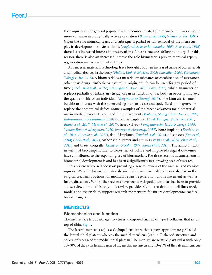
MENISCUSBiomechanics and functionThe menisci are fibrocartilage structures, composed mainly of type 1 collagen, that sit ontop of tibia, Fig. 1.
The lateral meniscus (e) is a C-shaped structure that covers approximately 80% ofthe lateral tibial plateau whereas the medial meniscus (c) is a U-shaped structure andcovers only 60% of the medial tibial plateau. The menisci are relatively avascular with only10–30% of the peripheral region of themedial meniscus and 10–25% of the lateral meniscus
Kean et al. (2017), PeerJ, DOI 10.7717/peerj.4076 2/26
Figure 1 Superior view of the right tibia in the knee joint illustrating the menisci and cruciate liga-ments. (A) anterior cruciate ligament, (B) articular cartilage on medial tibial condyle, (C) medial menis-cus, (D) posterior cruciate ligament, (E) lateral meniscus, (F) articular cartilage on lateral tibial condyle.
Full-size DOI: 10.7717/peerj.4076/fig-1
being vascular (Arnoczky & Warren, 1982). Based on its vascularisation, the menisci canbe divided into three zones: the red-red vascular zone (outer peripheral region), thewhite-white avascular zone (inner region) and the red-white zone which lies between ofthe two other zones and has characteristics of other two zones. The red vascular region isthick and convex and attaches to the capsule of the joint whereas the white-white innerregion is thin, concave and is a free edge unattached to the joint.
The menisci effectively deepen the tibial plateau and allow smooth articulation betweenthe tibial and femoral condyles and the transmission of loads across the tibiofemoral joint.In full knee extension, the medial meniscus transmits approximately 50% of the load on themedial compartment, while lateral meniscus transmits approximately 70% of the load inthe lateral compartment (Walker & Erkman, 1975). As knee flexion increases the amountof load transmitted to the lateral meniscus increases such that when the knee is flexedbeyond 75◦ the entire load that passes through the lateral compartment, is transmittedby the lateral meniscus (Walker & Erkman, 1975). For the medial meniscus the increasein load transmission as the knee flexes is less apparent (Walker & Erkman, 1975). Whenthe meniscus is intact, the load is well distributed across the tibiofemoral compartment;however when part or the entire meniscus is removed there is considerable alterations toload distribution such that there is a decrease in the contact area and increases in peakcontact forces (Bedi et al., 2012; Lee et al., 2006; Ihn, Kim & Park, 1993).
Kean et al. (2017), PeerJ, DOI 10.7717/peerj.4076 3/26
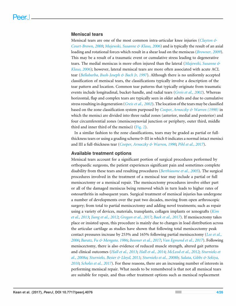
Meniscal tearsMeniscal tears are one of the most common intra-articular knee injuries (Clayton &Court-Brown, 2008;Majewski, Susanne & Klaus, 2006) and is typically the result of an axialloading and rotational forces which result in a shear load on the meniscus (Browner, 2009).This may be a result of a traumatic event or cumulative stress leading to degenerativetears. The medial meniscus is more often injured than the lateral (Majewski, Susanne &Klaus, 2006); however, lateral meniscal tears are more often associated with acute ACLtear (Bellabarba, Bush-Joseph & Bach Jr, 1997). Although there is no uniformly acceptedclassification of meniscal tears, the classifications typically involve a description of thetear pattern and location. Common tear patterns that typically originate from traumaticevents include longitudinal, bucket-handle, and radial tears (Greis et al., 2002). Whereashorizontal, flap and complex tears are typically seen in older adults and due to cumulativestress resulting in degeneration (Greis et al., 2002). The location of the tearsmay be classifiedbased on the zone classification system purposed by Cooper, Arnoczky & Warren (1990) inwhich the menisci are divided into three radial zones (anterior, medial and posterior) andfour circumferential zones (meniscosynovial junction or periphery, outer third, middlethird and inner third of the menisci) (Fig. 2).In a similar fashion to the zone classifications, tears may be graded as partial or full-
thickness tears or using a grading scheme 0–III in which 0 indicates a normal intact menisciand III a full-thickness tear (Cooper, Arnoczky & Warren, 1990; Pihl et al., 2017).
Available treatment optionsMeniscal tears account for a significant portion of surgical procedures performed byorthopaedic surgeons, the patient experiences significant pain and sometimes completedisability from these tears and resulting procedures (Berthiaume et al., 2005). The surgicalprocedures involved in the treatment of a meniscal tear may include a partial or fullmeniscectomy or a meniscal repair. The meniscectomy procedures involve either partor all of the damaged meniscus being removed which in turn leads to higher rates ofosteoarthritis in subsequent years. Surgical treatment of meniscal injuries has undergonea number of developments over the past two decades, moving from open arthroscopicsurgery; from total to partial meniscectomy and adding novel treatments; such as repairusing a variety of devices, materials, transplants, collagen implants or xenografts (Kimet al., 2013; Jiang et al., 2012; Grogan et al., 2017; Baek et al., 2017). If meniscectomy takesplace or insisted upon, this procedure is mainly due to changes in load distribution acrossthe articular cartilage as studies have shown that following total meniscectomy peakcontact pressures increase by 253% and 165% following partial meniscectomy (Lee et al.,2006; Baratz, Fu & Mengato, 1986; Beamer et al., 2017; Van Egmond et al., 2017). Followingmeniscectomy, there is also evidence of reduced muscle strength, altered gait patternsand clinical outcomes (Hall et al., 2013; Hall et al., 2014; McLeod et al., 2012; Sturnieks etal., 2008a; Sturnieks, Besier & Lloyd, 2011; Sturnieks et al., 2008b; Salata, Gibbs & Sekiya,2010; Scholes et al., 2017). For these reasons, there are an increasing number of interests inperforming meniscal repair. What needs to be remembered is that not all meniscal tearsare suitable for repair, and thus other treatment options such as meniscal replacement
Kean et al. (2017), PeerJ, DOI 10.7717/peerj.4076 4/26
Figure 2 Schematic diagram highlighting the various types of meniscal tears, Bucket Handle MRI im-age taken fromHan et al. (2015) (CC BYNC 3.0), Radial Tear, MRI image taken from Jung et al. (2012),and longitudinal (photograph taken from Feucht et al. (2015) (CC BY 4.0)) and horizontal tears (MRItaken fromOhishi et al. (2010) (CC BY 2.0)) all with permission.
Full-size DOI: 10.7717/peerj.4076/fig-2
and regeneration are of considerable interest when a surgical intervention is necessary toimprove any pain and symptoms.
BIOMATERIALSCurrent treatment modalities for meniscal repair tears still carry their drawbacks andnovel, robust and effective solutions are required. Some recent advances in meniscusresearch suggest that low cellularity, (King et al., 2017) dense ECM and poor vascularisationcoupled with the inflammatory responses (King et al., 2017) in the knee joint areresponsible for a lack of healing. Recently, biomaterials in the form of tissue adhesiveshave become available for clinical use: fibrin glue, (Bochyńska et al., 2016a) TissuGlu R©,
Kean et al. (2017), PeerJ, DOI 10.7717/peerj.4076 5/26
Dermabond R©, (Balakrishnan et al., 2017) where the development of these new adhesivebiomaterials has improved the properties of existing biomaterials alone (TissuGlu R©,Raleigh, NC, USA; Ethicon Inc., Somerville, NJ, USA). Furthermore, these materials andstrategies are not always a given success, presenting limitations to the accomplishment ofthe meniscal reparation.
Tissue engineering using biomaterialsOf late, tissue-engineering and cellular biomaterial interactive concepts have beenintroduced to develop cellular-based reparation for cartilage regeneration (Temenoff &Mikos, 2000). The type of cell used to engineer cartilage is critical as a future goal ofbiomaterial development. Various cell populations that have been investigated for theseroles include: chondrocytes (King et al., 2017; Chen & Cheng, 2006), mesenchymal stemcells, bone marrow stromal cells and perichondrocytes (Bruns et al., 1998). The choice ofbiomaterial is critical to the success of tissue engineering approaches for cartilage repair.The concept of ‘tissue engineering’ was first introduced and postulated by Green Jr (1977)where chondrocytes grown ex vivo could be transplanted into a region of tissue defect.Recently, tissue and biomaterial engineering concepts have been initiated to develop cellularbased approaches for tissue repair (Freed et al., 1993). Typically, the process for engineeringtissue involves the isolation of chondrocytes which are then seeded into a biocompatiblematrix or scaffold and finally cultivated for implantation into the defected region. A largevariety of biomaterials, natural and synthetic, have been employed as potential cell-carriersfor tissue regeneration. The most common naturally occurring materials include type Iand type II collagen-based biomaterials. Furthermore, some of the contrasting syntheticapproaches include: polyglycolic acid or poly-L-lactic acid or other various compositemixtures (Chen & Cheng, 2006). In essence, an ideal candidate biomaterial would be acell-carrier substance which closely mimics the natural environment in the surroundingmatrix—as given by the definition of a biomaterial.
Regenerative approaches to meniscus repair occurs in a series of precise stages. It istypically understood that the low cellularity (endogenous meniscus cells and meniscusprogenitors) (Mauck & Burdick, 2015), the dense ECM, poor vascularisation potential andthe inflammatory responses typically linked to meniscus wounds all contribute to thesuccess or failure of the meniscus healing and regeneration alone. This success of healingprocess is without a biomaterial introduced into the site. Based on these principles, thepotential use of a biomaterial to develop and deliver a viable solution requires thoughtaround this repair process.
Biomaterials are typically promoters of tissue repair through provision of scaffold layersfor cellular attachment and growth and differentiation further acting as a vehicle for proteinand gene transfer to regenerate functional tissue approaches (Chen, Zhang & Wu, 2010).Biomaterials in this area should have several properties to support viable repair. Typically,this is achieved through:(1) The material must act as a support structure for cell lines (i.e., cells that are seeded
in vitro are compatible, adhere to the material if required or certain cell linesare not required; filtered out). For meniscal repair the biomaterial must provide
Kean et al. (2017), PeerJ, DOI 10.7717/peerj.4076 6/26
appropriate biomechanical functions after implantation to shield cells from damagingor compressive forces;
(2) Possess sufficient mechanical strength to protect the surrounding cells (cells shouldbe mechanically stable i.e., cell attachment is maintained). For meniscal repair thebiomaterial must maintain their shape and integrity, mechanical stability and strengthfor the defect area in questionuntil newhost tissue has been regenerated. Furthermore, itmay be important to provide biological and mechanical context for cell differentiation,proliferation and attachment when a biomaterial is introduced into the knee. Forexample, it is now very well understood that cells are influenced by the local externalenvironment including the adhesive and biophysical properties (Engler et al., 2006);
(3) Withstand in vivo forces during the joint movement operation (mechanical andstructural stability of the biomaterial in the meniscus area needs to be able to withstandcompressive and tensile forces (these forces have been aptly described in Paschos et al.(2017));
(4) Bioactivity should be provided to accommodate cellular attachment and cellularmigration (the biomaterial in the meniscus will therefore be able to promote tissueregeneration). Furthermore, providing directional cues, such as chemotactic gradientsto guide cells like endogenous cells to the injury site. Recently, some studies have shownthat allowing migration of cells provides a motivation for the cells to attach and drivesthe cellular colonisation process (Mauck & Burdick, 2015; Greiner et al., 2014);
(5) The biomaterial should have biodegradable properties and be able to remodel asthe novel cartilage grows, embeds and replaces the original construct; therefore, thematrix must be non-toxic, non-adhering and non-stimulating for inflammatory cells.The biomaterial for a meniscus should therefore facilitate host tissue integration andprovide the appropriate biomechanical function in the knee.
(6) Furthermore, they should be non-immunogenic as this is catastrophic for thebiomaterial insertion. For any biomaterial, this is important, to prevent rejectionthe appropriate level of biocompatibility and non-toxic ability needs to be considered.
BiocompatibilityOne of the most important non-mechanical requirements of orthopaedic biomaterialsis biocompatibility. Biocompatibility is the ability of a substrate to exist in contactwith tissues of the human body without causing an unacceptable degree of harm inthe body. The biomaterial domain has been aptly described by Mardis and Kroeger,‘‘the utopian state where a biomaterial presents an interface with physiologic environmentwithout the material adversely affecting the environment or environment adversely affectingthe biomaterial’’ (Mardis & Kroeger, 1988). An understanding of biocompatibility requiresan appreciation of tissue cell, bacterial cell and host defence response to the insertion of abiomaterial in particular for this review—for meniscal interventions. Once the biomaterialhas been placed into the body, a conditioning film containing biomolecules such as; water,electrolytes, cholesterol, vitamins, lipids and proteins (Chapman et al., 2013) (albumin,igG, fibronectin, fibrinogen, laminin, collagen and osteopontin) form on the surface longbefore cells are present and reach the state of equilibrium (Thevenot, Hu & Tang, 2008).
Kean et al. (2017), PeerJ, DOI 10.7717/peerj.4076 7/26
In the very early implantation period or injury for this matter, inflammatory cells beginto proliferate, this is an immediate response (Anderson, Rodriguez & Chang, 2008). Thefirst contact with tissue, proteins in blood and the interstitial fluids adsorb on to thebiomaterial surface. An injury to vascularised connective tissue initiates the inflammatoryresponse but also leads to the process of thrombus formation involving the activation of theextrinsic and also intrinsic coagulation, complement, fibrinolytic, kinin-generating systemsand platelets (Anderson, Rodriguez & Chang, 2008). The conditioning layer represents adynamic, ever-changing layer due to differential diffusion and mass transport of moleculesin and out of the implant surface. Later stages of competitive binding then occur on thesurface of the material owing to functional groups within the molecules. Cells thereforenever see the ‘true’ surface of the biomaterial, but more correctly, respond and interact toa conditioned film that has consequently developed in-situ.
Following the conditioning sequence of the biomaterial, attachment cells securethemselves to the protein and protein matrices using integrin receptors. Thus, thisconditioning layer is vital to the reaction of cells to the surface of the implanted biomaterial.The introduction of the biomaterial, the conditioning and immune response sequence isnot always obvious as proteins have the ability to conform and expose epitopes that arenot always identified as self-produced by the body’s immune system. Immune cells reactas they detect what were once normal proteins and recognise them as foreign bodies. Thisprocess can result in a cascade of blood coagulation and chronic inflammation that can leadto occlusion of nutrients, changes in oxygen and fibrous capsule formation—operatingtoward total rejection by the body of the implanted biomaterial (Nasab & Hassan, 2010).The extent of the deformation process for proteins has been remedied based on the selectionof material type. Surfaces are made more ‘‘passive’’ where chemical treatments are added tothemanufacturing process. Passivation with acids such as nitric acid of stainless steel createsa less reactive oxide layer; this has been shown to improve the biocompatibility process.One added benefit to passivation is it serves as a means for removing foreign material fromthe surface, such as bacteria or biofilms (Blumenfeld & Bargar, 2006). Passivation can alsobe used to surface-modify natural or synthetic polymer biomaterial substrates for meniscaltear applications. For example, albumin, where the resulting surface passivation has beenshown to reduce and prevent clotting (Kaur, 2017; Hanker & Giammara, 1988).
Role of biomaterials in meniscal repairAn article by Abrams et al. (2013) has shown that while there was no increase in the overallnumber of meniscal procedures, over a seven-year period there has been an 11.4% increasein isolated meniscal repairs and a 48.3% increase in meniscal repairs in combination withACL reconstruction. This sharp increase in meniscal repair treatment is mainly due to theincreased knowledge in the importance of the preservation of the meniscus to maintainnormal knee function and prevent osteoarthritis. It has been shown that following meniscalrepair, peak contact pressures are similar to that experienced with an intact meniscus (Bediet al., 2010). Unfortunately, it is estimated that currently only 20% of all meniscal tearsare repairable. Tears in the meniscal periphery (i.e., the red-red vascular zone) are mostlikely to heal whereas those in the meniscal avascular zone (i.e., the white-white zone)
Kean et al. (2017), PeerJ, DOI 10.7717/peerj.4076 8/26
are unlikely to heal and those in the red-white zone have the potential to heal (Belzer &Cannon, 1993;Noyes & Barber-Westin, 2012). Besides vascularisation, tear type and variouspatient characteristics can influence decision making on treatment options and success of ameniscal repair. Typically, tears that are less than 2 cm in length, longitudinal and acute aremore amendable to repair than larger tears (Taylor & Rodeo, 2013; Laible, Stein & Kiridly,2013). Meniscal repairs are also not typically recommended for degenerative tears and thusrepair success is typically superior in young patients (less than 50 years of age) (Laible, Stein& Kiridly, 2013). When appropriately performed, meniscal repairs provide considerableimprovements in terms of clinical outcome and osteoarthritis prevention compared to apartial meniscectomy (Stein et al., 2010). Thus, finding ways to increase the number ofmeniscal tears that can be treated by meniscal reparation is of great importance.
Vascularisation in the meniscus tissue is of high relevance to biomaterial design. Fromprenatal development up until after birth, the meniscus is fully vascularised. Following this,from the age of ten, vascularisation reduces to 30% of the meniscus and at maturity themeniscus only in the peripheral region of approximately 10% of the tissue. Vascularisationrepresents another challenge in meeting the requirements of success for biomaterialimplantation as a meniscus operation. Vascular endothelial growth factor enhances theblood vessel density in peri-implant spaces. Biomaterial scaffolds of knee menisci exist in ahighly challenging environment as little vascular support is provided in this region of thebody. Electrospinning of polymeric fibres can be produced to support other engineeringapplications such as blood vessel, tendons, meniscus and cartilage (Xu et al., 2004). Someauthors have used unique biodegradable nanofibers as a scaffold to support blood vesselengineering. They have demonstrated that fibres of 500 nm with an aligned topography isable to mimic the circumferential orientation of cells and fibrils (Leong et al., 2009). Theauthors have postulated that macrophage within the CES produce angiogeneic growthfactors that potentially stimulates vascularisation.
Role of biomaterials in meniscal replacement/regenerationOwing to the limited percentage of meniscal tears that can be repaired and the poorclinical results with untreated symptomatic meniscal injuries and partial meniscectomy,biomaterial synthetic and allogeneic (genetically dissimilar) interacting biomaterials havebeen investigated to serve as a matrix to lead meniscal regeneration medicine, particularlyas a cellular support.
HydrogelsUsing a biomaterial that has the ability to seamlessly integrate in to water matrices isanother attractive property in regenerative medicine applications for meniscal repair.Hydrogels are one such material with a considerable water based content; using hydratedpolymer networks capable of absorbing and retaining fluids. Hydrogels are determinedby their monomeric composition, crosslinking density and polymerisation ability. Due tothe crosslinking chemistry the polymer remains insoluble in solution. The insolubility,along with the high hydration threshold, make hydrogels appealing to use for humantissue mimetics (Kobayashi, Chang & Oka, 2005). As an example, some authors have used a
Kean et al. (2017), PeerJ, DOI 10.7717/peerj.4076 9/26
poly (vinyl alcohol) hydrogel with a water content of approximately 90% to produce kneeimplants using a rabbit model (Makris, Hadidi & Athanasiou, 2011). The implant replacedthe whole lateral meniscus over two years. In a subsequent study, the hydrogel implantwas not able to prevent damage to articular cartilage but was able to reduce progressionof meniscal decay. Some of the new and emerging biomaterial types have been shown inTable 1.
ChitinChitin sutures are an emerging material of choice for improvement of the mechanicalproperties of a knee healing process (Brittberg et al., 1994). Owing to its favourablemechanical properties, chitin has been used for applications that require exceptionalintegrity and physical strength in surgical sutures, some new medical textiles and even asbone substitute materials.
NanofibresElectrospun scaffolds are also another emerging biomaterial that has begun to be usedfor cellular adhesion applications in regenerative medicine. The fibres have the ability tomimic both anisotropy of fibrous tissues and withstand high load forces that are imposedon the tissue during physiological motions (Ionescu & Mauck, 2012). The electrospunbiomaterials can also be tailored to produce various size, shapes and makeup (for examplecoaxial materials) will influence cell interactions and the cells will begin to proliferateand adhere and finally deposit matrix on to the fibre network. These interactions provideimproved mechanical properties for the biomaterial scaffold over time. Fibres can becollected on to rotating drums or flat collection plates, depending on the order, orientationand architectures that they are required. Cells typically are seeded on to these scaffoldsand cultured over time in vitro. In a study by Passaretti et al. (2001) tensile modulus wasseen to improve on fibre aligned scaffolds some 7-fold higher than disorganised fibresapproaching the value of a normal meniscus. Essentially, the authors determined that cellsprefer to align on ordered scaffold fibres rather than disorganised arrangements. Furtherto these findings, internal organisation in the form of sheet fibres can also be arrangedfor tissue-mimicking structures. Specifically, for meniscal tissue engineering, cells can beisolated, expanded and manually seeded on to the surfaces of electrospun scaffolds prior toan implantation operation, expediting the regenerative process. Cells along with host cellswill migrate on to the newly implanted scaffold and deposit proteoglycan and collagen.Some implantation methods require surgery prior to this implant step to isolate the cellsprior to seeding, maturation and implantation.
Biodegradable polymersSome of the more current treatment methods for repair of meniscal tears are somewhatindifferent for positive results and outcomes. Tissue adhesives are a promising alternative,owing to their ease of application andminimal tissue trauma. Co-polymeric tissue adhesiveshave been shown to have adhesive strengths of 40–50 kPa and hold edges of meniscal tearstogether during healing periods. These results indicate that copolymers are able to improvetissue capacity for self-repair specific for meniscal applications. Other authors have used
Kean et al. (2017), PeerJ, DOI 10.7717/peerj.4076 10/26
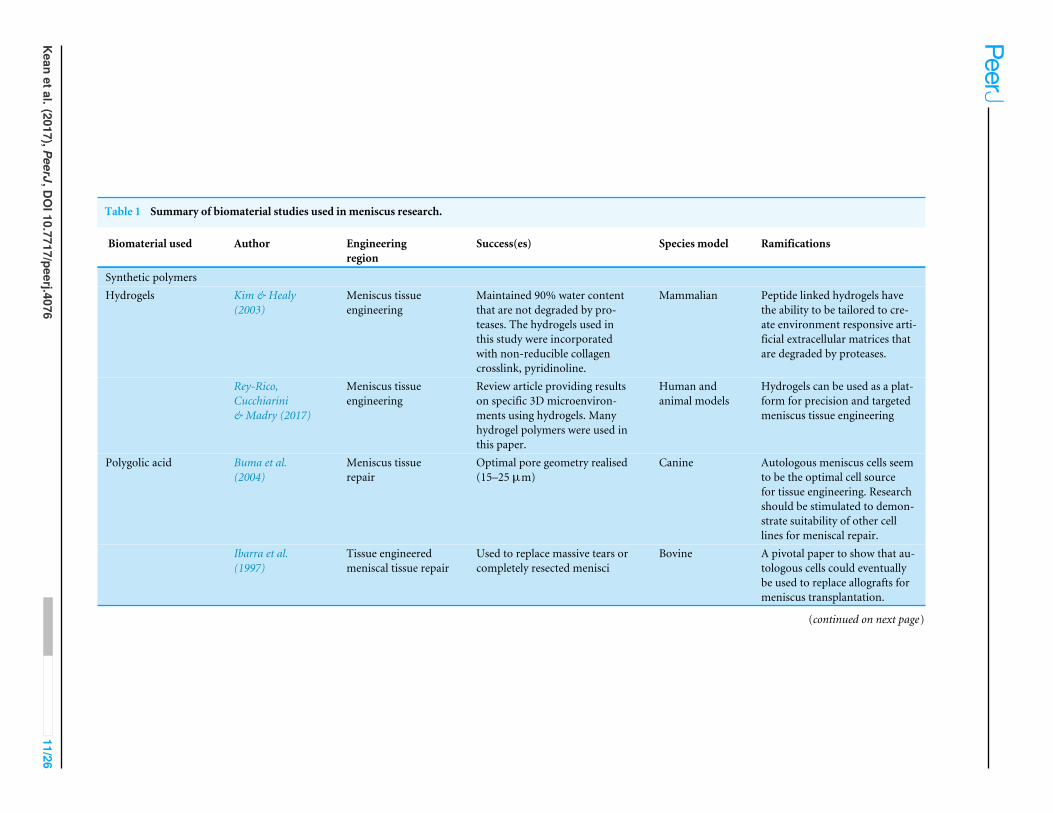
Table 1 Summary of biomaterial studies used in meniscus research.
Biomaterial used Author Engineeringregion
Success(es) Species model Ramifications
Synthetic polymersHydrogels Kim & Healy
(2003)Meniscus tissueengineering
Maintained 90% water contentthat are not degraded by pro-teases. The hydrogels used inthis study were incorporatedwith non-reducible collagencrosslink, pyridinoline.
Mammalian Peptide linked hydrogels havethe ability to be tailored to cre-ate environment responsive arti-ficial extracellular matrices thatare degraded by proteases.
Rey-Rico,Cucchiarini& Madry (2017)
Meniscus tissueengineering
Review article providing resultson specific 3D microenviron-ments using hydrogels. Manyhydrogel polymers were used inthis paper.
Human andanimal models
Hydrogels can be used as a plat-form for precision and targetedmeniscus tissue engineering
Polygolic acid Buma et al.(2004)
Meniscus tissuerepair
Optimal pore geometry realised(15–25 µm)
Canine Autologous meniscus cells seemto be the optimal cell sourcefor tissue engineering. Researchshould be stimulated to demon-strate suitability of other celllines for meniscal repair.
Ibarra et al.(1997)
Tissue engineeredmeniscal tissue repair
Used to replace massive tears orcompletely resected menisci
Bovine A pivotal paper to show that au-tologous cells could eventuallybe used to replace allografts formeniscus transplantation.
(continued on next page)
Keanetal.(2017),PeerJ,D
OI10.7717/peerj.4076
11/26
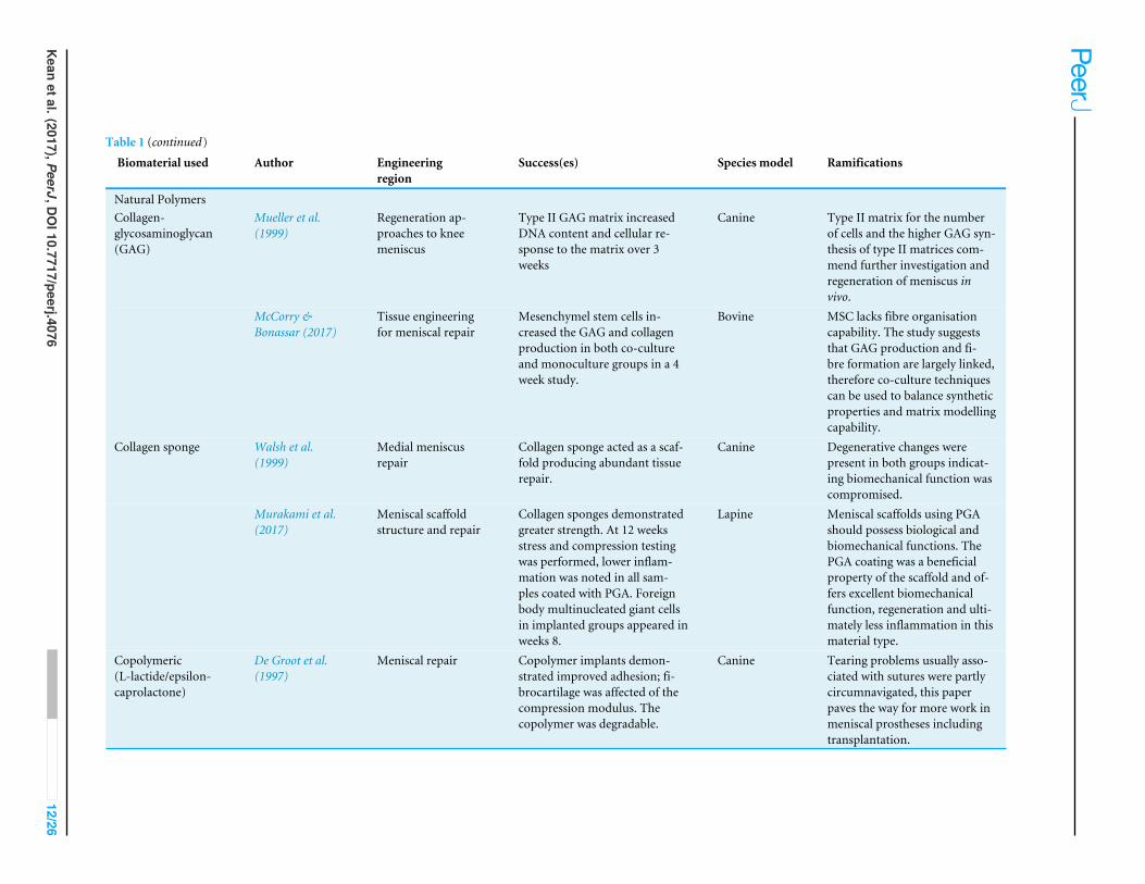
Table 1 (continued)
Biomaterial used Author Engineeringregion
Success(es) Species model Ramifications
Natural PolymersCollagen-glycosaminoglycan(GAG)
Mueller et al.(1999)
Regeneration ap-proaches to kneemeniscus
Type II GAG matrix increasedDNA content and cellular re-sponse to the matrix over 3weeks
Canine Type II matrix for the numberof cells and the higher GAG syn-thesis of type II matrices com-mend further investigation andregeneration of meniscus invivo.
McCorry &Bonassar (2017)
Tissue engineeringfor meniscal repair
Mesenchymel stem cells in-creased the GAG and collagenproduction in both co-cultureand monoculture groups in a 4week study.
Bovine MSC lacks fibre organisationcapability. The study suggeststhat GAG production and fi-bre formation are largely linked,therefore co-culture techniquescan be used to balance syntheticproperties and matrix modellingcapability.
Collagen sponge Walsh et al.(1999)
Medial meniscusrepair
Collagen sponge acted as a scaf-fold producing abundant tissuerepair.
Canine Degenerative changes werepresent in both groups indicat-ing biomechanical function wascompromised.
Murakami et al.(2017)
Meniscal scaffoldstructure and repair
Collagen sponges demonstratedgreater strength. At 12 weeksstress and compression testingwas performed, lower inflam-mation was noted in all sam-ples coated with PGA. Foreignbody multinucleated giant cellsin implanted groups appeared inweeks 8.
Lapine Meniscal scaffolds using PGAshould possess biological andbiomechanical functions. ThePGA coating was a beneficialproperty of the scaffold and of-fers excellent biomechanicalfunction, regeneration and ulti-mately less inflammation in thismaterial type.
Copolymeric(L-lactide/epsilon-caprolactone)
De Groot et al.(1997)
Meniscal repair Copolymer implants demon-strated improved adhesion; fi-brocartilage was affected of thecompression modulus. Thecopolymer was degradable.
Canine Tearing problems usually asso-ciated with sutures were partlycircumnavigated, this paperpaves the way for more work inmeniscal prostheses includingtransplantation.
Keanetal.(2017),PeerJ,D
OI10.7717/peerj.4076
12/26
amphiphilic copolymers based on polyethylene glycol, trimethylene carbonate and citricacid to synthesise end-functionalised hexamethylene diisocyanate to form biodegradablehyper-branched tissue adhesives. The work showcases resorbable tissue materials formeniscus repair. The materials have excellent mechanical and adhesive properties thatcould be adjusted through variation of the composition of the copolymers (Bochyńska etal., 2016c). Regenerative engineering converges a number of research areas and is trulymultidisciplinary inclusive of tissue engineering, advanced materials, stem cell scienceand developmental biology to regenerate complex tissues from menisci to whole limbs(Narayanan et al., 2016). Clinical applications of tissue engineering technologies arestill relatively restricted owing in part to the limited number of biomaterials that areapproved for human use. While many biomaterials have been developed, their translationinto practice has been extremely slow. Consequently, many researchers are still usingbiodegradable choices that were approved some 30 years ago. Most degradable biomaterialsused to date comprise of synthetic polyesters:
• Poly(L-lactic acid) PLLA;• Poly(L–glyolic acid) PLGA; and• Biological polymers such as: alginate or chitosan, collagen or fibrin (Middleton & Tipton,2000).
Polyester-based polymers are clearly an excellent candidate as a synthetic biodegradableand bio-absorbable material for medical applications. The use of synthetic polyesters asbiomaterials allow the unique control of the morphology, mechanical properties anddegradation profiles measured through the monomer selection, polymer compositioninformed through the copolymer and homopolymer, stereo-complexation and also themolecular weight. In an excellent review by Brannigan and Dove, degradation mechanismshas been discussed in detail, in a clinical research capacity—these parameters are ofparamount importance to understand the behaviour of the material in vitro or in vivo.The authors discuss enzymatic, oxidative, and physical degradation. Brannigan & Dove(2017) discuss the use and importance of polyester type Poly-HDPE scaffolds with aninterconnected porous structure for cartilage regeneration. In their work, neocartilageformation within a synthetic polyester scaffold based on polymerisation of high internalphase emulsions were used. The fabrication of polyHIPE polymers (PHP) was ordered tohave highly porous giving structure to the cartilage with a higher potential in force wear.Another example of the use of biodegradable polymers inmeniscal repair research includes,poly lactic acid or L-PLA is used in menisca reconstruction in a study using canines, thepresence of macrophages, fibroblasts, giant cells and lymphocytes were observed to beattaching to the material. From this study it seems that biocompatibility reduces when thedegradation process ensues. This degradation property therefore promoted inflammatoryresponses and thus rejection (Jones et al., 2002).
Kean et al. (2017), PeerJ, DOI 10.7717/peerj.4076 13/26
Discovery of new biomaterials—beyond state of the artThe next phase in developing knee meniscal biomaterials for replacement and orregeneration applications extends to the design, discovery and evaluation of bioactivematerials.
Bioactive meniscal materials have been used with some significantly exciting andpromising results. For example, bioactive scaffolds have been shown to modulate localECM density to improve repair (Shin, Jo & Mikos, 2003). A novel biphasic collagen scaffoldand shown to support meniscal repair in vivo to support meniscal cell ingrowth butalso producing ECM in vitro by Howard et al. (2016). The authors have shown that theaddition of PRP enhanced scaffold enhanced healing (Howard et al., 2016). Other emergingmaterials which could show potential in meniscal repair include: cartilage matrix is alsoa promising material for cartilage regeneration given the emerging evidence supportingits chondroinductive character. The cartilage matrix is a promising material for hyalinecartilage tissue engineering applications and has been shown that cell derived matrixand ECM materials and have been demonstrated to show established decellularisation,representing an excellent and promising choice of new material for future direction.(Redman, Oldfield & Archer, 2005). A drawback so far is that the FDA regulatory approvalmay affect the decision to use a native or cell-derived matrix. To expedite FDA approval,a full chemical decellularisation of allogeneic matrix may be used—this way, removal ofcells ensures no cross-species interactions (Sutherland et al., 2015). For example, allogeneiccells from bone marrow can be used in cardiac repair (Lemcke et al., 2017).
Initially, this is a relatively straightforward process whereby advanced synthesis of newmaterials can be performed. The difficulty lies with producing the novel activity andevaluation of the behaviour of the material in the biological system. Adapting the surfaceproperties through the addition of synthetic peptides and or molecular drugs can yieldthousands of candidate materials for testing. This approach has already been realised in theform of library derived screening techniques using commercially available methacrylatemonomers—influencing attachment, growth, proliferation and differentiation of humanembryonic stem cells (Anderson, Levenberg & Langer, 2004).
Further developments in biomaterials will continue to expand at the interface ofnanotechnology. Understanding the tribological interaction with the surroundinginterface of the human body is an approach that is being realised using the ‘‘bottom-up’’approach (Zur et al., 2011). The bottom up approach will develop novel, self-assemblingand environment reactive biomaterials. In particular, self-assembling peptides offer anew approach owing to the large variety of sequences that can be produced by chemicalsynthesis. These advances include the design of short peptides that have the ability toresemble nanofilaments which are compatible in vitro, without rejection. The use ofpeptides in polymeric materials allows for resistance in concentration, pH or level ofdivalent cation variability (Hartgerink, Beniash & Stupp, 2002).
The use of combinational gene therapy and biomaterial approaches is a recenttechnique to remedy meniscal lesions formed when orthopaedic surgery and loss ofthe meniscus has accelerated in the patient. The lack of therapeutic options suggests thereis a need for improved treatments to enhance meniscal tear repair treatments/operations.
Kean et al. (2017), PeerJ, DOI 10.7717/peerj.4076 14/26
Combinational approaches may also provide strategies to support this remedy (Cucchiariniet al., 2016). Gene therapy, can be directly applied as a combination or direct approachto meniscal repair strategies. A recent evaluation on gene therapy with cell and tissueengineering-based approaches demonstrates a six strategy approach: (a) directly using genetransfer vectors (Elsler et al., 2012), (b) administering genetically modified cells (Nakagawaet al., 2015), which could be fraught upon in some researching countries, continents, (c)implanting the biocompatible material that can deliver the recombinant factor, as wehave seen rejection may be a potential problem with this result, (d) applying autologousplatelet-rich plasma or fibrin clotting factors, (e) providing a biomaterial that delivers agene transfer vector, (f) transplanting a material seeded with cells, again, we envisage apotential rejection with this treatment.
Stem cell approachesExciting new techniques are emerging as non-invasive approaches to meniscal tearcorrection using stem cells. The promising use of new tissue engineering approaches haveincorporated natural biomaterials made from extracellularmatrices of decellularised tissuesfrom the heart, lung and bone for example (Yuan et al., 2017). The use of a scaffold or ‘shell’to align stem cells upon in a given feature is fast becoming attractive. Decullularisationpreserves themolecular composition with tissue specific molecules including structural andmechanical features present in the original tissue. The preservation step will aid in guidingthe behaviour of the therapeutic cells and facilitate tissue development when implanted,non-invasively to the meniscal tear region.
In vitro studies have also been used to investigate tissue surface modification withcollagenase to prime the surface where the addition of the TGF-beta3 cells has been provento increase the number of cells present in meniscal tears repaired with newly developedtissue adhesives such as isocyanate-terminated block polymers. For example, Bochynskeet al. have used cylindrical explants harvested from bovine menisci, the explants weresimulated to possesses a full thickness-tear where the explants were then removed andglued back to the defect. In addition, the repair constructs were then culture with andwithout the addition of TGF-beta3 and assessed for their histological appearances. Thehistological staining of the constructs confirmed that cytotoxicity was not an issue andafter 28 days, meniscal cells were present in the contact glues (Bochyńska et al., 2016b). Theresults demonstrate that the use of TGF-beta 3 induces thicker cell numbers round theedges of the annulus of the explants and also appears to be a promising treatment for tearsusing these glue types.
BiomimeticsOne final, prominent field emerging in material science lies with biomimetic biomaterialapproaches (Chapman et al., 2014). Biomimetic materials are materials that have beendirectly replicated from nature to produce a solution to a specific problem. Somesynthetic polymers may be able to provide a more biomimetic environment than thepreviously discussed hydrogel approach. Functionalising hydrogels using chemistry is onestrategy that requires future investigation. Hydrogels have the ability to create a more‘native’ microenvironment for cells in a particular area of the body—i.e., the knee. For
Kean et al. (2017), PeerJ, DOI 10.7717/peerj.4076 15/26
example, scaffolds with biomimetics have been developed for tissue engineering based on amultidisciplinary approach using engineering of biomaterials and nano/micro structuringof the defect tissues. The use of 3D bioprinting is considered to be conventional however,the technique has allowed for traditional fabricationmethods for porous bone and cartilageregeneration to be taken in new directions using gas forming, soluble particle leaching orfreeze drying. Newer methods to generate porous scaffolds using biodegradable polymersinclude using gas forming of porogens (ammonium bicarbonate particles). Injectablehydrogels using click chemistry (high yielding, wide in scope molecules) have also shownto be highly advantageous for local delivery of bioactive molecules, ease of handing andreduced invasiveness, these techniques have been demonstrated to be potentially used in3D bioprinting (Jo, Kim & Noh, 2012). The use of hand held 3D matrix printing usinga bio-pen has allowed for in-situ printing and repair to take place. This will be a majordevelopment in regenerativemedicine (Di Bella et al., 2017). Themost recent and emergingareas for biomimetic medical materials are (Chen et al., 2016): (1) 3D bioprinting (focussedon medical materials); (2) designing nano/micro technologies; (3) surface modificationof biomaterials for their cellular interaction ability; (4) clinical aspects of biomaterialsfor cartilage focussing on cells, scaffolds and cytokines (Fratzl & Weinkamer, 2007). Thetraditional methods still have many advantages (Chen et al., 2016), but as 3D printingtechniques develop coupled with new developments in chemistry of the biomaterial, theuse of biomimetic design and the inherent properties linked to biocompatibility will enablemore advanced developments in the future of meniscal repair.
CONCLUSIONSEvidently, the diversity of biomaterials for meniscal applications is immense. Manyapproaches to mimicking the structure and function of the ECM have been conceived. It iscrucial that these advances continue to be investigated for their ability to interact within abiological system. As biomaterials advance and new methods of delivery develop, inclusiveof minimal invasive surgery move forward—the field of meniscal tears and treatment willbe greatly advanced and if not greatly reduced in the coming decade.
ADDITIONAL INFORMATION AND DECLARATIONS
FundingThe authors received no funding for this work.
Competing InterestsRobert J. Brown is an employee of Advanced Medical Solutions, Plymouth, UK.
Author Contributions• CrystalO.Kean and JamesChapman conceived anddesigned the experiments, performedthe experiments, analyzed the data, contributed reagents/materials/analysis tools, wrotethe paper, prepared figures and/or tables, reviewed drafts of the paper.
Kean et al. (2017), PeerJ, DOI 10.7717/peerj.4076 16/26
• Robert J. Brown performed the experiments, analyzed the data, contributedreagents/materials/analysis tools, wrote the paper, prepared figures and/or tables,reviewed drafts of the paper.
Data AvailabilityThe following information was supplied regarding data availability:
No raw code exists for this work.
REFERENCESAbrams GD, Frank RM, Gupta AK, Harris JD, McCormick FM, Cole BJ. 2013. Trends
in meniscus repair and meniscectomy in the United States 2005–2011. The AmericanJournal of Sports Medicine 41(10):2333–2339 DOI 10.1177/0363546513495641.
Anderson DG, Levenberg S, Langer R. 2004. Nanoliter-scale synthesis of arrayedbiomaterials and application to human embryonic stem cells. Nature Biotechnology22(7):863–866 DOI 10.1038/nbt981.
Anderson JM, Rodriguez A, Chang DT. 2008. Foreign body reaction to biomaterials.Seminars in Immunology 20(2):86–100 DOI 10.1016/j.smim.2007.11.004.
Apicella A, Apicella D, Syed J, Aversa R. 2017. Innovative biomaterials in bone tissueengineering and regenerative medicine. In: Tatullo M, ed.MSCs and innovativebiomaterials in dentistry. Stem cell biology and regenerative medicine. New York:Humana Press.
Arnoczky SP,Warren RF. 1982.Microvasculature of the human meniscus. The AmericanJournal of Sports Medicine 10(2):90–95 DOI 10.1177/036354658201000205.
Baek J, Sovani S, ChoiW, Jin S, Grogan SP, D’Lima DD. 2017.Meniscal tis-sue engineering using aligned collagen fibrous scaffolds: comparison of dif-ferent human cell sources. Tissue Engineering Part A Epub ahead of printDOI 10.1089/ten.TEA.2016.0205.
BahraminasabM, Farahmand F. 2017. State of the art review on design and manufactureof hybrid biomedical materials: hip and knee prostheses. Proceedings of the Institutionof Mechanical Engineers, Part H: Journal of Engineering in Medicine 231(9):785–813DOI 10.1177/0954411917705911.
Baino F, Perero S, Miola M, Ferraris M. 2017. Advances in science and technology. Vol.102. Zurich, 24–28.
Baker BE, Peckham AC, Pupparo F, Sanborn JC. 1985. Review of meniscal in-jury and associated sports. The American Journal of Sports Medicine 13(1):1–4DOI 10.1177/036354658501300101.
Balakrishnan B, Soman D, PayanamU, Laurent A, Labarre D, Jayakrishnan A. 2017.A novel injectable tissue adhesive based on oxidized dextran and chitosan. ActaBiomaterialia 53:343–354 DOI 10.1016/j.actbio.2017.01.065.
Baratz ME, Fu FH, Mengato R. 1986.Meniscal tears: the effect of meniscectomyand of repair on intraarticular contact areas and stress in the human knee. Apreliminary report. The American Journal of Sports Medicine 14(4):270–275DOI 10.1177/036354658601400405.
Kean et al. (2017), PeerJ, DOI 10.7717/peerj.4076 17/26
Beamer BS,Walley KC, Okajima S, Manoukian OS, Perez-Viloria M, DeAngelis JP,Ramappa AJ, Nazarian A. 2017. Changes in contact area in meniscus horizontalcleavage tears subjected to repair and resection. Arthroscopy: The Journal of Arthro-scopic & Related Surgery 33(3):617–624 DOI 10.1016/j.arthro.2016.09.004.
Bedi A, Kelly NH, BaadM, Fox AJ, Brophy RH,Warren RF, Maher SA. 2010. Dynamiccontact mechanics of the medial meniscus as a function of radial tear repair, andpartial meniscectomy. The Journal of Bone and Joint Surgery. American Volume92(6):1398–1408 DOI 10.2106/JBJS.I.00539.
Bedi A, Kelly N, BaadM, Fox AJ, Ma Y,Warren RF, Maher SA. 2012. Dynamic contactmechanics of radial tears of the lateral meniscus: implications for treatment.Arthroscopy 28(3):372–381 DOI 10.1016/j.arthro.2011.08.287.
Bellabarba C, Bush-Joseph C, Bach Jr B. 1997. Patterns of meniscal injury in the anteriorcruciate-deficient knee: a review of the literature. American Journal of Orthopedics26(1):18–23.
Belzer JP, CannonW. 1993.Meniscus tears: treatment in the stable and unstableknee. Journal of the American Academy of Orthopaedic Surgeons 1(1):41–47DOI 10.5435/00124635-199309000-00006.
Bergmann CP, Stumpf A. 2013.Dental ceramics: microstructure, properties and degrada-tion. VII. Berlin Heidelberg: Springer-Verlag, 84.
BerthiaumeM-J, Raynauld J-P, Martel-Pelletier J, Labonté F, Beaudoin G, Bloch DA,Choquette D, Haraoui B, Altman RD, HochbergM. 2005.Meniscal tear and ex-trusion are strongly associated with progression of symptomatic knee osteoarthritisas assessed by quantitative magnetic resonance imaging. Annals of the RheumaticDiseases 64(4):556–563 DOI 10.1136/ard.2004.023796.
Blumenfeld TJ, BargarWL. 2006. Early aseptic loosening of a modern acetabularcomponent secondary to a change in manufacturing. The Journal of Arthroplasty21(5):689–695 DOI 10.1016/j.arth.2005.10.010.
Bochyńska AI, Hannink G, Janssen D, Buma P, Grijpma DW. 2016a. Development ofa fast curing tissue adhesive for meniscus tear repair. Journal of Materials Science:Materials in Medicine 28(1):Article 1 DOI 10.1007/s10856-016-5790-6.
Bochyńska AI, Hannink G, Verhoeven R, Grijpma DW, Buma P. 2016b. The effect oftissue surface modification with collagenase and addition of TGF- β3 on the healingpotential of meniscal tears repaired with tissue glues in vitro. Journal of MaterialsScience: Materials in Medicine 28(1):Article 22 DOI 10.1007/s10856-016-5832-0.
Bochyńska AI, Van Tienen TG, Hannink G, Buma P, Grijpma DW. 2016c. Develop-ment of biodegradable hyper-branched tissue adhesives for the repair of meniscustears. Acta Biomaterialia 32:1–9 DOI 10.1016/j.actbio.2015.12.018.
Brannigan RP, Dove AP. 2017. Synthesis properties and biomedical applications ofhydrolytically degradable materials based on aliphatic polyesters and polycarbonates.Biomaterials Science 5(1):9–21 DOI 10.1039/C6BM00584E.
Kean et al. (2017), PeerJ, DOI 10.7717/peerj.4076 18/26
Brittberg M, Lindahl A, Nilsson A, Ohlsson C, Isaksson O, Peterson L. 1994.Treatment of deep cartilage defects in the knee with autologous chondro-cyte transplantation. New England Journal of Medicine 331(14):889–895DOI 10.1056/NEJM199410063311401.
Bròdano GB, Giavaresi G, Lolli F, Salamanna F, Parrilli A, Martini L, Griffoni C, GreggiT, Arcangeli E, Pressato D. 2014.Hydroxyapatite-based biomaterials versus autol-ogous bone graft in spinal fusion: an in vivo animal study. Spine 39(11):E661–E668DOI 10.1097/BRS.0000000000000311.
Browner BD. 2009. Skeletal trauma: basic science, management, and reconstruction.London: Elsevier Health Sciences.
Bruns J, Kahrs J, Kampen J, Behrens P, PlitzW. 1998. Autologous perichondraltissue for meniscal replacement. Journal of Bone & Joint Surgery, British Volume80(5):918–923 DOI 10.1302/0301-620X.80B5.8023.
Buma P, Ramrattan NN, Van Tienen TG, Veth RPH. 2004. Tissue engineering of themeniscus. Biomaterials 25(9):1523–1532 DOI 10.1016/S0142-9612(03)00499-X.
Calvo JN-M, Elices M, Guinea GV, Pérez-Rigueiro J, Arroyo-HernándezM. 2017.Stability and activity of lactate dehydrogenase on biofunctional layers deposited byactivated vapor silanization (AVS) and immersion silanization (IS). Applied SurfaceScience 416:965–970 DOI 10.1016/j.apsusc.2017.04.123.
Cameron JC, Saha S. 1997.Meniscal allograft transplantation for unicompartmentalarthritis of the knee. Clinical Orthopaedics and Related Research 337:164–171DOI 10.1097/00003086-199704000-00018.
Chapman J, Hellio C, Sullivan T, Brown R, Russell S, Kiterringham E, Le Nor L,Regan F. 2014. Bioinspired synthetic macroalgae: examples from nature forantifouling applications. International Biodeterioration & Biodegradation 86:6–13DOI 10.1016/j.ibiod.2013.03.036.
Chapman J, Le Nor L, Brown R, Kitteringham E, Russell S, Sullivan T, Regan F.2013. Antifouling performances of macro-to micro-to nano-copper materials forthe inhibition of biofouling in its early stages. Journal of Materials Chemistry B1(45):6194–6200 DOI 10.1039/c3tb21285h.
Chen C, Bang S, Cho Y, Lee S, Lee I, Zhang S, Noh I. 2016. Research trends inbiomimetic medical materials for tissue engineering: 3D bioprintingsurface modi-fication, nano/micro-technology and clinical aspects in tissue engineering of cartilageand bone. Biomaterials Research 20(1):Article 10 DOI 10.1186/s40824-016-0057-3.
Chen JP, Cheng TH. 2006. Thermo-responsive chitosan-graft-poly (N-isopropylacry-lamide) injectable hydrogel for cultivation of chondrocytes and meniscus cells.Macromolecular Bioscience 6(12):1026–1039 DOI 10.1002/mabi.200600142.
Chen F-M, ZhangM,Wu Z-F. 2010. Toward delivery of multiple growth factors in tissueengineering. Biomaterials 31(24):6279–6308 DOI 10.1016/j.biomaterials.2010.04.053.
Chevalier J. 2006.What future for zirconia as a biomaterial? Biomaterials 27(4):535–543DOI 10.1016/j.biomaterials.2005.07.034.
Clayton RA, Court-Brown CM. 2008. The epidemiology of musculoskeletal tendinousand ligamentous injuries. Injury 39(12):1338–1344 DOI 10.1016/j.injury.2008.06.021.
Kean et al. (2017), PeerJ, DOI 10.7717/peerj.4076 19/26
Cooper DE, Arnoczky SP,Warren RF. 1990. Arthroscopic meniscal repair. Clinics inSports Medicine 9(3):589–607.
Cucchiarini M, McNulty AL, Mauck RL, Setton LA, Guilak F, Madry H. 2016. Advancesin combining gene therapy with cell and tissue engineering-based approaches toenhance healing of the meniscus. Osteoarthritis and Cartilage 24(8):1330–1339DOI 10.1016/j.joca.2016.03.018.
De Groot J, Zijlstra F, Kuipers H, Pennings A, Klompmaker J, Veth R, Jansen H. 1997.Meniscal tissue regeneration in porous 5050 copoly (l-lactide/ ε-caprolactone)implants. Biomaterials 18(8):613–622 DOI 10.1016/S0142-9612(96)00169-X.
Di Bella C, Duchi S, O’Connell CD, Blanchard R, Augustine C, Yue Z, ThompsonF, Richards C, Beirne S, Onofrillo C. 2017. In-situ handheld 3D bioprinting forcartilage regeneration. Journal of Tissue Engineering and Regenerative Medicine Epubahead of print DOI 10.1002/term.2476.
Elsler S, Schetting S, Schmitt G, Kohn D, Madry H, Cucchiarini M. 2012. Effectivesafe nonviral gene transfer to preserve the chondrogenic differentiation potential ofhuman mesenchymal stem cells. Journal of Gene Medicine 14(7):501–511.
Emmert MY, Hoerstrup SP. 2017. Challenges in translating tissue engineeredheart valves into clinical practice. European Heart Journal 38(9):619–621DOI 10.1093/eurheartj/ehx075.
Engler AJ, Sen S, Sweeney HL, Discher DE. 2006.Matrix elasticity directs stem celllineage specification. Cell 126(4):677–689 DOI 10.1016/j.cell.2006.06.044.
EnglundM, Roos EM, Lohmander LS. 2003. Impact of type of meniscal tear onradiographic and symptomatic knee osteoarthritis: a sixteen-year followup ofmeniscectomy with matched controls. Arthritis and Rheumatism 48(8):2178–2187DOI 10.1002/art.11088.
Feucht MJ, Bigdon S, Bode G, Salzmann GM, Dovi-Akue D, Südkamp NP, Niemeyer P.2015. Associated tears of the lateral meniscus in anterior cruciate ligament injuries:risk factors for different tear patterns. Journal of Orthopaedic Surgery and Research10(1):Article 34 DOI 10.1186/s13018-015-0184-x.
Fratzl P, Weinkamer R. 2007. Nature’s hierarchical materials. Progress in MaterialsScience 52(8):1263–1334 DOI 10.1016/j.pmatsci.2007.06.001.
Freed LE, Marquis J, Nohria A, Emmanual J, Mikos A, Langer R. 1993. Neocartilage for-mation in vitro and in vivo using cells cultured on synthetic biodegradable polymers.Journal of Biomedical Materials Research 27(1):11–23 DOI 10.1002/jbm.820270104.
Gage BE, McIlvain NM, Collins CL, Fields SK, Comstock RD. 2012. Epidemiol-ogy of 6.6 million knee injuries presenting to United States emergency depart-ments from 1999 through 2008. Academic Emergency Medicine 19(4):378–385DOI 10.1111/j.1553-2712.2012.01315.x.
Green JrWT. 1977. Articular cartilage repair: behavior of rabbit chondrocytes duringtissue culture and subsequent allografting. Clinical Orthopaedics and Related Research124:237–250.
Greiner AM, Jäckel M, Scheiwe AC, StamowDR, Autenrieth TJ, Lahann J, FranzCM, Bastmeyer M. 2014.Multifunctional polymer scaffolds with adjustable pore
Kean et al. (2017), PeerJ, DOI 10.7717/peerj.4076 20/26
size and chemoattractant gradients for studying cell matrix invasion. Biomaterials35(2):611–619 DOI 10.1016/j.biomaterials.2013.09.095.
Greis PE, Bardana DD, HolmstromMC, Burks RT. 2002.Meniscal injury: I. Basicscience and evaluation. Journal of the American Academy of Orthopaedic Surgeons10(3):168–176 DOI 10.5435/00124635-200205000-00003.
Grogan SP, Pauli C, Lotz MK, D’Lima DD. 2017. Relevance of meniscal cell regionalphenotype to tissue engineering. Connective Tissue Research 58(3-4):259–270DOI 10.1080/03008207.2016.1268604.
Hall M,Wrigley TV, Metcalf BR, Hinman RS, Dempsey AR, Mills PM, CicuttiniFM, Lloyd DG, Bennell KL. 2013. A longitudinal study of strength and gait afterarthroscopic partial meniscectomy.Medicine and Science in Sports and Exercise45(11):2036–2043 DOI 10.1249/MSS.0b013e318299982a.
Hall M,Wrigley TV, Metcalf BR, Hinman RS, Dempsey AR, Mills PM, Cicuttini FM,Lloyd DG, Bennell KL. 2014. A longitudinal study of impact and early stance loadsduring gait following arthroscopic partial meniscectomy. Journal of Biomechanics47(12):2852–2857 DOI 10.1016/j.jbiomech.2014.07.029.
Hallab N, Link HD,McAfee PC. 2003. Biomaterial optimization in total disc arthro-plasty. Spine 28 20S:S139–S152.
Han JH, Song JG, Kwon JH, Kang KW, Shah D, Nha K-W. 2015. Spontaneous healingof a displaced bucket-handle tear of the lateral meniscus in a child. Knee Surgery &Related Research 27(1):65–67 DOI 10.5792/ksrr.2015.27.1.65.
Hanker JS, Giammara BL. 1988. Biomaterials and biomedical devices. Science242:885–892 DOI 10.1126/science.3055300.
Hartgerink JD, Beniash E, Stupp SI. 2002. Peptide-amphiphile nanofibers: a versa-tile scaffold for the preparation of self-assembling materials. Proceedings of theNational Academy of Sciences of the United States of America 99(8):5133–5138DOI 10.1073/pnas.072699999.
Howard D, Tanase E, Wardale J, Henson F. 2016. A novel biphasic scaffold supportsmeniscal tissue repair in ex vivo and in vivomodels.Musculoskeletal Regeneration2:e1411 DOI 10.14800/mr.1411.
Ibarra C, Jannetta C, Vacanti CA, Cao Y, Kim TH, Upton J, Vacanti JP. 1997. Tissueengineered meniscus: a potential new alternative to allogeneic meniscus transplanta-tion. Transplantation Proceedings 29(1–2):986–988DOI 10.1016/S0041-1345(96)00337-5.
Ihn JC, Kim SJ, Park IH. 1993. In vitro study of contact area and pressure distributionin the human knee after partial and total meniscectomy. International Orthopaedics17(4):214–218.
Ionescu LC, Mauck R.L. 2012. Porosity and cell preseeding influence electrospun scaffoldmaturation and meniscus integration in vitro. Tissue Engineering Part A 19(3-4):538–547 DOI 10.1089/ten.tea.2012.0052.
Jiang D, Zhao L-H, TianM, Zhang J-Y, Yu J-K. 2012.Meniscus transplantation usingtreated xenogeneic meniscal tissue: viability and chondroprotection study in
Kean et al. (2017), PeerJ, DOI 10.7717/peerj.4076 21/26
rabbits. Arthroscopy: The Journal of Arthroscopic & Related Surgery 28(8):1147–1159DOI 10.1016/j.arthro.2012.01.001.
Jo S, Kim S, Noh I. 2012. Synthesis of in situ chondroitin sulfate hydrogel throughphosphine-mediated Michael type addition reaction.Macromolecular Research20:968–976 DOI 10.1007/s13233-012-0138-7.
Jones HP, LemosMJ,Wilk RM, Smiley PM, Gutierrez R, Schepsis AA. 2002. Two-yearfollow-up of meniscal repair using a bioabsorbable arrow. Arthroscopy 18(1):64–69.
Jung J-Y, JeeW-H, ParkMY, Lee S-Y, Kim J-M. 2012.Meniscal tear configurations:categorization with 3D isotropic turbo spin-echo MRI compared with con-ventional MRI at 3 T. American Journal of Roentgenology 198(2):W173–W180DOI 10.2214/AJR.11.6979.
Kaur G. 2017. Biomaterials influencing human lives. In: Bioactive glasses. Series inBioEngineering, Cham: Springer.
Kim S, Healy KE. 2003. Synthesis and characterization of injectable poly (N-isopropylacrylamide-co-acrylic acid) hydrogels with proteolytically degradable cross-links. Biomacromolecules 4(5):1214–1223 DOI 10.1021/bm0340467.
Kim JG, Lee YS, Bae TS, Ha JK, Lee DH, Kim YJ, Ra HJ. 2013. Tibiofemoral contactmechanics following posterior root of medial meniscus tear repair, meniscec-tomy, and allograft transplantation. Knee Surg, Sports Traumatol, Arthroscopy21(9):2121–2125 DOI 10.1007/s00167-012-2182-4.
KingW, Bendele A, Marohl T,Woodell-May J. 2017.Human blood-based anti-inflammatory solution inhibits osteoarthritis progression in a meniscal-tear ratstudy. Journal of Orthopaedic Research 35(10):2260–2268 DOI 10.1002/jor.23528.
Kobayashi M, Chang Y-S, OkaM. 2005. A two year in vivo study of polyvinylalcohol-hydrogel (PVA-H) artificial meniscus. Biomaterials 26(16):3243–3248DOI 10.1016/j.biomaterials.2004.08.028.
Laible C, Stein DA, Kiridly DN. 2013.Meniscal repair. The Journal of the AmericanAcademy of Orthopaedic Surgeons 21:204–213 DOI 10.5435/JAAOS-21-04-204.
Lee SJ, Aadalen KJ, Malaviya P, Lorenz EP, Hayden JK, Farr J, Kang RW, Cole BJ.2006. Tibiofemoral contact mechanics after serial medial meniscectomies in thehuman cadaveric knee. The American Journal of Sports Medicine 34(8):1334–1344DOI 10.1177/0363546506286786.
Lemcke H, Gaebel R, Skorska A, Voronina N, Lux CA, Petters J, Sasse S, Zarniko N,Steinhoff G, David R. 2017.Mechanisms of stem cell based cardiac repair-gapjunctional signaling promotes the cardiac lineage specification of mesenchymal stemcells. Scientific Reports 7(1):Article 9755 DOI 10.1038/s41598-017-10122-6.
LeongMF, RasheedMZ, Lim TC, Chian KS. 2009. In vitro cell infiltration and in vivo cellinfiltration and vascularization in a fibrous highly porous poly (D, L-lactide) scaffoldfabricated by cryogenic electrospinning technique. Journal of Biomedical MaterialsResearch Part A 91(1):231–240 DOI 10.1002/jbm.a.32208.
Lloyd AW, Faragher RG, Denyer SP. 2001. Ocular biomaterials and implants. Biomateri-als 22(8):769–785 DOI 10.1016/S0142-9612(00)00237-4.
Kean et al. (2017), PeerJ, DOI 10.7717/peerj.4076 22/26
Majewski M, Susanne H, Klaus S. 2006. Epidemiology of athletic knee injuries: a 10-yearstudy. The Knee 13(3):184–188 DOI 10.1016/j.knee.2006.01.005.
Makris EA, Hadidi P, Athanasiou KA. 2011. The knee meniscus: structure–functionpathophysiology, current repair techniques, and prospects for regeneration.Biomaterials 32(30):7411–7431 DOI 10.1016/j.biomaterials.2011.06.037.
Mardis HK, Kroeger R. 1988. Ureteral stents materials. 15(3):471–479.Mauck RL, Burdick JA. 2015. From repair to regeneration: biomaterials to repro-
gram the meniscus wound microenvironment. Annals of Biomedical Engineering43(3):529–542 DOI 10.1007/s10439-015-1249-z.
McCorryMC, Bonassar LJ. 2017. Fiber development and matrix production in tissue-engineered menisci using bovine mesenchymal stem cells and fibrochondrocytes.Connective Tissue Research 58(3–4):329–341 DOI 10.1080/03008207.2016.1267152.
McDermott ID, Masouros SD, Amis AA. 2008. Biomechanics of the menisci of the knee.Current Orthopaedics 22(3):193–201 DOI 10.1016/j.cuor.2008.04.005.
McLeodMM, Gribble P, Pfile KR, Pietrosimone BG. 2012. Effects of arthroscopicpartial meniscectomy on quadriceps strength: a systematic review. Journal of SportRehabilitation 21(3):285–295 DOI 10.1123/jsr.21.3.285.
Middleton JC, Tipton AJ. 2000. Synthetic biodegradable polymers as orthopedic devices.Biomaterials 21(23):2335–2346 DOI 10.1016/S0142-9612(00)00101-0.
Mota C, Labardi M, Trombi L, Astolfi L, D’AcuntoM, Puppi D, Gallone G, ChielliniF, Berrettini S, Bruschini L. 2017. Design fabrication and characterization ofcomposite piezoelectric ultrafine fibers for cochlear stimulation.Materials & Design122:206–219 DOI 10.1016/j.matdes.2017.03.013.
Mueller SM, Shortkroff S, Schneider TO, Breinan HA, Yannas IV, Spector M. 1999.Meniscus cells seeded in type I and type II collagen–GAG matrices in vitro. Bioma-terials 20(8):701–709 DOI 10.1016/S0142-9612(98)00189-6.
Murakami T, Otsuki S, Nakagawa K, Okamoto Y, Inoue T, Sakamoto Y, Sato H,NeoM. 2017. Establishment of novel meniscal scaffold structures using polygly-colic and poly-l-lactic acids. Journal of Biomaterials Applications 32(2):150–161DOI 10.1177/0885328217713631.
Nakagawa Y, Muneta T, Kondo S, MizunoM, Takakuda K, Ichinose S, Tabuchi T, KogaH, Tsuji K, Sekiya I. 2015. Synovial mesenchymal stem cells promote healing aftermeniscal repair in microminipigs. Osteoarthritis and Cartilage 23(6):1007–1017DOI 10.1016/j.joca.2015.02.008.
Narayanan G, Vernekar VN, Kuyinu EL, Laurencin CT. 2016. Poly (lactic acid)-basedbiomaterials for orthopaedic regenerative engineering. Advanced Drug DeliveryReviews 107:247–276 DOI 10.1016/j.addr.2016.04.015.
NasabMB, HassanMR. 2010.Metallic biomaterials of knee and hip—a review. Trends inBiomaterials & Artificial Organs 24(1):69–82.
Nielsen AB, Yde J. 1991. Epidemiology of acute knee injuries: a prospective hospitalinvestigation. The Journal of Trauma 31(12):1644–1648DOI 10.1097/00005373-199112000-00014.
Kean et al. (2017), PeerJ, DOI 10.7717/peerj.4076 23/26
Noyes FR, Barber-Westin SD. 2012.Management of meniscus tears that extend into theavascular region. Clinics in Sports Medicine 31(1):65–90DOI 10.1016/j.csm.2011.08.009.
Ohishi T, Torikai E, Suzuki D, Banno T, Honda Y. 2010. Arthroscopic treatmentof a medial meniscal cyst using a posterior trans-septal approach: a case report.Sports Medicine, Arthroscopy, Rehabilitation, Therapy & Technology 2(1):Article 25DOI 10.1186/1758-2555-2-25.
Paschos NK, LimN, Hu JC, Athanasiou KA. 2017. Functional properties of native andtissue-engineered cartilage toward understanding the pathogenesis of chondrallesions at the knee: a bovine cadaveric study. Journal of Orthopaedic Research35(11):2452–2464 DOI 10.1002/jor.23558.
Passaretti D, Silverman RP, HuangW, Kirchhoff CH, Ashiku S, RandolphMA,YaremchukMJ. 2001. Cultured chondrocytes produce injectable tissue-engineeredcartilage in hydrogel polymer. Tissue Engineering 7(6):805–815DOI 10.1089/107632701753337744.
Pihl K, EnglundM, Lohmander LS, Jørgensen U, Nissen N, Schjerning J, Thorlund JB.2017. Signs of knee osteoarthritis common in 620 patients undergoing arthroscopicsurgery for meniscal tear. Acta Orthopaedica 88(1):90–95DOI 10.1080/17453674.2016.1253329.
Redman S, Oldfield S, Archer C. 2005. Current strategies for articular cartilage repair.European Cells & Materials 9:23–32 DOI 10.22203/eCM.v009a04.
Reid D, LeighW,Wilkins S, Willis R, Twaddle B,Walsh S. 2017. A 10-year retrospectivereview of functional outcomes of adolescent anterior cruciate ligament reconstruc-tion. Journal of Pediatric Orthopaedics 37(2):133–137DOI 10.1097/BPO.0000000000000594.
Rey-Rico A, Cucchiarini M, Madry H. 2017.Hydrogels for precision meniscus tissueengineering: a comprehensive review. Connective Tissue Research 58(3–4):317–328DOI 10.1080/03008207.2016.1276576.
Roos H, LaurenM, Adalberth T, Roos EM, Jonsson K, Lohmander LS. 1998. Knee os-teoarthritis after meniscectomy: prevalence of radiographic changes after twenty-oneyears compared with matched controls. Arthritis and Rheumatism 41(4):687–693DOI 10.1002/1529-0131(199804)41:4<687::AID-ART16>3.0.CO;2-2.
Salata MJ, Gibbs AE, Sekiya JK. 2010. A systematic review of clinical outcomes inpatients undergoing meniscectomy. The American Journal of Sports Medicine38(9):1907–1916 DOI 10.1177/0363546510370196.
Sanen K, MartensW, GeorgiouM, Ameloot M, Lambrichts I, Phillips J. 2017. Engi-neered neural tissue with Schwann cell differentiated human dental pulp stem cells:potential for peripheral nerve repair? Journal of Tissue Engineering and RegenerativeMedicine Epub ahead of print DOI 10.1002/term.2249.
Scholes CJ, Lynch JT, Ebrahimi M, Fritsch BA, Parker DA. 2017. Gait adaptationsfollowing multiple-ligament knee reconstruction occur with altered knee kine-matics during level walking. Knee Surgery, Sports Traumatology, Arthroscopy25(5):1489–1499 DOI 10.1007/s00167-016-4104-3.
Kean et al. (2017), PeerJ, DOI 10.7717/peerj.4076 24/26
Seedhom BB, Dowson D,Wright V. 1974. Functions of the menisci. A preliminarystudy. Annals of the Rheumatic Diseases 33(1):111 DOI 10.1136/ard.33.1.111.
Shin H, Jo S, Mikos AG. 2003. Biomimetic materials for tissue engineering. Biomaterials24(24):4353–4364 DOI 10.1016/S0142-9612(03)00339-9.
Stein T, Mehling AP,Welsch F, Von Eisenhart-Rothe R, Jager A. 2010. Long-term out-come after arthroscopic meniscal repair versus arthroscopic partial meniscectomy fortraumatic meniscal tears. The American Journal of Sports Medicine 38(8):1542–1548DOI 10.1177/0363546510364052.
Sturnieks DL, Besier TF, Hamer PW, Ackland TR, Mills PM, Stachowiak GW, Podsi-adlo P, Lloyd DG. 2008a. Knee strength and knee adduction moments followingarthroscopic partial meniscectomy.Medicine and Science in Sports and Exercise40(6):991–997 DOI 10.1249/MSS.0b013e318167812a.
Sturnieks DL, Besier TF, Lloyd DG. 2011.Muscle activations to stabilize the kneefollowing arthroscopic partial meniscectomy. Clinical Biomechanics 26(3):292–297DOI 10.1016/j.clinbiomech.2010.11.003.
Sturnieks DL, Besier TF, Mills PM, Ackland TR, Maguire KF, Stachowiak GW,Podsiadlo P, Lloyd DG. 2008b. Knee joint biomechanics following arthro-scopic partial meniscectomy. Journal of Orthopaedic Research 26(8):1075–1080DOI 10.1002/jor.20610.
Sun C,Wang X, Mao C, Shen J. 2014. Novel biomaterials for human health: hemocom-patible polymeric micro-and nanoparticles and their application in biosensor. In:Tiwari A, ed. Advanced healthcare materials. Hoboken: John Wiley & Sons, Inc.
Sutherland AJ, Converse GL, Hopkins RA, DetamoreMS. 2015. The bioactivity ofcartilage extracellular matrix in articular cartilage regeneration. Advanced HealthcareMaterials 4(1):29–39 DOI 10.1002/adhm.201400165.
Tamimi F, Torres J, Al-Abedalla K, Lopez-Cabarcos E, Alkhraisat MH, Bassett DC,Gbureck U, Barralet JE. 2014. Osseointegration of dental implants in 3D-printedsynthetic onlay grafts customized according to bone metabolic activity in recipientsite. Biomaterials 35(21):5436–5445 DOI 10.1016/j.biomaterials.2014.03.050.
Taylor SA, Rodeo SA. 2013. Augmentation techniques for isolated meniscal tears.Current Reviews in Musculoskeletal Medicine 6(2):95–101DOI 10.1007/s12178-013-9165-z.
Temenoff JS, Mikos AG. 2000. Review: tissue engineering for regeneration of articularcartilage. Biomaterials 21(5):431–440 DOI 10.1016/S0142-9612(99)00213-6.
Thevenot P, HuW, Tang L. 2008. Surface chemistry influence implant biocompatibility.Current Topics in Medicinal Chemistry 8(4):270–280 DOI 10.2174/156802608783790901.
Vander Roest MJ, MerrymanWD. 2016. A developmental approach to inducedpluripotent stem cells-based tissue engineered heart valves. Future Cardiology 13(1)DOI 10.2217/fca-2016-0071.
Van Egmond N, Hannink G, Janssen D, Vrancken A, Verdonschot N, Van Kampen A.2017. Relaxation of the MCL after an Open-Wedge High Tibial Osteotomy resultsin decreasing contact pressures of the knee over time. Knee Surg, Sports Traumatol,Arthroscopy 25(3):800–807 DOI 10.1007/s00167-017-4438-5.
Kean et al. (2017), PeerJ, DOI 10.7717/peerj.4076 25/26
VongpatanasinW, Hillis LD, Lange RA. 1996. Prosthetic heart valves. New EnglandJournal of Medicine 335(6):407–416 DOI 10.1056/NEJM199608083350607.
Waizy H, Diekmann J, Weizbauer A, Reifenrath J, Bartsch I, Neubert V, Schavan R,Windhagen H. 2014. In vivo study of a biodegradable orthopedic screw (MgYREZr-alloy) in a rabbit model for up to 12 months. Journal of Biomaterials Applications28(5):667–675 DOI 10.1177/0885328212472215.
Walczak J, Shahgaldi F, Heatley F. 1998. In vivo corrosion of 316L stainless-steelhip implants: morphology and elemental compositions of corrosion products.Biomaterials 19(1):229–237 DOI 10.1016/S0142-9612(97)00208-1.
Walker PS, ErkmanMJ. 1975. The role of the menisci in force transmission across theknee. Clinical Orthopaedics and Related Research 109:184–192DOI 10.1097/00003086-197506000-00027.
Walsh CJ, Goodman D, Caplan AI, Goldberg VM. 1999.Meniscus regenerationin a rabbit partial meniscectomy model. Tissue Engineering 5(4):327–337DOI 10.1089/ten.1999.5.327.
Xu C, Inai R, Kotaki M, Ramakrishna S. 2004. Aligned biodegradable nanofibrous struc-ture: a potential scaffold for blood vessel engineering. Biomaterials 25(5):877–886DOI 10.1016/S0142-9612(03)00593-3.
Yamamoto K, Takagi M, Ito H. 2016. Emerging insights on surgical techniques andbiomaterials for total hip and knee arthroplasty. BioMed Research International2016:Article 1496529 DOI 10.1155/2016/1496529.
Yuan X,Wei Y, Villasante A, Ng JJD, Arkonac DE, Chao P-HG, Vunjak-Novakovic G.2017. Stem cell delivery in tissue-specific hydrogel enabled meniscal repair in an or-thotopic rat model. Biomaterials 132:59–71 DOI 10.1016/j.biomaterials.2017.04.004.
Zhao D,Witte F, Lu F,Wang J, Li J, Qin L. 2017. Current status on clinical applicationsof magnesium-based orthopaedic implants: a review from clinical translationalperspective. Biomaterials 112:287–302 DOI 10.1016/j.biomaterials.2016.10.017.
Zur G, Linder-Ganz E, Elsner JJ, Shani J, Brenner O, Agar G, Hershman EB, ArnoczkySP, Guilak F, Shterling A. 2011. Chondroprotective effects of a polycarbonate-urethane meniscal implant: histopathological results in a sheep model. Knee Surgery,Sports Traumatology, Arthroscopy 19(2):255–263 DOI 10.1007/s00167-010-1210-5.
Kean et al. (2017), PeerJ, DOI 10.7717/peerj.4076 26/26


![th Anniversary Special Issues (5): Knee Treatment of meniscal … · 2017-05-05 · clinical symptoms[14]. They are usually mechanically stable but may give rise to flap tears. Their](https://img.pdfslide.us/doc/110x75/5f853869eb8ec247bc3650c6/th-anniversary-special-issues-5-knee-treatment-of-meniscal-2017-05-05-clinical.jpg)