Embed Size (px)
Citation preview
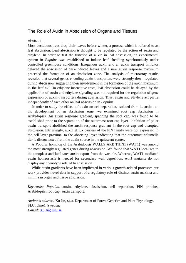
The Role of Auxin in Abscission of Organs and Tissues
Xu Jin Faculty of Forest Sciences
Department of Forest Genetics and Plant Physiology Umeå
Doctoral Thesis Swedish University of Agricultural Sciences
Umeå 2015
Acta Universitatis agriculturae Sueciae 2015:54
ISSN 1652-6880 ISBN (print version) 978- 91-576-8306-9 ISBN (electronic version) 978- 91-576-8307-6 © 2015 Xu Jin, Umeå Print: Arkitektkopia 2015
Cover: Abscission zone of a Populus leaf with GUS staining (photo: Xu Jin)
The Role of Auxin in Abscission of Organs and Tissues
Abstract Most deciduous trees drop their leaves before winter, a process which is referred to as leaf abscission. Leaf abscission is thought to be regulated by the action of auxin and ethylene. In order to test the function of auxin in leaf abscission, an experimental system in Populus was established to induce leaf shedding synchronously under controlled greenhouse conditions. Exogenous auxin and an auxin transport inhibitor delayed the abscission of dark-induced leaves and a new auxin response maximum preceded the formation of an abscission zone. The analysis of microarray results revealed that several genes encoding auxin transporters were strongly down-regulated during abscission, suggesting their involvement in the formation of the auxin maximum in the leaf axil. In ethylene-insensitive trees, leaf abscission could be delayed by the application of auxin and ethylene signaling was not required for the regulation of gene expression of auxin transporters during abscission. Thus, auxin and ethylene act partly independently of each other on leaf abscission in Populus.
In order to study the effects of auxin on cell separation, isolated from its action on the development of an abscission zone, we examined root cap abscission in Arabidopsis. An auxin response gradient, spanning the root cap, was found to be established prior to the separation of the outermost root cap layer. Inhibition of polar auxin transport abolished the auxin response gradient in the root cap and disrupted abscission. Intriguingly, auxin efflux carriers of the PIN family were not expressed in the cell layer proximal to the abscising layer indicating that the outermost columella tier is disconnected from the auxin source in the quiescent center.
A Populus homolog of the Arabidopsis WALLS ARE THIN1 (WAT1) was among the most strongly regulated genes during abscission. We found that WAT1 localizes to the tonoplast and facilitates auxin export from the vacuole. Whereas, WAT1-mediated auxin homeostasis is needed for secondary wall deposition, wat1 mutants do not display any phenotype related to abscission.
While auxin gradients have been implicated in various growth-related processes our work provides novel data in support of a regulatory role of distinct auxin maxima and minima in organ and tissue abscission.
Keywords: Populus, auxin, ethylene, abscission, cell separation, PIN proteins, Arabidopsis, root cap, auxin transport.
Author’s address: Xu Jin, SLU, Department of Forest Genetics and Plant Physiology, SLU, Umeå, Sweden. E-mail: [email protected]
It's fine to celebrate success but it is more important to heed the lessons of failure.
- Bill Gates
Contents List of Publications 7
Abbreviations 9
1 Introduction 11 1.1 Leaf abscission 12
1.1.1 Autumnal leaf abscission in deciduous trees 13 1.1.2 Retardation of abscission by auxin 13 1.1.3 Acceleration of abscission by ethylene 15 1.1.4 Cross talk between ethylene and auxin 18
1.2 Separation of root border-like cells in Arabidopsis 20
2 Objectives 25 2.1 Leaf abscission in Populus (Manuscript I) 25 2.2 Root cap abscission in Arabidopsis (Manuscript II) 25 2.3 WAT1 function in plant development and auxin homeostasis (Manuscript
III) 25
3 Materials and methods 27 3.1 Populus as a model tree species to study leaf abscission 27 3.2 The Arabidopsis root as a model to study root-cap abscission 28 3.3 Dark induction of leaf abscission in Populus 29 3.4 Gene expression analysis 29
4 Results and discussion 31 4.1 Application of auxin delayed the formation and maturation of the AZ
(Manuscript I) 31 4.2 Ethylene-auxin crosstalk (Manuscript I) 32 4.3 Expression of auxin efflux carriers during floral organ abscission in
Arabidopsis (Manuscript I) 34 4.4 Increased PtIDA expression levels in the AZ during leaf abscission
(Manuscript I) 34 4.5 Pectin remodeling during leaf abscission (Manuscript II) 36 4.6 Expression of WAT1 in vascular tissue but not in lateral root caps
(Manuscript III) 36
5 Conclusion and future perspectives 41

References 43
Acknowledgements 55
List of Publications This thesis is based on the work contained in the following papers, referred to by Roman numerals in the text:
I Xu Jin, Jorma Zimmermann, Andrea Polle and Urs Fischer (2015). Auxin is a long-range signal that acts independently of ethylene signaling on leaf abscission in Populus. submitted.
II Xu Jin, Urs Fischer. Establishment of a new auxin gradient prior to abscission of the lateral root cap. (Manuscript).
III Philippe Ranocha, Oana Dima, Réka Nagy, Judith Felten, Claire Corratgé-Faillie, Ondřej Novák, Kris Morreel, Benoît Lacombe, Yves Martinez, Stephanie Pfrunder, Xu Jin, Jean-Pierre Renou, Jean-Baptiste Thibaud, Karin Ljung, Urs Fischer, Enrico Martinoia, Wout Boerjan and Deborah Goffner (2013). Arabidopsis WAT1 is a vacuolar auxin transport facilitator required for auxin homoeostasis. Nature Communications 4, 2625.
Paper III is reproduced with the permission of the publishers.
7
The contribution of Xu Jin to the papers included in this thesis was as follows:
I Planning, performance of all the experiments, analysis and summary of the results, preparation and writing part of the manuscript.
II Planning, performance of all the experiments, analysis and summary of the results, preparation and writing part of the manuscript.
III Performance of all the experiments for Figure 3 and part of the experiments for Figure 4 and Figure S3.
8
Abbreviations 1-MCP 1-methyl-cyclopropene 2,4-D 2,4-dichlorophenoxyacetic acid ACC 1-aminocyclopropane-1-carboxylic acid ACS 1-AMINOCYCLOPROPANE-1-CARBOXYLATESYNTHASE AGL AGAMOUS-LIKE AGP ARABINOGALACTAN-PROTEIN ARF AUXIN RESPONSE FACTOR ARP ACTIN-RELATED PROTEIN ATAF ARABIDOPSIS TRANSCRIPTION ACTIVATION FACTOR AUX1 AUXIN RESISTANT1 AVG amino-ethoxyvinyl glycine AZ abscission zone BC border cells BLC border-like cells BRN BEARSKIN CF 9-hydroxyfluorene-9-carboxylic acid CTR CONSTITUTIVE TRIPLE RESPONSE CUC CUP-SHAPED COTYLEDON DAB DELAYED FLORAL ORGAN ABSCISSION EIN ETHYLENE-INSENSITIVE ERS ETHYLENE RESPONSE SENSOR ETR ETHYLENE RECEPTOR Gr Green-ripe HAE HAESA HSL HAESA-like IAA Indole-3-acetic acid iaaL IAA-Lys synthetase iaaM Trp-2-monooxygenase
9
ida inflorescence deficient in abscission ind indehiscent KD1 KNOTTED1-LIKE HOMEOBOX PROTEIN1 LAX1 LIKE AUX1 Le Lycopersicon esculentum LRC lateral root cap LRR-RLK LEUCINE-RICH REPEATS- RECEPTOR-LIKE KINASE LX LX ribonuclease MKK MITOGEN-ACTIVATED PROTEIN KINASE KINASE MPK MITOGEN-ACTIVATED PROTEIN MS Murashige and Skoog NAA 1-naphthaleneacetic acid NAC NAM, ATAF, and CUC transcription factor NAM NO APICAL MERISTEM NPA 1-N-Naphthylphthalamic acid Nr Never-ripe PG POLYGALACTURONASE PI propidium iodide PIN PIN-FORMED PK Pro transporter QC quiescent centre RNAi RNA interference RNase ribonuclease SMB SOMBRERO TNK4 TOMATO KNOTTED4 WAT WALLS ARE THIN WEI WEAK ETHYLENE INSENSITIVE
10
1 Introduction Abscission is a widespread physiological process in plants that plays a variety of roles in development. During their life cycle, plants can shed entire organs and tissues, e. g. leaves, flowers, fruits and the bark in response to developmental or environmental cues (Siwecki & Kozlowski, 1973; Addicott, 1982; Sexton & Roberts, 1982; Osborne & Morgan, 1989; Lewis et al., 2006). As diverse the nature of the shed organs is, as different is the significance or function of separation for plant development. For example, shedding the leaves in autumn minimizes water loss in the winter; separating seeds from fruits permits propagation and rapidly detaching damaged or infected organs is a successful defense strategy of the plant.
Organ abscission usually occurs at a histologically distinct boundary between the organ and the plant body referred to as the abscission zone (AZ). The AZ is comprised of layers of functionally specialized cells with morphologically distinct features like smaller, square-shaped cells, interconnected by branched plasmodesmata and dense cytoplasm (Brown & Addicott, 1950; Gawadi & Avery, 1950; Sexton & Roberts, 1982; Taylor & Whitelaw, 2001). Two types of AZs have been reported, the primary and secondary AZ. Primary AZs differentiate early in development, simultaneously with the development of the lateral organ at a predefined position (Addicott, 1982; Osborne & Morgan, 1989; Taylor & Whitelaw, 2001). By contrast, secondary AZs, also referred to as adventitious AZs, are formed as a response to non-developmental cues, usually late in the development of the lateral organ (Addicott, 1982; Webster & Leopold, 1972; Pierik, 1977, 1980). The number of AZ layers is highly variable between species (Taylor & Whitelaw, 2001; Estornell et al., 2013). While maturation of AZs may be reached early during development, induction of the separation process may occur only in late stages of an organ’s life span (Estornell et al., 2013). During the actual separation process cells within the AZ lose cell-to-cell adhesion due to the dissolution of
11
the pectic matrix in the middle lamella by the secretion of hydrolytic enzymes into the apoplast (Sexton, 1997).
Patterson et al. (2001) proposed a general model for abscission processes (reviewed in Estornell et al., 2013), which defines four major steps:
I Differentiation of the AZ in the future position of organ detachment. II Competence acquisition by cells of the AZ to react to abscission signals. III Activation of the abscission process within the AZ and organ
detachment. IV Differentiation of a protective layer. Although the various abscission processes have common features, upstream
signaling, which induces the formation of distinct separation structures and activation of the separation process are thought to be different (Taylor & Whitelaw, 2001). Many lines of research indicate that different plant hormones play important roles in the regulation of AZ formation and activation of abscission. Whereas some hormones act as inhibitors of abscission, like auxin, gibberellins and brassinosteroids (Stutte & Gage, 1990; Ben-Cheikh et al., 1997; Khripach et al., 1999; Taylor & Whitelaw, 2001; Aziz, 2003) others, such as ethylene, abscisic acid, jasmonic acid and cytokinins were shown to accelerate abscission (Sipes & Einset, 1983; Hartmond et al., 2000; Taylor & Whitelaw, 2001; Dal Cin et al., 2007). It is not only the synthesis of these hormones but also their transport and perception, which can be modulated in order to regulate the activity of AZs (Hoad, 1995).
1.1 Leaf abscission
Trees can shed their leaves in response to environmental changes, as seasonal changes in temperature and day-length, or to stress cues, as toxic concentrations of acids or salts, wounding of the leaf blade, pathogen invasion and attack or drought (Taylor & Whitelaw, 2001). Both environmental and stress factors can initiate leaf senescence and as a consequence leaves may generate an abscission-inducing signal, which is transported to the AZ in order to trigger the separation process in primary AZs or to induce the formation of a secondary abscission zone (Addicott, 1982). However, in some cases of stress induced leaf abscission, e.g. drought and pathogen attack, leaf separation can occur in absence of leaf senescence (Taylor & Whitelaw, 2001). Leaf AZs form usually at the base of leaf petioles or leaflets (Addicott, 1982). Simultaneously, or slightly delayed, with the separation phase a protective layer is formed on the proximal site of the fracture. The protective layer reduces water-loss and prevents pathogens from entry (Addicott, 1982; Meirs et al., 2010, 2011; Estronell et al. 2013).
12
1.1.1 Autumnal leaf abscission in deciduous trees
In autumn, plants of many different species drop all of their leaves within short time resulting in seasonally bare, leafless plants. These plants are referred to as deciduous plants. Decreasing day-length and colder temperature in autumn can initiate senescence of leaves in deciduous trees. With the beginning of senescence chlorophyll levels decrease and the photosynthetic capacity of leaves sharply declines (Keskitalo et al., 2005; Fracheboud et al., 2009). Subsequently, anthocyanins accumulate in leaves causing the typical change in colors associated with autumnal senescence (from green to red or yellow; Mol et al. 1996; Hoch et al., 2001). Later, the plants remobilize nutrients from the degradation of nucleic acids, proteins and starch in leaves and store them for later use in their roots and stems (Clausen & Apel, 1991; Gan & Amasino, 1997; reviewed in Fischer, 2007).
Comparing with evergreen plants, seasonal leaf abscission in deciduous plants is an evolutionary adaptation to overwinter freezing periods (Zanne et al., 2014). This adaptation allowed deciduous plants to invade vast areas (Frelich & Reich, 2002). In addition, leaf fall can help the plant to generate favorable soil conditions by increasing nutrient release from the soil and regulate the size and composition of microbial communities in the soil (Aponte et al., 2013).
In deciduous trees, e.g. Populus, the AZ is formed at the base of the petiole. Secretion of pectinases and cellulases into the apoplast and subsequent dissolution of the middle lamellae are thought to lead to cell separation in the AZ of leaves (Sexton, 1997). After cells of the AZ lose cell-to-cell adhesion, leaves remain attached only through the vascular strands, which are not digested by the secreted hydrolytic enzymes. Completion of abscission by breaking the vascular strands is finally reached by mechanical forces, e.g. wind. The leaf scar, the proximal side of the fracture, is then protected by one or several suberinized cell layers.
1.1.2 Retardation of abscission by auxin
In leaf abscission, auxin and ethylene have been pointed out as prominent regulators of abscission (Addicott, 1951; Beyer, 1975). In 1936, La Rue demonstrated that application of auxin onto petioles with excised leaf blades delays petiole abscission in Coleus (La Rue, 1936). Later, Addicott reported that the ratio of applied auxin between distal and proximal sides of the AZ is relevant for the timing of abscission and not the absolute concentration of auxin (Addicott et al., 1955; Louie & Addicott 1970). Low auxin concentrations distal to the abscission zone and high concentration proximal favors abscission, whereas the opposite experiment with higher concentrations
13
on the distal site delays abscission (Louie & Addicott, 1970). Hence, it was suggested that an auxin gradient is spanning the abscission zone and regulates the induction of abscission (Addicott et al., 1955). As a consequence of decreasing auxin concentrations on the distal side either ethylene biosynthesis or/and sensitivity are increased and induce the activity of hydrolytic enzymes degrading cell walls and middle lamellae in the abscission zone (Sexton, 1997).
In Arabidopsis, mutations in two AUXIN RESPONSE FACTORS (ARFs) have been described to affect petal abscission (Ellis et al., 2005; Okushima et al., 2005). An arf2 mutation causes delays in the abscission of floral organs, senescence of rosette leaves, flowering time and silique dehiscence, whereas single arf1 mutants do not display the same phenotypes (Ellis et al., 2005). However, in double mutants of arf1 and arf2, the arf2 phenotype was strongly enhanced indicating that ARF1 and ARF2 have redundant functions in the regulation of abscission and senescence in Arabidopsis (Ellis et al., 2005). Additionally, in arf2 flowers three members of the 1-AMINOCYCLOPROPANE-1-CARBOXYLATESYNTHASE (ACS) family – ACS2, ACS6 and ACS8 show decreased transcript levels (Okushima et al., 2005).
In the auxin influx facilitators mutants aux1 (auxin resistant1), lax1 (like aux1) and lax3 the petal break strength at flower position 3 and 4 is lower than in wild type indicating that the reduction of auxin concentration within the AZ, at the site of their expression, can accelerate petal abscission (Basu et al., 2013). Surprisingly, in an aux1 lax3 double mutant and an aux1 lax1 lax2 lax3 quadruple break strength is not more drastically decreased than in the single mutants. In order to test if the manipulation of auxin concentration in the petal AZ affects organ abscission Basu and co-workers expressed two bacterial genes, iaaM (Trp-2-monooxygenase) and iaaL (IAA-Lys synthetase), under the control of a polygalacturonase, ADPG2 (ARABIDOPSIS DEHISCENCE ZONE POLYGALACTURONASE2), promoter in Arabidopsis (Basu et al., 2013). The ADPG2 promoter drives gene expression locally restricted to floral abscission zones (González-Carranza et al., 2002). AZ-specific expression of iaaM, which converts tryptophan into indole-3-acetamide, causes increased petal break strength. By contrast, expression of iaaL, which inactivates IAA, under the control of pADPG2 decreases petal break strength. These results indicate that the manipulation of auxin concentration in the AZ is sufficient to regulate petal break strength.
Similar as in Addicottt’s experiments, which led to the auxin gradient hypothesis, distal application of auxin (IAA) to floral explants delays organ separation in tomato (Roberts et al., 1984). Exogenous auxin, however, has no effect on the morphology of the AZ (Roberts et al., 1984). Microarray gene expression analysis from RNA of tomato flower AZs revealed that seven
14
AUXIN/INDOLE-3-ACETIC ACID (Aux/IAA) genes are down-regulated upon flower removal, whereas three homologs of the IAA-amino acid conjugate hydrolase IAA-LEUCINE RESISTANT (ILR) gene family are up-regulated within two hours after flower removal (Meir et al., 2010). The same authors found for two members of the class I KNOTTED1-LIKE HOMEOBOX (KNOX) homeodomain transcription factor gene family, TOMATO KNOTTED4 (TNK4) and KNOTTED1-LIKE HOMEOBOX PROTEIN1 (KD1), increased expression in the pedicel AZ of tomato (Meir et al., 2010). Ma et al. (2015) showed that KD1 expression is restricted to the AZ of petioles and petals and that overexpression of KD1 results in accelerated petal abscission. Conversely, when KD1 is silenced, abscission is delayed, correlating with higher endogenous auxin concentrations and expression of auxin-related genes (Ma et al., 2015).
The above summarized results are consistent with those reported by Hänisch Ten Cate and Bruinsma (1973) who found that although auxin application delays abscission in pedicels of Begonia flower buds, it does not affect the anatomy of the abscission zone. Collectively, research on the role of auxin in abscission suggests that auxin can delay cell separation by inhibiting the expression of certain cell wall degrading enzymes (Hong et al., 2000; Tucker et al., 2002), but does not affect AZ differentiation (Ellis et al., 2005).
While direct evidence for auxin or auxin response distribution across the AZ zone in leaf axils still is scarce, Sorefan et al. (2009) described a local auxin minimum to be required for the differentiation of a separation zone in the valve margins of the Arabidopsis silique. In wild type, the separation layers of the valve margin differentiate at the position of a local auxin response minimum before fruit opening. By contrast, in the indehiscent (ind) mutant, which is defective in valve separation, increased auxin response occurs at the position, where normally valve margins differentiate. Similarly, expression of the bacterial auxin biosynthesis gene iaaM under the control of the IND promoter prevents the differentiation of valve margins (Sorefan et al., 2009). Together, these results support the hypothesis that a local auxin minimum is required for the formation of a functional separation zone.
1.1.3 Acceleration of abscission by ethylene
In 1931, Zimmerman et al. reported that high concentration of ethylene caused premature opening and dropping of petals in rose flowers (Zimmerman et al., 1931). Since then, ethylene function in abscission has been intensely researched and various independent lines of evidence show that ethylene is a positive regulator of abscission, which accelerates separation of different organs, such as leaves, flowers and fruits (Brown, 1997; Jackson & Osborne,
15
1970). During petiole abscission, ethylene concentration in the AZ often increases just before the onset of organ separation (Jackson et al., 1972, 1973). Increasing ethylene concentrations are believed to activate the expression of genes encoding cell wall remodeling enzymes, such as cellulases and pectinases, and their secretion into the apoplast of AZ cells (Addicott, 1982; Brown, 1997).
Genetic evidence for an involvement of ethylene response in abscission came from the study of the Arabidopsis ETHYLENE RECEPTOR1 (ETR1), which contains an amino-terminal domain that possesses ethylene-binding activity (Schaller & Bleecker, 1995). The carboxy-terminal domain of ETR1 exhibits sequence homology to bacterial two-component regulators (Chang et al. 1993), which are predominantly sensors and signal transducers of environmental stimuli in a variety of adaptation responses in bacteria (Parkinson & Kofoid, 1992). In the dominant etr1-1 mutant, which has a missense mutation in the amino-terminal ethylene-binding domain and which is completely insensitive to exogenous ethylene (Chang et al. 1993), floral organ abscission is delayed (Bleecker & Patterson, 1997). Similarly, in ethylene-insensitive2-1 (ein2-1), a mutant of the transmembrane protein EIN2 involved in ethylene signaling, floral organ abscission is delayed (Patterson & Bleecker, 1997 and 2004).
In tomato, when floral explants are exposed to atmospheric ethylene, abscission of pedicels is more rapid than in the ethylene-free atmosphere. By contrast, when the explants are pretreated with amino-ethoxyvinyl glycine (AVG), an inhibitor of ethylene biosynthesis, abscission was delayed (Roberts et al., 1984). In line with this, the pretreatment of AZ from tomato explants with an inhibitor of ethylene action, 1-methyl-cyclopropene (1-MCP), causes retardation of abscission, too (Meir et al., 2010).
Significant inhibition of leaf abscission has been observed in LX-deficient (LX ribonuclease (RNase)) transgenic tomato. Expression of the LX gene is AZ-specific in tomato and is activated by ethylene in detaching young leaves (Lers et al., 1998 and 2006). In LX-deficient antisense lines, both leaf senescence and abscission are delayed (Lers et al., 2006). Furthermore, when the leaves are pretreated with auxin for 30 min and then moved into ethylene containing atmosphere for 48 hours, LX protein levels of auxin pretreated material are lower than the levels of only ethylene-gas treated leaves. This indicates that auxin nullifies the ethylene-induced expression of the LX protein in the AZ of tomato leaves (Bar-Dror et al., 2011).
Three partially ethylene-insensitive, dominant mutants, Never-ripe (Nr), Never-ripe 2 (Nr-2) and Green-ripe (Gr), show defects in fruit ripening in tomato. Nr, Nr-2 and Gr also affect abscission at the floral pedicel (Wilkinson
16
et al., 1995; Lashbrook et al., 1998; Barry et al., 2005). Nr encodes a homologue of the Arabidopsis ERS (ETHYLENE RESPONSE SENSOR) and affects the ethylene receptor LeETR3 as an inhibitor of ethylene signal transduction (Wilkinson et al., 1995; Lashbrook et al., 1998).
The tomato genes ETHYLENE-INSENSITIVE3 (EIN3)-LIKE LeEIL1, 2 and 3, homologs of the Arabidopsis EIN3 that encodes a nuclear-localized component of the ethylene signal-transduction pathway with DNA-binding activity, are known to be involved in the ethylene-mediated initiation of abscission (Tieman et al., 2001). LeEIL antisense plants are ethylene insensitive, resulting in flower organ abscission to be delayed until the point when fruit development begins (Tieman et al., 2001). Okabe et al. also reported two ethylene intensive mutants of ETR1 in tomato, Sletr1-1 and Sletr1-2. Both Sletr1 mutants showed delayed petal abscission (Okabe et al., 2011).
In both, Arabidopsis and tomato, most of the reported ethylene mutants, which show delayed organ abscission, are dominant or semi-dominant. In absence of genetic data from recessive mutants, with the notable exception of ein2, this may suggest that although ethylene can modulate the timing of abscission, it may not be essential for the activation of organ abscission (reviewed in Estornell et al., 2013). Therefore and since the inhibition of abscission in completely ethylene-insensitive mutants is incomplete, an ethylene-independent pathway participating in the regulation of organ abscission has been proposed (Bleecker & Patterson, 1997).
In Arabidopsis, a T-DNA mutant of INFLORESCENCE DEFICIENT IN ABSCISSION (IDA), was the first ethylene-sensitive mutant, with a phenotype in floral organ shedding. The ida mutant develops a normal AZ, but is deficient in floral abscission. In ida, exogenous ethylene induces senescence of floral organs but is not sufficient for their abscission (Butenko et al., 2003). Since then, the IDA signaling pathway in Arabidopsis has been studied intensively. The mutants of the receptor-like protein kinases HAESA (HAE), the double mutant of HAESA-LIKE 2 (HSL2) and HAE, a tandem RNAi transgenic line of MKK4 and MKK5 (MITOGEN-ACTIVATED PROTEIN KINASE KINASE), and the mutated MPK6 (MITOGEN-ACTIVATED PROTEIN KINASE6) in the mpk3 background, all have an abscission defective phenotype (Jinn et al., 2000; Cho et al., 2008). Genetic interaction studies have shown that there is a signaling cascade from the putative ligand (IDA) to receptors (HAE HSL2) to cytoplasmic effectors (MKK4, MKK5, MPK3, and MPK6) regulating floral organ abscission in Arabidopsis (Stenvik et al., 2008; Cho et al., 2008; Butenko et al., 2012).
17
Subsequently, the recessive mutants delayed floral organ abscission 1 (dab1), dab2 and dab3 were reported to delay floral organ separation (Patterson & Bleecker, 2004). Delayed floral organ detachment in Arabidopsis was also observed in several other mutants and transgenic lines, such as in plants with constitutive expression of AGL15 (AGAMOUS-LIKE 15), a MADS domain transcription factor (Fernandez et al., 2000), and in knock-down lines of ARP7 (ACTIN-RELATED PROTEIN7; Kandasamy et al., 2005). All of the above referred mutants senesce normally and are ethylene-sensitive.
The available evidence clearly shows that ethylene plays an important role during organ abscission but also that ethylene signaling is not a universal requirement or prerequisite for abscission to take place (Addicott, 1982; Taylor & Whitelaw, 2001; Lewis et al., 2006; Estornell et al., 2013). Signaling, which leads to the separation of an organ might therefore not be performed by a single hormonal pathway or a linear sequence of processes (Patterson & Bleecker, 2004).
1.1.4 Cross talk between ethylene and auxin
It is commonly accepted that abscission of diverse organs is regulated by the contrary cooperation between auxin and ethylene action. It is believed that decreased flux of auxin through the AZ triggers increased ethylene signaling and in turn increases the expression and secretion of hydrolytic enzymes involved in the dissolution of the middle lamellae (Roberts & Osborne, 1981; Sexton & Roberts, 1982; Osborne, 1989; Sexton, 1997; Taylor & Whitelaw, 2001). On the other hand, when the flux of auxin through the AZ is high, cell separation is inhibited resulting in organ retention (Addicott, 1982; Sexton et al., 1985). Hence, free auxin in the AZ regulates the sensitivity to ethylene, and therefore any factor, which modulates auxin biosynthesis, homeostasis, signaling or transport in AZ, can also affect the sensitivity of cells in the AZ to ethylene. In order to investigate the cooperation between auxin and ethylene on a molecular and biochemical base, Meir and coworkers examined the abscission-related gene expression in tomato flower AZs after detaching the flowers (Meir et al., 2010). Removal of the flower, the sole auxin source, is expected to deplete the AZ of auxin (Meir et al., 2010). The expression levels of some AUX/IAAs, which are directly regulated by auxin, decreases sharply within 2 h after flower removal and remains low thereafter (Meir et al., 2010). As the AZ becomes sensitive to ethylene because of auxin depletion, some ethylene related genes and transcription factors increase their expression levels, such as ETHYLENE-RESPONSIVE1 (ER1), ETHYLENE RESISTANT4 (ETR4), CONSTITUTIVE TRIPLE RESPONSE1 (CTR1), and ETHYLENE-RESPONSIVE FACTORS (ERF1c). Along with ethylene signaling-related
18
genes, genes encoding cell wall-modifying proteins are up-regulated (Meir et al., 2010).
Many lines of evidence indicate that ethylene can inhibit auxin transport and biosynthesis, in other words, ethylene can render the AZ more sensitive to itself via positive feedback (Beyer & Morgan, 1971; Beyer, 1973). Beyer and Morgan concluded that following leaf senescence the up-regulation of ethylene signaling reduces auxin transport. Such reduced auxin transport could lead to increased sensitivity to ethylene of the cells in the AZ, and could also induce the production of the enzymes required for abscission and regulate their secretion into the cell wall (Beyer & Morgan, 1971). More complicatedly, auxin itself can stimulate ethylene production and thus accelerate abscission (Abeles & Rubinstein, 1964; Morgan & Hall, 1964).
In Arabidopsis, the inhibition of auxin transport and biosynthesis by ethylene has been studied, too. WEAK ETHYLENE INSENSITIVE 2 (WEI2) and WEI7 regulate auxin biosynthesis in root tip. WEI2 and WEI7 encode subunits of the anthranilate synthase, a rate-limiting enzyme in tryptophan biosynthesis. Overexpression of WEI2 and WEI7 results in the accumulation of auxin in the tip of primary root, whereas in the loss-of-function mutants auxin levels are decreased (Stepanova et al., 2005). Subsequently, Swarup et al. (2007) reported that IAA biosynthesis in the root tip was inhibited by AVG treatment indicating that ethylene regulates auxin biosynthesis positively in order to enhance the inhibition of root cell elongation. Furthermore, expression of auxin efflux carriers, such as PIN-FORMED1 (PIN1), PIN2, PIN4, and the influx carrier AUX1 increases in response to ethylene (Ruzicka et al., 2007). All these results elucidate that ethylene increases the auxin biosynthesis rate and modulates the capacity of polar auxin transport (Ruzicka et al., 2007).
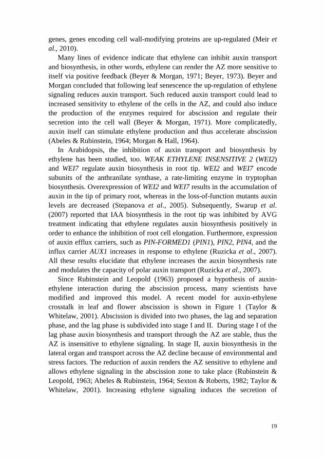
Since Rubinstein and Leopold (1963) proposed a hypothesis of auxin-ethylene interaction during the abscission process, many scientists have modified and improved this model. A recent model for auxin-ethylene crosstalk in leaf and flower abscission is shown in Figure 1 (Taylor & Whitelaw, 2001). Abscission is divided into two phases, the lag and separation phase, and the lag phase is subdivided into stage I and II. During stage I of the lag phase auxin biosynthesis and transport through the AZ are stable, thus the AZ is insensitive to ethylene signaling. In stage II, auxin biosynthesis in the lateral organ and transport across the AZ decline because of environmental and stress factors. The reduction of auxin renders the AZ sensitive to ethylene and allows ethylene signaling in the abscission zone to take place (Rubinstein & Leopold, 1963; Abeles & Rubinstein, 1964; Sexton & Roberts, 1982; Taylor & Whitelaw, 2001). Increasing ethylene signaling induces the secretion of
19
pectinases and cellulases to hydrolyze the middle lamellae and the cell walls in the AZ (Addicott, 1982; Brown, 1997).
Figure 1. Auxin and ethylene interplay in leaf and flower abscission. Picture reproduced from Taylor and Whitelaw (2001) with the kind permission of the publisher.
1.2 Separation of root border-like cells in Arabidopsis
Like organ separation processes, the separation of the outermost cell layers from the root tip is controlled by phytohormones and environmental factors, and involves the degradation of cell walls and middle lamella (reviewed in Barlow, 2002). Expression of hydrolytic enzymes catalyzing the dissolution of the cell wall and pectin is increased during root cap separation (Driouich et al., 2007).
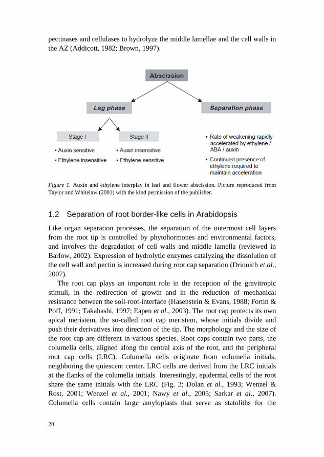
The root cap plays an important role in the reception of the gravitropic stimuli, in the redirection of growth and in the reduction of mechanical resistance between the soil-root-interface (Hasenstein & Evans, 1988; Fortin & Poff, 1991; Takahashi, 1997; Eapen et al., 2003). The root cap protects its own apical meristem, the so-called root cap meristem, whose initials divide and push their derivatives into direction of the tip. The morphology and the size of the root cap are different in various species. Root caps contain two parts, the columella cells, aligned along the central axis of the root, and the peripheral root cap cells (LRC). Columella cells originate from columella initials, neighboring the quiescent center. LRC cells are derived from the LRC initials at the flanks of the columella initials. Interestingly, epidermal cells of the root share the same initials with the LRC (Fig. 2; Dolan et al., 1993; Wenzel & Rost, 2001; Wenzel et al., 2001; Nawy et al., 2005; Sarkar et al., 2007). Columella cells contain large amyloplasts that serve as statoliths for the
20
perception of gravity (Moore et al., 1986; Yoder et al., 2001; Saiki and Sato, 2004). The outer two cell layers of the LRC and the tip of the root cap synthesize and secrete the polysaccharide-containing mucilage (Bacic et al., 1986; Morel et al., 1986; Staehelin et al., 1990; Iijima et al., 2004; Cai et al., 2013). The mucilage can lubricate the root so that it penetrates the soil with less mechanical resistance. Prevention of desiccation and facilitating the establishment of mycorrhizae and the interaction with symbiotic bacteria are other functions of the mucilage (McCully & Sealey, 1996; Staehelin et al., 1990; Iijima et al., 2004; Cai et al., 2013).
Figure 2. Scheme of the Arabidopsis root tip. The primary root meristem consists of the QC (white), and the undifferentiated initial cells of the columella (light blue), the lateral root cap and epidermis (light red), cortex and endodermis (light green). Picture reproduced from Nawy et al., (2005) with the kind permission of the publisher, www.plantcell.org. Copyright American Society of Plant Biologists.
Two types of cells sloughed from the root cap of higher plants have been observed, the border cells (BC; Hawes et al., 2000) and the border-like cells (BLC; Vicré et al., 2005). The BCs are released as dispersed single cells after the mucilage swells due to contact with the soil water (Hawes et al., 1991; Brigham et al., 1998; Hawes et al., 2000). The BCs play an important role to protect the root system against biotic and abiotic stress (Miasaka & Hawes, 2001; Pan, 2004; Gunawardena & Hawes, 2002; Gunawardena, 2005). Unlike the BC, the BLC are released in blocks of associated cells. Despite this, BLCs have the same functions as BCs. The root cap of the model plant Arabidopsis thaliana is shed in blocks of associates cells of single layers (Vicré et al., 2005; Hamamoto et al., 2006). The BLC of Arabidopsis share characteristic features with BLC/BC from other species, such as they are rich in mitochondria, endoplasmic reticulum, multi-vesicular bodies, Golgi stacks with many Golgi-
21
derived vesicles, suggesting that these cells actively secrete pectic polysaccharides and arabinogalactan-proteins (AGPs) to their cell walls and surrounding medium (Vicré et al., 2005; Nguema-Ona et al., 2014). While the traditional model species to study BC function are cotton (Gossypium hirsutum), wheat (Triticum aestivum), maize (Zea mays), tomato (Lycopersicon esculentum) and pea (Pisum sativum), research on BLC can be performed in Arabidopsis (Vicré et al., 2005).
The separation of BLC in Arabidopsis starts at the age of 5 to 7 days. Before, the root cap stays intact (Vicré et al., 2005). Lateral root cap layers are shed one by one, starting from the outermost, oldest layer. In 13-days-old seedlings, at least 3 layers of BLC are shed (Vicré et al., 2005). In Arabidopsis, separation of the BLC occurs before cell death and BLC remain viable for at least 24 hours after separation (Vicré et al., 2005). Similarly, BC also can survive after the separation from the root cap and even survive in vitro in similar conditions as in nature. Interestingly, BC can divide and develop into callus tissue in vitro (Hawes et al., 2000; Hawes & Lin, 1990).
The separation of BC in pea (Pisum sativum) revealed that a specific polygalacturonase enzyme, which can hydrolyze polygalacturonic acid to disassemble primary cell walls, is activated during the BC separation process (Hawes & Lin, 1990). In comparison with BC, BLC in Arabidopsis secrete significant amounts of homogalacturonan and AGPs into their cell walls so that they do not become dispersed individually into suspension. The Arabidopsis root epidermal bulger1-1 (reb1-1) mutant, which has reduced levels of AGPs, releases less BLC with altered morphology compared to wild-type roots (Driouich et al, 2007).This has been interpreted in such a way that there is a specific cell wall composition or structure in the BLC, that confers resistance to hydrolysis of cell walls or that the BLC have no functional pectolytic enzymes capable to hydrolyze the pectic matrix of cell walls (Driouich et al, 2007).
So far research on the separation processes of the BC and BLC has more focused on regulatory functions of the cell wall modifying enzymes than on the effects of phytohormones. For the BC separation in maize, Ponce demonstrated that the interruption of polar auxin transport by the application of the polar auxin transport inhibitor NPA (1-N-naphthylphthalamic acid) increased the BC shedding (Ponce et al., 2005). Surprisingly, application of ACC (1-aminocyclopropane-1-carboxylic acid) and of the ethylene synthesis inhibitor AVG (aminovinylglycin) decreased the BC shedding significantly (Ponce et al., 2005). These results indicate that the BC separation is regulated by ethylene and by auxin, and that auxin and ethylene may act coordinately to regulate BC differentiation and separation, in a similar fashion as in other
22
abscission processes (Ponce et al., 2005; Driouich et al., 2007). For the BLC separation in Arabidopsis, no such data is available. But for a mutant of the auxin efflux carrier PIN4, an increased number of columella layers has been reported (Friml et al., 2002).
In Arabidopsis, the NAC domain transcription factors, SOMBRERO (SMB), BEARSKIN1 (BRN1), and BRN2, are required for the BLC separation (Bennett et al., 2010). The Arabidopsis genome contains more than 100 members of the NAC domain transcription factors (Olsen et al., 2005) and many of them have been reported to be regulated by phytohormones (He et al., 2005). SMB, BRN1 and BRN2 regulate independently and redundantly the differentiation of root cap cells, including their morphology and their ability to separate from the root. In the smb-3 mutant, the LRC cells display an abnormal morphology and fail to detach from the root tip. In the brn1-1 brn2-1 double mutant, the separation of BLC is defective, but the shape of the BLC was normal. Surprisingly, in the smb-3 brn1-1 brn2-1 triple mutant both the shape and the detachment of the BLC are altered (Bennett et al., 2010).
23

2 Objectives The main aim of the work described in this thesis was to understand how auxin regulates leaf and root cap abscission.
2.1 Leaf abscission in Populus (Manuscript I)
The specific objectives of this project were: - the identification of auxin carriers involved in leaf abscission. - the examination of the function of auxin and ethylene during leaf abscission. - the study of auxin - ethylene crosstalk in the abscission zone.
2.2 Root cap abscission in Arabidopsis (Manuscript II)
The specific objectives of this project were: - the study of auxin response during root cap abscission. - testing the plausibility of a regulatory function of an auxin gradient in root
cap abscission. - the examination of cell wall remodeling during root cap abscission.
2.3 WAT1 function in plant development and auxin homeostasis (Manuscript III)
In Manuscript I, a Populus homolog of the Arabidopsis WALLS ARE THIN1 (WAT1), a vacuolar auxin transport facilitator, was identified as being regulated during leaf abscission in a microarray experiment. We wanted to study:
- the expression pattern of WAT1 in Arabidopsis. - the subcellular localization of WAT1. - the function of WAT1 in development.
25

3 Materials and methods
3.1 Populus as a model tree species to study leaf abscission
The genus Populus consists of deciduous flowering tree species, including poplar, aspen, and cottonwood species. In most cases, the flowers are dioicous and the predominant pollination syndrome is anemophily (wind pollination). Natural Populus populations occur in temperate climate regions of the northern hemisphere, where these trees shed their leaves in autumn (autumnal leaf abscission). Many Populus species are considered to be fast growing with straight, massive stems and can be easily clonally propagated. In 1986, Parsons et al. produced the first transgenic woody plants by transforming P. trichocarpa × deltoids (Parsons et al., 1986). Soon thereafter the transformation protocol had been optimized and other Populus hybrids became transformable, such as P. alba L. × P. tremula (referred to as Populus × canescens; Devillard, 1992; Leple et al., 1992) and P. tremula × P. tremuloides (Ptt, referred to as T89; Nilsson et al., 1992).
In 2006, a complete sequence of the Populus trichocharpa genome, the first completed tree genome, was published by the Joint Genome Institute together with researchers at the Umeå Plant Science Centre (Tuskan et al., 2006). Populus trichocarpa has a comparatively small genome of about 535 mega base pairs. The availability of a full genome sequence facilitated molecular breeding approaches, as genome-wide association mapping, to exploit the huge natural variation within more than 30 Populus species (Porth et al., 2013).
After the publication of the P. trichocarpa genome (Tuskan et al., 2006), the importance of Populus sp. as a model for various aspects of tree physiology has steadily increased. Nowadays, Populus sp. is used as a model species for e.g., wood formation, dormancy, drought stress, adventitious root formation, organic volatile emissions and mycorrhiza (Jansson & Douglas, 2007; Fischer & Polle, 2011; Legué et al., 2014; Douglas & Polle, 2010; Ditengou et al.,
27
2015). Rapid growth and easy and efficient clonal mass propagation makes Populus interesting as a possible sustainable feedstock for bioenergy production (Porth & El-Kassaby, 2015).
In Manuscript I, two different Populus hybrids were employed to study the regulation of leaf abscission, i.e. hybrid aspen T89 and Populus × canescens.
3.2 The Arabidopsis root as a model to study root-cap abscission
Arabidopsis is a popular model organism for most aspects of plant biology. Despite being a complex multicellular eukaryote, Arabidopsis thaliana has a relatively small genome of approximately 135 mega base pairs. The small size of its genome made Arabidopsis attractive since the beginning of plant molecular biology. In fact, the Arabidopsis genome was the first plant genome to be fully sequenced in 2000 by the ‘Arabidopsis Genome Initiative’. In addition, Arabidopsis provides many other advantages, such as its short life cycle, prolific seed production, easy cultivation, highly efficient and simple transformation protocols with A. tumefaciens and a large number of publicly available mutant lines and genetic resources.
In Manuscript II, we made use of some of the benefits Arabidopsis provides in order to study root cap abscission. Arabidopsis root tips display a simple and for plant organs highly ordered cellular organization. Aspects of patterning, cell specification, growth and differentiation, meristem maintenance, polarity, lateral root formation and physiological and environmental responses have been studied in Arabidopsis in great detail during the last 25 years (Benfey et al., 2010). Arabidopsis roots can grow on solid growth media in transparent petri dishes, which facilities the visual observation of mutant phenotypes or growth responses. Due to their small diameter Arabidopsis roots are fully transparent in confocal and light microscopy and various protocols have been established to exploit the benefits of live imaging (Petricka et al., 2012).
In Arabidopsis, the root cap consists of the lateral root cap (LRC) and columella. The root cap forms protective layers of cells, which are continuously separated from the root tip (Driouich et al., 2007). The root cap is involved in the reception of gravity and regulates the gravitropic response (Sato et al., 2014). The investigation of lateral root cap cells, the so called border like cells (BLC), that are released to the surrounding media has been pioneered by Vicré et al. in 2005. Examination of the morphology, cell wall composition of BLC and of bacteria attachment to BLC in Arabidopsis seedlings has been reported (Driouich et al., 2007).
28
In Manuscript II, we employed Arabidopsis in order to study cell separation in the root cap. All the transgenics and mutants used in Manuscript II are in the Col-0 background.
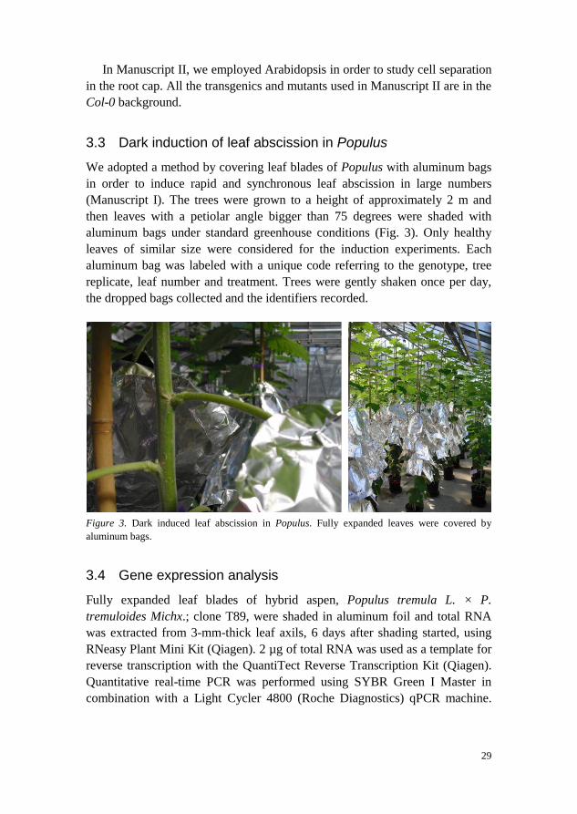
3.3 Dark induction of leaf abscission in Populus
We adopted a method by covering leaf blades of Populus with aluminum bags in order to induce rapid and synchronous leaf abscission in large numbers (Manuscript I). The trees were grown to a height of approximately 2 m and then leaves with a petiolar angle bigger than 75 degrees were shaded with aluminum bags under standard greenhouse conditions (Fig. 3). Only healthy leaves of similar size were considered for the induction experiments. Each aluminum bag was labeled with a unique code referring to the genotype, tree replicate, leaf number and treatment. Trees were gently shaken once per day, the dropped bags collected and the identifiers recorded.
Figure 3. Dark induced leaf abscission in Populus. Fully expanded leaves were covered by aluminum bags.
3.4 Gene expression analysis
Fully expanded leaf blades of hybrid aspen, Populus tremula L. × P. tremuloides Michx.; clone T89, were shaded in aluminum foil and total RNA was extracted from 3-mm-thick leaf axils, 6 days after shading started, using RNeasy Plant Mini Kit (Qiagen). 2 µg of total RNA was used as a template for reverse transcription with the QuantiTect Reverse Transcription Kit (Qiagen). Quantitative real-time PCR was performed using SYBR Green I Master in combination with a Light Cycler 4800 (Roche Diagnostics) qPCR machine.
29
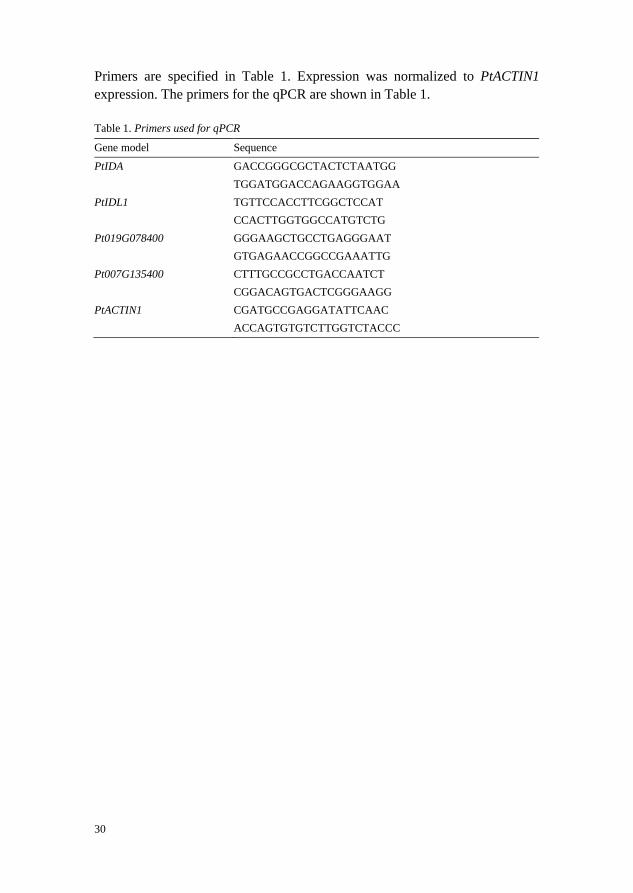
Primers are specified in Table 1. Expression was normalized to PtACTIN1 expression. The primers for the qPCR are shown in Table 1.
Table 1. Primers used for qPCR
Gene model Sequence
PtIDA GACCGGGCGCTACTCTAATGG TGGATGGACCAGAAGGTGGAA PtIDL1 TGTTCCACCTTCGGCTCCAT CCACTTGGTGGCCATGTCTG Pt019G078400 GGGAAGCTGCCTGAGGGAAT GTGAGAACCGGCCGAAATTG Pt007G135400 CTTTGCCGCCTGACCAATCT CGGACAGTGACTCGGGAAGG PtACTIN1 CGATGCCGAGGATATTCAAC ACCAGTGTGTCTTGGTCTACCC
30
4 Results and discussion In Manuscript I, dark-induced leaf abscission in Populus was described. We studied the auxin response during abscission and identified auxin transporters, whose expression was regulated during leaf abscission in intact trees. Expression patterns of these auxin transporters were examined and it was also shown that regulation of their expression is independent of ethylene signaling. In the following chapters, additional data in support of the findings in Manuscript I are presented.
4.1 Application of auxin delayed the formation and maturation of the AZ (Manuscript I)
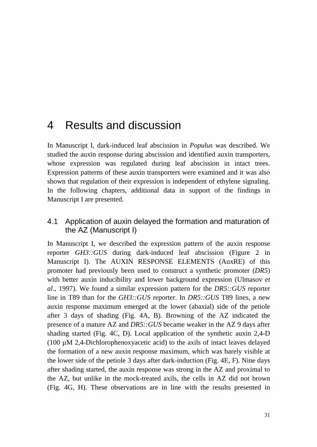
In Manuscript I, we described the expression pattern of the auxin response reporter GH3::GUS during dark-induced leaf abscission (Figure 2 in Manuscript I). The AUXIN RESPONSE ELEMENTS (AuxRE) of this promoter had previously been used to construct a synthetic promoter (DR5) with better auxin inducibility and lower background expression (Ulmasov et al., 1997). We found a similar expression pattern for the DR5::GUS reporter line in T89 than for the GH3::GUS reporter. In DR5::GUS T89 lines, a new auxin response maximum emerged at the lower (abaxial) side of the petiole after 3 days of shading (Fig. 4A, B). Browning of the AZ indicated the presence of a mature AZ and DR5::GUS became weaker in the AZ 9 days after shading started (Fig. 4C, D). Local application of the synthetic auxin 2,4-D (100 µM 2,4-Dichlorophenoxyacetic acid) to the axils of intact leaves delayed the formation of a new auxin response maximum, which was barely visible at the lower side of the petiole 3 days after dark-induction (Fig. 4E, F). Nine days after shading started, the auxin response was strong in the AZ and proximal to the AZ, but unlike in the mock-treated axils, the cells in AZ did not brown (Fig. 4G, H). These observations are in line with the results presented in
31
Manuscript I (compare with Figure 4 in Manuscript I), where local application of 2,4-D to the axil of shaded leaves delayed separation by approximately 4 days. Additionally, the results presented here suggest that auxin acts on the formation of the abscission zone.
Figure 4. Exogenous auxin delayed the formation of a local auxin response maximum and the maturation of the leaf AZ. Expression of DR5::GUS in the AZ of 2,4-D-treated and control transgenic plants. A) to D), dark induction without auxin treatment; A) and B), 3 days after shading started, black frame highlights the AZ; C) and D), 9 days after shading started. Mock treatment, lanolin paste locally applied to the AZ. E) to H), dark induction with application of 2,4-D; E) and F), 3 days after shading started; G) and H), 9 days after shading started. Lanolin paste containing 100 µM 2,4-D was glued to the leaf axils locally . Scale bars, 1 mm (A, C, E, G); 200 µm (B, D, F, H).
4.2 Ethylene-auxin crosstalk (Manuscript I)
Auxin and ethylene have antagonistic functions during the abscission of various organs (Hong et al., 2000). To test if auxin acts upstream of ethylene on leaf abscission, we transformed DR5::eto3 and DR5::etr1-1 into T89. eto3 is a dominant mutant of the ethylene biosynthetic gene 1-AMINOCYCLOPROPANE-1-CARBOXYLATE SYNTHASE 9 (ACS9) and can induce higher concentrations of ethylene in Arabidopsis (Chae et al., 2003). By contrast, the mutant of the dominant etr1-1 allele in Arabidopsis is insensitive to ethylene (Bleecker et al.,
32
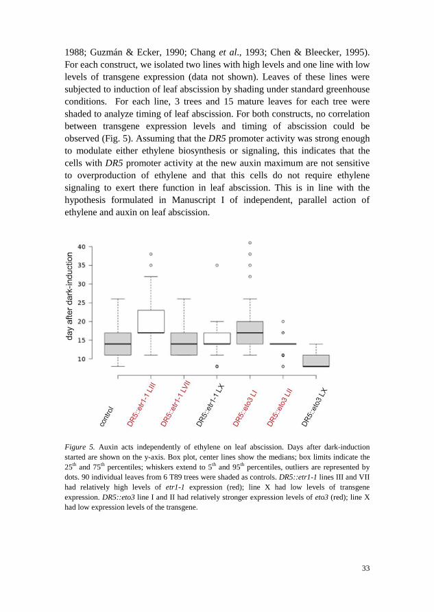
1988; Guzmán & Ecker, 1990; Chang et al., 1993; Chen & Bleecker, 1995). For each construct, we isolated two lines with high levels and one line with low levels of transgene expression (data not shown). Leaves of these lines were subjected to induction of leaf abscission by shading under standard greenhouse conditions. For each line, 3 trees and 15 mature leaves for each tree were shaded to analyze timing of leaf abscission. For both constructs, no correlation between transgene expression levels and timing of abscission could be observed (Fig. 5). Assuming that the DR5 promoter activity was strong enough to modulate either ethylene biosynthesis or signaling, this indicates that the cells with DR5 promoter activity at the new auxin maximum are not sensitive to overproduction of ethylene and that this cells do not require ethylene signaling to exert there function in leaf abscission. This is in line with the hypothesis formulated in Manuscript I of independent, parallel action of ethylene and auxin on leaf abscission.
Figure 5. Auxin acts independently of ethylene on leaf abscission. Days after dark-induction started are shown on the y-axis. Box plot, center lines show the medians; box limits indicate the 25th and 75th percentiles; whiskers extend to 5th and 95th percentiles, outliers are represented by dots. 90 individual leaves from 6 T89 trees were shaded as controls. DR5::etr1-1 lines III and VII had relatively high levels of etr1-1 expression (red); line X had low levels of transgene expression. DR5::eto3 line I and II had relatively stronger expression levels of eto3 (red); line X had low expression levels of the transgene.
33
4.3 Expression of auxin efflux carriers during floral organ abscission in Arabidopsis (Manuscript I)
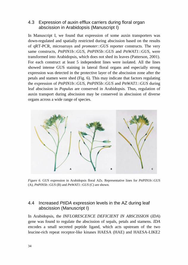
In Manuscript I, we found that expression of some auxin transporters was down-regulated and spatially restricted during abscission based on the results of qRT-PCR, microarrays and promoter::GUS reporter constructs. The very same constructs, PttPIN1b::GUS, PttPIN5b::GUS and PttWAT1::GUS, were transformed into Arabidopsis, which does not shed its leaves (Patterson, 2001). For each construct at least 5 independent lines were isolated. All the lines showed intense GUS staining in lateral floral organs and especially strong expression was detected in the protective layer of the abscission zone after the petals and stamen were shed (Fig. 6). This may indicate that factors regulating the expression of PttPIN1b::GUS, PttPIN5b::GUS and PttWAT1::GUS during leaf abscission in Populus are conserved in Arabidopsis. Thus, regulation of auxin transport during abscission may be conserved in abscission of diverse organs across a wide range of species.
Figure 6. GUS expression in Arabidopsis floral AZs. Representative lines for PttPIN1b::GUS (A), PttPIN5b::GUS (B) and PttWAT1::GUS (C) are shown.
4.4 Increased PtIDA expression levels in the AZ during leaf abscission (Manuscript I)
In Arabidopsis, the INFLORESCENCE DEFICIENT IN ABSCISSION (IDA) gene was found to regulate the abscission of sepals, petals and stamens. IDA encodes a small secreted peptide ligand, which acts upstream of the two leucine-rich repeat receptor-like kinases HAESA (HAE) and HAESA-LIKE2
34
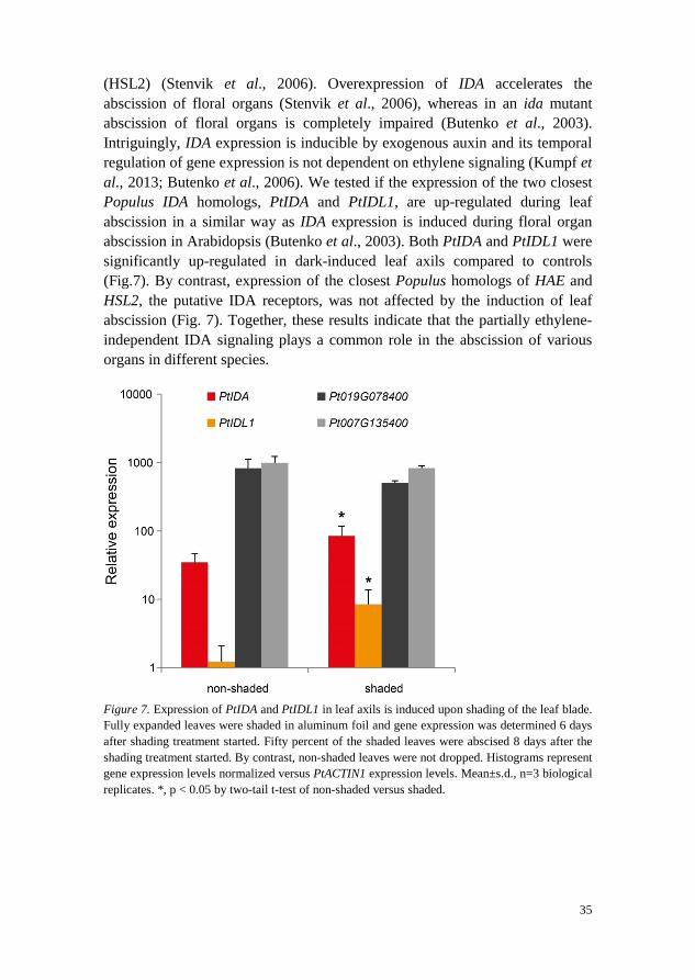
(HSL2) (Stenvik et al., 2006). Overexpression of IDA accelerates the abscission of floral organs (Stenvik et al., 2006), whereas in an ida mutant abscission of floral organs is completely impaired (Butenko et al., 2003). Intriguingly, IDA expression is inducible by exogenous auxin and its temporal regulation of gene expression is not dependent on ethylene signaling (Kumpf et al., 2013; Butenko et al., 2006). We tested if the expression of the two closest Populus IDA homologs, PtIDA and PtIDL1, are up-regulated during leaf abscission in a similar way as IDA expression is induced during floral organ abscission in Arabidopsis (Butenko et al., 2003). Both PtIDA and PtIDL1 were significantly up-regulated in dark-induced leaf axils compared to controls (Fig.7). By contrast, expression of the closest Populus homologs of HAE and HSL2, the putative IDA receptors, was not affected by the induction of leaf abscission (Fig. 7). Together, these results indicate that the partially ethylene-independent IDA signaling plays a common role in the abscission of various organs in different species.
Figure 7. Expression of PtIDA and PtIDL1 in leaf axils is induced upon shading of the leaf blade. Fully expanded leaves were shaded in aluminum foil and gene expression was determined 6 days after shading treatment started. Fifty percent of the shaded leaves were abscised 8 days after the shading treatment started. By contrast, non-shaded leaves were not dropped. Histograms represent gene expression levels normalized versus PtACTIN1 expression levels. Mean±s.d., n=3 biological replicates. *, p < 0.05 by two-tail t-test of non-shaded versus shaded.
35
4.5 Pectin remodeling during leaf abscission (Manuscript II)
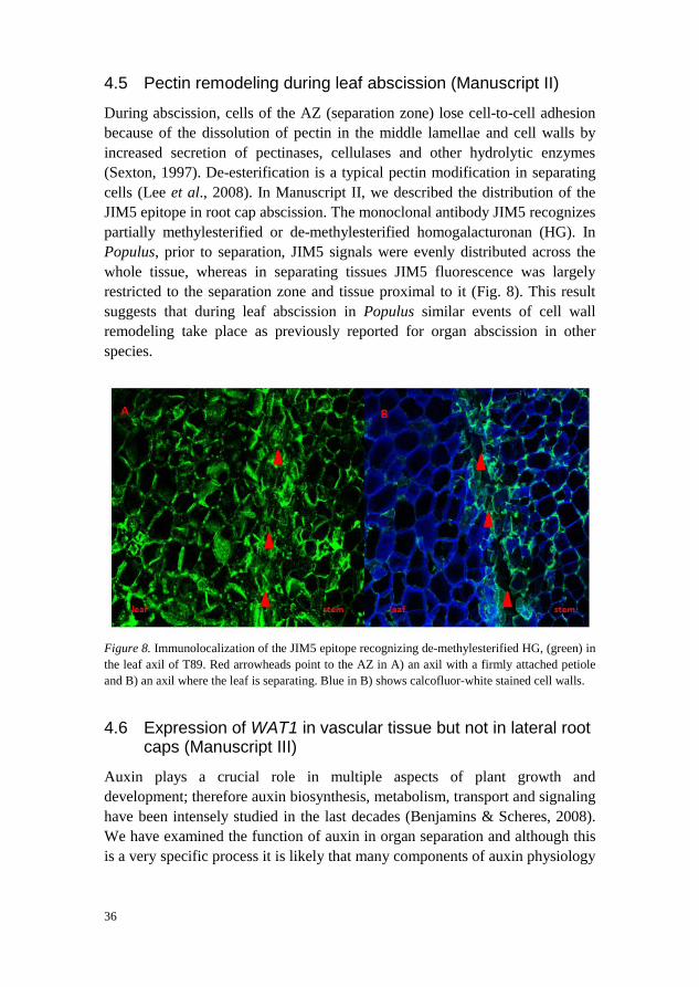
During abscission, cells of the AZ (separation zone) lose cell-to-cell adhesion because of the dissolution of pectin in the middle lamellae and cell walls by increased secretion of pectinases, cellulases and other hydrolytic enzymes (Sexton, 1997). De-esterification is a typical pectin modification in separating cells (Lee et al., 2008). In Manuscript II, we described the distribution of the JIM5 epitope in root cap abscission. The monoclonal antibody JIM5 recognizes partially methylesterified or de-methylesterified homogalacturonan (HG). In Populus, prior to separation, JIM5 signals were evenly distributed across the whole tissue, whereas in separating tissues JIM5 fluorescence was largely restricted to the separation zone and tissue proximal to it (Fig. 8). This result suggests that during leaf abscission in Populus similar events of cell wall remodeling take place as previously reported for organ abscission in other species.
Figure 8. Immunolocalization of the JIM5 epitope recognizing de-methylesterified HG, (green) in the leaf axil of T89. Red arrowheads point to the AZ in A) an axil with a firmly attached petiole and B) an axil where the leaf is separating. Blue in B) shows calcofluor-white stained cell walls.
4.6 Expression of WAT1 in vascular tissue but not in lateral root caps (Manuscript III)
Auxin plays a crucial role in multiple aspects of plant growth and development; therefore auxin biosynthesis, metabolism, transport and signaling have been intensely studied in the last decades (Benjamins & Scheres, 2008). We have examined the function of auxin in organ separation and although this is a very specific process it is likely that many components of auxin physiology
36
are not specific for organ abscission but are common players in other auxin-regulated processes. One of the most significantly down-regulated genes during leaf abscission was PtWAT1 (Manuscript I). At the time we obtained this result, the biochemical function of WAT1 was unknown. However, co-regulation with auxin regulated genes and the predicted transmembrane structures suggested that WAT1 could be a so far unknown auxin transporter. In order to better understand the WAT1 function, we collaborated with the Goffner group, which had previously described the wat1 loss-of-function phenotype (Ranocha et al., 2010). wat1 mutants are defective in secondary cell wall deposition in interfascicular fibers and xylem vessels and fibers (Ranocha et al., 2010). We localized WAT1-GFP to the tonoplast and our collaborators found that WAT1 transports auxin from the vacuole across the tonoplast to the cytoplasm (Ranocha et al., 2013, Manuscript III). We could rescue the cell wall phenotype by local application of synthetic auxins (Ranocha et al., 2013, Manuscript III). This strongly suggests that auxin is a WAT1 transport substrate in planta.
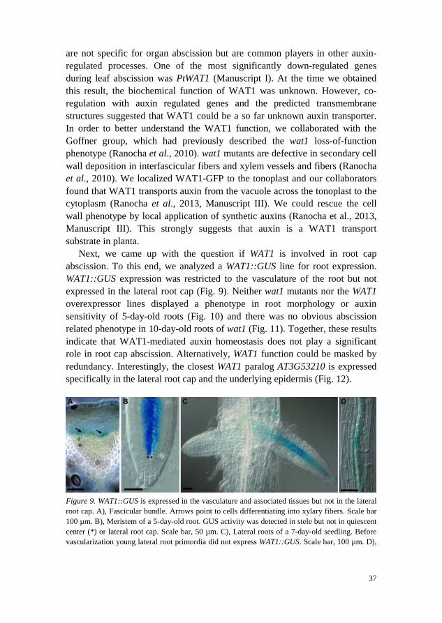
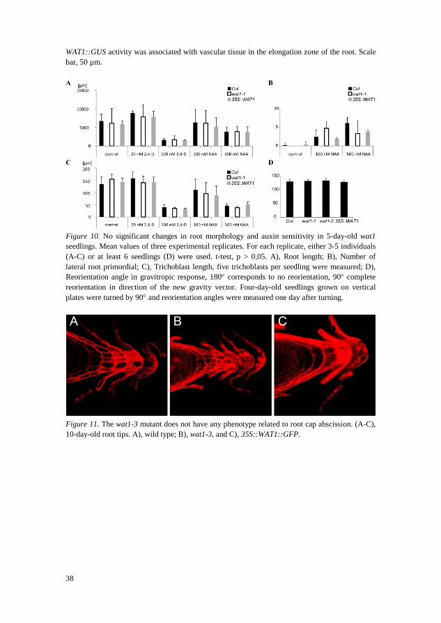
Next, we came up with the question if WAT1 is involved in root cap abscission. To this end, we analyzed a WAT1::GUS line for root expression. WAT1::GUS expression was restricted to the vasculature of the root but not expressed in the lateral root cap (Fig. 9). Neither wat1 mutants nor the WAT1 overexpressor lines displayed a phenotype in root morphology or auxin sensitivity of 5-day-old roots (Fig. 10) and there was no obvious abscission related phenotype in 10-day-old roots of wat1 (Fig. 11). Together, these results indicate that WAT1-mediated auxin homeostasis does not play a significant role in root cap abscission. Alternatively, WAT1 function could be masked by redundancy. Interestingly, the closest WAT1 paralog AT3G53210 is expressed specifically in the lateral root cap and the underlying epidermis (Fig. 12).
Figure 9. WAT1::GUS is expressed in the vasculature and associated tissues but not in the lateral root cap. A), Fascicular bundle. Arrows point to cells differentiating into xylary fibers. Scale bar 100 µm. B), Meristem of a 5-day-old root. GUS activity was detected in stele but not in quiescent center (*) or lateral root cap. Scale bar, 50 µm. C), Lateral roots of a 7-day-old seedling. Before vascularization young lateral root primordia did not express WAT1::GUS. Scale bar, 100 µm. D),
37
WAT1::GUS activity was associated with vascular tissue in the elongation zone of the root. Scale bar, 50 µm.
Figure 10. No significant changes in root morphology and auxin sensitivity in 5-day-old wat1 seedlings. Mean values of three experimental replicates. For each replicate, either 3-5 individuals (A-C) or at least 6 seedlings (D) were used. t-test, p > 0,05. A), Root length; B), Number of lateral root primordial; C), Trichoblast length, five trichoblasts per seedling were measured; D), Reorientation angle in gravitropic response, 180° corresponds to no reorientation, 90° complete reorientation in direction of the new gravity vector. Four-day-old seedlings grown on vertical plates were turned by 90° and reorientation angles were measured one day after turning.
Figure 11. The wat1-3 mutant does not have any phenotype related to root cap abscission. (A-C), 10-day-old root tips. A), wild type; B), wat1-3, and C), 35S::WAT1::GFP.
38
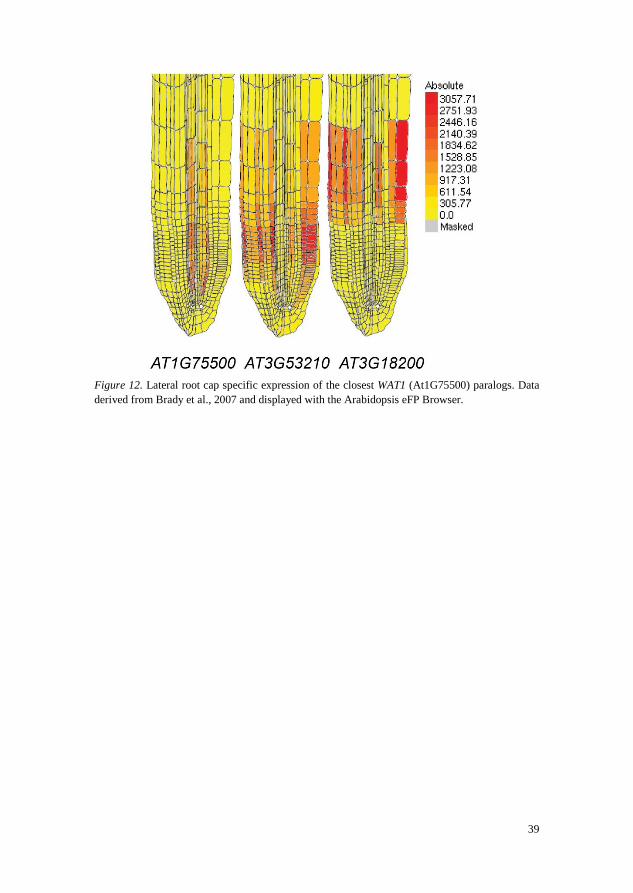
Figure 12. Lateral root cap specific expression of the closest WAT1 (At1G75500) paralogs. Data derived from Brady et al., 2007 and displayed with the Arabidopsis eFP Browser.
39

5 Conclusion and future perspectives Although the season when leaves separate from deciduous trees – “the fall” – is named after “the fall of the leaf” little attention has been paid to how seasonal leaf abscission is regulated. Current models of leaf abscission almost uniquely derive from the study of annual plants. These models, reproduced in popular plant biology text books (e.g. Taiz and Zeiger), suggest that reduced auxin flow from the leaf blade into the leaf axil leads to increased ethylene signaling and consequently to hydrolysis of middle lamellae.
In Manuscript I, we described the establishment of an experimental model for leaf abscission in Populus trees. We found that prior to the formation of an abscission zone, a new auxin maximum is established, which likely provides positional cues for the formation of an abscission zone. Inhibition of polar auxin transport, as well as exogenous auxin application, delays abscission. In contrast to the current text book opinion, auxin acts independently of ethylene signaling on leaf abscission.
We identified auxin transport facilitators, which are among the most significantly regulated genes during leaf abscission. We described their expression patterns during leaf abscission (Manuscript I). Functional analysis of these auxin transport facilitators and the determination of their subcellular localization will shed light on how the local auxin maximum is established in the leaf axil.
Unlike in leaf abscission, separation of the root cap does not involve the formation of a morphologically complex abscission zone. This makes the root cap a more accessible model than leaf axils in order to study the basic principles of cell separation. We found that root cap cell layers at the minimum of the columella-spanning auxin gradient undergo abscission. Transport of auxin from its source in the quiescent center to the periphery is hindered due to the absence of auxin efflux carrier expression in the outer layers of the columella (Manuscript II). Solely based on physiological experiments, Addicott (1955) proposed an auxin gradient to regulate organ abscission. Here,
41
we provide novel, molecular evidence, which is in line with Addicott’s hypothesis. Future experiments should include the genetic analysis of auxin gradient formation in the root cap, e.g. by the means of a mutant screen or inducible silencing of PIN expression.
One of the most significantly regulated genes during abscission is a Populus homolog of the Arabidopsis WAT1. At the time of our gene expression study, the biochemical function of WAT1 was still unknown. In collaboration with other groups, we found that WAT1 is a tonoplast-localized auxin transporter regulating cellular auxin homeostasis (Manuscript III). WAT1, which belongs to a large gene family of transmembrane proteins, is not expressed in the root cap and the perturbation of this gene does not lead to any phenotype related to abscission. Future work should address expression patterns and functions of the close WAT1 homologs in order to test if auxin homeostasis mediated by WAT1 homologs plays any role in organ abscission.
42
References Abeles, F.B. & Rubinstein, B. (1964). Regulation of ethylene evolution and leaf
abscission by auxin. Plant Physiology, 39(6), pp. 963-969. Addicott, F.T. (1982). Abscission. University of California Press, Berkeley Addicott, F.T., Lynch, R.S. & Carns, H.R. (1955). Auxin gradient theory of
abscission regulation. Science, 121(3148), pp. 644-645. Aponte, C., García, L.V. & Marañón, T. (2013). Tree species effects on nutrient
cycling and soil biota: A feedback mechanism favouring species coexistence. Forest Ecology and Management, 309(0), pp. 36-46.
Aziz, A. (2003). Spermidine and related-metabolic inhibitors modulate sugar and amino acid levels in Vitis vinifera L.: possible relationships with initial fruitlet abscission. Journal of Experimental Botany, 54(381), pp. 355-363.
Bacic, A., Moody, S.F. & Clarke, A.E. (1986). Structural Analysis of Secreted Root Slime from Maize (Zea mays L.). Plant Physiology, 80(3), pp. 771-777.
Bar-Dror, T., Dermastia, M., Kladnik, A., Znidaric, M.T., Novak, M.P., Meir, S., Burd, S., Philosoph-Hadas, S., Ori, N., Sonego, L., Dickman, M.B. & Lers, A. (2011). Programmed Cell Death Occurs Asymmetrically during Abscission in Tomato. Plant Cell, 23(11), pp. 4146-4163.
Barlow, P.W. (2002). The root cap: Cell dynamics, cell differentiation and cap function. Journal of Plant Growth Regulation, 21(4), pp. 261-286.
Barry, C.S., McQuinn, R.P., Thompson, A.J., Seymour, G.B., Grierson, D. & Giovannoni, J.J. (2005). Ethylene insensitivity conferred by the Green-ripe and Never-ripe 2 ripening mutants of tomato. Plant Physiology, 138(1), pp. 267-275.
Basu, M.M., González-Carranza, Z.H., Azam-Ali, S., Tang, S., Shahid, A.A. & Roberts, J.A. (2013). The Manipulation of Auxin in the Abscission Zone Cells of Arabidopsis Flowers Reveals That Indoleacetic Acid Signaling Is a Prerequisite for Organ Shedding. Plant Physiology, 162(1), pp. 96-106.
BenCheikh, W., PerezBotella, J., Tadeo, F.R., Talon, M. & PrimoMillo, E. (1997). Pollination increases gibberellin levels in developing ovaries of seeded varieties of citrus. Plant Physiology, 114(2), pp. 557-564.
43
Benfey, P.N., Bennett, M. & Schiefelbein, J. (2010). Getting to the root of plant biology: impact of the Arabidopsis genome sequence on root research. Plant Journal, 61(6), pp. 992-1000.
Benjamins, R. & Scheres, B. (2008). Auxin: The looping star in plant development. In: Annual Review of Plant Biology. (Annual Review of Plant Biology, 59). Palo Alto: Annual Reviews, pp. 443-465.
Bennett, T., van den Toorn, A., Sanchez-Perez, G.F., Campilho, A., Willemsen, V., Snel, B. & Scheres, B. (2010). SOMBRERO, BEARSKIN1, and BEARSKIN2 Regulate Root Cap Maturation in Arabidopsis. The Plant Cell, 22(3), pp. 640-654.
Beyer, E.M. (1975). Abscission - initial effect of ethylene is in leaf blade. Plant Physiology, 55(2), pp. 322-327.
Beyer, E.M., Jr. (1973). Abscission. Support for a role of ethylene modification of auxin transport. Plant Physiology, 52(1), pp. 1-5.
Beyer, E.M. & Morgan, P.W. (1971). Abscission - role of ethylene modification of auxin transport. Plant Physiology, 48(2), pp. 208-212.
Bleecker, A.B., Estelle, M.A., Somerville, C. & Kende, H. (1988). Insensitivity to Ethylene Conferred by a Dominant Mutation in Arabidopsis thaliana. Science, 241(4869), pp. 1086-1089.
Bleecker, A.B. & Patterson, S.E. (1997). Last exit: Senescence, abscission, and meristem arrest in Arabidopsis. Plant Cell, 9(7), pp. 1169-1179.
Brady, S.M., Orlando, D.A., Lee, J.Y., Wang, J.Y., Koch, J., Dinneny, J.R., Mace, D., Ohler, U. & Benfey, P.N. (2007). A high-resolution root spatiotemporal map reveals dominant expression patterns. Science, 318(5851), pp. 801-806.
Brigham, L.A., Woo, H.H., Wen, F. & Hawes, M.C. (1998). Meristem-specific suppression of mitosis and a global switch in gene expression in the root cap of pea by endogenous signals. Plant Physiology, 118(4), pp. 1223-1231.
Brown, H.S. & Addicott, F.T. (1950). The anatomy of experimental leaflet abscission in Phaseolus vulgaris. American Journal of Botany, 37(8), pp. 650-656.
Brown, K.M. (1997). Ethylene and abscission. Physiologia Plantarum, 100(3), pp. 567-576.
Butenko, M.A., Patterson, S.E., Grini, P.E., Stenvik, G.E., Amundsen, S.S., Mandal, A. & Aalen, R.B. (2003). INFLORESCENCE DEFICIENT IN ABSCISSION controls floral organ abscission in arabidopsis and identifies a novel family of putative ligands in plants. Plant Cell, 15(10), pp. 2296-2307.
Butenko, M.A., Shi, C.-L. & Aalen, R.B. (2012). KNAT1, KNAT2 and KNAT6 act downstream in the IDA-HAE/HSL2 signaling pathway to regulate floral organ abscission. Plant Signaling & Behavior, 7(1), pp. 135-8.
Butenko, M.A., Stenvik, G.E., Alm, V., Saether, B., Patterson, S.E. & Aalen, R.B. (2006). Ethylene-dependent and -independent pathways controlling floral abscission are revealed to converge using promoter::reporter gene constructs in the ida abscission mutant. Journal of Experimental Botany, 57(14), pp. 3627-3637.
44
Cai, M.Z., Wang, N., Xing, C.H., Wang, F.M., Wu, K. & Du, X. (2013). Immobilization of aluminum with mucilage secreted by root cap and root border cells is related to aluminum resistance in Glycine max L. Environmental Science and Pollution Research, 20(12), pp. 8924-8933.
Chae, H.S., Faure, F. & Kieber, J.J. (2003). The eto1, eto2, and eto3 Mutations and Cytokinin Treatment Increase Ethylene Biosynthesis in Arabidopsis by Increasing the Stability of ACS Protein. The Plant Cell, 15(2), pp. 545-559.
Chang, C., Kwok, S.F., Bleecker, A.B. & Meyerowitz, E.M. (1993). Arabidopsis ethylene-response gene ETR1: similarity of product to two component regulators. Science, 262(5133), pp. 539-544.
Chen, Q.H.G. & Bleecker, A.B. (1995). Analysis of ethylene signal-transduction kinetics associated with seedling-growth response and chitinase induction in wild-type and mutant Arabidopsis. Plant Physiology, 108(2), pp. 597-607.
Cho, S.K., Larue, C.T., Chevalier, D., Wang, H.C., Jinn, T.L., Zhang, S.Q. & Walker, J.C. (2008). Regulation of floral organ abscission in Arabidopsis thaliana. Proceedings of the National Academy of Sciences of the United States of America, 105(40), pp. 15629-15634.
Clausen, S. & Apel, K. (1991). Seasonal changes in the concentration of the major storage protein and its mRNA in xylem ray cells of poplar trees. Plant Molecular Biology, 17(4), pp. 669-678.
Dal Cin, V., Boschetti, A., Dorigoni, A. & Ramina, A. (2007). Benzylaminopurine application on two different apple cultivars (Malus domestica) displays new and unexpected fruitlet abscission features. Annals of Botany, 99(6), pp. 1195-1202.
Devillard, C. (1992). Genetic-transformation of aspen (populus-tremulaxpopulus-alba) by agrobacterium-rhizogenes and regeneration of plants tolerant to herbicide. Comptes Rendus De L Academie Des Sciences Serie Iii-Sciences De La Vie-Life Sciences, 314(6), pp. 291-298.
Ditengou, F.A., Muller, A., Rosenkranz, M., Felten, J., Lasok, H., van Doorn, M.M., Legue, V., Palme, K., Schnitzler, J.P. & Polle, A. (2015). Volatile signalling by sesquiterpenes from ectomycorrhizal fungi reprogrammes root architecture. Nature Communications, 6.
Dolan, L., Janmaat, K., Willemsen, V., Linstead, P., Poethig, S., Roberts, K. & Scheres, B. (1993). Cellular organisation of the Arabidopsis thaliana root. Development, 119(1), pp. 71-84.
Driouich, A., Durand, C. & Vicré-Gibouin, M. (2007). Formation and separation of root border cells. Trends in Plant Science, 12(1), pp. 14-19.
Eapen, D., Barroso, M.L., Campos, M.E., Ponce, G., Corkidi, G., Dubrovsky, J.G. & Cassab, G.I. (2003). A no hydrotropic response root mutant that responds positively to gravitropism in Arabidopsis. Plant Physiology, 131(2), pp. 536-546.
Ellis, C.M., Nagpal, P., Young, J.C., Hagen, G., Guilfoyle, T.J. & Reed, J.W. (2005). AUXIN RESPONSE FACTOR1 and AUXIN RESPONSE FACTOR2 regulate senescence and floral organ abscission in Arabidopsis thaliana. Development, 132(20), pp. 4563-4574.
45
Estornell, L.H., Agustí, J., Merelo, P., Talón, M. & Tadeo, F.R. (2013). Elucidating mechanisms underlying organ abscission. Plant Science, 199–200(0), pp. 48-60.
Fernandez, D.E., Heck, G.R., Perry, S.E., Patterson, S.E., Bleecker, A.B. & Fang, S.-C. (2000). The Embryo MADS Domain Factor AGL15 Acts Postembryonically: Inhibition of Perianth Senescence and Abscission via Constitutive Expression. The Plant Cell, 12(2), pp. 183-198.
Fischer, A.M. (2007). Nutrient Remobilization During Leaf Senescence. In: Annual Plant Reviews Volume 26: Senescence Processes in Plants Blackwell Publishing Ltd, pp. 87-107.
Fischer, U. & Polle, A. (2010). Populus Responses to Abiotic Stress. (Genetics and Genomics of Populus, 8). New York: Springer.
Fortin, M.C. & Poff, K.L. (1991). Characterization of thermotropism in primary roots of maize - dependence on temperature and temperature-gradient, and interaction with gravitropism. Planta, 184(3), pp. 410-414.
Fracheboud, Y., Luquez, V., Bjorken, L., Sjodin, A., Tuominen, H. & Jansson, S. (2009). The Control of Autumn Senescence in European Aspen. Plant Physiology, 149(4), pp. 1982-1991.
Frelich, L.E. & Reich, P.B. (2002). Dynamics of old-growth oak forests in the Eastern United States. (Oak forest ecosystems: ecology and management for wildlife. Baltimore,: Johns Hopkins University Press.
Friml, J., Benková, E., Blilou, I., Wisniewska, J., Hamann, T., Ljung, K., Woody, S., Sandberg, G., Scheres, B., Jürgens, G. & Palme, K. (2002). AtPIN4 Mediates Sink-Driven Auxin Gradients and Root Patterning in Arabidopsis. Cell, 108(5), pp. 661-673.
Gan, S.S. & Amasino, R.M. (1997). Making sense of senescence - Molecular genetic regulation and manipulation of leaf senescence. Plant Physiology, 113(2), pp. 313-319.
Gawadi, A.G. & Avery, G.S. (1950). LEAF ABSCISSION AND THE SO-CALLED ABSCISSION LAYER. American Journal of Botany, 37(2), pp. 172-180.
González-Carranza, Z.H., Whitelaw, C.A., Swarup, R. & Roberts, J.A. (2002). Temporal and Spatial Expression of a Polygalacturonase during Leaf and Flower Abscission in Oilseed Rape and Arabidopsis. Plant Physiology, 128(2), pp. 534-543.
Gunawardena, U. & Hawes, M.C. (2002). Tissue specific localization of root infection by fungal pathogens: Role of root border cells. Molecular Plant-Microbe Interactions, 15(11), pp. 1128-1136.
Gunawardena, U., Rodriguez, M., Straney, D., Romeo, J.T., VanEtten, H.D. & Hawes, M.C. (2005). Tissue-specific localization of pea root infection by Nectria haematococca. Mechanisms and consequences. Plant Physiology, 137(4), pp. 1363-1374.
Guzman, P. & Ecker, J.R. (1990). Exploiting the triple response of Arabidopsis to identify ethylene-related mutants. Plant Cell, 2(6), pp. 513-523.
Hamamoto, L., Hawes, M.C. & Rost, T.L. (2006). The production and release of living root cap border cells is a function of root apical meristem type in dicotyledonous angiosperm plants. Annals of Botany, 97(5), pp. 917-923.
46
Hanischt.Ch & Bruinsma, J. (1973). Abscission of flower bud pedicels in begonia .1. Effects of plant-growth regulating substances on abscission with intact plants and with explants. Acta Botanica Neerlandica, 22(6), pp. 666-674.
Hartmond, U., Yuan, R.C., Burns, J.K., Grant, A. & Kender, W.J. (2000). Citrus fruit abscission induced by methyl-jasmonate. Journal of the American Society for Horticultural Science, 125(5), pp. 547-552.
Hasenstein, K.H. & Evans, M.L. (1988). Effects of cations on hormone transport in primary roots of Zea-mays. Plant Physiology, 86(3), pp. 890-894.
Hawes, M.C., Gunawardena, U., Miyasaka, S. & Zhao, X.W. (2000). The role of root border cells in plant defense. Trends in Plant Science, 5(3), pp. 128-133.
Hawes, M.C. & Lin, H.-J. (1990). Correlation of Pectolytic Enzyme Activity with the Programmed Release of Cells from Root Caps of Pea (Pisum sativum). Plant Physiology, 94(4), pp. 1855-1859.
Hawes, M., Smith, L. & Stephenson, M. (1991). Root organogenesis from single cells released from the root cap of Medicago sp. Plant Cell, Tissue and Organ Culture, 27(3), pp. 303-308.
He, X.-J., Mu, R.-L., Cao, W.-H., Zhang, Z.-G., Zhang, J.-S. & Chen, S.-Y. (2005). AtNAC2, a transcription factor downstream of ethylene and auxin signaling pathways, is involved in salt stress response and lateral root development. The Plant Journal, 44(6), pp. 903-916.
Hoad, G.V. (1995). Transport of hormones in the phloem of higher plants. Plant Growth Regulation, 16(2), pp. 173-182.
Hoch, W.A., Zeldin, E.L. & McCown, B.H. (2001). Physiological significance of anthocyanins during autumnal leaf senescence. Tree Physiology, 21(1), pp. 1-8.
Hong, S.B., Sexton, R. & Tucker, M.L. (2000). Analysis of gene promoters for two tomato polygalacturonases expressed in abscission zones and the stigma. Plant Physiology, 123(3), pp. 869-881.
Iijima, M., Higuchi, T. & Barlow, P.W. (2004). Contribution of root cap mucilage and presence of an intact root cap in maize (Zea mays) to the reduction of soil mechanical impedance. Annals of Botany, 94(3), pp. 473-477.
Jackson, M.B., Hartley, C.B. & Osborne, D.J. (1973). Timing abscission in Phaseolus vulgaris L. by controlling ethylene production and sensitivity to ethylene. New Phytologist, 72(6), pp. 1251-1260.
Jackson, M.B. & Osborne, D.J. (1970). Ethylene, the Natural Regulator of Leaf Abscission. Nature, 225(5237), pp. 1019-1022.
Jackson, M.B. & Osborne, D.J. (1972). Abscisic acid, auxin, and ethylene in explant abscission. Journal of Experimental Botany, 23(3), pp. 849-862.
Jansson, S. & Douglas, C.J. (2007). Populus: A model system for plant biology. In: Annual Review of Plant Biology. (Annual Review of Plant Biology, 58). Palo Alto: Annual Reviews, pp. 435-458.
Jinn, T.L., Stone, J.M. & Walker, J.C. (2000). HAESA, an Arabidopsis leucine-rich repeat receptor kinase, controls floral organ abscission. Genes & Development, 14(1), pp. 108-117.
Kandasamy, M.K., McKinney, E.C., Deal, R.B. & Meagher, R.B. (2005). Arabidopsis ARP7 Is an Essential Actin-Related Protein Required for
47
Normal Embryogenesis, Plant Architecture, and Floral Organ Abscission. Plant Physiology, 138(4), pp. 2019-2032.
Keskitalo, J., Bergquist, G., Gardestrom, P. & Jansson, S. (2005). A cellular timetable of autumn senescence. Plant Physiology, 139(4), pp. 1635-1648.
Khripach, V.A., Zhabinskii, V.N. & Groot, A.E.d. (1999). Brassinosteroids: a new class of plant hormones. (Brassinosteroids: a new class of plant hormones. San Diego, California,: Academic Press.
Kumpf, R.P., Shi, C.L., Larrieu, A., Sto, I.M., Butenko, M.A., Peret, B., Riiser, E.S., Bennett, M.J. & Aalen, R.B. (2013). Floral organ abscission peptide IDA and its HAE/HSL2 receptors control cell separation during lateral root emergence. Proceedings of the National Academy of Sciences of the United States of America, 110(13), pp. 5235-5240.
La Rue, C.D. (1936). The Effect of Auxin on the Abscission of Petioles. Proceedings of the National Academy of Sciences of the United States of America, 22(5), pp. 254-259.
Lashbrook, C.C., Tieman, D.M. & Klee, H.J. (1998). Differential regulation of the tomato ETR gene family throughout plant development. Plant Journal, 15(2), pp. 243-252.
Lee, Y.N., Gao, Y. & Wang, H.Y. (2008). Differential mediation of the Wnt canonical pathway by mammalian Dishevelleds-1,-2, and-3. Cellular Signalling, 20(2), pp. 443-452.
Legue, V., Rigal, A. & Bhalerao, R.P. (2014). Adventitious root formation in tree species: involvement of transcription factors. Physiologia Plantarum, 151(2), pp. 192-198.
Leple, J.C., Brasileiro, A.C.M., Michel, M.F., Delmotte, F. & Jouanin, L. (1992). Transgenic poplars - expression of chimeric genes using 4 different constructs. Plant Cell Reports, 11(3), pp. 137-141.
Lers, A., Khalchitski, A., Lomaniec, E., Burd, S. & Green, P. (1998). Senescence-induced RNases in tomato. Plant Molecular Biology, 36(3), pp. 439-449.
Lers, A., Sonego, L., Green, P.J. & Burd, S. (2006). Suppression of LX ribonuclease in tomato results in a delay of leaf senescence and abscission. Plant Physiology, 142(2), pp. 710-721.
Lewis, M.W., Leslie, M.E. & Liljegren, S.J. (2006). Plant separation: 50 ways to leave your mother. Current Opinion in Plant Biology, 9(1), pp. 59-65.
Louie, D.S. & Addicott, F.T. (1970). Applied Auxin Gradients and Abscission in Explants. Plant Physiology, 45(6), pp. 654-657.
Ma, C., Meir, S., Xiao, L., Tong, J., Liu, Q., Reid, M.S. & Jiang, C.-Z. (2015). A KNOTTED1-LIKE HOMEOBOX Protein Regulates Abscission in Tomato by Modulating the Auxin Pathway. Plant Physiology, 167(3), pp. 844-853.
McCully, M.E. & Sealey, L.J. (1996). The expansion of maize root-cap mucilage during hydration .2. Observations on soil-grown roots by cryo-scanning electron microscopy. Physiologia Plantarum, 97(3), pp. 454-462.
Meir, S., Philosoph-Hadas, S., Sundaresan, S., Selvaraj, K.S.V., Burd, S., Ophir, R., Kochanek, B., Reid, M.S., Jiang, C.-Z. & Lers, A. (2010). Microarray Analysis of the Abscission-Related Transcriptome in the Tomato Flower
48
Abscission Zone in Response to Auxin Depletion. Plant Physiology, 154(4), pp. 1929-1956.
Meir, S., Philosoph-Hadas, S., Sundaresan, S., Selvaraj, K.S.V., Burd, S., Ophir, R., Kochanek, B., Reid, M.S., Jiang, C.-Z. & Lers, A. (2011). Identification of defense-related genes newly associated with tomato flower abscission. Plant Signaling & Behavior, 6(4), pp. 590-593.
Miyasaka, S.C. & Hawes, M.C. (2001). Possible role of root border cells in detection and avoidance of aluminum toxicity. Plant Physiology, 125(4), pp. 1978-1987.
Mol, J., Jenkins, G., Schafer, E. & Weiss, D. (1996). Signal perception, transduction, and gene expression involved in anthocyanin biosynthesis. Critical Reviews in Plant Sciences, 15(5-6), pp. 525-557.
Moore, R., Fondren, W.M., Koon, E.C. & Wang, C.L. (1986). The Influence of Gravity on the Formation of Amyloplasts in Columella Cells of Zea mays L. Plant Physiology, 82(3), pp. 867-868.
Morel, J.L., Mench, M. & Guckert, A. (1986). Measurement of Pb2+, Cu2+ and Cd2+ binding with mucilage exudates from maize (Zea mays L.) roots. Biology and Fertility of Soils, 2(1), pp. 29-34.
Morgan, P.W. & Hall, W.C. (1964). Accelerated Release of Ethylene by Cotton following Application of Indolyl-3-acetic Acid. Nature, 201(4914), pp. 99-99.
Nawy, T., Lee, J.Y., Colinas, J., Wang, J.Y., Thongrod, S.C., Malamy, J.E., Birnbaum, K. & Benfey, P.N. (2005). Transcriptional profile of the Arabidopsis root quiescent center. Plant Cell, 17(7), pp. 1908-1925.
Nguema-Ona, E., Vicre-Gibouin, M., Gotte, M., Plancot, B., Lerouge, P., Bardor, M. & Driouich, A. (2014). Cell wall O-glycoproteins and N-glycoproteins: aspects of biosynthesis and function. Frontiers in Plant Science, 5.
Nilsson, O., Alden, T., Sitbon, F., Little, C.H.A., Chalupa, V., Sandberg, G. & Olsson, O. (1992). Spatial pattern of cauliflower mosaic virus 35S promoter-luciferase expression in transgenic hybrid aspen trees monitored by enzymatic assay and non-destructive imaging. Transgenic Research, 1(5), pp. 209-220.
Okabe, Y., Asamizu, E., Saito, T., Matsukura, C., Ariizumi, T., Bres, C., Rothan, C., Mizoguchi, T. & Ezura, H. (2011). Tomato TILLING Technology: Development of a Reverse Genetics Tool for the Efficient Isolation of Mutants from Micro-Tom Mutant Libraries. Plant and Cell Physiology, 52(11), pp. 1994-2005.
Okushima, Y., Mitina, I., Quach, H.L. & Theologis, A. (2005). AUXIN RESPONSE FACTOR 2 (ARF2): a pleiotropic developmental regulator. Plant Journal, 43(1), pp. 29-46.
Olsen, A.N., Ernst, H.A., Leggio, L.L. & Skriver, K. (2005). NAC transcription factors: structurally distinct, functionally diverse. Trends in Plant Science, 10(2), pp. 79-87.
Osborne, D.J. (1989). ABSCISSION. Critical Reviews in Plant Sciences, 8(2), pp. 103-129.
49
Pan, J.-W., Ye, D., Wang, L.-L., Hua, J., Zhao, G.-F., Pan, W.-H., Han, N. & Zhu, M.-Y. (2004). Root Border Cell Development is a Temperature-Insensitive and Al-Sensitive Process in Barley. Plant and Cell Physiology, 45(6), pp. 751-760.
Parkinson, J.S. & Kofoid, E.C. (1992). Communication modules in bacterial signaling proteins. Annual Review of Genetics, 26, pp. 71-112.
Parsons, T.J., Sinkar, V.P., Stettler, R.F., Nester, E.W. & Gordon, M.P. (1986). Transformation of Poplar by Agrobacterium tumefaciens. Nature Biotechnology, 4(6), pp. 533-536.
Patterson, S.E. (2001). Cutting Loose. Abscission and Dehiscence in Arabidopsis. Plant Physiology, 126(2), pp. 494-500.
Patterson, S.E. & Bleecker, A.B. (2004). Ethylene-dependent and -independent processes associated with floral organ abscission in Arabidopsis. Plant Physiology, 134(1), pp. 194-203.
Petricka, J.J., Schauer, M.A., Megraw, M., Breakfield, N.W., Thompson, J.W., Georgiev, S., Soderblom, E.J., Ohler, U., Moseley, M.A., Grossniklaus, U. & Benfey, P.N. (2012). The protein expression landscape of the Arabidopsis root. Proceedings of the National Academy of Sciences of the United States of America, 109(18), pp. 6811-6818.
Pierik, R.L.M. (1977). Induction of secondary abscission in apple pedicels invitro. Physiologia Plantarum, 39(4), pp. 271-274.
Pierik, R.L.M. (1980). Hormonal-regulation of secondary abscission in pear pedicels in vitro. Physiologia Plantarum, 48(1), pp. 5-8.
Polle, A. & Douglas, C. (2010). The molecular physiology of poplars: paving the way for knowledge-based biomass production. Plant Biology, 12(2), pp. 239-241.
Ponce, G., Barlow, P.W., Feldman, L.J. & Cassab, G.I. (2005). Auxin and ethylene interactions control mitotic activity of the quiescent centre, root cap size, and pattern of cap cell differentiation in maize. Plant, Cell & Environment, 28(6), pp. 719-732.
Porth, I. & El-Kassaby, Y.A. (2015). Using Populus as a lignocellulosic feedstock for bioethanol. Biotechnology journal, 10(4), pp. 510-24.
Porth, I., Klapste, J., Skyba, O., Friedmann, M.C., Hannemann, J., Ehlting, J., El-Kassaby, Y.A., Mansfield, S.D. & Douglas, C.J. (2013). Network analysis reveals the relationship among wood properties, gene expression levels and genotypes of natural Populus trichocarpa accessions. New Phytologist, 200(3), pp. 727-742.
Ranocha, P., Denancé, N., Vanholme, R., Freydier, A., Martinez, Y., Hoffmann, L., Köhler, L., Pouzet, C., Renou, J.-P., Sundberg, B., Boerjan, W. & Goffner, D. (2010). WALLS ARE THIN 1 (WAT1), an Arabidopsis homolog of Medicago truncatula NODULIN21, is a tonoplast-localized protein required for secondary wall formation in fibers. The Plant Journal, 63(3), pp. 469-483.
Ranocha, P., Dima, O., Nagy, R., Felten, J., Corratgé-Faillie, C., Novák, O., Morreel, K., Lacombe, B., Martinez, Y., Pfrunder, S., Jin, X., Renou, J.-P., Thibaud, J.-B., Ljung, K., Fischer, U., Martinoia, E., Boerjan, W. & Goffner, D. (2013). Arabidopsis WAT1 is a vacuolar auxin transport
50
facilitator required for auxin homoeostasis. Nature Communications, 4, p. 2625.
Roberts, J., Schindler, C.B. & Tucker, G. (1984). Ethylene-promoted tomato flower abscission and the possible involvement of an inhibitor. Planta, 160(2), pp. 159-163.
Roberts, J.A. & Osborne, D.J. (1981). Auxin and the Control of Ethylene Production during the Development and Senescence of Leaves and Fruits. Journal of Experimental Botany, 32(130), pp. 875-887.
Rubinstein, B. & Leopold, A.C. (1963). Analysis of the Auxin Control of Bean Leaf Abscission. Plant Physiology, 38(3), pp. 262-267.
Ruzicka, K., Ljung, K., Vanneste, S., Podhorska, R., Beeckman, T., Friml, J. & Benkova, E. (2007). Ethylene regulates root growth through effects on auxin biosynthesis and transport-dependent auxin distribution. Plant Cell, 19(7), pp. 2197-2212.
Saiki, M. & Sato, S. (2004). Root-cap columella with movable amyloplasts may cause gravitropism of primary roots of Brassica rapa. Biologia, 59(4), pp. 505-512.
Sarkar, A.K., Luijten, M., Miyashima, S., Lenhard, M., Hashimoto, T., Nakajima, K., Scheres, B., Heidstra, R. & Laux, T. (2007). Conserved factors regulate signalling in Arabidopsis thaliana shoot and root stem cell organizers. Nature, 446(7137), pp. 811-814.
Sato, M., Dehvari, N., Oberg, A.I., Dallner, O.S., Sandstrom, A.L., Olsen, J.M., Csikasz, R.I., Summers, R.J., Hutchinson, D.S. & Bengtsson, T. (2014). Improving Type 2 Diabetes Through a Distinct Adrenergic Signaling Pathway Involving mTORC2 That Mediates Glucose Uptake in Skeletal Muscle. Diabetes, 63(12), pp. 4115-4129.
Schaller, G.E. & Bleecker, A.B. (1995). Ethylene-binding sites generated in yeast expressing the Arabidopsis ETR1 gene. Science, 270(5243), pp. 1809-1811.
Sexton, R. (1997). The role of ethylene and auxin in abscission. In: Guardiola, J.L. & Martinez, J.L.G. (eds) Eight International Symposium on Plant Bioregulators in Fruit Production. (Acta Horticulturae. Leuven 1: International Society Horticultural Science), pp. 435-444.
Sexton, R., Lewis, L.N., Trewavas, A.J. & Kelly, P. (1985). Ethylene and abscission. Ethylene and plant development, pp. 173-196.
Sexton, R. & Roberts, J.A. (1982). Cell Biology of Abscission. Annual Review of Plant Physiology, 33(1), pp. 133-162.
Sipes, D.L. & Einset, J.W. (1983). Cytokinin stimulation of abscission in lemon pistil explants. Journal of Plant Growth Regulation, 2(1), pp. 73-80.
Siwecki, R. & Kozlowski, T.T. (1973). Leaf anatomy and water relations of excised leaves of six Populus clones. Arboretum Kornickie, 18, pp. 83-105.
Sorefan, K., Girin, T., Liljegren, S.J., Ljung, K., Robles, P., Galvan-Ampudia, C.S., Offringa, R., Friml, J., Yanofsky, M.F. & Ostergaard, L. (2009). A regulated auxin minimum is required for seed dispersal in Arabidopsis. Nature, 459(7246), pp. 583-586.
51
Staehelin, L.A., Giddings, T.H., Kiss, J.Z. & Sack, F.D. (1990). Macromolecular differentiation of Golgi stacks in root tips of Arabidopsis and Nicotiana seedlings as visualized in high pressure frozen and freeze-substituted samples. Protoplasma, 157(1-3), pp. 75-91.
Stenvik, G.E., Butenko, M.A., Urbanowicz, B.R., Rose, J.K.C. & Aalen, R.B. (2006). Overexpression of INFLORESCENCE DEFICIENT IN ABSCISSION activates cell separation in vestigial abscission zones in Arabidopsis. Plant Cell, 18(6), pp. 1467-1476.
Stenvik, G.E., Tandstad, N.M., Guo, Y., Shi, C.L., Kristiansen, W., Holmgren, A., Clark, S.E., Aalen, R.B. & Butenko, M.A. (2008). The EPIP peptide of INFLORESCENCE DEFICIENT IN ABSCISSION is sufficient to induce abscission in Arabidopsis through the receptor-like kinases HAESA and HAESA-LIKE2. Plant Cell, 20(7), pp. 1805-1817.
Stepanova, A.N., Hoyt, J.M., Hamilton, A.A. & Alonso, J.M. (2005). A link between ethylene and auxin uncovered by the characterization of two root-specific ethylene-insensitive mutants in Arabidopsis. Plant Cell, 17(8), pp. 2230-2242.
Stutte, G.W. & Gage, J. (1990). Gibberellin inhibits fruit abscission following seed abortion in peach. Journal of the American Society for Horticultural Science, 115(1), pp. 107-110.
Swarup, R., Perry, P., Hagenbeek, D., Van Der Straeten, D., Beemster, G.T.S., Sandberg, G., Bhalerao, R., Ljung, K. & Bennett, M.J. (2007). Ethylene upregulates auxin biosynthesis in Arabidopsis seedlings to enhance inhibition of root cell elongation. Plant Cell, 19(7), pp. 2186-2196.
Takahashi, H. (1997). Hydrotropism: The current state of our knowledge. Journal of Plant Research, 110(1098), pp. 163-169.
Taylor, J.E. & Whitelaw, C.A. (2001). Signals in abscission. New Phytologist, 151(2), pp. 323-339.
Tieman, D.M., Ciardi, J.A., Taylor, M.G. & Klee, H.J. (2001). Members of the tomato LeEIL (EIN3-like) gene family are functionally redundant and regulate ethylene responses throughout plant development. Plant Journal, 26(1), pp. 47-58.
Tucker, M.L., Whitelaw, C.A., Lyssenko, N.N. & Nath, P. (2002). Functional analysis of regulatory elements in the gene promoter for an abscission-specific cellulase from bean and isolation, expression, and binding affinity of three TGA-Type basic leucine zipper transcription factors. Plant Physiology, 130(3), pp. 1487-1496.
Tuskan, G.A., DiFazio, S., Jansson, S., Bohlmann, J., Grigoriev, I., Hellsten, U., Putnam, N., Ralph, S., Rombauts, S., Salamov, A., Schein, J., Sterck, L., Aerts, A., Bhalerao, R.R., Bhalerao, R.P., Blaudez, D., Boerjan, W., Brun, A., Brunner, A., Busov, V., Campbell, M., Carlson, J., Chalot, M., Chapman, J., Chen, G.L., Cooper, D., Coutinho, P.M., Couturier, J., Covert, S., Cronk, Q., Cunningham, R., Davis, J., Degroeve, S., Dejardin, A., Depamphilis, C., Detter, J., Dirks, B., Dubchak, I., Duplessis, S., Ehlting, J., Ellis, B., Gendler, K., Goodstein, D., Gribskov, M., Grimwood, J., Groover, A., Gunter, L., Hamberger, B., Heinze, B., Helariutta, Y., Henrissat, B., Holligan, D., Holt, R., Huang, W., Islam-
52
Faridi, N., Jones, S., Jones-Rhoades, M., Jorgensen, R., Joshi, C., Kangasjarvi, J., Karlsson, J., Kelleher, C., Kirkpatrick, R., Kirst, M., Kohler, A., Kalluri, U., Larimer, F., Leebens-Mack, J., Leple, J.C., Locascio, P., Lou, Y., Lucas, S., Martin, F., Montanini, B., Napoli, C., Nelson, D.R., Nelson, C., Nieminen, K., Nilsson, O., Pereda, V., Peter, G., Philippe, R., Pilate, G., Poliakov, A., Razumovskaya, J., Richardson, P., Rinaldi, C., Ritland, K., Rouze, P., Ryaboy, D., Schmutz, J., Schrader, J., Segerman, B., Shin, H., Siddiqui, A., Sterky, F., Terry, A., Tsai, C.J., Uberbacher, E., Unneberg, P., Vahala, J., Wall, K., Wessler, S., Yang, G., Yin, T., Douglas, C., Marra, M., Sandberg, G., Van de Peer, Y. & Rokhsar, D. (2006). The genome of black cottonwood, Populus trichocarpa (Torr. & Gray). Science, 313(5793), pp. 1596-1604.
Ulmasov, T., Murfett, J., Hagen, G. & Guilfoyle, T.J. (1997). Aux/IAA proteins repress expression of reporter genes containing natural and highly active synthetic auxin response elements. Plant Cell, 9(11), pp. 1963-1971.
Webster, B.D. & Leopold, A.C. (1972). Stem abscission in Phaseolus vulgaris explants. Botanical Gazette, 133(3), pp. 292-298.
Wenzel, C.L. & Rost, T.L. (2001). Cell division patterns of the protoderm and root cap in the “closed” root apical meristem of Arabidopsis thaliana. Protoplasma, 218(3-4), pp. 203-213.
Wenzel, C.L., Tong, K.L. & Rost, T.L. (2001). Modular construction of the protoderm and peripheral root cap in the “open” root apical meristem ofTrifolium repens cv. Ladino. Protoplasma, 218(3-4), pp. 214-224.
Vicre, M., Santaella, C., Blanchet, S., Gateau, A. & Driouich, A. (2005). Root border-like cells of Arabidopsis. Microscopical characterization and role in the interaction with rhizobacteria. Plant Physiology, 138(2), pp. 998-1008.
Wilkinson, J.Q., Lanahan, M.B., Yen, H.C., Giovannoni, J.J. & Klee, H.J. (1995). An Ethylene-Inducible Component of Signal Transduction Encoded by Never-ripe. Science, 270(5243), pp. 1807-1809.
Yoder, T.I., Zheng, H.Q., Todd, P. & Staehelin, L.A. (2001). Amyloplast sedimentation dynamics in maize columella cells support a new model for the gravity-sensing apparatus of roots. Plant Physiology, 125(2), pp. 1045-1060.
Zanne, A.E., Tank, D.C., Cornwell, W.K., Eastman, J.M., Smith, S.A., FitzJohn, R.G., McGlinn, D.J., O/'Meara, B.C., Moles, A.T., Reich, P.B., Royer, D.L., Soltis, D.E., Stevens, P.F., Westoby, M., Wright, I.J., Aarssen, L., Bertin, R.I., Calaminus, A., Govaerts, R., Hemmings, F., Leishman, M.R., Oleksyn, J., Soltis, P.S., Swenson, N.G., Warman, L. & Beaulieu, J.M. (2014). Three keys to the radiation of angiosperms into freezing environments. Nature, 506(7486), pp. 89-92.
Zimmermann, P., HITCHCOCK, A., & CROCKER, W. (1931). The effect of ethylene and illuminating gas on roses. Contributions from Boyce Thompson Institutet, 3: 459-481.
53

Acknowledgements I would like to thank the following people: Urs, that he has given me the opportunity to work with him and the assessment of my work, and for his great support throughout the entire thesis and for the opportunity to attend many meetings. Above all, particularly that he was always there for me, for all the encouragement, for the great freedom that he has given me and for his patience. Björn Sundberg, for being my co-supervisor, for helpful discussions and for the positive feedback. Totte Niittylä, my co-supervisor for providing helpful advice and feedback. The members of the Fischer group: Daniela Liebsch, Hardy Hall, Weiya Xue, Christoffer Johnsson and all the members of the cell wall group for the great atmosphere in the laboratory, the fun activities and for the support in scientific issues. Special thanks to Hardy Hall as my committee member and for the help with the immunolocalization experiment. Judith Felten, for helpful advice and technical support. Tao Yang, Vicky Guo, Jin Pan, Biyue Tan, Jing Wang, Baosheng Wang and Qian Ma, for helping me with the poplar experiments. Alexander Makoveychuk and Jeanette Nilsson for the transformation and caring of poplars.
55

The members of the KB4C-labor, Anna Gustavsson, Thomas Dobrenel, Anne Honsel, Hailiang Mao, Christian Kiefer, Moritaka Nakamura, Thomas Stanislas and Corrado Viotti for creating a nice work atmosphere and for helping with any kind of issue. Special thanks to Anna Gustavsson for the introduction to confocal laser-scanning microscopy and resolving its problems. Thomas Moritz, for the support of my work. Andrea Polle, for having the opportunity to work in her group and for being my co-supervisor at the University of Göttingen, Germany. Ivo Feussner, for being part of my thesis committee at the University of Göttingen, Germany. Deepest thanks to Daniela Liebsch and Qian Ma for the proofreading of my publications and thesis. Bo Zhang, Anne Honsel, Christian Kiefer and Corrado Viotti for helping to solve technical problems. Maria Israelsson Nordström, for the guidelines and advice about the graduate studies. Britt-Marie Wrede and her team at Wallenberg greenhouse, for arrangement and caring of my trees. Inga-Lis Johansson and Gun-Britt Långnabba for many kind words and help. Everybody at UPSC, for the wonderful environment. My parents, my family and all my friends for the support and backing throughout my studies.
56