Embed Size (px)
Citation preview
fl. exp. Biol. 139, 135-150 (1988) 135Printed in Great Britain © The Company of Biologists Limited 1988
THE ROLE OF PHOSPHOINOSITIDE METABOLISM INSIGNAL TRANSDUCTION IN SECRETORY CELLS
BY JAMES W. PUTNEY, JR
Calcium Regulation Section, Laboratory of Cellular and MolecularPharmacology, National Institute of Environmental Health Sciences,
PO Box 12233, Research Triangle Park, NC 27709, USA
Summary
Activation of a variety of cell surface receptors results in a biphasic increase inthe cytoplasmic Ca2+ concentration, due to the release, or mobilization, ofintracellular Ca2+ stores and to the entry of Ca2+ from the extracellular space.Stimulation of these same receptors also results in the phospholipase-C-catalysedhydrolysis of the minor plasma membrane phospholipid, phosphatidylinositol 4,5-bisphosphate, with the concomitant formation of inositol 1,4,5-trisphosphate[Ins(l,4,5)/>3] and diacylglycerol. Analogous to the adenylyl cyclase signallingsystem, receptor-mediated stimulation of phospholipase C also appears to occurthrough one or more intermediary guanine nucleotide-dependent regulatoryproteins. It is well established that phosphatidylinositol 4,5-bisphosphate hydroly-sis is responsible for the changes in Ca2+ homeostasis. There is strong evidencethat Ins(l,4,5)P3 stimulates Ca2+ release from intracellular stores. The Ca2+-releasing actions of Ins(l,4,5)/>
3 are terminated by its metabolism through twodistinct pathways. Ins(l,4,5)P3 is dephosphorylated by a 5-phosphatase toIns(l,4)/3
2; alternatively, Ins(l,4,5)F3 can also be phosphorylated toIns(l,3,4,5)/>
4 by a 3-kinase. Whereas the mechanism of Ca2+ mobilization isunderstood, the precise mechanisms involved in Ca2+ entry are not known; arecent proposal that Ins(l,4,5)P3, by emptying an intracellular Ca2+ pool,secondarily elicits Ca2+ entry will be considered. This review summarizes ourcurrent understanding of the mechanisms by which inositol phosphates regulatecytoplasmic Ca2+ concentrations.
Introduction
Over 30 years have now passed since the original report by Hokin & Hokin ofreceptor-stimulated turnover of inositol lipids (Hokin & Hokin, 1953). Today, theimpact of this phenomenon on a multitude of important biological systems iswidely appreciated. Of significance to the theme of this review is not only thecentral role of the inositides in secretory mechanisms, but also the major roleBodel secretory systems have played in uncovering the meaning and significance
Key words: inositol phosphates, phosphoinositides, calcium mobilization, secretory cells.
136 J. W. PUTNEY, JR
of Hokin & Hokin's 'phospholipid effect'. Thus, we note that this phenomenonwas originally discovered in studies of a model secretory system, the exocrinepancreas, and that the experimental evidence disclosing its physiological signifi-cance (discussed below) was also obtained in this same tissue, although almost 30years later and some 4000 miles away (Streb, Irvine, Berridge & Schulz, 1983).Since the first papers by the Hokins, the phosphoinositides have enjoyed periodsof interest, neglect, rekindled interest, controversy and finally acceptance asimportant intermediaries in biological signalling processes in a wide variety ofsystems. The pivotal contributions which resulted in this tumultuous history camefrom a number of different laboratories. Bob Michell's (Michell, 1975) hypothesisthat the phosphoinositides somehow served to couple receptors to cellular calciummobilization provoked considerable research and criticism. However, furtherprogress in understanding the exact role of inositol lipid turnover in receptormechanisms was hindered by an imprecise knowledge of the biochemical pathwaysinvolved. Only in the early 1980s did the experimental evidence begin to indicatethat the initial reaction in stimulated phosphoinositide turnover was the break-down not of the major known inositide, phosphatidylinositol, but rather a minorphosphorylated derivative, phosphatidylinositol 4,5-bisphosphate (Abdel-Latif,Akhtar & Hawthorne, 1977; Kirk, Creba, Downes & Michell, 1981). Berridge(1983) realized that the water-soluble product of this reaction, inositol 1,4,5-trisphosphate [Ins(l,4,5)P3], was a likely candidate for a second messenger toactivate the release of Ca2+ from intracellular stores. Shortly thereafter, he andRobin Irvine, in collaboration with Irene Schulz and Hans-Peter Streb inFrankfurt, demonstrated that this molecule in fact had the predicted biologicalactivity: Ins(l,4,5)P3 in micromolar concentrations rapidly released Ca2+ from anon-mitochondrial store in permeabilized pancreatic acinar cells (Streb et al.1983). This result was quickly confirmed in a number of different laboratories(Berridge, 1986). It now seems very likely that Ins(l,4,5)P3 is indeed the secondmessenger signalling the release of intracellular Ca2+ that occurs on activation ofCa2+-mobilizing receptors.
A parallel story evolved from the work in Nishizuka's laboratory (Nishizuka,1983, 1984a,/?), which demonstrated that the other product of inositol lipidbreakdown, diacylglycerol, was also a cellular messenger; this apparently in-nocuous intermediate of phospholipid metabolism was shown to be a potent andspecific activator of a ubiquitous protein kinase which Nishizuka designated as C-kinase. Again, a model secretory system, the platelet, proved ideal in uncoveringthe role of this important enzyme in signal transduction.
This article summarizes recent work from a number of laboratories whichaddresses fundamental questions about this important biological signalling system,with emphasis on the inositol phosphates and their relationship to cellular Ca2+
metabolism. These questions include the nature of the coupling of receptors tophospholipase C, the metabolism of the inositol phosphates, and the mechanisor mechanisms by which the inositol phosphates regulate cellular calciumetabolism.
1
Phosphoinositide metabolism in secretory cells 137
Receptor control of phospholipase C
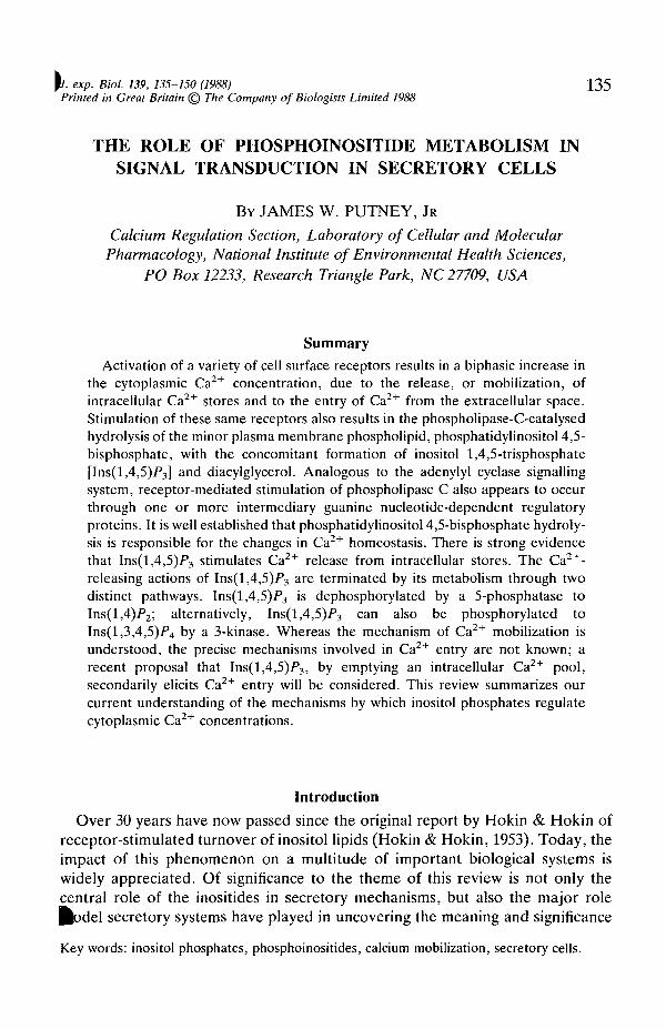
The first receptor-effector system for which a role of guanine nucleotides wasrecognized was the adenylate cyclase system (Rodbell, 1980; Smigel etal. 1984). Itis now recognized that receptor coupling to adenylate cyclase involves anintermediate guanine nucleotide-dependent regulatory protein (G-protein) desig-nated Gs (s for stimulation of cyclase). In addition, a second G-protein, Gi; is alsoknown to mediate the actions of receptors which inhibit adenylate cyclase. OtherG-proteins have been described, including transducin, a G-protein which mediatesthe coupling of photon excitation to the activation of a cyclic GMP phosphodi-esterase, and Go (o for other), purified from brain, whose function is not yetclearly understood. All these proteins are believed to exist naturally as hetero-trimers, composed of a, /? and y subunits. Receptor interaction changes theconformation of the G-protein such that GTP displaces bound GDP from the asubunit and dissociates from the /3/y complex. The dissociated GTP-bound asubunit of Gs activates adenylate cyclase (Fig. 1). However, direct inhibition of
Hormone • TReceptor •
G-protein G,
Enzyme
Intracellular messenger
Effects
(
AC PLC
\Cyclic AMP InsP3 DAG
\A kinase Ca2+ f C kinase Arachidon
1 1 ) 1CELL RESPONSE
Fig. 1. G-proteins and signal transduction. Known G-proteins (Gs and G[) regulatethe adenylate cyclase system, and there is strong circumstantial evidence that a similarmechanism regulates the polyphosphoinositide phospholipase C (PLC). AC, adenylatecyclase; DAG, diacylglycerol; InsF3, inositol trisphosphate.
138 J. W. PUTNEY, JR
adenylate cyclase by the a subunit has not been demonstrated. It has beensuggested that inhibition of adenylate cyclase occurs through the liberation of astoichiometric excess of /3/y subunits which associate with the free a subunits ofGs, resulting in inhibition (Smigel et al. 1984). Nevertheless, it is apparent for thecase of the receptors that regulate adenylate cyclase that the dissociation of theG-protein into separate a and f5/y subunits is a necessary prerequisite foraccomplishing the necessary effector function. Because receptor activation in-creases the affinity of the G-proteins for GTP, agonists and GTP generallyproduce synergistic effects in activating or inhibiting adenylate cyclase. Thissynergism is especially apparent when non-hydrolysable derivatives of GTP areused. This is because the reassociation of G-protein subunits is normally precededby the hydrolysis of the bound GTP to GDP. In fact, these non-hydrolysablederivatives of GTP [i.e. GTP-y-S, guanylyl-5'-imidodiphosphate (GppNHp)] bindto G-proteins quite well in the absence of agonists; they can thus produce subunitdissociation and G-protein activation in the absence of receptor activation.
A characteristic of receptors that couple to their effectors through G-proteins isthat, in membrane preparations, the binding of agonists (but not antagonists) isinhibited by GTP and non-hydrolysable guanine nucleotides through a decrease inapparent agonist affinity. The earliest indication that receptor coupling tophospholipase C might involve a G-protein mechanism similar to the adenylatecyclase system was the demonstration by a number of investigators that guaninenucleotides decrease the apparent affinity of agonists for receptors of this type (El-Refai, Blackmore & Exton, 1979; Goodhardt, Ferry, Geynet & Hanoune, 1982;Snavely & Insel, 1982; Evans, Martin, Hughes & Harden, 1985). In electricallypermeabilized platelets, the effects of thrombin on secretion were potentiated byguanine nucleotides (Haslam & Davidson, 1984a,b). More recently, severalinvestigators have demonstrated an activation by guanine nucleotides of phospha-tidylinositol 4,5-bisphosphate [PtdIns(4,5)F2] phospholipase C in membranes orin permeable cells (Cockcroft & Gomperts, 1985; Litosch & Fain, 1985; Litosch,Wallis & Fain, 1985; Gonzales & Crews, 1985; Lucas, Bujjalieh, Kowalchyk &Martin, 1985; Uhing, Jiang, Prpic & Exton, 1985; Uhing, Prpic, Jiang & Exton,1986; Merritt, Taylor, Rubin & Putney, 1986; Taylor, Merritt, Putney & Rubin,1986; Smith, Cox & Snyderman, 1986). In many cases, the appropriate synergisticinteractions between agonists and guanine nucleotides have been reported.Similarly, activation of phospholipase C shows the same relative sensitivity toguanine nucleotide analogues as for regulation of adenylate cyclase: GTP-y-S > GppNHp > GTP (Merritt et al. 1986).
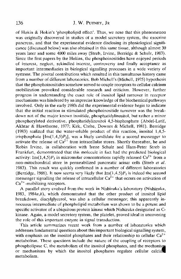
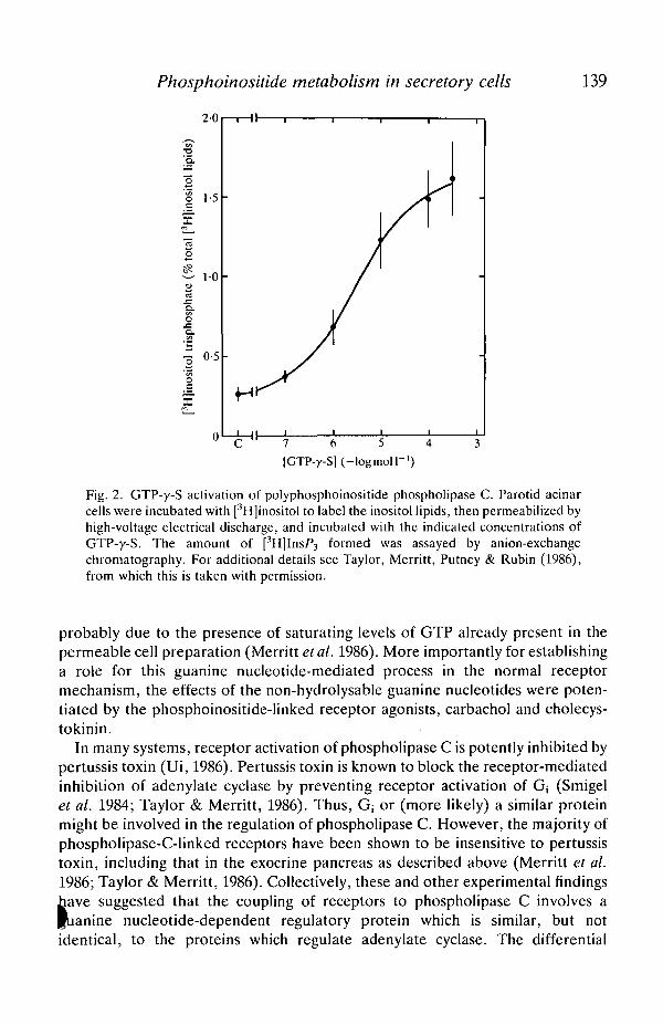
An example of guanine nucleotide regulation of phospholipase C is illustrated inFig. 2. In this experiment, rat pancreatic acinar cells were pre-incubated with[3H]inositol to label the endogenous inositol lipids, and then electrically per-meabilized (Baker & Knight, 1978) to permit experimental access to theintracellular side of the plasma membrane (Merritt et al. 1986). In this prenaaration, GTP-y-S stimulated the formation of [3H]InsP3. GppNHp also stimuladjInsP3 formation, but was less potent. GTP itself was without effect, but this was
Phosphoinositide metabolism in secretory cells 139
7 6 5 4 3[GTP-y-S] (-logmoll"1)
Fig. 2. GTP-y-S activation of polyphosphoinositide phospholipase C. Parotid acinarcells were incubated with [3H]inositol to label the inositol lipids, then permeabilized byhigh-voltage electrical discharge, and incubated with the indicated concentrations ofGTP-y-S. The amount of [3H]InsP3 formed was assayed by anion-exchangechromatography. For additional details see Taylor, Merritt, Putney & Rubin (1986),from which this is taken with permission.
probably due to the presence of saturating levels of GTP already present in thepermeable cell preparation (Merritt et al. 1986). More importantly for establishinga role for this guanine nucleotide-mediated process in the normal receptormechanism, the effects of the non-hydrolysable guanine nucleotides were poten-tiated by the phosphoinositide-linked receptor agonists, carbachol and cholecys-tokinin.
In many systems, receptor activation of phospholipase C is potently inhibited bypertussis toxin (Ui, 1986). Pertussis toxin is known to block the receptor-mediatedinhibition of adenylate cyclase by preventing receptor activation of Gj (Smigelet al. 1984; Taylor & Merritt, 1986). Thus, Gj or (more likely) a similar proteinmight be involved in the regulation of phospholipase C. However, the majority ofphospholipase-C-linked receptors have been shown to be insensitive to pertussistoxin, including that in the exocrine pancreas as described above (Merritt et al.1986; Taylor & Merritt, 1986). Collectively, these and other experimental findingshave suggested that the coupling of receptors to phospholipase C involves apuanine nucleotide-dependent regulatory protein which is similar, but notidentical, to the proteins which regulate adenylate cyclase. The differential
140 J. W. PUTNEY, JR
sensitivity to pertussis toxin may indicate that different G-proteins are involved inthe regulation of inositol lipid metabolism in different systems.
Metabolism of inositol phosphatesJust 4 years ago, the formation of inositol tris- and bisphosphates in response to
the activation of surface membrane receptors was demonstrated (Berridge et al.1983). At this time a rather simple biochemical pathway was envisaged involvingthe sequential dephosphorylation of Ins(l,4,5)P3 to inositol 1,4-bisphosphate[Ins(l,4)P2] to inositol 1-phosphate (InslP) and finally to free inositol by alithium-sensitive inositol 1-phosphatase. Today, the picture of inositol phosphatemetabolism is exceedingly complex (Putney, 1987). This is largely attributable tothe power of HPLC analytical procedures that can separate inositol phosphateswith only subtle structural differences. Ins(l,4,5)P3 is dephosphorylated by anextremely active 5-phosphatase to Ins(l,4)P2, as originally suggested (Downes,Mussat & Michel, 1982), but it now seems that Ins(l,4)P2 is dephosphorylatedalmost exclusively to inositol 4-phosphate (Ins4P) by a relatively non-specificinositol polyphosphate 1-phosphatase (Inhorn, Bansal & Majerus, 1987). Inaddition to the dephosphorylation of Ins(l,4,5)P3 by the 5-phosphatase, thereexists in most tissues thus far examined a 3-kinase which transfers a phosphatefrom ATP to the 3 position of Ins(l,4,5)P3 to form inositol 1,3,4,5-tetrakisphos-phate [Ins(l,3,4,5)P4] (Irvine, Letcher, Heslop & Berridge, 1986). This moleculeis then, probably, dephosphorylated by the same 5-phosphatase that degradesIns(l,4,5)P3 to form an isomeric inositol trisphosphate, Ins(l,3,4)P3. Ins(l,3,4)P3
is then dephosphorylated by the inositol polyphosphate 1-phosphatase to inositol3,4-bisphosphate [Ins(3,4)P2] (Inhorn et al. 1987), and to a lesser extent toIns(l,3)P2 (Bansal, Inhorn & Majerus, 1987) by an enzyme that has not been aswell characterized. These bisphosphates are then dephosphorylated primarily to amixture of InslP and Ins3P, which are stereoisomers, and not resolved byconventional HPLC. The complexity of this metabolic pathway suggests that somebiological function may be regulated by one of these metabolites. There isevidence for biological activity of Ins(l,3,4,5)P4 in either potentiating the action ofIns(l,4,5)P3 or modulating Ca2+ entry (see below).
An additional complication in the metabolism of the inositol phosphates is thedemonstration that in vitro the soluble product of phospholipase C action onphosphatidylinositol 4,5-bisphosphate is a mixture of Ins(l,4,5)P3 and a derivativewith the 1-phosphate cyclized between the 1- and 2-hydroxyls, inositol (1:2-cyclic,4,5)-trisphosphate [Ins(l:2cyc,4,5)P3] (Connolly, Wilson, Bross & Majer-us, 1986). In platelets, this Ins(l:2cyc,4,5)P3 also mobilizes intracellular Ca2+ withabout the same potency as the non-cyclic variety (Wilson et al. 1985); it is not asubstrate for the 3-kinase, and is only slowly dephosphorylated by the 5-phosphatase (Connolly et al. 1987; Hawkins, Berrie, Morris & Downes, 1987^Thus, if formed in vivo, this compound might cause persistent activation of Ca2^mobilization. In fact, reports on the production of Ins(l:2cyc,4,5)P3 in cells are
Phosphoinositide metabolism in secretory cells 141
2500
<3
l:2c
yc,4
,5)f
lns(
$
Neutral
1 t
(l:2
cyc,
4,5)
f
(AC VI
C
Acid
VA!80 85 90 95 100 80
Time (min)85 90 95 100
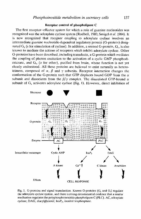
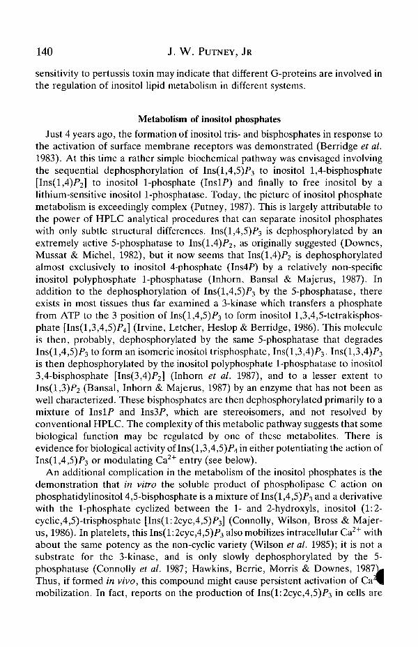
Fig. 3. HPLC analysis of [3H]inositol trisphosphates formed in parotid acinar cells.The data show radioactivity as a function of elution time from a Whatman Partisil SAXcolumn. The cells were incubated with [3H]inositol to label the inositol lipids, and thentreated with lOO^moll""1 methacholine for 40min. Samples extracted under neutralconditions, and then analysed by conventional strong anion-exchange HPLC, showthree peaks corresponding to standards for Ins(l,4,5)P3, Ins(l,3,4)P3 andIns(l:2cyc,4,5)P3 (left). If extracted under acid conditions (right) no Ins(l:2cyc4,5)P3
is seen, the amount of Ins(l,4,5)P3 is increased and a small peak corresponding toIns(2,4,5)P3 appears.
somewhat conflicting; it appears that on brief stimulation little of the cyclicderivative is formed (Hawkins et al. 1987) but, on prolonged stimulation, itaccumulates (Sekar, Dixon & Hokin, 1987), presumably due to its rather slow rateof metabolism. In most published studies to date, biological samples are treatedwith acid prior to analysis. Under these conditions, Ins(l:2cyc,4,5)P3 is convertedto Ins(l,4,5)P3, and a small amount of Ins(2,4,5)P3 (Fig. 3). Estimates fromkinetics of Ins(l,4,5)P3 and Ins(l:2cyc,4,5)F3 turnover in parotid gland suggestthat as little as 1 % of the PtdIns(4,5)P2 phospholipase C product may be cyclic(J. W. Putney, unpublished observation).
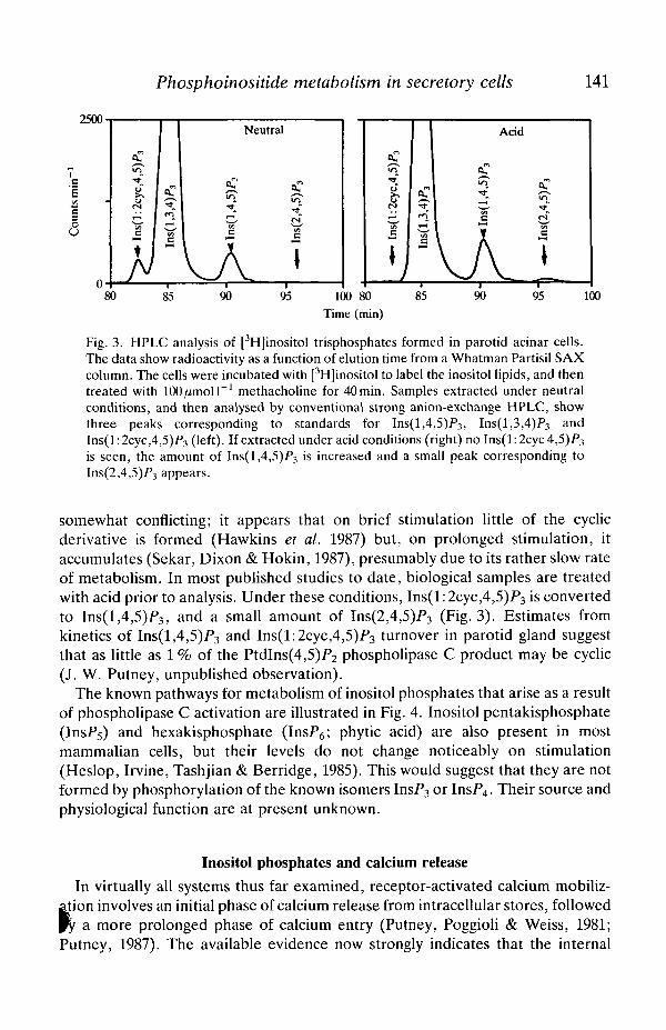
The known pathways for metabolism of inositol phosphates that arise as a resultof phospholipase C activation are illustrated in Fig. 4. Inositol pentakisphosphate(InsP5) and hexakisphosphate (InsP6; phytic acid) are also present in mostmammalian cells, but their levels do not change noticeably on stimulation(Heslop, Irvine, Tashjian & Berridge, 1985). This would suggest that they are notformed by phosphorylation of the known isomers InsP3 or InsP4. Their source andphysiological function are at present unknown.
Inositol phosphates and calcium release
In virtually all systems thus far examined, receptor-activated calcium mobiliz-tion involves an initial phase of calcium release from intracellular stores, followed
a more prolonged phase of calcium entry (Putney, Poggioli & Weiss, 1981;Putney, 1987). The available evidence now strongly indicates that the internal
142 J. W. PUTNEY, JR
Ins(l,3,4,5)/>4
• \Ins(l,3,4)P3 2
Ins(l:2cyc)F
Ins(l,3)P2 Ins(3,4)P2 Ins(l,4)P2
IIns4/>
Inositol
Fig. 4. Major (known) pathways for metabolism of Ins(l ,4,5)P3 andIns(l:2cyc,4,5)/>3. R, receptor; G, G-protein; PIC, phosphoinositidase C;DAG, diacylglycerol; PtdIns(4,5)P2, phosphatidylinositol 4,5-bisphosphate. See textfor details.
release is signalled by Ins(l,4,5)P3 (Berridge, 1983, 1986). Thus, resolution of thesite of action of Ins(l,4,5)P3 in cells would provide information on the locus ofreceptor-regulated calcium pools. Prior to the discovery of the second messengerfunction of Ins(l,4,5)P3, a number of laboratories approached this problem byexamining the distribution of total and radiolabelled calcium in subcellularfractions prepared as rapidly as possible following agonist treatment, and withprecautions to minimize redistribution during fractionation. This approach wasemployed by several laboratories in studies of calcium-mobilizing hormones in theliver. The general finding was that, following receptor activation, mitochondrialand microsomal fractions show a net loss of calcium (Exton, 1980; Williamson,Cooper & Hoek, 1981). Other reports questioned the likelihood of mitochondrialinvolvement on the basis of data suggesting that normal, healthy cells do noAgenerally contain appreciable quantities of calcium in their mitochondria (Burge*et al. 1983).

Phosphoinositide metabolism in secretory cells 143
For Ins(l,4,5)P3 the results are more straightforward. Since the action of thismediator can be tested in permeable cells and subcellular fractions, specificpoisons and other experimental manipulations can be used to determine unam-biguously the contributions of mitochondrial and non-mitochondrial pools. Underthese conditions, the results from a number of different laboratories concur thatIns(l,4,5)P3 induces release of calcium from an intracellular pool which isinsensitive to inhibitors of mitochondrial calcium uptake and is thus likely to be acomponent of the endoplasmic reticulum (ER) (Streb et al. 1983,1984; Burgess etal. 1984a,6; Joseph etal. 1984; Prentki, Wollheim & Lew, 1984). However, in somecells there is evidence that not all of the ER stores of calcium may be regulated byIns(l,4,5)P3 (Taylor & Putney, 1985; Leslie, Burgess & Putney, 1988). Forexample, in hepatocytes, only about 30-40% of exchangeable calcium could bereleased by Ins(l,4,5)P3 (Burgess etal. 1984ft). Taylor & Putney (1985) examinedthe effects of Ins(l,4,5)P3 on efflux of 45Ca2+ from fully loaded ER following rapiddepletion of ATP by glucose and hexokinase. Under these conditions, calcium exitfrom the ER of the permeable cells resembles a monoexponential process with ahalf-time of about 5 min. This rate was dramatically increased by Ins(l,4,5)P3 suchthat about 40 % of the 45Ca2+ content is lost in 30 s. Thereafter, in the continuedpresence of Ins(l,4,5)P3, the remaining 60% is lost at a rate similar to thatobserved in the absence of Ins(l,4,5)P3. This remaining fraction of accumulatedcalcium presumably resides in a pool, or component of the ER, which is notsubject to regulation by Ins(l,4,5)P3.
Experiments by Dawson and by Gill suggest that Ca2+ in the compartments ofthe ER insensitive to Ins(l,4,5)P3 can be released by GTP (Dawson, 1985;Dawson, Comerford & Fulton, 1986; Ueda, Chueh, Noel & Gill, 1986; Gill, Ueda,Chueh & Noel, 1986; Chueh & Gill, 1986; Chueh et al. 1987; Mullaney, Chueh,Ghosh & Gill, 1987). This guanine nucleotide effect is substantially different fromthat for receptor-effector coupling, in that non-hydrolysable analogues cannotsubstitute for GTP and, in fact, will block the response to GTP. Gill has shownthat there are also a number of differences between the mechanism of action ofGTP and that of Ins(l,4,5)P3, most notable being the marked temperature-dependence of the GTP-induced release, and the marked temperature-indepen-dence of the Ins(l,4,5)P3-induced release. In recent experiments, it was shownthat GTP could, in fact, induce the translocation of Ca2+ from an Ins(l,4,5)P3-insensitive to an Ins(l,4,5)P3-sensitive pool (Mullaney et al. 1987). The ability ofGTP to regulate the size of the Ins(l,4,5)P3-sensitive pool, and the concept that itis possible to induce reversible communication between membrane-limitedcompartments, has profound implications and may be directly relevant toproposals discussed below on the mechanisms for receptor-regulated Ca2+ entryinto cells.
Based on the low concentration of Ins(l,4,5)P3 necessary to release calcium,
f d certain structural requirements for activity, it was suggested that Ins(l,4,5)P3
ted by binding to a specific receptor on the ER (Burgess et al. 1984a,b). Thus,the failure of a component of the ER to respond to Ins(l,4,5)P3 may result from
144 J. W. PUTNEY, JR
the absence of the receptor in that fraction. Spat et al. (1986a), Spat, Fabiato &Rubin (19866) and Baukal et al. (1985) prepared 32P-labelled Ins(l,4,5)P3 of highspecific activity and demonstrated the presence of specific binding sites inmicrosomes and permeable cells. In permeable hepatocytes, there was a goodcorrelation between occupancy of these sites and the mobilization of calcium byboth Ins(l,4,5)P3 and Ins(2,4,5)P3. There was also a reasonably good correlationbetween Ins(l,4,5)F3 binding and calcium release in rabbit neutrophils, the latterbeing about 10 times more sensitive to Ins(l,4,5)P3 than hepatocytes. Recently, ahigh-affinity binding site for Ins(l,4,5)P3 has been purified to homogeneity frombovine brain (Worley et al. 1987). This protein has a relative molecular mass ofaround 250000, and has many of the same properties of the putative Ins(l,4,5)P3
receptor. However, no functional data are yet available to confirm the identity ofthis binding site with the Ins(l,4,5)P3 receptor involved in mediating intracellularrelease. What is sorely needed is a better selection of potent pharmacologicalprobes to aid in the biochemical characterization of this important receptor site.
Calcium entry
By comparison with calcium release, the regulation of the second phase ofcalcium mobilization, calcium entry, is poorly understood (Putney, 1986). There isstrong, albeit indirect, evidence that inositol lipid metabolism is important forcalcium entry (Berridge & Fain, 1979). Recently, the injection of inositolphosphates into sea urchin eggs and lacrimal gland cells produced a responsepattern suggestive of an activation of both intracellular calcium release and entryof calcium from the extracellular space (Slack, Bell & Benos, 1986; Irvine & Moor,1986, 1987; Morris, Gallacher, Irvine & Petersen, 1987). However, the directapplication of Ins(l,4,5)P3 to plasma membranes does not increase their per-meability to calcium (Delfert, Hill, Pershadsingh & Sherman, 1986; Ueda et al.1986). Collectively, these observations suggest that Ins(l,4,5)P3 may activatecalcium entry into cells, but not by acting directly on the plasma membrane.
Recently, a mechanism that is consistent with this idea has been proposed.According to this hypothesis, the emptying of an Ins(l,4,5)F3-sensitive poolsecondarily signals calcium entry. A detailed discussion of the circumstantialevidence supporting this idea has been presented elsewhere (Putney, 1986). Themajor evidence is derived from studies of the kinetics of emptying and refilling of areceptor-regulated calcium pool in the rat parotid gland. In the parotid glandunder resting conditions, this intracellular calcium pool is resistant to depletion byextracellular chelating agents. However, when emptied by agonist stimulation,this pool can rapidly be filled from outside the cell, even in the absence of agonistsand (presumably) second messengers, such as inositol phosphates (Aub, McKin-ney & Putney, 1982). These results suggest that the loss of calcium from this poolhas somehow activated a pathway for entry into the pool from the extracellulspace. Thus, in the continued presence of agonist, when Ins(l,4,5)F3 levels abeing continuously maintained, the pool would presumably be held empty, the
Phosphoinositide metabolism in secretory cells 145
pathway from the extracellular space would be open, and calcium would enter theER and subsequently the cytosol through the Ins(l,4,5)P3-activated channels.This has been termed a capacitative mechanism for calcium entry, because the ERis seen as behaving as a Ca2+ capacitance between the extracellular space and thecytosol.
Recently, evidence has been presented suggesting that Ins(l,3,4,5)P4 could actas a specific signal for calcium entry in sea urchin eggs and in lacrimal gland acinarcells (Irvine & Moor, 1986, 1987; Morris et al. 1987). As discussed above,Ins(l,3,4,5)P4 is formed by phosphorylation of Ins(l,4,5)P3. In sea urchin eggs,injection of Ins(l,4,5)P3 caused full activation of the eggs (i.e. raising of afertilization envelope), a response believed to involve intracellular calcium releaseas well as calcium entry (Slack et al. 1986). The data of Irvine & Moor (1986,1987)show that Ins(2,4,5)P3, an inositol phosphate that causes calcium release in othersystems, but which, they argue, may not be phosphorylated to an InsP4, does notfully activate eggs when injected intracellularly. Also, they found thatIns(l,3,4,5)P4 did not activate eggs when injected alone. However, whenIns(l,3,4,5)P4 was injected together with the calcium-releasing inositol phos-phate, Ins(2,4,5)P3, a full fertilization response (i.e. raised envelope) wasobtained. They suggest that when Ins(l,4,5)P3 is injected, it causes calciumrelease, but that activation of calcium entry requires its phosphorylation toIns(l,3,4,5)P4. But since calcium release seemed to be a prerequisite for activationof calcium entry [i.e. Ins(l,3,4,5)P4 only worked together with the calcium-releasing Ins(2,4,5)P3], they concluded that the emptying of the Ins(l,4,5)P3-regulated calcium pool was necessary for activation of calcium entry byIns(l,3,4,5)P4. These findings may represent the first demonstrated biologicalaction of Ins(l,3,4,5)P4. Whether the specific mechanism suggested by Irvine &Moor (1986) is correct depends heavily on the assumptions made which werebased on known properties of the inositol phosphates in other biological systems.There also seems to be no basis for attributing a specific action of InsP4 on Ca2+
entry as opposed to Ca2+ release. In more recent experiments, Morris etal. (1987)demonstrated that, in perfused lacrimal acinar cells, InsP4 was required in theperfusate to see effects of Ins(l,4,5)P3 on either the initial transient or latersustained phase of Ca2+ mobilization. Thus the role of InsP4 in these cells may be apermissive one for Ins(l,4,5)P3, and the actual biphasic mobilization of Ca2+ maythen be accomplished through the capacitative mechanism discussed above; thatis, the emptying of the Ins(l,4,5)P3-regulated pool serves as a signal regulatingcalcium entry. It should be remembered that Ins(l,4,5)P3 is fully capable ofactivating Ca2+ release in permeable cells, or in microsomal preparations in theabsence of InsP4, although it is not known whether this occurs in the concentrationrange actually achieved in activated cells. Recall also that, with prolongedactivation of exocrine gland cells, Ins(l:2cyc,4,5)P3 accumulates. If this inositolphosphate is, in fact, fully capable of mimicking the actions of Ins(l,4,5)P3, thenW> slow degradation would prevent rapid restoration of basal Ca2+ levels in cellsfollowing the removal of a stimulus. However, if InsP4 were necessary for the

146 J. W. PUTNEY, JR
Ins(l,4,5)P3
Ins(l:2cyc,4,5)/'3
Fig. 5. Model depicting some suggested biological actions of inositol polyphosphates.Ins(l,4,5)P3 and Ins(l:2cyc,4,5)P3may both be able to release Ca2+ from endoplasmicreticulum, and this may secondarily signal entry of Ca2+ from the extracellular space.This process may be amplified or modulated in some manner by Ins(l,3,4,5)/>4;alternatively, Ins(l,3,4,5)F4 may also act on the entry mechanism more directly. RI,Ins(l,4,5)f3 receptor. For other abbreviations, see legend to Fig. 4.
Ins(l:2cyc,4,5)F3 to act, then the rapid fall in cellular levels of InsP4 would stilllead to a rapid termination of the Ca2+ signal. Some of the proposed roles ofinositol polyphosphates in Ca2+ signalling are illustrated in Fig. 5.
Conclusions
In the past few years, our understanding of the relationships between receptors,inositol lipids and calcium has advanced at an incredible rate. Indeed, just morethan 4 years before the writing of this review, Streb et al. (1983) first demonstratedthe biological activity of Ins(l,4,5)P3; this molecule has now gained generalacceptance as an important and ubiquitous second messenger. It is apparent thatthis second messenger system is exceedingly complex and possesses manypotential sites of regulation. As with most important biochemical pathways, ourknowledge raises numerous challenging questions. What is the nature of theG-protein (or proteins) that couples receptors to phospholipase C? What is thesignificance of the alternative pathway (phosphorylation/dephosphorylation) ofIns(l,4,5)P3 metabolism? What are the nature, mechanism and locus of theIns(l,4,5)P3 receptor? How is calcium entry regulated? Indeed, how is the entiresystem regulated, and how does it interact with other signalling systems such asthose which utilize cyclic nucleotides as messengers? We hope that the substantialprogress we have made in the past few years will continue towards the resoluti(^of these issues and also pose new questions.

Phosphoinositide metabolism in secretory cells 147
ReferencesABDEL-LATIF, A. A., AKHTAR, R. & HAWTHORNE, J. N. (1977). Acetylcholine increases the
breakdown of triphosphoinositide of rabbit iris muscle prelabelled with [32P]phosphate.Biochem. J. 162, 61-73.
AUB, D. L., MCKINNEY, J. S. & PUTNEY, J. W., JR (1982). Nature of the receptor-regulatedcalcium pool in the rat parotid gland. J. Physiol., Lond. 331, 557-565.
BAKER, P. F. & KNIGHT, D. E. (1978). Calcium-dependent exocytosis in bovine adrenalmedullary cells with leaky plasma membranes. Nature, Lond. 276, 620-622.
BANSAL, V. S., INHORN, R. C. & MAJERUS, P. W. (1987). The metabolism of inositol 1,3,4-trisphosphate to inositol 1,3-bisphosphate. /. biol. Chem. 262, 9444-9447.
BAUKAL, A. J., GUILLEMETTE, G., RUBIN, R., SPAT, A. & CATT, K. J. (1985). Binding sites forinositol trisphosphate in the bovine adrenal cortex. Biochem. biophys. Res. Commun. 133,532-538.
BERRIDGE, M. J. (1983). Rapid accumulation of inositol trisphosphate reveals that agonistshydrolyse polyphosphoinositides instead of phosphatidylinositol. Biochem. J. 212, 849-858.
BERRIDGE, M. J. (1986). Inositol phosphates as second messengers. In Phosphoinositides andReceptor Mechanisms (ed. J. W. Putney, Jr), pp. 25-46. New York: Alan R. Liss, Inc.
BERRIDGE, M. J., DAWSON, R. M., DOWNES, C. P., HESLOP, J. P. & IRVINE, R. F. (1983).Changes in the levels of inositol phosphates after agonist-dependent hydrolysis of membranephosphoinositides. Biochem. J. 212, 473-482.
BERRIDGE, M. J. & FAIN, J. N. (1979). Inhibition of phosphatidylinositol synthesis and theinactivation of calcium entry after prolonged exposure of the blowfly salivary gland to5-hydroxytryptamine. Biochem. J. 178, 59-69.
BURGESS, G. M., GODFREY, P. P., MCKINNEY, J. S., BERRIDGE, M. J., IRVINE, R. F. & PUTNEY,J. W., JR (1984a). The second messenger linking receptor activation to internal Ca2+ releasein liver. Nature, Lond. 309, 63-66.
BURGESS, G. M., MCKINNEY, J. S., FABIATO, A., LESLIE, B. A. & PUTNEY, J. W., JR (1983).Calcium pools in saponin-permeabilized guinea-pig hepatocytes. /. biol. Chem. 258,15 336-15345.
BURGESS, G. M., IRVINE, R. F., BERRIDGE, M. J., MCKINNEY, J. S. & PUTNEY, J. W., JR (1984ft).Actions of inositol phosphates on Ca2+ pools in guinea-pig hepatocytes. Biochem. J. 224,741-746.
CHUEH, S.-H. & GILL, D. L. (1986). Inositol 1,4,5-trisphosphate and guanine nucleotidesactivate calcium release from endoplasmic reticulum via distinct mechanisms. J. biol. Chem.261,13 883-13 886.
CHUEH, S.-H., MULLANEY, J. M., GHOSH, T. K., ZACHARY, A. L. & GILL, D. L. (1987). GTP-and inositol 1,4,5-trisphosphate-activated intracellular calcium movements in neuronal andsmooth muscle cell lines. / . biol. Chem. 262, 13857-13864.
COCKCROFT, S. & GOMPERTS, B. (1985). Role of guanine nucleotide binding protein in theactivation of polyphosphoinositide phosphodiesterase. Nature, Lond. 314, 534-536.
CONNOLLY, T. M., BANSAL, V. S., BROSS, T. E., IRVINE, R. F. & MAJERUS, P. W. (1987). Themetabolism of tris- and tetraphosphates of inositol by 5-phosphomonoesterase and 3-kinaseenzymes. J. biol. Chem. 262, 2146-2149.
CONNOLLY, T. M., WILSON, D. B., BROSS, T. E. & MAJERUS, P. W. (1986). Isolation andcharacterization of the inositol cyclic phosphate products of phosphoinositide cleavage byphospholipase C. Metabolism in cell-free extracts. J. biol. Chem. 261, 122-126.
DAWSON, A. P. (1985). GTP enhances inositol trisphosphate-stimulated Ca2+ release from ratliver microsomes. FEBS Letts 185, 147-150.
DAWSON, A. P., COMERFORD, J. G. & FULTON, D. V. (1986). The effect of GTP on inositol 1,4,5-trisphosphate-stimulated Ca2+ efflux from a rat liver microsome fraction: is a GTP-dependentprotein phosphorylation involved? Biochem. J. 234, 311-315.
DELFERT, D. M., HILL, S., PERSHADSINGH, H. A. & SHERMAN, W. R. (1986). wyo-Inositol 1,4,5-trisphosphate mobilizes Ca2+ from isolated adipocyte endoplasmic reticulum but not from
lasma membranes. Biochem. J. 236, 37-44.ES, C. P., MUSSAT, M. C. & MICHELL, R. H. (1982). The inositol trisphosphate
phosphomonoesterase of the human erythrocyte membrane. Biochem. J. 203, 169-177.
148 J. W. PUTNEY, JR
EL-REFAI, M. F., BLACKMORE, P. F. & EXTON, J. H. (1979). Evidence for two a/p/ia-adrenergicbinding sites in liver plasma membranes. Studies with [3H]epinephrine and [3H]dihydro-ergocryptine. J. biol. Chem. 254, 4375-4386.
EVANS, T., MARTIN, M. W., HUGHES, A. R. & HARDEN, T. K. (1985). Guanine nucleotidesensitive, high affinity binding of carbachol to muscarinic cholinergic receptors of 1321N1astrocytoma cells is insensitive to pertussis toxin. Molec. Pharmac. 27, 32-37.
EXTON, J. H. (1980). Mechanisms involved in alpha-adrenergic phenomena: Role of calciumions in actions of catecholamines in liver and other tissues. Am. J. Physiol. 238, E3-E12.
GILL, D. L., UEDA, T., CHUEH, S.-H. & NOEL, M. W. (1986). Ca2+ release from endoplasmicreticulum is mediated by a guanine nucleotide regulatory mechanism. Nature, Lond. 320,461-464.
GONZALES, R. A. & CREWS, F. T. (1985). Guanine nucleotides stimulate production of inositoltrisphosphate in rat cortical membranes. Biochem. J. 232, 799-804.
GOODHARDT, M., FERRY, N., GEYNET, P. & HANOUNE, J. (1982). Hepatic alphaj-adrenergicreceptors show agonist-specific regulation by guanine nucleotides. Loss of nucleotide effectafter adrenalectomy. J. biol. Chem. 257, 11577-11583.
HASLAM, R. J. & DAVIDSON, M. M. L. (1984a). Receptor-induced diacylglycerol formation inpermeabilized platelets; possible role for a GTP-binding protein. J. Receptor Res. 4, 605-629.
HASLAM, R. J. & DAVIDSON, M. M. L. (1984i>). Potentiation by thrombin of the secretion ofserotonin from permeabilized platelets equilibrated with Ca2+ buffers. Relationship toprotein phosphorylation and diacylglycerol formation. Biochem. J. 222, 351-361.
HAWKINS, P. T., BERRIE, C. P., MORRIS, A. J. & DOWNES, C. P. (1987). Inositol 1,2-cyclic 4,5-trisphosphate is not a product of muscarinic receptor-stimulated phosphatidylinositol 4,5-bisphosphate hydrolysis in rat parotid glands. Biochem. J. 243, 211-218.
HESLOP, J. P., IRVINE, R. F., TASHJIAN, A. H. & BERRIDGE, M. J. (1985). Inositol tetrakis- andpentakisphosphates in GH4 cells. /. exp. Biol. 119, 395-401.
HOKIN, M. R. & HOKIN, L. E. (1953). Enzyme secretion and the incorporation of P32 intophospholipides of pancreas slices. J. biol. Chem. 203, 967-977.
INHORN, R. C , BANSAL, V. S. & MAJERUS, P. W. (1987). Pathway for 1,3,4-trisphosphate and1,4-bisphosphate metabolism. Proc. natn. Acad. Sci. U.S.A. 84, 2170-2174.
IRVINE, R. F., LETCHER, A. J., HESLOP, J. P. & BERRIDGE, M. J. (1986). The inositoltris/tetrakisphosphate pathway - demonstration of Ins(l,4,5)P3 3-kinase activity in animaltissues. Nature, Lond. 320, 631-634.
IRVINE, R. F. & MOOR, R. M. (1986). Micro-injection of inositol 1,3,4,5-tetrakisphosphateactivates sea urchin eggs by a mechanism dependent on external Ca2+. Biochem. J. 240,917-920.
IRVINE, R. F. & MOOR, R. M. (1987). Inositol(l,3,4,5)tetrakisphosphate-induced activation ofsea urchin eggs requires the presence of inositol trisphosphate. Biochem. biophys. Res.Commun. 146, 284-290.
JOSEPH, S. K., THOMAS, A. P., WILLIAMS, R. J., IRVINE, R. F. & WILLIAMSON, J. R. (1984). myo-Inositol 1,4,5-trisphosphate. A second messenger for the hormonal mobilization ofintracellular Ca2+ in liver. J. biol. Chem. 259, 3077-3081.
KIRK, C. J., CREBA, J. A., DOWNES, C. P. & MICHELL, R. H. (1981). Hormone-stimulatedmetabolism of inositol lipids and its relationship to hepatic receptor function. Biochem. Soc.Trans. 9, 377-379.
LESLIE, B. A., BURGESS, G. M. & PUTNEY, J. W., JR. (1988). Persistent inhibition by inositol1,4,5-trisphosphate of oxalate-dependent 45calcium uptake in permeable guinea-pighepatocytes. Cell Calcium (in press).
LITOSCH, I. & FAIN, J. N. (1985). 5-Methyltryptamine stimulates phospholipase C-mediatedbreakdown of exogenous phosphoinositides by blowfly salivary gland membranes. J. biol.Chem. 260, 16052-16055.
LITOSCH, I., WALLIS, C. & FAIN, J. N. (1985). 5-Hydroxytryptamine stimulates inositolphosphate production in a cell-free system from blowfly salivary glands. Evidence for a role ofGTP in coupling receptor activation to phosphoinositide breakdown. /. biol. Chem. 260,5464-5471. M
LUCAS, D. O., BUJJALIEH, S. M., KOWALCHYK, J. A. & MARTIN, T. F. J. (1985). DirdjJstimulation by thyrotropin-releasing hormone (TRH) of polyphosphoinositide hydrolysis in
Phosphoinositide metabolism in secretory cells 149
GH3 cell membranes by a guanine nucleotide-modulated mechanism. Biochem. biophys. Res.Commun. 132, 721-728.
MERRITT, J. E., TAYLOR, C. W., RUBIN, R. P. & PUTNEY, J. W., JR (1986). Evidence suggestingthat a novel guanine nucleotide regulatory protein couples receptors to phospholipase C inexocrine pancreas. Biochem. J. 236, 337-343.
MICHELL, R. H. (1975). Inositol phospholipids and cell surface receptor function. Biochim.biophys. Ada 415, 81-147.
MORRIS, A. P., GALLACHER, D. V., IRVINE, R. F. & PETERSEN, O. H. (1987). Internal cellperfusion with inositol 1,4,5-trisphosphate combined with inositol 1,3,4,5-tetrakisphosphatemimics muscarinic receptor activation of Ca2+-dependent K+ channels. Nature, Lond.(in press).
MULLANEY, J. M., CHUEH, S.-H., GHOSH, T. K. & GILL, D. L. (1987). Intracellular calciumuptake activated by GTP: Evidence for a possible guanine nucleotide-inducedtransmembrane conveyance of intracellular calcium. J. biol. Chem. 262, 13865-13872.
NISHIZUKA, Y. (1983). Calcium, phospholipid turnover and transmembrane signalling. Phil.Trans. R. Soc. Ser. B 302, 101-112.
NISHIZUKA, Y. (1984a). Turnover of inositol phospholipids and signal transduction. Science 225,1365-1370.
NISHIZUKA, Y. (19846). The role of protein kinase C in cell surface signal transduction andtumour promotion. Nature, Lond. 308, 693-698.
PRENTKI, M., WOLLHEIM, C. B. & LEW, P. D. (1984). Ca2+ homeostasis in permeabilized humanneutrophils. Characterization of Ca2+-sequestering pools and the action of inositol 1,4,5-trisphosphate. J. biol. Chem. 259, 13777-13782.
PUTNEY, J. W'., JR (1986). A model for receptor-regulated calcium entry. Cell Calcium 7, 1-12.PUTNEY, J. W., JR (1987). Calcium-mobilizing receptors. Trends pharmac. Sci. 8, 481-486.PUTNEY, J. W., JR, POGGIOLI, J. & WEISS, S. J. (1981). Receptor regulation of calcium release
and calcium permeability in parotid gland cells. Phil. Trans. R. Soc. Ser. B 296, 37-45.RODBELL, M. (1980). The role of hormone receptors and GTP-regulatory proteins in membrane
transduction. Nature, Lond. 284, 17-22.SEKAR, M. C , DIXON, J. F. & HOKIN, L. E. (1987). The formation of inositol 1,2-cyclic 4,5-
trisphosphate and inositol 1,2-cyclic 4-bisphosphate on stimulation of mouse pancreaticminilobules with carbamylcholine. /. biol. Chem. 262, 340-344.
SLACK, B. E., BELL, J. E. & BENOS, D. J. (1986). Inositol 1,4,5-trisphosphate injection mimicsfertilization potentials in sea urchin eggs. Am. J. Physiol. 250, C340-C344.
SMIGEL, M., KATADA, T., NORTHUP, J. K., BOKOCH, G. M., UI , M. & GILMAN, A. G. (1984).Mechanisms of guanine nucleotide-mediated regulation of adenylate cyclase activity. Adv.cyclic Nucleotide Prot. Phosphoryl. Res. 17', 1-18.
SMITH, C. D., COX, C. C. & SNYDERMAN, R. (1986). Receptor-coupled activation ofphosphoinositide-specific phospholipase C by an N protein. Science 232, 97-100.
SNAVELY, M. D. & INSEL, P. A. (1982). Characterization of a/p/ia-adrenergic receptor subtypesin the rat renal cortex. Differential regulation oialpha^- and a/p/ia2-adrenergic receptors byguanyl nucleotides and Na+. Molec. Pharmac. 22, 532-543.
SPAT, A., BRADFORD, P. G., MCKINNEY, J. S., RUBIN, R. P. & PUTNEY, J. W., JR (1986a).A saturable receptor for 32P-inositol-l,4,5-triphosphate in hepatocytes and neutrophils.Nature, Lond. 319, 514-516.
SPAT, A., FABIATO, A. & RUBIN, R. P. (19866). Binding of inositol trisphosphate by a livermicrosomal fraction. Biochem. J. 233, 929-932.
STREB, H., BAYERDORFFER, E., HAASE, W., IRVINE, R. F. & SCHULZ, I. (1984). Effect of inositol-1,4,5-trisphosphate on isolated subcellular fractions of rat pancreas. J. Membr. Biol. 81,241-253.
STREB, H., IRVINE, R. F., BERRIDGE, M. J. & SCHULZ, I. (1983). Release of Ca2+ from anonmitochondrial store in pancreatic cells by inositol-l,4,5-trisphosphate. Nature, Lond. 306,
^67-68.LYLOR, C. W. & MERRITT, J. E. (1986). Receptor coupling to polyphosphoinositide turnover:A parallel with the adenylate cyclase system. Trends pharmac. Sci. 7, 238-242.
150 J. W. PUTNEY, JR
TAYLOR, C. W., MERRITT, J. E., PUTNEY, J. W., JR & RUBIN, R. P. (1986). A guanine nucleotide-dependent regulatory protein couples substance P receptors to phospholipase C in rat parotidgland. Biochem. biophys. Res. Commun. 136, 362-368.
TAYLOR, C. W. & PUTNEY, J. W., JR (1985). Size of the inositol 1,4,5-trisphosphate-sensitivecalcium pool in guinea-pig hepatocytes. Biochem. J. 232, 435-438.
UEDA, T., CHUEH, S.-H., NOEL, M. W. & GILL, D. L. (1986). Influence of inositol 1,4,5-trisphosphate and guanine nucleotides on intracellular calcium release within the N1E-115neuronal cell line. J. biol. Chem. 261, 3184-3192.
UHING, R. J., JIANG, H., PRPIC, V. & EXTON, J. H. (1985). Regulation of a liver plasmamembrane phosphoinositide phosphodiesterase by guanine nucleotides and calcium. FEBSLetts 188, 317-320.
UHING, R. J., PRPIC, V., JIANG, H. & EXTON, J. H. (1986). Hormone-stimulatedpolyphosphoinositide breakdown in rat liver membranes. J. biol. Chem. 621, 2140-2146.
Ui, M. (1986). Pertussis toxin as a probe of receptor coupling to inositol lipid metabolism.In Phosphoinositid.es and Receptor Mechanisms (ed. J. W. Putney, Jr), pp. 163-196. NewYork: Alan R. Liss, Inc.
WILLIAMSON, J. R., COOPER, R. H. & HOEK, J. B. (1981). Role of calcium in the hormonalregulation of liver metabolism. Biochim. biophys. Ada 639, 243-295.
WILSON, D. B., CONNOLLY, T. M., BROSS, T. E., MAJERUS, P. W., SHERMAN, W. R., TYLER,A. N., RUBIN, L. J. & BROWN, J. E. (1985). Isolation and characterization of the inositolcyclic phosphate products of polyphosphoinositide cleavage by phospholipase C.Physiological effects in permeabilized platelets and Limulus photoreceptor cells. J. biol.Chem. 260, 13496-13501.
WORLEY, P. F., BARABAN, J. M., SUPATTAPONE, S., WILSON, V. S. & SNYDER, S. H. (1987).Characterization of inositol trisphosphate receptor binding in brain: Regulation by pH andcalcium. /. biol. Chem. 262, 12132-12136.


![Metabolis m Photosynthesis [8.2] Cell Respiration [8.3] Fermentation [8.3]](https://img.pdfslide.us/doc/110x75/56649ef95503460f94c0b06c/metabolis-m-photosynthesis-82-cell-respiration-83-fermentation-83.jpg)


![A Direct Linkage between the Phosphoinositide 3 …...[CANCER RESEARCH 60, 3504–3513, July 1, 2000] A Direct Linkage between the Phosphoinositide 3-Kinase-AKT Signaling Pathway and](https://img.pdfslide.us/doc/110x75/5ea4ac155453582a17137598/a-direct-linkage-between-the-phosphoinositide-3-cancer-research-60-3504a3513.jpg)