Embed Size (px)
Citation preview

.
DOI 10.1002/bies
Unite d’Ecologie, SystCNRS UMR8079, UnivFrance
*Corresponding authPurificaci�on L�opez-GaE-mail: puri.lopez@u-p
468 www.bioes
Thinkagain
Insights & Perspectives
The rise and fall of Picobiliphytes:How assumed autotrophs turnedout to be heterotrophs
David Moreira and Purificaci�on L�opez-Garc�ıa*
Algae are significant members of Earth’s biodiversity. Having been studied for a
long time, the discovery of new algal phyla is extremely unusual. Recently, the
enigmatic Picobiliphyta, a group of uncultured eukaryotes unveiled using
molecular tools, were claimed to represent an unrecognized early branching
algal lineage with a nucleomorph (remnant nucleus of a secondary algal
endosymbiont) in their plastids. However, subsequent studies rejected the
presence of a nucleomorph, and single-cell genomic studies failed to detect any
plastid-related genes, ruling out the possibility of plastid occurrence. The
isolation of the first picobiliphyte, Picomonas judraskeda, a tiny organism that
feeds on very small (<150nm) organic particles, came as final proof of their non-
photosynthetic lifestyle. Consequently, the group has been renamed Picozoa.
The passage from picobiliphytes to picozoa illustrates the crucial role that
classical protistology should play to provide sound biological context for the
wealth of data produced by modern molecular techniques.
algae; eukaryotic evolution; phylogen
Keywords:
y; Picobiliphyte; Picozoa; protist
Introduction
Cyanobacteria, plants, and a variety ofeukaryotic algae are responsible for therelease of oxygen to the Earth’s atmo-sphere and also for the most significantpart of CO2 fixation into organic matter[1–4]. Among these organisms, cyano-bacteria and many unicellular algae are
.201300176
ematique et Evolution,ersite Paris-Sud, Orsay,
or:rcıasud.fr
says-journal.com
microscopic, but the presence of photo-synthetic and other accessory pigmentsin their cells makes them easily visibleunder the microscope. This is why, tothe detriment of the many colorlessheterotrophic species that coexist withthem, microalgae became a favoredtarget of study since the early days ofoptical microscopy and why they figureamong the best knownmicroorganisms.A vast amount of microalgal morpholo-gy and cell ultrastructural data has beencollected during the last five decadesand has served for phenotype-basedtaxonomy. In more recent years, thisbody of information has been completedby molecular phylogeny analyses ofconserved gene markers, which has
Bioessays 36: 46
been instrumental in attempting toestablish a natural, phylogeny-basedtaxonomy. These studies led to classifyeukaryotic algae into a relatively smallnumber of lineages [5]. Three algalgroups (Glaucophyta, Rhodophyta,and Chlorophyta) harbor plastids thatderive from the original endosymbiosisof a cyanobacterium within an initialheterotrophic eukaryotic cell (primaryendosymbiosis) [6, 7]. The rest of theeukaryotic algae have plastids acquiredthrough the endosymbiosis of red orgreen algae within different eukaryotichosts (secondary endosymbioses), al-though in some occasions tertiaryendosymbioses or secondary plastidreplacements may also have oc-curred [8]. These secondary and tertiaryevents gave rise to a variety of algalgroups that are distributed across theeukaryotic tree. These include theEuglenida, Chlorarachniophyta, Crypto-phyta, Haptophyta, various groupswithin the Heterokonta or Strameno-piles (Bacillariophyta, Phaeophyta,Xantophyta, etc.) and, within the Alveo-lata, the Dinophyceae and a group thatsecondarily lost the photosyntheticcapacity, the parasitic Apicomplexa.As mentioned above, these groups havebeen known for a long time, so thediscovery of new algal lineages canbe regarded as highly improbable,something only comparable to thediscovery of new mammalian speciesor new animal groups. In fact, a recentanalysis using accumulation curves forhigh level algal taxa (families, orders,and classes) has showed that the curvesare already close to saturation, which
8–474,� 2014 WILEY Periodicals, Inc.

..... Insights & Perspectives D. Moreira and P. L�opez-GarcıaThinkagain
suggests that most of those taxa havealready been discovered [9].
Unexpectedly, the existence of threenew groups of putative algae has beenreported recently. One of them, theChromerida, represented by its firstdescribed species Chromera velia, isrelated to the once-photosynthetic Api-complexa and contains plastids derivedfrom red algae [10–12]. This algal groupis now firmly established based onabundant sequence information as wellas morphological, ultrastructural, andbiochemical data, and its diversity hasbeen expanded with the isolation of thenew species Vitrella brassicaformis [13–15]. The second group, informally called“rappemonads”, has been characterizedby a combination of the analysis ofplastid 16S rRNA gene sequences re-trieved from environmental surveys ofmarine and freshwater environmentsand fluorescent in situ hybridization(FISH) experiments with specific plas-tid-targeted probes [16]. Rappemonadsappear to contain several plastids intheir cells and branch as sister-group ofthe haptophytes in plastid rRNA genephylogenies. Thus, it is unclear whetherthese organisms actually represent anew algal group or just an uncharac-terized deeply diverging haptophytelineage. The third group of putativenew algae was probably the mostenigmatic. Initially detected in 18SrDNA environmental libraries as alineage with unclear affinity to anyother eukaryotic group, their memberswere subsequently visualized usingFISH with specific 18S rDNA-targetedprobes. FISH experiments and fluores-cent microscopy revealed very smallcells (<5mm) showing orange auto-fluorescence, which might indicate thepresence of phycobilin pigments(known to occur in cyanobacteria andin plastids of glaucophytes, red algae,and some other algae containing plas-tids derived from red algae [17]). Sur-prisingly, DNA staining with fluorescentintercalating agents appeared to showthe presence of a DNA-containingorganelle resembling a plastid with anucleomorph. Nucleomorphs are rem-nants of red or green algal nuclei inplastids derived from secondary endo-symbionts [18]. Their occurrence inthese tiny organisms would indicatethat they are algae containing plastidsacquired by secondary endosymbiosis.
Bioessays 36: 468–474,� 2014 WILEY Pe
Accordingly, the authors of these obser-vations concluded that these organismswere possibly photosynthetic andcoined for them the term Picobiliphyta,in reference to their small size, pigmen-tation, and presumed algal nature [19].
However, these conclusions werebased on indirect observations in natu-ral environments, since no picobili-phyte representative was available inculture. Nonetheless, the discovery of anew algal group attracted much atten-tion and efforts were made to gain moreknowledge from this potentially pivotalalgal group. As data began to accumu-late, doubts were raised as to thephotosynthetic nature of these organ-isms and, finally, a new conclusion onthese organisms’ lifestyle was reached.We will discuss here these recentadvances as an example that illustratesthe necessity of a renaissance of classi-cal protistology to complement modernmolecular methods of microbial diver-sity analysis in order to fully understandthe biology of new microbial eukaryoticlineages.
“Picobiliphytes” arewidespread in oceans butlack a nucleomorph
As mentioned above, “picobiliphytes”(also known as “biliphytes”), were firstdetected as a separate group of sequen-ces in 18S rDNA libraries constructedfrom marine surface plankton DNAsamples [19]. A subsequent study byCuvelier et al. [20] retrieved additional“picobiliphyte” sequences in warm andcold surface samples from very distantmarine locations, indicating that thisgroup was globally distributed, sincethey were present in very differentoceanic regions. Phylogenetic analysisof the complete set of 18S rDNAsequences led these authors to dividethe “picobiliphytes” into three groups(BP1, BP2, and BP3) containing sequen-ces from different locations, withoutany apparent pattern of distributionrelated to geography or temperatureadaptation. In contrast with the strongstatistical support found for group BP2,the groups BP1 and BP3 were weaklysupported [20]. The subsequent accu-mulation of new “picobiliphyte” 18SrDNA sequences allowed more accurate
riodicals, Inc.
phylogenetic analyses that confirmedthe monophyly of group BP2 and thatBP3 and, especially, BP1 were notmonophyletic and could be subdividedinto several smaller clades [21]. Never-theless, the most interesting result ofCuvelier et al. was the absence of clearevidence for the presence of a nucleo-morph. This was tested by direct cellobservation after FISH and DNA stain-ing, which did not reveal any DNA inthe cell area showing phycobilin-likefluorescence (the putative plastid). Inaddition, primers designed to amplifynucleomorph and/or rhodophyte 18SrDNA failed to yield any sequence fromthe “picobiliphyte” cells. Thus, theauthors concluded that “understandingthe origin of biliphyte plastids willlikely require ultrastructural, molecularphylogenetic, and genomic investiga-tions” [20]. Such investigations came afew years later thanks to the work ofseveral teams.
Single-cell genomics anddeep-sea sequences –where is the plastid?
Recent technical advances in single-cellmanipulation and in workingwith smallamounts of starting material havefacilitated the development of single-cell genomics, a discipline that is likelyto revolutionizemany scientific areas, inparticular microbiology [22]. Using thisapproach, Yoon et al. isolated single“picobiliphyte” cells by fluorescenceactivated cell sorting (FACS) and usedmultiple displacement amplification toamplify their genomic DNA. They used454 pyrosequencing to obtain�5Mbp ofassembled contigs per cell followed byIllumina sequencing on one of theamplified genomes, which led to �28Mbp of assembled contigs [23]. Theythen looked for plastid and nuclear-encoded plastid-targeted gene sequen-ces in all these datasets but were unableto identify any convincing hit. As isoften the case for this type of approach,the single-cell genome data obtainedwere partial. However, given that se-quenced genomes of eukaryotic plantsand algae contain a significant propor-tion of genes related to photosynthesisand other plastid-related activities scat-tered in the genomes [24, 25], they
469
D. Moreira and P. L�opez-Garcıa Insights & Perspectives.....Thinkagain
interpreted the complete absence ofphotosynthesis-related genes from sin-gle-cell genome sequence data as evi-dence against a photosynthetic lifestylefor the “picobiliphytes”. Moreover, thethree “picobiliphyte” cells examinedhad been retrieved from the colorless,chlorophyll-lacking planktonic fractionof a seawater sample, reinforcing theidea that these organisms were notphotosynthetic. This result was consis-tent with the finding of “picobiliphyte”cells recognized by specific FISH probesbut lacking the orange phycobilin-likefluorescence [16].
In addition to those studies, theuse of “picobiliphyte”-specific primersallowed us to detect a large diversity of“picobiliphyte” 18S rDNA sequences indifferent marine samples, includingnot only surface but also deep-seaones. Thus, surprisingly, we found verydiverse sequences belonging to thegroups BP1 and BP3 defined by Cuvelieret al. [20] not only in surface waters,as expected, but also in very deepsamples, down to 3,000m (Fig. 1). Sofar, only one “picobiliphyte” sequence,SSRPB47, a partial sequence retrieved at500m depth in the Sargasso Sea andbelonging to group BP1, had been foundin deep-sea samples [19]. Thus, whereasgroup BP2 appears to occur in surfacewaters, groups BP1 and BP3 do not showany clear distribution pattern related todepth, being found all along the watercolumn. In addition to BP1 and BP3sequences, we also retrieved sequencesthat define two new early-diverginggroups sister to the already knownpicobiliphyte clades (deep-branchinggroups 1 and 2, see Fig. 1). One of them,group 2, appears to be dominated bydeep ocean sequences, although thishas to be confirmed by more extensivesampling Altogether, these results sug-gest that “picobiliphytes” are at least asdiverse in the deep dark oceanic regionsas in surface waters, which is difficult toimagine for supposedly photosyntheticorganisms.
An intriguing phylogeneticposition
The first insights into the phylogeneticposition of “picobiliphytes” were basedon trees reconstructed using 18S rDNA
470
sequences [19]. In these trees, “picobi-liphytes” branched close to the crypto-phytes and a group of essentiallyheterotrophic flagellates that had beenconsidered incertae sedis for a longtime, the katablepharids, althoughwith moderate statistical support(Fig. 2A). In a subsequent analysis,Cuvelier et al. confirmed this result andalso the proximity of a clade containingthe “picobiliphytes”, cryptophytes andkatablepharids to the glaucophytes(Fig. 2B) although, once again, withweak statistical support [20]. This rela-tionship was unexpected since glauco-phytes resulted from a primaryendosymbiotic event with cyanobacte-ria and are assumed to form a mono-phyletic group with red algae and greenalgae and plants rather than withcryptophytes [6, 7]. Indeed, 18S rDNAalone does not contain enough phylo-genetic signal as to allow reconstructionof all evolutionary relationships amongthe different eukaryotic groups: accessto additional genomic information from“picobiliphytes” was necessary to try toresolve their phylogenetic position.
Thus, in addition to providingmaterial to test the presence of aputative plastid, the single-cell genomestudy also offered, in principle, abun-dant sequence data to address thisquestion. Among approximately 2,000picobiliphyte predicted proteins bearingsimilarity with sequences in othereukaryotic species, Yoon et al. selectedseven conserved markers to carry outa phylogenetic analysis. It retrievedthe “picobiliphytes” as a deep branchrelated to the telonemids (anothergroup of heterotrophic protists of unre-solved position), within a clade alsocontaining the cryptophytes, hapto-phytes and katablepharids (Fig. 2C),although with moderate statistical sup-port [23]. This study still used arelatively small amount of data, whichcould explain such limited resolution. Amore comprehensive phylogenetic anal-ysis was carried out by Burki et al. usinga much richer dataset containing 258protein markers. In their tree, “picobi-liphytes” emerged close to the primary-plastid-bearing glaucophytes (Fig. 2D),far from the telonemids [26]. Surpris-ingly, despite the large amount ofsequence data used, the statisticalsupport for the position of the “picobi-liphytes” remained low. This could be
Bioessays 36: 4
due, at least in part, to the large amountof missing data for this taxon (66% ofthe genes employed by Burki et al. werenot found in the single-cell genome dataavailable for the “picobiliphytes”), asmissing data can reduce the resolvingpower of phylogenetic analysis [27]. Infact, one problem frequently encoun-tered in single-cell genomic experi-ments is the incomplete and biasedamplification of the genomic DNA bymultiple displacement amplification orrelated amplification techniques, whichleads to incomplete genome cover-age [28]. Thus, genome sequence datafrom other picobiliphyte species, ideallyfrom cultured strains to avoid theDNA limitation inherent to single-cellapproaches, or from single-cell ampli-fied genomes using improved amplifi-cation methods, remain necessary tosolve the phylogenetic position of thisgroup. In this sense, the recovery ofgenome sequence data from the newlyidentified deep-branching groups 1 and2 (Fig. 1) would be of particular interest.
Classical protistologydiscloses a lifestylemystery
Since their discovery, “picobiliphytes”were comprehensively studied using18S rDNA libraries, FISH and evensingle-cell genomics, but the majorquestion of whether they had a photo-synthetic or heterotrophic lifestyleremained open since only indirect andpartial evidence was available. A formalproof remained necessary. Such aproof has been provided very recentlythanks to the isolation of the first“picobiliphyte” species, Picomonasjudraskeda [21]. It was cultured froma surface seawater sample previouslyfiltered through 2mm pore size filters.Accordingly, Picomonas cells turned outto be of tiny size, 2.5–3.8� 2–2.5mm onaverage. These cells had a very unusualstructure, as they were divided intotwo very distinct parts separated by adeep groove (Fig. 3). One part containedall the major cell organelles (the nucle-us, a single mitochondrion, a Golgibody and the flagellar apparatus) andthe second part was specialized infeeding, containing the “mouth” (cytos-tome) and a number of vacuoles and
68–474,� 2014 WILEY Periodicals, Inc.

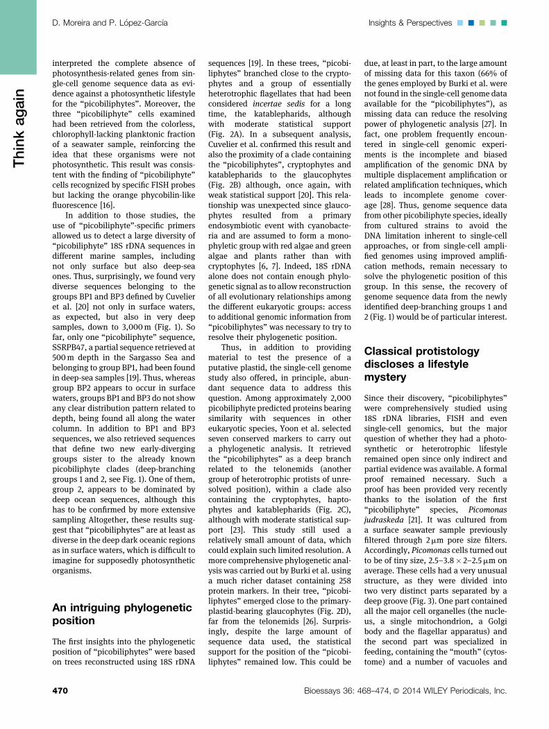
Figure 1. Phylogenetic analysis of “pycobiliphyte” 18S rDNA sequences. Environmental sequences retrieved from the epipelagic regionare shown in light blue; deep-sea sequences are highlighted in dark blue. Sequences from our study are in bold (accession numbersKJ173835–KJ173876). 18S rDNA was amplified using the “picobiliphyte”-specific primer pico1F (50-CGTTTATTTGATGATCTCTTG) andgeneral eukaryotic reverse primers from a collection of marine plankton DNA from the South Atlantic/Southern Ocean and the Marmara Seaused in previous studies following the same technical procedures [33, 34]. The name of the only isolated species, Picomonas judraskeda, isindicated by a black background. Groups BP1, BP2, and BP3 are named according to Cuvelier et al. [20]. The tree was reconstructed byBayesian analysis using 1,057 positions. The scale bar indicates the estimated number of nucleotide substitutions per site.
..... Insights & Perspectives D. Moreira and P. L�opez-Garcıa
471Bioessays 36: 468–474,� 2014 WILEY Periodicals, Inc.
Thinkagain

Figure 2. Schematic phylogenetic trees showing the positions proposed for the “picobiliphytes”by A: Not et al. [19], based on 18S rDNA sequence analyses; B: Cuvelier et al. [20], based on18S rDNA sequence analyses; C: Yoon et al. [23], based on the analysis of seven conservedproteins; and D: Burki et al. [26], based on the analysis of 258 conserved proteins. Circles atbranches indicate the statistical support (bootstrap proportions, BP) found by maximumparsimony [19], maximum likelihood [20, 23], and Bayesian inference [26].
D. Moreira and P. L�opez-Garcıa Insights & Perspectives.....
472 Bioessays 36: 4
Thinkagain
vesicles involved in digestion. Thesetwo parts were separated by a largevacuolar cisterna of unknown function,though it has been speculated that itmay serve as a physical barrier toprotect the cell against infection byviruses ingested by the cell regionspecialized in feeding. In addition tothis unusual structure, Picomonasdisplayed an atypical movement thatwas described as a “jump, drag, andskedaddle” cycle [21]. The combinationof all these morphological and behav-ioral traits is unique to Picomonasjudraskeda.
An important observation in thenicely detailed ultrastructural charac-terization carried out by Seenivasanet al. was to show that plastids wereabsent from Picomonas. In addition toprevious studies that failed to findany typical photosynthetic trait in the“picobiliphytes”, ultrastructure provid-ed the missing proof required to ascer-tain the heterotrophic nature of theseorganisms. Moreover, the food vacuolesfound in Picomonas appeared to containonly particles of less than 150 nm in size,much less than the typical size ofprokaryotic cells, which suggested thatPicomonas probably feeds on smallcolloidal food particles (such as bacte-rial membrane debris) and, perhaps,also on small viruses. This is animportant observation because it rulesout the possibility that “picobiliphytes”may ingest eukaryotic algae to recoverand enslave their plastids (a processcalled kleptoplastidy) or even cyano-bacteria, as both types of cells arelarger than 150 nm in diameter. Thisalso excludes the possibility that thepattern of orange autofluorescenceobserved in a number of unculturedpicobiliphyte cells is due to the presenceof ingested photosynthetic cells, ashad been advanced [19], and rathersuggests some kind of fixation orstaining artifact [21].
The morphological characteristicsof Picomonas are unique among theknown eukaryotic species and do notshow any specific affinity to those foundin groups such as cryptophytes, kata-blepharids, or glaucophytes, whichappear to be related to “picobiliphytes”in phylogenetic analyses, albeit alwayswith weak support (Fig. 2). The unique-ness of the cell structure and the lackof close phylogenetic relatives justify
68–474,� 2014 WILEY Periodicals, Inc.

Figure 3. Scanning electron microscopy image of a cell of Picomonas judraskeda. Anoptical microscopy image is shown in the inset. The difference in cell size between the twoimages is due to the chemical fixation prior to optical microscopy, which slightly increasesthe actual cell size. Photographs courtesy of R. Seenivasan and M. Melkonian.
..... Insights & Perspectives D. Moreira and P. L�opez-GarcıaThinkagain
the inauguration of a new phylum,which Seenivasan et al. proposed tocall “Picozoa” in order to highlightthe heterotrophic nutrition mode of theseorganisms, in clear contrast with thesemantics of the previously proposedname “Picobiliphyta”. The dismissal ofthe photosynthetic nature of theseorganisms may be perceived as adisappointing result. However, Picozoaconstitute a bona fide new eukaryoticlineage, widespread across very differentoceanic locations and depths. Thismakes them good candidates to play arelevant ecological role in marine eco-systems, may be as specialists in thedegradation of very small particles,including marine colloids which, inci-dentally, are the most abundant par-ticles in the ocean and represent
Bioessays 36: 468–474,� 2014 WILEY Pe
up to 50% of the dissolved organiccarbon [29]. Although quantitative dataare scarce for this group, it has alreadybeen shown that picozoan cells canbe abundant in certain locations.For example, FISH experiments haverevealed that they can reach densitiesbetween 80 and 300 cells/mL andaccount for >1% of all planktonicpicoeukaryotic cells in several surfacemarine locations [19]. Thus, they aremost likely significant actors oforganic matter recycling in oceans. Inaddition, they seem to encompass alarge diversity, which remains to befully explored, and branch in a verydifficult-to-resolve region of the eukary-otic tree, where photosynthetic andheterotrophic lineages appear inter-mixed [26]. Thus, isolating new species
riodicals, Inc.
and sequencing their genomes will becrucial to understand the evolution ofthese eukaryotic lineages and resolvetheir phylogenetic relationships as wellas to understand their ecological role.
Conclusions and outlook
Picozoa represent a novel diverse groupof small marine heterotrophic protistsbranching deeply in the eukaryotic tree.Unfortunately, the Picomonas strainisolated by Seenivasan et al. [21] waslost after a relatively short time ofsuccessful cultivation in the laboratory.Nevertheless, their work demonstratedtwo important things. First, that neweukaryotic lineages can be put intoculture not only by chance but alsotargeting them using FISH, flow cytom-etry sorting or other techniques toenrich the desired group from naturalsamples. Second, that, as could beexpected, having the organisms inculture allows their accurate character-ization, including morphological, ultra-structural, behavioral, genomic, andother aspects. This illustrates that,despite the amazing power of newmolecular techniques, traditional pro-tistology remains fundamental to un-derstand the biology of eukaryoticmicroorganisms. Regrettably, at thesame time as molecular approachesbecame more and more prevalent,classical specialists about many micro-bial eukaryotic groups retired, so thatthe vast body of classical knowledgeon these organisms became highlyendangered and can get lost forever.Of course, this problem is not limitedto protistology but to all branches ofbiology, as traditional whole-organismapproaches are being increasinglydeserted [30–32]. The story of the raiseand fall of the Picobiliphyta, a poten-tially novel unique algal group, thatsuddenly became the Picozoa, a phylumof heterotrophic protists feeding on tinyparticles, is a paradigmatic example ofthe importance of combining molecularand classical tools to fully characterizenew lineages.
AcknowledgementsWe thank Ramkumar Seenivasan andMichael Melkonian (University of Co-logne) for the generous gift of Picomonas
473

D. Moreira and P. L�opez-Garcıa Insights & Perspectives.....Thinkagain
judraskeda pictures, and Giselle Walkerand two anonymous reviewers foruseful comments. The research leadingto these results has received fundingfrom the European Research Councilunder the European Union’s SeventhFramework Programme ERC GrantAgreement 322669 “ProtistWorld”.
References
1. Field CB, Behrenfeld MJ, Randerson JT,Falkowski P. 1998. Primary production of thebiosphere: integrating terrestrial and oceaniccomponents. Science 281: 237–40.
2. Arrigo KR. 2005. Marine microorganisms andglobal nutrient cycles. Nature 437: 349–55.
3. Falkowski PG, Fenchel T, Delong EF. 2008.The microbial engines that drive Earth’sbiogeochemical cycles. Science 320: 1034–9.
4. Jiao N, Zheng Q. 2011. The microbial carbonpump: from genes to ecosystems. ApplEnviron Microbiol 77: 7439–44.
5. Adl SM, Simpson AG, Lane CE, Lukes J,et al. 2013. The revised classification ofeukaryotes. J Eukaryot Microbiol 59: 429–93.
6. Moreira D, Le Guyader H, Philippe H. 2000.The origin of red algae and the evolution ofchloroplasts. Nature 405: 69–72.
7. Rodrıguez-Ezpeleta N, Brinkmann H,Burey SC, Roure B, et al. 2005. Monophylyof primary photosynthetic eukaryotes: greenplants, red algae, and glaucophytes. Curr Biol15: 1325–30.
8. Keeling PJ. 2013. The number, speed, andimpact of plastid endosymbioses in eukaryot-ic evolution. Annu Rev Plant Biol 64: 583–607.
9. DeClerckO,GuiryMD, Leliaert F,Samyn Y,et al. 2013. Algal taxonomy: a road tonowhere? J Phycol 49: 215–25.
10. Moore RB, Obornik M, Janouskovec J,Chrudimsky T, et al. 2008. A photosyntheticalveolate closely related to apicomplexanparasites. Nature 451: 959–63.
474
11. Janouskovec J, Horak A, Obornik M,Lukes J, et al. 2010. A common red algalorigin of the apicomplexan, dinoflagellate,and heterokont plastids. Proc Natl Acad SciUSA 107: 10949–54.
12. Obornik M, Vancova M, Lai DH, Janous-kovec J, et al. 2011. Morphology andultrastructure of multiple life cycle stages ofthe photosynthetic relative of apicomplexa,Chromera velia. Protist 162: 115–30.
13. ObornikM,Modry D, LukesM,Cernotikova-Stribrna E, et al. 2012. Morphology, ultrastruc-ture and life cycle of Vitrella brassicaformis n.sp., n. gen., a novel chromerid from the GreatBarrier Reef. Protist 163: 306–23.
14. Woehle C, Dagan T, Martin WF, Gould SB.2011. Red and problematic green phylogeneticsignals among thousands of nuclear genes fromthe photosynthetic and apicomplexa-relatedChromera velia. Genome Biol Evol 3: 1220–30.
15. Burki F, Flegontov P, Obornik M, Cihlar J,et al. 2012. Re-evaluating the green versusred signal in eukaryotes with secondaryplastid of red algal origin. Genome Biol Evol4: 626–35.
16. Kim E, Harrison JW, Sudek S, Jones MD,et al. 2011. Newly identified and diverseplastid-bearing branch on the eukaryotic treeof life.Proc Natl Acad Sci USA 108: 1496–500.
17. Oheocha C. 1965. Biliproteins of algae. AnnuR Plant Physiol 16: 415–34.
18. Cavalier-Smith T. 2002. Nucleomorphs:enslaved algal nuclei. Curr Opin Microbiol 5:612–9.
19. Not F, Valentin K, Romari K, Lovejoy C,et al. 2007. Picobiliphytes: a marine pico-planktonic algal group with unknown affinitiesto other eukaryotes. Science 315: 253–5.
20. Cuvelier ML, Ortiz A, Kim E, Moehlig H, et al.2008.Widespreaddistribution of a uniquemarineprotistan lineage. Environ Microbiol 10: 1621–34.
21. Seenivasan R, Sausen N, Medlin LK,Melkonian M. 2013. Picomonas judraskedagen. et sp. nov.: the first identified member ofthe Picozoa phylum nov., a widespread groupof picoeukaryotes, formerly known as ‘pico-biliphytes’. PLoS One 8: 26.
22. Stepanauskas R, Sieracki ME. 2007.Matching phylogeny and metabolism in the
Bioessays 36: 4
uncultured marine bacteria, one cell at a time.Proc Natl Acad Sci USA 104: 9052–7.
23. YoonHS,Price DC, Stepanauskas R,RajahVD, et al. 2011. Single-cell genomics revealsorganismal interactions in uncultivated ma-rine protists. Science 332: 714–7.
24. Timmis JN, Ayliffe MA, Huang CY, MartinW. 2004. Endosymbiotic gene transfer: or-ganelle genomes forge eukaryotic chromo-somes. Nat Rev Genet 5: 123–35.
25. Reyes-Prieto A, Hackett JD, Soares MB,Bonaldo MF, et al. 2006. Cyanobacterialcontribution to algal nuclear genomes isprimarily limited to plastid functions. CurrBiol 16: 2320–5.
26. Burki F, Okamoto N, Pombert JF, KeelingPJ. 2012. The evolutionary history of hapto-phytes and cryptophytes: phylogenomic evi-dence for separate origins. Proc Biol Sci 279:2246–54.
27. Roure B, Baurain D, Philippe H. 2013.Impact of missing data on phylogeniesinferred from empirical phylogenomic datasets. Mol Biol Evol 30: 197–214.
28. Worden AZ, Dupont C, Allen AE. 2011.Genomes of uncultured eukaryotes: sortingFACS from fiction. Genome Biol 12: 2011–2.
29. Wells M. 1998. Marine colloids: a neglecteddimension. Nature 391: 530–1.
30. Samyn Y, Massin C. 2002. Taxonomists’requiem? Science 295: 276–7.
31. Wagele H, Klussmann-Kolb A, KuhlmannM,Haszprunar G, et al. 2011. The taxonomist– an endangered race. A practical proposalfor its survival. Front Zool 8: 25.
32. Heger TJ, Edgcomb VP, Kim E, Lukes J,et al. 2014. A resurgence in field research isessential to better understand the diversity,ecology, and evolution of microbial eukar-yotes. J Eukaryot Microbiol, in press, doi:10.1111/jeu.12095
33. L�opez-Garcıa P, Rodrıguez-Valera F,Pedr�os-Ali�o C, Moreira D. 2001. Unexpect-ed diversity of small eukaryotes in deep-seaAntarctic plankton. Nature 409: 603–7.
34. Lara E, Moreira D, Vereshchaka A, Lopez-Garcia P. 2009. Pan-oceanic distribution ofnew highly diverse clades of deep-seadiplonemids. Environ Microbiol 11: 47–55.
68–474,� 2014 WILEY Periodicals, Inc.