Embed Size (px)
Citation preview
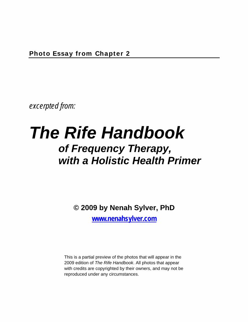
Photo Essay from Chapter 2
excerpted from: The Rife Handbook of Frequency Therapy, with a Holistic Health Primer
© 2009 by Nenah Sylver, PhD www.nenahsylver.com
This is a partial preview of the photos that will appear in the 2009 edition of The Rife Handbook. All photos that appear with credits are copyrighted by their owners, and may not be reproduced under any circumstances.
Rife’s first high-powered microscope, built in 1920.
Courtesy of Rife Research Group of Canada
Rife’s Microscope No. 5. This was his last model.
Rife’s Microscope No. 4, intended for commercial production.
Courtesy of Rife Research Group of Canada
The Rife Handbook – Chapter 2 photo essay excerpt © 2009 by Nenah Sylver, PhD
1
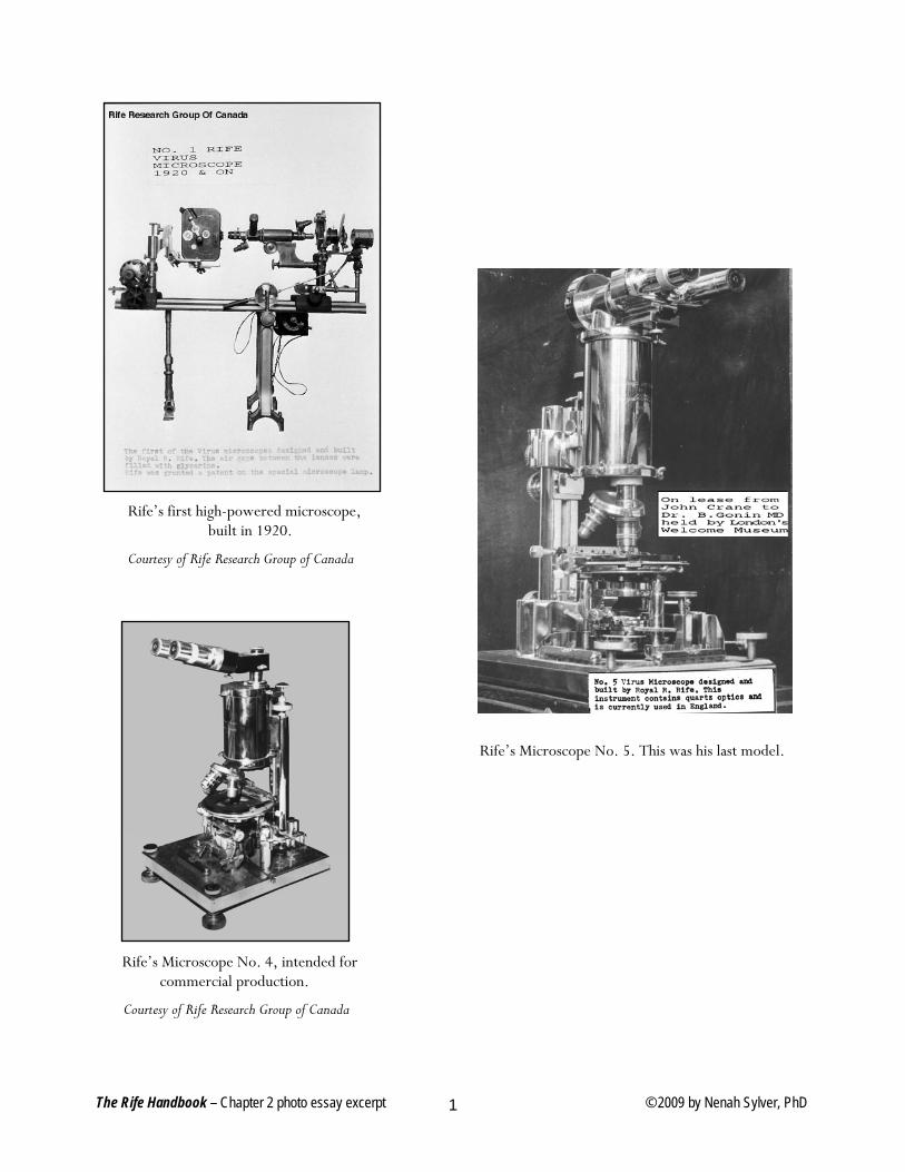
Rife Universal Microscope No. 3. Inscription on top reads, “Designed and built by Royal R. Rife, 1933.”
Plaque at base reads, “Property of Rife Research Lab.”
The Rife Handbook – Chapter 2 photo essay excerpt © 2009 by Nenah Sylver, PhD
2
The Rife Handbook – Chapter 2 photo essay excerpt © 2009 by Nenah Sylver, PhD
3
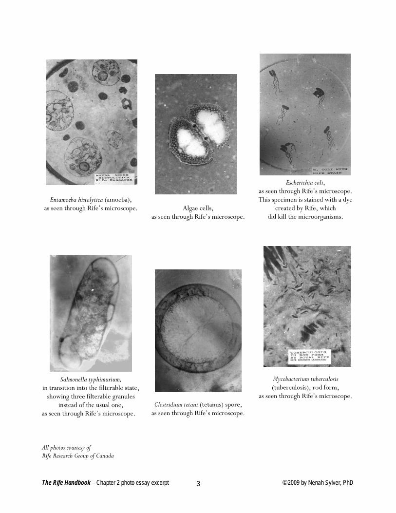
Entamoeba histolytica (amoeba), as seen through Rife’s microscope.
Salmonella typhimurium,
in transition into the filterable state, showing three filterable granules
instead of the usual one, as seen through Rife’s microscope. All photos courtesy of Rife Research Group of Canada
Algae cells, as seen through Rife’s microscope.
Clostridium tetani (tetanus) spore, as seen through Rife’s microscope.
Escherichia coli, as seen through Rife’s microscope. This specimen is stained with a dye
created by Rife, which did kill the microorganisms.
Mycobacterium tuberculosis (tuberculosis), rod form,
as seen through Rife’s microscope.
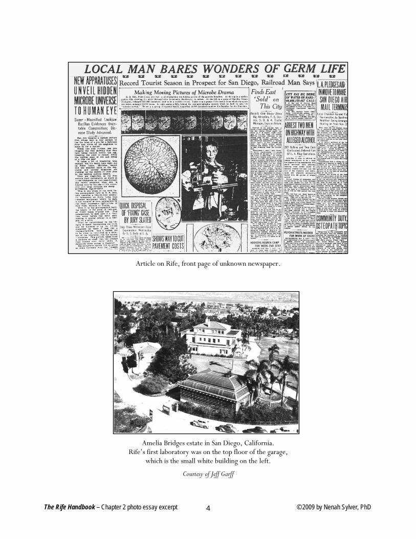
Article on Rife, front page of unknown newspaper.
Amelia Bridges estate in San Diego, California. Rife’s first laboratory was on the top floor of the garage,
which is the small white building on the left.
Courtesy of Jeff Garff
The Rife Handbook – Chapter 2 photo essay excerpt © 2009 by Nenah Sylver, PhD
4
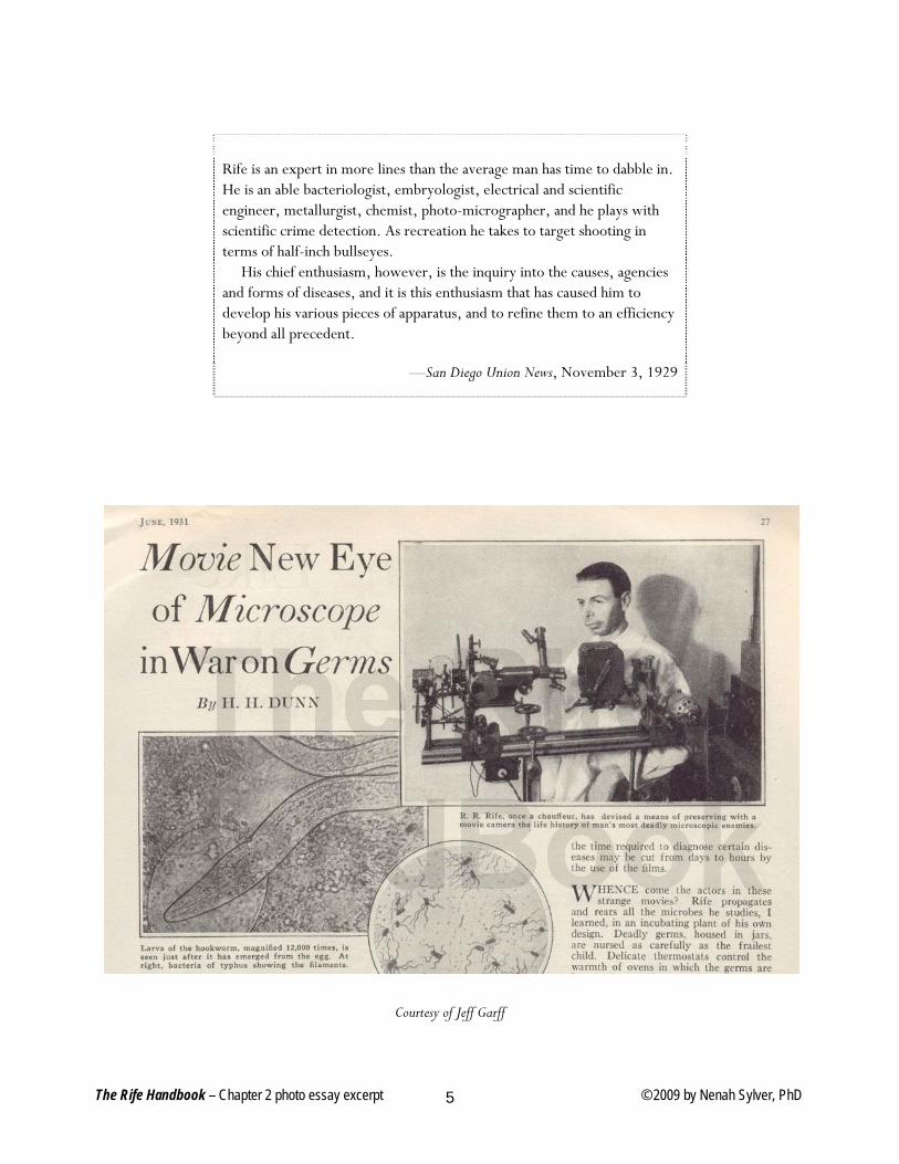
Rife is an expert in more lines than the average man has time to dabble in. He is an able bacteriologist, embryologist, electrical and scientific engineer, metallurgist, chemist, photo-micrographer, and he plays with scientific crime detection. As recreation he takes to target shooting in terms of half-inch bullseyes. His chief enthusiasm, however, is the inquiry into the causes, agencies and forms of diseases, and it is this enthusiasm that has caused him to develop his various pieces of apparatus, and to refine them to an efficiency beyond all precedent.
—San Diego Union News, November 3, 1929
Courtesy of Jeff Garff
The Rife Handbook – Chapter 2 photo essay excerpt © 2009 by Nenah Sylver, PhD 5
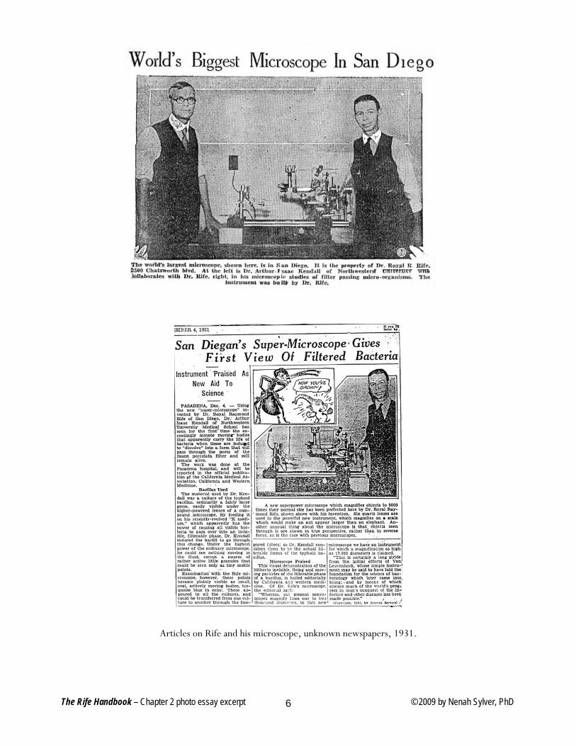
Articles on Rife and his microscope, unknown newspapers, 1931.
The Rife Handbook – Chapter 2 photo essay excerpt © 2009 by Nenah Sylver, PhD 6
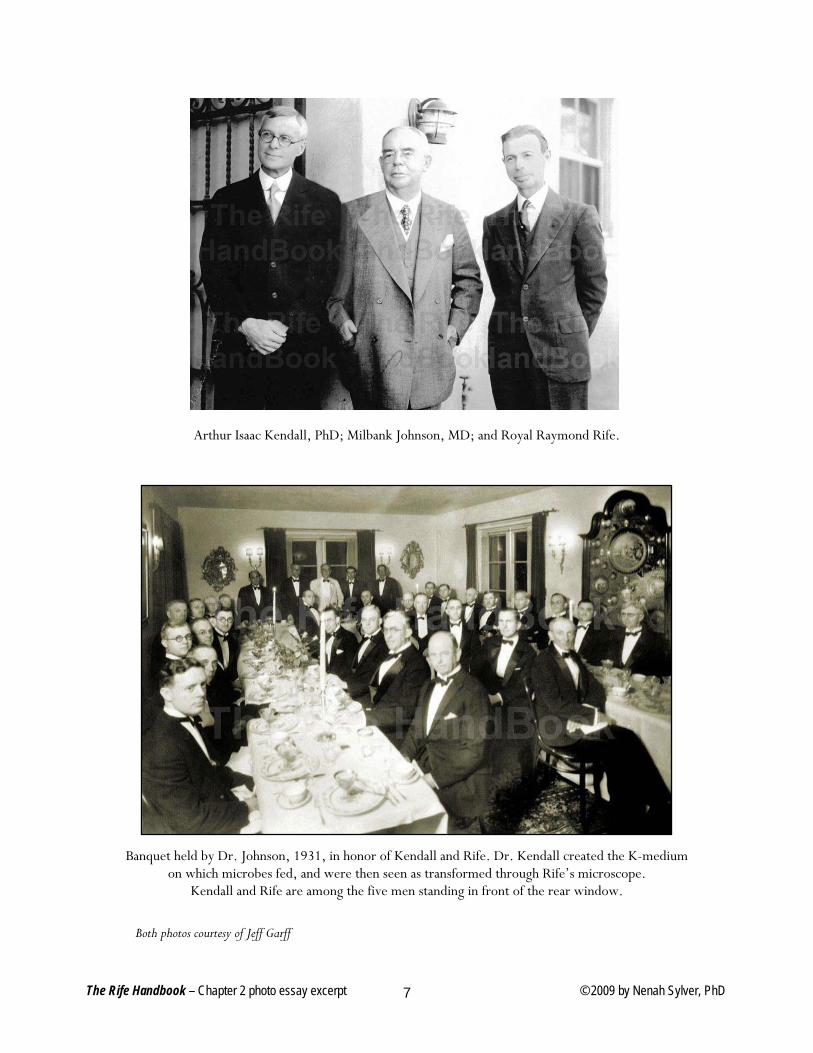
Arthur Isaac Kendall, PhD; Milbank Johnson, MD; and Royal Raymond Rife.
Banquet held by Dr. Johnson, 1931, in honor of Kendall and Rife. Dr. Kendall created the K-medium on which microbes fed, and were then seen as transformed through Rife’s microscope.
Kendall and Rife are among the five men standing in front of the rear window.
Both photos courtesy of Jeff Garff
The Rife Handbook – Chapter 2 photo essay excerpt © 2009 by Nenah Sylver, PhD 7
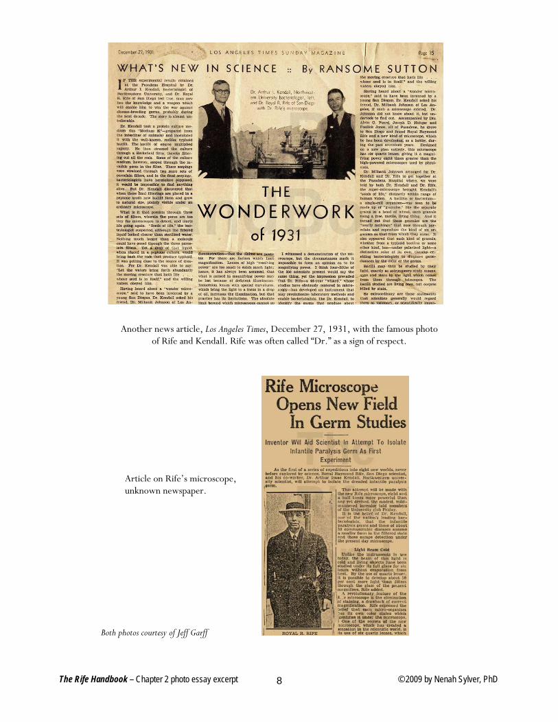
Another news article, Los Angeles Times, December 27, 1931, with the famous photo of Rife and Kendall. Rife was often called “Dr.” as a sign of respect.
Article on Rife’s microscope, unknown newspaper.
Both photos courtesy of Jeff Garff
The Rife Handbook – Chapter 2 photo essay excerpt © 2009 by Nenah Sylver, PhD 8
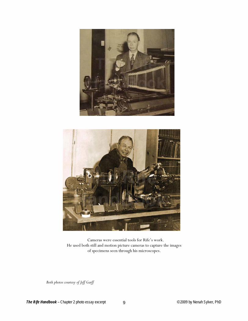
Cameras were essential tools for Rife’s work.
He used both still and motion picture cameras to capture the images of specimens seen through his microscopes.
Both photos courtesy of Jeff Garff
The Rife Handbook – Chapter 2 photo essay excerpt © 2009 by Nenah Sylver, PhD 9
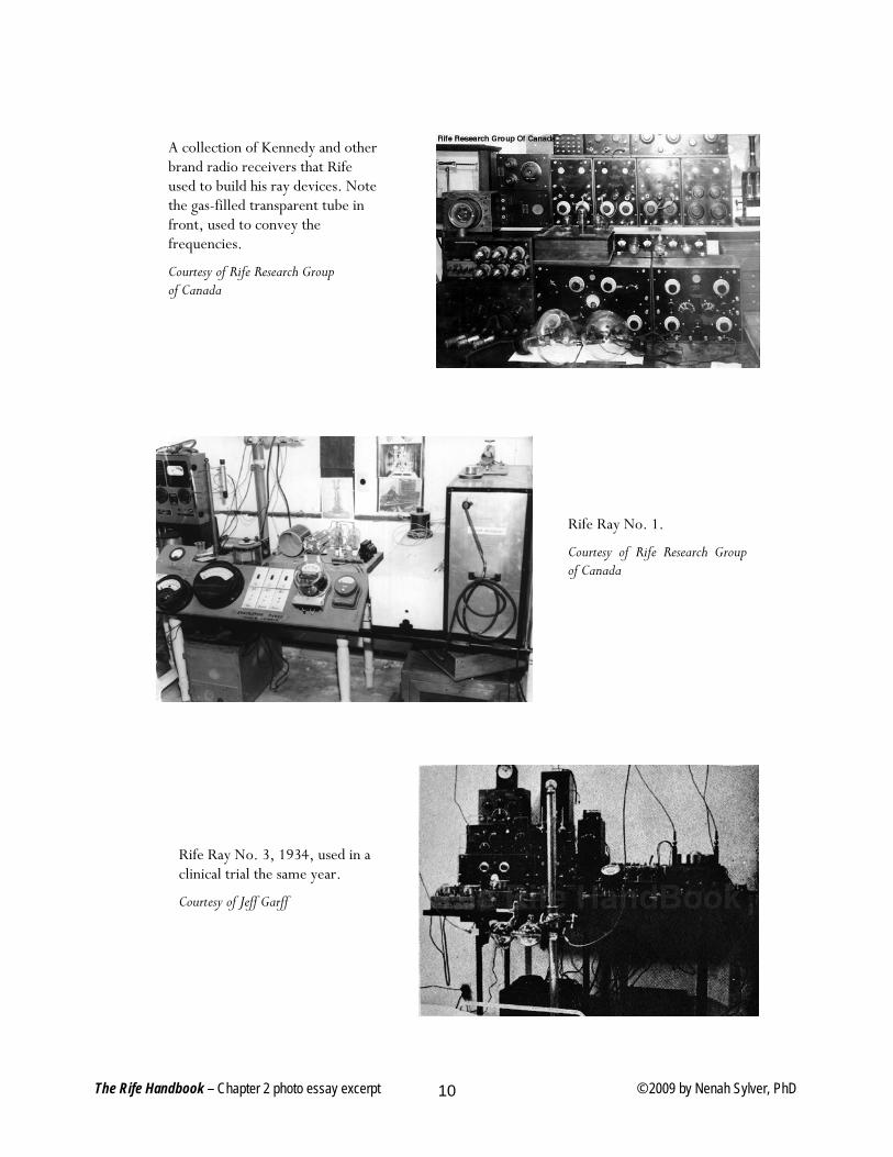
A collection of Kennedy and other brand radio receivers that Rife used to build his ray devices. Note the gas-filled transparent tube in front, used to convey the frequencies.
Courtesy of Rife Research Group of Canada
Rife Ray No. 1.
Courtesy of Rife Research Group of Canada
Rife Ray No. 3, 1934, used in a clinical trial the same year.
Courtesy of Jeff Garff
The Rife Handbook – Chapter 2 photo essay excerpt © 2009 by Nenah Sylver, PhD 10
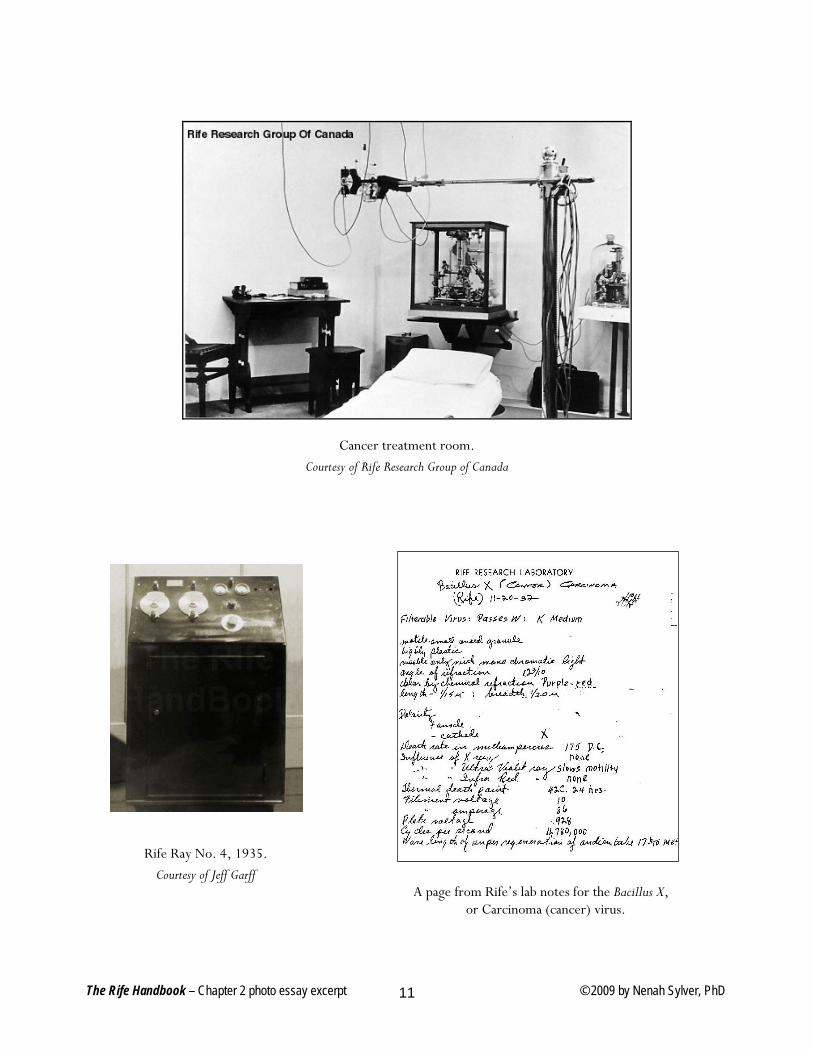
Cancer treatment room. Courtesy of Rife Research Group of Canada
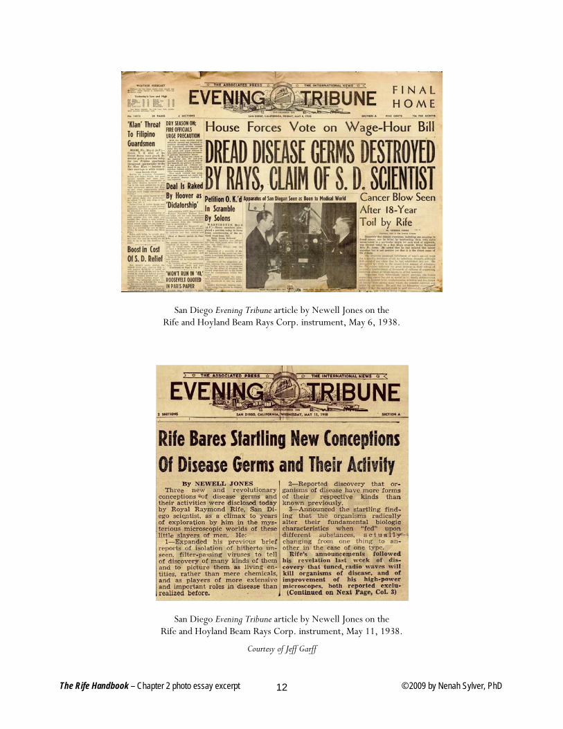
Rife Ray No. 4, 1935. Courtesy of Jeff Garff
A page from Rife’s lab notes for the Bacillus X, or Carcinoma (cancer) virus.
The Rife Handbook – Chapter 2 photo essay excerpt © 2009 by Nenah Sylver, PhD 11
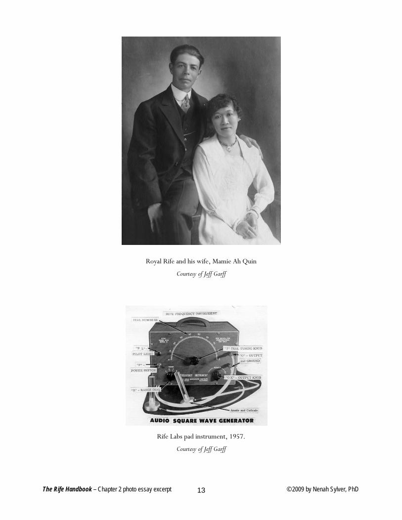
San Diego Evening Tribune article by Newell Jones on the Rife and Hoyland Beam Rays Corp. instrument, May 6, 1938.
San Diego Evening Tribune article by Newell Jones on the Rife and Hoyland Beam Rays Corp. instrument, May 11, 1938.
Courtesy of Jeff Garff
The Rife Handbook – Chapter 2 photo essay excerpt © 2009 by Nenah Sylver, PhD 12
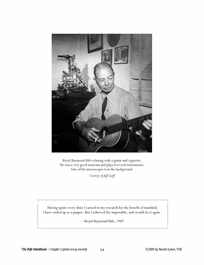
Royal Rife and his wife, Mamie Ah Quin
Courtesy of Jeff Garff
Rife Labs pad instrument, 1957.
Courtesy of Jeff Garff
The Rife Handbook – Chapter 2 photo essay excerpt © 2009 by Nenah Sylver, PhD 13
Royal Raymond Rife relaxing with a guitar and cigarette. He was a very good musician and played several instruments.
One of his microscopes is in the background.
Courtesy of Jeff Garff
Having spent every dime I earned in my research for the benefit of mankind, I have ended up as a pauper. But I achieved the impossible, and would do it again.
—Royal Raymond Rife, 1967
The Rife Handbook – Chapter 2 photo essay excerpt © 2009 by Nenah Sylver, PhD 14