Embed Size (px)
Citation preview
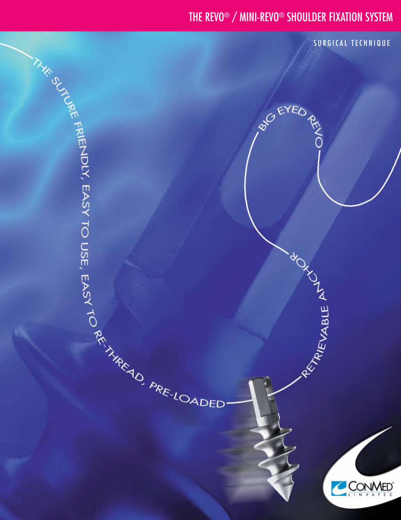
the revo® / mini-revo® shoulder fixation system
s u r g i c a l t e c h n i q u e
revo
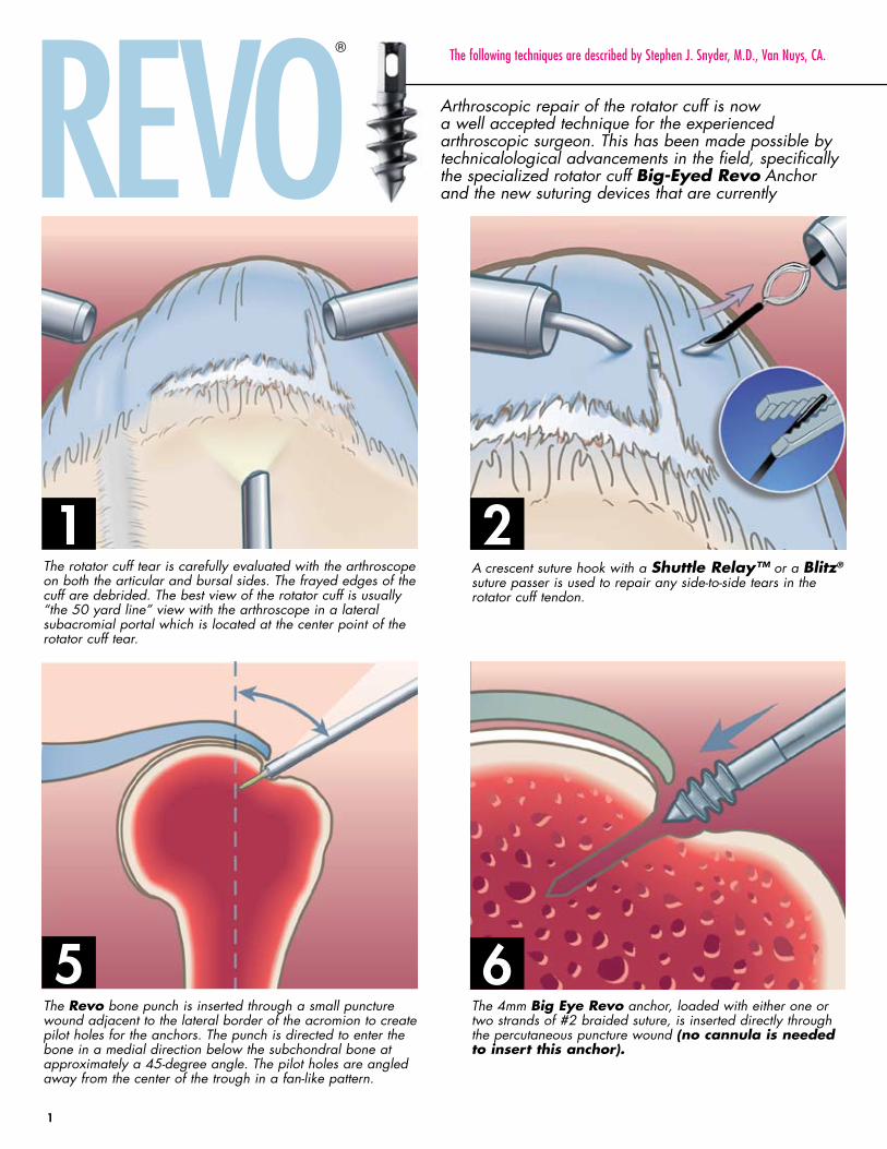
The rotator cuff tear is carefully evaluated with the arthroscope on both the articular and bursal sides. The frayed edges of the cuff are debrided. The best view of the rotator cuff is usually “the 50 yard line” view with the arthroscope in a lateral subacromial portal which is located at the center point of the rotator cuff tear.
The Revo bone punch is inserted through a small puncture wound adjacent to the lateral border of the acromion to create pilot holes for the anchors. The punch is directed to enter the bone in a medial direction below the subchondral bone at approximately a 45-degree angle. The pilot holes are angled away from the center of the trough in a fan-like pattern.
A crescent suture hook with a Shuttle Relay™ or a Blitz® suture passer is used to repair any side-to-side tears in the rotator cuff tendon.
The 4mm Big Eye Revo anchor, loaded with either one or two strands of #2 braided suture, is inserted directly through the percutaneous puncture wound (no cannula is needed to insert this anchor).
Arthroscopic repair of the rotator cuff is now a well accepted technique for the experienced arthroscopic surgeon. This has been made possible by technicalological advancements in the field, specifically the specialized rotator cuff Big-Eyed Revo Anchor and the new suturing devices that are currently
® the following techniques are described by stephen J. snyder, m.d., van nuys, ca.
1
1
2
5 6
rotator cuff repair surgical techniquesteps in repair technique
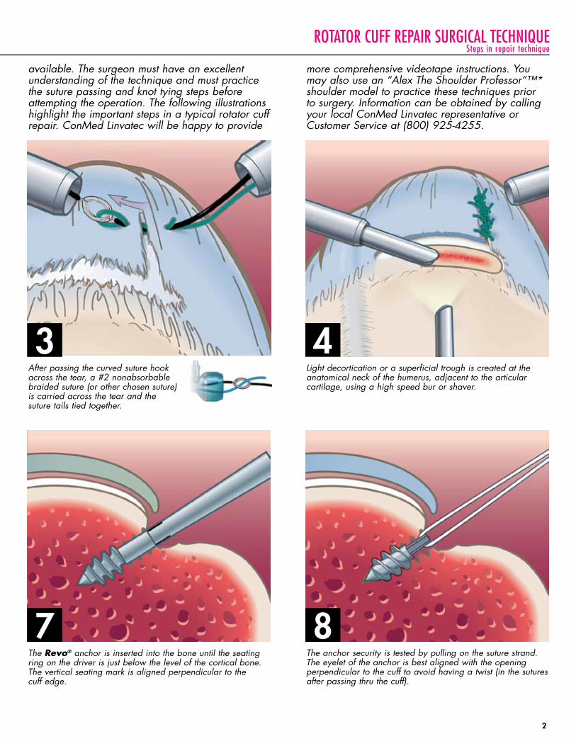
Light decortication or a superficial trough is created at the anatomical neck of the humerus, adjacent to the articular cartilage, using a high speed bur or shaver.
The Revo® anchor is inserted into the bone until the seating ring on the driver is just below the level of the cortical bone. The vertical seating mark is aligned perpendicular to the cuff edge.
After passing the curved suture hook across the tear, a #2 nonabsorbable braided suture (or other chosen suture) is carried across the tear and the suture tails tied together.
The anchor security is tested by pulling on the suture strand. The eyelet of the anchor is best aligned with the opening perpendicular to the cuff to avoid having a twist (in the sutures after passing thru the cuff).
available. The surgeon must have an excellent understanding of the technique and must practice the suture passing and knot tying steps before attempting the operation. The following illustrations highlight the important steps in a typical rotator cuff repair. ConMed Linvatec will be happy to provide
more comprehensive videotape instructions. You may also use an “Alex The Shoulder Professor”™* shoulder model to practice these techniques prior to surgery. Information can be obtained by calling your local ConMed Linvatec representative or Customer Service at (800) 925-4255.
2
3 4
7 8
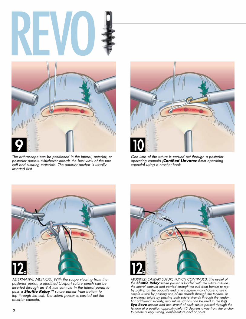
ModIfIed CASPArI SuTure PunCh ConTInued: The eyelet of the Shuttle Relay suture passer is loaded with the suture outside the lateral cannula and carried through the cuff from bottom to top by pulling on the opposite end. The surgeon may choose to use a simple suture by passing one of the strands through the tendon, or a mattress suture by passing both suture strands through the tendon. for additional security, two suture strands can be used in the Big Eye Revo anchor and one strand of each suture passed through the tendon at a position approximately 45 degrees away from the anchor to create a very strong, double-suture anchor point.
revo
The arthroscope can be positioned in the lateral, anterior, or posterior portals, whichever affords the best view of the torn cuff and suturing materials. The anterior anchor is usually inserted first.
one limb of the suture is carried out through a posterior operating cannula (ConMed Linvatec 6mm operating cannula) using a crochet hook.
ALTernATIVe MeThod: With the scope viewing from the posterior portal, a modified Caspari suture punch can be inserted through an 8.4 mm cannula in the lateral portal to pass a Shuttle Relay™ suture passer from bottom to top through the cuff. The suture passer is carried out the anterior cannula.
12
9 10
12 ba
3
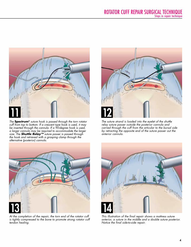
The Spectrum® suture hook is passed through the torn rotator cuff from top to bottom. If a crescent type hook is used, it may be inserted through the cannula. If a 90-degree hook is used, a larger cannula may be required to accommodate the larger size. The Shuttle Relay™ suture passer is passed through the hook and retrieved with a grasping clamp through the alternative (posterior) cannula.
At the completion of the repair, the torn end of the rotator cuff is tightly compressed to the bone to promote strong rotator cuff tendon healing.
This illustration of the final repair shows a mattress suture anterior, a suture in the middle and a double suture posterior. notice the final side-to-side repair.
The suture strand is loaded into the eyelet of the shuttle relay suture passer outside the posterior cannula and carried through the cuff from the articular to the bursal side by retracting the opposite end of the suture passer out the anterior cannula.
11 12
13 14
rotator cuff repair surgical techniquesteps in repair technique
4
mini-revo®
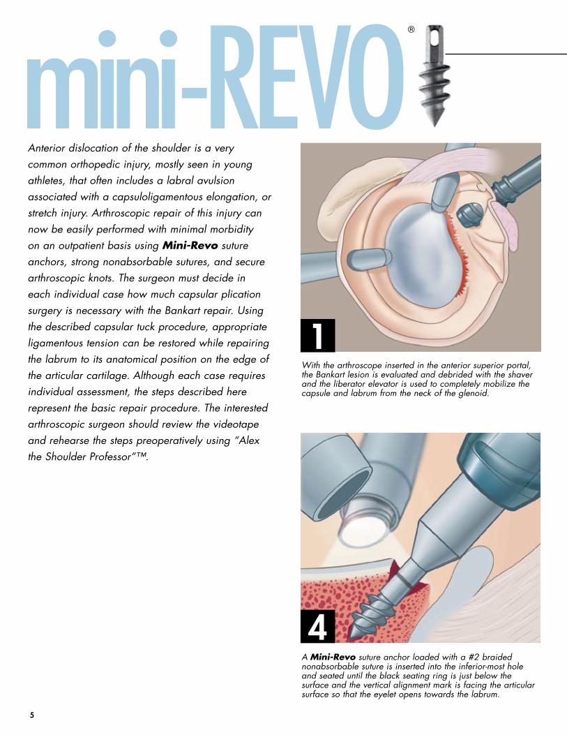
With the arthroscope inserted in the anterior superior portal, the Bankart lesion is evaluated and debrided with the shaver and the liberator elevator is used to completely mobilize the capsule and labrum from the neck of the glenoid.
A Mini-Revo suture anchor loaded with a #2 braided nonabsorbable suture is inserted into the inferior-most hole and seated until the black seating ring is just below the surface and the vertical alignment mark is facing the articular surface so that the eyelet opens towards the labrum.
Anterior dislocation of the shoulder is a very common orthopedic injury, mostly seen in young athletes, that often includes a labral avulsion associated with a capsuloligamentous elongation, or stretch injury. Arthroscopic repair of this injury can now be easily performed with minimal morbidity on an outpatient basis using Mini-Revo suture anchors, strong nonabsorbable sutures, and secure arthroscopic knots. The surgeon must decide in each individual case how much capsular plication surgery is necessary with the Bankart repair. using the described capsular tuck procedure, appropriate ligamentous tension can be restored while repairing the labrum to its anatomical position on the edge of the articular cartilage. Although each case requires individual assessment, the steps described here represent the basic repair procedure. The interested arthroscopic surgeon should review the videotape and rehearse the steps preoperatively using “Alex the Shoulder Professor”™.
1
4
5
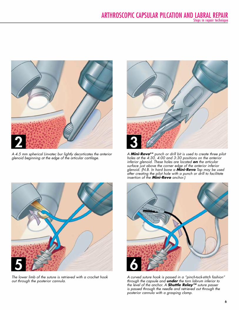
A curved suture hook is passed in a “pinch-tuck-stitch fashion” through the capsule and under the torn labrum inferior to the level of the anchor. A Shuttle Relay™ suture passer is passed through the needle and retrieved out through the posterior cannula with a grasping clamp.
arthroscopic capsular pilcation and labral repair
A 4.5 mm spherical Linvatec bur lightly decorticates the anterior glenoid beginning at the edge of the articular cartilage.
A Mini-Revo®* punch or drill bit is used to create three pilot holes at the 4:30, 4:00 and 3:30 positions on the anterior inferior glenoid. These holes are located on the articular surface just above the corner edge of the anterior inferior glenoid. (n.B. In hard bone a Mini-Revo Tap may be used after creating the pilot hole with a punch or drill to facilitate insertion of the Mini-Revo anchor.)
The lower limb of the suture is retrieved with a crochet hook out through the posterior cannula.
steps in repair technique
2 3
5 6
6
using the blitZ suture retriever
®
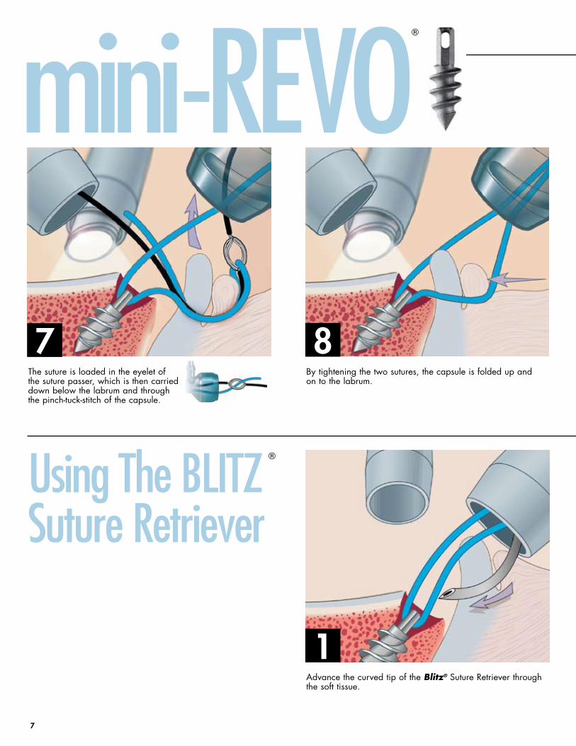
The suture is loaded in the eyelet of the suture passer, which is then carried down below the labrum and through the pinch-tuck-stitch of the capsule.
By tightening the two sutures, the capsule is folded up and on to the labrum.
Advance the curved tip of the Blitz® Suture Retriever through the soft tissue.
mini-revo
7 8
1
7
®
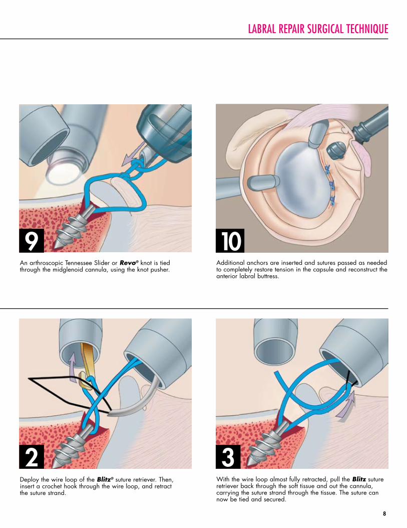
An arthroscopic Tennessee Slider or Revo® knot is tied through the midglenoid cannula, using the knot pusher.
Additional anchors are inserted and sutures passed as needed to completely restore tension in the capsule and reconstruct the anterior labral buttress.
Deploy the wire loop of the Blitz® suture retriever. Then, insert a crochet hook through the wire loop, and retract the suture strand.
With the wire loop almost fully retracted, pull the Blitz suture retriever back through the soft tissue and out the cannula, carrying the suture strand through the tissue. The suture can now be tied and secured.
labral repair surgical technique
9
2 3
10
8
revo Knot
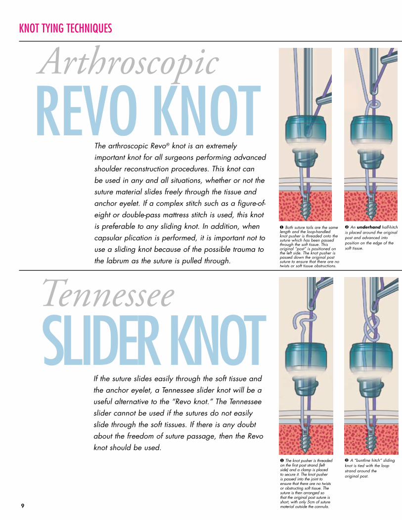
slider Knot❶ The knot pusher is threaded on the first post strand (left side) and a clamp is placed to secure it. The knot pusher is passed into the joint to ensure that there are no twists or obstructing soft tissue. The suture is then arranged so that the original post suture is short, with only 5cm of suture material outside the cannula.
❷ A “buntline hitch” sliding knot is tied with the loop strand around the original post.
Knot tying techniques
Arthroscopic
Tennessee
If the suture slides easily through the soft tissue and the anchor eyelet, a Tennessee slider knot will be a useful alternative to the “revo knot.” The Tennessee slider cannot be used if the sutures do not easily slide through the soft tissues. If there is any doubt about the freedom of suture passage, then the revo knot should be used.
❶ Both suture tails are the same length and the loop-handled knot pusher is threaded onto the suture which has been passed through the soft tissue. This original “post” is positioned on the left side. The knot pusher is passed down the original post suture to ensure that there are no twists or soft tissue obstructions.
The arthroscopic revo® knot is an extremely important knot for all surgeons performing advanced shoulder reconstruction procedures. This knot can be used in any and all situations, whether or not the suture material slides freely through the tissue and anchor eyelet. If a complex stitch such as a figure-of-eight or double-pass mattress stitch is used, this knot is preferable to any sliding knot. In addition, when capsular plication is performed, it is important not to use a sliding knot because of the possible trauma to the labrum as the suture is pulled through.
❷ An underhand half-hitch is placed around the original post and advanced into position on the edge of the soft tissue.
9
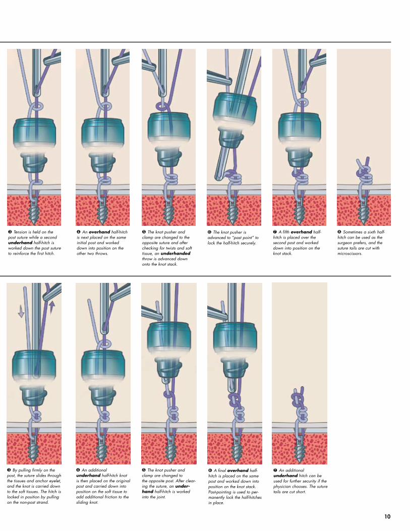
❸ By pulling firmly on the post, the suture slides through the tissues and anchor eyelet, and the knot is carried down to the soft tissues. The hitch is locked in position by pulling on the non-post strand.
❹ An additional underhand half-hitch knot is then placed on the original post and carried down into position on the soft tissue to add additional friction to the sliding knot.
❺ The knot pusher and clamp are changed to the opposite post. After clear-ing the suture, an under-hand half-hitch is worked into the joint.
❻ A final overhand half-hitch is placed on the same post and worked down into position on the knot stack. Past-pointing is used to per-manently lock the half-hitches in place.
❼ An additional underhand hitch can be used for further security if the physician chooses. The suture tails are cut short.
❸ Tension is held on the post suture while a second underhand half-hitch is worked down the post suture to reinforce the first hitch.
❹ An overhand half-hitch is next placed on the same initial post and worked down into position on the other two throws.
❺ The knot pusher and clamp are changed to the opposite suture and after checking for twists and soft tissue, an underhanded throw is advanced down onto the knot stack.
❻ The knot pusher is advanced to “past point” to lock the half-hitch securely.
❼ A fifth overhand half-hitch is placed over the second post and worked down into position on the knot stack.
❽ Sometimes a sixth half-hitch can be used as the surgeon prefers, and the suture tails are cut with microscissors.
10
© 2000, 2003, 2005 Linvatec Corporation, a subsidiary of ConMed Corporation, 10/05, CST 3013 Rev. 4
ImplantsRevo Suture Anchor, 4mm O.D. (1.8mm I.D.) x 12mm C6101A (pre-loaded with #2 braided, polyester suture)
Mini-Revo Suture Anchor, 2.7mm O.D. (1.5mm I.D.) C6109A x 8.5mm (pre-loaded with #2 braided, polyester suture)
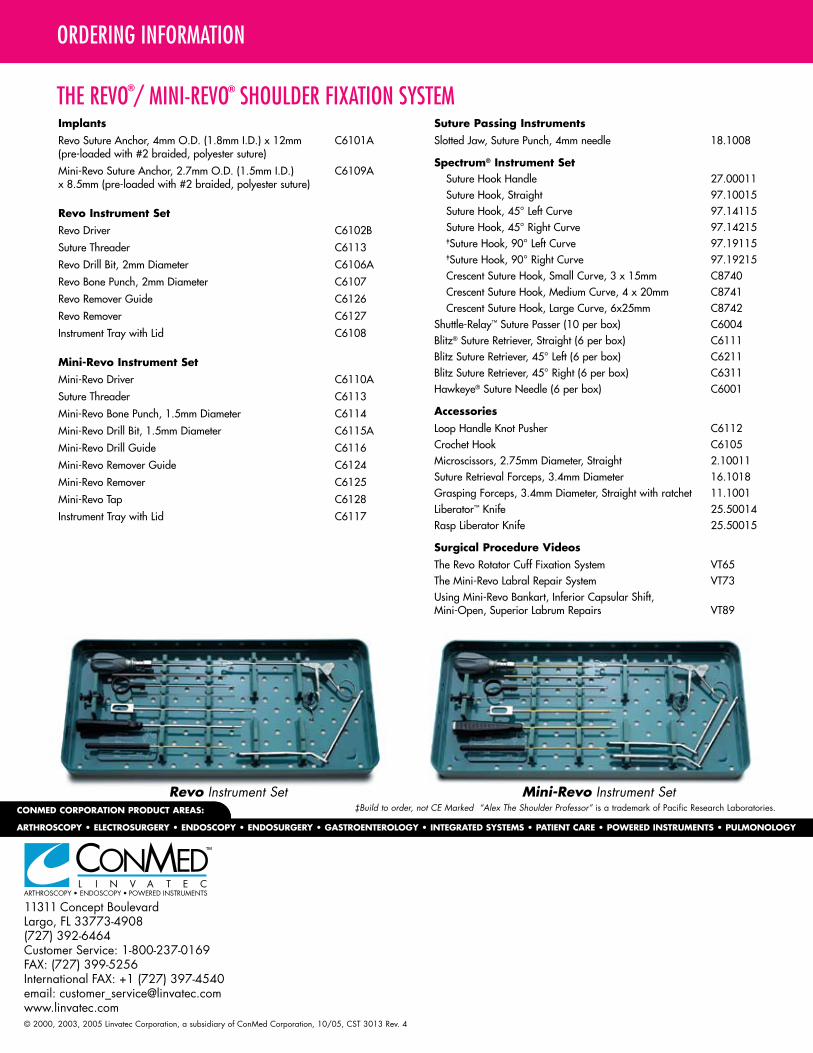
Revo Instrument SetRevo Driver C6102B
Suture Threader C6113
Revo Drill Bit, 2mm Diameter C6106A
Revo Bone Punch, 2mm Diameter C6107
Revo Remover Guide C6126
Revo Remover C6127
Instrument Tray with Lid C6108
Mini-Revo Instrument SetMini-Revo Driver C6110A
Suture Threader C6113
Mini-Revo Bone Punch, 1.5mm Diameter C6114
Mini-Revo Drill Bit, 1.5mm Diameter C6115A
Mini-Revo Drill Guide C6116
Mini-Revo Remover Guide C6124
Mini-Revo Remover C6125
Mini-Revo Tap C6128
Instrument Tray with Lid C6117
Suture Passing InstrumentsSlotted Jaw, Suture Punch, 4mm needle 18.1008
Spectrum® Instrument Set Suture Hook Handle 27.00011 Suture Hook, Straight 97.10015 Suture Hook, 45° Left Curve 97.14115 Suture Hook, 45° Right Curve 97.14215 ‡Suture Hook, 90° Left Curve 97.19115 ‡Suture Hook, 90° Right Curve 97.19215 Crescent Suture Hook, Small Curve, 3 x 15mm C8740 Crescent Suture Hook, Medium Curve, 4 x 20mm C8741 Crescent Suture Hook, Large Curve, 6x25mm C8742Shuttle-Relay™ Suture Passer (10 per box) C6004Blitz® Suture Retriever, Straight (6 per box) C6111Blitz Suture Retriever, 45° Left (6 per box) C6211Blitz Suture Retriever, 45° Right (6 per box) C6311Hawkeye® Suture Needle (6 per box) C6001
AccessoriesLoop Handle Knot Pusher C6112Crochet Hook C6105Microscissors, 2.75mm Diameter, Straight 2.10011Suture Retrieval Forceps, 3.4mm Diameter 16.1018Grasping Forceps, 3.4mm Diameter, Straight with ratchet 11.1001Liberator™ Knife 25.50014Rasp Liberator Knife 25.50015
Surgical Procedure VideosThe Revo Rotator Cuff Fixation System VT65The Mini-Revo Labral Repair System VT73Using Mini-Revo Bankart, Inferior Capsular Shift, Mini-Open, Superior Labrum Repairs VT89
Mini-Revo Instrument SetRevo Instrument Set
ordering information
the revo / mini-revo shoulder fixation system® ®
‡Build to order, not Ce Marked “Alex The Shoulder Professor” is a trademark of Pacific Research Laboratories.