Embed Size (px)
Citation preview

Volumen VIII No.2 Junio 2009
GIANT RESERVOIR CYST FOLLOWING MOLTENO IMPLANT IN STURGE-WEBER SYNDROMECamila Cassiano Simões MD; Sérgio Henrique Sampaio Meirelles MD PhD; Ana Carolina de Arantes Frota MDPhD
THE RETURN OF LAMELLAR KERATOPLASTYW. Barry Lee MD; Mark J. Mannis MD
CURRENT MANAGEMENT OF ANOPHTHALMIC ENOPHTHALMOSChun Cheng Lin Yang MD MSc; M. Reza Vagefi MD; Richard L. Anderson MD FACS; John McCann MD PhD
LEBER’S HEREDITARY OPTIC NEUROPATHY: PROJECT BRAZIL/LHON - 8 YEAR SUMMARYCarlos F. Chicani MD PhD; Valerio Carelli MD PhD; Solange R. Salomao PhD; Peter A. Quiros MD; Adriana Berezovsky PhD; Eric Sutter PhD; Jerome Sherman OD; Dora F. Ventura PhD; Piero Barboni MD; Carolina Ramos MD PhD; Federico Sadun MD; Anna Maria De Negri MD; Milton Moraes MD; Milton Moraes Filho MD; Celina Tamaki PhD; Josenilson M. Pereira MSc; Paula Y. Sacai COMT; Fred N. Ross-Cisneros; Rubens Belfort Jr. MD PhD; Alfredo A. Sadun MD PhD
DETERMINACIÓN DE LA HISTÉRESIS CORNEAL PRE Y POST ANILLOS INTRACORNEALESMiguel Angel Quiroz González MD; Raúl Suárez MD; Luisa Maria Loustaunau Godoy MD; Arturo Gómez MD; Alberto Haber MD; Gabriela Ortiz MD; Alejandro Navas MD; Abel Martinez MD; Jerónimo Álvarez MD; Cristian Tinoco MD
OPTIC DISC CAVERNOUS HEMANGIOMA: CASE REPORTDalila Coelho MD; Isabel Pires MD; Maria L. Cachulo MD; João Figueira MD; Rufino Silva MD PhD; José R. Faria Abreu MD PhD; José Cunha-Vaz MD PhD

Febrero 2009
PAN-AMERICA : B
Junio 2009
PAN-AMERICA
Mark J. Mannis, MDUniversity of California, Davis
Sacramento, CaliforniaEditor-in-Chief
Cristian Luco, MDSantiago, ChileAssociate Editor
Teresa J. BradshawArlington, TexasManaging Editor
Terri L. GrassiArlington, TexasProduction Editor
EDITORIAL BOARD
Eduardo Alfonso, MDMiami, Florida USA
Eduardo Arenas, MDBogotá, Colombia
J. Fernando Arévalo, MDCaracas, Venezuela
José A. Roca Fernández, MDLima, Perú
Denise de Freitas, MDSão Paulo, Brazil
Marian Macsai, MDChicago, Illinois USA
David E. Pelayes, MD PhDBuenos Aires, Argentina
Alfredo Sadun, MDLos Angeles, California USA
José Benítez del Castillo Sánchez, MDMadrid, Spain
Allan Slomovic, MDToronto, Ontario, Canada
Luciene Barbosa de Sousa, MDSão Paulo, Brazil
Lihteh Wu, MDSan José, Costa Rica
Paulo Dantas, MDSão Paulo, Brazil
Chun Cheng Lin Yang, MD MScSan José, Costa Rica
OFFICERS
Richard L. Abbott, MDSan Francisco, USA
President, Pan-American Association of Ophthalmology
Rubens Belfort, MDSão Paulo, Brazil
Chairman of the Board,Pan-American Ophthalmological Foundation
PRODUCTION STAFF
Javier JiménezGraphic Design
Eliana BarbosaDirector of Production and Distribution
PAOF INDUSTRY SPONSORS
• Advanced Medical Optics Inc.• Alcon Inc.• Allergan Inc.• Bausch & Lomb Inc.• Carl Zeiss Meditec Inc.
• Johnson & Johnson Vision Care Latin America• Merck & Co Inc.• Novartis International AG.• Santen Inc.
Prepress Creative Latin Media. Printed in Printer Colombiana - Colombia
CREATIVE LATIN MEDIA, LLC.P.M.B 117
2901 Clint Moore, Boca Raton, FL 33496Tel.: (561) 495 4728Fax: (561) 865 1934
E-mail: [email protected]@clatinmedia.com
Special thanks to Ana Carolina Vieira, Enrique Graue Hernández, Citlali Gurrusquieta, Mapy Padilla, and Cristián Luco for assistance in translation to Spanish and Portuguese.

PAN-AMERICA162 : PAN-AMERICA
Richard L. Abbott MDPresidente PAAO PAAO President
MENSAJE DEL PRESIDENTE MESSAGE FROM THE PRESIDENT
Pan American Research Day at ARVO…..It has come a long way!!
This year in Ft Lauderdale, Florida, the Pan-American Research Day was outstanding! Although this program has previously taken place for many years at ARVO, this year was particu-larly special. Under the outstanding leadership of Drs Peter Quiros, J. Fernando Arevalo, Lihteh Wu, Paulo Dantas, and Rubens Belfort Jr., the concept of dedicating a place and time to share new and innovative vision research among our colleagues and to provide an opportunity for our younger in-vestigators to have a forum to present their work has significantly expanded. The PAAO Research Day program took place at the Ft Lauderdale Re-nassaince Hotel on May 2, 2009 and, thanks to generous support from Allergan, the entire event was free of charge. In addition, this year, becau-se of the overwhelming interest, there were two meeting rooms used; one for anterior segment papers and posters and one for the posterior seg-ment.
Adding prestige and interest to the program sessions were invited keynote speakers who pre-sented cutting edge research work from both North America and Latin America. Among the speakers from North America were Michael R. Robinson MD
(Senior Medical Director, Ophthalmology Clinical Research, Allergan), and Peter McDonnell MD (De-partment Chair at the Wilmer Eye Institute). From Latin America, Dr. Hugo Quiroz-Mercado (Mexico & Denver Health, Colorado) and Dr. Miguel Burnier (Mc Gill University, Montreal & UNIFESP, Sao Paulo) addressed the meeting.
A second opportunity to participate in the Pan-American Research Day encouraged participants to display their abstracts similar to a poster! The abs-tracts were discussed by subspecialty groups, and individuals had a unique opportunity to present their work as well as invite colleagues to visit their poster at the ARVO meeting later in the week.
For 2009, the PAAO offered more travel awards and scholarships than ever before. A total of 16 tra-vel awards in the amount of $1,500 were presented this year to deserving young investigators.
Finally, we encourage all who participated in the PAAO ARVO Research Day to submit their work for peer review in our outstanding publication -- Vision Pan-America. This is a great opportunity to support our own Journal and to share your work with your colleagues across the Americas.
PAN-AMERICAPAN-AMERICA : 163PAN-AMERICA
Junio 2009
Día Panamericano de la Investigación en ARVO….ha sido un largo camino!!
Este año en Fort Lauderdale, Florida, el Día Panamericano de la Investigación fue extraordinario! Aunque este programa se ha realizado por muchos años en el ARVO, este año fue particularmente especial. Bajo el excelente liderazgo de los doctores Peter Quiros, Fernando Arévalo, Lihteh Wu, Paulo Dantas y Rubens Belfort Jr., se ha expandido significativamente el concepto de dedicar un lugar y momento para compartir con los colegas lo nuevo e innovador en investiga-ción de la visión y brindar una oportunidad a nuestros jóvenes investigadores para que cuenten con un foro para presentar sus tra-bajos. El programa del Día Panamericano de la Investigación tuvo lugar en el Hotel Renaissance de Fort Lauderdale el día 2 de Mayo del 2009 y gracias al generoso apo-yo de Allergan, todo el evento fue gratuito. Además, este año, dado el gran interés, se
utilizaron dos salas de reuniones; una para posters y trabajos libres en segmento ante-rior y una para segmento posterior.
Se invitaron connotados expositores, quienes por su prestigio añadieron interés a las sesiones del programa, presentando trabajos de investigación de última genera-ción tanto de Norte América como de Lati-no América. Entre los expositores de Norte América estaban el Dr. Michael R. Robinson (Director Médico de Investigación Clínica en Oftalmología de Allergan) y el Dr. Peter McDonnell (Jefe de Departamento en el Wilmer Eye Institute). Por Latino América, el Dr. Hugo Quiroz-Mercado (Mexico y Denver Health, Colorado) y el Dr. Miguel Burnier (Universidad de McGill, Montreal y UNIFESP, Sao Paulo).
Una segunda oportunidad de participar en el Día Panamericano de la Investigación
motivó a los participantes a exhibir su re-sumen similar a un poster! Los resúmenes fueron discutidos por grupos de subespe-cialidad y tuvieron una gran oportunidad de presentar su trabajo así como de invitar a sus colegas a visitar su poster en la reunión de ARVO durante la semana.
Para el 2009, la Panamericana ofreció más premios de viajes y becas que nunca antes. Se otorgaron un total de 16 premios de viajes por US$1,500.00 a jóvenes inves-tigadores que lo merecían.
Finalmente, motivamos a todos los que participaron en el Día Panamericano de la In-vestigación en ARVO a enviarnos su trabajo para revisión por expertos en nuestra destacada publicación -- Vision Pan-America. Esta es una gran oportunidad para apoyar nuestra propia revista y compartir su trabajo con los colegas a través de las Américas.
Dia Panamericano da Pesquisa na ARVO.... Uma longa trajetória!
Este ano em Fort Lauderdale, Flórida, o Dia Panamericano da Pesquisa foi excelente! Apesar deste programa já acontecer há mui-tos anos na ARVO, este ano foi particularmen-te especial. Sob a fantástica liderança dos Doutores Peter Quiros, J. Fernando Arevalo, Lihteh Wu, Paulo Dantas e Rubens Belfort Jr, a idéia de dedicar um determinado local e tempo especialmente para compartilhar pes-quisas inovadoras na área da oftalmologia e dar a jovens pesquisadores a oportunidade de terem um forum para apresentação de seus trabalhos expandiu enormemente. O Dia da Pesquisa da PAAO aconteceu no Fortt Lauderdale Renaissance Hotel no dia 2 de Maio de 2009 e, graças ao generoso apoio da Allergan, o evento foi inteiramente gra-tuito. Além disso, este ano, devido à enorme procura, duas salas foram utilizadas: uma para trabalhos e pôsteres sobre o segmento
anterior e outra para o segmento posterior.Para acrescentar ainda mais prestígio e
interesse às sessões do programa, pales-trantes dignos de nota foram convidados a apresentar projetos de pesquisa de ponta realizados nas Américas do Norte e Latina. Entre os convidados da América do Norte estavam Michael R. Robinson MD, (Diretor Medico de Pesquisa Clinica em Oftalmolo-gia, Allergan); e Peter McDonnell MD, (Che-fe do Departamento de Oftalmologia, Wilmer Eye Institute). Da América Latina, Dr Hugo Queiroz-Mercado (México & Denver Health, Colorado) e Dr Miguel Burnier (McGill Uni-versity, Montreal & UNIFESP, São Paulo) de-ram palestras.
Uma outra maneira incentivada para participar-se do Dia Panamericano da Pes-quisa foi através da exposição dos resumos dos trabalhos em formato semelhante a um
poster. Esses resumos foram analisados por grupos das subespecialidades e os partici-pantes tiveram a oportunidade única não só de apresentar seus trabalhos como também de convidar colegas para visitarem seus pôs-teres no Encontro da ARVO posteriormente durante a semana.
2009 foi o ano em que a PAAO mais ofereceu bolsas e auxílios-viagem. Um total de 16 auxílios-viagem no valor de US$1500 foram oferecidos esse ano a jovens investi-gadores que os mereceram.
Finalmente, incentivamos todos aque-les que participaram do Dia da Pesquisa da PAAO ARVO a submeterem seus trabalhos para nossa excelente publicação - Vision Pan America. Trata-se de uma ótima opor-tunidade de apoiar nossa própria revista e de compartilhar seu trabalho com colegas pelas Américas.

PAN-AMERICAPAN-AMERICA164 :
REVIEW
PAN-AMERICA
W. Barry Lee MD1, Mark J. Mannis MD2
1 Eye Consultants of Atlanta & Piedmont Hospital, Cornea, External Disease & Refractive Surgery Service, Atlanta, Georgia2 Department of Ophthalmology & Vision Science, Cornea, External Disease & Refactive Surgery Service, University of California, Davis Eye Center, Sacramento, California
The Return of Lamellar Keratoplasty
ResumenLa queratoplastía lamelar ha renacido
en el nuevo milenio tras una era en la que la queratoplastía penetrante se convirtió en el estándar de oro para el tratamiento de patología corneal. Realizamos una revisión histórica queratoplastía y discutimos varias técnicas de queratoplastía lamelar anterior profunda (de sus siglas en ingles: DALK).
AbstractLamellar keratoplasty has seen a resur-
gence in the new millennium from an era where penetrating keratoplasty has become the gold standard for surgical treatment for corneal disorders. We provide a historical review of keratoplasty and discuss various techniques of deep anterior lamellar kerato-plasty (DALK).
IntroductionA review of the history of keratoplasty
is essential to understand the significance of lamellar keratoplasty’s rebirth. Although Reisinger coined the term keratoplasty in 1824, it was not until 1878 that Sellerbeck performed the first human cornea trans-plant, followed by the first human lamel-lar keratoplasty by deWecker in 1879.1 A decade later in 1888, Von Hippel reported the first successful human lamellar kerato-plasty.1 Lamellar techniques continued to evolve in the early to mid-20th century with Barraquer, Filatov, Paufique, and others.2-6 Lamellar keratoplasty eventually lost fa-vor due to interface issues such as haze, scarring, epithelial ingrowth, and subse-quent decreased post-operative vision.7 These complications led to the theory of deep stromal lamellar dissection, yet as dissections deepened, surgical time and intra-operative risks such as perforation dramatically increased.8-13 Despite these novel techniques of deep lamellar dissec-tion such as “air lamellar keratoplasty”,14,15
it was not until the new millennium that multiple reports of DALK techniques found reproducible success with improved vision outcomes in the setting of limited scar-ring, haze, and lower intra-operative and post-operative complications.16-29
IndicationsDeep anterior lamellar keratoplasty
(DALK) is indicated for disorders of the an-terior cornea in the setting of a healthy pos-terior cornea (endothelium and Descemet’s membrane). The goal of the procedure is to remove the anterior corneal pathology in its entirety with replacement of healthy donor corneal epithelium and stroma. Indications for DALK include corneal scars from healed infectious keratitis or superficial trauma, an-terior corneal dystrophies, stromal corneal dystrophies, and ectatic disorders such as keratoconus and pellucid marginal degen-eration.
DALK ConsiderationsDisadvantages of DALK include in-
terface scarring/interface irregularities, a learning curve, donor/recipient mismatch, and the persistent risk of astigmatism and suture-related complications as with pen-etrating surgery. De-epithelialization of donor corneal epithelium may lead to epi-thelial ingrowth, diffuse lamellar keratitis, and/or trophic ulcers, thus epithelial sta-tus is critical as with penetrating surgery.30
Interface opacities and scarring occur less often with Descemet’s baring techniques but in techniques such as Anwar’s Big-bubble, the big bubble is not always pro-ducible leading to a difficult layer-by-layer dissection. A double anterior chamber can also result from intra-operative microp-erforation with Descemet’s baring tech-niques. Long-term effects to the corneal endothelium remain unknown with deep tissue dissection using instruments or air, femtosecond laser energy, and potential
endothelial-iris touch during Descemet’s detachment following air injection.
The steep learning curve for the Big-bubble technique deserves special men-tion. The procedure is technically difficult and carries a significant intra-operative perforation risk with potential need for conversion to penetrating keratoplasty. The operating time is also significantly increased when first learning this tech-nique so surgical scheduling may need adjustments. As the learning curve pro-gresses, surgical time and perforation risk will decrease accordingly for most surgeons. Fortunately, it is not difficult to convert to penetrating procedures in cases where intra-operative perforation occurs. If the perforation is small and peripheral, stromal hydration with BSS may prevent conversion.16-18 Avoidance of a double anterior chamber post-operatively is im-perative in cases of microperforation and can usually be circumvented by placing a 40-50% anterior chamber air bubble for tamponade after completion of the case.
The major DALK advantages include the preservation of the host’s corneal endothelium with elimination of endothe-lial immune rejection and reduced vision-threatening intra-operative complications such as endophthalmitis and expulsive hemorrhage. The technique also affords utilization of tissue with poor endothe-lium, thus increasing the donor pool. Additionally, automated DALK actually decreases surgical time with creation of a much smoother interface compared to hand dissection techniques of the past, thus interface scarring and haze are sig-nificantly decreased. The Big-bubble tech-nique reduces the risk of interface scarring even more as it restores similar corneal anatomy as with penetrating grafts with complete elimination of the stroma-stroma interface created with automated DALK. Most importantly, visual acuity results are improved as interface issues decline with
Address Correspondence: W. Barry Lee, MDPiedmont Hospital, 95 Collier Road, Suite 3000, Atlanta, GA 30309Tel: 404-351-2220 Extension 1216; Fax: 404-591-2936 (FAX)Email: [email protected]
The authors have no financial interest in the material presented.
PAN-AMERICA : 165PAN-AMERICA
Junio 2009
PAN-AMERICA
DALK. Finally, topical corticosteroid side-effects of cataracts and glaucoma are reduced as patients can taper medications more rapidly in DALK.
Surgical techniquesAutomated DALK
Microkeratome-assisted or femtosecond laser-as-sisted DALK utilizes either a microkeratome or a fem-tosecond laser as with LASIK along with an artificial anterior chamber for donor tissue preparation. Initially the donor cornea is prepared with creation of an anterior donor corneal free cap on an artificial anterior chamber (Figure 1). A similar microkeratome or femtosecond la-ser pass is made on the host cornea after preparation of the donor. It is imperative to know the depth of the corneal pathology with this technique so pathological stromal disease is not left in the host bed. Anterior seg-ment imaging devices are useful pre-operatively to de-termine depth of disease.
Microkeratome blades have a variety of cutting depths depending on the depth desired to remove all pathology. Conversely, the femtosecond laser can be set at the exact depth desired for creation of the host corneal bed (Figure 2). The dimensions of the host corneal cap are typically set at the same thickness and flap diam-eter as the donor tissue or a slightly smaller dimension and larger thickness in ectatic disorders. Donor-recipient mismatch must remain a concern in determining tissue sizing. Once the host cap is created, it is removed and secured to the donor cap with the surgeon’s preferred suture technique.
Anwar’s Big-Bubble Keratoplasty (BBK)
The BBK begins with donor tissue preparation as with penetrating keratoplasty with an 8.25 to 8.75 mm full-thickness trephination using the surgeon’s desired trephine punch system. The host cornea is trephinated at the same diameter using a 300 or 350-micron depth depending on pre-operative pachymetry. Either the Han-na or Hessburg-Barron trephine can be used. A 27/30 gauge needle is attached firmly to an air-filled syringe with tip bent to a 30-40 degree angle 5mm away from the tip edge. The tip is inserted bevel-down deep into the trephination groove and advanced deep into the stroma and parallel to the posterior corneal surface (Figure 3).
Once the tip is well buried into the corneal stro-ma with a 3-4mm advancement from the trephina-tion groove, air is injected with a moderate amount of force. The stroma becomes opaque as the air advances away from the tip (Figure 4). A sudden explosive appearance of a large air bubble appears and injection is stopped when the border of the big bubble reaches the trephination area. A keratec-tomy is performed with a crescent blade removing nearly 50% of stromal tissue anterior to the bubble
Figure 1: A donor cornea has been placed on the artificial anterior chamber from the Moria ALTK® system using balanced salt solution as the chamber media.
Figure 2: A femtosecond laser cut in the corneal stroma using the Intralase. The opaque layer represents the air bubbles formed at the resection plane.
Figure 3: Anwar’s Big-bubble technique: A bent 30 G needle is inserted in the deep corneal stroma starting within the trephination groove with passage parallel to the posterior corneal layers.
Figure 4: Anwar’s Big-bubble technique: A big bubble is shown after forceful injection of air into the corneal stroma.
Figure 5: A crescent blade is used to remove the anterior corneal stroma after creation of the big bubble as described by Anwar.

REVIEW
166 : PAN-AMERICA
(Figure 5), followed by a limbal para-centesis to lower the intraocular pres-sure. A small air bubble can be injected into the anterior chamber at this point as described by Parthasarathy et al.31 If the small bubble remains in the periphery,
Descemet’s membrane has detached ap-propriately.
If the small bubble floats centrally, the big bubble has not been created and Descemet’s membrane is not detached, thus the air injection step should be done
in another location. Once the bubble has been confirmed, a super sharp blade is then inserted into the stroma towards the air bubble and quickly withdrawn so that a perforation does not occur as the air bubble collapses. Blunt dissection with a
Article DissectionTechnique
Eyes (n)
Follow-up(months) Haze Perf
(n1/n)BCVA
(x)Bib-bubble
formation or bare Descemet’s
PK conversion(n)
Sugita et al (1999) Fluid and manual 120 6 - 47/120 20/50 61% 7
Amayem et al (2000) Fluid 26 12 0 2/26 20/30 12% 2
Anwar et al (2002) Air 181 6 - 16/181 ≥20/40(89%)
Shimazaki et al (2002) Air or fluid 13 24 - 2/13 - - 0
Fogla et al (2006) Air 13 5 0 2/13 20/25 69.2% 0
Al-Torbak et al (2006) Air and manual 127 11 7/127 16/127 ≥20/50
(74%) 37% 2
Fontana et al (2007) Air 81 24 2/81 11/81 20/30 64% 3
Vajpayee et al (2007) Air 10 8 0 0 20/25 100% 0
Borderie et al (2008 Air and MK 77 23 23/69* 22 20/40 40% 8
Table 1: Published Results of DALK using techniques that involve baring of Descemet’s Membrane (BCVA = Best-corrected visual acuity; x = average; PK = penetrating keratoplasty; perf = micro or macroperforation)
* This study was initially started with 77 eyes but the 8 perforation eyes, which were converted to PK, were dropped out of the study leaving 69 eyes
Figure 8: Anwar’s big bubble technique: A) A pre-operative photo of a patient with a dense corneal scar from Acanthamoeba keratitis and count fingers vision. B) A post-operative photo at 2 months depicting complete removal of the corneal scar, a clear lamellar graft, and no interface scarring with best-corrected vision of 20/25 following the big-bubble technique.
A B
Figure 6: Depiction of bare Descemet’s membrane after removal of anterior stromal tissue.
Figure 7: Descemet’s membrane is peeled from the donor cornea prior to lamellar transplantation.
REFERENCES
PAN-AMERICA : 167PAN-AMERICA
Junio 2009
1. Mannis MJ, Mannis AA. Corneal Transplantation: A History in Profiles, J.P. Wayenborgh, Belgium, 1999.
2. Filatov VP. Transplantation of the cornea. Arch Ophthalmol 1935;13:321-323.
3. Barraquer JI. Queratoileusis para la correccion de la myopia. Arch Soc Am Oftalmol Optom 1964;5:27-48.
4. Barraquer JI. Lamellar keratoplasty (special technique). Ann Ophthalmol 1972;4(6):437-469.
5. Paufique L, Charleux J. Lamellar keratoplasty. In Casey T, (Ed.): Corneal Grafting. Appleton-Century-Crofts, New York, 1972, pp. 121-176.
6. Polack FM. Lamellar keratoplasty. Malbran’s “peeling off” technique.
Arch Ophthalmol. 1971;86(3):293-5.
7. Richard JM, Paton D, Gassett AR. Comparison of penetrating keratoplasty and lamellar keratoplasty in the surgical management of keratoconus. Am J Ophthalmol 1978;86 (6):807-811.
8. Hallermann W. Vershciendenes uber keratoplastik. Klin Monatshl Augenheilkd 1959;135:252-259.
9. McCulloch C, Thompson GA, Basu PK. Lamellar keratoplasty using full thickness donor material. Trans Am Ophthalmol Soc 1963;61:154-180.
10. Malbran E. Lamellar keratoplasty in keratoconus. In King JH, McTigue JW, (Eds.): The Cornea-World Congress. Butterworths Inc., Washington DC, 1965, pp. 511-518.
11. Malbran E, Stefani C. Lamellar keratoplasty in corneal ectasias. Ophthalmologica. 1972;164(1): 50-58.
12. Malbran E, Stefani C. Lamellar keratoplasty in corneal ectasias. Ophthalmologica. 1972;164(1):59-70.
13. Anwar M. Dissection technique in lamellar keratoplasty. Br J Ophthalmol 1972;56(9):711-713.
14. Archila EA. Deep lamellar keratoplasty dissection of host tissue with intrastromal air injection. Cornea. 1984-1985;3(3):217-8.
15. Price FW. Air lamellar keratoplasty. Refract Corneal Surg. 1989;5(4):240-3.
16. Amayem AF, Anwar M. Fluid lamellar keratoplasty in keratoconus. Ophthalmology. 2000;107(1):76-9.
17. Anwar M, Teichmann KD. Big bubble technique to bare Descemet’s membrane in anterior lamellar keratoplasty. J Cataract Refract Surg. 2002;28(3):398-403.
18. Anwar M, Teichmann KD. Deep lamellar keratoplasty: surgical techniques for anterior lamellar keratoplasty with and without baring of Descemet’s membrane. Cornea 2002;21(4):374-83.
19. Shimazaki J, Shimmura S, Ishioka M, Tsubota K. Randomized clinical trial of deep lamellar kerato-plasty vs penetrating keratoplasty. Am J Ophthalmol 2002;134(2):159-165.
20. Busin M. Zambianchi L, Arffa RC. Microkeratome-assisted lamellar keratoplasty for the surgical treatment of keratoconus. Ophthalmology 2005;112(6):987-997.
21. Mosca L, Fasciani R, Tamburelli C, et al. Femtosecond laser-assisted lamellar keratoplasty: early results. Cornea. 2008;27(6):668-672.
22. Fogla R, Padmanabhan P. Results of deep lamellar keratoplasty using the big-bubble technique in patients with keratoconus. Am J Ophthalmol 2006;141(2):254-259.
23. Hsu HY, Culbertson WW, Alfonso EC. Staged automated lamellar keratoplasty for anterior stromal corneal dystrophies. Ophthalmic Surg Lasers Imaging 2008;39(3):196-202.
24. Vajpayee RB, Tyagi J, Sharma N, et al. Deep anterior lamellar keratoplasty by big-bubble technique for treatment corneal stromal opacities. Am J Ophthalmol 2007;143(6):954-957.
25. Borderie VM, Werthel AL, Touzeau O, et al. Comparison of techniques used for removing the recipi-ent stroma in anterior lamellar keratoplasty. Am J Ophthalmol 2008;126(1):31-37.
26. Fontana L, Parente G, Tassinari G. Clinical outcomes after deep anterior lamellar keratoplasty using the big-bubble technique in patients with keratoconus. Am J Ophthalmol 2007;143(1):117-124.
27. Panda A, Bageshwar LM, Ray M, et al. Deep lamellar keratoplasty versus penetrating keratoplasty for corneal lesions. Cornea 1999;18(2):172-175.
28. Al-Torbak AA, Al-Motowa S, Al-Assiri A, et al. Deep anterior lamellar keratoplasty for keratoconus. Cornea 2006;25(4):408-412.
29. Sugita J, Kondo J. Deep lamellar keratoplasty with complete removal of pathological stroma for vi-sion improvement. Br J Ophthalmol 1997;81(3):184-188.
30. Machado RA, Mannis MJ, Mandel HA, et al. The relationship between first postoperative day epithelial status and eventual health of the ocular surface in penetrating keratoplasty. Cornea 2002;21(6):574-7.
31. Parthasarathy A, Por YM, Tan DT. Using a “small bubble technique” to aid in success in Anwar’s “big bubble technique” of deep lamellar keratoplasty with complete baring of Descemet’s membrane. Br J Ophthalmol 2008;92(3):422.
spatula can then be achieved to create a plane between the stroma and Descemet’s membrane, taking care to avoid perforation. The corneal stroma is removed completely within the trephination circle leaving ex-posed Descemet’s membrane (Figure 6). Descemet’s membrane is peeled from the donor cornea (Figure 7) and secured to the host tissue. (Figure 8A & 8B).
DiscussionDALK results have remained favorable
in a number of published reports using the deep lamellar dissection techniques with excellent visual acuity outcomes and clear lamellar grafts (Table 1). In fact, several comparison studies have shown better vi-sual acuity outcomes, higher endothelial cell density and subsequent graft survival in head-to-head comparisons between DALK and PK patients, with the BBK pro-cedure faring the best in regards to final visual acuity, astigmatism, and interface clarity of the lamellar techniques.19,25,27
Amayem et al16 found an average BCVA of 20/40 in 26 patients undergoing DALK and Anwar et al17 found 89% of 181 pa-tients had BCVA of 20/40 or better with BBK. Busin et al found 88% of 50 patients saw 20/40 or better using microkeratome-assisted DALK.20 Both microkeratome and Big-bubble DALK techniques have proven successful in these various studies, yet the risk of perforation, PK conversion, interface haze, and lack of bubble formation remain realistic risks with the BBK technique as with the risk of donor perforation with the microkeratome technique.
With the advent of mechanical micro-keratomes, femtosecond lasers, and novel lamellar surgical techniques, a new vigor for lamellar keratoplasty has returned and may well overtake penetrating keratoplasty as the most common corneal transplant technique performed in the next several years. Early clinical results with femto-second lasers suggest the future remains bright for lamellar keratoplasty. The con-tinued development of new devices to increase the efficiency, predictability, and ultimate improvement in corneal clarity and vision will likely continue to improve DALK as corneal surgery advances from the days of deWecker and von Hippel into the new millennium.
REVIEW
168 : PAN-AMERICA
Abstracto
Objetivo: Revisar las opciones terapéuticas actuales en el manejo del enoftalmo asociado al socket anoftálmico.
ResumenEl manejo del enoftalmo asociado al socket
anoftálmico has sido siempre un reto para el of-talmólogo. Con el pasar del tiempo, diferentes opciones han emergido para el tratamiento de esta compleja entidad. Actualmente, opciones mínimamente invasivas con buenos resultados están disponibles para mejorar el enoftalmo asociado al socket anoftálmico.
Abstract
Purpose: To review the current therapeutic options to address anophthalmic eno-phthalmos.
Summary: Anophthalmic enophthalmos has always been a challenge for the ophthalmo-logists. Over time, different options have emerged to address this complex entity associated with the anophthalmic socket. Recently, minimally invasive options with great results have become available to help patients with anophthalmic enophthalmos.
Introduction: Treatment of anophthalmic enophthalmos is an ongoing challenge for the ophthalmologist, especially for the oculoplastics and orbital specialist. Ano-phthalmic enophthalmos is attributed to orbital soft-tissue volume loss secondary to atrophy of muscle and fat, causing a downward, posterior, and medial shift of the prosthesis.1 This orbital volume deficit gives a sunken aspect of the eye socket, which is cosmetically unacceptable to the patient and the treating specialist. Current options to improve anophthalmic eno-phthalmos include: alloplastic materials,1,2
hydrogel implants,3-7 and dermal fillers, in-
cluding injectable calcium hydroxylapatite8 and injectable hyaluronic acid.9-10
Alloplastic Materials
Alloplastic materials are artificial im-plants used in the orbit to replace volume loss or bony defects. The advantages of using these materials are its availability, bio-compability, and lack of immunogenic acti-vity. Some alloplastic materials commonly utilized for orbital repair are cyanoacrylate, nylon mesh or sheets (Supramid), solid silicone, curable methymethacrylate (Cra-nioplastic), calcium phosphate derivatives, polymethylmethacrylate (PMMA), polyte-trafluoroethylene (PTEF or Teflon), porous or expanded polytetrafluoroethylene (eP-TFE or Gore-Tex), particulate hydroxyapatite (HA), and porous polyethylene (Medpor).1
Recently, the FDA approved NovaBone-C/M (distributed by Porex Surgical, Inc., Newnan, GA, USA) for orbito-facial use. This new alloplastic implant is a bioactive glass ceramic material (Bioglass) that promotes both intracellular and extracellular bone for-mation.1 Amato et al.’s study shows promi-sing results with NovaBone for volume aug-mentation for enophthalmos, with the only inconveniences of material migration and volume loss over time.
Another positive result with alloplastic ma-terial to correct enophthalmos is the use of Bio-plant (Bioplant Inc., New York, NY, USA) hard tissue replacement (HTR) synthetic bone. Bio-plant combines a polymethylmethcrylate inner core with an outer layer of polyhydroxyethylme-thacrylate coated with a layer of barium sulfate and calcium hydroxide. It presents as granules that hydrate with blood from the surgical site providing bone interface and conjugate in a filler.2 Huang et al. report encouraging results injecting Bioplant hard tissue replacement syn-thetic bone in the subperiosteal space of orbital
floor and lateral and medial wall for orbital vo-lume augmentation.
Injectable Hydrogel PelletsAnother solution to improve volume defi-
cit in enophthalmos is the use of hydrogel pe-llets, self-expanding and hydrophilic osmotic expanders. The use of hydrogel orbital expan-ders started in Europe during the late 1990’s.3
Recently, these have been introduced and marketed in the United States (Osmed GmbH, Illmenau, Germany, distributed in the United States by IOP, Inc., Costa Mesa, CA). The pe-llet expander is made of a highly hydrophilic hydrogel consisting of N-vinyl pyrrolidone and methyl methacrylate. These augment in size by osmotic hydration.3,4 In the dry state the pellet expander is 8 mm in length and 2 mm in diameter with a volume of 0.025 ml. The swelling capacity of the pellet is approxi-mately 10 fold. The pellets can be injected via cutaneous approach using a transcutaneous trocar directed into the intraconal space.3
Li T, McCann JD, et al. reported in the American Society of Ophthalmic Plastics & Reconstructive Surgery (ASOPRS) meeting of 2003 positive experiences utilizing this method for orbital volume augmentation. Schittkowski et al. and Mazzoli et al. from Europe have recently reported gratifying re-sults utilizing the hydrogel expander in the pe-diatric population based on Li and McCann’s initial report in adult population. Given the long-term complications of overswelling of hydrogel scleral buckles seen in retinal sur-gery, the long-term results of hydrogel pellets in volume augmentation of the orbit remains to be seen and should be used with caution.3
Dermal. FillersThe newest concept in addressing ano-
phthalmic enophthalmos is a minimally inva-sive technique to restore orbital volume that
Current Management of Anophthalmic EnophthalmosChun Cheng Lin Yang MD MSc1; M. Reza Vagefi MD2 ; Richard L. Anderson MD FACS3; John McCann MD PhD3
Institutional Affiliations:1 Costa Rica Oculoplastics Inc., Hospital Cima San José, San José, Costa Rica2 Scheie Eye Institute, University of Pennsylvania, Philadelphia, Pennsylvania, United States 3 Center for Facial Appearances, Salt Lake City, Utah, United States
Corresponding Author: Chun Cheng Lin Yang MD MScCosta Rica Oculoplastics Inc., Hospital CIMA San José, Torre 3, Cons. 322, Escazú, Costa RicaTel: (506) 2208-8322Fax: (506) 2208-8372Email: [email protected]
Propriety interest statement:None of the authors have any financial interest on the subject
Financial Support: None
Junio 2009
has proven to be effective in the senior authors’ experience. This innovative technique consists of injecting a FDA approved dermal filler, Ra-diesse® (Bioform Medical, Inc., San Mateo, CA), into the medial, inferior, and lateral ex-traconal orbital space to help restore volume. Radiesse® is made out of 30% hydroxylapatite (HA) microspheres (25-45μm) in a carrying vehicle (1.3% sodium carboxymethylcellulose, 6.4% glycerin and 36.6% sterile water for injec-tion). We obtained encouraging enophthalmos correction of 3 mm for every 1.3 ml of Radies-se® application, lasting in average of one year, with no major complications; however, it is uncertain how frequently patients will require re-injection.8
Hyaluronic Acid is another type of dermal filler used to enhance orbital volume to address sighted and anophthalmic enophthalmos.9-10
The first nonanimal stabilized hyaluronic acid approved by the Food and Drug Administration for soft tissue augmentation was Restylane (Q-Med, Uppsala, Sweden). Nonanimal stabilized hyaluronic acid offers longer lasting effects than bovine collagen and potentially lower risks of immunogenicity and hypersensitivity reactions with the added benefit of being dissolved by
Current Management of Anophthalmic Enophthalmos
REFERENCES1. Amato MM, Blaydon SM, FW Scribbick. Use of Bioglass for orbital volume augmentation in enophthal-mos: a rabbit model (Oryctolagus Cuniculus). Ophthal Plast Reconstr Surg 2003;19:455-4652. Huang ZL, Ma L. Restoration of enophthalmos in anophthalmic socket by HTR polymer. Ophthal Plast Reconstr Surg 2005;21:318-3213. Mazzoli RA, Raymond WR, Ainbinder DJ, Hansen EA. Use of self-expanding, hydrophilic osmotic expanders (hydrogel) in the reconstruction of congenital clinical anophthalmos. Curr Opin Ophthalmol 2004;15:426-4314. Schittkowski MP, Guthoff RF. Injectable self inflating hydrogel pellet expanders for the treatment of orbital volume deficiency in congenital microphthalmos: preliminary results with a therapeutic approach. Br. J. Ophthalmol 2006;90:1173-11775. Li GT, McCann JD, Goldberg RA. Orbital volume augmentation in anophthalmic patients using inject-able hydrogel implant. ASOPRS Abstracts 2003:916. Bacskulin A, Vogel M, Wiese KG, et al. New osmotically active hydrogel expander for enlargement of the contracted anophthalmic socket. Graefes Arch Clin Exp Ophthalmol 2000;238:24-277. Tse DT, Pinchuk L, Davis S, et al. Evaluation of an integrated orbital tissue expander in an anophthalmic feline model. Am J Ophthalmol 2007;143:317-3278. Vagefi MR, McMullen TF, Burroughs JR, McCann JD, Anderson RL. Injectable Hydroxylapatite for Orbital Volume Augmentation. Arch Facial Plast Surg. 2007;9:439-442. 9. Malhotra R. Deep orbital sub-Q Restylane (non-animal stabilized hyaluronic acid) for orbital volume enhancement in sighted and anophthalmic orbits. Arch Ophthalmol 2007;125:1623-1629.10. Tay E, Olver J. Intraorbital hyaluronic acid for enophthalmos. Ophthalmology 2008; 115:1101-1102.e2.
hyaluronidase.9 Malhotra reported corrections of 1 mm of enophthalmus per 1 ml of hyaluro-nic acid injection, with no further improvement beyond 2 mm with 2 ml of injection.
ConclusionIn summary, even though the mana-
gement of anophthalmic enophthalmos is
an ongoing challenge for the ophthalmo-logist, current innovations offer minima-lly invasive alternatives with great results to address this entity; and the available options include: alloplastic implants, hydrogel pellets, and injectable dermal fillers such as hyaluronic acid, and injec-table calcium hydroxylapatite.
AGOSTO 31 A SEPTIEMbRE 6 DE 2009MEDELLíN - COLOMbIA
Asociación Panamericanade Bancos de Ojos - APABO
Pan-American Association of Eye BanksAsociación Pan Americana de Bancos de OjosAssociaçâo Pan Americana de Bancos de Olhos
XI Curso InternaCIonalCERTIFICADO DE
ENTRETENIMIENTO TéCNICO y
CIENTíFICO EN bANCOS DE OJOS
MAyOR INFORMACIóN: [email protected] / Tel. (57-4) 230 22 88
Abstrato:
Objetivo: Resumir achados de um time de pesquisadores voluntários internacionais, cientistas e médicos, do Brasil, Estados Un-idos e Itália, que realizaram oito expedições a uma área rural do Brasil para estudar um pedigree muito grande de NOHL. Métodos: O pedigree mais extenso do mundo de NOHL, uma família com 328 membros, foi descoberto em Colatina, estado do Espírito Santo, Brasil. Todos os indivíduos foram submetidos a extenso questionário e inves-tigação neuro-oftalmologica. Resultados: 2001-3: Os pesquisadores localizaram, or-ganizaram e definiram o pedigree de NOHL que foi iniciado por uma imigrante italiana nascida em 1861. A mutação mitocondrial era 11778, haplogrupo J homoplasmico. Estudos epidemiológicos e analise gené-tica foram realizados. Sofisticados equi-pamentos foram trazidos para realização de testes eletrofisiologicos e psicofísicos, complementares a extenso e completo ex-ame neuro-oftalmologico. 2004-5: Medi-das subclínicas e sorológicas se mostraram úteis no acompanhamento da progressão de portadores de LHON. 2006-8: Achados de OCT (espessura da camada de fibras ner-vosas da retina), complementaram achados psicofísicos e demonstraram ser uma fer-ramenta boa e objetiva na determinação de alterações pré clinicas observada na maio-ria dos portadores de NOHL. Conclusões: Estabelecemos dados de incidência e num pedigree gigante de NOHL 11778. Tabaco e álcool foram considerados fatores de risco. Alterações psicofísicas e de OCt foram freqüentes em portadores assintomáticos de NOHL.
Resumen
Objetivo: Resumir los hallazgos encontra-dos por un grupo internacional de inves-tigadores, científicos y médicos voluntar-ios de Brasil, Estados Unidos e Italia que han realizado ocho viajes anuales para estudiar un pedigrí gigante de LHON en una comunidad rural de Brasil. Método: El pedigrí de LHON más grande del mundo, una familia de 328 miembros localizada en la comunidad de Colatina, en el es-tado de Espirito Santo , Brasil. Todos los sujetos recibieron un cuestionario detal-lado y fueron investigados desde el punto de vista neuro-oftalmológico. Resultados: 2001-3: Los investigadores encontraron, organizaron y definieron el pedigrí de LHON que fue fundado por un inmigrante italiano nacido en 1861. La mutación mi-tocondrial fue 11778, homoplásmica y de haplogrupo-J. Se realizaron estudios epi-demiológicos y de ligamiento genético. Para complementar el examen neuro-of-talmológico integral, equipo sofisticado fue trasladado para realizar los estudios psicofísicos. 2004-5 Se encontro que las mediciones subclínicas y serológicas son útiles en el monitoreo de la progresión en portadores de LHON. 2006-8: Hallazgos de OCT (grosor de CFN) complementaron los estudios psicofísicos y se demostró que éste es el cambio preclínico más objetivo hallado en la mayoría de los portadores. Conclusión: Fuimos capaces de generar información con respecto a la incidencia y penetrancia de un pedigrí gi-gante de LHON 11778. Como factores de riesgo fueron identificados el consumo de tabaco y alcohol . Es común que entre
los portadores asintomáticos se encuen-tren hallazgos psicofísicos y en la OCT.
Abstract:
Purpose: To summarize the findings of an international team of volunteer research-ers, scientists and physicians, from Bra-zil, United States of America and Italy, who have made eight yearly field inves-tigations in rural Brazil to study a giant pedigree of LHON. Methods: The world’s largest pedigree of LHON, a family of 328-members, was found in Colatina, Espirito Santo state, Brazil. All subjects underwent a detailed questionnaire and neuro-ophthalmologic investigation. Results: 2001-3: The researchers found, organized and defined the pedigree of LHON that was founded by an Italian im-migrant born in 1861. The mitochondrial mutation was 11778, homoplasmic, J-haplogroup. Epidemiological studies and gene linkage analysis were performed on this pedigree. Sophisticated equipment was brought for psychophysical and elec-trophysiological testing to complement the comprehensive neuro-ophthalmo-logical examinations. 2004-5: Subclini-cal and serological measurements were found to be useful in following the pro-gression of LHON in carriers. 2006-8: OCT findings (nerve fiber layer (NFL) thickness), complemented psychophys-ics and was demonstrated to be an excel-lent the best objective preclinical diag-nostic tool to determine changing seen in most LHON carriers. Conclusions: We were able to establish incidence and pen-etrance data on a giant pedigree of 11778 LHON. Smoking and drinking alcohol were
CLINICAL SCIENCES
170 : PAN-AMERICA
Leber’s Hereditary Optic Neuropathy: Project Brazil/LHON - 8 year summaryCarlos F Chicani MD PhD1,3, Valerio Carelli MD PhD2, Adriana Berezovsky PhD3, Peter A Quiros MD1, Eric Sutter PhD4 , Jerome Sherman OD5, Dora F. Ventura PhD6, Piero Barboni MD7, Carolina Ramos MD PhD3, Federico Sadun MD8, Anna Maria De Negri MD9, Milton Moraes MD10, Milton Moraes Filho MD3, Celina Tamaki PhD3, Josenilson M. Pereira MSc3, Paula Y Sacai COMT3, Fred N Ross-Cisneros1, Rubens Belfort Jr. MD PhD3, Solange R. Salomao PhD3*, Alfredo A Sadun MD, PhD1*
*Dr. Solange Salomao and Dr. Alfredo Sadun both contributed equally as senior level supervisors for this large scale field investigation and are the co-senior investigator/authors.1. Doheny Eye Institute, Keck - University of Southern California, Los Angeles, CA, USA2. Dipartimento di Scienze Neurologiche, Alma Mater Studiorum-University of Bologna, Bologna, Italy3. Vision Institute Department of Ophthalmology , Federal University of São Paulo (UNIFESP), São Paulo, SP, Brazil
4. The Smith-Kettlewell Eye Research Institute, San Francisco, CA, USA5. State University of New York, State College of Optometry, NY, USA6. Department of Experimental Psychology, University of São Paulo, São Paulo, SP, Brazil7. Dipartimento di Scienze Neurologiche, University of Bologna, Bologna, Italy8. Ospedale S. Giovanni Evangelista, Tivoli, Italy9. Azienda Ospedaliera San Camilo – Forlanini, Rome, Italy10. Instituto de Olhos de Colatina, Colatina, ES, Brazil
: 171PAN-AMERICA
Junio 2009
identified as risk factors. Psychophysical and OCT findings were frequent amongst asymptomatic carriers.
Introduction:Leber’s Hereditary Optic Neuropathy
(LHON)LHON was first described in 1871 by
Theodore Leber.1 Later von Hippel, Gow-ers and Collins refined our understanding and introduced the term “hereditary op-tic atrophy”2, 3. As recently as the 1980’s though, LHON was considered to be an inherited genetic disorder of non-Mende-lian inheritance, since there was no male-to-male transmission.4-7 In 1988, Douglas Wallace demonstrated LHON as the first maternally inherited disease to be associ-ated with point mutations in mitochondrial DNA: it is now considered the most preva-lent mitochondrial disorder.7
LHON typically manifests as a subacute central loss of vision that affects predomi-nantly young adult males. Age of onset is usually between 15 and 35 years; however it has been reported to occur as young as 2 and as old as 80 years of age. Almost invari-ably the second eye is affected within weeks to months.6, 8-10 LHON is usually due to one of three pathogenic mitochondrial DNA (mtDNA) point mutations8-10. These muta-tions affect nucleotide positions 11778, 3460, and 14484, respectively, in the ND4, ND1, and ND6 subunit genes of complex I which is integral for oxidative phosphory-lation. These three primary mutations are responsible for about 95% of LHON cas-es.10 Other, rarer mutations continue to be described.11-17 In some pedigrees of LHON, associated systemic features have been re-ported; these include cardiac abnormalities (pre-excitation syndromes and hypertro-phic cardiomyopathy), reflex and sensory changes, Charcot Marie Tooth disease and skeletal disorders.18-20 The retinal ganglion cell degeneration and axonal loss occurs predominantly in the papillomacular bundle of the optic nerve with predictable oph-thalmological consequences21-25. It is also suggested that oxidative stress induced apoptosis is higher in LHON than in control healthy cells.26-33
In LHON, the pathologic mutation may either be homoplasmic (involving all the mitochondria) or heteroplasmic (involving only a fraction of the mitochondria). Most heteroplasmic pedigrees have much lower
penetrance but surprisingly, the disease is not milder in form.14 Even with homoplas-mic families, penetrance is highly vari-able33. The rate of penetrance varies with the mutation and pedigree, though it is always greater in males. Hence, in a typi-cal family with 11778 mtDNA, 8-10% of the women and 40%-50% of the men may suf-fer devastating and sudden visual loss in young adulthood34-36.
In LHON, fundus changes, such as mi-croangiopathy and nerve fiber layer swell-ing, have been described to immediately precede or accompany the onset of visual loss. This process, though usually bilateral, occurs asynchronously over the course of several weeks to months and eventu-ally evolves to severe optic atrophy and irreversible impairments of vision.37-41 The smaller-caliber fibers of the papillomacular bundle (PMB) are selectively lost at a very early stage of the pathological process, which eventually extends to most of the rest of the nerve leading to optic atrophy.25
The acute stage of LHON usually lasts a few weeks. The affected eye character-istically demonstrates an early dropout of the PMB; an edematous appearance of the rest of the NFL, especially in the arcuate bundles; and enlarged or telangiectatic and tortuous peripapillary vessels (microan-giopathy). There is absence of leakage from the optic disc or peripapillary region on flu-orescein angiography. These main features seen on fundus examination are visible just before or subsequent to the onset of visual loss.42-3
In LHON affected patients, the clinical examination reveals decreased visual acu-ity, dyschromatopsia, and cecocentral sco-toma on visual field examination.38-41 There are a few reports of spontaneous recovery, especially with the uncommon 14484/ND6 mutation (up to 60% of cases) and in younger patients. For this mutation, visual recovery, may occur in one or both eyes and may happen as late as 10 years after the onset of visual loss.42-44 However, most often in LHON affected, severe blindness stabilizes within a year, with associated op-tic atrophy.
It remains enigmatic that only some members of a family with identical mtDNA mutations become blind, that this occurs suddenly after decades without symptoms, and that the optic nerve is particularly sus-ceptible. This multiyear, international set of
field investigations attempted to address these and other remarkable features of this intriguing mitochondrial disease.
Methods:The authors became aware of this ex-
tremely large pedigree when contacted by the mother of the index case in the summer of 2001. Her 14-year-old son had suddenly lost vision in one eye, and she reached us through the internet via the International Foundation for Optic Nerve Disease (IF-OND). She and her family were examined, photographed, and evaluated by the Neuro-Ophthalmology Unit at the Department of Ophthalmology of the Federal University of São Paulo (UNIFESP). Blood samples con-firmed the clinical impression that they had LHON and were homoplasmic for 11778 J-haplotype.
The seven-generation maternal lineage was reconstructed descended from a female ancestor born in Verona, Italy in 1861. Start-ing in 2001, we assembled a large team of international and Brazilian investigators who made yearly field investigations to Colatina, Espirito Santo state, 650 km north of Rio de Janeiro, Brazil. Five hundred and seventy eyes from about 285 of the 328 living family members of this very large LHON pedigree were evaluated. All maternally related family members were invariably homoplasmic for 11778/ND4 mutation with a haplogroup-J mtDNA background, 33 being affected, of which 22 were still living.
This study population allowed us to prospectively examine carriers as well as affected members of this large pedigree with extensive testing. Spouses of the ma-ternally related individuals having neither the mtDNA mutation nor any significant vi-sual problems were examined as controls. Epidemiologic interviews were conducted that emphasized possible environmental risk factors. A full neuro-ophthalmologic examination was performed on each pa-tient. Best-corrected visual acuity was as-sessed with the ETDRS visual acuity chart. Ophthalmoscopy was performed with high-intensity red-free light, and fundus pictures were captured on 35-mm color slides with a 30° fundus camera. Optic disc photographs were independently reviewed by at least two neuro-ophthalmologists; they commented on presence or absence of six itemized fun-dus features (optic disc pallor, optic disc hyperemia, microangiopathy, nerve fiber

CLINICAL SCIENCES
172 : PAN-AMERICA
layer swelling, nerve fiber layer deficit, peri-papillary atrophy) and indicated a grade of severity (0=absent, 1=mild, 2=moderate, 3=severe). The observers were masked re-garding the patients’ data and the opinions of the other reviewers. The visual field ex-aminations were performed with the SITA threshold strategy for program 24-2 on the Humphrey Field Analyzer (Humphrey Sys-tems, Inc, Dublin, California). For evalua-tion and classification of each visual field, the methods reported by the Optic Neuritis Treatment Trial (ONTT) were used. In spe-cial cases, with some abnormal findings in other psychophysical examination and nor-mal VF exam, another exam was performed with strategy 10-2. Many of the subjects underwent sophisticated psychophysical examination, including Cambridge Systems colour vision and contrast sensitivity test-ing, nerve fiber layer analysis either by op-tical coherence tomography (OCT), or GDx, and electrophysiologic tests such as mul-tifocal visual evoked responses (mfVERs) and electroretinograms (ERGs). Blood test-ing for mitochondrial genetic analysis, and serologic measures of oxidative stress and neurologic distress were performed. Quan-titative measurements were obtained by Op-tical Coherence Tomography (Stratus OCT,
Carl Zeiss Ophthalmic Systems Inc).All data were eventually organized in a
fully comprehensive database and sub-jected to statistical analysis. We computed standard deviations for numerical data and tested by chi-square test or Fisher exact test. Four groups of patients were indepen-dently evaluated: (1) carriers (those carry-ing the 11778 mutation and with no visual complains), (2) affected (those with the 11778 mutation and the optic neuropathy), (3) controls (spouses of the maternally re-lated individuals having neither the mtDNA mutation nor any significant visual prob-lems), and (4) male descendents (offspring of affected or carrier males).
Results:Year I -III – 2001-2003: The researchers
found, organized and defined the world’s largest pedigree (seven generations) of LHON established to be 11778, homoplas-mic, J-haplogroup, confirmed by blood ex-ams (figure 1).
The first reported findings were on 20 affected patients, 75 carriers, and 68 controls. Subsequent field expeditions allowed us to see a few more subjects in each category.
We found that the penetrance of disease
expression changed with these generations from over 70% in the early generations (I is the founder, so consider II), down to below 20% in the later generations V and VI. We also found that the percentage of cases that were male changed with time. In the first generations after the immigrant founder, it ranged between 50% and 70%; however, in the last three generations it rose to nearly 100%.45-6
Several general findings can be sum-marized for the first two groups. The affect-ed group consisted of 17 males and three females. The average age at onset of visual loss was 26 ± 10 years (range, 10 to 41 years). The age at onset did not change sig-nificantly over the generations. The average visual acuity of those affected was 0.0125 (5/400; range, light perception to 20/400). Three quarters of the patients recalled their visual loss as occurring either simultane-ously in both eyes or occurring in the second eye within a couple of weeks from the event in the first eye. The carrier group consisted of 27 males and 49 females. Very extensive epidemiologic investigations were conduct-ed in attempts to correlate exposures, risk factors, and lifestyle to the affected status. Generally, two risk factors stood out: 65% of these 20 affected patients smoked, 60% drank heavily and 50% did both. This was statistically significant in comparison to the carriers (14%, 34% and 10%, respectively; P < .01) or controls (26%, 38% and 12%, respectively; P < .05).45-6
Sophisticated equipment was brought in for psychophysical examinations of members of this pedigree to complement the comprehensive neuro-ophthalmological examinations. Based on these results we found the existence of subclinical mani-festations of the disease that allowed us to follow chronic progression in non-affected carriers. This made clear that the disease was more chronic and complicated than commonly believed.
Many of the carriers demonstrated changes on at least some of the neuro-ophthalmologic and psychophysical test-ing. None of the carriers were aware of any visual impairment. All specifically denied problems with visual acuity, visual field, and color vision. Nonetheless, many as-ymptomatic carriers had abnormalities in these and other psychophysical areas as well as signs on clinical examination.
In regard to ophthalmologic examina-
Figure 1: Seven-generation pedigree of 11778/ND4 J-haplogroup Leber’s hereditary optic neuropathy (LHON). This Brazilian family descends from a female Italian founder, born in Verona in 1861 and later moved to Brazil.
: 173PAN-AMERICA
Junio 2009
tion, we included 75 carriers, of which 26 were male and 49 were female. The mean age of this group was 31, and the mean vi-sual acuity (excluding five with cataracts) was 20/20-. Most remarkably, of these 75 carriers, microangiopathy of the optic disc was seen in 13% of the eyes and in 21% of the subjects (Figure 2). Two of these car-riers with abnormal telangiectatic vessels of the optic disc were less than 12 years old. Focal nerve fiber layer swelling was also found in about 14% of the eyes and 21% of the subjects (Figure 2). Eighty-six percent of the eyes with focal nerve fiber layer swelling also had microangiopathy. Optic disc atrophy was uncommon (3% of the eyes).47
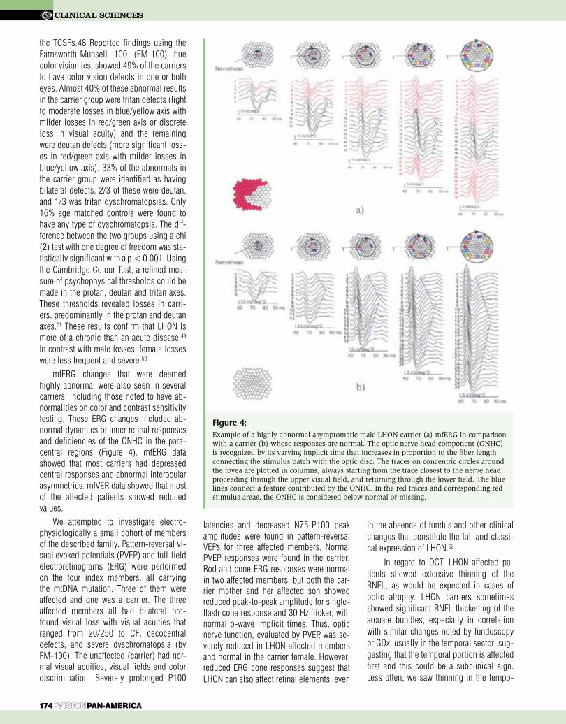
Most of the eyes with abnormal fundus findings also had other abnormalities, such as relative paracentral or arcuate scotomas on HVF testing. Although 30% of the car-riers had abnormalities on HVF, reliability was not very good. However, those patients with abnormal fundus findings most often showed visual field defects on HVF testing that conformed to the very same abnormali-ties noted on funduscopy. Further corrobo-ration of such changes could be observed on serial GDx studies (Figure 3).47
Novel testing was developed includ-ing cutting edge electrophysiological techniques such as multifocal ERGs with emphasis on the optic nerve head com-ponent (ONHC). Studies of color vision began (2002) utilizing pseudoisochro-matic color test plates. The following year, Fansworth-Munsell 100 (FM-100) studies were added. Subsequently, the color vi-sion testing was improved by employing the Cambridge Colour Test (Cambridge Re-search Systems) and also contrast sensitiv-ity was measured with the PSYCHO software (Cambridge Research Systems). Compared to controls, LHON carriers had significant losses in color vision affecting mostly the red-green system and reduction in spatial but not temporal contrast sensitivity.48-51 It was determined that asymptomatic carri-ers demonstrated impairments in chromatic red/green (R/G) and blue/yellow (B/Y) contrast sensitivity functions (CSF) and in luminance contrast sensitivity functions in the spatial CSF (SCSF) and temporal CSF (TCSF) domains. The differences between carriers and controls were statistically sig-nificant for all spatial frequencies of chro-matic and luminance SCSFs, but not for
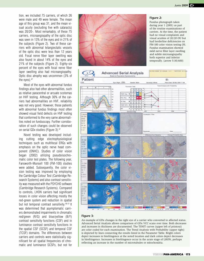
Figure 2: Fundus photograph taken during year 1 (2001) as part of the routine examinations of carriers. At the time, the patient had no visual complaints and visual acuities of 20/20 OU but had borderline deficiencies on FM-100 color vision testing OS. Fundus examination showed mild nerve fiber layer swelling and subtle microangiopathy both superior and inferior temporally. (arrow 5:30/600)
Figure 3: An example of GDx changes in the right eye of a carrier who converted to affected status. Advanced Serial Analysis allows comparison of GDx VCC scans over time. Both decreases and increases in thickness are documented. The TSNIT curves (upper left of printout) are color coded for each examination. The Trend Analysis with Probability (upper right) is depicted by lines connecting the results listed in the Parameter Table. Bright colors depict increases in birefringence at the noted location and dark colors depict decreases in birefringence. Increases in birefringence occur in the acute stage of LHON, perhaps reflecting an increase in the number of microtubules or mitochondria.
CLINICAL SCIENCES
PAN-AMERICA174 :
the TCSFs.48 Reported findings using the Farnsworth-Munsell 100 (FM-100) hue color vision test showed 49% of the carriers to have color vision defects in one or both eyes. Almost 40% of these abnormal results in the carrier group were tritan defects (light to moderate losses in blue/yellow axis with milder losses in red/green axis or discrete loss in visual acuity) and the remaining were deutan defects (more significant loss-es in red/green axis with milder losses in blue/yellow axis). 33% of the abnormals in the carrier group were identified as having bilateral defects. 2/3 of these were deutan, and 1/3 was tritan dyschromatopsias. Only 16% age matched controls were found to have any type of dyschromatopsia. The dif-ference between the two groups using a chi (2) test with one degree of freedom was sta-tistically significant with a p < 0.001. Using the Cambridge Colour Test, a refined mea-sure of psychophysical thresholds could be made in the protan, deutan and tritan axes. These thresholds revealed losses in carri-ers, predominantly in the protan and deutan axes.51 These results confirm that LHON is more of a chronic than an acute disease.49 In contrast with male losses, female losses were less frequent and severe.50
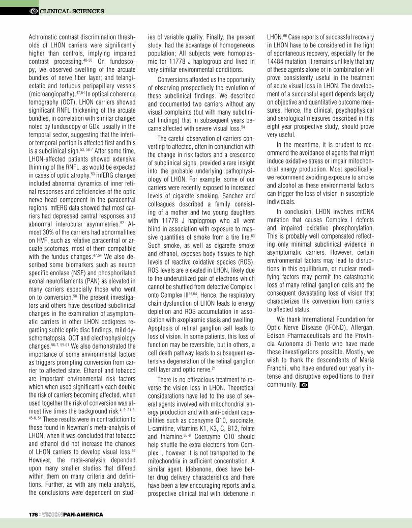
mfERG changes that were deemed highly abnormal were also seen in several carriers, including those noted to have ab-normalities on color and contrast sensitivity testing. These ERG changes included ab-normal dynamics of inner retinal responses and deficiencies of the ONHC in the para-central regions (Figure 4). mfERG data showed that most carriers had depressed central responses and abnormal interocular asymmetries. mfVER data showed that most of the affected patients showed reduced values.
We attempted to investigate electro-physiologically a small cohort of members of the described family. Pattern-reversal vi-sual evoked potentials (PVEP) and full-field electroretinograms (ERG) were performed on the four index members, all carrying the mtDNA mutation. Three of them were affected and one was a carrier. The three affected members all had bilateral pro-found visual loss with visual acuities that ranged from 20/250 to CF, cecocentral defects, and severe dyschromatopsia (by FM-100). The unaffected (carrier) had nor-mal visual acuities, visual fields and color discrimination. Severely prolonged P100
latencies and decreased N75-P100 peak amplitudes were found in pattern-reversal VEPs for three affected members. Normal PVEP responses were found in the carrier. Rod and cone ERG responses were normal in two affected members, but both the car-rier mother and her affected son showed reduced peak-to-peak amplitude for single-flash cone response and 30 Hz flicker, with normal b-wave implicit times. Thus, optic nerve function, evaluated by PVEP, was se-verely reduced in LHON affected members and normal in the carrier female. However, reduced ERG cone responses suggest that LHON can also affect retinal elements, even
in the absence of fundus and other clinical changes that constitute the full and classi-cal expression of LHON.52
In regard to OCT, LHON-affected pa-tients showed extensive thinning of the RNFL, as would be expected in cases of optic atrophy. LHON carriers sometimes showed significant RNFL thickening of the arcuate bundles, especially in correlation with similar changes noted by funduscopy or GDx, usually in the temporal sector, sug-gesting that the temporal portion is affected first and this could be a subclinical sign. Less often, we saw thinning in the tempo-
Figure 4: Example of a highly abnormal asymptomatic male LHON carrier (a) mfERG in comparison with a carrier (b) whose responses are normal. The optic nerve head component (ONHC) is recognized by its varying implicit time that increases in proportion to the fiber length connecting the stimulus patch with the optic disc. The traces on concentric circles around the fovea are plotted in columns, always starting from the trace closest to the nerve head, proceeding through the upper visual field, and returning through the lower field. The blue lines connect a feature contributed by the ONHC. In the red traces and corresponding red stimulus areas, the ONHC is considered below normal or missing.

ral and inferior quadrants, as well as in the 360° average measurement.53 (Table 1)
This study population also allowed us to prospectively examine carriers as well as affected members of this large pedigree with this extensive testing. The subclinical findings described in several of our carriers can be best demonstrated in the summari-zation of two unaffected carriers who were followed for years and then underwent fairly classic conversion.
Year IV- V – 2004-2005: Subclinical measurements were demonstrated to be useful to follow the progression of LHON in carriers. Peripapillary NFL swellings associated with mild microangiopathy at the superior and inferior poles of the op-tic disc were found on routine screening. GDx confirmed the NFL swelling. There were also mild central depressions on HVF testing. Both patients lost vision in a fairly classical way to become affected. In the moment of noticeable visual loss, GDx testing in the affected eye confirmed dramatic thinning of the NFL. However, surprisingly, in the nonaffected eye (OD), GDx also showed that temporally there
was marked thinning of the NFL. After a few months and loss of vision in OU, GDx testing showed a marked decrease of thickening of both superior and inferior NFL OU. In summary, GDx results showed sequentially: (1) thickening of the PMB bundle, (2) thickening of RNFL in the arcuate bundles, (3) loss of most of the PMB, followed by (4) loss of most of the RNFL. This was a very nice way to docu-ment the natural history of this conver-sion with a variety of very sophisticated measures.54
Specialized necropsy of eye, brain and peripheral nerve tissues were performed on tissue from two affected patients who died. New technology was used to refine the validity and reliability of several quantita-tive psychophysical tests (GDx) and Opti-cal Coherence Tomography (OCT) for the quantification of retinal nerve fiber layer thickening and losses. Serological markers were obtained in a larger cohort of the pedi-gree to make comparisons between these values and the psychophysical impairments noted. New modalities of therapy such as long wavelength (670 um) light to modu-
late mitochondrial activity were attempted in affected members with the most reliable outcome measures.
Year VI-VIII – 2006-2008: OCT data was gathered to better understand the objec-tive preclinical change seen in most LHON carriers (NFL thickness), and compare this with funduscopic results, to validate these changes as risk markers of developing the disease.45 (Table 1) Blood specimens were collected for four sets of assessments neu-ron specific enolase and axonal heavy chain neurofilaments (serum), and RNA and DNA phosphorylated changes (blood cells) to identify abnormal blood values specific for carriers of LHON and to provide additional easy and objective outcome measures for future management strategies.53
Achromatic contrast discrimination was studied in asymptomatic carriers and con-trols and it was found that contrast discrim-ination thresholds of LHON carriers were significantly higher than controls, imply-ing impaired contrast processing. Carriers' thresholds manifested significantly longer temporal integration than controls.51
A novel LHON susceptibility locus on the long arm of the X-chromosome (Xq25-q27.2) was identified using both linkage analysis (parametric and non-parametric) and transmission disequilibrium testing. These results suggest genetic heterogene-ity for X-linked modifiers of LHON.55
Discussion:This multinational longitudinal eight
year study was performed with the world’s largest documented LHON pedigree. Most of the 332 family members were located in Colatina, Espirito Santo state, Brazil. The findings of these investigations allowed for three general conclusions regarding affect-ed, carriers and the nature of conversion:
Affected members demonstrated a long-term continuous process for losing vision. This is corroborated by histopathology that showed ongoing generation 50 years after the affected individual had become blind.21
Carriers, even when totally asymptom-atic, usually demonstrated one or several subclinical signs, manifested on different examinations such as: color vision and contrast sensitivity testing, fundoscopy, OCT, electrophysiology, visual field test-ing and blood biomarkers.47-54 Almost 50% of asymptomatic carriers showed color vision defects in one or both eyes.
Junio 2009
PAN-AMERICA : 175
RNFL Thickness (μ)
AfectedN=18
CarriersN=51
ControlN=75
Global Mean (DP)Global Median
51.6 (18.7)46.0
107.4 (10.5)104.9 (9.4)
109.2106.0
Temporal (DP)Temporal Median
36.5 (11.1)34.0
80.4 (11.3)79.0
69.9 (10.5)70.0
Superior (DP)Superior Median
60.4 (28.8)47.5
128.5 (20.2)128.0
129.7 (16.4)128.0
Nasal (DP)Nasal Median
47.4 (14.8)43.5
78.6 (14.5)80.0
83.1 (15.6)82.0
Inferior (DP)Inferior Median
59.8 (32.7)49.0
141.9 (13.7)142.0
137.2 (16.6)138.0
Year 1 2 3 4 5 6 7 8Pedigree/Risk factors X X
Fundus X X X X X X X X
Color/Contrast X X X X X X
Mf ERG X X X
GDx X X
OCT X X X X
Blood markers X X X
Table 1: Retinal nerve fiber layer (RNFL) thickness in different groups.
Table 2: Presents each of our findings/conclusions year by year.
Summary of our results/findings year by year. The subclinical findings were discovered on years 3, 4, 5 and refined on years 6, 7, 8.
Achromatic contrast discrimination thresh-olds of LHON carriers were significantly higher than controls, implying impaired contrast processing.48-50 On fundosco-py, we observed swelling of the arcuate bundles of nerve fiber layer; and telangi-ectatic and tortuous peripapillary vessels (microangiopathy).47,54 In optical coherence tomography (OCT), LHON carriers showed significant RNFL thickening of the arcuate bundles, in correlation with similar changes noted by funduscopy or GDx, usually in the temporal sector, suggesting that the inferi-or temporal portion is affected first and this is a subclinical sign.53, 56-7 After some time, LHON-affected patients showed extensive thinning of the RNFL, as would be expected in cases of optic atrophy.53 mfERG changes included abnormal dynamics of inner reti-nal responses and deficiencies of the optic nerve head component in the paracentral regions. mfERG data showed that most car-riers had depressed central responses and abnormal interocular asymmetries.52 Al-most 30% of the carriers had abnormalities on HVF, such as relative paracentral or ar-cuate scotomas, most of them compatible with the fundus changes.47,54 We also de-scribed some biomarkers such as neuron specific enolase (NSE) and phosphorilated axonal neurofilaments (PAN) as elevated in many carriers especially those who went on to conversion.58 The present investiga-tors and others have described subclinical changes in the examination of asymptom-atic carriers in other LHON pedigrees re-garding subtle optic disc findings, mild dy-schromatopsia, OCT and electrophysiology changes.56-7, 59-61 We also demonstrated the importance of some environmental factors as triggers prompting conversion from car-rier to affected state. Ethanol and tobacco are important environmental risk factors which when used significantly each double the risk of carriers becoming affected, when used together the risk of conversion was al-most five times the background risk.4, 9, 21-3,
45-6, 54 These results were in contradiction to those found in Newman’s meta-analysis of LHON, when it was concluded that tobacco and ethanol did not increase the chances of LHON carriers to develop visual loss.62 However, the meta-analysis depended upon many smaller studies that differed within them on many criteria and defini-tions. Further, as with any meta-analysis, the conclusions were dependent on stud-
ies of variable quality. Finally, the present study, had the advantage of homogeneous population; All subjects were homoplas-mic for 11778 J haplogroup and lived in very similar environmental conditions.
Conversions afforded us the opportunity of observing prospectively the evolution of these subclinical findings. We described and documented two carriers without any visual complaints (but with many subclini-cal findings) that in subsequent years be-came affected with severe visual loss.54
The careful observation of carriers con-verting to affected, often in conjunction with the change in risk factors and a crescendo of subclinical signs, provided a rare insight into the probable underlying pathophysi-ology of LHON. For example; some of our carriers were recently exposed to increased levels of cigarette smoking. Sanchez and colleagues described a family consist-ing of a mother and two young daughters with 11778 J haplogroup who all went blind in association with exposure to mas-sive quantities of smoke from a tire fire.63 Such smoke, as well as cigarette smoke and ethanol, exposes body tissues to high levels of reactive oxidative species (ROS). ROS levels are elevated in LHON, likely due to the underutilized pair of electrons which cannot be shuttled from defective Complex I onto Complex III25,64. Hence, the respiratory chain dysfunction of LHON leads to energy depletion and ROS accumulation in asso-ciation with axoplasmic stasis and swelling. Apoptosis of retinal ganglion cell leads to loss of vision. In some patients, this loss of function may be reversible, but in others, a cell death pathway leads to subsequent ex-tensive degeneration of the retinal ganglion cell layer and optic nerve.21
There is no efficacious treatment to re-verse the vision loss in LHON. Theoretical considerations have led to the use of sev-eral agents involved with mitochondrial en-ergy production and with anti-oxidant capa-bilities such as coenzyme Q10, succinate, L-carnitine, vitamins K1, K3, C, B12, folate and thiamine.65-8 Coenzyme Q10 should help shuttle the extra electrons from Com-plex I, however it is not transported to the mitochondria in sufficient concentration. A similar agent, Idebenone, does have bet-ter drug delivery characteristics and there have been a few encouraging reports and a prospective clinical trial with Idebenone in
LHON.66 Case reports of successful recovery in LHON have to be considered in the light of spontaneous recovery, especially for the 14484 mutation. It remains unlikely that any of these agents alone or in combination will prove consistently useful in the treatment of acute visual loss in LHON. The develop-ment of a successful agent depends largely on objective and quantitative outcome mea-sures. Hence, the clinical, psychophysical and serological measures described in this eight year prospective study, should prove very useful.
In the meantime, it is prudent to rec-ommend the avoidance of agents that might induce oxidative stress or impair mitochon-drial energy production. Most specifically, we recommend avoiding exposure to smoke and alcohol as these environmental factors can trigger the loss of vision in susceptible individuals.
In conclusion, LHON involves mtDNA mutation that causes Complex I defects and impaired oxidative phosphorylation. This is probably well compensated reflect-ing only minimal subclinical evidence in asymptomatic carriers. However, certain environmental factors may lead to disrup-tions in this equilibrium, or nuclear modi-fying factors may permit the catastrophic loss of many retinal ganglion cells and the consequent devastating loss of vision that characterizes the conversion from carriers to affected status.
We thank International Foundation for Optic Nerve Disease (IFOND), Allergan, Edison Pharmaceuticals and the Provín-cia Autonoma di Trento who have made these investigations possible. Mostly, we wish to thank the descendents of Maria Franchi, who have endured our yearly in-tense and disruptive expeditions to their community.
CLINICAL SCIENCES
176 : PAN-AMERICA
REFERENCES
1. Leber T Uber hereditare and congenitalangelegte Seh-nervenleiden. Grafes Arch Ophthalmol 1871, 17:249-91.2. Gowers WR. A letter on abiotrophy. Lancet 1902:1003-21.3. Collins ET. Hereditary ocular degenerations. Int Cong Ophthalmol Washington 1922: 103-38.4. Sadun AA. Metabolic optic neuropathies. Semin Opthalmol. 2002; 17 (1): 29-32.5. Seedorff T. The inheritance of Leber’s disease: a gene-alogical follow-up study. Acta Ophthalmol1985: 135-45.6. Nikoskelainen E. New aspects of genetic, etiologic and clinical puzzle of Leber’s disease. Neurology 1984: 1482-4.7. Wallace DC, Singh G, Lott MT, et al. Mitochondrial DNA mutation associated with Leber’s hereditary optic neuropathy. Science. 1988; 242:1427–1430.8. Chalmers RM, Schapira AHV. Clinical, biochemi-cal and molecular genetic features of Leber’s heredi-tary optic neuropathy. Biochem Biophys Acta. 1999; 1410:147–158.9. Carelli V, Ross-Cisneros FN, Sadun AA. Optic nerve degeneration and mitochondrial dysfunction: genetic and acquired optic neuropathies. Neurochem Int. 2002; 40(6): 573-84.10. Howell N, Bindoff LA, McCullough DA, et al. Leber’s hereditary optic neuropathy: identification of the same mitochondrial ND1 mutation in six pedigrees. Am J Hum Genet. 1991; 49:939–950.11. Huponen K, Vilkki J, Aula P, et al. A new mtDNA mutation associated with Leber’s hereditary optic neu-ropathy. Am J Hum Genet. 1991; 48:1147–1153.12. Mackey D, Howell N. A variant of Leber’s heredi-tary optic neuropathy characterized by recovery of vision and by an unusual mitochondrial genetic etiology. Am J Hum Genet. 1992; 51:1218–1228.13. Man PY, Turnbull DM, and Chinnery PF. Leber’s he-reditary optic neuropathy. J Med Genet 2002; 39(3): 162-9.14. Wang AG, Wei YH. Leber’s hereditary optic neu-ropathy: a multifactorial disease. Prog Retin Eye Res. 2006; 25(4):381-96.15. Kerrison JB, Howell N, Miller NR, et al. Leber’s hereditary optic neuropathy. Electron microscopy and molecular genetic analysis of a case. Ophthalmology. 1995; 102:1509–1516.16. Fauser S, Luberichs J, Besch D et al. sequence analysis of the complete mitochondrial genome in pa-tients with Leber’s hereditary optic neuropathy lacking the three most common pathogenic DNA mutations. Bio-chem Biophys Res Commun 2002; 295(2): 342-7.17. Cock HR, Tabrizi SJ, Cooper JM, Schapira AHV. The influence of nuclear background on the biochemical expression of 3460 Leber’s hereditary optic neuropathy. Ann Neurol. 1998; 44:187–193.18. Sorajja P, Sweeney MG, Chalmers R, et al. Cardiac Abnormalities in patients with Leber’s hereditary optic neuropathy. Heart 2003: 791-2.19. Carelli V, Valentino ML, Liguori R, et al. Leber’s hereditary optic neuropathy (LHON/11778) with myo-clonus: report of two cases. J Neurol Neurosurgery Psy-chiatry 2001; 71(6): 813-6.20. Chalmers RM, Harding AE. A case-control study of Leber’s hereditary optic neuropathy. Brain. 1996; 119:1481–1486.21. Carelli V, Ross-Cisneros F, Sadun A. Mitochondrial dysfunction as a cause of optic neuropathies. Prog Reti-nal Eye Res. 2004; 23:53–89.22. Sadun AA, Kashima Y, Wurdeman AE, et al. Mor-phological findings in the visual system in a case of Leb-er’s’ hereditary optic neuropathy. Clin Neurosci. 1994; 2:165–172.23. Sadun AA, Win PH, Ross-Cisneros FN, et al. Leber’s hereditary optic neuropathy differentially affects smaller axons in the optic nerve. Trans Am Acad Oph-thalmol. 2000; 98:881–923.24. Howel N. Leber’s hereditary optic neuropathy: how do mitochondrial DNA mutations cause degeneration of the optic nerve? J Bioenerg biomembr. 1997; 29(2): 165-73. 25. Carelli V, Rugolo M, Sgarbi G et al. Bioenergetics shapes cellular death pathways in Leber’s hereditary op-tic neuropathy: a model of mitochondrial neurodegenera-tion. Biochim Biophys Acta. 2004;1658(1-2):172-9.
26. Rizzo JF III. Adenosine triphosphate deficiency: a genre of optic neuropathy. Neurology. 1995; 45:11–16.27. Battisti C, Cardaioli E, et al. Cell response to oxi-dative stress induced apoptosis in patients with Leber’s hereditary optic neuropathy. J Neurol Neurosurg Psy-chiatry. 2004; 75(12):1731-6.28. Wang JY, Gu YS, Wang J, et al. Oxidative stress in Chinese patients with Leber’s hereditary optic neuropa-thy. J Int Med Res. 2008; 36(3):544-50.29. Yu Wai Man CY, Chinnery PF, Griffiths PG. Optic neuropathies--importance of spatial distribution of mi-tochondria as well as function. Med Hypotheses. 2005; 65(6): 1038-42.30. Mroczek-Tonska K, Kisiel B, Piechota J et al. Leb-er’s hereditary optic neuropathy – a disease with a known molecular basis but a mysterious mechanism of pathol-ogy. J Apple Genet 2003; 44(4): 529-38.31. Ghelli A, Zanna C, Porcelli AM et al. Leber’s he-reditary optic neuropathy (LHON) pathogenic mutations induce mitochondrial-dependent apoptotic death in transmitochondrial cells incubated with galactose me-dium. J Biol Chem. 2003; 278(6):4145-50.32. Napoli E, et al. Antioxidant defenses in cybrids har-boring mtDNA mutations associated with Leber’s hereditary optic neuropathy. FEBS J. 2005; 272(5):1124-35.33. Yen MY, Kao SH, Wang AG et al. Increased 8-hy-droxy-2’-deoxyguanosine in leukocyte DNA in Leber’s hereditary optic neuropathy. Invest Ophthalmol Vis Sci. 2004; 45(6):1688-91.34. Carelli V, La Morgia C, Iommarini L et al. Mitochon-drial optic neuropathies: how two genomes may kill the same cell type? Biosci Rep. 2007; 27(1-3):173-84.35. Wang HW, Jia X, Ji Y, Strikingly different pen-etrance of LHON in two Chinese families with primary mutation G11778A is independent of mtDNA haplo-group background and secondary mutation G13708A. Mutat Res. 2008 Jun 21. [Epub ahead of print] 36. Howell N, Mackey DA. Low-penetrance branches in matrilinear pedigrees with Leber’s hereditary optic neuropathy. Am J Hum Genet. 1998; 63:1220–1224.37. Cullom ME, Heher KL, Miller NR, et al. Leber’s he-reditary optic neuropathy masquerading as tobacco-alcohol amblyopia. Arch Ophthalmol. 1993; 111:1482–1485.38. Smith JL, Hoyt WF, Susac JO. Ocular fundus in acute Leber Optic Neuropathy. Arch Ophthalmol 1973:349-54.39. Nikoskelainen EK. Clinical picture of LHON. Clin Neurosci. 1994; 2:115–120.40. Newman NJ, Lott MT Wallace DC. The clinical char-acteristics of Leber’s hereditary optic neuropathy with the 11778 mutation. Am J Ophthalmol 1991: 750-62.41. Riordan-Eva P, Sander MD, Govan GC, et al. The clinical features of Leber’s hereditary optic neuropathy defined by the presence of pathogenic mitochondrial DNA mutation. Brain 1995: 319-37.42. Mashima Y, Sato EA, Ohde H, et al. Macular nerve fibers temporal to fovea may have a greater potential to recover function in patients with Leber’s hereditary optic neuropathy. Jpn J Ophthalmol. 2002; 46:660-7.43. Lessel S, Gise RL, Krohel GB. Bilateral optic neu-ropathy with remission in young men: variation on a theme by Leber? Arch Neurol 1983: 2-6.44. Acaroglu G, Kansu T, Dogulu CF. Visual recovery patterns in children with Leber’s hereditary optic neu-ropathy. Int Ophthalmol 2001: 349-55.45. Sadun AA, Carelli V, Salomao SR et al. Extensive investigation of large Brazilian pedigree of Italian ances-try (SOA-BR) with 117788/Haplogroup J Leber’s heredi-tary optic neuropathy (LHON). Am J Ophthalmol. 2003; 136:231–238.46. Sadun AA, Carelli V, Salomao SR et al. A very large Brazilian pedigree with 11778 Leber’s hereditary optic neu-ropathy. Trans Am Ophthalmol Soc. 2002; 100: 169–179.47. Sadun F, De Negri A, Carelli V, et al. Ophthalmo-logic findings in large pedigree of 11778/Haplogroup J Leber’s hereditary optic neuropathy. Am J Ophthalmol. 2004; 137:271–277.48. Ventura DF, Quiros P, Carelli V, et al. Chromatic and luminance contrast sensitivities in asymptomatic carriers from a large Brazilian pedigree of 11778 Leb-er’s hereditary optic neuropathy. Invest Ophthalmol
Vis Sci. 2005; 46:4809–4814.49. Torres RJ, Salomao S et al. Colour vision defects in asymptomatic carriers of the Leber’s hereditary optic neuropathy (LHON) mtDNA 11778 mutation from a large Brazilian LHON pedigree: a case-control study. Br J Ophthalmol. 2006; 90(2):150-3.50. Ventura DF, Gualtieri M, Oliveira AG, et al. Male prevalence of acquired color vision defects in asymp-tomatic carriers of Leber’s hereditary optic neuropathy. Invest Ophthalmol Vis Sci. 2007; 48(5):2362-70.51. Gualtieri M, Bandeira M, Hamer RD et al. Psy-chophysical analysis of contrast processing segregated into magnocellular and parvocellular systems in asymp-tomatic carriers of 11778 Leber’s hereditary optic neu-ropathy. Vis Neurosci. 2008; 25(3):469-74.52. Salomao SR, Berezovsky A, Andrade RE, et al. Visual electrophysiologic findings in patients from an extensive Brazilian family with Leber’s hereditary optic neuropathy. Doc Ophthalmol. 2004; 108:147–155.53. Ramos CVF, Chicani CF, Salomao SR et al. Optic coherence tomography for optic nerve and retinal nerve fiber layer analysis in carriers of 11778 mutation for Leber’s hereditary optic neuropathy.(Doctorate/PhD Thesis in press)54. Sadun A, Salomao SR, Berezovsky A, et al. Sub-clinical carriers and conversions in Leber’s hereditary optic neuropathy: a prospective psychophysical study. Trans Am Ophthalmol Soc. 2006; 104: 51–61.55. Shankar SP, Fingert JH, Carelli V et al. Evidence for a novel x-linked modifier locus for Leber’s hereditary optic neuropathy. Ophthalmic Genet. 2008; 29:17-24.56. Barboni P, Savini G, Valentino ML, et al. Retinal nerve fiber layer evaluation by optical coherence tomog-raphy in Leber’s hereditary optic neuropathy. Ophthal-mology 2005; 112: 120-6.57. Savini G, Barboni P, Valentino ML, et al. Retinal nerve fiber layer evaluation by optical coherence tomogra-phy in unafected carriers of Leber’s hereditary optic neu-ropathy mutations. Ophthalmology 2005; 112: 127-31.58. Guy J, Shaw G, Ross-Cisneros FN et al. Phosphory-lated neurofilament heavy chain is a marker of neurode-generation in Leber hereditary optic neuropathy (LHON). Mol Vis. 2008; 14: 2443-50. Epub 2008 Dec 22.59. Carrol WM, Mastalgia FL. Leber’s optic neuropathy: a clinical and visual evoked potential study of affected and asymptomatic members of a six generation family. Brain 1979: 559-80.60. Mashima Y, Kigasawa K, Wakakura M, et al. Do idebenone and vitamin therapy shorten the time to achieve visual recovery in Leber hereditary optic atro-phy? J Neurophthalmol 2000; 20(3):166-70.61. Oguchi Y. [Past, present, and future in Leber’s he-reditary optic neuropathy] Nippon Ganka Gakkai Zasshi. 2001; 105(12):809-27.62. Newman NJ, Biousse V, David R, et al. Prophylaxis for second eye involvement in Leber’s hereditary optic neuropathy: a open-labeled, non randomized multicenter trial of topical brimonidine purite. Am J Ophthalmol 2005; 140(3): 407-15.63. Sanchez RN, Smith AJ, Carelli V, et al. Leber’s he-reditary optic neuropathy possibly triggered by exposure to tire fire. J Neuroophthalmol 2006; 26(4): 268-72.64. Wong A, Cavalier L, Collins-Schramm HE, et al. Differentiation-specific effects of LHON mutations intro-duced into neuronal NT2 cells. Hum Mol Genet 2002; 11 (4): 431-8.65. Qi X, Sun L, Hauswirth WW, et al. Use of mito-chondrial antioxidant defenses for rescue of cells with Leber’s hereditary optic neuropathy- causing mutations. Arc Ophthalmol 2006; 125(2): 268-72.66. Barnils N, Mesa E, Munoz S, et al. Response to Ide-benone and multivitamin therapy in Leber’s hereditary optic neuropathy. Arch Soc Esp Oftalmol 2007; 82(6): 377-80.67. Ghelli A, Porcelli AM, Zanna C, et al. Protection against oxidant induced apoptosis by exogenous gluta-thione in Leber’s hereditary optic neuropathy cybrids. Invest Ophthalmol Vis Sci 2008; 49(2): 671-6.68. Johns DR, Colby KA. Treatment of Leber’s heredi-tary optic neuropathy: theory to practice. Semin Ophthal-mol 2002; 17(1): 33-8.
: 177PAN-AMERICA
Junio 2009
CLINICAL SCIENCES
178 : PAN-AMERICA
Abstract
Introduction: Corneal hysteresis quan-tifies the biomechanical properties of corneal tissue, both static and dynamic, that influence the quantification of ocular pathology such as in corneal ectasia and after surgical procedures. It may also be important for the diagnosis and treatment of corneal disease.
Objective: To determine corneal hysteresis and the corneal resistance factor in patients with the diagnosis of corneal ectasia pre- and post-surgical implantation of intracor-neal rings.
Method: This is a prospective, comparative and longitudinal study. The statistical analy-sis was performed using a Student t- test for paired samples. We studied a series of 14 patients (16 eyes) with the diagnosis of cor-neal ectasia, fourteen with the diagnosis of keratoconus, one with the diagnosis of pellu-cid marginal degeneration, and one with the diagnosis of post-LASIK ectasia. The mean age was 25 years (range 16-39), 50% were female and 50% were male. This study was performed in the Cornea and Refractive Sur-gery Department of the Instituto de Oftalmo-logía, Fundación Conde de Valenciana I.A.P., Mexico City. In this series, the quantification of the biomechanics was made with the ORA (Ocular Response Analyzer) by Reichert´s, pre- and post-surgical implantation of intra-corneal rings one month after surgery. Other variables that were included were visual acui-ty, visual capacity, refraction error by spheri-cal equivalent and keratometry average.
Results: The visual acuity prior to sur-gery varied from count fingers at one foot to 20/50 on the Snellen chart or its equivalen-ce of 3.00 to .40 in LogMAR. After surgical implantation of the rings, the visual acuity was better by an average of four lines with a decrease in refractive error by a spherical equivalent of three diopters and an average keratometric flattening of four diopters. With
regard to the corneal biomechanics, hystere-sis and corneal resistance factor did not vary, (pre = 6.5 mmHg and post = 6.4 mmHg).
Discussion: Despite our hypothesis that corneal hysteresis and the corneal resis-tance factor should be greater after surgical implantation of intracorneal rings, our study demonstrated no change in the viscoelastic properties of the cornea. Nonetheless, the results of the biomechanics were better as reflected in visual acuity, a decreased refrac-tive error, flatter keratometric curvature and the promise of long range corneal stability as suggested in the literature.
Conclusion: The expected biomechanical property changes of the cornea were not statistically significantly different in terms of quantification of hysteresis and corneal re-sistance factor using the ORA.
Resumen
Introducción: La histéresis corneal es la cuantificación de las propiedades biome-cánicas del tejido corneal, estas caracterís-ticas son estáticas y dinámicas, las cuales influyen en los estados patológicos como en las ectasias corneales y procedimientos oculares, actualmente importantes para el diagnóstico y manejo de las enfermedades del tejido corneal.
Objetivo: Determinar la histéresis corneal y factor de resistencia corneal en pacientes con el diagnóstico de ectasia corneal pre y post-quirúrgico a la implantación de anillos.
Método: Es un estudio prospectivo, compa-rativo y longitudinal, del análisis estadístico de una serie de muestras pareadas a través de una prueba t de student. Se estudiaron una serie de 14 pacientes, un total de 16 ojos de pacientes con el diagnóstico de ec-tasia corneal, catorce con el diagnostico de queratocono, uno con el diagnóstico de de-generación marginal pelucida, y otro con el diagnóstico de ectasia post-LASIK. Este es-
tudio se realizó en el Departamento de Cor-nea del Instituto de Oftalmología, Fundación Conde de Valenciana, I.A.P., México D.F. De esta serie de casos se les realizó la cuan-tificación de su biomecánica a través ORA (ocular response analyzer) de Reichert´s, pre y post implante de anillos intracorneales a los 30 días. Otras variables que se inclu-yeron son; agudeza visual, capacidad visual, error refractivo en equivalente esférico y pro-medio queratométrico.
Resultados: Un total de 16 ojos, el prome-dio de edad fue de 25 años (rango de 16-39), 50% del sexo femenino y 50% del sexo masculino. La agudeza visual previa la ciru-gía fue de cuenta dedos a 20/50 según car-tilla de Snellen o su equivalencia de 3.00 a .40 en LogMAR, después de la implantación de los anillos, la agudeza visual mejoró en un promedio de 4 líneas, menor error refrac-tivo en equivalente esférico de tres dioptrías, aplanamiento de la queratometría promedio de cuatro dioptrías, y en lo que respecta a las características de la biomecánica corneal, la histéresis corneal y el factor de resistencia corneal no presentaron diferencias significa-tivas, pre 6.5 mmHg y post 6.4 mmHg.
Discusión: A pesar de inferir que la histére-sis y el factor de resistencia corneal debieran de aumentar con la implantación quirúrgica de los anillos intracorneales, este estudio demuestra que no hay cambios en las pro-piedades viscoelásticas de la cornea, sin embargo los resultados biomecánicos fue-ron mejores en el aspecto de la agudeza vi-sual, menor error refractivo, menor curvatura corneal y prometen estabilidad corneal a lo largo del tiempo de acuerdo con la literatura mundial.
Conclusión: Los cambios esperados en las propiedades de la biomecánica corneal no mostraron diferencia estadísticamente sig-nificativa en la cuantificación de histéresis corneal y factor de resistencia corneal uti-lizando el ORA.
Determinación de la Histéresis Corneal Pre y Post Anillos IntracornealesMiguel Angel Quiroz González MD, Raúl Suárez MD, Luisa Maria Loustaunau Godoy MD, Arturo Gómez MD, Alberto Haber MD, Gabriela Ortiz MD, Alejandro Navas MD, Abel Martinez MD, Jerónimo Álvarez MD y Cristian Tinoco MD
Instituto de OftalmologíaFundación Conde de ValencianaDepartamento de Cornea y Cirugía Refractiva
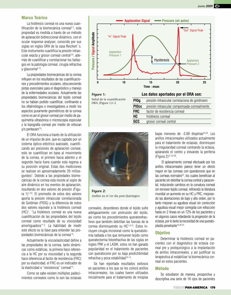
Marco TeóricoLa histéresis corneal es una nueva cuan-
tificación de la biomecánica corneal1,2, esta propiedad es medida a través de un método de aplanación bidireccional dinámico, con el ocular response analyser, conocido por sus siglas en ingles ORA de la casa Reichert´s. Este instrumento cuantifica la presión intrao-cular exacta y grosor corneal central3-6, ade-más de cuantificar y correlacionar los hallaz-gos en la patología corneal, cirugía refractiva y glaucoma6,13.
La propiedades biomecánicas de la cornea influyen en los resultados de las cuantificacio-nes y procedimientos oculares, obscureciendo pistas esenciales para el diagnóstico y manejo de la enfermedades oculares. Actualmente las propiedades biomecánicas del tejido corneal no se habían podido cuantificar, confinando a los oftalmólogos e investigadores a medir los aspectos puramente geométricos de la cornea como es así el grosor corneal por medio de pa-quimetría ultrasónica o microscopia especular y la topografía corneal por medio de orbscan y/o pentacam7,8.
El ORA funciona a través de la utilización de un impulso de aire, que es captado por un sistema óptico-eléctrico avanzado, cuantifi-cando así presiones de aplanación corneal, esto se cuantifican en base al movimiento de la cornea, el primero hacia adentro y el segundo hacia fuera cuando esta regresa a su posición original. Estas dos mediciones se realizan en aproximadamente 20 milise-gundos1. Debido a las propiedades biome-cánicas de la cornea esta resiste al soplo de aire dinámico en los eventos de aplanación, resultando en dos valores de presión (Figu-ra 1)1,2,6. El promedio de estos dos valores aporta la presión intraocular correlacionada de Goldman (PIOG) y la diferencia de estos dos valores equivale a la histéresis corneal (HC)1. “La histéresis corneal es una nueva cuantificación de las propiedades del tejido corneal como resultado de su viscosidad amortiguadora”1,2. La habilidad de medir este efecto es la llave para entender las pro-piedades biomecánicas de la cornea1,6.
Actualmente la viscoelasticidad define a las propiedades de la cornea, tanto dinámi-cas como estáticas, la primera hace referen-cia a la HC por su viscosidad y la segunda hace referencia al factor de resistencia (FRC) por su elasticidad, el FRC es un indicador de la elasticidad o “resistencia” corneal14.
Como se sabe existen múltiples padeci-mientos corneales como lo son las ectasias
corneales, desordenes donde el tejido sufre adelgazamiento con protrusión del tejido, asi como los procedimientos queratorefrac-tivos que también debilitan las fuerzas de la cornea disminuyendo su HC1-6,15. Estos in-cluyen cirugía incisional como la queratoto-mia radiada o los que remueven tejido como queratectomia fotorefractiva de las siglas en ingles PRK y el LASIK, estos no han ganado popularidad en el tratamiento de pacientes con queratocono por su baja predictibilidad refractiva y poca estabilidad10-13.
Se han reportado resultados exitosos en pacientes a los que se les colocó anillos intracorneales, los cuales fueron utilizados inicialmente para el tratamiento de miopías
bajas menores de -3.00 dioptrías23,24. Los anillos intracorneales utilizados actualmente para el tratamiento de ectasias, disminuyen la irregularidad corneal centrando la ectasia, aplanando el centro y elevando la periferia (Figura 2)21,23-26.
El aplanamiento corneal efectuado por los anillos intracorneales parece tener un efecto mayor en las corneas con queratocono que en las corneas normales22, los cuales benefician al paciente sin debilitar la cornea central y paracen-tal, induciendo cambios en la curvatura corneal sin remover tejido corneal, refiriendo la literatura mundial un aumento en su HC y FRC, mejoran-do las aberraciones de bajo y alto orden, por lo tanto mejoran su agudeza visual sin corrección y agudeza visual mejor corregida con refracción hasta en 2 líneas en un 72% de los pacientes y en algunos casos retardando la progresión de la ectasia, por lo tanto la necesidad de una querato-plastia penetrante15,20-22.
ObjetivoDeterminar la histéresis corneal en pa-
cientes con el diagnóstico de ectasia cor-neal pre y postquirúrgico a la implantación de anillos intracorneales, y así justificar su terapéutica al estabilizar la biomecánica cor-neal en estos pacientes.
MétodoSe estudiaron de manera, prospectiva y
descriptiva una serie de 16 ojos de pacientes
: 179PAN-AMERICA
Junio 2009
Figure 1: Señal de la cuantificación ORA (Figura 1)1-2
Figure 2: Anillos en el 1er día post-Quirúrgico
Los datos aportados por el ORA son:PIOg presión intraocular correlaciona de goldmann
PIOcc presión intraocular compensada-cornealmenteFRC factor de resistencia cornealHC histéresis cornealGCC grosor corneal central
0 10 15 20 25 Time - msec
Applanation Signal
“In” Signal Peak
ApplantionPressure 1
ApplantionPressure 2
“Out” Signal Peak
Hysteresis
Pressure (air pulse)
Pres
sure
/ Si
gnal
Am
plitu
de
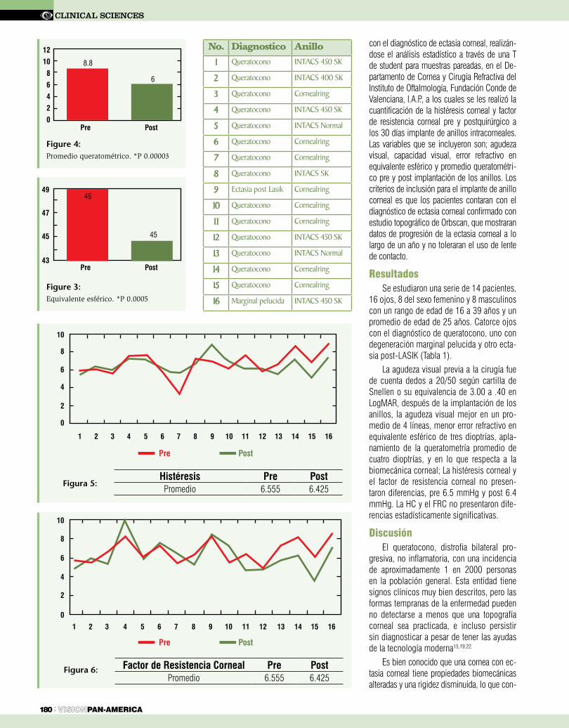
CLINICAL SCIENCES
180 : PAN-AMERICA
con el diagnóstico de ectasia corneal, realizán-dose el análisis estadístico a través de una T de student para muestras pareadas, en el De-partamento de Cornea y Cirugía Refractiva del Instituto de Oftalmología, Fundación Conde de Valenciana, I.A.P., a los cuales se les realizó la cuantificación de la histéresis corneal y factor de resistencia corneal pre y postquirúrgico a los 30 días implante de anillos intracorneales. Las variables que se incluyeron son; agudeza visual, capacidad visual, error refractivo en equivalente esférico y promedio queratométri-co pre y post implantación de los anillos. Los criterios de inclusión para el implante de anillo corneal es que los pacientes contaran con el diagnóstico de ectasia corneal confirmado con estudio topográfico de Orbscan, que mostraran datos de progresión de la ectasia corneal a lo largo de un año y no toleraran el uso de lente de contacto.
ResultadosSe estudiaron una serie de 14 pacientes,
16 ojos, 8 del sexo femenino y 8 masculinos con un rango de edad de 16 a 39 años y un promedio de edad de 25 años. Catorce ojos con el diagnóstico de queratocono, uno con degeneración marginal pelucida y otro ecta-sia post-LASIK (Tabla 1).
La agudeza visual previa a la cirugía fue de cuenta dedos a 20/50 según cartilla de Snellen o su equivalencia de 3.00 a .40 en LogMAR, después de la implantación de los anillos, la agudeza visual mejor en un pro-medio de 4 líneas, menor error refractivo en equivalente esférico de tres dioptrías, apla-namiento de la queratometría promedio de cuatro dioptrías, y en lo que respecta a la biomecánica corneal; La histéresis corneal y el factor de resistencia corneal no presen-taron diferencias, pre 6.5 mmHg y post 6.4 mmHg. La HC y el FRC no presentaron dife-rencias estadísticamente significativas.
DiscusiónEl queratocono, distrofia bilateral pro-
gresiva, no inflamatoria, con una incidencia de aproximadamente 1 en 2000 personas en la población general. Esta entidad tiene signos clínicos muy bien descritos, pero las formas tempranas de la enfermedad pueden no detectarse a menos que una topografía corneal sea practicada, e incluso persistir sin diagnosticar a pesar de tener las ayudas de la tecnología moderna15,19,22.
Es bien conocido que una cornea con ec-tasia corneal tiene propiedades biomecánicas alteradas y una rigidez disminuida, lo que con-
No. Diagnostico Anillo
1 Queratocono INTACS 450 SK
2 Queratocono INTACS 400 SK
3 Queratocono Cornealring
4 Queratocono INTACS 450 SK
5 Queratocono INTACS Normal
6 Queratocono Cornealring
7 Queratocono Cornealring
8 Queratocono INTACS SK
9 Ectasia post Lasik Cornealring
10 Queratocono Cornealring
11 Queratocono Cornealring
12 Queratocono INTACS 450 SK
13 Queratocono INTACS Normal
14 Queratocono Cornealring
15 Queratocono Cornealring
16 Marginal pelucida INTACS 450 SK
Figure 4: Promedio queratométrico. *P 0.00003
Pre Post
12
10
8
6
4
2
0
8.8
6
Figure 3: Equivalente esférico. *P 0.0005
Pre Post
49
47
45
43
49
45
Figura 5: Histéresis Pre PostPromedio 6.555 6.425
Pre Post
16151413121110987654321
10
8
6
4
2
0
Figura 6: Factor de Resistencia Corneal Pre PostPromedio 6.555 6.425
Pre Post
16151413121110987654321
10
8
6
4
2
0
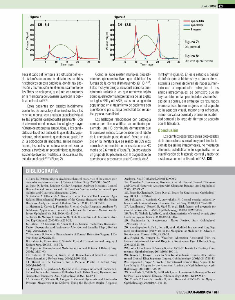
bIbLIOGRAFíA1. Luce D. Determining in vivo biomechanical properties of the cornea with an ocular response analyzer. J Cataract Refract Surg. 2005;31:156-62.2. Luce D, Taylor. Reichert Ocular Response Analyser Measures Corneal Biomechanical Properties and IOP. Provides New Indicador for Corneal Spe-cialities and Glaucoma Managment. 2006.3. Kotecha A, Elbsbeikb A, Roberts C, et al. Corneal Thickness and Age-Related Biomechanical Properties of the Cornea Measured with the Ocular Response Analyzer. Invest Opthalmol Vis Sci. 2006; 47:5337-47.4. Martínez J, García J, Fernández A, et al. Ocular Response Analyzer Vs Goldmann Applanation Tonometry for Intraocular Pressure Measurements. Invest Opthalmol Vis Sci. 2006; 47:4410-4.5. Torres R, Merayo J, Jaramillo M, et al. Biomecánica de la cornea. Arch Soc Esp Oftalmol. 2005:80(4):215-23.6. Gatinel D, Chaabouni S, Adam P, et al. Corneal Hysteresis, Resistance Factor, Topography, and Pachymetry After Corneal Lamellar Flap. J Refract Surg. 2007;23:76-84.7. Reinstein D, Roberts. Biomechanics of Corneal Refractive Surgery. J Re-fract Surg. 2007;22:285.8. Grabner G, Eilmsteiner R, Steindel C, et al. Dynamic corneal imaging. J Refract Surg. 2005;31:163-74.9. Dupps W. Biomechanical Modeling of Corneal Ectasia. J Refract Surg. 2005;21:186-90.10. Cabrera D, Niazy A, Kurtz, et al. Biomechanical Model of Corneal Transplantation. J Refract Surg. 2006;22:293-302.11. Robert C. The Cornea is Not a Piece of Plastic. J Refract Surg. 2000;16:407-13.12. Peptose J, Feigenbaum S, Qazi M, et al. Changes in Corneal Biomechan-ics and Intraocular Pressure Following Lasik Using Static, Dynamic, and Noncontact Tonometry. Am J Ophtahlmol. 2007;143:39-47.13. Kirwan C, O´Keefe M, Lanigan B. Corneal Hysteresis and Intraocular Pressure Measurement in Children Using the Reichert Ocular Response
Analyzer. Am J Opthalmol.2006;142:990-2.14. Congdon N, Broman A, Bandeen K, et al. Central Corneal Thickness and Corneal Hysteresis Associate with Glaucoma Damage. Am J Opthalmol. 2006;142:990-2.15. Boxer B, Chandra N, Chou D, et al. Intacs for Keratoconus. Ophthalmol-ogy. 2003;110:1031-40.16. Pallikaris I, Kymionis G, Astyrakakis N. Corneal ectasia induced by laser in situ keratomileusis. J Cataract Refract Surg. 2001;27:1796-180217. Randleman J, Russell B, Ward M, et al. Risk factors and prognosis for corneal ectasia after LASIK. Ophthalmology. 2003;110:267-275.18. Twa M, Nichols J, Joslin C, et al. Characteristics of corneal ectasia after Lasik for myopia. Cornea. 2004;23:447-457.19. Rabinowitz Y. Keratoconus. Major Review. Surv Ophthalmol. 1998;42:297-319.20. Kanellopoulos A, Pe L, Perry H, et al. Modified Intracorneal Ring Seg-ment Implantations (INTACS) for the Mangment of Moderate to Advanced Keratoconus. Cornea. 2006;25:29-33.21. Regina M, Krueger R, Wavefront Aberration Associated With ths Ferrara Intrastromal Corneal Ring in a Keratoconic Eye. J Refract Surg. 2004;20:823-30.22. Colin J, Cochener B, Savary G, et al. INTACS Inserts for Treating Kera-toconus. Ophthalmology. 2001;108:1409-14.23. Gomez L, Chayet. Laser In Situ Keratomileusis Results after Intras-tromal Croneal Ring Segments (Intacs). Ophthalmology. 2001;108:1738-43.24. Rapuano C, Sugar A, Koch D. Intrastromal Corneal Ring Segments for Low Miopía. AReport by the American Academy of Ophthalmology. Oph-thalmology. 2001;108:1922-28.25. Kymionis G, Tsildis N, Pallikaris I, et al. Long-term Follow-up of Intacs for Post-Lasik Corneal Ectasia. Ophthalmology. 2006;113:1909-17.26. Clinch T, Lemp M, Foulks G, et al. Removal of INTACS for Myopia. Ophthalmology. 2002;109:1441-46.
Marzo 2009
: 181PAN-AMERICA
Junio 2009
lleva al cabo del tiempo a la protrusión del teji-do. Además se conoce en detalle los cambios histológicos en esta patología, donde hay alte-ración y disminución en el entrecruzamiento de las fibras de colágeno, que junto con rupturas en la membrana de Bowman favorecen la debi-lidad estructural16-18.
Estos pacientes son tratados inicialmente con lentes de contacto y al ser intolerantes a los mismos o cursar con una baja capacidad visual se les proponía queratoplastia penetrante. Con el advenimiento de nuevas tecnologías y mayor número de propuestas terapéuticas, a los candi-datos se les ofrece antes de la queratoplastía pe-netrante, principalmente queratoconos grado 1 y 2, la colocación de implantes, anillos intracor-neales, los cuales son colocados en el estroma corneal a través de un procedimiento quirúrgico, existiendo diversos modelos, a los cuales se les estudia su eficacia20,22 (Figura 2).
Como se sabe existen múltiples procedi-mientos queratorefractivos que debilitan las fuerzas de la cornea disminuyendo su HC1-6,15. Estos incluyen cirugía incisional como la que-ratotomia radiada o los que remueven tejido como queratectomia fotorefractiva de las siglas en ingles PRK y el LASIK, estos no han ganado popularidad en el tratamiento de pacientes con queratocono por su baja predictibilidad refrac-tiva y poca estabilidad.
Los hallazgos relacionados con patología corneal permiten cuantificar su condición, por ejemplo; una HC disminuida demuestran que la cornea es menos capaz de absorber el rebote de la energía del pulso de aire6. Existe un estu-dio en la literatura que se realizó en 339 ojos normales2 que mostró como resultado una HC media de 9.6 mmHg (Figura 7). En otro estudio un grupo de 60 pacientes con el diagnóstico de queratocono presentaron una HC media de 8.1
mmHg6,9 (Figura 8). En este estudio a pensar de inferir que la histéresis y el factor de re-sistencia corneal debieran de haber aumen-tado con la implantación quirúrgica de los anillos intracorneales, se demostró que no hay cambios en las propiedades viscoelásti-cas de la cornea, sin embargo los resultados biomecánicos fueron mejores en el aspecto de la agudeza visual, menor error refractivo, menor curvatura corneal y prometen estabili-dad corneal a lo largo del tiempo de acuerdo con la literatura.
ConclusiónLos cambios esperados en las propiedades
de la biomecánica corneal pre y post-implanta-ción de los anillos intracorneales, no mostraron diferencia estadísticamente significativa en la cuantificación de histéresis corneal y factor de resistencia corneal utilizando el ORA.
Figura 7: Ojo normal
Figura 8: Ojo con queratocono
Figura 7 Figura 8
700
600
500
CH - 6.4
400
300
200
100
05 10 15 20 25 30
Pres
sure
/ Si
gnal
Time - msec
700
600
500
CH - 12.5
400
300
200
100
05 10 15 20 25 30
Pres
sure
/ Si
gnal
Time - msec
Pressure
app-filteredapp-no filter

Figure 1. Ultrasound OS: B-scan (slightly irregular highly reflective dome shaped lesion adjacent to the optic disc) and A-scan (2,3 mm elevated mass with a high reflectivity and a slightly irregular internal structure). (a) 1998. (b) 2008. 10 years later, the lesion has been stable and didn’t grow, comparing with the first ultrasound.
CASE REpoRt
182 : PAN-AMERICA
Optic Disc Cavernous Hemangioma: Case ReportDalila Coelho MD; Isabel Pires MD; Maria L Cachulo MD; João Figueira MD; Rufino Silva MD PhD; José R. Faria Abreu MD PhD; José Cunha-Vaz MD PhD
Ophthalmology Department of Coimbra University HospitalPraceta Dr. Mota Pinto, 3000-075 Coimbra, Portugal
Please address all correspondence to Dalila Coelho MD, at the above address or email [email protected]
The authors have no proprietary or financial interest in the material discussed in this paper.
ResumoIntrodução: O hemangioma cavernoso da retina é um hamartoma vascular congénito raro, geralmente unifocal e unilateral. Os autores apresentam um caso raro de hemangioma cavernoso do disco óptico e fazem uma revisão desta patologia.
Caso Clínico: Doente do sexo masculino, de 59 anos, surge com perda súbita de visão à esquerda, causada por hemovitreo espontâneo. Apresentava uma lesão no fundo ocular compatível com heman-gioma do disco óptico, hipótese apoiada por estudo ecográfico e exames funcionais e confirmada por estudo angiográfico. Foi feito o despiste de lesões cutâneas associadas e de lesões do sistema nervoso central. A lesão tem-se mantido estável com boa AV, tendo apenas ocorrido um segundo episódio de he-movítreo, com reabsorção total.
Discussão E Conclusóes: O hemangioma caver-noso da retina localiza-se geralmente na retina peri-férica ao longo do trajecto de uma veia, mas pode surgir em qualquer localização, incluindo mácula e disco óptico. O hemovítreo é uma das complicações mais frequentes, podendo levar ao seu diagnóstico. A maioria destes doentes é apenas vigiada regular-mente. No entanto, complicações como hemovítreo recorrente frequente ou denso que não reabsorve, poderão necessitar de tratamento com crioterapia, laser ou vitrectomia posterior.
AbstractIntroduction: Retinal cavernous hemangioma is a rare congenital vascular hamartoma, usually unifo-cal and unilateral. The authors present a rare case of an exuberant cavernous hemangioma located at the optic disc and review the pathology.
Case Report: a 59-year-old male presented with sudden vision loss in the left eye (OS), caused by spontaneous vitreous hemorrhage. A lesion was found in the ocular fundus OS consistent with a cavernous hemangioma of the optic disc. This di-agnosis was supported by ultrasound and functional evaluation of the optic disc and was confirmed by angiography. Skin lesions and central nervous sys-tem involvement were excluded. The lesion has been stable with a good visual acuity. Meanwhile, there was a second vitreous hemorrhage with complete spontaneous resolution.
Discussion and Conclusions: Although the usual location of the retinal cavernous hemangioma is the peripheral retina following the course of a vein,
it can appear in any location, including macula and optic disc. Vitreous hemorrhage is the most com-mon complication and can lead to the diagnosis. The majority of patients only need regular follow-up, but complications as vitreous hemorrhage without spontaneous resolution or with frequent recurrences may need treatment with cryotherapy, laser photoco-agulation or posterior vitrectomy.
IntroductionAlthough reported previously by Davies and
Thumim1, cavernous hemangioma of the retina and optic disc, was described as a distinct retinal vascu-lar hamartoma by Gass2 in 1971.
Retinal cavernous hemangiomas usually appear between ages 10 and 40 and mean age at presenta-tion is 23 years. They are typically unilateral (<10% are bilateral) and are more prevalent in females than males by a 3-2 ratio. These slowly progressive tumors resemble a cluster of grapes and are com-posed of clusters of thin-walled and saccular aneu-rysms. They are typically 1-2 disc diameters in size, isolated and widely distributed across the fundus, but mainly in mid-peripheral or peripheral retina. They can appear anywhere, but are usually located along the epicentre of a vein or venule, and they do not have a feeder artery.5 Rarely, they can involve only the optic disc.6 These tumors do not grow, do not produce exudation and may have white fibroglial tissue overlying their surface.3-8 Histopathologically, they have a fibroglial membrane that attaches to the internal limiting membrane and connects with the inner retinal surface. The presence of glial protein, both in the gray membrane and the inner retina, is evidence that the membrane is of glial origin.2,3,5
The pathognomonic characteristic of retinal cav-ernous hemangioma is its appearance, since many patients are asymptomatic. Visual acuity (VA) is af-fected in cases in which the macula is involved or in cases that exhibit spontaneous intravitreal bleeding.9 A visual field defect may arise if the tumor is near the optic nerve head, usually causing an enlarged blind spot.2-5 Other clinical findings may include: strabis-mus, chorioretinal striae/folds, optic atrophy, vitreo-retinal traction and tractional retinal detachment.
In early frames of the fluorescein angiography (FA), the vascular channels within retinal cavernous hemangioma remain hypofluorescent. As the angio-gram progresses, these vascular channels fill slowly with dye. In late frames, a blood-fluorescein interface is typically observed in the saccular dilations within the tumor. This phenomenon seems to be related to sedimentation of erythrocytes in the inferior aspect of the venous aneurysm, which appears hypofluores-cent. The presence of plasma in the superior aspect of this vascular space, which stains with fluorescein, appears hyperfluorescent. There is no extravascular leakage of dye in retinal cavernous hemangioma.
Most patients do not require treatment and as-ymptomatic patients should be followed every 6-12 months.8,11 Laser photocoagulation or cryotherapy can be used when the tumor causes recurrent vitre-ous hemorrhage.9 Excision of the hemangioma via posterior vitrectomy is indicated when vitreous trac-tional or dense vitreous hemorrhage occurs, when optic nerve compression is present or if visual func-tion is compromised.9
Diagnosing retinal cavernous hemangioma requires ruling out central nervous system (CNS) involvement, referring the patient for computed to-
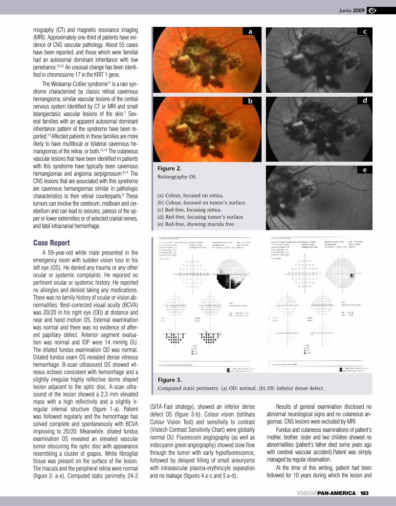
mography (CT) and magnetic resonance imaging (MRI). Approximately one-third of patients have evi-dence of CNS vascular pathology. About 55 cases have been reported, and those which were familial had an autosomal dominant inheritance with low penetrance.10,12 An unusual change has been identi-fied in chromosome 17 in the KRIT 1 gene.
The Weskamp-Cotlier syndrome13 is a rare syn-drome characterized by classic retinal cavernous hemangioma, similar vascular lesions of the central nervous system identified by CT or MRI and small telangiectasic vascular lesions of the skin.2 Sev-eral families with an apparent autosomal dominant inheritance pattern of the syndrome have been re-ported.13 Affected patients in these families are more likely to have multifocal or bilateral cavernous he-mangiomas of the retina, or both.13,14 The cutaneous vascular lesions that have been identified in patients with this syndrome have typically been cavernous hemangiomas and angioma serpiginosum.8,14 The CNS lesions that are associated with this syndrome are cavernous hemangiomas similar in pathologic characteristics to their retinal counterparts.8 These tumors can involve the cerebrum, midbrain and cer-ebellum and can lead to seizures, paresis of the up-per or lower extremities or of selected cranial nerves, and fatal intracranial hemorrhage.
Case ReportA 59-year-old white male presented in the
emergency room with sudden vision loss in his left eye (OS). He denied any trauma or any other ocular or systemic complaints. He reported no pertinent ocular or systemic history. He reported no allergies and denied taking any medications. There was no family history of ocular or vision ab-normalities. Best-corrected visual acuity (BCVA) was 20/20 in his right eye (OD) at distance and near and hand motion OS. External examination was normal and there was no evidence of affer-ent papillary defect. Anterior segment evalua-tion was normal and IOP were 14 mmHg OU. The dilated fundus examination OD was normal. Dilated fundus exam OS revealed dense vitreous hemorrhage. B-scan ultrasound OS showed vit-reous echoes consistent with hemorrhage and a slightly irregular highly reflective dome shaped lesion adjacent to the optic disc. A-scan ultra-sound of the lesion showed a 2,3 mm elevated mass with a high reflectivity and a slightly ir-regular internal structure (figure 1-a). Patient was followed regularly and the hemorrhage has solved complete and spontaneously with BCVA improving to 20/20. Meanwhile, dilated fundus examination OS revealed an elevated vascular tumor obscuring the optic disc with appearance resembling a cluster of grapes. White fibroglial tissue was present on the surface of the lesion. The macula and the peripheral retina were normal (figure 2: a-e). Computed static perimetry 24-2
(SITA-Fast strategy), showed an inferior dense defect OS (figure 3-b). Colour vision (Ishihara Colour Vision Test) and sensitivity to contrast (Vistech Contrast Sensitivity Chart) were globally normal OU. Fluorescein angiography (as well as indocyanin green angiography) showed slow flow through the tumor with early hypofluorescence, followed by delayed filling of small aneurysms with intravascular plasma-erythrocyte separation and no leakage (figures 4 a-c and 5 a-d).
Results of general examination disclosed no abnormal neurological signs and no cutaneous an-giomas. CNS lesions were excluded by MRI.
Fundus and cutaneous examinations of patient’s mother, brother, sister and two children showed no abnormalities (patient’s father died some years ago with cerebral vascular accident).Patient was simply managed by regular observation.
At the time of this writing, patient had been followed for 10 years during which the lesion and
: 183PAN-AMERICA
Junio 2009
(a) Colour, focused on retina. (b) Colour, focused on tumor’s surface. (c) Red-free, focusing retina. (d) Red-free, focusing tumor’s surface. (e) Red-free, showing macula free.
a
b
c
d
e
Figure 3. Computed static perimetry. (a) OD: normal. (b) OS: inferior dense defect.
Figure 2. Retinography OS.
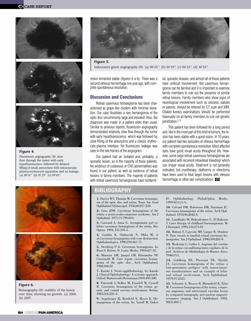
CASE REpoRt
184 : PAN-AMERICA
bIbLIOGRAPHy1. Davies WS, Thumim M. Cavernous hemangio-ma of the optic disc and retina. Trans Am Acad Ophthalmol Otolaryngol. 1956;60:217-218.
2. Gass JDM. Cavernous hemangioma of the retina: a neuro-oculo-cutaneous syndrome. Am J Opthalmol. 1971;71:799-814.
3. Gurwood A, Amos G. Asymptomatic girl ex-hibits cavernous hemangioma of the retina. Rev Optom. 1994; 131:101-5.
4. Gunduz K, Ozzbayrak N, Okka M, et al.Cavernous hemangioma with cone dysfunction. Ophthalmologica. 1996;210:367-71.
5. Sternberg P Jr. Cavernous hemangioma. In: Ryan S. Retina. St. Louis: Mosby, 1994:627-32.
6. Mansour AM, Jampol LM, Hrisomalos NF, Greenwald M. Case report: cavernous heman-gioma of the optic disc. Arch Ophthalmol. 1988;106:22.
7. Kanski J. Neuro-opphthalmology. In: Kanski J. Clinical Ophyhalmology: A systemic approach. Oxford: Butterworth-Heinemann,1994:495-500.
8. Pancurak J, Bolber M, Frendell M, Crowell R. Cavernous hemangioma of the retina: ge-netic and central nervous involvement. Retina. 1985;5(4):221-4.
9. Augsburger JJ, Bornfeld N, Meyer K. He-mangiomas of the retina. In: Yanoff M, Duker
JS. Ophthalmology. Philadelphia: Mosby, 1999;9(11):1-4.
10. Colvard DM, Robertson DM, Trautman JC. Cavernous hemangioma of the retina. Arch Oph-thalmol. 1978;96:2042-4.
11. Landthaler M, Hohenleutner U, El-Raheem T. Laser therapy of childhood haemangiomas. Br J Dermatol. 1995;133:275-81.
12. Bottoni F, Canevini MP, Canger R, Orzalesi N. Twin vessels in familial retinal cavernous he-mangioma. Am J Opthalmol. 1990;109:285-9.
13. Weskamp C, Cotlier I. Angioma del cerebro y de la retina con malformaciones capilares de la prel. Archivos de Oftalmologia de Buenos Aires. 1940;15:1.
14. Goldberg RE, Pheasant TR, Shields JA. Cavernous hemangioma of the retina: a four-generation pedigree with neurocutane-ous manifestations and an example of bilat-eral retinal involvement. Arch Ophthalmol. 1979;97:2321.
15. Schwartz A, Weaver R, Bloomfield R, Tyler M. Cavernous hemangioma of the retina, cutane-ous angiomas, and intracranial vascular lesion by computed tomography and nuclear magnetic resonance imaging. Am J Ophthalmol. 1984, 98(3):483-7.
Figure 6.Retinography OS: stability of the lesion over time, showing no growth. (a) 2004. (b) 2007.
Figure 4. Fluorescein angiography OS: slow flow through the tumor with early hypofluorescence, followed by delayed filling of small aneurysms with intravascular plasma-erythrocyte separation and no leakage. (a) 00’31”. (b) 01’29”. (c) 09’45”.
Figure 5. Indocianyn green angiography OS. (a) 00’23’’. (b) 03’39”. (c) 06’23”. (d) 30’33”.
vision remained stable (figures 6 a-b). There was a second vitreous hemorrhage one year ago, with com-plete spontaneous resolution.
Discussion and ConclusionsRetinal cavernous hemangioma has been char-
acterized as grape-like clusters with minimal eleva-tion. Our case illustrates a rare hemangioma of the optic disc uncommonly large and elevated. Also, the diagnosis was made in a patient older than usual. Similar to previous reports, fluorescein angiography demonstrated relatively slow flow through the tumor with early hypofluorescence, which was followed by slow filling of the aneurysms and a classic erythro-cyte-plasma interface. No fluorescein leakage was seen in the late frames of the angiogram.
Our patient had an isolated and, probably, a sporadic lesion, as in the majority of these patients. No evidence of cutaneous or CNS abnormalities was found in our patient, as well as evidence of retinal lesions in family members. The majority of patients with retinal cavernous hemangiomas have nonfamil-
ial, sporadic disease, and almost all of these patients have unifocal involvement. But cavernous heman-gioma can be familial and it is important to examine family members to rule out the presence of similar retinal lesions. Family members who show signs of neurological involvement such as seizures, palsies or paresis, should be referred for CT scan and MRI. Dilated fundus examinations should be performed biannually on all family members to rule out genetic penetration.3,15
This patient has been followed for a long period and, like in the most part of this kind of tumors, the le-sion has been stable with a good vision. In 10 years, our patient had two episodes of vitreous hemorrhage with complete spontaneous resolution. Most affected eyes have good visual acuity throughout life. How-ever, some large retinal cavernous hemangiomas are associated with recurrent intravitreal bleeding2 which can impair visual acuity. Treatment is not generally indicated, but cryotherapy, diathermy or vitrectomy have been used to treat larger lesions with vitreous hemorrhage or other rare complications.

: 185PAN-AMERICA
Junio 2009
Resumo
Objetivo: Relatar um caso de cisto gigan-te de retenção (CGR) após a realização de cirurgia filtrante com implante de tubo em um paciente com síndrome de Sturge-Weber (SWS) e glaucoma refratário.
Desenho do estudo: Relato de caso
Métodos: Um caso de aparecimento de CGR Após cirurgia antiglaucomatosa, sem intercorrências, para implante de tubo de Molteno não valvulado (em quadrante tem-poral superior) em uma criança de 4 anos, sexo masculino, portadora de SWS e que apresentava glaucoma secundário, será descrito. após insucesso com o tratamento medicamentoso tópico a criança foi subme-tida a cirurgia para implante de tubo de dre-nagem usando a técnica padrão. Devido ao retardo mental da criança, não foi possível a avaliação da acuidade visual.
Resultados: Após o procedimento, o pa-ciente foi reavaliado semanalmente durante o primeiro mês sendo, então, diagnostica-do atalamia. A câmara anterior foi refeita com sucesso e após este procedimento o paciente não compareceu aos retornos por um mês quando, então, retornou apresen-tando: proptose, desvio nasal-inferior do globo e restrição da motilidade ocular. Foi submetido a ultrassonografia ocular (US) e ressonância nuclear magnética (RNM) com-patíveis com CGR. O paciente foi pronta-mente tratado sendo restabelecido o correto posicionamento do globo, mas infelizmente houve provável comprometimento da visão, uma vez que foi diagnosticado descolamen-to de retina.
Conclusão: CGR é uma complicação rara que pode ocorrer após cirurgia filtrante com
implante de tubo. O reconhecimento precoce e a rápida intervenção são fundamentais para se conseguir o melhor resultado possível na tentativa de preservar a função visual.
Abstract
Background: To report a case of giant re-servoir cyst (GRC) following aqueous shunt (AS) surgery in a Sturge-Weber Syndrome (SWS) patient with refractory glaucoma.
Design: Interventional case report
Methods: One case of GRC occurring af-ter superotemporal quadrant implantation of nonvalved single plate Molteno drainage device, without intraoperative complications, is described. In the case, a four-year-old boy who has SWS developed secondary glauco-ma and after unsuccessful topical treatment he was underwent a valve implant surgery using a standard technique. Visual acuity was never possible to be measured because of mental retardation.
Results: The patient was followed every week during the first month, when athalamia was diagnosed. The anterior chamber was re-forming and after that patient missed follow-up for one month when returned to the clinic with proptosis, inferonasal dystopia and res-triction in ocular motility. Ultrasound (USB) and magnetic resonance imaging (CT) was consistent with GRC. Patient was promptly treated and the eye returned to its normal position. Unfortunately it was not possible to reestablish the patient’s vision once a total retinal detachment was further diagnosed.
Conclusion: GRC is a rare complication that may occur following AS surgery. Early recog-nition and intervention are of great relevance to achieve the best outcome possible and try to preserve visual function.
IntroductionSturge-Weber Syndrome (SWS) is a
rare congenital neurocutaneous disorder characterized by facial angioma and ip-silateral leptomeningeal hemangioma. This sporadic phacomatosis, present at birth, can be classified in trisystem when involves face, leptomeninges and eye or bisystem, when involves face and eyes or face and leptomeninges1,2. Ipsilateral secondary glaucoma has been reported in about 30% of patients and it is the most commonly reported ocular association in SWS1,2. In 60% of patients glaucoma presents in the first two years of life with buphthalmia, epiphora and photophobia, but it may also manifest at any time from childhood to adulthood in 40% of patients3. Although the precise pathogenesis of glauco-ma is unknown, the majority of publications claim that raised episcleral venous pressure would be the responsible mechanism4,5. Cronenberg et al. performed ultrasonic bio-microscopy (UBM) in eyes with SWS and found images of dilated intrasclerous ves-sels, dilated intraciliary vessels compatible with hemangioms, and supraciliary effusion in most of the glaucomatous eyes studied. These findings should explain not only the pathogenesis of the raised episcleral venous pressure but also the peri- and postoperative complications eventually present in these patients like choroidal effusions6.
Children with SWS often develop pro-gressive neurologic problems7, 8.
The current approach of glaucoma as-sociated with SWS is trabeculotomy or goniotomy in the ones that develop at birth and trabeculectomy or aqueous shunt sur-gery in the ones that present later in life3. However, surgical procedures are known to cause serious sight-threatening complica-tions in these patients as choroidal effusion or hemorrhage5.
Giant Reservoir Cyst Following Molteno Implant in Sturge-Weber Syndrome
Camila Cassiano Simões MD; Sérgio Henrique Sampaio Meirelles MD PhD; Ana Carolina de Arantes Frota MD PhD
Universidade Gama Filho, Rio de Janeiro, RJ, Brazil1 Corresponding Author:Camila Cassiano SimõesR: Wanderkey 637#35 – Perdizes, São Paulo, SP 05011-001 BrazilTel: (55-11) 3774-2917Fax: (55-11) 2509-2291Email: [email protected]
The authors do not have any financial relationship
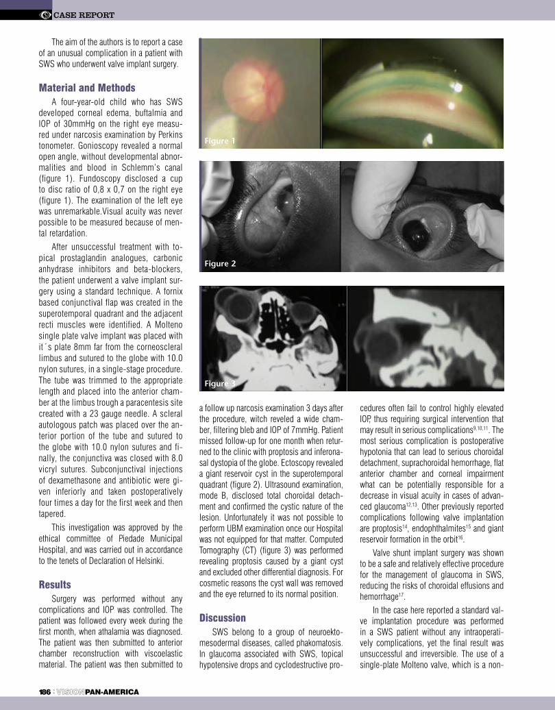
The aim of the authors is to report a case of an unusual complication in a patient with SWS who underwent valve implant surgery.
Material and MethodsA four-year-old child who has SWS
developed corneal edema, buftalmia and IOP of 30mmHg on the right eye measu-red under narcosis examination by Perkins tonometer. Gonioscopy revealed a normal open angle, without developmental abnor-malities and blood in Schlemm’s canal (figure 1). Fundoscopy disclosed a cup to disc ratio of 0,8 x 0,7 on the right eye (figure 1). The examination of the left eye was unremarkable.Visual acuity was never possible to be measured because of men-tal retardation.
After unsuccessful treatment with to-pical prostaglandin analogues, carbonic anhydrase inhibitors and beta-blockers, the patient underwent a valve implant sur-gery using a standard technique. A fornix based conjunctival flap was created in the superotemporal quadrant and the adjacent recti muscles were identified. A Molteno single plate valve implant was placed with it´s plate 8mm far from the corneoscleral limbus and sutured to the globe with 10.0 nylon sutures, in a single-stage procedure. The tube was trimmed to the appropriate length and placed into the anterior cham-ber at the limbus trough a paracentesis site created with a 23 gauge needle. A scleral autologous patch was placed over the an-terior portion of the tube and sutured to the globe with 10.0 nylon sutures and fi-nally, the conjunctiva was closed with 8.0 vicryl sutures. Subconjunctival injections of dexamethasone and antibiotic were gi-ven inferiorly and taken postoperatively four times a day for the first week and then tapered.
This investigation was approved by the ethical committee of Piedade Municipal Hospital, and was carried out in accordance to the tenets of Declaration of Helsinki.
ResultsSurgery was performed without any
complications and IOP was controlled. The patient was followed every week during the first month, when athalamia was diagnosed. The patient was then submitted to anterior chamber reconstruction with viscoelastic material. The patient was then submitted to
a follow up narcosis examination 3 days after the procedure, witch reveled a wide cham-ber, filtering bleb and IOP of 7mmHg. Patient missed follow-up for one month when retur-ned to the clinic with proptosis and inferona-sal dystopia of the globe. Ectoscopy revealed a giant reservoir cyst in the superotemporal quadrant (figure 2). Ultrasound examination, mode B, disclosed total choroidal detach-ment and confirmed the cystic nature of the lesion. Unfortunately it was not possible to perform UBM examination once our Hospital was not equipped for that matter. Computed Tomography (CT) (figure 3) was performed revealing proptosis caused by a giant cyst and excluded other differential diagnosis. For cosmetic reasons the cyst wall was removed and the eye returned to its normal position.
DiscussionSWS belong to a group of neuroekto-
mesodermal diseases, called phakomatosis. In glaucoma associated with SWS, topical hypotensive drops and cyclodestructive pro-
cedures often fail to control highly elevated IOP, thus requiring surgical intervention that may result in serious complications9,10,11. The most serious complication is postoperative hypotonia that can lead to serious choroidal detachment, suprachoroidal hemorrhage, flat anterior chamber and corneal impairment what can be potentially responsible for a decrease in visual acuity in cases of advan-ced glaucoma12,13. Other previously reported complications following valve implantation are proptosis14, endophthalmites15 and giant reservoir formation in the orbit16.
Valve shunt implant surgery was shown to be a safe and relatively effective procedure for the management of glaucoma in SWS, reducing the risks of choroidal effusions and hemorrhage17.
In the case here reported a standard val-ve implantation procedure was performed in a SWS patient without any intraoperati-vely complications, yet the final result was unsuccessful and irreversible. The use of a single-plate Molteno valve, which is a non-
CASE REpoRt
186 : PAN-AMERICA
Figure 1
Figure 2
Figure 3
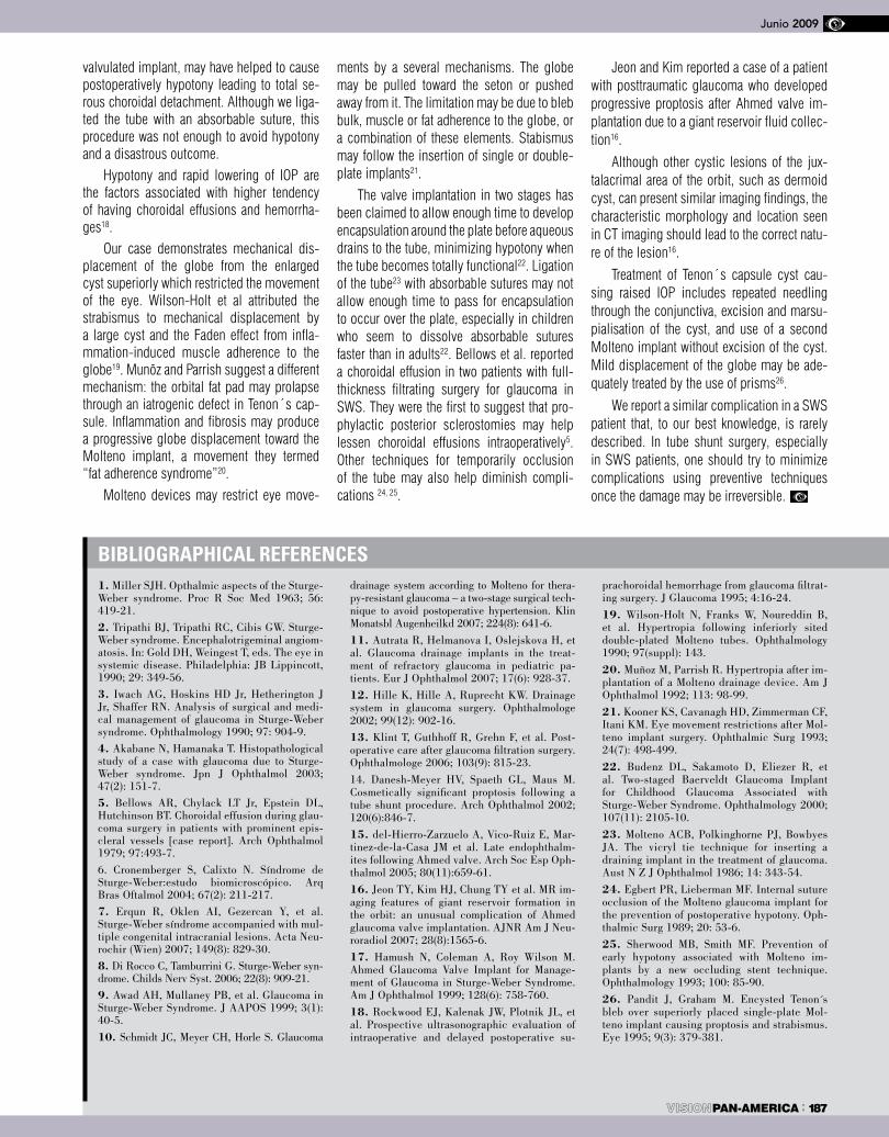
bIbLIOGRAPHICAL REFERENCES1. Miller SJH. Opthalmic aspects of the Sturge-Weber syndrome. Proc R Soc Med 1963; 56: 419-21.
2. Tripathi BJ, Tripathi RC, Cibis GW. Sturge-Weber syndrome. Encephalotrigeminal angiom-atosis. In: Gold DH, Weingest T, eds. The eye in systemic disease. Philadelphia: JB Lippincott, 1990; 29: 349-56.
3. Iwach AG, Hoskins HD Jr, Hetherington J Jr, Shaffer RN. Analysis of surgical and medi-cal management of glaucoma in Sturge-Weber syndrome. Ophthalmology 1990; 97: 904-9.
4. Akabane N, Hamanaka T. Histopathological study of a case with glaucoma due to Sturge-Weber syndrome. Jpn J Ophthalmol 2003; 47(2): 151-7.
5. Bellows AR, Chylack LT Jr, Epstein DL, Hutchinson BT. Choroidal effusion during glau-coma surgery in patients with prominent epis-cleral vessels [case report]. Arch Ophthalmol 1979; 97:493-7.
6. Cronemberger S, Calixto N. Síndrome de Sturge-Weber:estudo biomicroscópico. Arq Bras Oftalmol 2004; 67(2): 211-217.
7. Erqun R, Oklen AI, Gezercan Y, et al. Sturge-Weber síndrome accompanied with mul-tiple congenital intracranial lesions. Acta Neu-rochir (Wien) 2007; 149(8): 829-30.
8. Di Rocco C, Tamburrini G. Sturge-Weber syn-drome. Childs Nerv Syst. 2006; 22(8): 909-21.
9. Awad AH, Mullaney PB, et al. Glaucoma in Sturge-Weber Syndrome. J AAPOS 1999; 3(1): 40-5.
10. Schmidt JC, Meyer CH, Horle S. Glaucoma
drainage system according to Molteno for thera-py-resistant glaucoma – a two-stage surgical tech-nique to avoid postoperative hypertension. Klin Monatsbl Augenheilkd 2007; 224(8): 641-6.
11. Autrata R, Helmanova I, Oslejskova H, et al. Glaucoma drainage implants in the treat-ment of refractory glaucoma in pediatric pa-tients. Eur J Ophthalmol 2007; 17(6): 928-37.
12. Hille K, Hille A, Ruprecht KW. Drainage system in glaucoma surgery. Ophthalmologe 2002; 99(12): 902-16.
13. Klint T, Guthhoff R, Grehn F, et al. Post-operative care after glaucoma filtration surgery. Ophthalmologe 2006; 103(9): 815-23.
14. Danesh-Meyer HV, Spaeth GL, Maus M. Cosmetically significant proptosis following a tube shunt procedure. Arch Ophthalmol 2002; 120(6):846-7.
15. del-Hierro-Zarzuelo A, Vico-Ruiz E, Mar-tinez-de-la-Casa JM et al. Late endophthalm-ites following Ahmed valve. Arch Soc Esp Oph-thalmol 2005; 80(11):659-61.
16. Jeon TY, Kim HJ, Chung TY et al. MR im-aging features of giant reservoir formation in the orbit: an unusual complication of Ahmed glaucoma valve implantation. AJNR Am J Neu-roradiol 2007; 28(8):1565-6.
17. Hamush N, Coleman A, Roy Wilson M. Ahmed Glaucoma Valve Implant for Manage-ment of Glaucoma in Sturge-Weber Syndrome. Am J Ophthalmol 1999; 128(6): 758-760.
18. Rockwood EJ, Kalenak JW, Plotnik JL, et al. Prospective ultrasonographic evaluation of intraoperative and delayed postoperative su-
prachoroidal hemorrhage from glaucoma filtrat-ing surgery. J Glaucoma 1995; 4:16-24.
19. Wilson-Holt N, Franks W, Noureddin B, et al. Hypertropia following inferiorly sited double-plated Molteno tubes. Ophthalmology 1990; 97(suppl): 143.
20. Muñoz M, Parrish R. Hypertropia after im-plantation of a Molteno drainage device. Am J Ophthalmol 1992; 113: 98-99.
21. Kooner KS, Cavanagh HD, Zimmerman CF, Itani KM. Eye movement restrictions after Mol-teno implant surgery. Ophthalmic Surg 1993; 24(7): 498-499.
22. Budenz DL, Sakamoto D, Eliezer R, et al. Two-staged Baerveldt Glaucoma Implant for Childhood Glaucoma Associated with Sturge-Weber Syndrome. Ophthalmology 2000; 107(11): 2105-10.
23. Molteno ACB, Polkinghorne PJ, Bowbyes JA. The vicryl tie technique for inserting a draining implant in the treatment of glaucoma. Aust N Z J Ophthalmol 1986; 14: 343-54.
24. Egbert PR, Lieberman MF. Internal suture occlusion of the Molteno glaucoma implant for the prevention of postoperative hypotony. Oph-thalmic Surg 1989; 20: 53-6.
25. Sherwood MB, Smith MF. Prevention of early hypotony associated with Molteno im-plants by a new occluding stent technique. Ophthalmology 1993; 100: 85-90.
26. Pandit J, Graham M. Encysted Tenon´s bleb over superiorly placed single-plate Mol-teno implant causing proptosis and strabismus. Eye 1995; 9(3): 379-381.
valvulated implant, may have helped to cause postoperatively hypotony leading to total se-rous choroidal detachment. Although we liga-ted the tube with an absorbable suture, this procedure was not enough to avoid hypotony and a disastrous outcome.
Hypotony and rapid lowering of IOP are the factors associated with higher tendency of having choroidal effusions and hemorrha-ges18.
Our case demonstrates mechanical dis-placement of the globe from the enlarged cyst superiorly which restricted the movement of the eye. Wilson-Holt et al attributed the strabismus to mechanical displacement by a large cyst and the Faden effect from infla-mmation-induced muscle adherence to the globe19. Munõz and Parrish suggest a different mechanism: the orbital fat pad may prolapse through an iatrogenic defect in Tenon´s cap-sule. Inflammation and fibrosis may produce a progressive globe displacement toward the Molteno implant, a movement they termed “fat adherence syndrome”20.
Molteno devices may restrict eye move-
ments by a several mechanisms. The globe may be pulled toward the seton or pushed away from it. The limitation may be due to bleb bulk, muscle or fat adherence to the globe, or a combination of these elements. Stabismus may follow the insertion of single or double-plate implants21.
The valve implantation in two stages has been claimed to allow enough time to develop encapsulation around the plate before aqueous drains to the tube, minimizing hypotony when the tube becomes totally functional22. Ligation of the tube23 with absorbable sutures may not allow enough time to pass for encapsulation to occur over the plate, especially in children who seem to dissolve absorbable sutures faster than in adults22. Bellows et al. reported a choroidal effusion in two patients with full-thickness filtrating surgery for glaucoma in SWS. They were the first to suggest that pro-phylactic posterior sclerostomies may help lessen choroidal effusions intraoperatively5.Other techniques for temporarily occlusion of the tube may also help diminish compli-cations 24, 25.
Jeon and Kim reported a case of a patient with posttraumatic glaucoma who developed progressive proptosis after Ahmed valve im-plantation due to a giant reservoir fluid collec-tion16.
Although other cystic lesions of the jux-talacrimal area of the orbit, such as dermoid cyst, can present similar imaging findings, the characteristic morphology and location seen in CT imaging should lead to the correct natu-re of the lesion16.
Treatment of Tenon´s capsule cyst cau-sing raised IOP includes repeated needling through the conjunctiva, excision and marsu-pialisation of the cyst, and use of a second Molteno implant without excision of the cyst. Mild displacement of the globe may be ade-quately treated by the use of prisms26.
We report a similar complication in a SWS patient that, to our best knowledge, is rarely described. In tube shunt surgery, especially in SWS patients, one should try to minimize complications using preventive techniques once the damage may be irreversible.
: 187PAN-AMERICA
Junio 2009
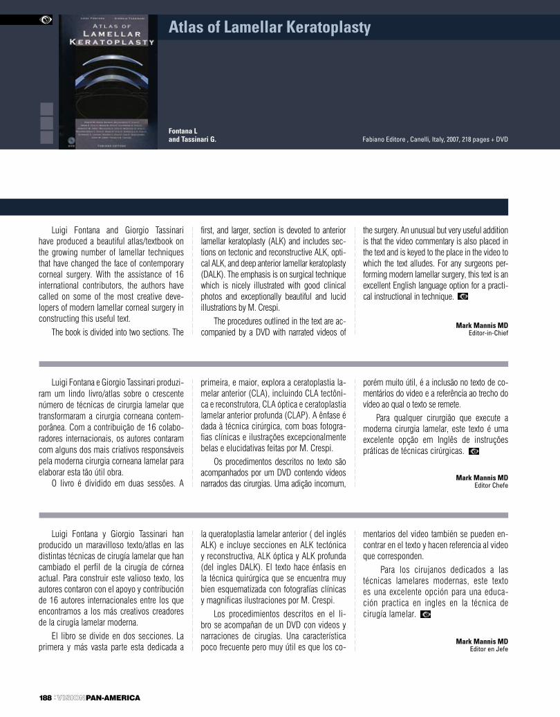
PAN-AMERICA188 :
Atlas of Lamellar Keratoplasty
Fontana Land Tassinari G. Fabiano Editore , Canelli, Italy, 2007, 218 pages + DVD
Luigi Fontana and Giorgio Tassinari have produced a beautiful atlas/textbook on the growing number of lamellar techniques that have changed the face of contemporary corneal surgery. With the assistance of 16 international contributors, the authors have called on some of the most creative deve-lopers of modern lamellar corneal surgery in constructing this useful text.
The book is divided into two sections. The
first, and larger, section is devoted to anterior lamellar keratoplasty (ALK) and includes sec-tions on tectonic and reconstructive ALK, opti-cal ALK, and deep anterior lamellar keratoplasty (DALK). The emphasis is on surgical technique which is nicely illustrated with good clinical photos and exceptionally beautiful and lucid illustrations by M. Crespi.
The procedures outlined in the text are ac-companied by a DVD with narrated videos of
the surgery. An unusual but very useful addition is that the video commentary is also placed in the text and is keyed to the place in the video to which the text alludes. For any surgeons per-forming modern lamellar surgery, this text is an excellent English language option for a practi-cal instructional in technique.
Mark Mannis MDEditor-in-Chief
Luigi Fontana e Giorgio Tassinari produzi-ram um lindo livro/atlas sobre o crescente número de técnicas de cirurgia lamelar que transformaram a cirurgia corneana contem-porânea. Com a contribuição de 16 colabo-radores internacionais, os autores contaram com alguns dos mais criativos responsáveis pela moderna cirurgia corneana lamelar para elaborar esta tão útil obra.
O livro é dividido em duas sessões. A
primeira, e maior, explora a ceratoplastia la-melar anterior (CLA), incluindo CLA tectôni-ca e reconstrutora, CLA óptica e ceratoplastia lamelar anterior profunda (CLAP). A ênfase é dada à técnica cirúrgica, com boas fotogra-fias clínicas e ilustrações excepcionalmente belas e elucidativas feitas por M. Crespi.
Os procedimentos descritos no texto são acompanhados por um DVD contendo vídeos narrados das cirurgias. Uma adição incomum,
porém muito útil, é a inclusão no texto de co-mentários do video e a referência ao trecho do vídeo ao qual o texto se remete.
Para qualquer cirurgião que execute a moderna cirurgia lamelar, este texto é uma excelente opção em Inglês de instruções práticas de técnicas cirúrgicas.
Mark Mannis MDEditor Chefe
Luigi Fontana y Giorgio Tassinari han producido un maravilloso texto/atlas en las distintas técnicas de cirugía lamelar que han cambiado el perfil de la cirugía de córnea actual. Para construir este valioso texto, los autores contaron con el apoyo y contribución de 16 autores internacionales entre los que encontramos a los más creativos creadores de la cirugía lamelar moderna.
El libro se divide en dos secciones. La primera y más vasta parte esta dedicada a
la queratoplastia lamelar anterior ( del inglés ALK) e incluye secciones en ALK tectónica y reconstructiva, ALK óptica y ALK profunda (del ingles DALK). El texto hace énfasis en la técnica quirúrgica que se encuentra muy bien esquematizada con fotografías clínicas y magnificas ilustraciones por M. Crespi.
Los procedimientos descritos en el li-bro se acompañan de un DVD con videos y narraciones de cirugías. Una característica poco frecuente pero muy útil es que los co-
mentarios del video también se pueden en-contrar en el texto y hacen referencia al video que corresponden.
Para los cirujanos dedicados a las técnicas lamelares modernas, este texto es una excelente opción para una educa-ción practica en ingles en la técnica de cirugía lamelar.
Mark Mannis MDEditor en Jefe
Manual de Condutas em OftalmologiaJunio 2009
PAN-AMERICA : 189
Atlas of Lamellar Keratoplasty
Faculty at the Federal University of São Paulo (UNIFESP), one of Brazil’s powerhouses of ophthalmology, have produced a multi-authored and detailed manual of practical eye care that spans the breadth of diagnosis and therapy for eye diseases of all categories. This Portuguese language text is divided into twenty sections covering abnormalities of the crystalline lens, external disease, glaucoma, neuro-ophthalmology, pediatric ophthalmolo-gy, orbit, oculoplastics, refraction and contact
lens, refractive surgery, retina and vitreous, trauma and ophthalmic emergencies, ocular oncology, uveitis, the lacrimal system, patho-logic anatomy, laboratory microbiology, ocular ultrasound, electrophysiology, ocular pharma-cology, and low vision.
The authors have organized the book so that each succinct chapter follows a similar format: definition of the disorder, signs and symptoms, examination, differential diagno-sis, and treatment. The chapters are short but
well referenced and provide the reader with a rapid but thorough discussion of the clinical entity, its diagnosis and management. Just as the manual devised at the Wills Eye Hospital in the United States has become a standard tool for almost every trainee in North Ameri-ca, this book will likely become a standard tool for Spanish and Portuguese speaking ophthalmologists. It is clear, comprehensive, and will surely be one of the most frequently used texts in Latin ophthalmology.
O corpo docente da Universidade Fede-ral de São Paulo (UNIFESP), um dos maiores centros de oftalmologia do Brasil, produziu um manual detalhado, de múltipla autoria, sobre cuidados práticos oftalmológicos, que engloba diagnóstico e terapia de todas as categorias de doenças oculares.
Esta obra em Língua Portuguesa é divi-dida em vinte sessões, cobrindo anormali-dades do cristalino, doenças externas, glau-coma, neuro-oftalmologia, oftalmopediatria, órbita, oculoplástica, refração e lentes de
contato, cirurgia refrativa, retina e vítreo, trau-ma e emergências oftalmológicas, oncologia ocular, uveíte, sistema lacrimal, anatomia patológica, microbiologia, ultra-sonografia ocular, eletrofisiologia, farmacologia ocular e visão subnormal.
Os autores organizaram o livro de modo que cada sucinto capítulo siga o mesmo for-mato: definição da doença, sinais e sintomas, exame clínico, diagnósticos diferenciais e tra-tamento. Os capítulos são curtos, mas com boa referência bibliográfica, fornecendo ao lei-
tor uma discussão rápida, porém detalhada, da entidade clínica, seu diagnóstico e manuseio.
Assim como o manual desenvolvido no Wills Eye Hospital nos Estados Unidos tornou-se uma ferramenta padrão para quase todos os oftalmologistas em treinamento na América do Norte, este livro provavelmente também tornar-se-á uma ferramenta padrão para oftalmologistas de Língua Espanhola e Portuguesa. A obra é clara, abrangente e certamente será uma das mais utilizadas na oftalmologia latina.
El profesorado de la Universidad de São Paulo (UNIFESP), un símbolo de la oftalmo-logía brasileña, ha producido un manual de prácticas oftalmológicas que cubre amplia-mente el diagnóstico y tratamiento de las enfermedades oculares en todas sus cate-gorías. Este texto en portugués se encuentra dividido en veinte secciones que abordan las anomalías del cristalino, las enfermedades externas, glaucoma, neuro-oftalmología, of-talmología pediátrica, órbita, oculo-plástica, refracción y lentes de contacto, cirugía re-
fractiva, retina y vítreo, trauma y urgencias oftalmológicas, oncología ocular, uveítis, vía lagrimal, anatomo-patología , microbiología, ecografía, electrofisiología, farmacología y visión baja.
Los autores han organizado este libro de forma tal que cada capítulo aborda el objeti-vo con un formato similar: definición , signos y síntomas, examen oftalmológico, diagnos-tico diferencial y tratamiento. Los capítulos son cortos pero bien referenciados, y proveen al lector de una discusión corta y concisa de
la entidad , su diagnóstico y tratamiento. De la misma manera que el manual concebi-do en el Wills Eye Hostpital en los Estados Unidos se ha convertido en una herramienta indispensable para los que se encuentran en entrenamiento en Norte America, este libro muy probablemente se convertirá en un ma-nual fundamental para los oftalmólogos de habla portuguesa e hispana. Este texto claro e integral seguramente se convertirá en uno de los libros más consultados en la oftalmo-logía latino-americana.
Hofling-Lima AL,Tobias de Aguiar Moeller CT,de Freitas D,and Nogueira Martins E Editora Atheneu, Sao Paulo, 2008, 1249 pages

190 : PAN-AMERICA
Thank you for giving me the chance to spend October 2008 with Dr. Martin Devoto, in Buenos Aires, and for your support with the Paul Kayser International Scholarship. As a re-sident planning a career in ophthalmic plastic and reconstructive surgery, I came across Dr. Devoto’s name in three completely independent ways. First, as I conveyed my interest to the Pan-American Foundation in learning about what this specialty was like in Argentina, it was suggested I spend some time with Dr. Devoto. Second, my oculoplastics faculty during my residency training, recommended I see what Dr. Devoto’s practice was like. Third, as a staff ophthalmologist for ORBIS International, the Flying Eye Hospital team members told me how remarkable it was to work with him. So, after finding that multiple roads to ophthalmic plastic and reconstructive surgery in Argentina led to Dr. Martin Devoto, I elected to apply my scholar-ship to learning why that was.
In the area of lacrimal disease, dacrycystorhinostomy is one of the most common proce-dures performed by ophthalmic plastic surgeons. Working with Dr. Devoto, I learned some of his particular approaches and reasons for his methods. The published prospective study about the type of incision he uses is an example of the scientific rigor that he applies in his practice. I also learned the importance of regular nasal osteotomy examination in patients who have undergone dacryocystorhinostomy, as often times the success or failure of this procedure can be so discovered. The variety in his practice was something that I had not seen here in the United States. From orbital tumors, his approach to patients presenting with active thryroid eye disease, lacrimal problems, and eyelid pathology, I had a chance to be exposed to the full breath of oculoplastics. Surprisingly, the origins of the patient population that presented to his clinic was equally broad—from Tierra del Fuego to even my own hometown of Highlands Ranch, Colorado, USA.
As digital photography has revolutionized ophthalmology, working with Dr. Devoto, I was able to appreciate how elegantly he was able to apply these techniques in explaining procedu-res to patients. As patients came into the clinic, his consistently systematic approach in pho-tographic documentation helped build an easily navigable library of pathology for me to learn from and allowed patients to better grasp the nature of their diagnosis. Patients also seemed to have a better understanding of what their post-operative course may be like.
In short, my first experience with the PAAO, was when I was a 4th year medical student heading to Peru. The PAAO and Dr. Francisco Contreras, at that time kindly put me in touch with Dr. Renee Vivanco, a comprehensive ophthalmologist in Cusco. It was there that I first learned some of the fundamentals of clinical ophthalmology, such as retinoscopy skills and biomicros-copy. Four years later, having finished my residency training, I have been fortunate to again benefit from the kindness and generosity of this association. The time I spent with Dr. Devoto will most certainly be helpful to me as I begin my fellowship. His well-known approaches to lacrimal and thyroid disease are concepts I will carry with me the rest of my career. His clinic, with its high level of technology, efficiency, scientific rigor, and particular attention to detail, are aspects I hope to one day emulate when I develop my own practice.
As I begin my fellowship in July, I look forward to maintaining my close affiliation with the Pan-American Association of Ophthalmology and Dr. Martin Devoto for many years to come. I was fortunate to be able to attend the PAAO President’s Reception this year at the AAO and amazed to see so many people whose names I had only read in textbooks. During my planned career in academic oculoplastic surgery, I plan to contribute to the PAAO Journal, participate in the scientific meetings, and look forward to the international collegiality that this group provi-des. I thank the scholarship committee for allowing me to increase my exposure to the research and clinical teaching practices of oculoplastics in Argentina as well as Dr. Martin Devoto and Consultores Oftalmologos for their kind hospitality.
Srinivas S. Iyengar MDPaul Kayser International Scholar Report
Srinivas S. Iyengar, MDPAAO Paul Kayser International Scholar 2008October 2008
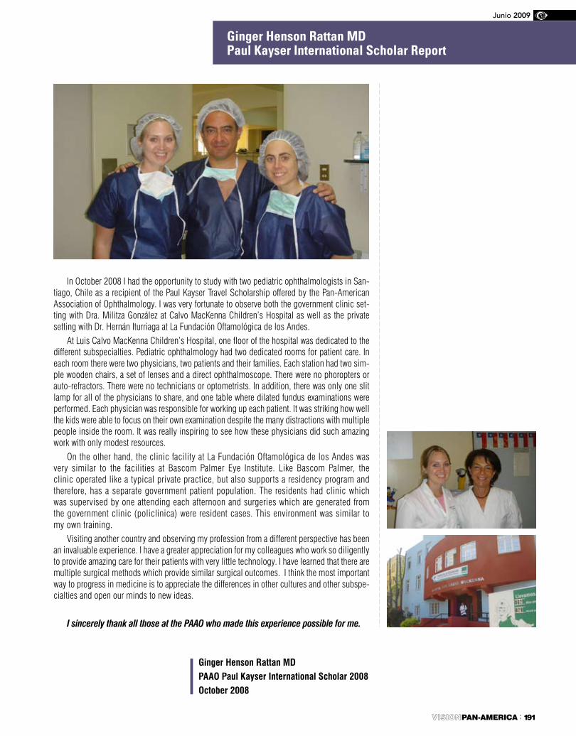
: 191PAN-AMERICA
Junio 2009
In October 2008 I had the opportunity to study with two pediatric ophthalmologists in San-tiago, Chile as a recipient of the Paul Kayser Travel Scholarship offered by the Pan-American Association of Ophthalmology. I was very fortunate to observe both the government clinic set-ting with Dra. Militza González at Calvo MacKenna Children’s Hospital as well as the private setting with Dr. Hernán Iturriaga at La Fundación Oftamológica de los Andes.
At Luis Calvo MacKenna Children’s Hospital, one floor of the hospital was dedicated to the different subspecialties. Pediatric ophthalmology had two dedicated rooms for patient care. In each room there were two physicians, two patients and their families. Each station had two sim-ple wooden chairs, a set of lenses and a direct ophthalmoscope. There were no phoropters or auto-refractors. There were no technicians or optometrists. In addition, there was only one slit lamp for all of the physicians to share, and one table where dilated fundus examinations were performed. Each physician was responsible for working up each patient. It was striking how well the kids were able to focus on their own examination despite the many distractions with multiple people inside the room. It was really inspiring to see how these physicians did such amazing work with only modest resources.
On the other hand, the clinic facility at La Fundación Oftamológica de los Andes was very similar to the facilities at Bascom Palmer Eye Institute. Like Bascom Palmer, the clinic operated like a typical private practice, but also supports a residency program and therefore, has a separate government patient population. The residents had clinic which was supervised by one attending each afternoon and surgeries which are generated from the government clinic (policlinica) were resident cases. This environment was similar to my own training.
Visiting another country and observing my profession from a different perspective has been an invaluable experience. I have a greater appreciation for my colleagues who work so diligently to provide amazing care for their patients with very little technology. I have learned that there are multiple surgical methods which provide similar surgical outcomes. I think the most important way to progress in medicine is to appreciate the differences in other cultures and other subspe-cialties and open our minds to new ideas.
I sincerely thank all those at the PAAO who made this experience possible for me.
Ginger Henson Rattan MDPaul Kayser International Scholar Report
Ginger Henson Rattan MDPAAO Paul Kayser International Scholar 2008October 2008
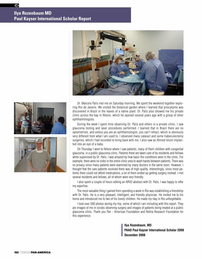
PAN-AMERICA192 :
Dr. Marcelo Palis met me on Saturday morning. We spent the weekend together explo-ring Rio de Janeiro. We visited the botanical garden where I learned that pilocarpine was discovered in Brazil in the leaves of a native plant. Dr. Palis also showed me his private clinic across the bay in Niteroi, which he opened several years ago with a group of other ophthalmologists.
During the week I spent time observing Dr. Palis and others in a private clinic. I saw glaucoma testing and laser procedures performed. I learned that in Brazil there are no optometrists, and unless you are an ophthalmologist, you can’t refract, which is obviously very different from what I am used to. I observed many cataract and some trabeculectomy surgeries, which I had recorded to bring back with me. I also saw an Ahmed shunt implan-ted into an eye of a baby.
On Thursday I went to Niteroi where I saw patients, many of them children with congenital glaucoma, in a public glaucoma clinic. Patients there are taken care of by residents and fellows while supervised by Dr. Palis. I was amazed by how basic the conditions were in the clinic. For example, there were no sinks in the entire clinic area to wash hands between patients. There was no privacy since many patients were examined by many doctors in the same room. However, I thought that the care patients received there was of high quality. Interestingly, since most pa-tients there could not afford medications, a lot of them ended up getting surgery instead. I met several residents and fellows, all of whom were very friendly.
I also spent a couple of hours editing an ARVO abstract with Dr. Palis. I was happy to offer my expertise.
The most valuable thing I gained from spending a week in Rio was establishing a friendship with Dr. Palis. He is a very pleasant, intelligent, and friendly physician. He invited me to his home and introduced me to two of his lovely children. He made my stay in Rio unforgettable.
I took over 500 photos during my trip, some of which I am including with this report. They are images of me in scrubs observing surgery and images of patients being treated at a public glaucoma clinic. Thank you Pan - American Foundation and Retina Research Foundation for this experience.
Ilya Rozenbaum MDPaul Kayser International Scholar Report
Ilya Rozenbaum, MDPAAO Paul Kayser International Scholar 2008December 2008
Mark Your Calendar
�e Foundation � the Pan American A�ociation � Ophthalmology Gala Yacht Event
San Francisco, California.Friday, October 23, 2009 7 PM - 10 PM
Food, dancing, cash bar! Tickets go on sale soon!www.paao.org
paofye 1 22/2/09 20:51:21


![VYSOKÉ UČENÍ TECHNICKÉ V BRNĚ · Vystrojení vrtů pro vertikální inklinometrii proběhlo 11.6. - 16.6.2009. [7] Geologické podmínky byly stanoveny z geologické mapy 1:50](https://img.pdfslide.us/doc/110x75/608beec7b8e7ce5c1d2adae9/vysok-uoeen-technick-v-brn-vystrojen-vrt-pro-vertikln-inklinometrii.jpg)