Embed Size (px)
Citation preview
The Respiratory System

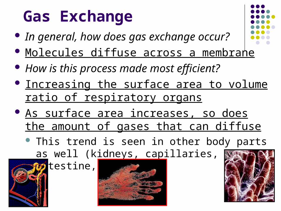
Gas Exchange In general, how does gas exchange occur? Molecules diffuse across a membrane How is this process made most efficient? Increasing the surface area to volume ratio of
respiratory organs As surface area increases, so does the amount of
gases that can diffuse This trend is seen in other body parts as well
(kidneys, capillaries, small intestine, etc.)
Gas Exchange cont.
What condition is necessary for gas exchange?
The surface for diffusion must be moist Ventilation – movement of gases between the
environment and the respiratory organ What is the difference between internal and
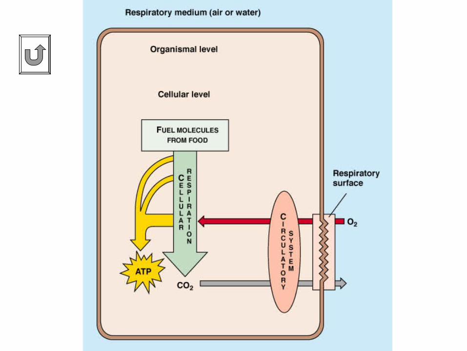
external respiration? External respiration = exchange of gases
between respiratory organ and blood Internal respiration = exchange between blood
and body cells
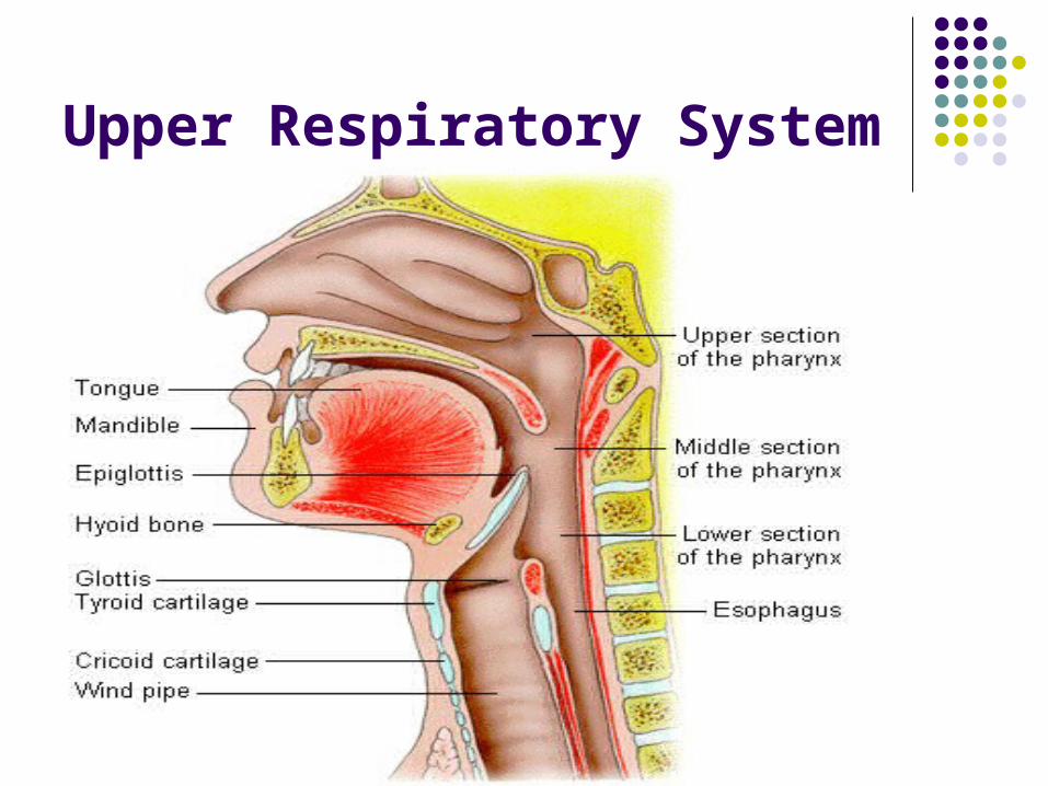
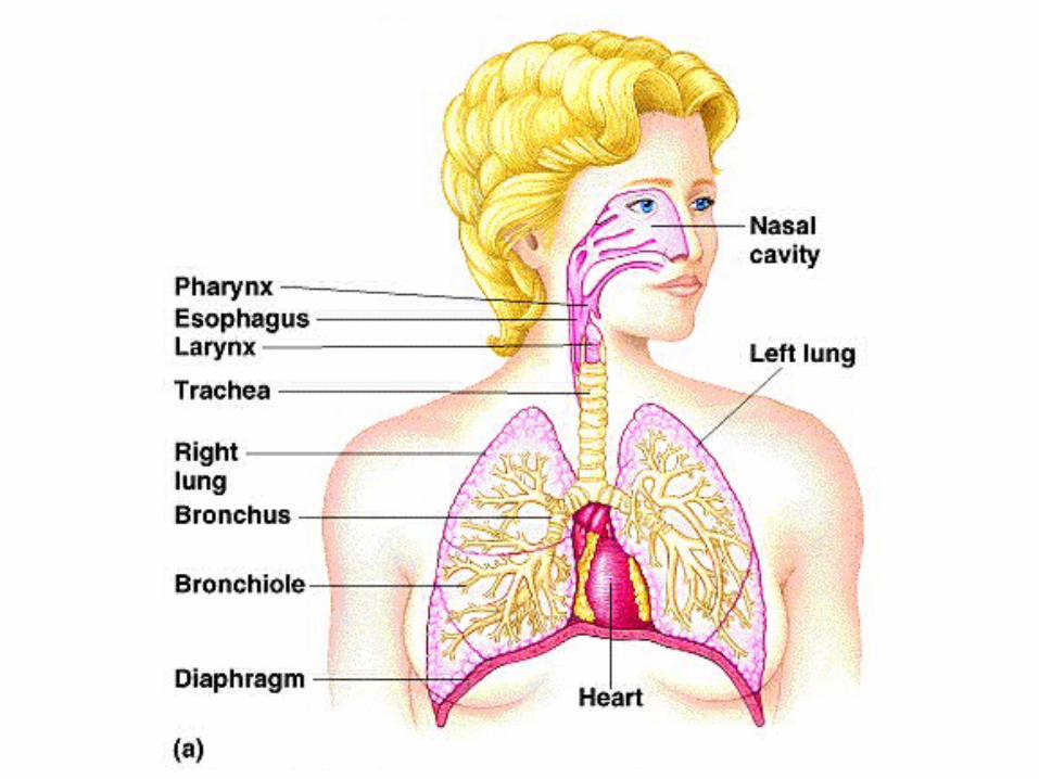
Upper Respiratory System
Respiratory System
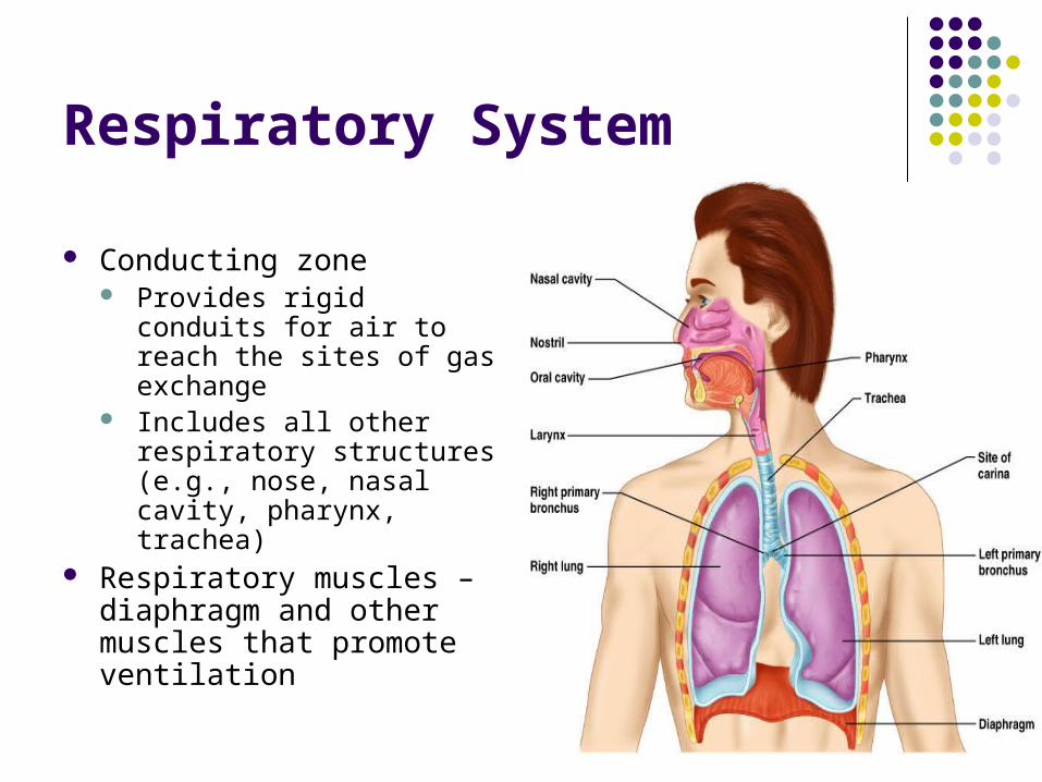
Conducting zone Provides rigid conduits for
air to reach the sites of gas exchange
Includes all other respiratory structures (e.g., nose, nasal cavity, pharynx, trachea)
Respiratory muscles – diaphragm and other muscles that promote ventilation
Major Functions of the Respiratory System
To supply the body with oxygen and dispose of carbon dioxide
Respiration – four distinct processes must happen Pulmonary ventilation – moving air into and out of
the lungs External respiration – gas exchange between the
lungs and the blood
Major Functions of the Respiratory System
Transport – transport of oxygen and carbon dioxide between the lungs and tissues
Internal respiration – gas exchange between systemic blood vessels and tissues
Function of the Nose
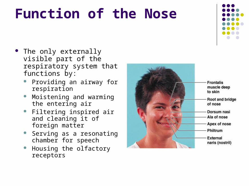
The only externally visible part of the respiratory system that functions by: Providing an airway for
respiration Moistening and warming
the entering air Filtering inspired air and
cleaning it of foreign matter Serving as a resonating
chamber for speech Housing the olfactory
receptors
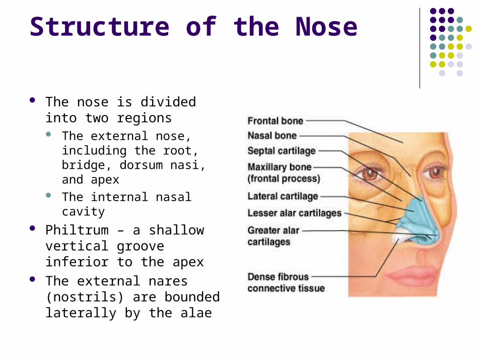
Structure of the Nose
The nose is divided into two regions The external nose,
including the root, bridge, dorsum nasi, and apex
The internal nasal cavity Philtrum – a shallow vertical
groove inferior to the apex The external nares (nostrils)
are bounded laterally by the alae
Nasal Cavity
Lies in and posterior to the external nose Is divided by a midline nasal septum Opens posteriorly into the nasal pharynx via
internal nares The ethmoid and sphenoid bones form the
roof The floor is formed by the hard and soft
palates
Nasal Cavity
Vestibule – nasal cavity superior to the nares Vibrissae – hairs that filter coarse particles from
inspired air Olfactory mucosa
Lines the superior nasal cavity Contains smell receptors
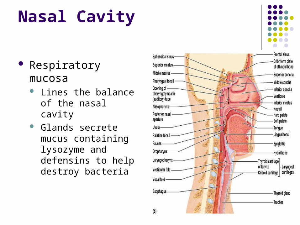
Nasal Cavity
Respiratory mucosa Lines the balance of the
nasal cavity Glands secrete mucus
containing lysozyme and defensins to help destroy bacteria
Nasal Cavity
Inspired air is: Humidified by the high water content in the nasal
cavity Warmed by rich plexuses of capillaries
Ciliated mucosal cells remove contaminated mucus
Nasal Cavity
Superior, medial, and inferior conchae: Protrude medially from the lateral walls Increase mucosal area Enhance air turbulence and help filter air
Sensitive mucosa triggers sneezing when stimulated by irritating particles
Functions of the Nasal Mucosa and Conchae
During inhalation the conchae and nasal mucosa: Filter, heat, and moisten air
During exhalation these structures: Reclaim heat and moisture Minimize heat and moisture loss
Paranasal Sinuses
Sinuses in bones that surround the nasal cavity
Sinuses lighten the skull and help to warm and moisten the air
Pharynx
Funnel-shaped tube of skeletal muscle that connects to the: Nasal cavity and mouth superiorly Larynx and esophagus inferiorly
Extends from the base of the skull to the level of the sixth cervical vertebra
Pharynx
It is divided into three regions Nasopharynx Oropharynx Laryngopharynx
Nasopharynx
Lies posterior to the nasal cavity, inferior to the sphenoid, and superior to the level of the soft palate
Strictly an air passageway Lined with pseudostratified columnar epithelium Closes during swallowing to prevent food from
entering the nasal cavity The pharyngeal tonsil lies high on the posterior wall Pharyngotympanic (auditory) tubes open into the
lateral walls
Oropharynx
Extends inferiorly from the level of the soft palate to the epiglottis
Opens to the oral cavity via an archway called the fauces
Serves as a common passageway for food and air The epithelial lining is protective stratified squamous
epithelium Palatine tonsils lie in the lateral walls of the fauces Lingual tonsil covers the base of the tongue
Laryngopharynx
Serves as a common passageway for food and air
Lies posterior to the upright epiglottis Extends to the larynx, where the respiratory
and digestive pathways diverge
Larynx (Voice Box)
Attaches to the hyoid bone and opens into the laryngopharynx superiorly
Continuous with the trachea posteriorly The three functions of the larynx are:
To provide a patent airway To act as a switching mechanism to route air and
food into the proper channels To function in voice production
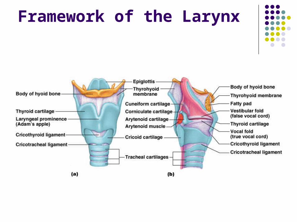
Framework of the Larynx
Cartilages (hyaline) of the larynx Shield-shaped anterosuperior thyroid cartilage
with a midline laryngeal prominence (Adam’s apple)
Signet ring–shaped anteroinferior cricoid cartilage Three pairs of small arytenoid, cuneiform, and
corniculate cartilages Epiglottis – elastic cartilage that covers the
laryngeal inlet during swallowing
Framework of the Larynx
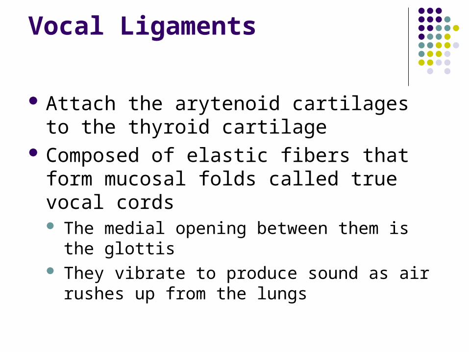
Vocal Ligaments
Attach the arytenoid cartilages to the thyroid cartilage
Composed of elastic fibers that form mucosal folds called true vocal cords The medial opening between them is the glottis They vibrate to produce sound as air rushes up
from the lungs
Vocal Ligaments
False vocal cords Mucosal folds superior to the true vocal cords Have no part in sound production
Vocal Production
Speech – intermittent release of expired air while opening and closing the glottis
Pitch – determined by the length and tension of the vocal cords
Loudness – depends upon the force at which the air rushes across the vocal cords
The pharynx resonates, amplifies, and enhances sound quality
Sound is “shaped” into language by action of the pharynx, tongue, soft palate, and lips
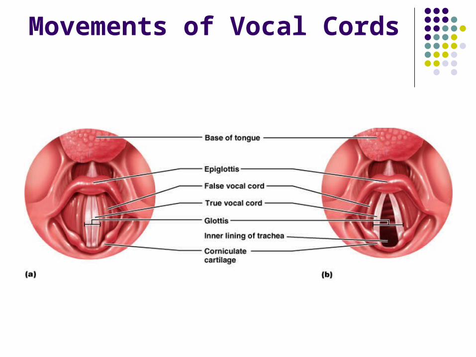
Movements of Vocal Cords
Sphincter Functions of the Larynx
The larynx is closed during coughing, sneezing, and Valsalva’s maneuver
Valsalva’s maneuver Air is temporarily held in the lower respiratory tract
by closing the glottis Causes intra-abdominal pressure to rise when
abdominal muscles contract Helps to empty the rectum Acts as a splint to stabilize the trunk when lifting
heavy loads

Larynx Video
Trachea
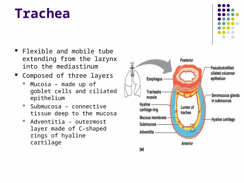
Flexible and mobile tube extending from the larynx into the mediastinum
Composed of three layers Mucosa – made up of
goblet cells and ciliated epithelium
Submucosa – connective tissue deep to the mucosa
Adventitia – outermost layer made of C-shaped rings of hyaline cartilage
Trachea Video
Conducting Zone: Bronchi
The carina of the last tracheal cartilage marks the end of the trachea and the beginning of the right and left bronchi
Air reaching the bronchi is: Warm and cleansed of impurities Saturated with water vapor
Bronchi subdivide into secondary bronchi, each supplying a lobe of the lungs
Air passages undergo 23 orders of branching in the lungs
Conducting Zone: Bronchial Tree
Tissue walls of bronchi mimic that of the trachea
As conducting tubes become smaller, structural changes occur Cartilage support structures change Epithelium types change Amount of smooth muscle increases
Conducting Zone: Bronchial Tree
Bronchioles Consist of cuboidal epithelium Have a complete layer of circular smooth muscle Lack cartilage support and mucus-producing cells
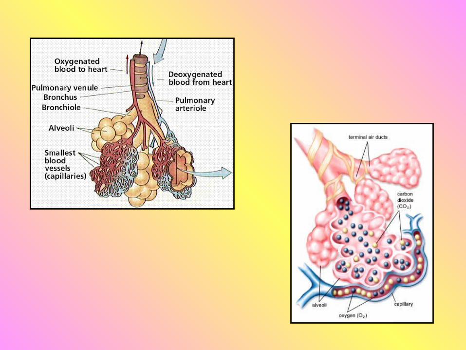
Respiratory Zone
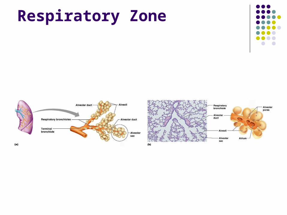
Defined by the presence of alveoli; begins as terminal bronchioles feed into respiratory bronchioles
Respiratory bronchioles lead to alveolar ducts, then to terminal clusters of alveolar sacs composed of alveoli
Approximately 300 million alveoli: Account for most of the lungs’ volume Provide tremendous surface area for gas
exchange
Respiratory Zone
Respiratory Membrane
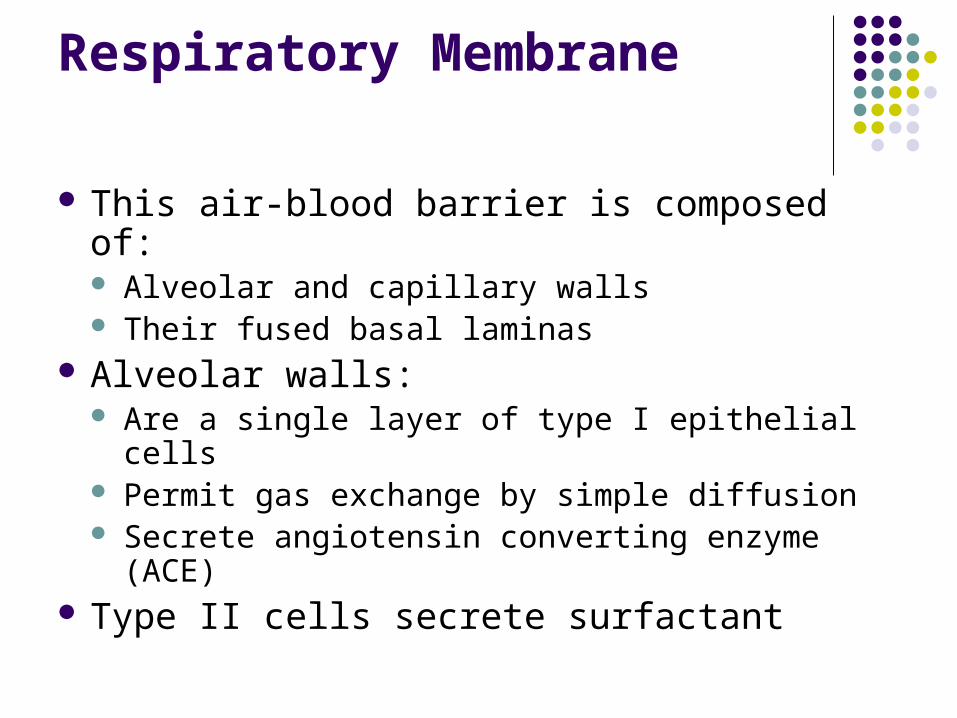
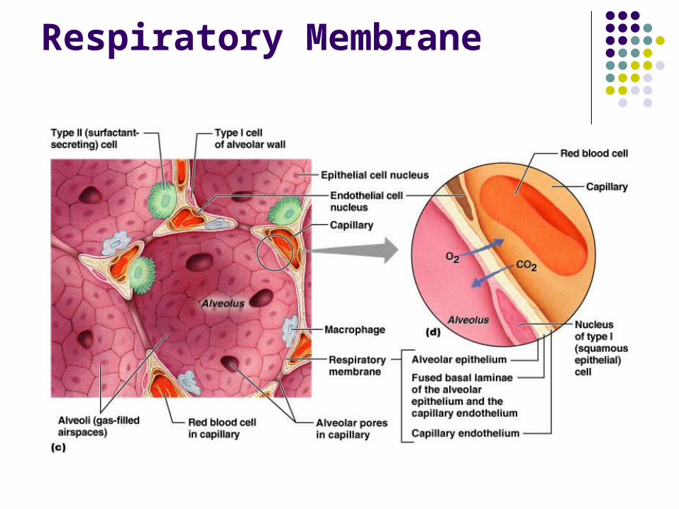
This air-blood barrier is composed of: Alveolar and capillary walls Their fused basal laminas
Alveolar walls: Are a single layer of type I epithelial cells Permit gas exchange by simple diffusion Secrete angiotensin converting enzyme (ACE)
Type II cells secrete surfactant
Alveoli
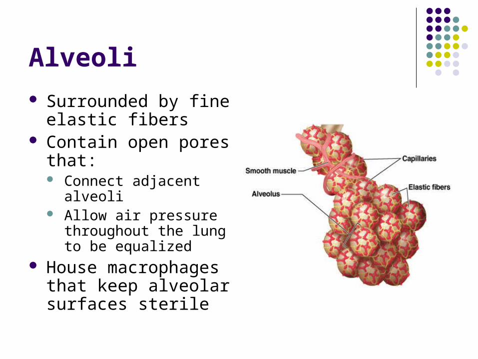
Surrounded by fine elastic fibers
Contain open pores that: Connect adjacent alveoli Allow air pressure
throughout the lung to be equalized
House macrophages that keep alveolar surfaces sterile
Respiratory Membrane
Lungs
Cardiac notch (impression) – cavity that accommodates the heart
Left lung – separated into upper and lower lobes by the oblique fissure
Right lung – separated into three lobes by the oblique and horizontal fissures
There are 10 bronchopulmonary segments in each lung
Blood Supply to Lungs
Lungs are perfused by two circulations: pulmonary and bronchial
Pulmonary arteries – supply systemic venous blood to be oxygenated Branch profusely, along with bronchi Ultimately feed into the pulmonary capillary
network surrounding the alveoli Pulmonary veins – carry oxygenated blood
from respiratory zones to the heart
Blood Supply to Lungs
Bronchial arteries – provide systemic blood to the lung tissue Arise from aorta and enter the lungs at the hilus Supply all lung tissue except the alveoli
Bronchial veins anastomose with pulmonary veins
Pulmonary veins carry most venous blood back to the heart
Pleurae
Thin, double-layered serosa Parietal pleura
Covers the thoracic wall and superior face of the diaphragm
Continues around heart and between lungs
Pleurae
Visceral, or pulmonary, pleura Covers the external lung surface Divides the thoracic cavity into three chambers
The central mediastinum Two lateral compartments, each containing a lung
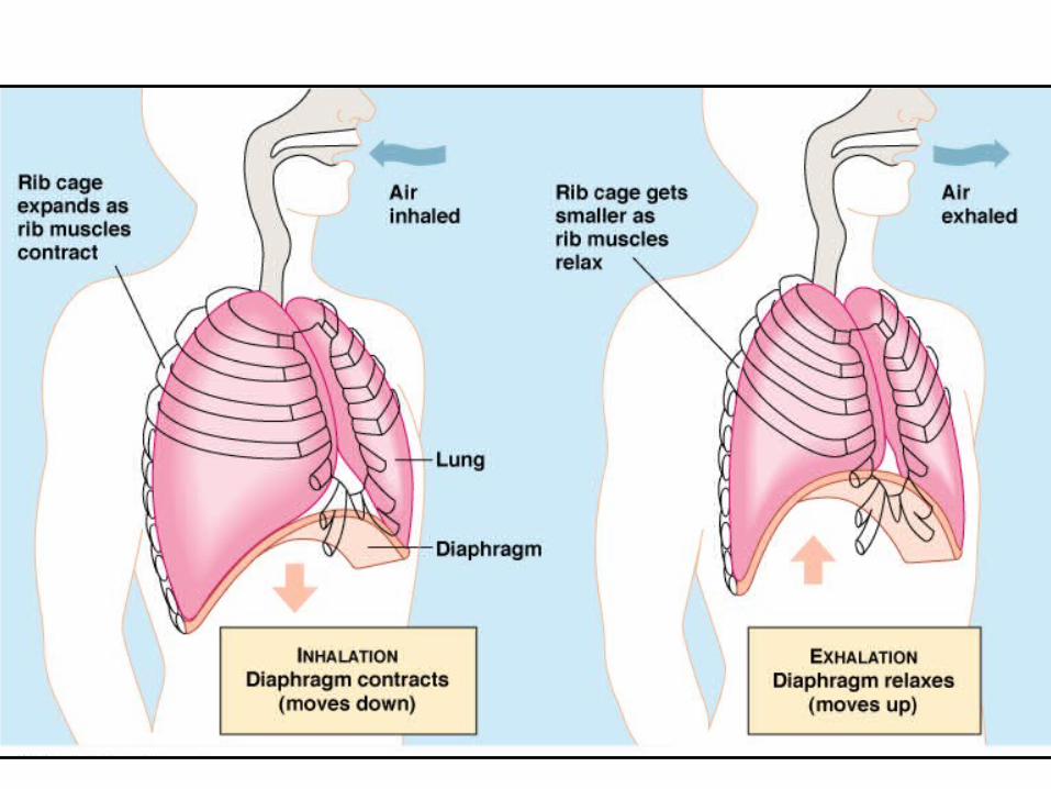
Breathing
Breathing, or pulmonary ventilation, consists of two phases Inspiration – air flows into the lungs Expiration – gases exit the lungs

Breathing Video
Pressure Relationships in the Thoracic Cavity
Respiratory pressure is always described relative to atmospheric pressure
Atmospheric pressure (Patm) Pressure exerted by the air surrounding the body Negative respiratory pressure is less than Patm
Positive respiratory pressure is greater than Patm
Pressure Relationships in the Thoracic Cavity
Intrapulmonary pressure (Ppul) – pressure within the alveoli
Intrapleural pressure (Pip) – pressure within the pleural cavity
Pressure Relationships
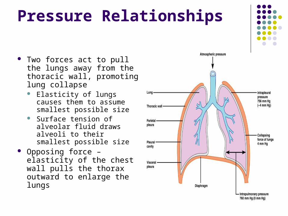
Two forces act to pull the lungs away from the thoracic wall, promoting lung collapse Elasticity of lungs causes
them to assume smallest possible size
Surface tension of alveolar fluid draws alveoli to their smallest possible size
Opposing force – elasticity of the chest wall pulls the thorax outward to enlarge the lungs
Inspiration
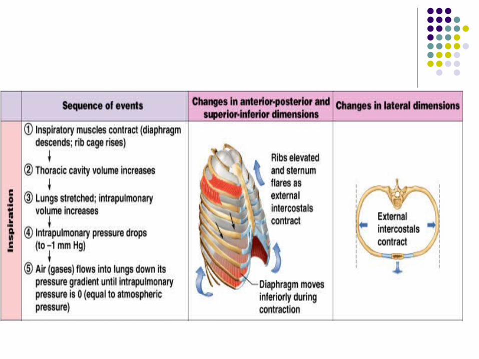
The diaphragm and external intercostal muscles (inspiratory muscles) contract and the rib cage rises
The lungs are stretched and intrapulmonary volume increases
Intrapulmonary pressure drops below atmospheric pressure (1 mm Hg)
Air flows into the lungs, down its pressure gradient, until intrapleural pressure = atmospheric pressure
Expiration
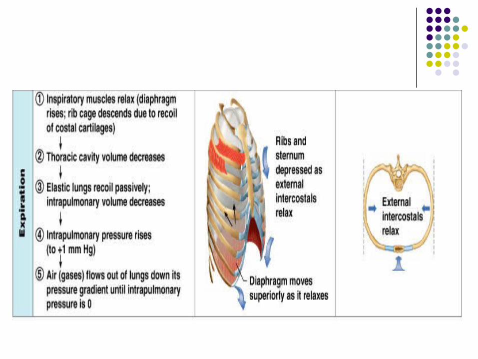
Inspiratory muscles relax and the rib cage descends due to gravity
Thoracic cavity volume decreases Elastic lungs recoil passively and intrapulmonary
volume decreases Intrapulmonary pressure rises above atmospheric
pressure (+1 mm Hg) Gases flow out of the lungs down the pressure
gradient until intrapulmonary pressure is 0
Alveolar Surface Tension
Surface tension – the attraction of liquid molecules to one another at a liquid-gas interface
The liquid coating the alveolar surface is always acting to reduce the alveoli to the smallest possible size
Surfactant, a detergent-like complex, reduces surface tension and helps keep the alveoli from collapsing
Respiratory Volumes
Tidal volume (TV) – air that moves into and out of the lungs with each breath (approximately 500 ml)
Inspiratory reserve volume (IRV) – air that can be inspired forcibly beyond the tidal volume (2100–3200 ml)
Expiratory reserve volume (ERV) – air that can be evacuated from the lungs after a tidal expiration (1000–1200 ml)
Residual volume (RV) – air left in the lungs after strenuous expiration (1200 ml)
Respiratory Capacities
Inspiratory capacity (IC) – total amount of air that can be inspired after a tidal expiration (IRV + TV)
Functional residual capacity (FRC) – amount of air remaining in the lungs after a tidal expiration (RV + ERV)
Vital capacity (VC) – the total amount of exchangeable air (TV + IRV + ERV)
Total lung capacity (TLC) – sum of all lung volumes (approximately 6000 ml in males)
Dead Space
Anatomical dead space – volume of the conducting respiratory passages (150 ml)
Alveolar dead space – alveoli that cease to act in gas exchange due to collapse or obstruction
Total dead space – sum of alveolar and anatomical dead spaces
Pulmonary Function Tests
Spirometer – an instrument consisting of a hollow bell inverted over water, used to evaluate respiratory function
Spirometry can distinguish between: Obstructive pulmonary disease – increased
airway resistance Restrictive disorders – reduction in total lung
capacity from structural or functional lung changes
Pulmonary Function Tests
Total ventilation – total amount of gas flow into or out of the respiratory tract in one minute
Forced vital capacity (FVC) – gas forcibly expelled after taking a deep breath
Forced expiratory volume (FEV) – the amount of gas expelled during specific time intervals of the FVC
Partial Pressure Gradients and Gas Solubilities
Although carbon dioxide has a lower partial pressure gradient: It is 20 times more soluble in plasma than oxygen It diffuses in equal amounts with oxygen
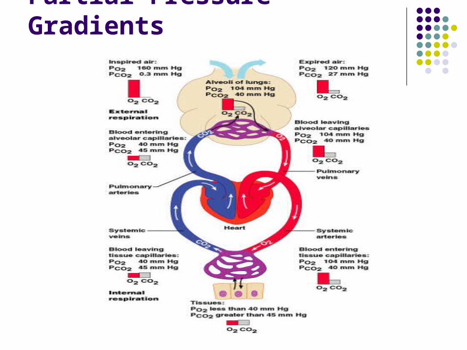
Partial Pressure Gradients
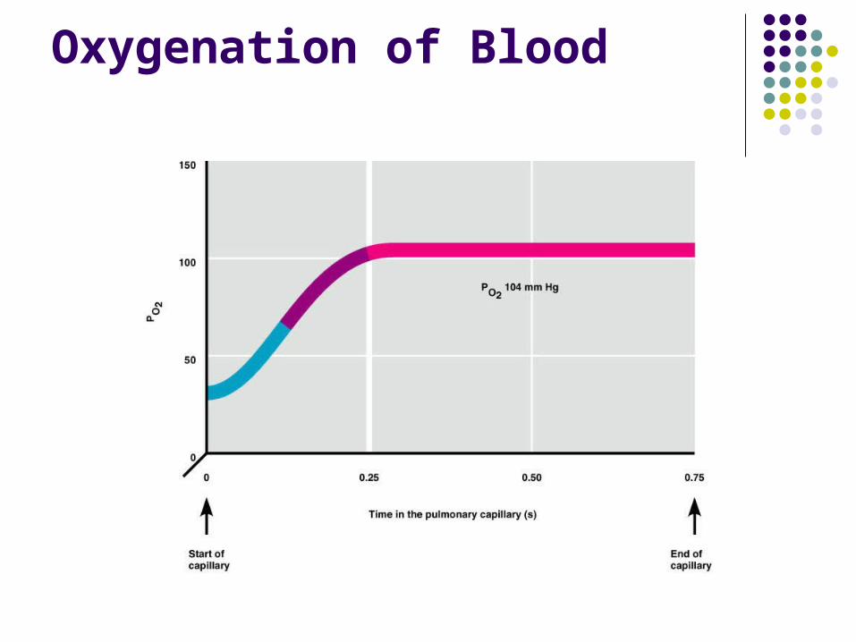
Oxygenation of Blood
Surface Area and Thickness of the Respiratory Membrane
Respiratory membranes: Are only 0.5 to 1 m thick, allowing for efficient
gas exchange Have a total surface area (in males) of about 60
m2 (40 times that of one’s skin) Thicken if lungs become waterlogged and
edematous, whereby gas exchange is inadequate and oxygen deprivation results
Decrease in surface area with emphysema, when walls of adjacent alveoli break through
Oxygen Transport
Molecular oxygen is carried in the blood: Bound to hemoglobin (Hb) within red blood cells Dissolved in plasma
Oxygen Transport: Role of Hemoglobin
Each Hb molecule binds four oxygen atoms in a rapid and reversible process
The hemoglobin-oxygen combination is called oxyhemoglobin (HbO2)
Hemoglobin that has released oxygen is called reduced hemoglobin (HHb)
Carbon Dioxide Transport
Carbon dioxide is transported in the blood in three forms Dissolved in plasma – 7 to 10% Chemically bound to hemoglobin – 20% is carried
in RBCs as carbaminohemoglobin Bicarbonate ion in plasma – 70% is transported
as bicarbonate (HCO3–)
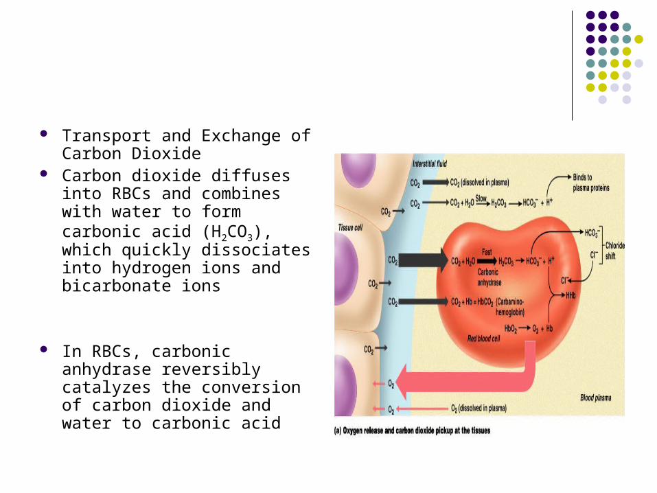
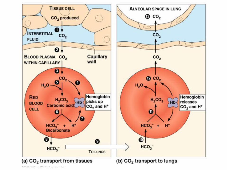
Transport and Exchange of Carbon Dioxide
Carbon dioxide diffuses into RBCs and combines with water to form carbonic acid (H2CO3), which quickly dissociates into hydrogen ions and bicarbonate ions
In RBCs, carbonic anhydrase reversibly catalyzes the conversion of carbon dioxide and water to carbonic acid
Transport and Exchange of Carbon DioxideAt the tissues:
Bicarbonate quickly diffuses from RBCs into the plasma
The chloride shift – to counterbalance the outrush of negative bicarbonate ions from the RBCs, chloride ions (Cl–) move from the plasma into the erythrocytes
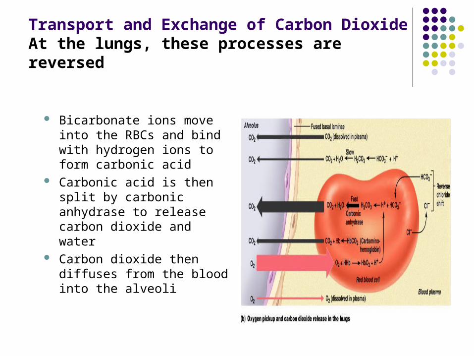
Transport and Exchange of Carbon DioxideAt the lungs, these processes are reversed
Bicarbonate ions move into the RBCs and bind with hydrogen ions to form carbonic acid
Carbonic acid is then split by carbonic anhydrase to release carbon dioxide and water
Carbon dioxide then diffuses from the blood into the alveoli
Influence of Carbon Dioxide on Blood pH
Changes in respiratory rate can also: Alter blood pH Provide a fast-acting system to adjust pH when
it is disturbed by metabolic factors
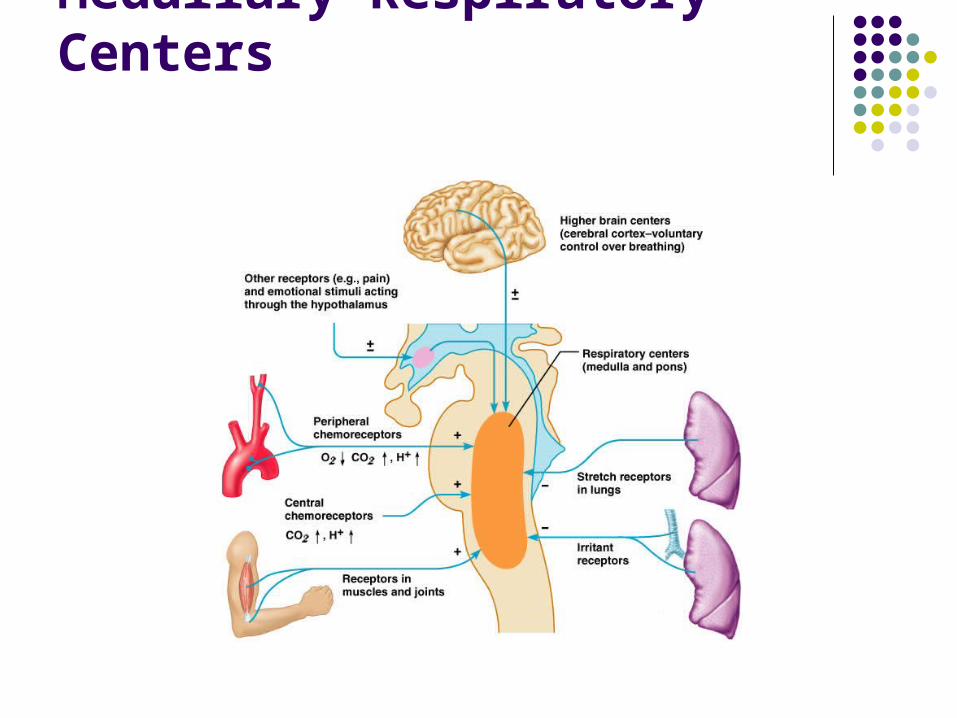
Medullary Respiratory Centers
Depth and Rate of Breathing: PCO2
Hyperventilation – increased depth and rate of breathing that: Quickly flushes carbon dioxide from the blood Occurs in response to hypercapnia
Though a rise CO2 acts as the original stimulus, control of breathing at rest is regulated by the hydrogen ion concentration in the brain
Depth and Rate of Breathing: PCO2
Hypoventilation – slow and shallow breathing due to abnormally low PCO2 levels Apnea (breathing cessation) may occur until PCO2
levels rise
Asthma
Characterized by dyspnea, wheezing, and chest tightness
Active inflammation of the airways precedes bronchospasms
Airway inflammation is an immune response caused by release of IL-4 and IL-5, which stimulate IgE and recruit inflammatory cells
Airways thickened with inflammatory exudates magnify the effect of bronchospasms
Tuberculosis
Infectious disease caused by the bacterium Mycobacterium tuberculosis
Symptoms include fever, night sweats, weight loss, a racking cough, and splitting headache
Treatment entails a 12-month course of antibiotics
Lung Cancer
Accounts for 1/3 of all cancer deaths in the U.S. 90% of all patients with lung cancer were smokers The three most common types are:
Squamous cell carcinoma (20-40% of cases) arises in bronchial epithelium
Adenocarcinoma (25-35% of cases) originates in peripheral lung area
Small cell carcinoma (20-25% of cases) contains lymphocyte-like cells that originate in the primary bronchi and subsequently metastasize
Respiration Video
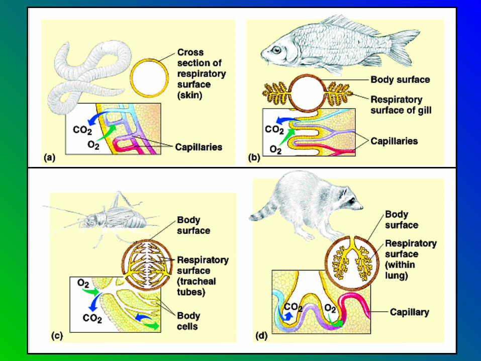
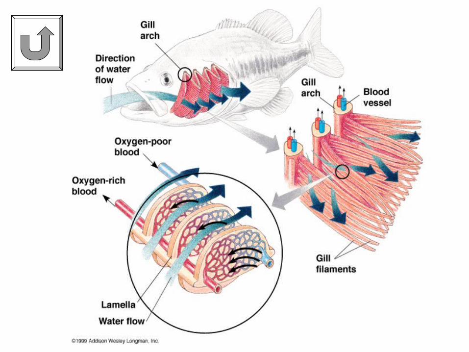
Gills Water has little dissolved oxygen Animals in the water need very efficient
respiratory organs What characteristics of gills make them
successful respiratory organs for aquatic animals?
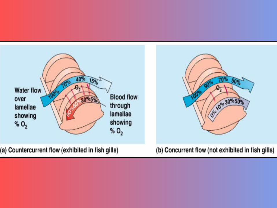
Large respiratory surface Continuous ventilation Countercurrent exchange
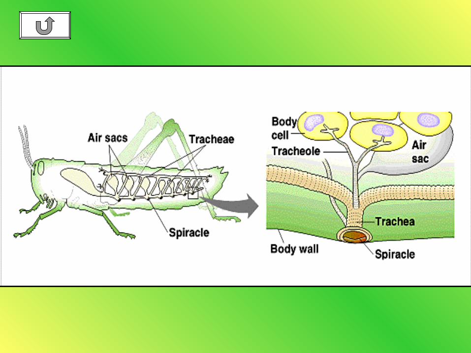
Gas Exchange in Terrestrial Organisms
More oxygen on land What problem regarding gas exchange is faced
on land that is not seen in the water? Dessication (drying out) of respiratory surfaces Gills would stick together on land What are the two main systems of gas
exchange on land? Trachea and Lungs
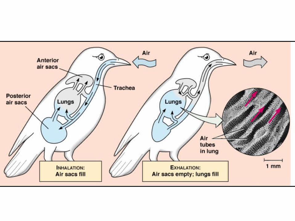
Lungs
What is the reason why the lungs are found within the body cavity?
To keep respiratory surfaces moist In what ways are lungs less efficient than
gills? No countercurrent exchange Mixing of oxygen rich and oxygen poor air Exchange of gases occurs at alveolar capil
lary membrane
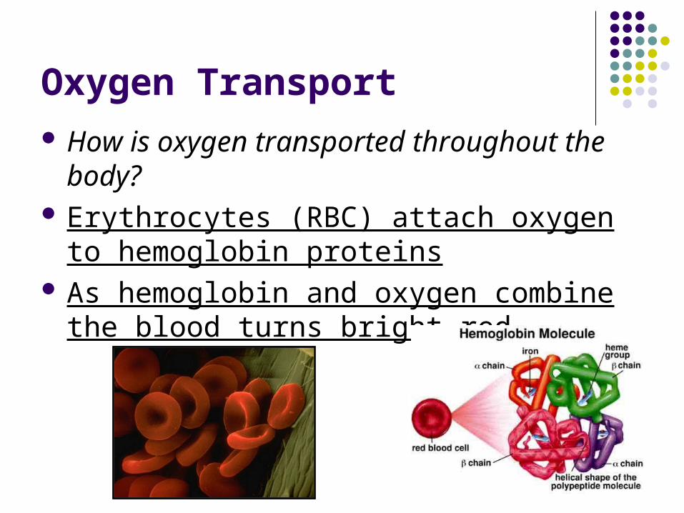
Oxygen Transport How is oxygen transported throughout the
body? Erythrocytes (RBC) attach oxygen to
hemoglobin proteins As hemoglobin and oxygen combine the
blood turns bright red
Carbon Dioxide Transport
Most CO2 is converted to bicarbonate. This conversion releases H+ ions into the
blood.
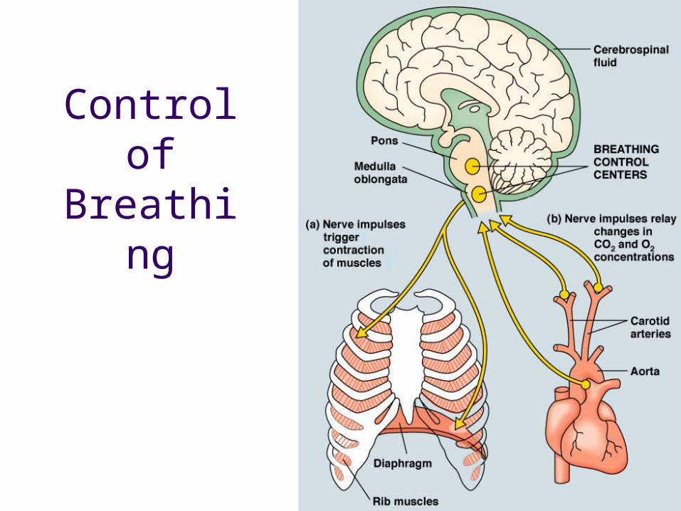
Control of
Breathing