Embed Size (px)
Citation preview
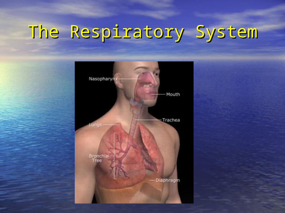
The The RespiratoryRespiratory System System
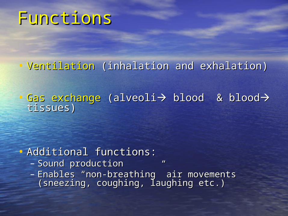
FunctionsFunctions
• Ventilation Ventilation (inhalation and exhalation)(inhalation and exhalation)
• Gas exchange Gas exchange (alveoli(alveoli blood & blood blood & blood tissues) tissues)
• Additional functions: Additional functions: – Sound productionSound production– Enables “non-breathing” air movements (sneezing, Enables “non-breathing” air movements (sneezing,
coughing, laughing etc.)coughing, laughing etc.)
CharacteristicsCharacteristics
• Usually 14-20 breaths/minuteUsually 14-20 breaths/minute– About 450 ml per breath at rest or up to 100 L/minute About 450 ml per breath at rest or up to 100 L/minute
during exerciseduring exercise
• Breathing is automatic and unconscious but one Breathing is automatic and unconscious but one can hold his/her breath up to a certain pointcan hold his/her breath up to a certain point
• If no air for:If no air for:– 4-5 minutes: loss of consciousness4-5 minutes: loss of consciousness– 7-8 minutes = brain damage7-8 minutes = brain damage– 10 minutes = impending death10 minutes = impending death
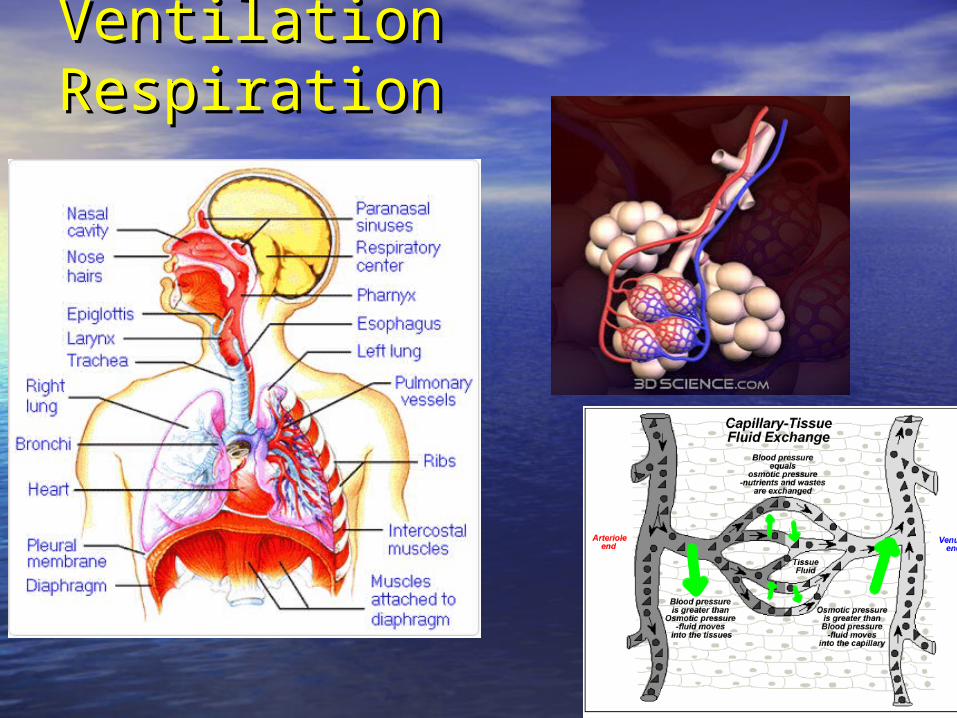
Ventilation Ventilation RespirationRespiration
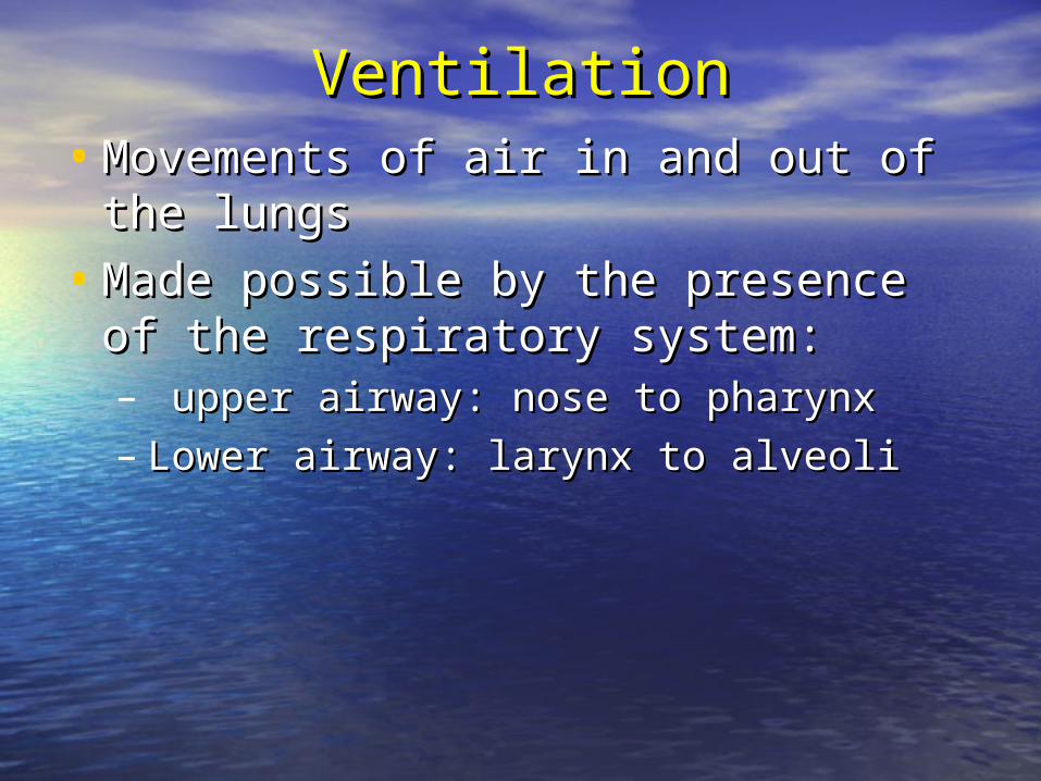
VentilationVentilation• Movements of air in and out of the Movements of air in and out of the
lungslungs
• Made possible by the presence of the Made possible by the presence of the respiratory system:respiratory system:– upper airway: nose to pharynxupper airway: nose to pharynx– Lower airway: larynx to alveoliLower airway: larynx to alveoli
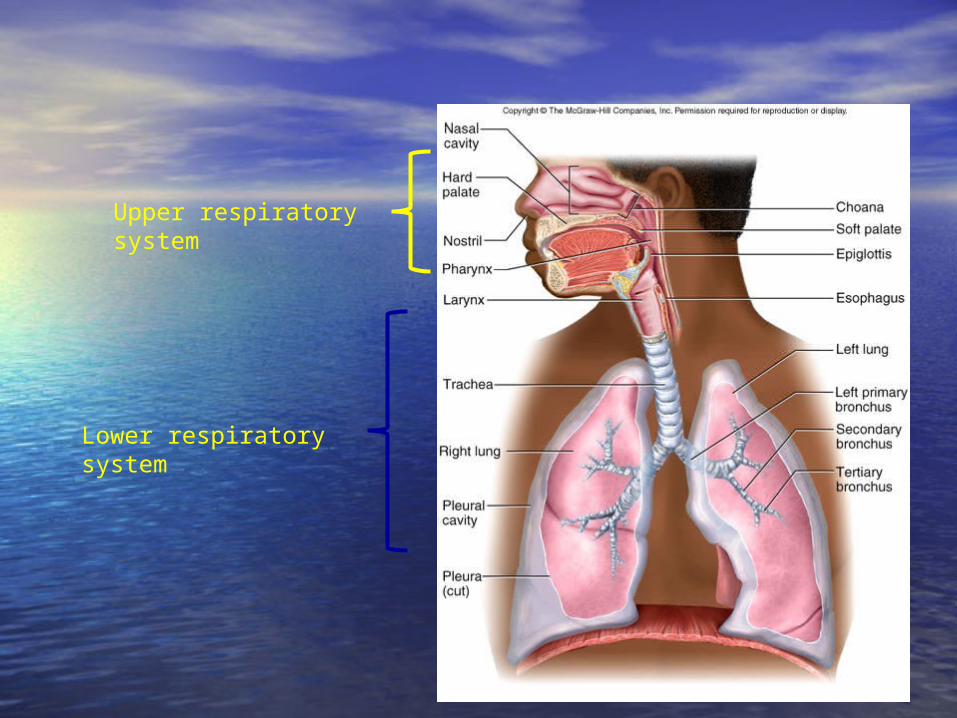
Upper respiratory system
Lower respiratory system
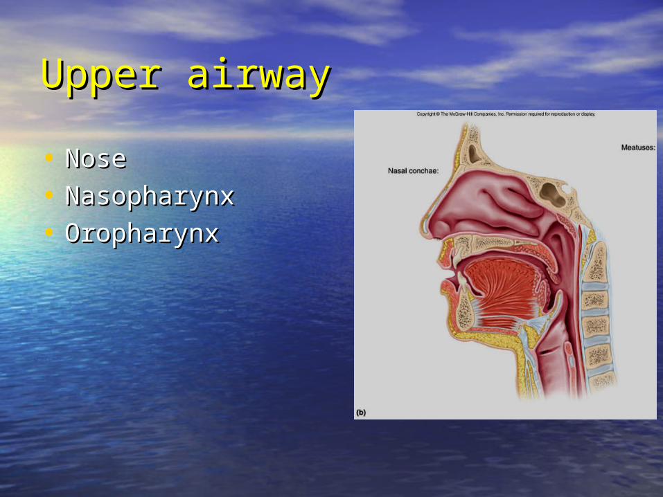
Upper airwayUpper airway
• NoseNose
• NasopharynxNasopharynx
• OropharynxOropharynx
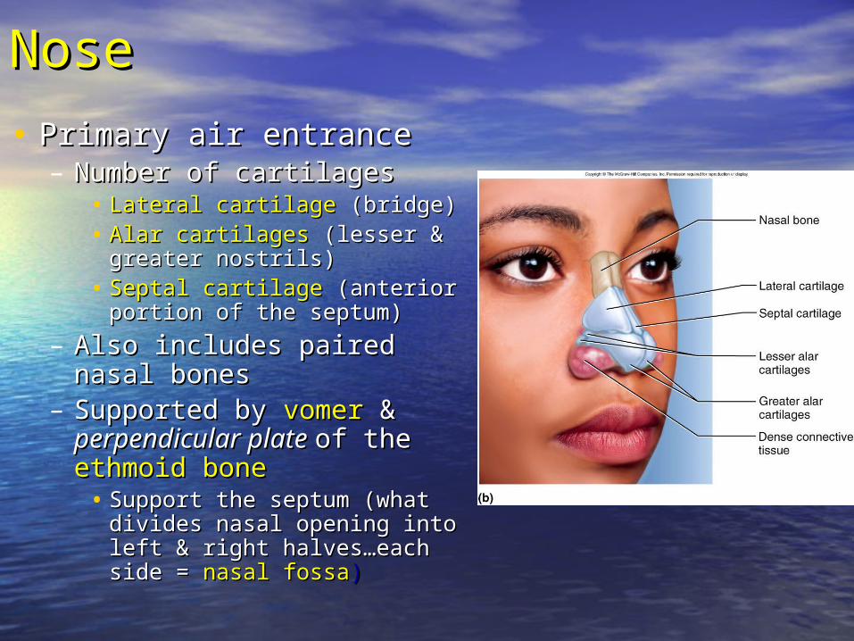
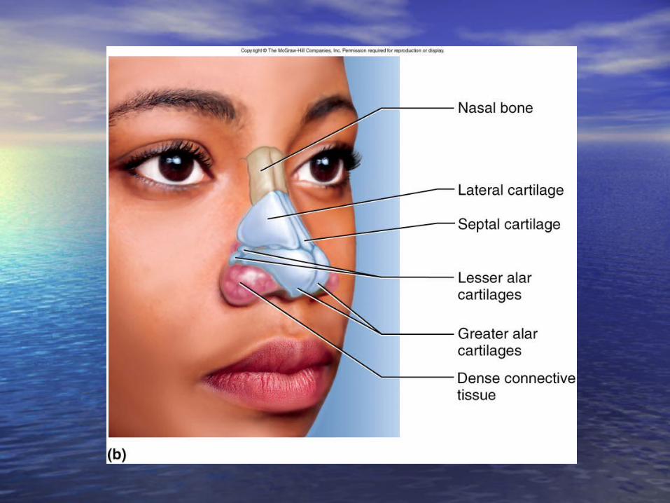
NoseNose
• Primary air entrancePrimary air entrance– Number of cartilagesNumber of cartilages
• Lateral cartilage Lateral cartilage (bridge)(bridge)• Alar cartilages Alar cartilages (lesser & greater (lesser & greater
nostrils)nostrils)• Septal cartilage Septal cartilage (anterior portion of (anterior portion of
the septum)the septum)
– Also includes paired nasal bonesAlso includes paired nasal bones– Supported bySupported by vomervomer & &
perpendicular plate perpendicular plate of theof the ethmoid boneethmoid bone
• Support the septum (what divides Support the septum (what divides nasal opening into left & right nasal opening into left & right halves…each side =halves…each side = nasal fossanasal fossa))
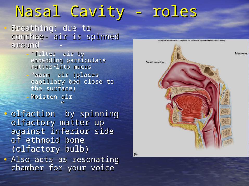
Nasal Cavity - rolesNasal Cavity - roles• Breathing: due to conchae- air Breathing: due to conchae- air
is spinned around is spinned around - ““filter” air by embedding filter” air by embedding
particulate matter into mucusparticulate matter into mucus
- ““warm” air (places capillary warm” air (places capillary bed close to the surface)bed close to the surface)
- Moisten airMoisten air
• olfaction” by spinning olfaction” by spinning olfactory matter up against olfactory matter up against inferior side of ethmoid bone inferior side of ethmoid bone (olfactory bulb)(olfactory bulb)
• Also acts as resonating Also acts as resonating chamber for your voicechamber for your voice
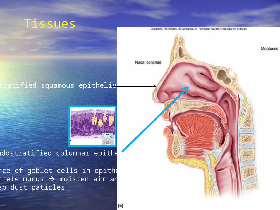
Tissues
Stratified squamous epithelium
Pseudostratified columnar epithelium
Presence of goblet cells in epithelium: secrete mucus moisten air and trap dust paticles

Paranasal SinusesParanasal Sinuses
• Paired air spaces in the Paired air spaces in the anterior skull:anterior skull:– Maxillary, frontal, Maxillary, frontal,
sphenoidal sphenoidal & & ethmoidalethmoidal
• Decrease skull weightDecrease skull weight
• Help with sound Help with sound resonance (both for resonance (both for hearing and voice)hearing and voice)
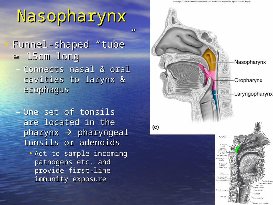
NasopharynxNasopharynx
• Funnel-shaped “tube” Funnel-shaped “tube” 15cm long15cm long– Connects nasal & oral Connects nasal & oral
cavities to larynx & cavities to larynx & esophagus esophagus
– One set of tonsils are located One set of tonsils are located in the pharynx in the pharynx pharyngeal tonsils or pharyngeal tonsils or adenoidsadenoids
• Act to sample incoming Act to sample incoming pathogens etc. and provide pathogens etc. and provide first-line immunity exposurefirst-line immunity exposure
NasopharynxNasopharynx
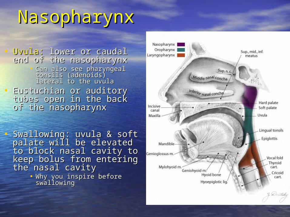
• Uvula:Uvula: lower or caudal end of lower or caudal end of the nasopharynxthe nasopharynx
• Can also see pharyngeal tonsils Can also see pharyngeal tonsils (adenoids) lateral to the uvula(adenoids) lateral to the uvula
• Eustuchian or auditory tubes Eustuchian or auditory tubes open in the back of the open in the back of the nasopharynxnasopharynx
• Swallowing: uvula & soft palate Swallowing: uvula & soft palate will be elevated to block nasal will be elevated to block nasal cavity to keep bolus from cavity to keep bolus from entering the nasal cavityentering the nasal cavity
• Why you inspire before Why you inspire before swallowingswallowing
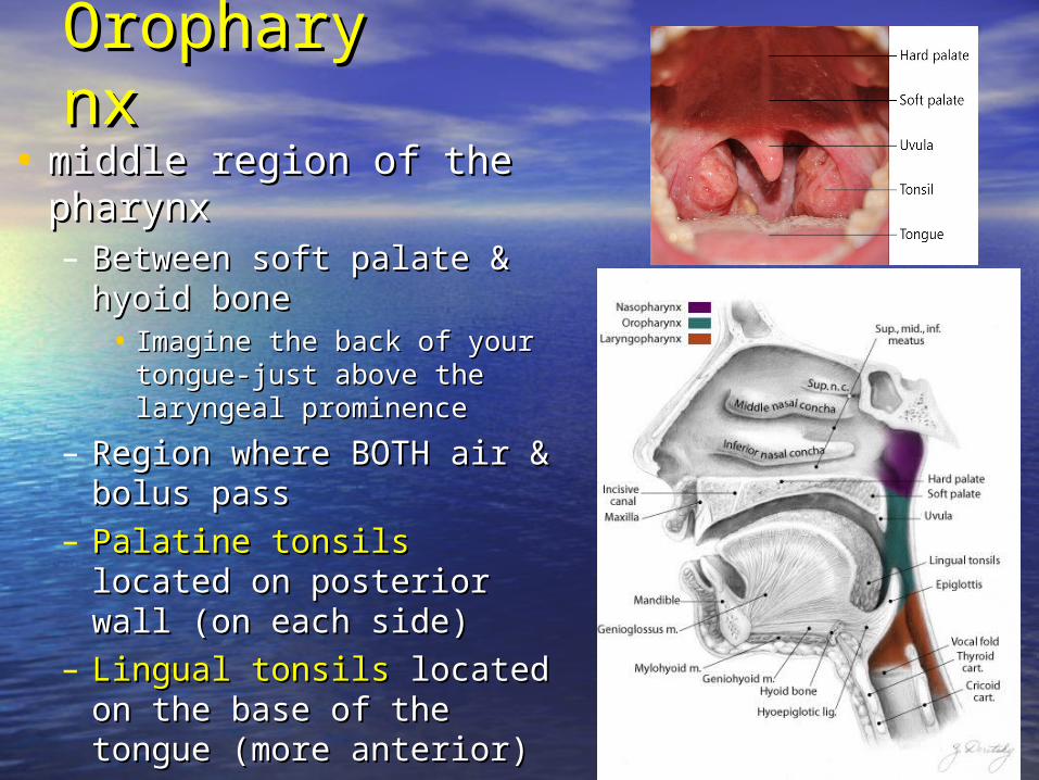
OropharynxOropharynx
• middle region of the pharynxmiddle region of the pharynx– Between soft palate & hyoid Between soft palate & hyoid
bone bone • Imagine the back of your tongue-Imagine the back of your tongue-
just above the laryngeal prominencejust above the laryngeal prominence
– Region where BOTH air & bolus Region where BOTH air & bolus pass pass
– Palatine tonsils Palatine tonsils located on located on posterior wall (on each side)posterior wall (on each side)
– Lingual tonsils Lingual tonsils located on the located on the base of the tongue (more base of the tongue (more anterior)anterior)
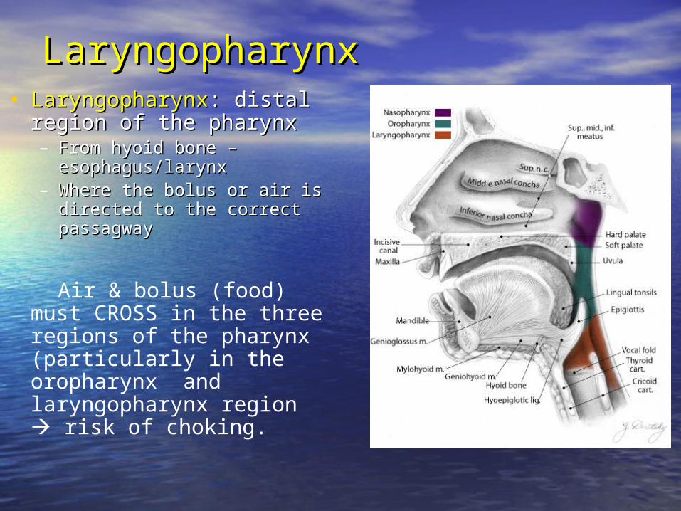
LaryngopharynxLaryngopharynx• LaryngopharynxLaryngopharynx: distal region : distal region
of the pharynxof the pharynx– From hyoid bone – From hyoid bone –
esophagus/larynxesophagus/larynx– Where the bolus or air is directed Where the bolus or air is directed
to the correct passagwayto the correct passagway
Air & bolus (food) must CROSS in the three regions of the pharynx (particularly in the oropharynx and laryngopharynx region risk of choking.
Lower airwayLower airway
• LarynxLarynx
• TracheaTrachea
• Bronchial treeBronchial tree
• AlveoliAlveoli
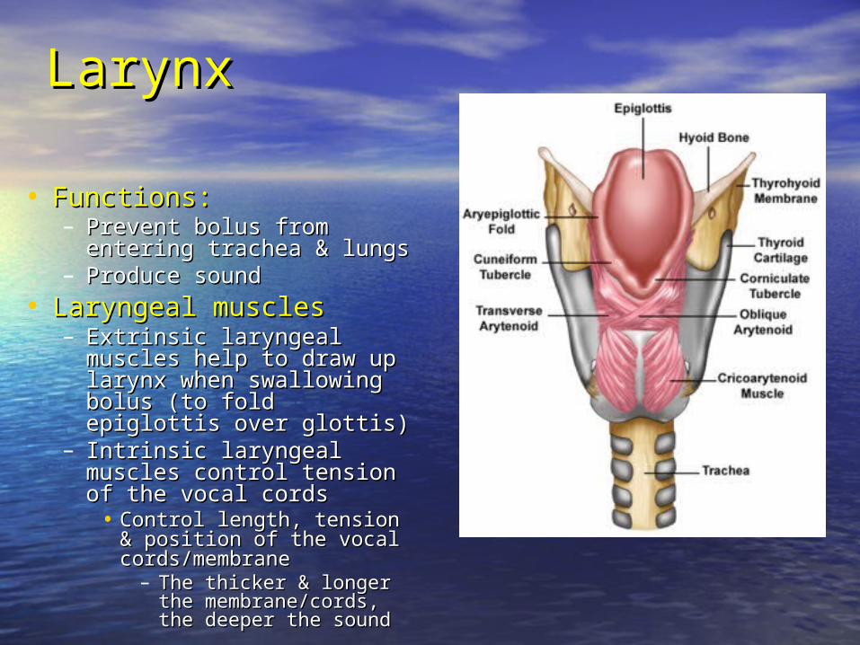
LarynxLarynx
• Functions:Functions:– Prevent bolus from entering Prevent bolus from entering
trachea & lungstrachea & lungs– Produce soundProduce sound
• Laryngeal musclesLaryngeal muscles – Extrinsic laryngeal muscles Extrinsic laryngeal muscles
help to draw up larynx when help to draw up larynx when swallowing bolus (to fold swallowing bolus (to fold epiglottis over glottis)epiglottis over glottis)
– Intrinsic laryngeal muscles Intrinsic laryngeal muscles control tension of the vocal control tension of the vocal cordscords
• Control length, tension & Control length, tension & position of the vocal position of the vocal cords/membranecords/membrane
– The thicker & longer the The thicker & longer the membrane/cords, the membrane/cords, the deeper the sounddeeper the sound
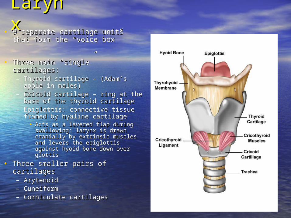
LarynxLarynx• 9 separate cartilage units that form the 9 separate cartilage units that form the
“voice box”“voice box”
• Three main “single” cartilages:Three main “single” cartilages:– Thyroid cartilage – (Adam’s apple in Thyroid cartilage – (Adam’s apple in
males)males)– Cricoid cartilage – ring at the base of Cricoid cartilage – ring at the base of
the thyroid cartilagethe thyroid cartilage– Epiglottis: connective tissue framed by Epiglottis: connective tissue framed by
hyaline cartilagehyaline cartilage• Acts as a levered flap during Acts as a levered flap during
swallowing: larynx is drawn cranially swallowing: larynx is drawn cranially by extrinsic muscles and levers the by extrinsic muscles and levers the epiglottis against hyoid bone down epiglottis against hyoid bone down over glottisover glottis
• Three smaller pairs of cartilagesThree smaller pairs of cartilages– ArytenoidArytenoid– CuneiformCuneiform– Corniculate cartilagesCorniculate cartilages
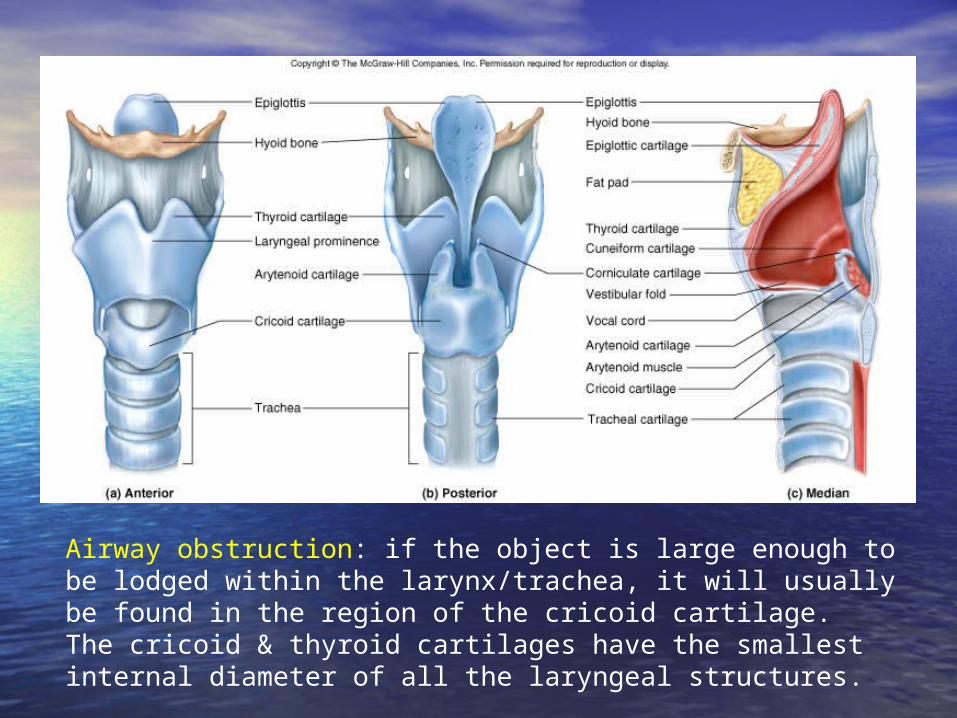
Airway obstruction: if the object is large enough to be lodged within the larynx/trachea, it will usually be found in the region of the cricoid cartilage. The cricoid & thyroid cartilages have the smallest internal diameter of all the laryngeal structures.
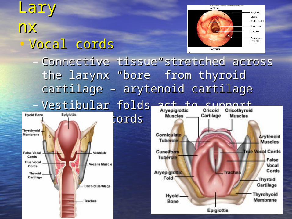
LarynxLarynx
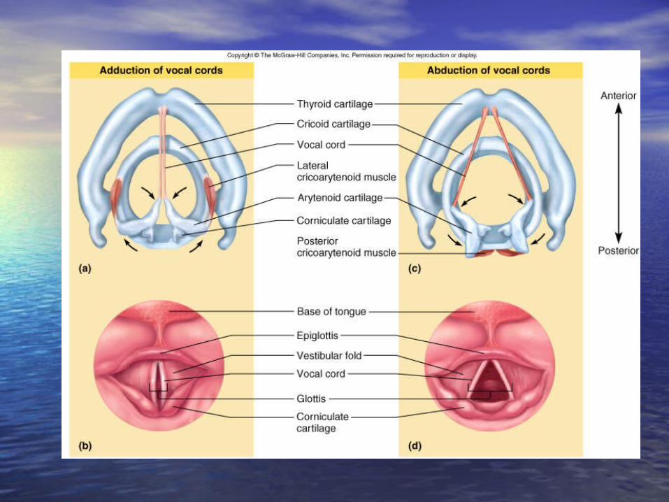
• Vocal cordsVocal cords– Connective tissue stretched across the larynx Connective tissue stretched across the larynx
“bore” from thyroid cartilage – arytenoid cartilage“bore” from thyroid cartilage – arytenoid cartilage
– Vestibular folds act to support the vocal cords & Vestibular folds act to support the vocal cords & keep them moistkeep them moist
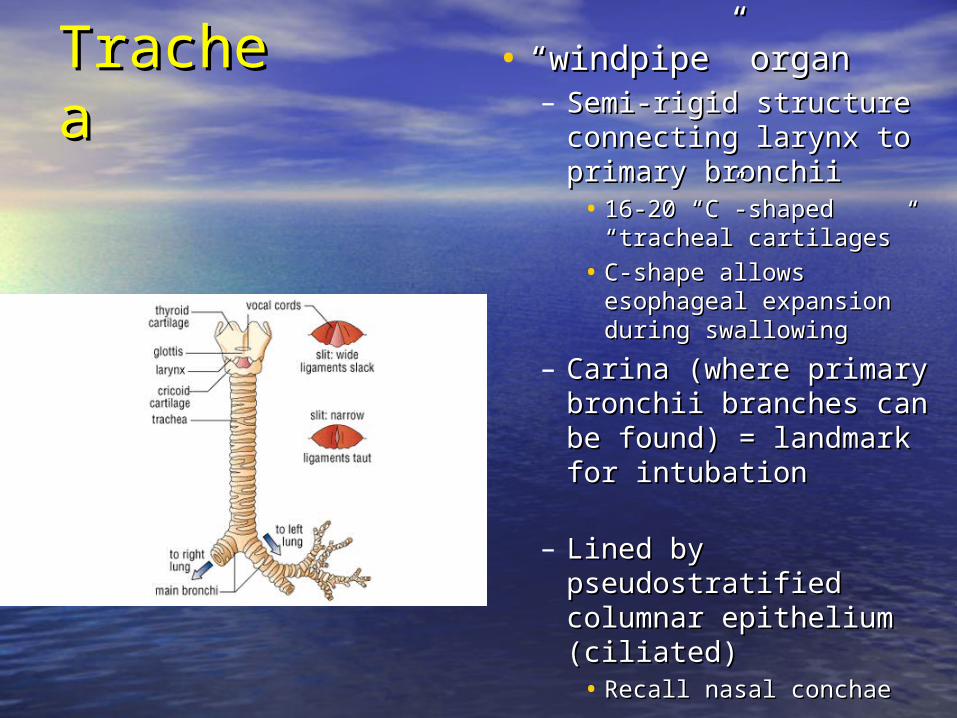
TracheaTrachea • ““windpipe” organwindpipe” organ– Semi-rigid structure Semi-rigid structure
connecting larynx to primary connecting larynx to primary bronchiibronchii
• 16-20 “C”-shaped “tracheal 16-20 “C”-shaped “tracheal cartilages”cartilages”
• C-shape allows esophageal C-shape allows esophageal expansion during swallowingexpansion during swallowing
– Carina (where primary Carina (where primary bronchii branches can be bronchii branches can be found) = landmark for found) = landmark for intubationintubation
– Lined by pseudostratified Lined by pseudostratified columnar epithelium columnar epithelium (ciliated)(ciliated)
• Recall nasal conchaeRecall nasal conchae
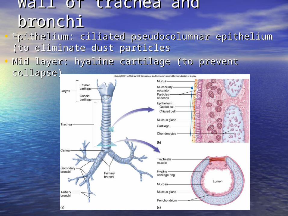
Wall of trachea and bronchiWall of trachea and bronchi
• Epithelium: ciliated pseudocolumnar epithelium (to Epithelium: ciliated pseudocolumnar epithelium (to eliminate dust particleseliminate dust particles
• Mid layer: hyaline cartilage (to prevent collapse)Mid layer: hyaline cartilage (to prevent collapse)
Bronchial TreeBronchial Tree
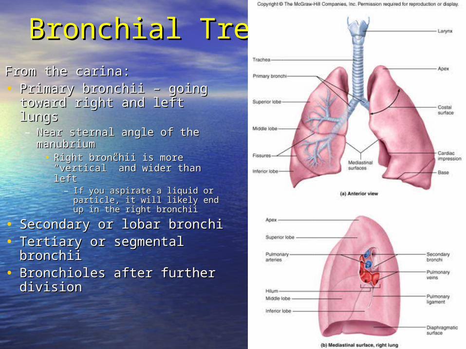
From the carina:From the carina:• Primary bronchii – going Primary bronchii – going
toward right and left lungs toward right and left lungs – Near sternal angle of the Near sternal angle of the
manubriummanubrium• Right bronchii is more “vertical” Right bronchii is more “vertical”
and wider than left and wider than left – If you aspirate a liquid or If you aspirate a liquid or
particle, it will likely end up in particle, it will likely end up in the right bronchiithe right bronchii
• Secondary or lobar bronchiSecondary or lobar bronchi• Tertiary or segmental bronchiiTertiary or segmental bronchii• Bronchioles after further Bronchioles after further
divisiondivision
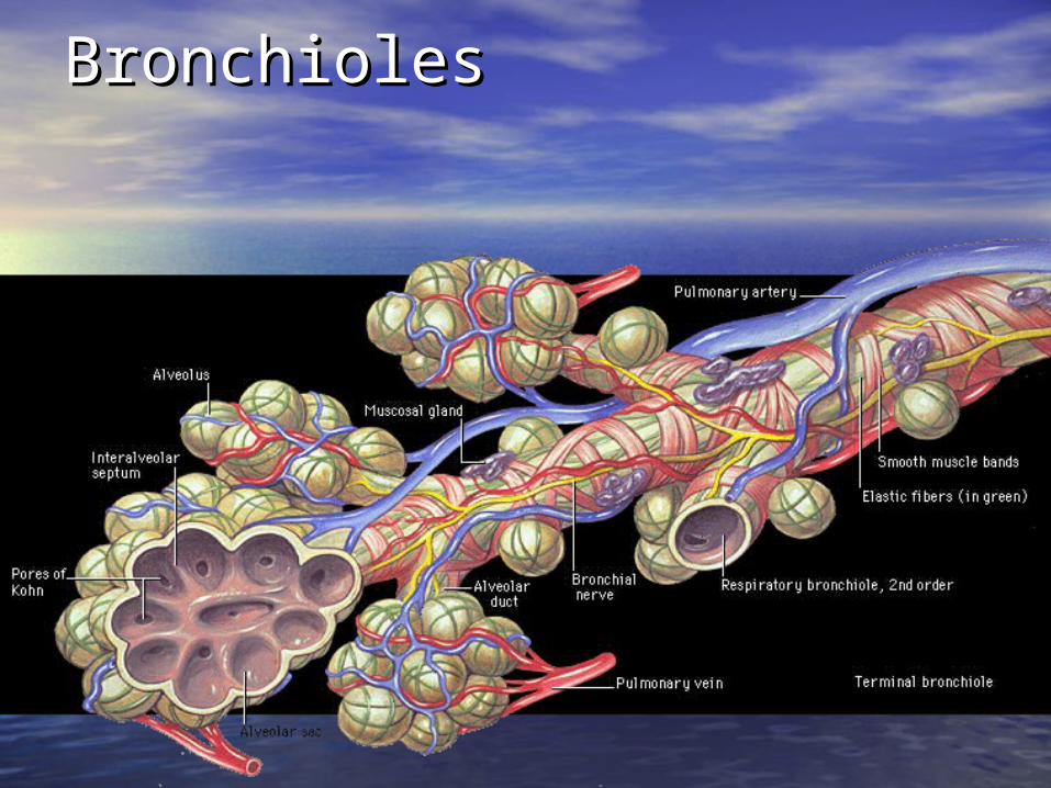
BronchiolesBronchioles
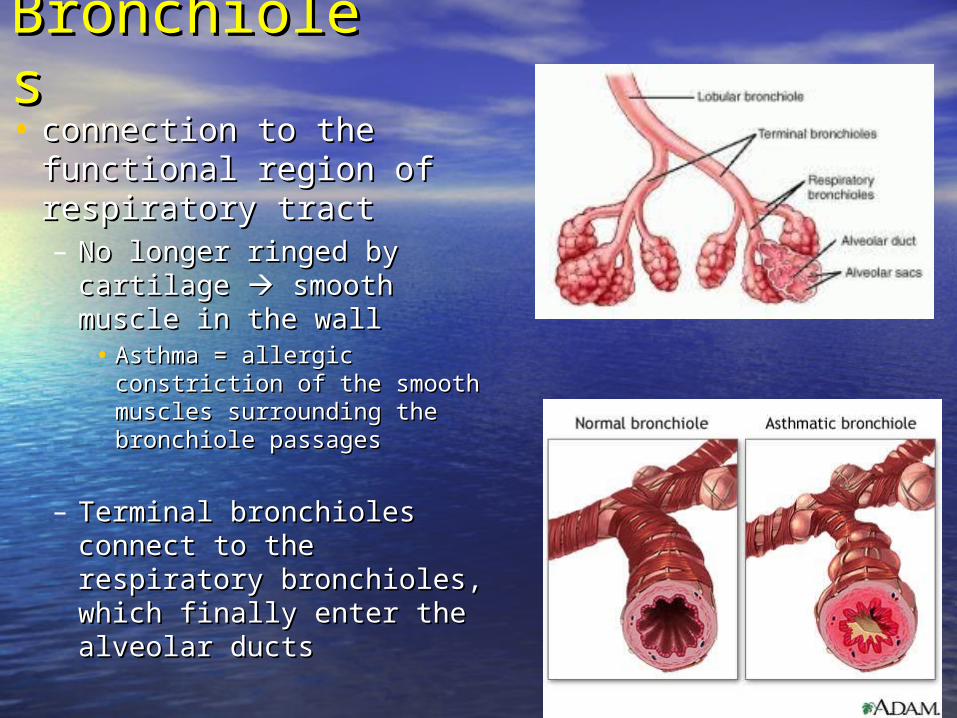
BronchiolesBronchioles• connection to the functional connection to the functional
region of respiratory tractregion of respiratory tract– No longer ringed by cartilage No longer ringed by cartilage
smooth muscle in the wall smooth muscle in the wall• Asthma = allergic constriction of Asthma = allergic constriction of
the smooth muscles surrounding the smooth muscles surrounding the bronchiole passagesthe bronchiole passages
– Terminal bronchioles connect Terminal bronchioles connect to the respiratory bronchioles, to the respiratory bronchioles, which finally enter the alveolar which finally enter the alveolar ductsducts
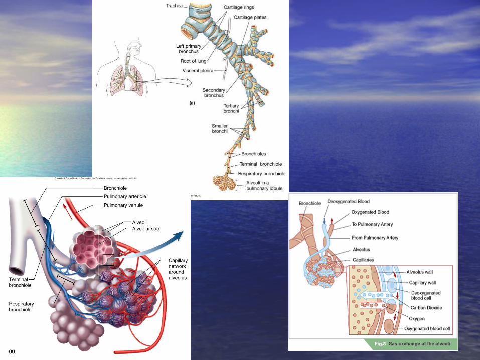
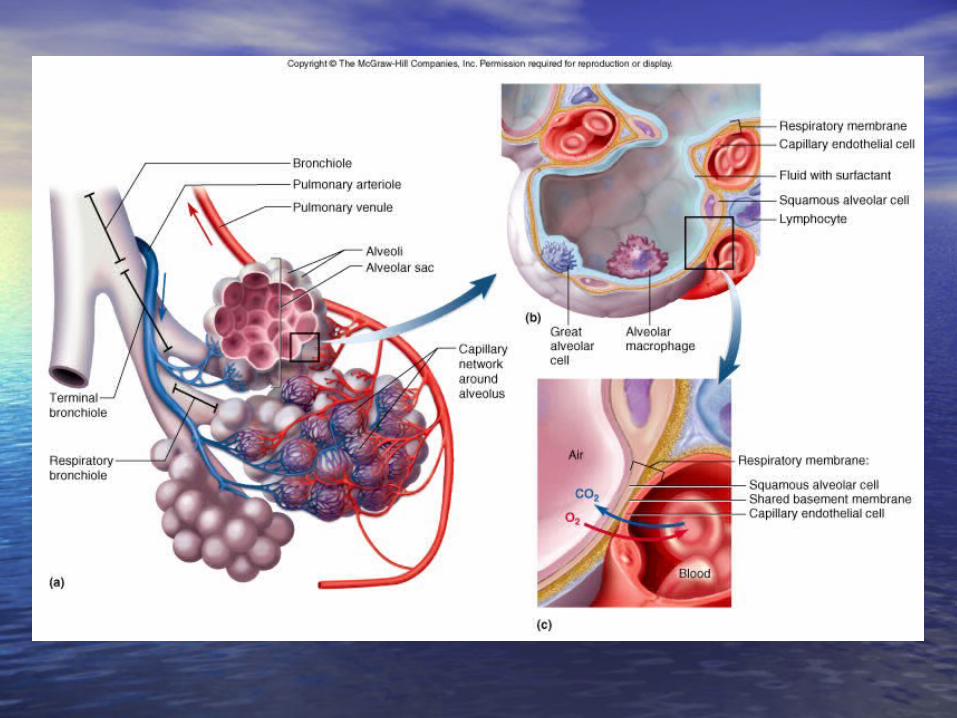
Pulmonary AlveoliPulmonary Alveoli
• Final destination for inspired air flowFinal destination for inspired air flow– Bear in mind that inspired/expired air is rarely “fully” Bear in mind that inspired/expired air is rarely “fully”
exchangedexchanged• Almost 85% of the volume of inspired air remains within the respiratory Almost 85% of the volume of inspired air remains within the respiratory
tract during the next cycle of expiration/inspirationtract during the next cycle of expiration/inspiration
– Region of gas exchangeRegion of gas exchange 350 million alveoli/lung (350 million alveoli/lung ( 70 M 70 M22))
• ““simple” squamous epitheliumsimple” squamous epithelium– ““air-blood” barrier = 1 cell thick + basement membrane + capillary air-blood” barrier = 1 cell thick + basement membrane + capillary
endotheliumendothelium
Pulmonary AlveoliPulmonary Alveoli
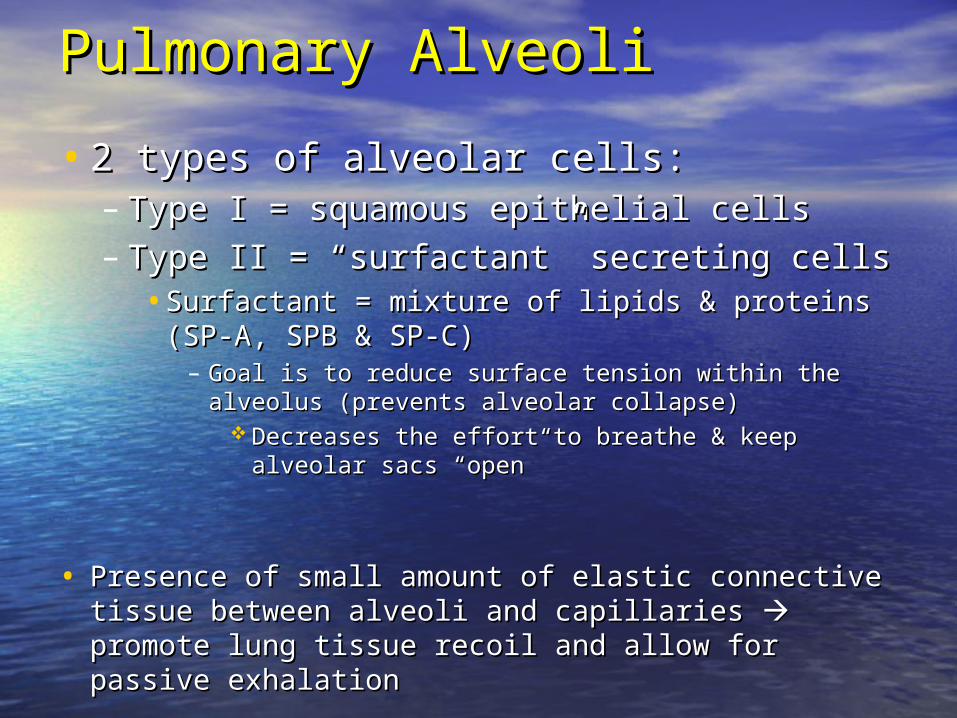
• 2 types of alveolar cells:2 types of alveolar cells:– Type I = squamous epithelial cellsType I = squamous epithelial cells
– Type II = “surfactant” secreting cellsType II = “surfactant” secreting cells• Surfactant = mixture of lipids & proteins (SP-A, SPB & Surfactant = mixture of lipids & proteins (SP-A, SPB &
SP-C)SP-C)– Goal is to reduce surface tension within the alveolus (prevents Goal is to reduce surface tension within the alveolus (prevents
alveolar collapse)alveolar collapse) Decreases the effort to breathe & keep alveolar sacs Decreases the effort to breathe & keep alveolar sacs
“open”“open”
• Presence of small amount of elastic connective tissue between Presence of small amount of elastic connective tissue between alveoli and capillaries alveoli and capillaries promote lung tissue recoil and allow promote lung tissue recoil and allow for passive exhalationfor passive exhalation
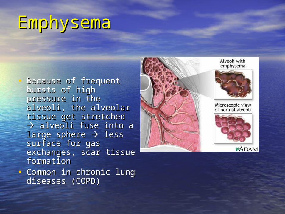
EmphyseEmphysemama
• Because of frequent Because of frequent bursts of high pressure bursts of high pressure in the alveoli, the in the alveoli, the alveolar tissue get alveolar tissue get stretched stretched alveoli alveoli fuse into a large fuse into a large sphere sphere less surface less surface for gas exchanges, for gas exchanges, scar tissue formationscar tissue formation
• Common in chronic Common in chronic lung diseases (COPD)lung diseases (COPD)
SurfactantSurfactant
• Remember that the surface of the alveoli is covered Remember that the surface of the alveoli is covered with a watery fluid with a watery fluid very high surface tensionvery high surface tension– Ex: The surfaces of a wet plastic bag will stick to each Ex: The surfaces of a wet plastic bag will stick to each
otherother– the walls of the alveoli tend to stick together the walls of the alveoli tend to stick together difficult to difficult to
re-open the alveoli)re-open the alveoli)
• Surfactant secreted by Type II alveolar cells decrease Surfactant secreted by Type II alveolar cells decrease this surface tensionthis surface tension– Permits more effective gas exchangePermits more effective gas exchange– Also permits alveolar expansion/contraction with less risk Also permits alveolar expansion/contraction with less risk
of “vacuum collapse”of “vacuum collapse”
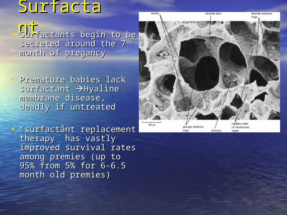
SurfactantSurfactant• Surfactants begin to be Surfactants begin to be
secreted around the 7secreted around the 7thth month month of pregancyof pregancy
• Premature babies lack Premature babies lack surfactant surfactant Hyaline Hyaline membrane disease, deadly if membrane disease, deadly if untreateduntreated
• ““surfactant replacement surfactant replacement therapy” has vastly improved therapy” has vastly improved survival rates among premies survival rates among premies (up to 95% from 5% for 6-6.5 (up to 95% from 5% for 6-6.5 month old premies) month old premies)
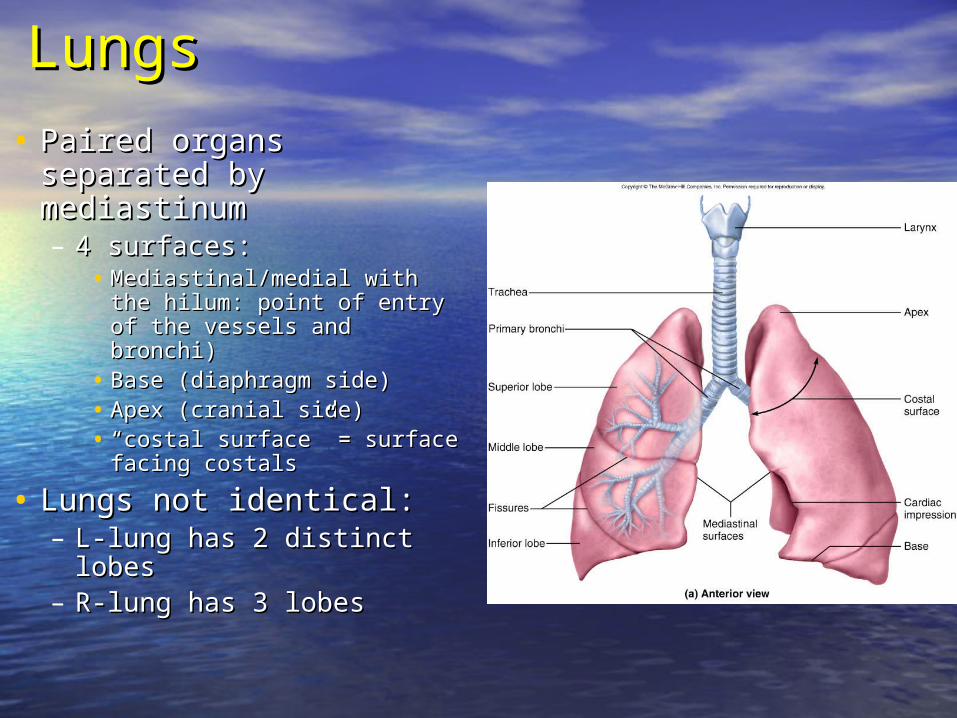
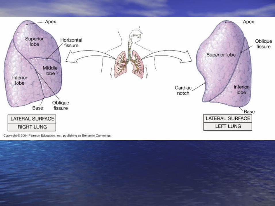
LungsLungs
• Paired organs separated by Paired organs separated by mediastinummediastinum– 4 surfaces: 4 surfaces:
• Mediastinal/medial with the Mediastinal/medial with the hilum: point of entry of the hilum: point of entry of the vessels and bronchi)vessels and bronchi)
• Base (diaphragm side)Base (diaphragm side)• Apex (cranial side)Apex (cranial side)• ““costal surface” = surface costal surface” = surface
facing costalsfacing costals
• Lungs not identical:Lungs not identical:– L-lung has 2 distinct lobesL-lung has 2 distinct lobes– R-lung has 3 lobesR-lung has 3 lobes
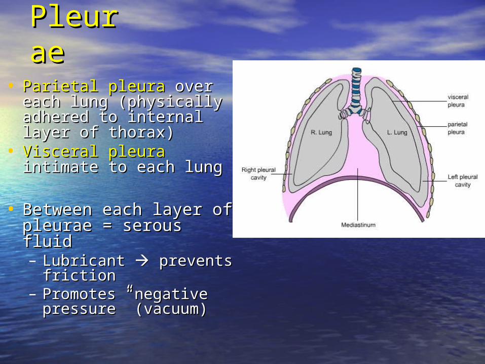
PleuraePleurae
• Parietal pleuraParietal pleura over each lung over each lung (physically adhered to internal (physically adhered to internal layer of thorax)layer of thorax)
• Visceral pleuraVisceral pleura intimate to intimate to each lungeach lung
• Between each layer of Between each layer of pleurae = serous fluidpleurae = serous fluid– Lubricant Lubricant prevents prevents
frictionfriction– Promotes “negative Promotes “negative
pressure” (vacuum)pressure” (vacuum)
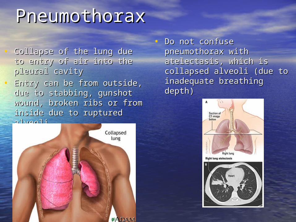
PneumothoraxPneumothorax
• Collapse of the lung due to Collapse of the lung due to entry of air into the pleural entry of air into the pleural cavitycavity
• Entry can be from outside, due Entry can be from outside, due to stabbing, gunshot wound, to stabbing, gunshot wound, broken ribs or from inside due broken ribs or from inside due to ruptured alveolito ruptured alveoli
• Do not confuse pneumothorax Do not confuse pneumothorax with atelectasis, which is with atelectasis, which is collapsed alveoli (due to collapsed alveoli (due to inadequate breathing depth)inadequate breathing depth)
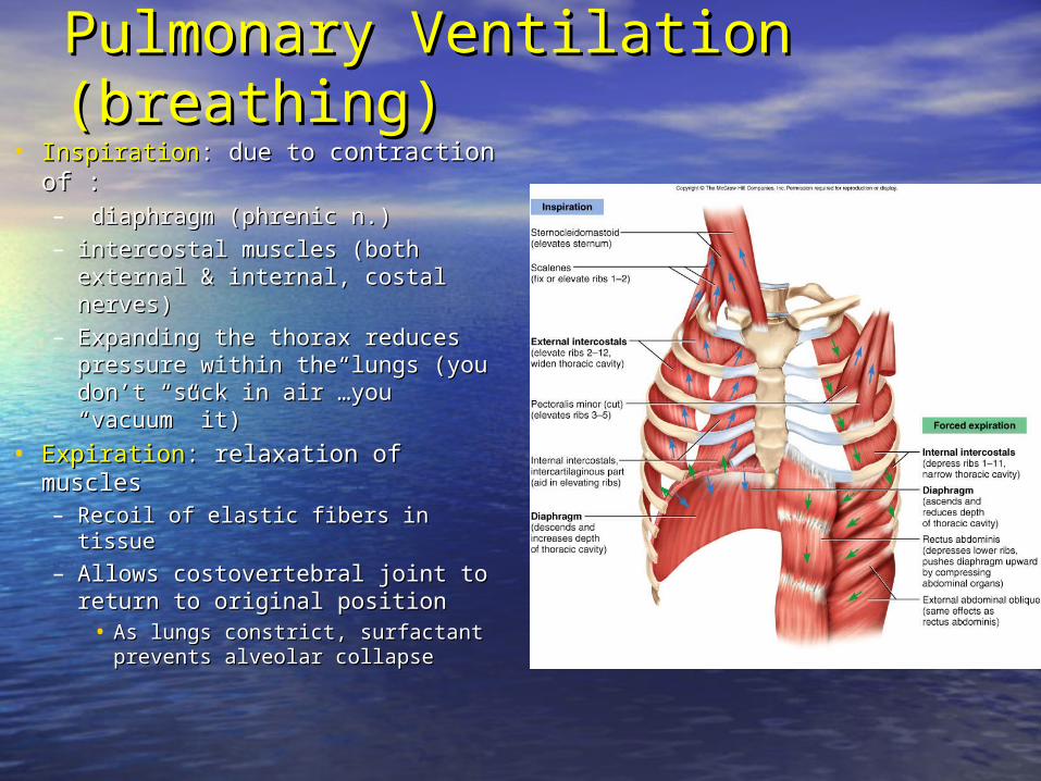
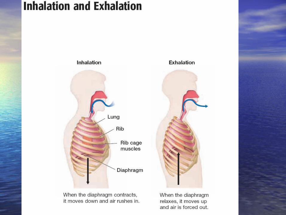
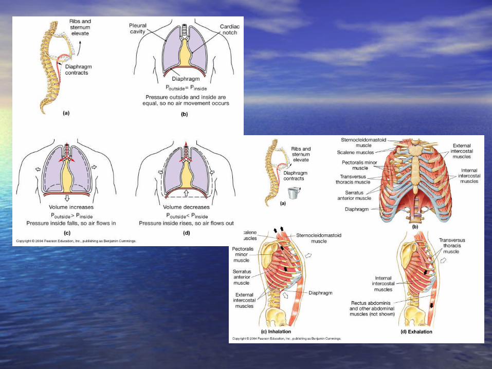
Pulmonary Ventilation (breathing)Pulmonary Ventilation (breathing)• InspirationInspiration: due to c: due to contraction of :ontraction of :
– diaphragm (phrenic n.)diaphragm (phrenic n.)
– intercostal muscles (both external intercostal muscles (both external & internal, costal nerves)& internal, costal nerves)
– Expanding the thorax reduces Expanding the thorax reduces pressure within the lungs (you pressure within the lungs (you don’t “suck in air”…you don’t “suck in air”…you “vacuum” it)“vacuum” it)
• ExpirationExpiration: relaxation of muscles: relaxation of muscles– Recoil of elastic fibers in tissueRecoil of elastic fibers in tissue
– Allows costovertebral joint to Allows costovertebral joint to return to original positionreturn to original position
• As lungs constrict, surfactant As lungs constrict, surfactant prevents alveolar collapseprevents alveolar collapse
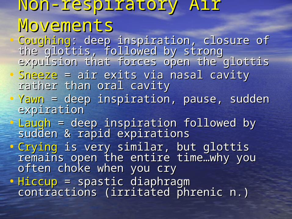
Non-respiratory Air MovementsNon-respiratory Air Movements• CoughingCoughing: deep inspiration, closure of the glottis, : deep inspiration, closure of the glottis,
followed by strong expulsion that forces open the followed by strong expulsion that forces open the glottis glottis
• SneezeSneeze = air exits via nasal cavity rather than oral = air exits via nasal cavity rather than oral cavitycavity
• YawnYawn = deep inspiration, pause, sudden expiration= deep inspiration, pause, sudden expiration• LaughLaugh = deep inspiration followed by sudden & = deep inspiration followed by sudden &
rapid expirationsrapid expirations• CryingCrying is very similar, but glottis remains open the is very similar, but glottis remains open the
entire time…why you often choke when you cryentire time…why you often choke when you cry• HiccupHiccup = spastic diaphragm contractions (irritated = spastic diaphragm contractions (irritated
phrenic n.)phrenic n.)
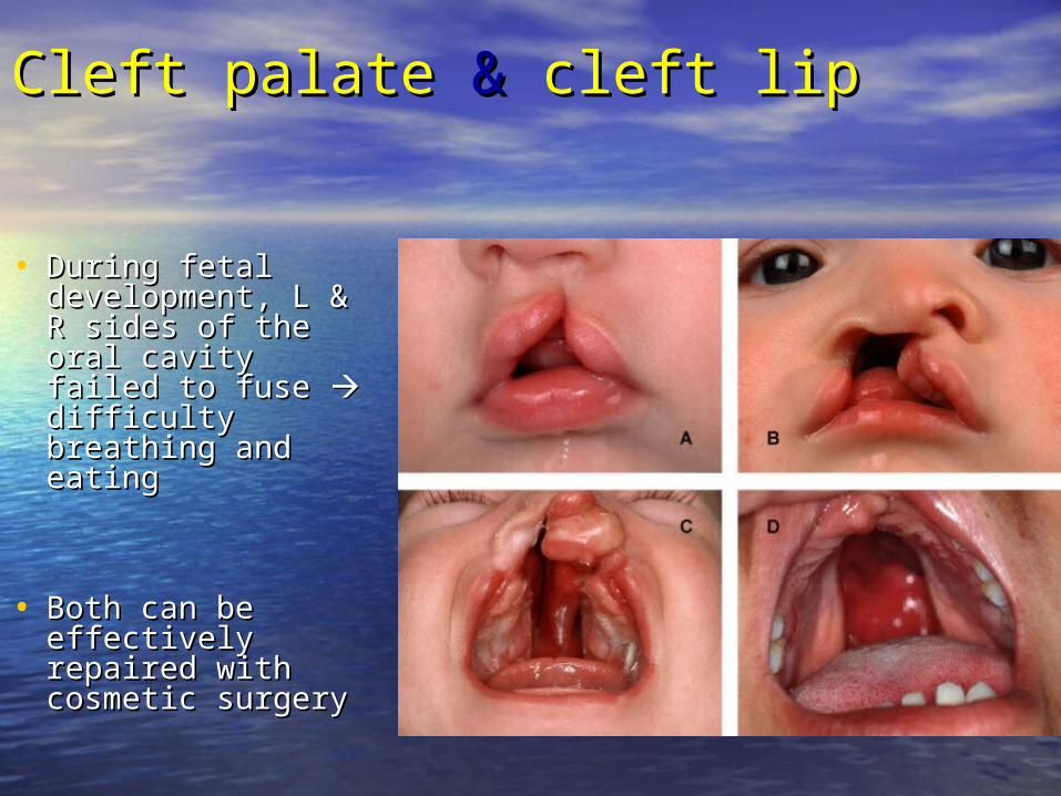
Cleft palate Cleft palate & & cleft lipcleft lip
• During fetal During fetal development, L & R development, L & R sides of the oral cavity sides of the oral cavity failed to fuse failed to fuse difficulty breathing difficulty breathing and eatingand eating
• Both can be Both can be effectively repaired effectively repaired with cosmetic surgerywith cosmetic surgery
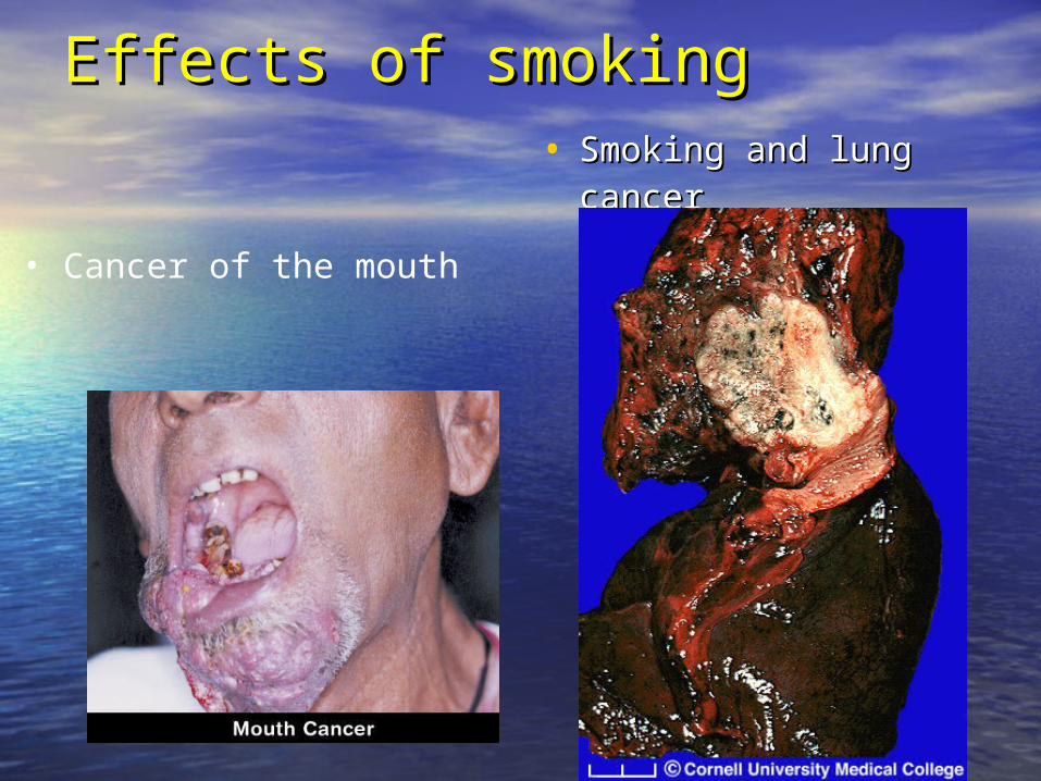
Effects of smokingEffects of smoking• Smoking and lung Smoking and lung
cancercancer
• Cancer of the mouth
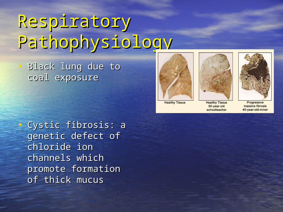
Respiratory PathophysiologyRespiratory Pathophysiology
• Black lung due to coal Black lung due to coal exposureexposure
• Cystic fibrosis: a Cystic fibrosis: a genetic defect of genetic defect of chloride ion channels chloride ion channels which promote which promote formation of thick formation of thick mucusmucus