Embed Size (px)
Citation preview
The Respiratory System
Chapter 23
(6th edition chapter 22)
Functions of the Respiratory System
1. Supply oxygen to the circulatory system for delivery to the tissues2. Remove CO2 (and some other wastes) from blood.
There are 4 processes that we call “respiration”.
1. Pulmonary ventilation - Movement of air into and out of the lungs(also referred to as “breathing”).
2. External respiration - Gas exchange in the lungs between the bloodof the capillaries and the spaces in the air sacs (alveoli)
3. Transport - The movement of gases by the circulatory systemStrictly speaking, a function of the blood.
4. Internal respiration - Gas exchange between the blood and the tissues of the body
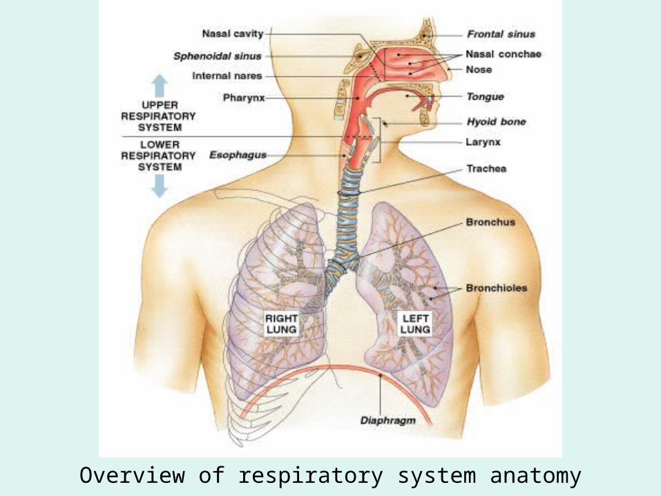
Overview of respiratory system anatomy
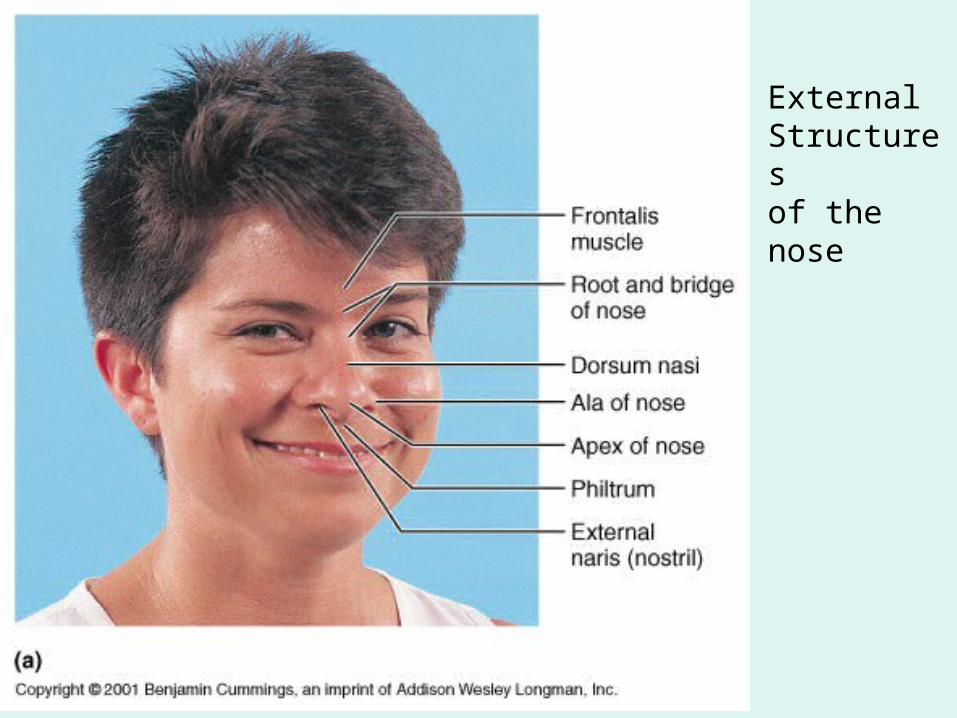
ExternalStructuresof the nose
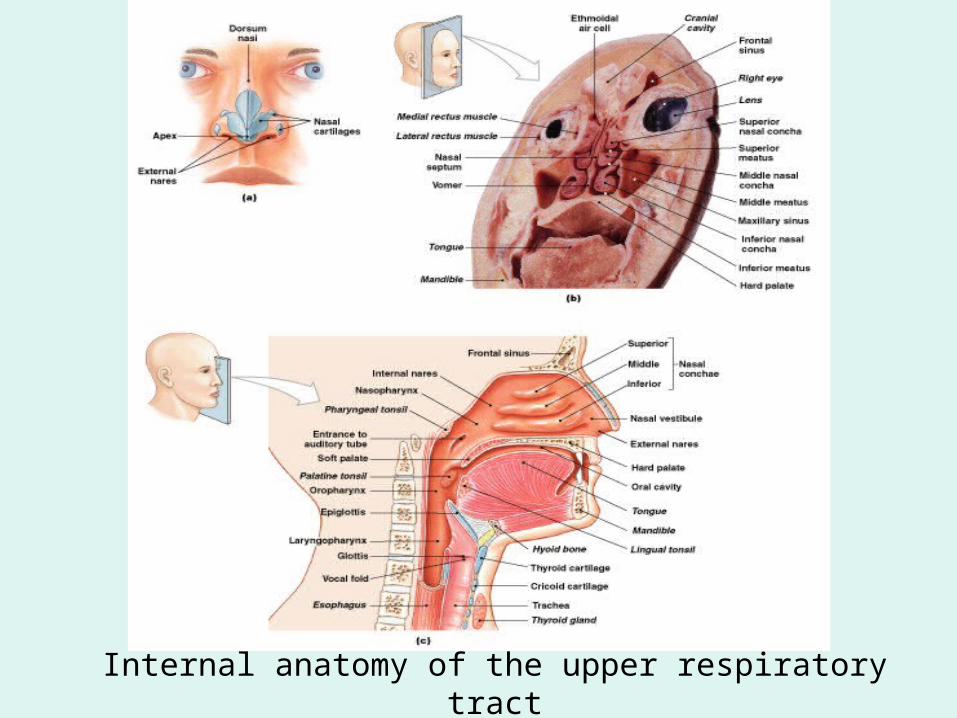
Internal anatomy of the upper respiratory tract
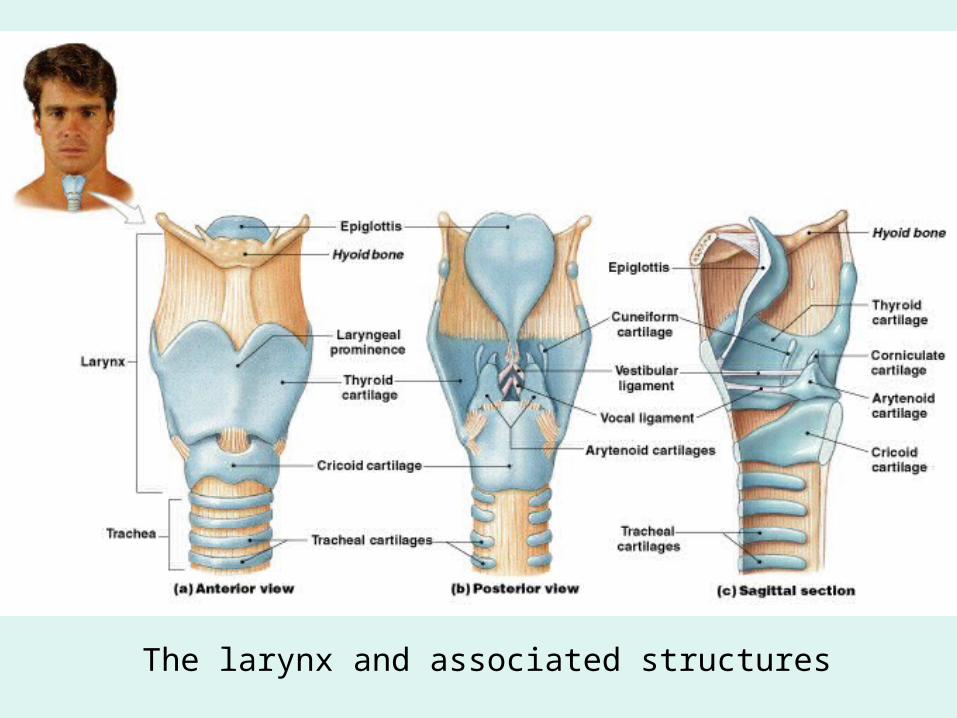
The larynx and associated structures
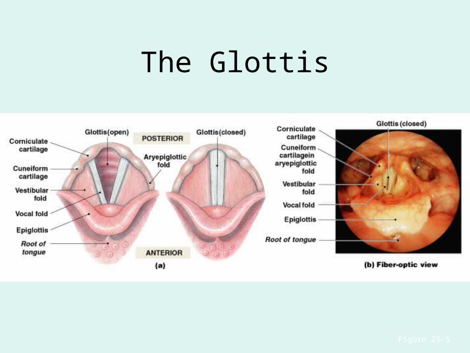
The Glottis
Figure 23–5
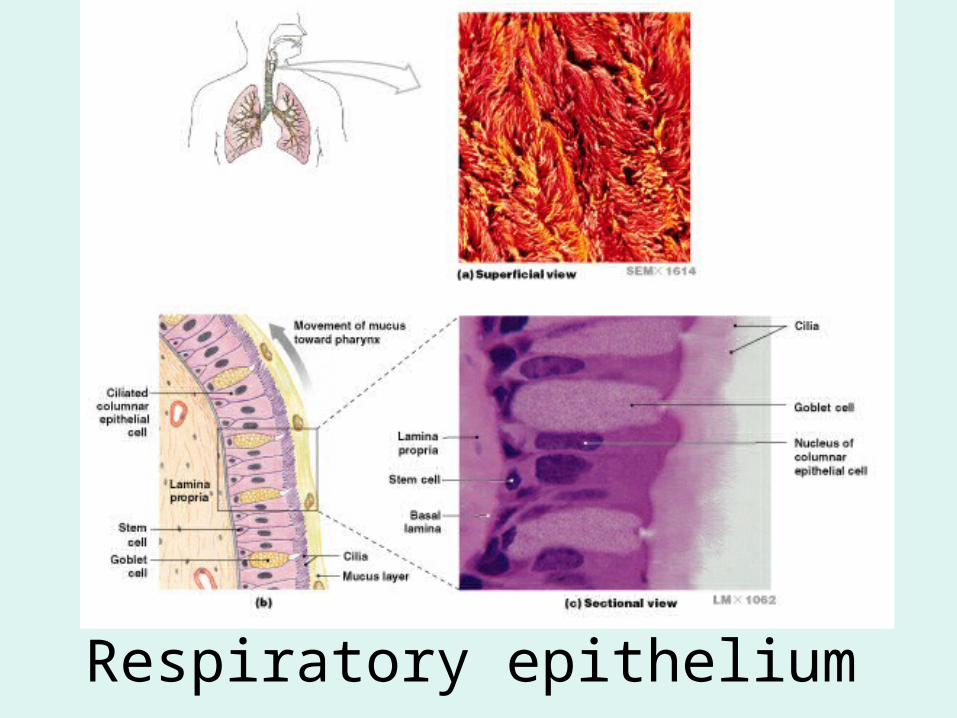
Respiratory epithelium
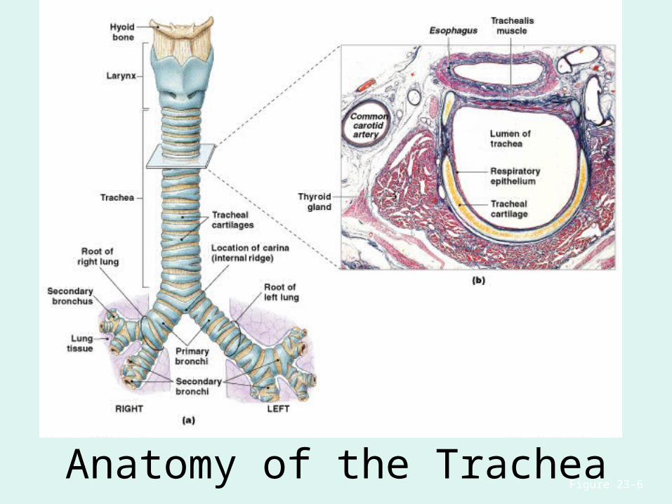
Anatomy of the TracheaFigure 23–6
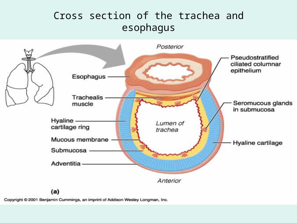
Cross section of the trachea and esophagus
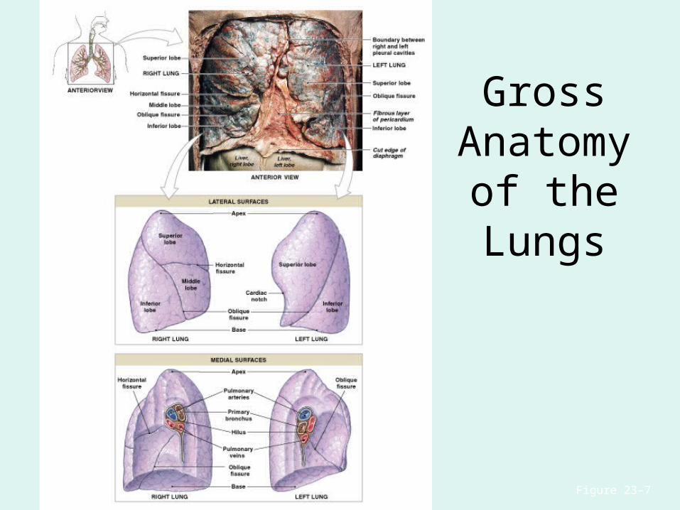
Figure 23–7
Gross Anatomy of the Lungs
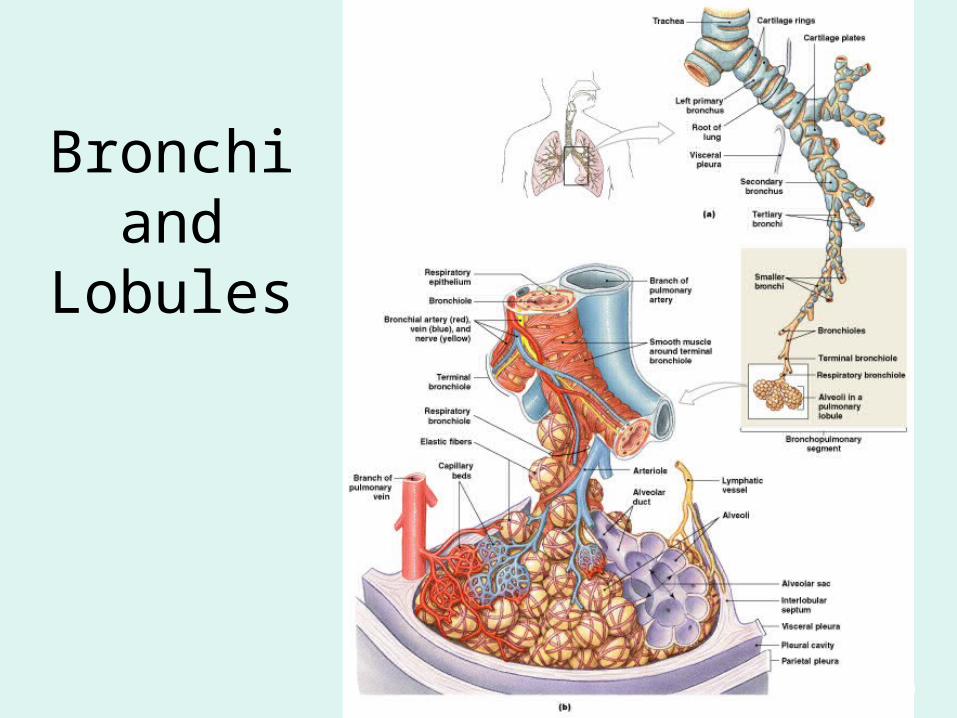
Bronchi and Lobules
Figure 23–9
Secondary Bronchi
• Branch to form tertiary bronchi, also called the segmental bronchi
• Each segmental bronchus:– supplies air to a single bronchopulmonary
segment
Bronchopulmonary Segments
• The right lung has 10
• The left lung has 8 or 9
Bronchial Structure
• The walls of primary, secondary, and tertiary bronchi:– contain progressively less cartilage and more
smooth muscle– increasing muscular effects on airway
constriction and resistance
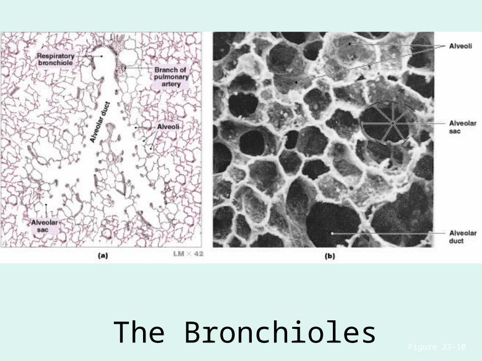
Figure 23–10
The Bronchioles
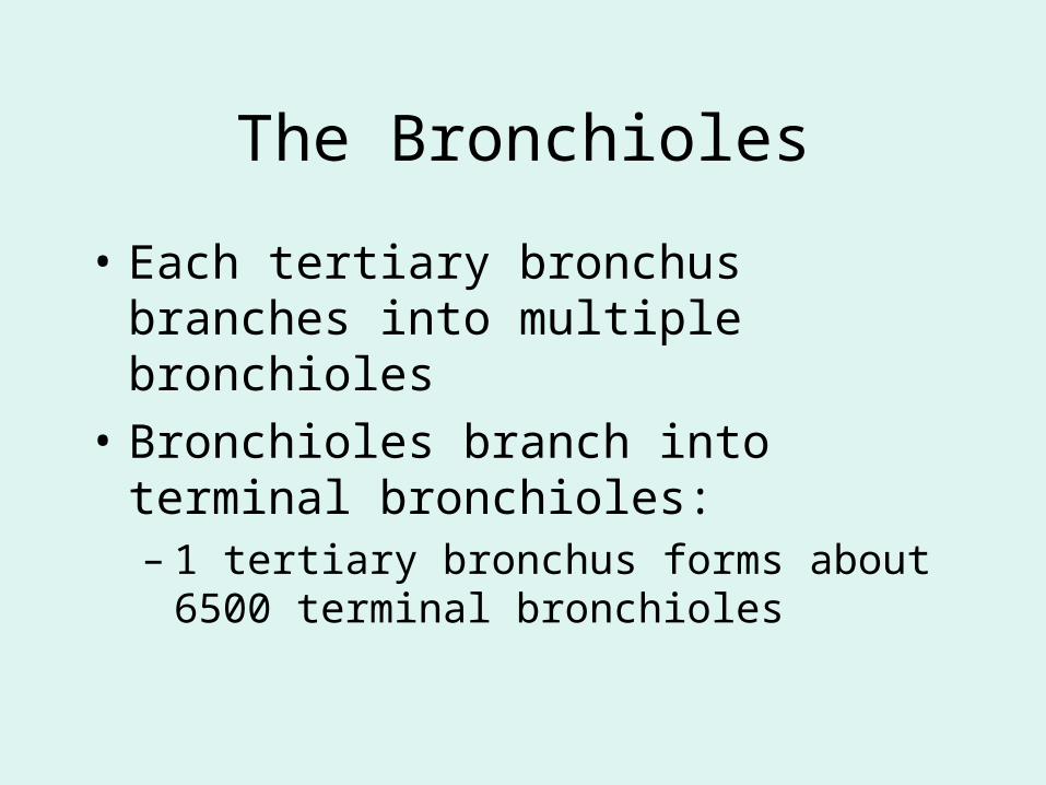
The Bronchioles
• Each tertiary bronchus branches into multiple bronchioles
• Bronchioles branch into terminal bronchioles: – 1 tertiary bronchus forms about 6500 terminal
bronchioles
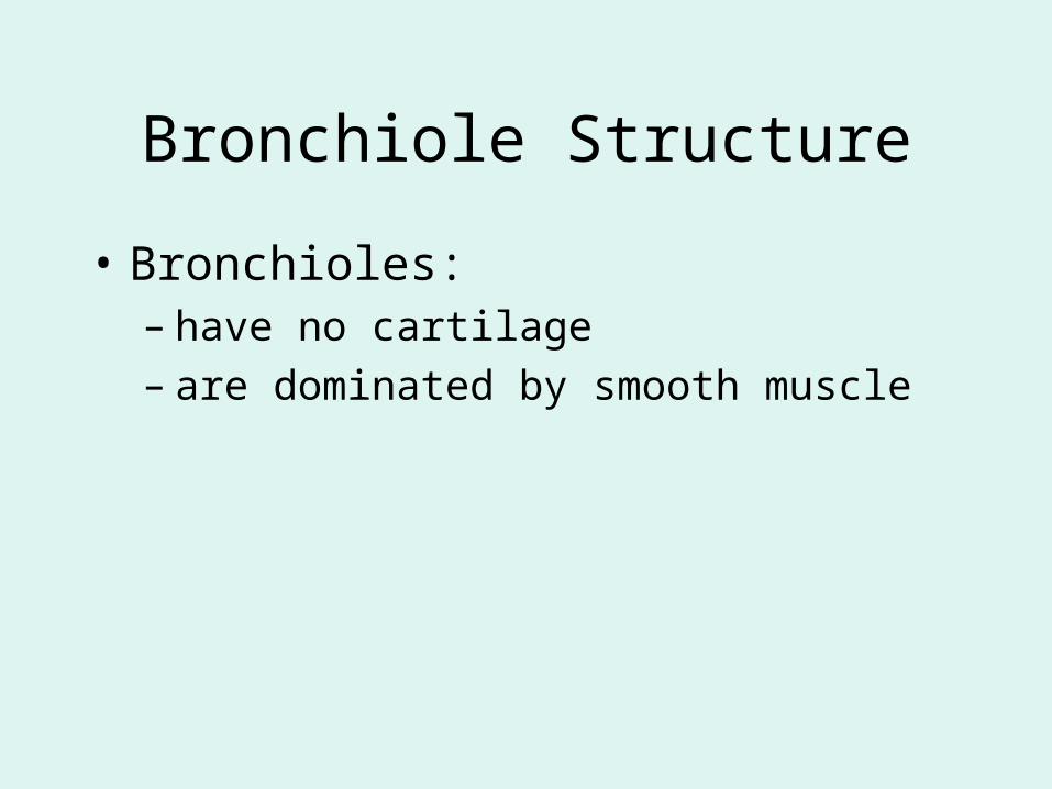
Bronchiole Structure
• Bronchioles:– have no cartilage– are dominated by smooth muscle
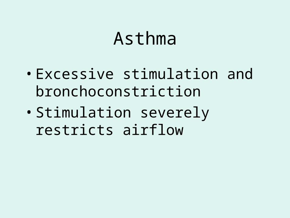
Asthma
• Excessive stimulation and bronchoconstriction
• Stimulation severely restricts airflow
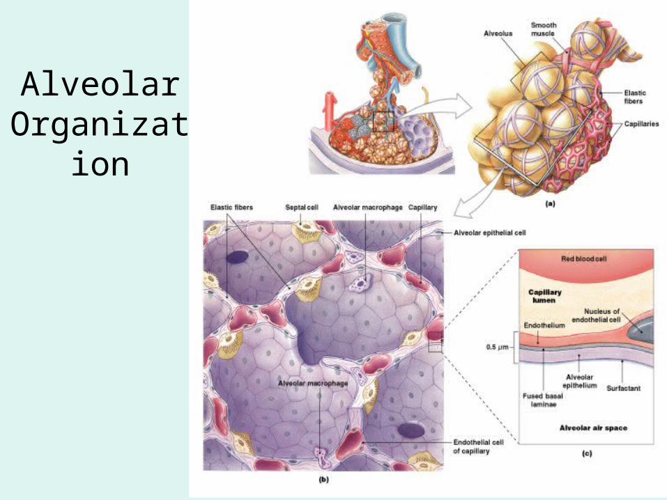
Figure 23–11
Alveolar Organization
Alveolar Epithelium
• Consists of simple squamous epithelium
• Consists of thin, delicate Type I cells
• Patrolled by alveolar macrophages, also called dust cells
• Contains septal cells (Type II cells) that produce surfactant
Surfactant
• Is an oily secretion
• Contains phospholipids and proteins
• Coats alveolar surfaces and reduces surface tension
Respiratory Distress
• Difficult respiration:– due to alveolar collapse – caused when septal cells do not produce enough
surfactant
Respiratory Membrane
• The thin membrane of alveoli where gas exchange takes place
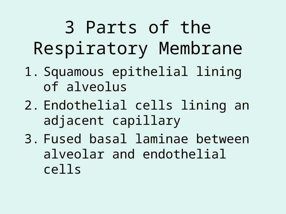
3 Parts of the Respiratory Membrane
1. Squamous epithelial lining of alveolus
2. Endothelial cells lining an adjacent capillary
3. Fused basal laminae between alveolar and endothelial cells
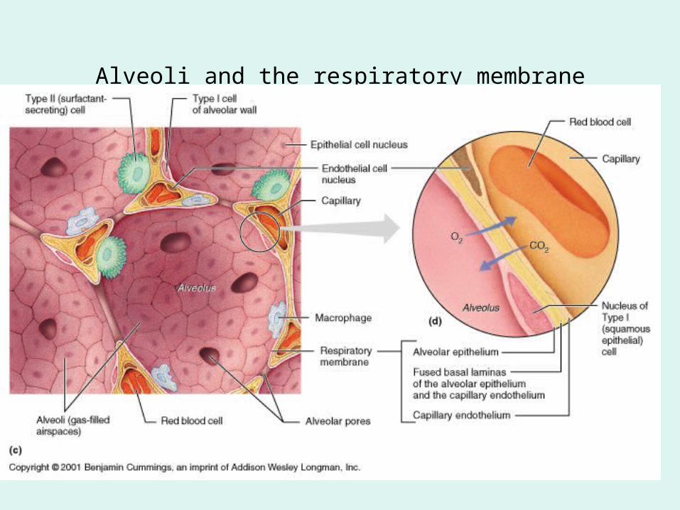
Alveoli and the respiratory membrane
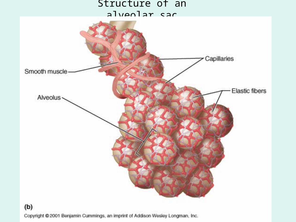
Structure of an alveolar sac
Pleural Cavities and Pleural Membranes
• 2 pleural cavities:– are separated by the mediastinum
• Each pleural cavity:– holds a lung – is lined with a serous membrane (the pleura)
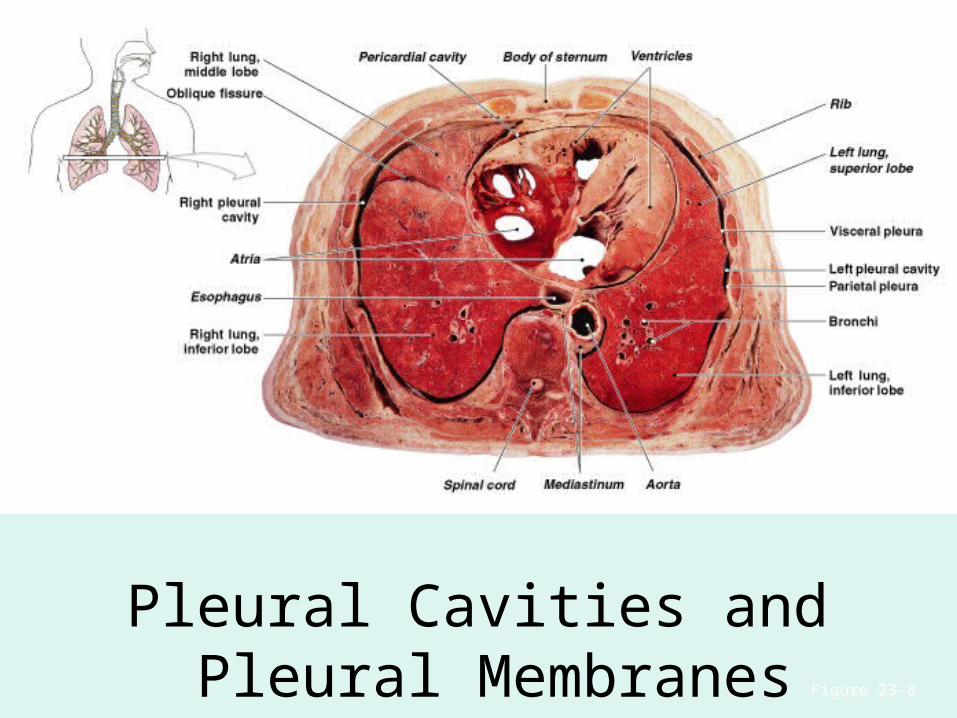
Figure 23–8
Pleural Cavities and Pleural Membranes
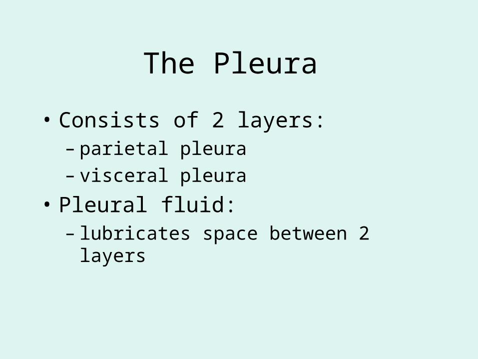
The Pleura
• Consists of 2 layers: – parietal pleura – visceral pleura
• Pleural fluid:– lubricates space between 2 layers
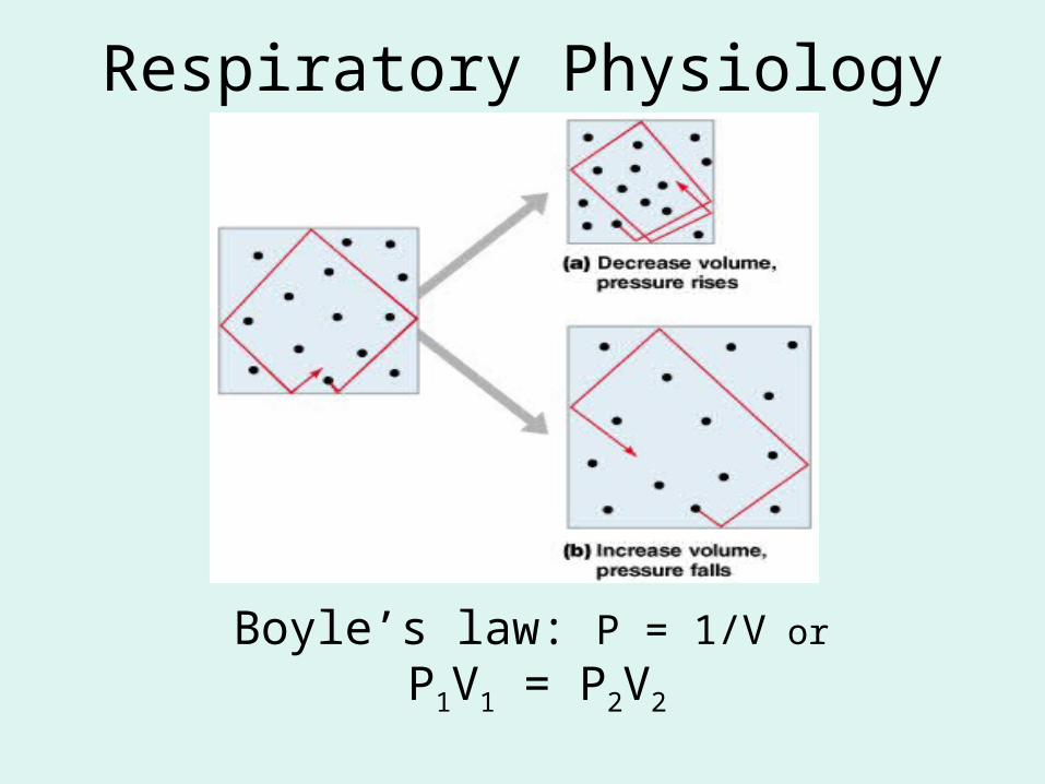
Respiratory Physiology
Boyle’s law: P = 1/V or
P1V1 = P2V2
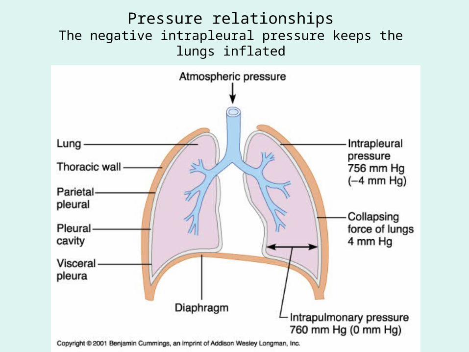
Pressure relationshipsThe negative intrapleural pressure keeps the lungs inflated
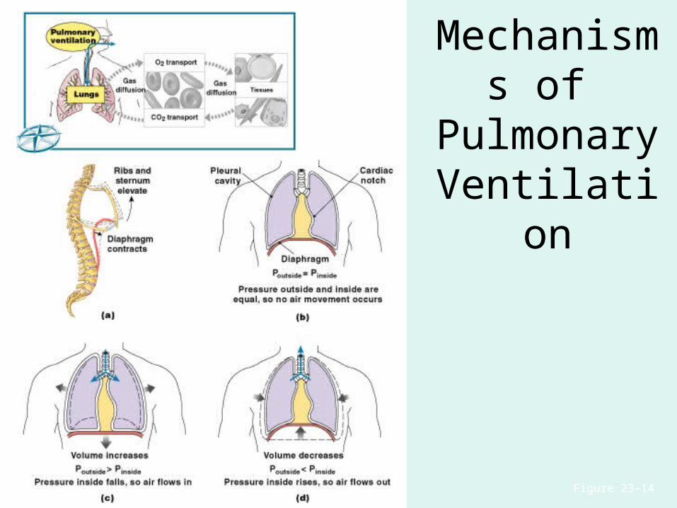
Figure 23–14
Mechanisms of
Pulmonary Ventilation
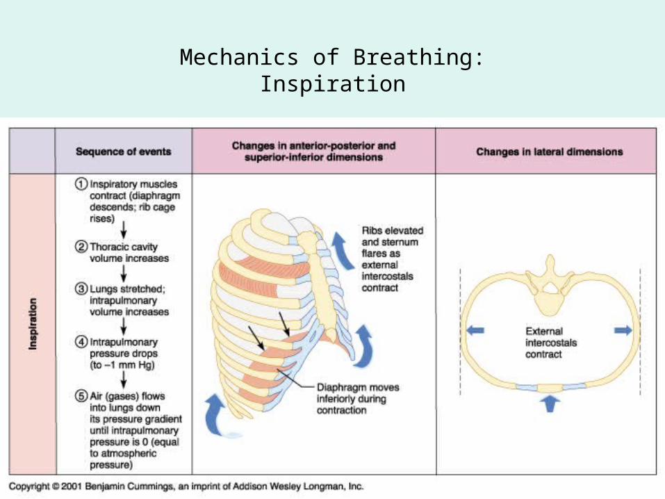
Mechanics of Breathing:Inspiration
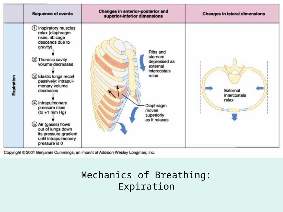
Mechanics of Breathing:Expiration
Compliance of the Lung
• An indicator of expandability
• Low compliance requires greater force
• High compliance requires less force
Factors That Affect Compliance
1. Connective-tissue structure of the lungs
2. Level of surfactant production
3. Mobility of the thoracic cage
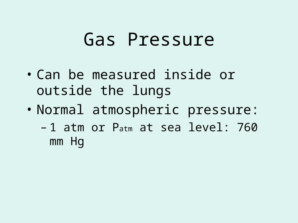
Gas Pressure
• Can be measured inside or outside the lungs
• Normal atmospheric pressure: – 1 atm or Patm at sea level: 760 mm Hg
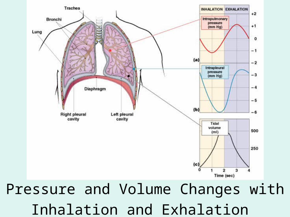
Pressure and Volume Changes with Inhalation
and Exhalation
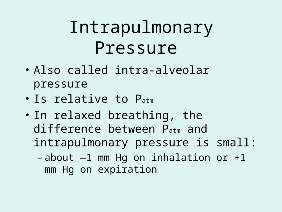
Intrapulmonary Pressure
• Also called intra-alveolar pressure• Is relative to Patm
• In relaxed breathing, the difference between Patm and intrapulmonary pressure is small:– about —1 mm Hg on inhalation or +1 mm Hg
on expiration
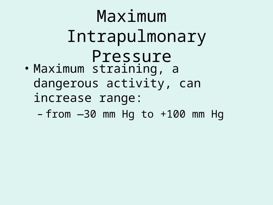
Maximum Intrapulmonary Pressure
• Maximum straining, a dangerous activity, can increase range:– from —30 mm Hg to +100 mm Hg
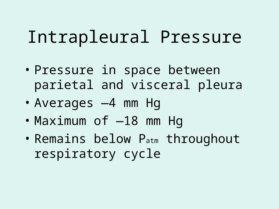
Intrapleural Pressure
• Pressure in space between parietal and visceral pleura
• Averages —4 mm Hg
• Maximum of —18 mm Hg
• Remains below Patm throughout respiratory cycle
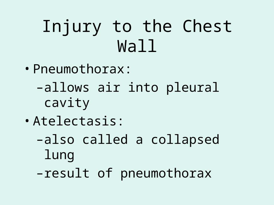
Injury to the Chest Wall
• Pneumothorax:
– allows air into pleural cavity
• Atelectasis:
– also called a collapsed lung
– result of pneumothorax
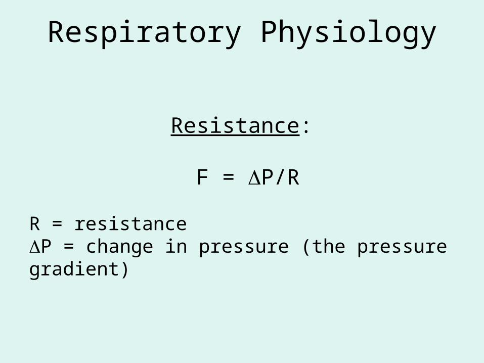
Respiratory Physiology
Resistance:
F = P/R
R = resistanceP = change in pressure (the pressure gradient)
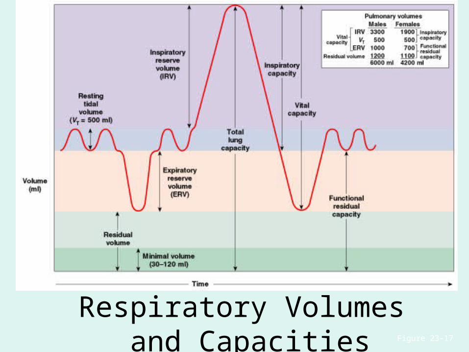
Respiratory Volumes and Capacities Figure 23–17
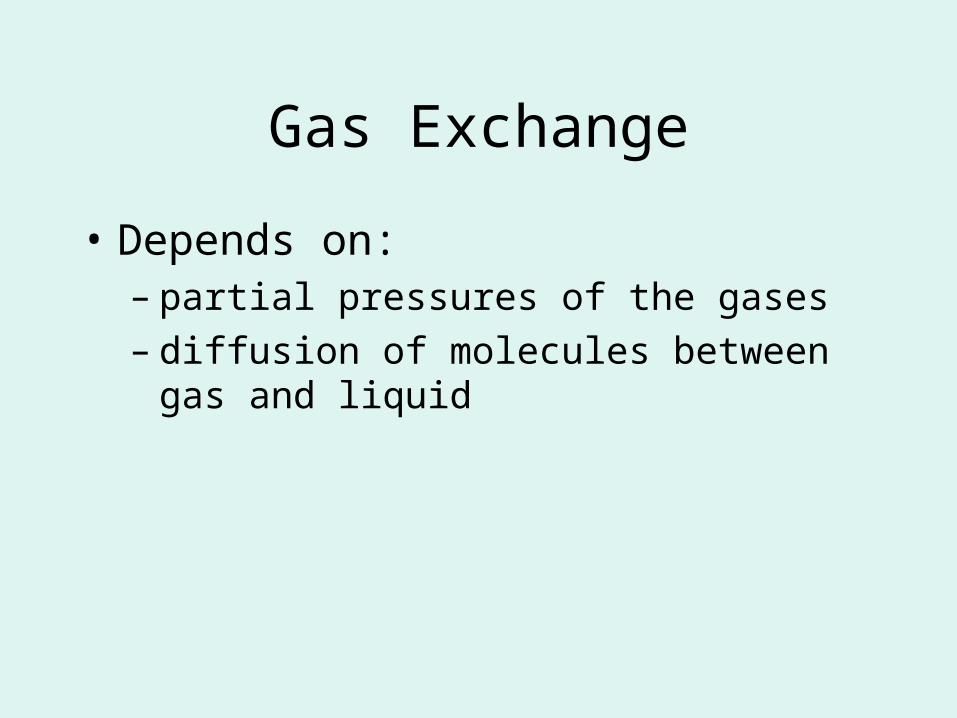
Gas Exchange
• Depends on:– partial pressures of the gases– diffusion of molecules between gas and liquid
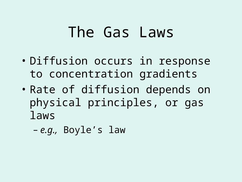
The Gas Laws
• Diffusion occurs in response to concentration gradients
• Rate of diffusion depends on physical principles, or gas laws– e.g., Boyle’s law
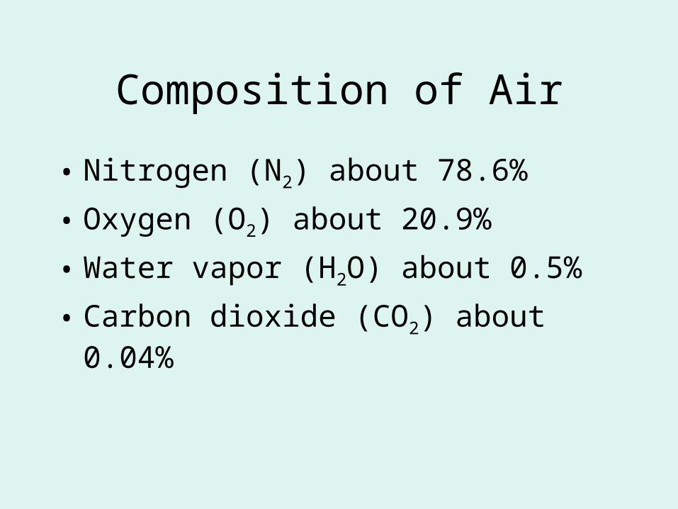
Composition of Air
• Nitrogen (N2) about 78.6%
• Oxygen (O2) about 20.9%
• Water vapor (H2O) about 0.5%
• Carbon dioxide (CO2) about 0.04%
Gas Pressure
• Atmospheric pressure (760 mm Hg):– produced by air molecules bumping into each
other
• Each gas contributes to the total pressure:– in proportion to its number of molecules
(Dalton’s law)
Partial Pressure
• The pressure contributed by each gas in the atmosphere
• All partial pressures together add up to 760 mm Hg
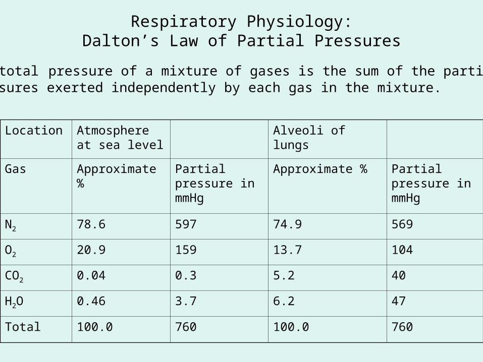
Respiratory Physiology:Dalton’s Law of Partial Pressures
The total pressure of a mixture of gases is the sum of the partialpressures exerted independently by each gas in the mixture.
Location Atmosphere at sea level
Alveoli of lungs
Gas Approximate %
Partial pressure in mmHg
Approximate % Partial pressure in mmHg
N2 78.6 597 74.9 569
O2 20.9 159 13.7 104
CO2 0.04 0.3 5.2 40
H2O 0.46 3.7 6.2 47
Total 100.0 760 100.0 760
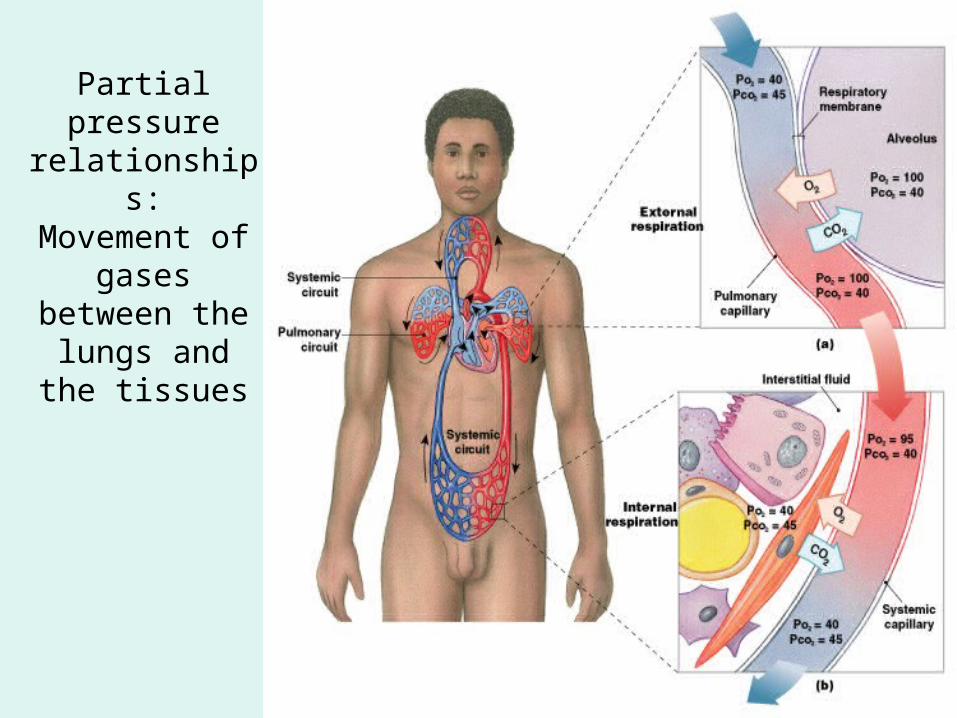
Partial pressure
relationships:Movement of
gases between the lungs and the
tissues
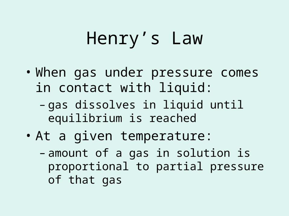
Henry’s Law
• When gas under pressure comes in contact with liquid:– gas dissolves in liquid until equilibrium is
reached
• At a given temperature:– amount of a gas in solution is proportional to
partial pressure of that gas
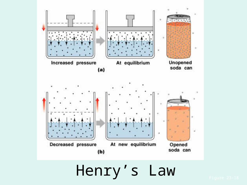
Henry’s LawFigure 23–18
Diffusion and the Respiratory Membrane
• Direction and rate of diffusion of gases across the respiratory membrane determine different partial pressures and solubilities
Efficiency of Gas Exchange
• Due to:– substantial differences in partial pressure across
the respiratory membrane
– distances involved in gas exchange are small
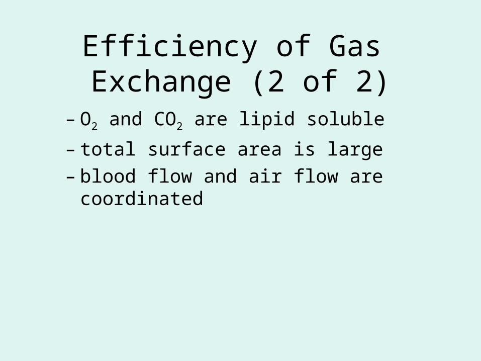
Efficiency of Gas Exchange (2 of 2)
– O2 and CO2 are lipid soluble
– total surface area is large
– blood flow and air flow are coordinated
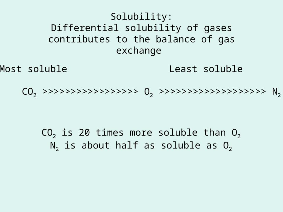
Solubility:Differential solubility of gases contributes to the
balance of gas exchange
Most soluble Least soluble
CO2 >>>>>>>>>>>>>>>>> O2 >>>>>>>>>>>>>>>>>>> N2
CO2 is 20 times more soluble than O2
N2 is about half as soluble as O2
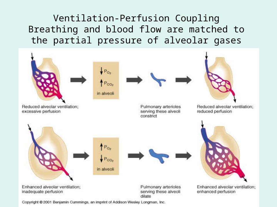
Ventilation-Perfusion CouplingBreathing and blood flow are matched to the
partial pressure of alveolar gases
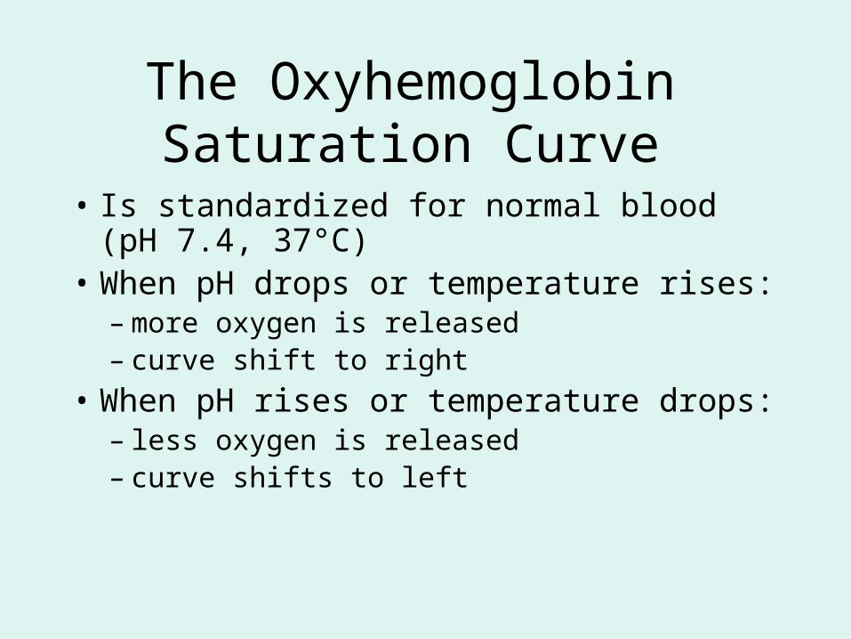
The Oxyhemoglobin Saturation Curve
• Is standardized for normal blood (pH 7.4, 37°C)
• When pH drops or temperature rises:– more oxygen is released– curve shift to right
• When pH rises or temperature drops:– less oxygen is released– curve shifts to left
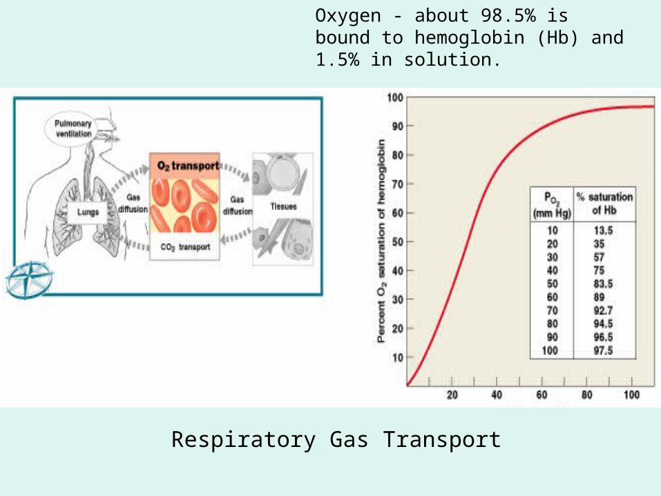
Respiratory Gas Transport
Oxygen - about 98.5% is bound to hemoglobin (Hb) and 1.5% in solution.
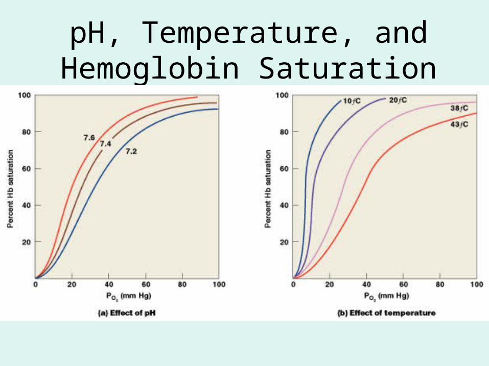
pH, Temperature, and Hemoglobin Saturation
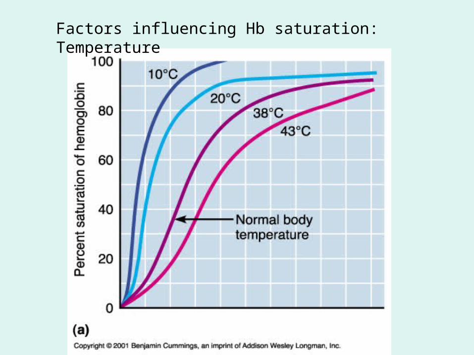
Factors influencing Hb saturation: Temperature
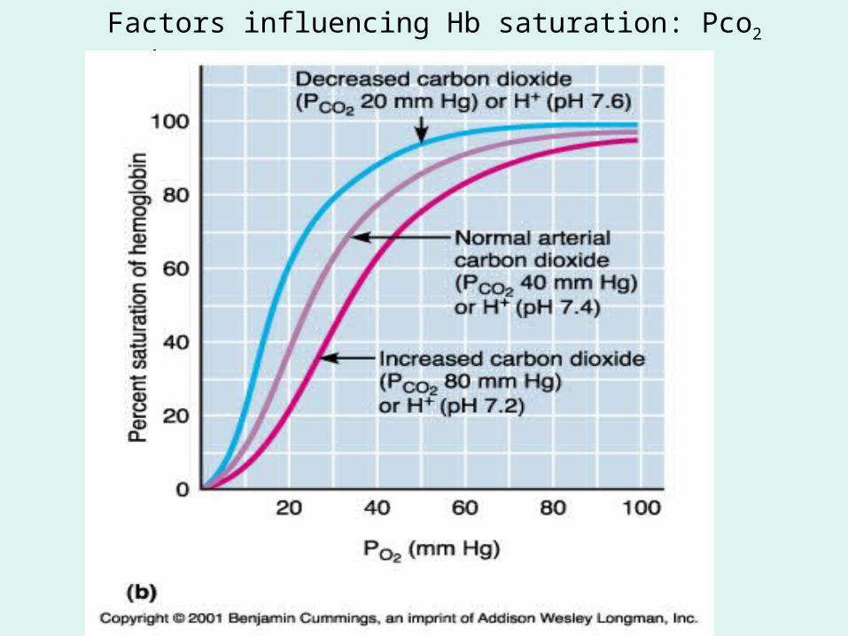
Factors influencing Hb saturation: Pco2 and pH
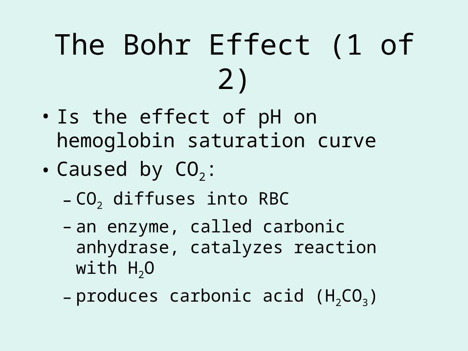
The Bohr Effect (1 of 2)
• Is the effect of pH on hemoglobin saturation curve
• Caused by CO2:
– CO2 diffuses into RBC
– an enzyme, called carbonic anhydrase, catalyzes reaction with H2O
– produces carbonic acid (H2CO3)
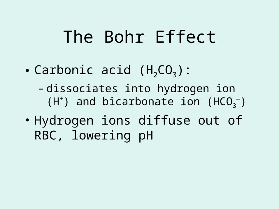
The Bohr Effect
• Carbonic acid (H2CO3):
– dissociates into hydrogen ion (H+) and bicarbonate ion (HCO3
—)
• Hydrogen ions diffuse out of RBC, lowering pH
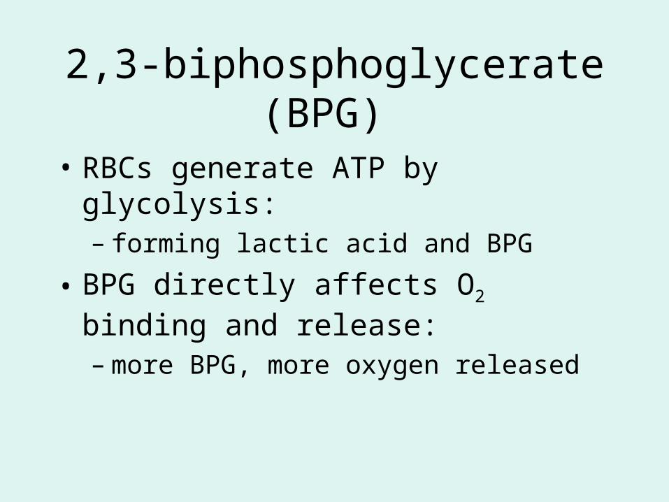
2,3-biphosphoglycerate (BPG)
• RBCs generate ATP by glycolysis:– forming lactic acid and BPG
• BPG directly affects O2 binding and release:
– more BPG, more oxygen released
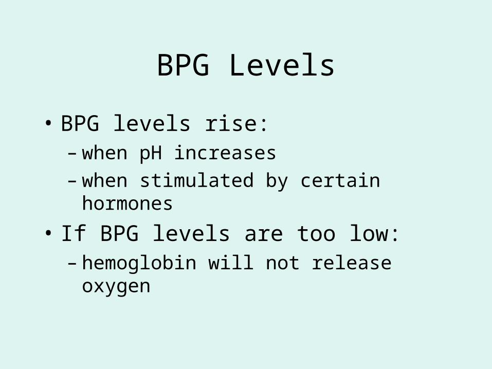
BPG Levels
• BPG levels rise:– when pH increases– when stimulated by certain hormones
• If BPG levels are too low:– hemoglobin will not release oxygen
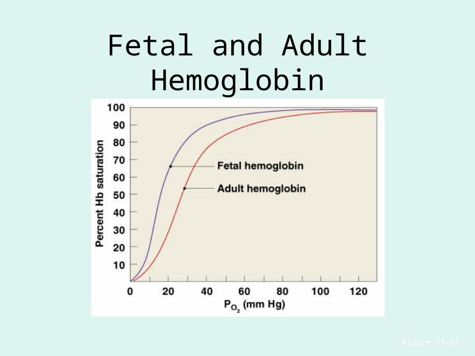
Fetal and Adult Hemoglobin
Figure 23–22
Fetal and Adult Hemoglobin
• The structure of fetal hemoglobin:– differs from that of adult Hb
• At the same PO2:
– fetal Hb binds more O2 than adult Hb
– which allows fetus to take O2 from maternal blood
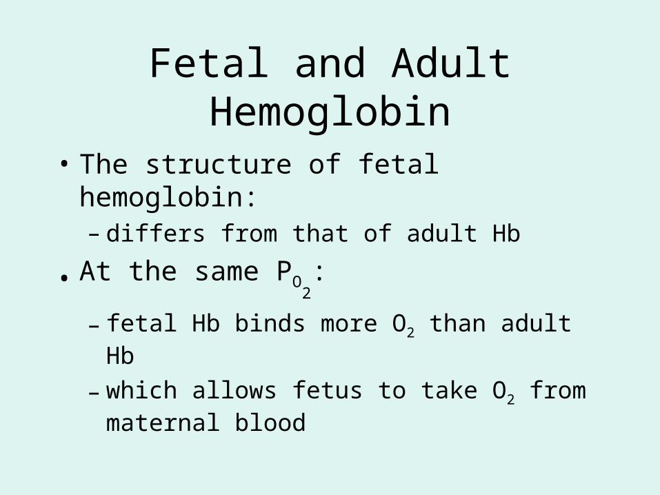
CO2 Transport
• 7 % dissolved in the plasma• ~ 23% bound to the amine groups of the Hb molecule as carbaminohemoglobin• ~ 70% as bicarbonate ion in the plasma
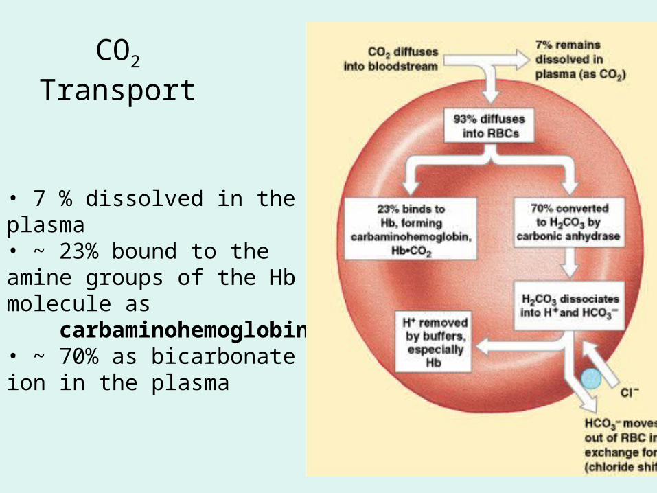
CO2 Transport & Exchange:at the tissues
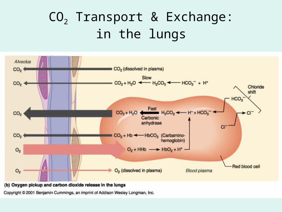
CO2 Transport & Exchange:in the lungs
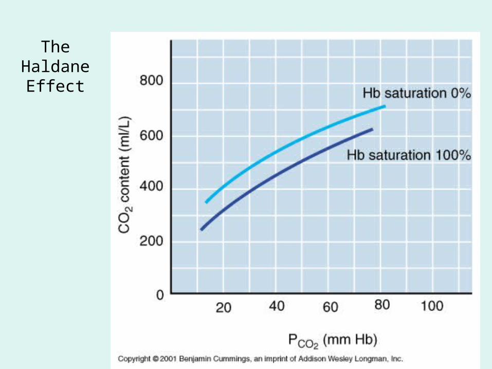
The Haldane
Effect

Control of Respiration
• Gas diffusion at peripheral and alveolar capillaries maintain balance by:
– changes in blood flow and oxygen delivery
– changes in depth and rate of respiration
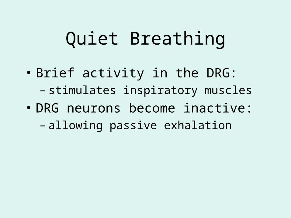
Quiet Breathing
• Brief activity in the DRG:– stimulates inspiratory muscles
• DRG neurons become inactive:– allowing passive exhalation
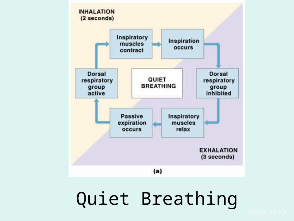
Figure 23–25a
Quiet Breathing
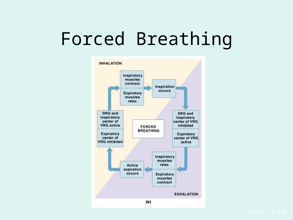
Figure 23–25b
Forced Breathing
The Apneustic and Pneumotaxic Centers of the Pons
• Paired nuclei that adjust output of respiratory rhythmicity centers:– regulating respiratory rate and depth of
respiration
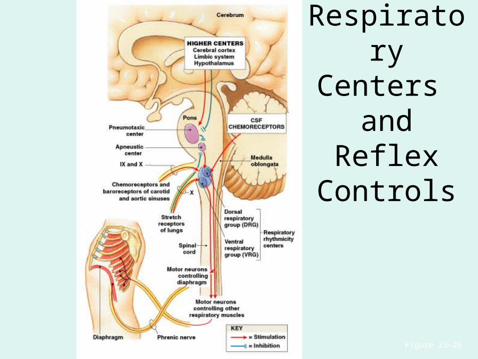
Respiratory Centers
and Reflex Controls
Figure 23–26
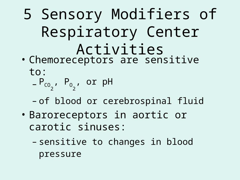
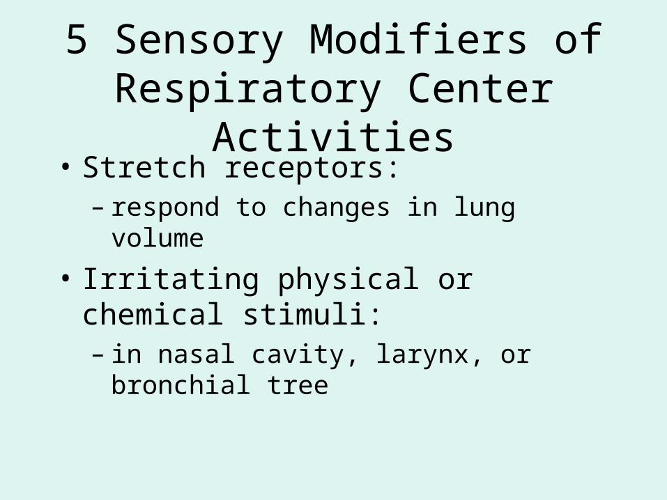
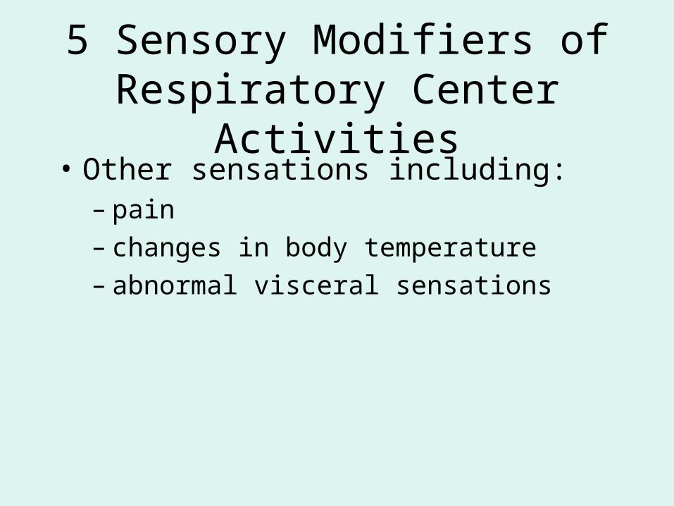
5 Sensory Modifiers of Respiratory Center Activities
• Chemoreceptors are sensitive to:– PCO
2, PO
2, or pH
– of blood or cerebrospinal fluid
• Baroreceptors in aortic or carotic sinuses:– sensitive to changes in blood pressure
5 Sensory Modifiers of Respiratory Center Activities
• Stretch receptors:– respond to changes in lung volume
• Irritating physical or chemical stimuli:– in nasal cavity, larynx, or bronchial tree
5 Sensory Modifiers of Respiratory Center Activities
• Other sensations including:– pain– changes in body temperature– abnormal visceral sensations
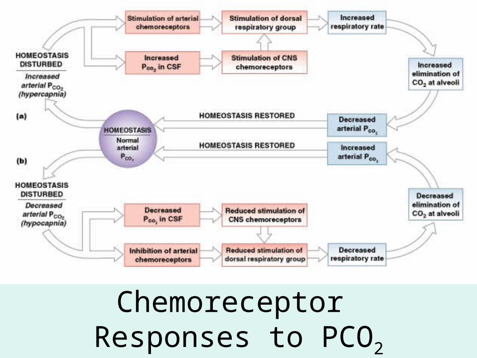
Chemoreceptor Responses to PCO2
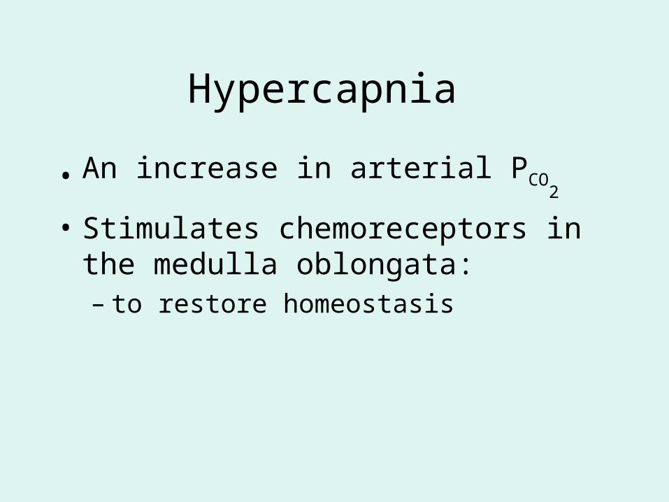
Hypercapnia
• An increase in arterial PCO2
• Stimulates chemoreceptors in the medulla oblongata:– to restore homeostasis
Hypoventilation
• A common cause of hypercapnia
• Abnormally low respiration rate:– allows CO2 build-up in blood
Hyperventilation
• Excessive ventilation
• Results in abnormally low PCO2
(hypocapnia)
• Stimulates chemoreceptors to decrease respiratory rate
Baroreceptor Reflexes
• Carotid and aortic baroreceptor stimulation:– affects blood pressure and respiratory centers
• When blood pressure falls:– respiration increases
• When blood pressure increases:– respiration decreases
The Hering–Breuer Reflexes
• 2 baroreceptor reflexes involved in forced breathing:– inflation reflex:
• prevents overexpansion of lungs
– deflation reflex:• inhibits expiratory centers
• stimulates inspiratory centers during lung deflation
Protective Reflexes
• Triggered by receptors in epithelium of respiratory tract when lungs are exposed to:– toxic vapors– chemicals irritants– mechanical stimulation
• Cause sneezing, coughing, and laryngeal spasm
Pathology and clinical considerations
Common homeostatic imbalances:• COPD (chronic obstructive pulmonary disease)• Asthma• Tuberculosis• Lung cancer
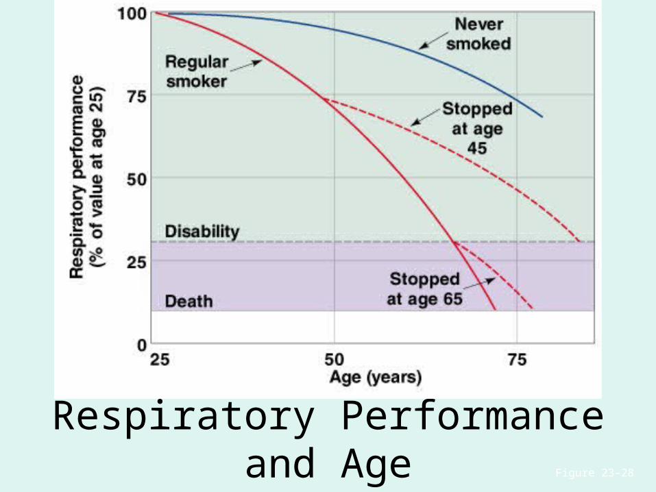
Respiratory Performance and Age
Figure 23–28
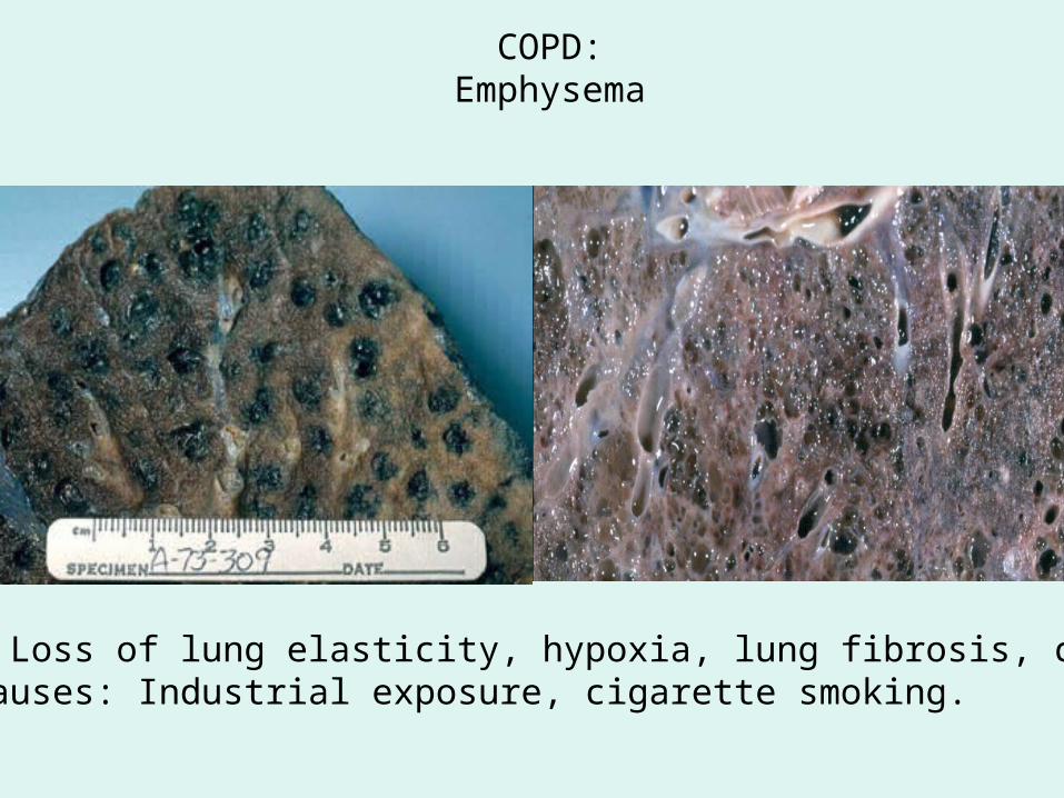
COPD:Emphysema
Results: Loss of lung elasticity, hypoxia, lung fibrosis, cyanosis.Common causes: Industrial exposure, cigarette smoking.
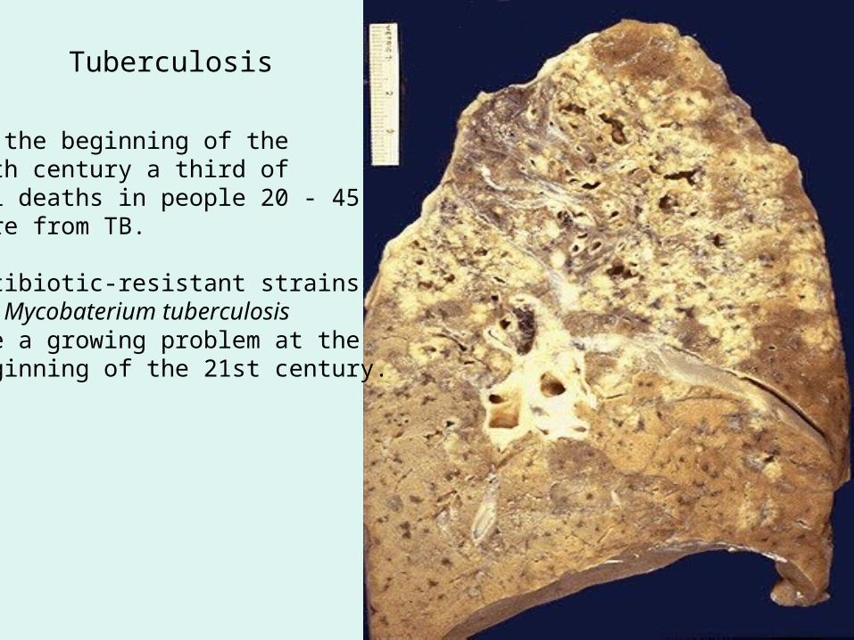
Tuberculosis
At the beginning of the20th century a third ofall deaths in people 20 - 45were from TB.
Antibiotic-resistant strainsof Mycobaterium tuberculosisare a growing problem at the beginning of the 21st century.
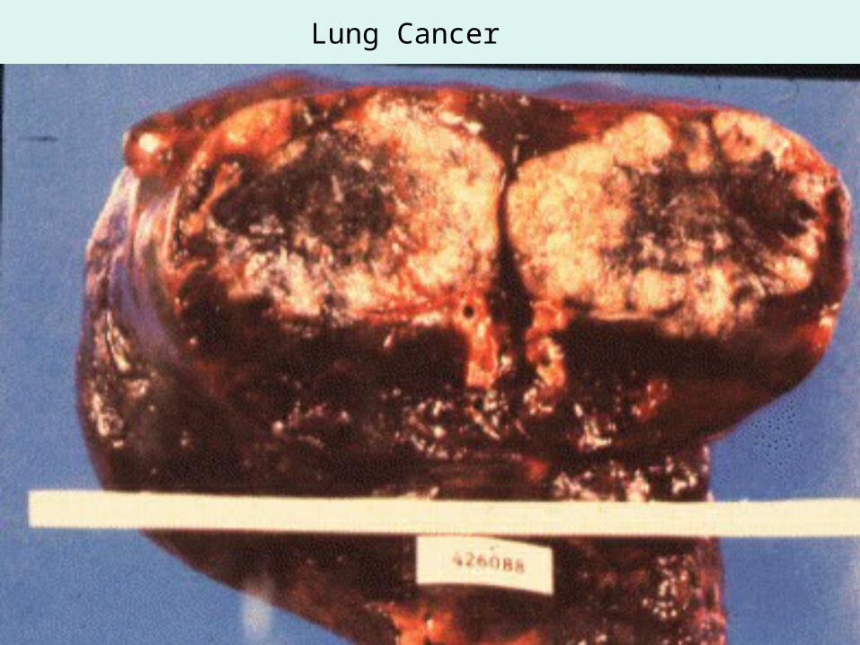
Lung Cancer
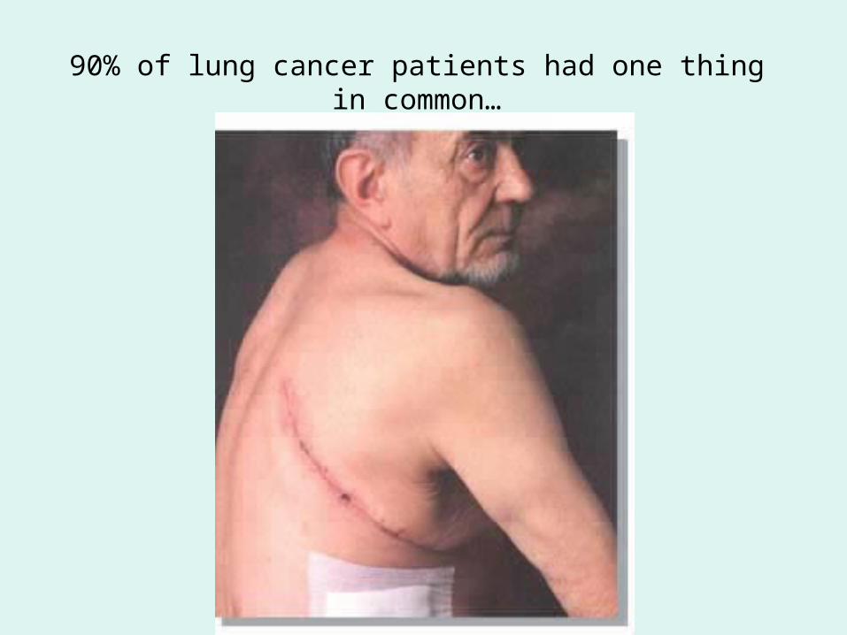
90% of lung cancer patients had one thing in common…
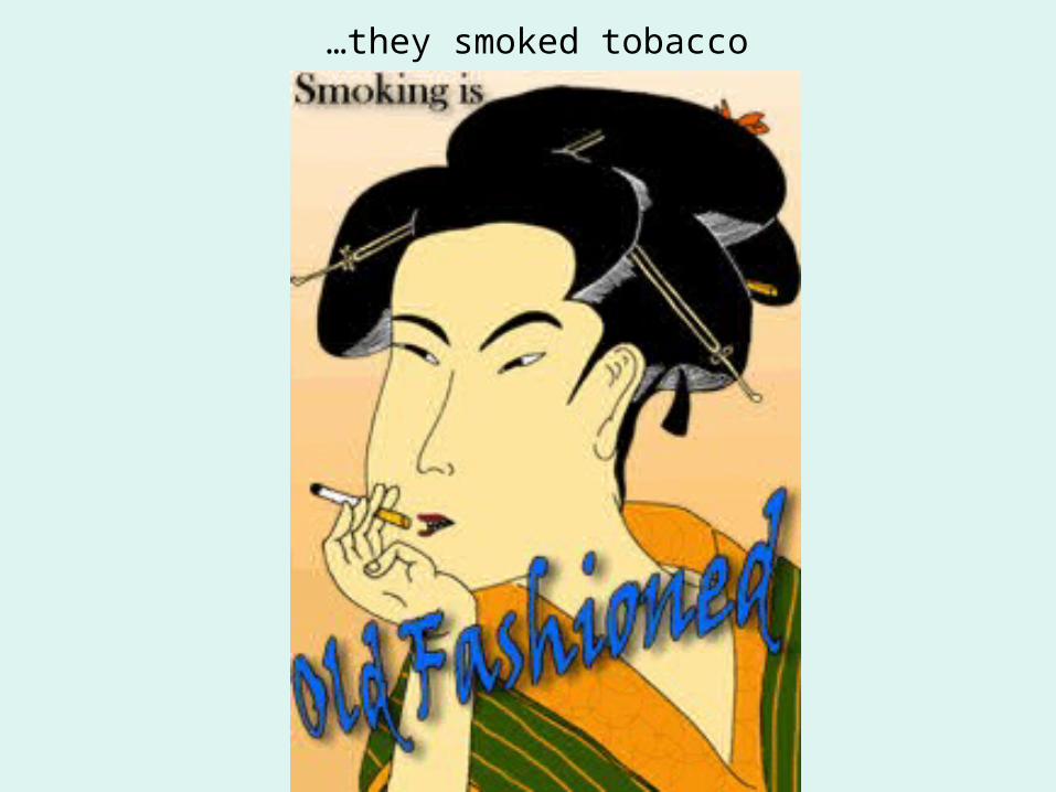
…they smoked tobacco

Fin