Embed Size (px)
Citation preview
The Respiratory SystemThe Respiratory SystemCh 22Ch 22
Human AnatomyHuman AnatomySonya Schuh-Huerta, Ph.D.Sonya Schuh-Huerta, Ph.D.
Leonardo Da Vinci

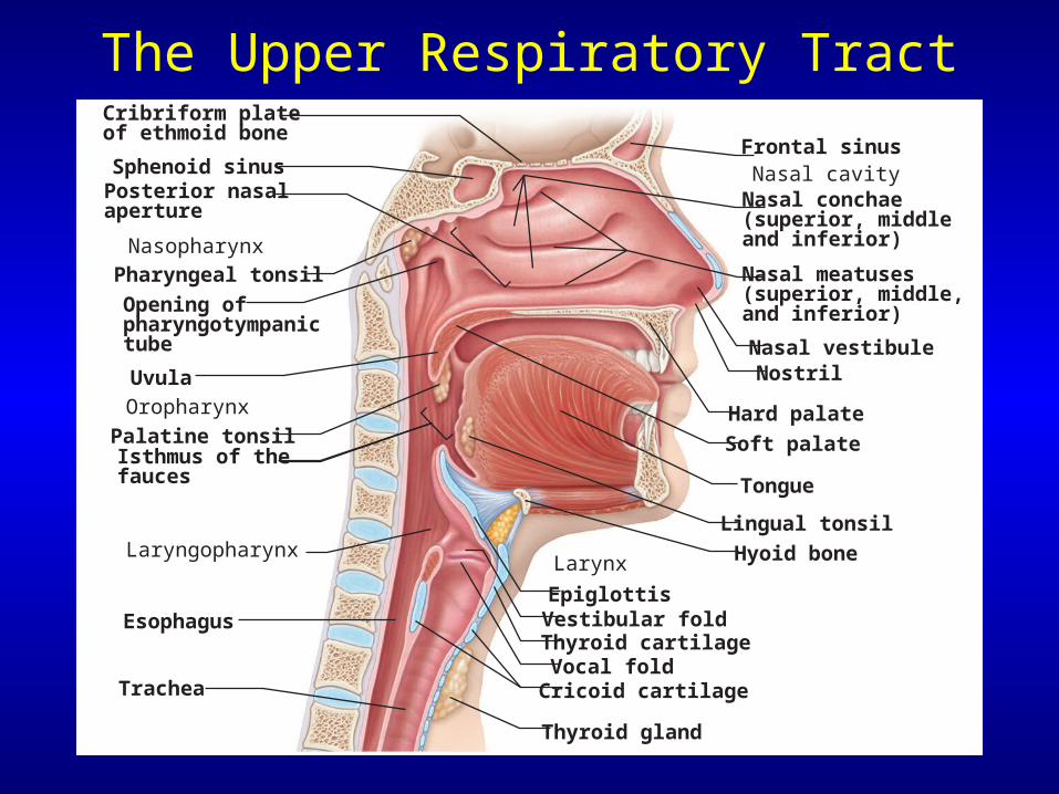
The Upper Respiratory Tract
Sphenoid sinusFrontal sinus
Nasal meatuses(superior, middle,and inferior)
Nasopharynx
Uvula
Palatine tonsilIsthmus of thefauces
Posterior nasalaperture
Opening ofpharyngotympanictube
Pharyngeal tonsil
Oropharynx
Laryngopharynx
Vocal fold
Esophagus
Nasal conchae(superior, middle and inferior)
Nasal vestibuleNostril
Nasal cavity
Hard palate
Soft palate
Tongue
Lingual tonsil
Epiglottis
Hyoid boneLarynx
Thyroid cartilageVestibular fold
Cricoid cartilage
Thyroid gland
Trachea
Cribriform plateof ethmoid bone
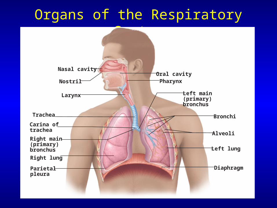
Organs of the Respiratory System
Nasal cavity
Trachea
Carina of trachea
Left main (primary) bronchus
Right main (primary) bronchus
Right lung
Parietalpleura
Left lung
Alveoli
Bronchi
NostrilOral cavityPharynx
Larynx
Diaphragm
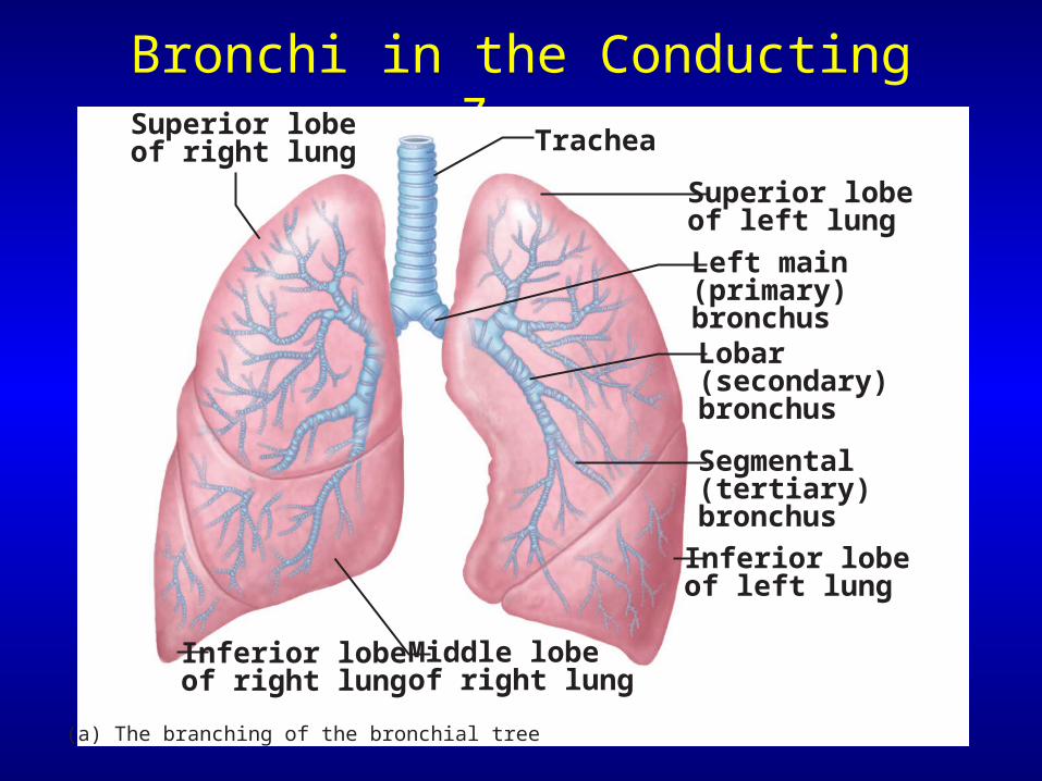
Bronchi in the Conducting Zone
TracheaSuperior lobe of right lung
Middle lobe of right lung
Inferior lobe of right lung
Superior lobe of left lung
Left main(primary) bronchusLobar(secondary)bronchus
Segmental(tertiary)bronchus
Inferior lobeof left lung
(a) The branching of the bronchial tree

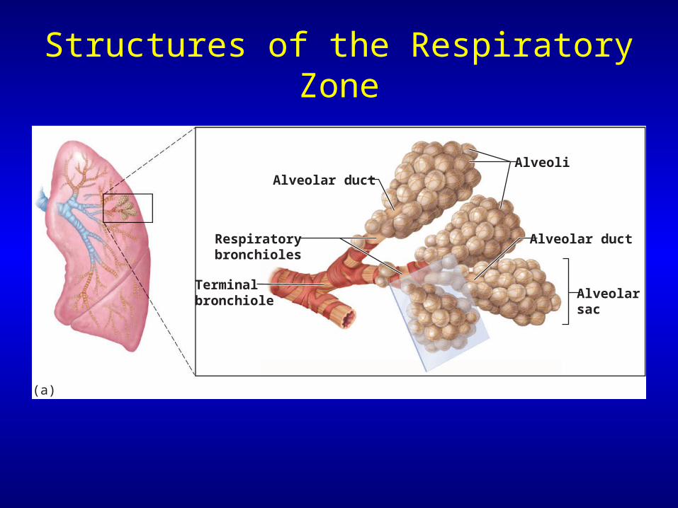
Structures of the Respiratory Zone
Alveolar duct
Alveolar ductAlveoli
Alveolarsac
Respiratory bronchioles
Terminalbronchiole
(a)
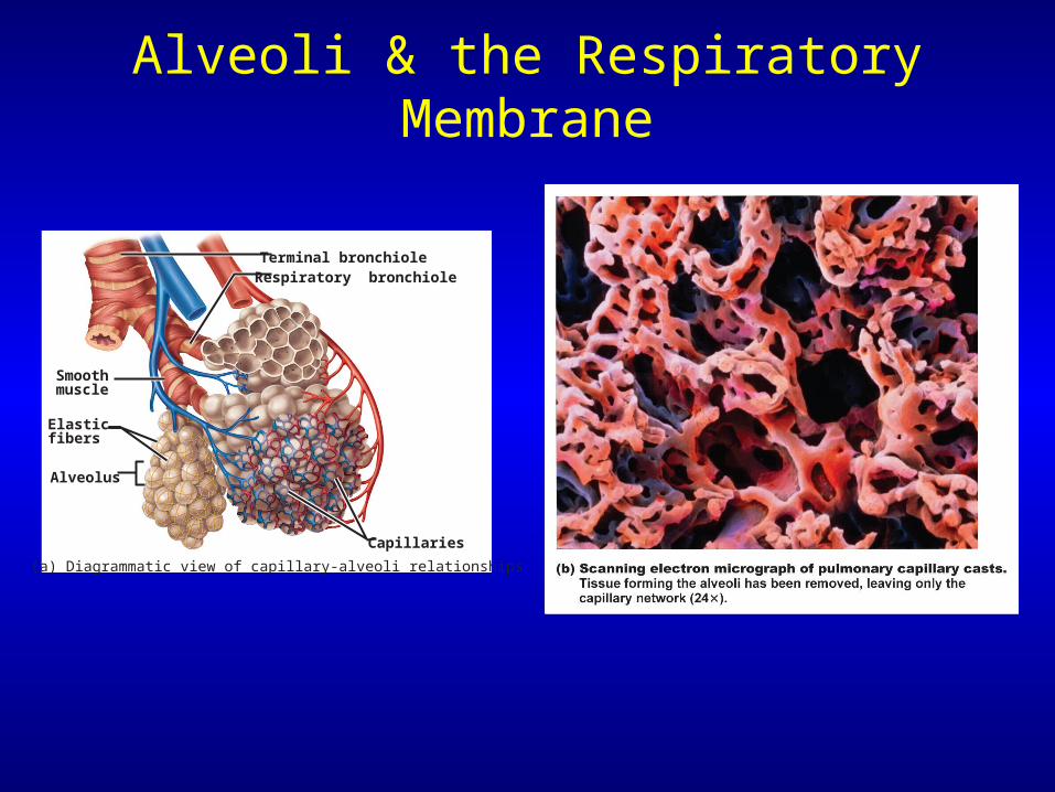
Alveoli & the Respiratory Membrane
Elasticfibers
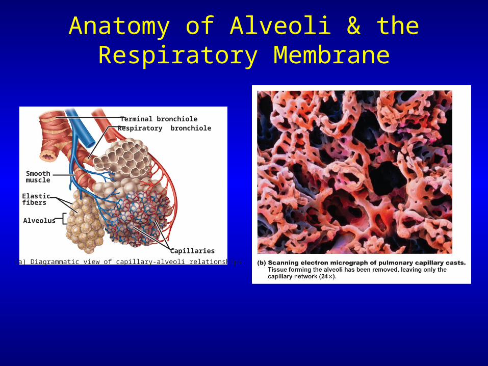
(a) Diagrammatic view of capillary-alveoli relationships
Smoothmuscle
Alveolus
Capillaries
Terminal bronchioleRespiratory bronchiole
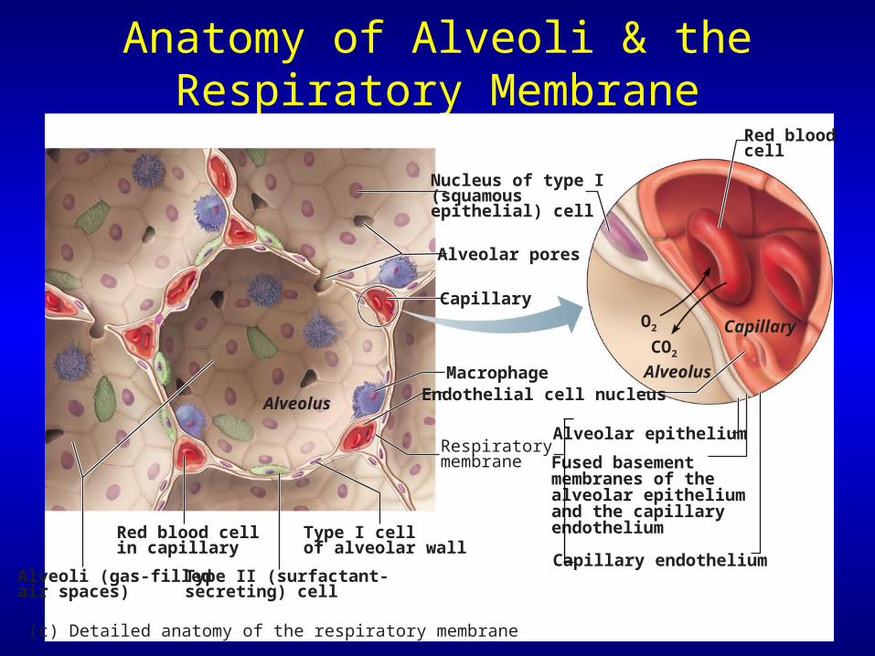
Anatomy of Alveoli & the Respiratory Membrane
Alveolus
Capillary
Type II (surfactant-secreting) cell
Type I cell of alveolar wall
Endothelial cell nucleusMacrophage
Alveoli (gas-filledair spaces)
Red blood cellin capillary
Alveolar pores
Capillary endothelium
Fused basement membranes of the alveolar epitheliumand the capillary endothelium
Alveolar epitheliumRespiratorymembrane
Red blood cell
O2
Alveolus
CO2
Capillary
Nucleus of type I(squamousepithelial) cell
(c) Detailed anatomy of the respiratory membrane
The Respiratory System
• Basic functions of the respiratory system– Supplies body with oxygen– Disposes of carbon dioxide
• 4 processes involved in respiration:– Pulmonary ventilation– External respiration– Transport of respiratory gases– Internal respiration
Functional Anatomy of the Respiratory System
• Respiratory organs– Nose, nasal cavity, & paranasal sinuses– Pharynx, larynx, & trachea– Bronchi & smaller branches– Lungs & alveoli
Organs of the Respiratory System
• Divided into– Conducting zone– Respiratory zone
The Nose
• Provides an airway for respiration
• Moistens & warms air (humidifies air)
• Filters inhaled air
• Resonating chamber for speech
• Houses olfactory receptors (olfaction)
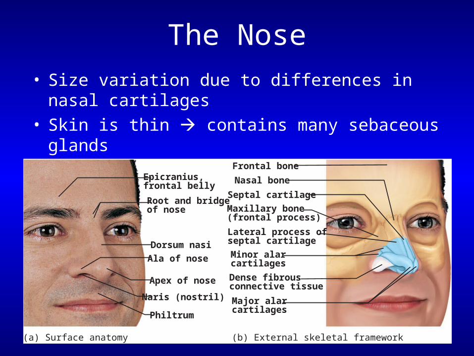
The Nose
• Size variation due to differences in nasal cartilages
• Skin is thin contains many sebaceous glands
Frontal bone
Nasal bone
Septal cartilage
Maxillary bone(frontal process)
Lateral process ofseptal cartilage
Minor alar cartilages
Major alarcartilages
Dense fibrousconnective tissue
(b) External skeletal framework
Epicranius,frontal belly
Ala of nose
Root and bridgeof nose
Dorsum nasi
Apex of nose
Philtrum
Naris (nostril)
(a) Surface anatomy
The Nasal Cavity
• External nares nostrils
• Divided by nasal septum
• Continuous with nasopharynx
Nasal Cavity
• 2 types of mucous membrane:– Olfactory mucosa
• Near roof of nasal cavity• Houses olfactory receptors
– Respiratory mucosa• Lines nasal cavity• Pseudostratified ciliated columnar epithelium
The Upper Respiratory Tract
Sphenoid sinusFrontal sinus
Nasal meatuses(superior, middle,and inferior)
Nasopharynx
Uvula
Palatine tonsilIsthmus of thefauces
Posterior nasalaperture
Opening ofpharyngotympanictube
Pharyngeal tonsil
Oropharynx
Laryngopharynx
Vocal fold
Esophagus
Nasal conchae(superior, middle and inferior)
Nasal vestibuleNostril
Nasal cavity
Hard palate
Soft palate
Tongue
Lingual tonsil
Epiglottis
Hyoid boneLarynx
Thyroid cartilageVestibular fold
Cricoid cartilage
Thyroid gland
Trachea
Cribriform plateof ethmoid bone
Respiratory Mucosa
• Consists of:– Pseudostratified ciliated columnar epithelium– Goblet cells within epithelium – Underlying layer of lamina propria
• Cilia move contaminated mucus posteriorly
Nasal Conchae
• Superior & middle nasal conchae – Part of the ethmoid bone
• Inferior nasal conchae– Separate bone
• Project medially from the lateral wall of the nasal cavity
• Particulate matter: – Deflected to mucus-coated surfaces
The Pharynx
• Funnel-shaped passageway
• Connects nasal cavity & mouth
• Divided into 3 sections by location:– Nasopharynx– Oropharynx– Laryngopharynx
• Type of mucosal lining changes along its length
The Nasopharynx
• Superior to the point where food enters• Only an air passageway• Closed off during swallowing• Pharyngeal tonsil (adenoids)
– Located on posterior wall– Destroys pathogens that enter
• Contains the opening to the pharyngotympanic tube (auditory or eustachian tube)– Tubal tonsil
• Provides some protection from infection
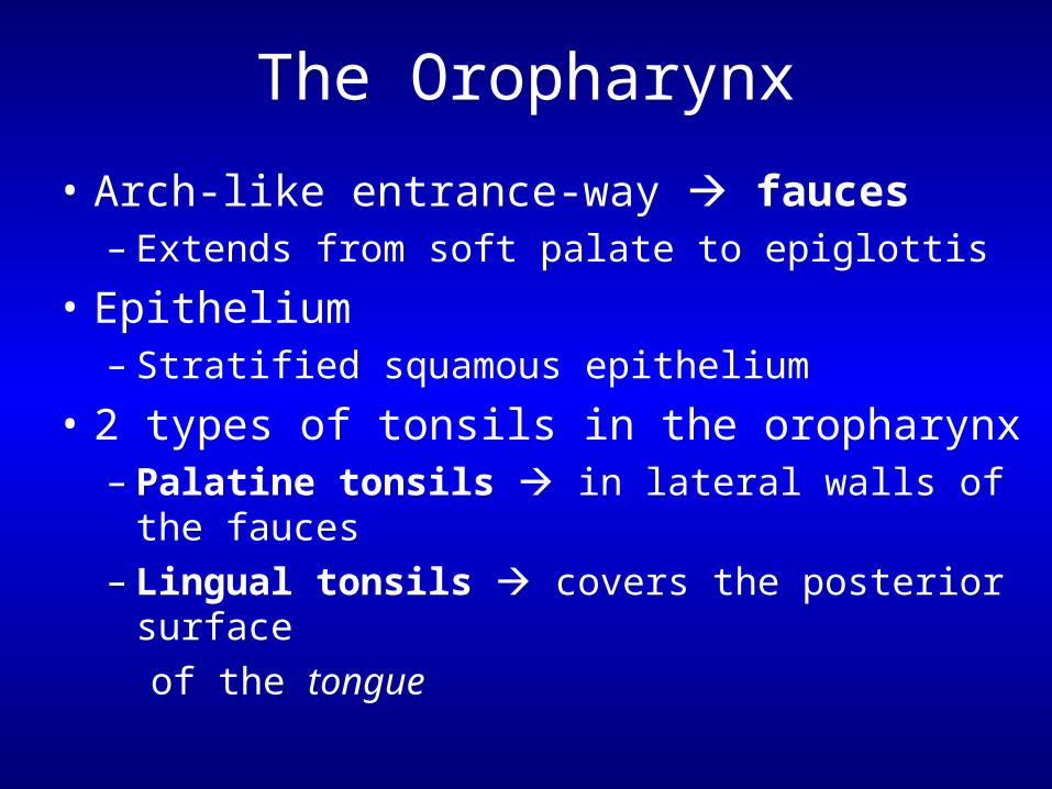
The Oropharynx
• Arch-like entrance-way fauces– Extends from soft palate to epiglottis
• Epithelium– Stratified squamous epithelium
• 2 types of tonsils in the oropharynx– Palatine tonsils in lateral walls of the fauces – Lingual tonsils covers the posterior surface
of the tongue
The Laryngopharynx
• Passageway for both food & air
• Epithelium– Stratified squamous epithelium
• Continuous with the esophagus & larynx
The Larynx
• 3 functions – Voice production– Provides an open airway– Routes air & food into the proper channels
• Superior opening (epiglotis) is:– Closed during swallowing– Open during breathing
9 Cartilages of the Larynx
• Thyroid cartilage– Shield-shaped, forms laryngeal prominence (=
Adam’s apple)
• 3 pairs of small cartilages– Arytenoid cartilages– Corniculate cartilages– Cuneiform cartilages
• Epiglottis– Tips inferiorly during swallowing
The Larynx
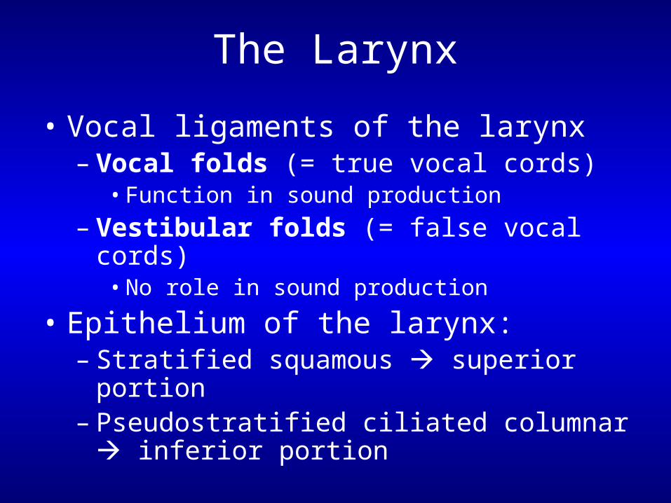
• Vocal ligaments of the larynx– Vocal folds (= true vocal cords)
• Function in sound production
– Vestibular folds (= false vocal cords)• No role in sound production
• Epithelium of the larynx:– Stratified squamous superior portion– Pseudostratified ciliated columnar inferior
portion
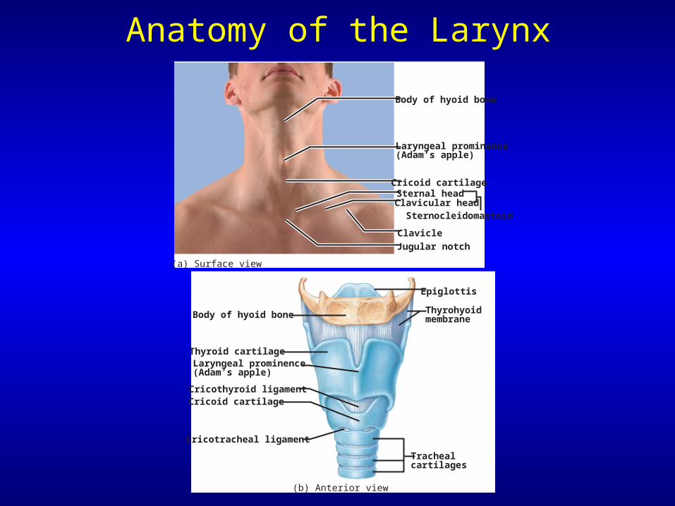
Anatomy of the Larynx
Body of hyoid bone
Cricoid cartilage
Laryngeal prominence(Adam’s apple)
Clavicle
Sternal headClavicular head
Sternocleidomastoid
Jugular notch
(a) Surface view
Body of hyoid bone
Epiglottis
Cricoid cartilage
Trachealcartilages
Thyroid cartilageLaryngeal prominence(Adam’s apple)
Cricothyroid ligament
Cricotracheal ligament
(b) Anterior view
Thyrohyoidmembrane
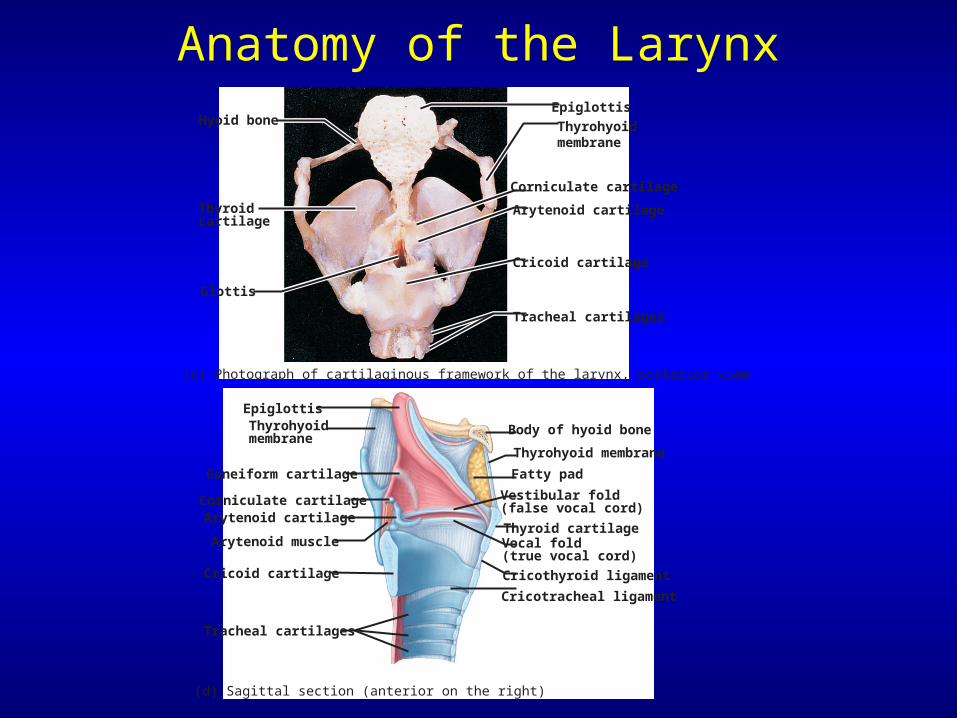
Anatomy of the LarynxHyoid bone
Thyroidcartilage
Glottis
(c) Photograph of cartilaginous framework of the larynx, posterior view
Epiglottis
Corniculate cartilage
Arytenoid cartilage
Cricoid cartilage
Tracheal cartilages
Thyrohyoidmembrane
Epiglottis
Body of hyoid bone
Thyrohyoid membrane
Vestibular fold(false vocal cord)
Vocal fold(true vocal cord)
Cricothyroid ligament
Cricotracheal ligament
Fatty pad
Thyroid cartilage
Cuneiform cartilage
Corniculate cartilageArytenoid cartilage
Cricoid cartilage
Tracheal cartilages
Arytenoid muscle
(d) Sagittal section (anterior on the right)
Thyrohyoidmembrane
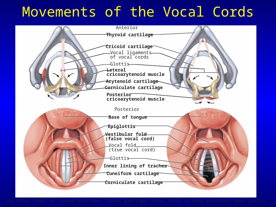
Movements of the Vocal Cords
(a) Vocal folds in closed position; closed glottis (b) Vocal folds in open position; open glottis
Base of tongue
Epiglottis
Vestibular fold (false vocal cord)
Vocal fold (true vocal cord)
Glottis
Inner lining of trachea
Cuneiform cartilage
Corniculate cartilage
Thyroid cartilage
Cricoid cartilageVocal ligaments of vocal cords
Lateral cricoarytenoid muscle
Arytenoid cartilage
Posterior cricoarytenoid muscle
Anterior
Posterior
Glottis
Corniculate cartilage
The Larynx
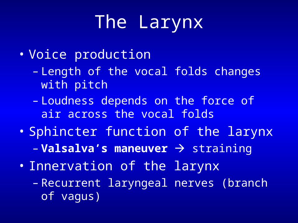
• Voice production – Length of the vocal folds changes with pitch– Loudness depends on the force of air across
the vocal folds
• Sphincter function of the larynx– Valsalva’s maneuver straining
• Innervation of the larynx– Recurrent laryngeal nerves (branch of vagus)
The Trachea
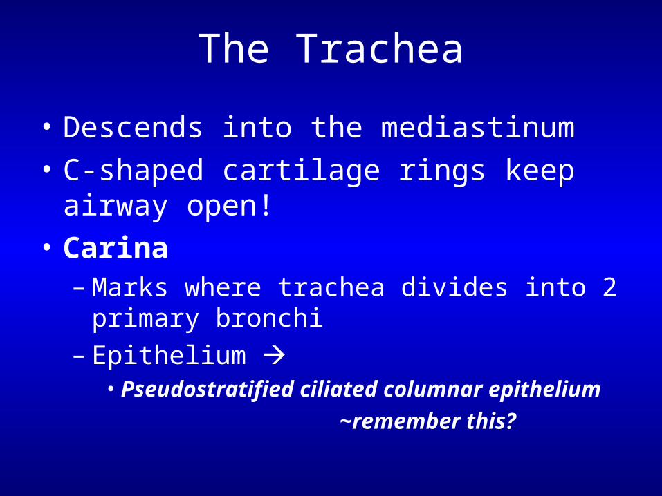
• Descends into the mediastinum
• C-shaped cartilage rings keep airway open!
• Carina– Marks where trachea divides into 2 primary
bronchi– Epithelium
• Pseudostratified ciliated columnar epithelium
~remember this?
The Trachea
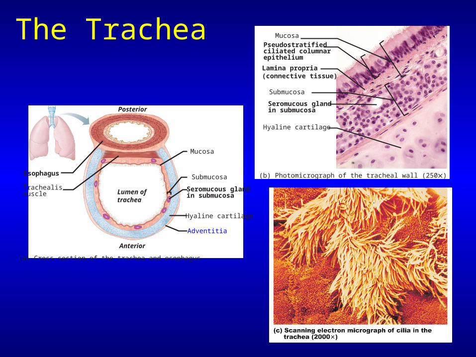
(a) Cross section of the trachea and esophagus
Hyaline cartilage
Submucosa
Mucosa
Seromucous glandin submucosa
Posterior
Lumen of trachea
Anterior
Esophagus
Trachealismuscle
Adventitia
(b) Photomicrograph of the tracheal wall (250)
Hyaline cartilage
Lamina propria(connective tissue)
Submucosa
Mucosa
Seromucous glandin submucosa
Pseudostratifiedciliated columnarepithelium
Bronchi in the Conducting Zone
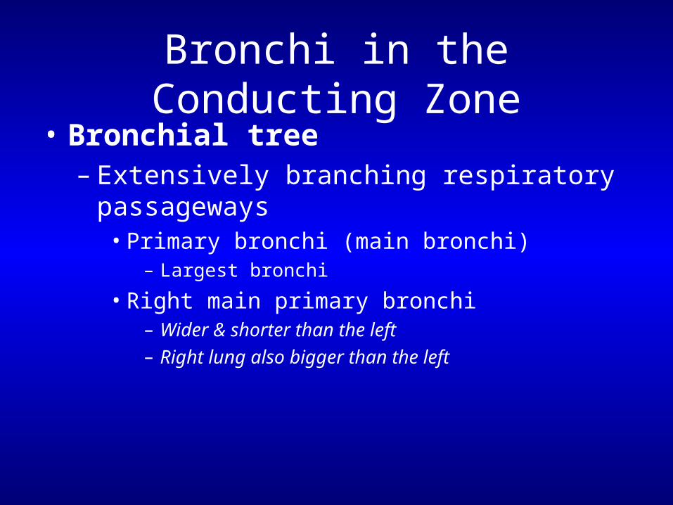
• Bronchial tree– Extensively branching respiratory
passageways• Primary bronchi (main bronchi)
– Largest bronchi
• Right main primary bronchi– Wider & shorter than the left– Right lung also bigger than the left

Bronchi in the Conducting Zone
TracheaSuperior lobe of right lung
Middle lobe of right lung
Inferior lobe of right lung
Superior lobe of left lung
Left main(primary) bronchusLobar(secondary)bronchus
Segmental(tertiary)bronchus
Inferior lobeof left lung
(a) The branching of the bronchial tree
Bronchi in the Conducting Zone
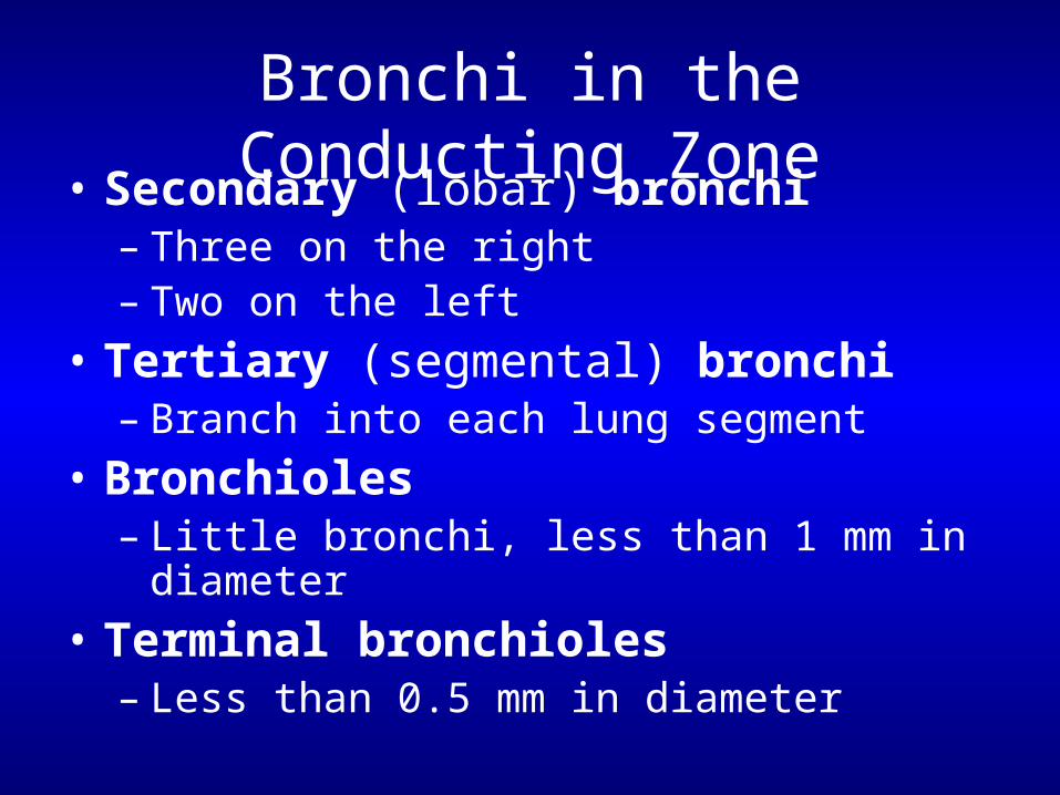
• Secondary (lobar) bronchi – Three on the right – Two on the left
• Tertiary (segmental) bronchi – Branch into each lung segment
• Bronchioles– Little bronchi, less than 1 mm in diameter
• Terminal bronchioles– Less than 0.5 mm in diameter
Bronchi in the Conducting Zone
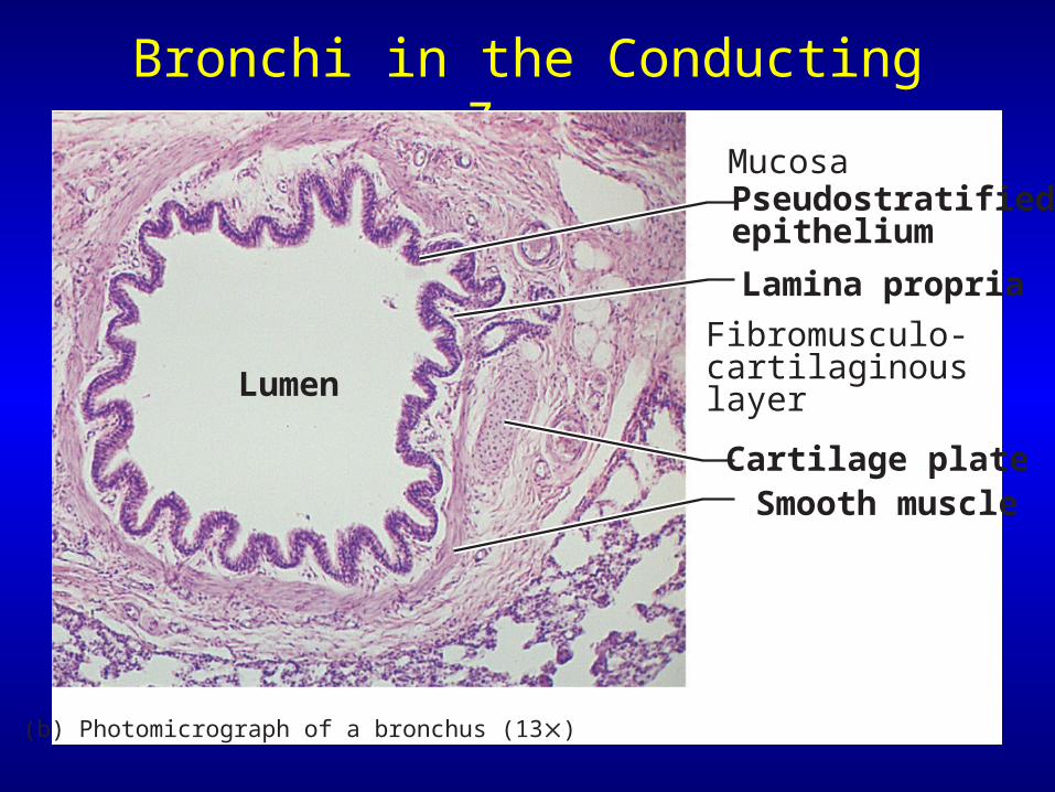
Mucosa Pseudostratified epithelium
Lamina propria
Fibromusculo-cartilaginous layer
Cartilage plate Smooth muscle
Lumen
(b) Photomicrograph of a bronchus (13)
Changes in Tissue Along Conducting Pathways
• Supportive connective tissues change– C-shaped rings replaced by cartilage plates
• Epithelium changes– First, pseudostratified ciliated columnar– Replaced by simple columnar, then simple
cuboidal epithelium• Smooth muscle becomes important:
– Airways widen with sympathetic stimulation– Airways constrict with parasympathetic stim.
Structures of the Respiratory Zone
• Consists of air-exchanging structures
• Respiratory bronchioles branch from terminal bronchioles– Lead to alveolar ducts
• Lead to alveolar sacs
Structures of the Respiratory Zone
Alveolar duct
Alveolar ductAlveoli
Alveolarsac
Respiratory bronchioles
Terminalbronchiole
(a)
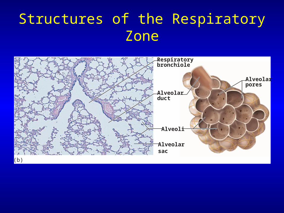
Structures of the Respiratory Zone
Alveolarpores
Alveolarduct
Respiratorybronchiole
Alveoli
Alveolarsac
(b)
Structures of the Respiratory Zone
• Alveoli– ~300 million alveoli account for tremendous
surface area of the lungs!• Surface area of alveoli is ~140 square meters!!!• Why such a large surface area?
Structures of the Respiratory Zone
• Structure of alveoli– Type I cells single layer of simple
squamous epithelial cells• Surrounded by basal lamina
– Alveolar & capillary walls plus their basal lamina form
• The Respiratory membrane
Anatomy of Alveoli & the Respiratory Membrane
Elasticfibers
(a) Diagrammatic view of capillary-alveoli relationships
Smoothmuscle
Alveolus
Capillaries
Terminal bronchioleRespiratory bronchiole
Structures of the Respiratory Zone
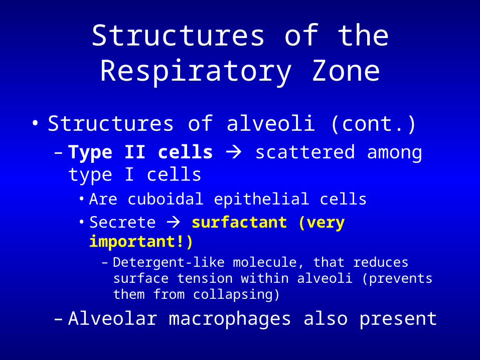
• Structures of alveoli (cont.)– Type II cells scattered among type I cells
• Are cuboidal epithelial cells• Secrete surfactant (very important!)
– Detergent-like molecule, that reduces surface tension within alveoli (prevents them from collapsing)
– Alveolar macrophages also present
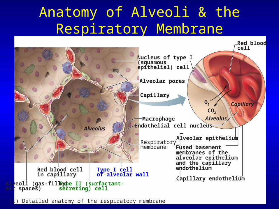
Anatomy of Alveoli & the Respiratory Membrane
Alveolus
Capillary
Type II (surfactant-secreting) cell
Type I cell of alveolar wall
Endothelial cell nucleusMacrophage
Alveoli (gas-filledair spaces)
Red blood cellin capillary
Alveolar pores
Capillary endothelium
Fused basement membranes of the alveolar epitheliumand the capillary endothelium
Alveolar epitheliumRespiratorymembrane
Red blood cell
O2
Alveolus
CO2
Capillary
Nucleus of type I(squamousepithelial) cell
(c) Detailed anatomy of the respiratory membrane
The Respiratory Zone
• Features of alveoli– Surrounded by elastic fibers– Interconnect by way of alveolar pores– Internal surfaces
• A site for free movement of alveolar macrophages
Gross Anatomy of the Lungs
• Major landmarks of the lungs– Apex, base, hilum, & root
• Left lung– Superior & inferior lobes
• Right lung– Superior, middle, & inferior lobes
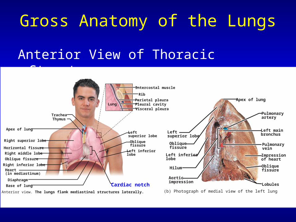
Leftsuperior lobe
Obliquefissure
Left inferiorlobe
(b) Photograph of medial view of the left lung
Left mainbronchus
Pulmonaryvein
Impressionof heart
Obliquefissure
Lobules
Pulmonary artery
Apex of lung
Hilum
Aorticimpression
Gross Anatomy of the Lungs
Anterior View of Thoracic Structures
Trachea
Apex of lung
Thymus
Right superior lobe
Horizontal fissure
Right middle lobe
Oblique fissure
Right inferior lobeHeart(in mediastinum)
Diaphragm
Base of lung
Leftsuperior lobe
Cardiac notch
Obliquefissure
Left inferiorlobe
Lung Pleural cavityParietal pleura
Rib
Intercostal muscle
Visceral pleura
(a) Anterior view. The lungs flank mediastinal structures laterally.
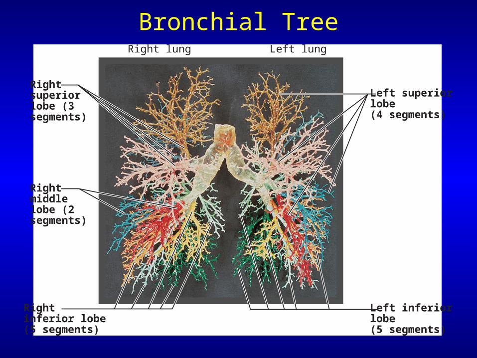
Bronchial Tree
Rightsuperiorlobe (3segments)
Rightmiddlelobe (2segments)
Rightinferior lobe(5 segments)
Left superiorlobe(4 segments)
Left inferiorlobe(5 segments)
Right lung Left lung
Blood Supply & Innervation of the Lungs
• Pulmonary arteries– Deliver oxygen-poor blood to the lungs
• Pulmonary veins– Carry oxygenated blood to the heart
• Innervation– Sympathetic, parasympathetic, & visceral
sensory fibers• Parasympathetic constrict airways• Sympathetic dilate airways
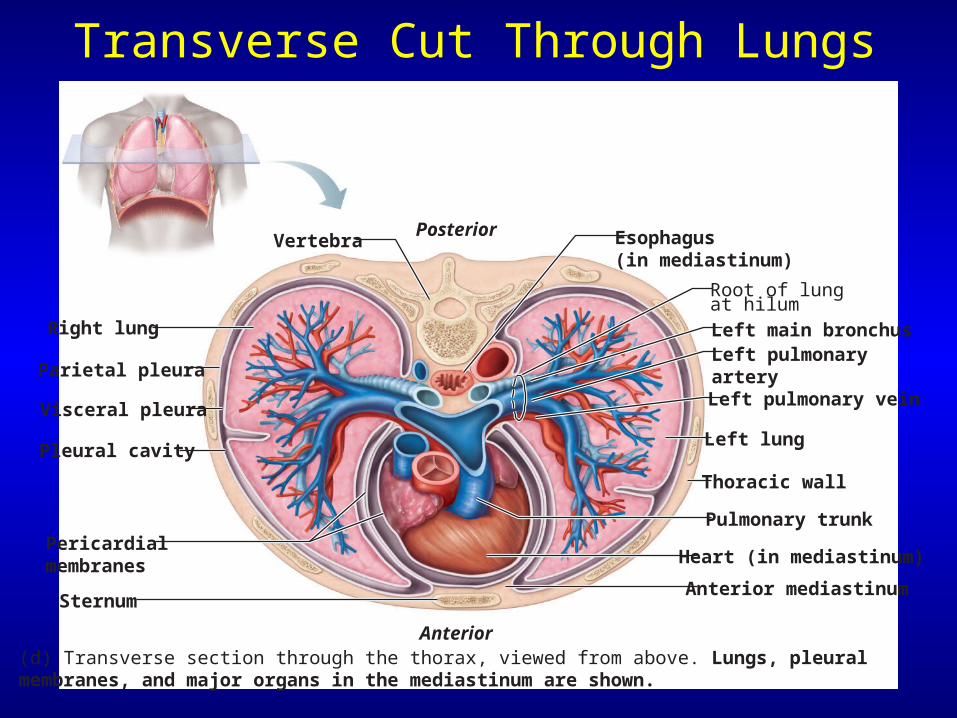
Transverse Cut Through Lungs
(d) Transverse section through the thorax, viewed from above. Lungs, pleural membranes, and major organs in the mediastinum are shown.
Esophagus(in mediastinum)
Right lung
Parietal pleura
Visceral pleura
Pleural cavity
Pericardial membranes
Sternum
Anterior
PosteriorVertebra
Root of lungat hilum
Left lung
Thoracic wall
Pulmonary trunk
Heart (in mediastinum)
Anterior mediastinum
Left main bronchusLeft pulmonary arteryLeft pulmonary vein
The Pleurae (review)
• A double-layered sac surrounding each lung– Parietal pleura– Visceral pleura
• Pleural cavity – Potential space between the visceral &
parietal pleurae
• Pleurae help divide the thoracic cavity – Central mediastinum – 2 lateral pleural compartments
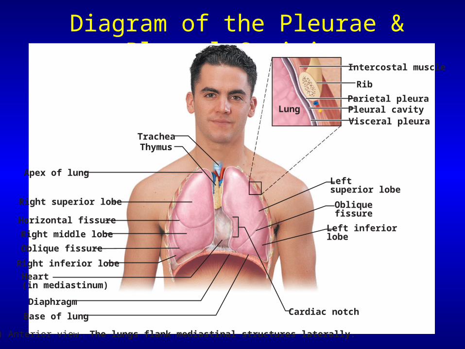
Diagram of the Pleurae & Pleural Cavities
Trachea
Apex of lung
Thymus
Right superior lobe
Horizontal fissure
Right middle lobe
Oblique fissure
Right inferior lobe
Heart(in mediastinum)
Diaphragm
Base of lung
Leftsuperior lobe
Cardiac notch
Obliquefissure
Left inferiorlobe
Lung Pleural cavityParietal pleura
Rib
Intercostal muscle
Visceral pleura
(a) Anterior view. The lungs flank mediastinal structures laterally.
The Mechanisms of Ventilation
• 2 phases of pulmonary ventilation– Inspiration inhalation – Expiration exhalation
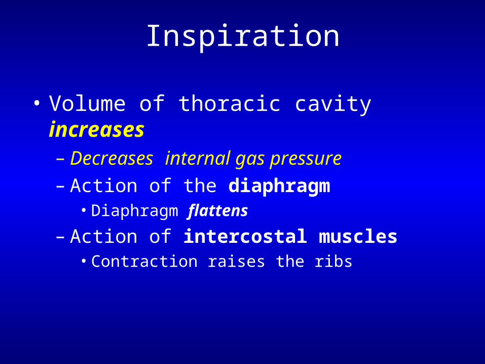
Inspiration
• Volume of thoracic cavity increases– Decreases internal gas pressure– Action of the diaphragm
• Diaphragm flattens
– Action of intercostal muscles• Contraction raises the ribs
Inspiration
• Deep inspiration requires – Scalenes– Sternocleidomastoid– Pectoralis minor– Erector spinae extends the back
Expiration
• Quiet expiration chiefly a passive process!– Inspiratory muscles relax– Diaphragm moves superiorly– Volume of thoracic cavity decreases
• Forced expiration an active process– Produced by contraction of
• Internal & external oblique muscles• Transverse abdominis muscles
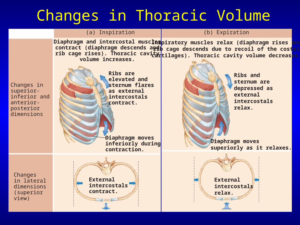
Changes in Thoracic Volume
Ribs areelevated and sternum flaresas externalintercostalscontract.
Diaphragm and intercostal musclescontract (diaphragm descends and
rib cage rises). Thoracic cavityvolume increases.
Diaphragm movesinferiorly duringcontraction.
Externalintercostalscontract.
Changes insuperior-inferior andanterior-posteriordimensions
Changesin lateraldimensions(superiorview)
(a) Inspiration
Inspiratory muscles relax (diaphragm rises andrib cage descends due to recoil of the costal
cartilages). Thoracic cavity volume decreases.
Ribs andsternum aredepressed asexternalintercostalsrelax.
Externalintercostalsrelax.
Diaphragm movessuperiorly as it relaxes.
(b) Expiration
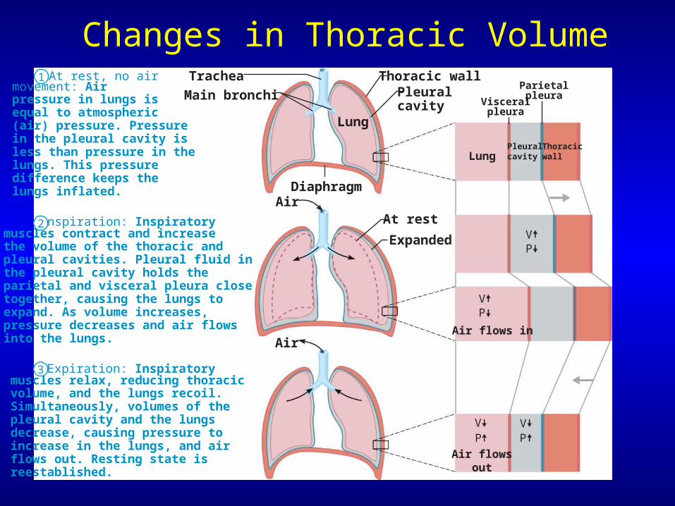
At rest, no air movement: Air pressure in lungs is equal to atmospheric (air) pressure. Pressure in the pleural cavity is less than pressure in the lungs. This pressure difference keeps the lungs inflated.
Inspiration: Inspiratory muscles contract and increase the volume of the thoracic and pleural cavities. Pleural fluid in the pleural cavity holds the parietal and visceral pleura close together, causing the lungs to expand. As volume increases, pressure decreases and air flows into the lungs.
Expiration: Inspiratory muscles relax, reducing thoracic volume, and the lungs recoil. Simultaneously, volumes of the pleural cavity and the lungs decrease, causing pressure to increase in the lungs, and air flows out. Resting state is reestablished.
Trachea
Diaphragm
Lung
Lung
Air flows in
Air flowsout
VP
VP
VP
VP
Pleuralcavity
Thoracicwall
Air
Air
Main bronchiParietalpleura
Visceralpleura
Thoracic wallPleural cavity
At rest
Expanded
1
2
3
Changes in Thoracic Volume
Neural Control of Ventilation
• Respiratory center– Generates baseline respiration rate– In the reticular formation of the medulla
oblongata
• Chemoreceptors– Sensitive to rising & falling oxygen levels – Central chemoreceptors located in
medulla– Peripheral chemoreceptors
• Aortic bodies • Carotid bodies
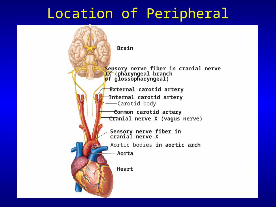
Location of Peripheral Chemoreceptors
Brain
Sensory nerve fiber in cranial nerve IX (pharyngeal branch of glossopharyngeal)
External carotid artery
Internal carotid arteryCarotid body
Common carotid arteryCranial nerve X (vagus nerve)
Sensory nerve fiber in cranial nerve X
Aortic bodies in aortic arch
Aorta
Heart
Disorders of Lower Respiratory Structures
• Bronchial asthma – A type of allergic inflammation
• Hypersensitivity to irritants in the air or to stress
– Asthma attacks characterized by• Contraction of bronchiole smooth muscle • Secretion of mucus in airways
Disorders of Lower Respiratory Structures
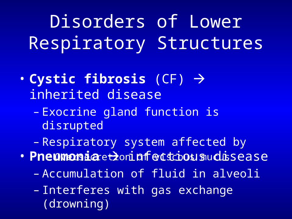
• Cystic fibrosis (CF) inherited disease – Exocrine gland function is disrupted– Respiratory system affected by
• Oversecretion of viscous mucus
• Pneumonia infectious disease– Accumulation of fluid in alveoli– Interferes with gas exchange (drowning)
Disorders of Lower Respiratory Structures
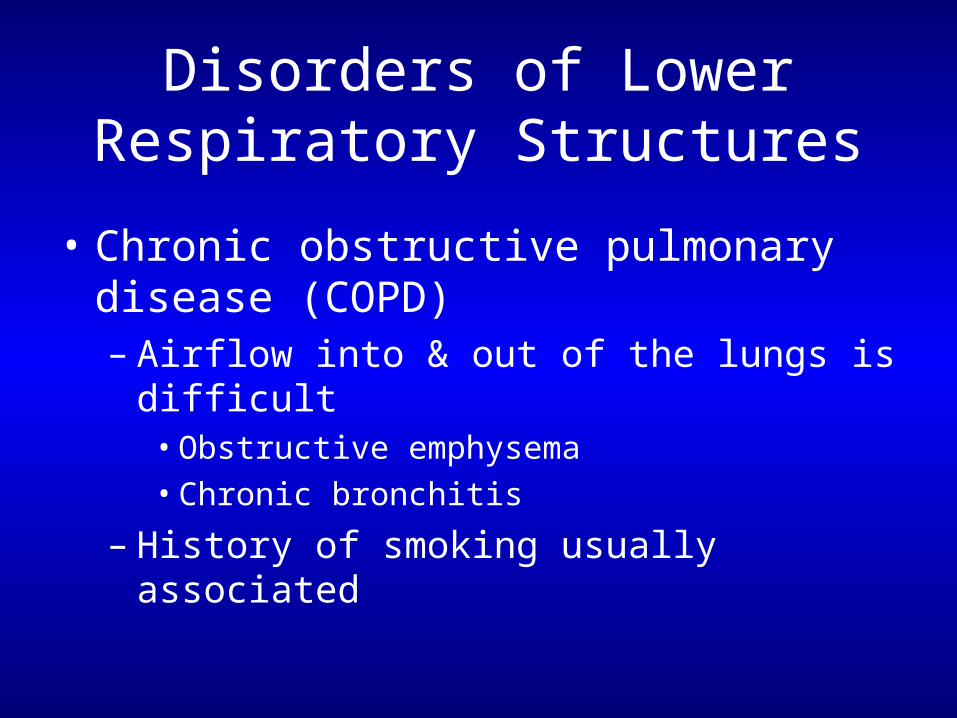
• Chronic obstructive pulmonary disease (COPD)– Airflow into & out of the lungs is difficult
• Obstructive emphysema• Chronic bronchitis
– History of smoking usually associated
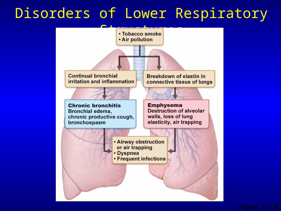
Disorders of Lower Respiratory Structures
Figure 22.18
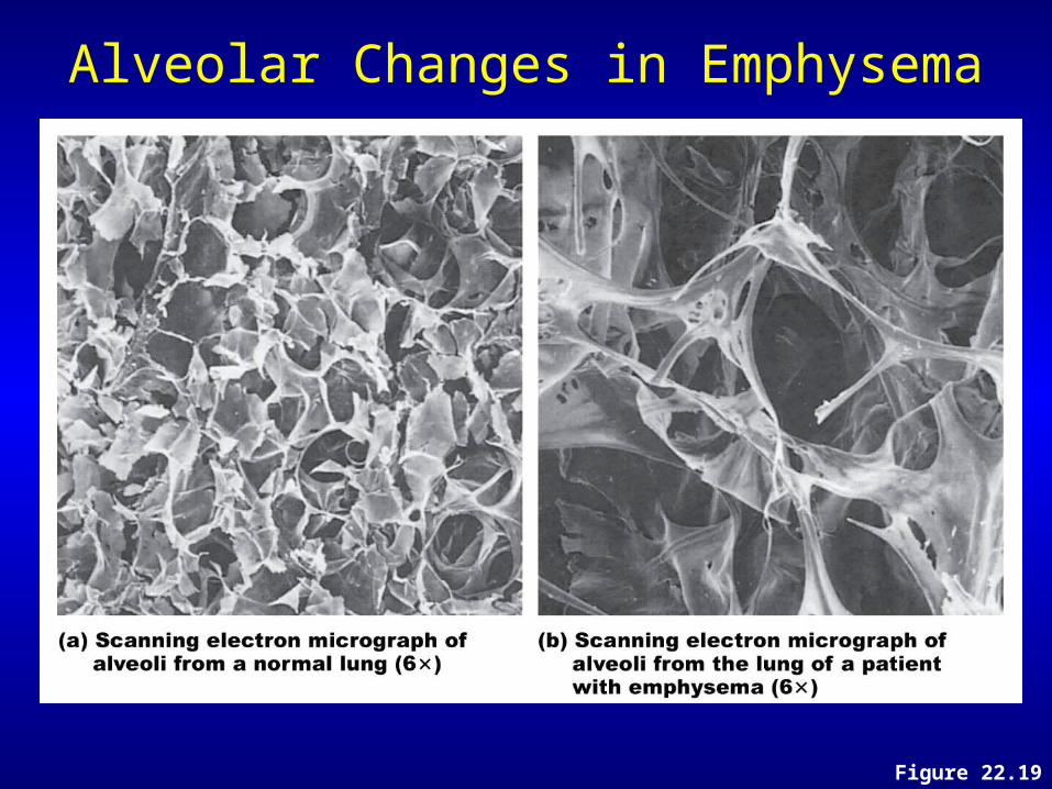
Alveolar Changes in Emphysema
Figure 22.19
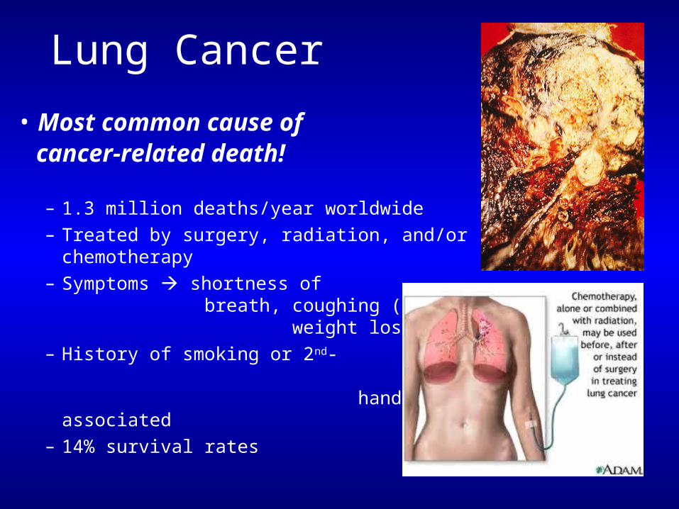
Lung Cancer
• Most common cause of cancer-related death!
– 1.3 million deaths/year worldwide– Treated by surgery, radiation, and/or
chemotherapy– Symptoms shortness of breath,
coughing (up blood), weight loss
– History of smoking or 2nd- hand smoke usually associated
– 14% survival rates
Aging of the Respiratory System
• The number of glands in nasal mucosa declines• Nose dries
– Produces thickened mucus
• Thoracic wall becomes more rigid• Lungs lose elasticity• Oxygen levels in the blood may fall
• Again…Exercise throughout life important for respiratory health!
Questions…?
What’s Next?Wed Lecture: Lecture Exam 4! Wed Lab: Start Digestive SystemMon Lecture: Digestive Sys. & class Potluck! ~We’ll eat while we learn about eating & digestion


![Schuh & [2009]](https://img.pdfslide.us/doc/110x75/55cf8c795503462b138cc6e9/schuh-2009.jpg)