Embed Size (px)
Citation preview

The rapid expansion of CT can be
adequately justified through the
existing framework of referral criteria
Denis Remedios
Consultant Radiologist, Northwick Park Hospital, London

Referral guidelines overview
• Imaging referral guidelines
– For whom are guidelines intended
– Which ones are available
– How are guidelines developed
• Tools to support guidelines
– Clinical Decision Support (CDS)
– Education and Awareness
– Audit for monitoring guideline use
• Evidence for reduction of utilisationAwareness, appropriateness and audit (Malone, 2011)

Guidelines: for whom? • For referring practitioners:
General Practitioners, doctors-in-
training & non-medically qualified
health professionals
• For radiology practitioners: ICRP
level 2 justification
• For patients: reinforcement of
advice
• For Healthcare organisations:
decision support, planning and
provision

Referral Guidelines:
Making the best use of clinical radiology
• The Royal College of Radiologists has published guidelines for >20
years since 1989. 7th edition 2012
• The guideline development process is accredited by NHS Evidence

EC Council Directive:
97/43 Euratom, Article 6.2http://ec.europa.eu/energy/nuclear/radioprotection/doc/legislation/9743_en.pdf


EC Referral Guidelines 2000


RCR Criteria for choice of
investigations
For a given clinical problem, imaging
modalities are listed in the order:
1. Evidence-based diagnostic impact
2. Effective dose
3. Cost-effectiveness

Levels of evidence for primary research question
Type of study
Therapeutic studies—investigating the results of treatment
Prognostic studies—investigating the effect of a patient characteristic on the
outcome of diseaseDiagnostic studies—
investigating a diagnostic test
Economic and decision analyses—developing an
economic or decision model
Level I High-quality randomised controlled trial with statistically significant difference or no statistically significant difference but narrow confidence intervals
Systematic review1 of level-I randomised controlled trials (and study results were homogeneous2)
High-quality prospective study3 (all patients were enrolled at the same point in their disease with ≥80% follow-up of enrolled patients)
Systematic review1 of level-I studies
Testing of previously developed diagnostic criteria in series of consecutive patients (with universally applied reference "gold" standard)
Systematic review1 of level-I studies
Sensible costs and alternatives; values obtained from many studies; multiway sensitivity analyses
Systematic review1 of level-I studies
Level II Lesser-quality randomised controlled trial (eg, <80% follow-up, no blinding, or imperfect randomisation)
Prospective3 comparative study4
Systematic review1 of level-II studies or level-I studies with inconsistent results
Retrospective5 studyUntreated controls from a
randomised controlled trial Lesser-quality prospective
study (e.g., patients enrolled at different points in their disease or <80% follow-up)
Systematic review1 of level-II studies
Development of diagnostic criteria on basis of consecutive patients (with universally applied reference "gold" standard)
Systematic review1 of level-II studies
Sensible costs and alternatives; values obtained from limited studies; multiway sensitivity analyses
Systematic review1 of level-II studies
Level III Case-control study6
Retrospective5 comparative study4
Systematic review1 of level-III studies
Case-control study6 Study of non-consecutive patients (without consistently applied reference "gold" standard)
Systematic review1 of level-III studies
Analyses based on limited alternatives and costs; imperfect estimates
Systematic review1 of level-III studies
Level IV Case series7 Case series Case-control studyPoor reference standard
No sensitivity analyses
Level V Expert opinion Expert opinion Expert opinion Expert opinion

Radiation doses The annual natural background radiation dose is 2.4mSv
Typical
effective dose
(mSv)
Examples
0 US, MRI
less than 1 CXR, XR limb, XR pelvis
1-5IVU, XR lumbar spine, NM (e.g.
bone scan), CT head and neck
5-10CT chest or abdomen, NM (e.g.
cardiac)
more than 10Extensive CT studies, some
NM studies (e.g. some PET)

NHS National Tariff 2008-9Tariff
(£)
Average
(£)
Reporting
Fee(£)
MRI, one area, no contrast 154
MRI
169*26
MRI, one area, post contrast only 199
MRI, one area, pre and post contrast only 228
MRI, 2 or 3 areas, no contrast 171
MRI, 2 or 3 areas, with contrast 260
CT, one area, no contrast 105
CT
131
24CT, one area, post contrast only 131
CT, one area, pre and post contrast only 152
CT, 2 or 3 areas, no contrast 132
CT, 2 areas with contrast 164
32CT, 3 areas with contrast 176
CT, More than 3 areas 223
Dexa Scans 49 49 13
Contrast fluoroscopy procedures <20 mins room usage 147159 N/A
Contrast fluoroscopy procedures >20 mins and <40 mins room usage 166
Ultrasound, scan 0-15 mins 63 US
69N/A
Ultrasound, scan > 15 mins 94
Nuclear Medicine Band 1 97
22823
Nuclear Medicine Band 2 151
Nuclear Medicine Band 3 302 64

RCR Recommendations
• Indicated- likely to contribute
• Specialised investigation- often complex,
time consuming or costly
• Indicated only in specific circumstances-
only done if appropriate for the individual
• Not indicated
• Grading A-C based on evidence level
– In 6e: 67 grade A, 409 B, 171 C.
– In 7e: 74 grade A, 633 B, 166 C.US DoH & Hum Services, Agency for Health Care Policy and Research. The Agency, 1993

MBUR7 EVIDENCE TABLE
MBUR7 Reference: -
Clinical/Diagnostic problem
Investigation Dose Recommendation [Grade]
Comment
Chronic lumbar back pain with no clinical or serological indicators of infection or neoplasia (ie, no red flags)
(For children see P11)
Lumbar imaging for low-back pain without suggestion of serious underlying conditions does not improve clinical outcomes.
MRI None Indicated only in specific circumstances [C]
MRI is the preferred investigation for the diagnosis of most spinal diseases and is helpful to identifying those patients who may benefit when planning surgical intervention.
XR
Indicated only in specific circumstances [C]
XR is only indicated if presentation suggests osteoporotic collapse in the elderly.
CT
Specialised investigation [C]
CT is used when MR is contraindicated and when further assessment of spondylolyses is required.
NM
Specialised investigation [C]
NM is non-specific, and has been largely supplanted by MR and CT in the assessment of chronic back pain. It may show occult osteoid osteomas and spondylolyses.
MeSH terms / keywords used for literature search
{back pain} OR {lower back pain} OR {low back pain} OR {backache}
Literature search: Search Period: 1999-2009
Refs Found: 130 Refs Used: 9 Refs from Previous Eds: 0
Literature search & Delphi Questionnaires (not for publication): M04-M06 back pain -
Literature search.doc M04 Round 1 Delphi Questionnaire.doc
M04 Delphi Round 2 Questionnaire ph&dr1.doc
DELPHI RESPONSE TABLE M04.xls
Composition of review panel (not for publication):
Dr A Pope (MSK) Dr J Bell (MSK) Dr J Rankine (MSK) Dr P Wilson (MSK)
Dr H El-Madbouh (MSK) Dr M Warren (MSK) Dr R Seymour (MSK)
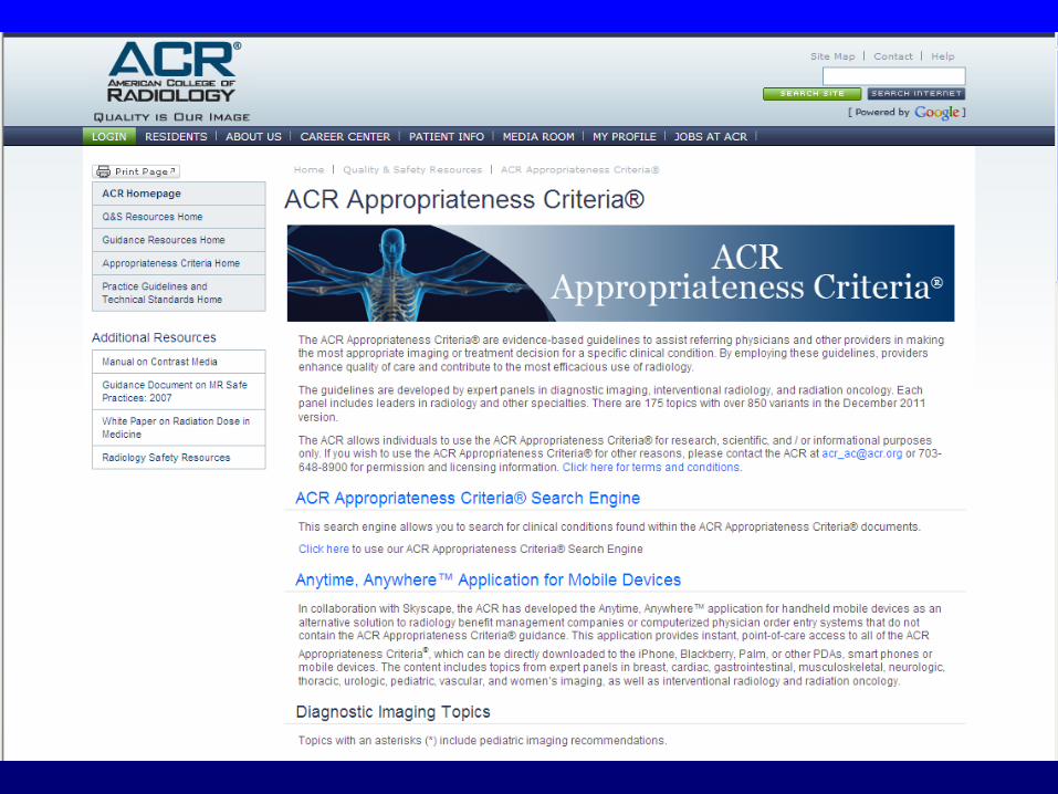
Existing NICE, SIGN & ACR Appropriateness Criteria:
Low Back Pain http://www.acr.org/SecondaryMainMenuCategories/quality_safety/app_criteria/pdf/ExpertPanelonNeurologicImaging/LowBackPainDoc7.aspx Low back pain; early management of persistent non-specific low back pain. NICE May 2009 http://www.nice.org.uk/nicemedia/pdf/CG88NICEGuidelineWord.doc
Highest level of evidence:
I


ACR Appropriateness
Criteria dose information

Guidelines appraisal
• Appraisal of Guidelines Research
& Evaluation (AGREE)- instrument http://www.agreetrust.org/
• Guidelines International Network
(GIN)- promotes systematic
approach http://www.g-i-n.net/
• NHS Evidence Accreditation
Scheme- quality mark (RCR MBUR
guideline process approved)
http://www.evidence.nhs.uk/Accreditation/Pages/Accreditation.aspx


EC Guidelines project:1. Study on implementation of imaging
referral guidelines in EU
2. European workshop for feed-back


Referral guidelines and clinician
involvement : the challenges• Dissemination of Referral Guidelines
– Widely and freely available to end-users “If they haven’t heard it you haven’t said it” McLuhan
• Implementation of guidance
– decision support tools? “We shape our tools and thereafter our tools shape us” McLuhan
• Uptake
– need buy-in by users and preferably ownership“Computers can do better than ever what needn’t be done at all. Making sense is still a
human monopoly” McLuhan
• Monitoring
– clinical audit, feedback and education “We drive into the future using only our rearview mirror ” McLuhan

Evidence for referral guidelines
• Following RCR guidelines, overall referrals fell 13%BMJ. 1993 Jan 9;306(6870):110-1
• RCGP Randomised controlled trial showed fewer referrals and better conformanceOakeshott, Kerry, Williams. Br J Gen Pract. 1994 Sep;44:427-8.
• Randomised trial with an educational reminder messages in reports is effective in reduction by up to 20% & does not affect quality of referrals.Eccles , Steen , Grimshaw , Thomas , McNamee , Soutter, Wilsdon , Matowe , Needham , Gilbert. The Lancet, 2001; 357: 1406 – 1409.
• Over 12 consecutive months no evidence of the effect of the intervention wearing off Ramsay, Eccles, Grimshaw, Steen. Clin Radiol. 2003 Apr;58(4):319-21
• Emerging evidence to show 2-20% improvement in conformance with clinical decision support tools.

http://www.hpa.org.uk/web/HPAwebFile/HPAweb_C/1287148001641

Ge
rma
ny
Be
lgiu
m
Sw
itze
rla
nd
Ice
lan
d
Fra
nc
e
No
rwa
y
Sw
ed
en
Ne
the
rla
nd
s
Lit
hu
an
ia
De
nm
ark
Fin
lan
d
UK
0
0.2
0.4
0.6
0.8
1
1.2
1.4
1.6
Per caput annual collective dose /mSv


1. Speed- sub-second “screen flips”
2. Anticipate needs, deliver in real time
3. Fit into users’ workflow
4. Little things make a big difference
5. Recognise physicians resist stopping
6. Changing direction better than stopping
7. Simple interventions work best
8. Ask for additional info only if essential
9. Monitor impact, get feedback, respond
10. Manage & maintain knowledge-based system

Sistrom et al. Radiology: Volume 251: Number 1—April 2009, 147
Limiting growth of CT usage with
guidelines & decision support

Out-patient & CT activity:
pre and post decision support guidance
Sistrom C L et al. Radiology 2009;251:147-155
CT
OP
CDS

Why do guidelines and
decision support work?
• “Gatekeeping effect” - new (and
sometimes more difficult) set of
steps are required to request exam
• “Educational effect” new process
attempts to change practice patterns
(and behaviour) or at least provide
some educational feedback


CDSS in Radiology:Advantages & barriers
Advantages
• Improved use of
effective test
• Reduced radiation
dose
• Reduction in
unnecessary tests
• Audit trail for
feedback
Barriers
• Computer access
to imaging request
• Guidelines do not
fit all patients
• Clinical condition
may have different
guidelines applied
• Clinician buy-in

Awareness: efficacy, safety, cost
Cancer plan undermined by PCTs
By Stephen Robinson, 08 September 2011
When the government unveiled its £750 million cancer
strategy for England in December 2010, the emphasis fell
squarely on early diagnosis.
Add to CPD Organiser
Be the first to comment
http://www.gponline.com/News/article/10
89232/cancer-plan-undermined-pcts/
Daily Mail, 8.9.11


Audit Live:
Guidelines for standardsYour IVU Radiograph Series
Number of radiographs obtained during intravenous urography (IVU).
IVU Examination Times
Examination times for intravenous urography (IVU).
Resuscitation Skills
Resuscitation skills within the Department of Clinical Radiology. A risk
management audit.
Resuscitation Awareness
Audit of practical knowledge of advanced resuscitation skills expected of
medical staff in a radiology department
Training in Gall Bladder Ultrasound
Adherence to departmental protocol during routine examination of the gall
bladder by those in training.
Needlestick injury
Contaminated needlestick injury to a member of staff or the public is a
serious health risk and could lead to litigation.
Pre-Op CXR for Elective Surgery
Pre-operative chest radiographs prior to elective surgery.
Bone Scan Images
Image quality of bone scans.
GP Chest Radiography
Appropriateness of requests for chest radiography from GPs.
Lumbar Spine
Lumbar spine radiography.
Out-of-Hours Imaging
Appropriateness of out-of-hours examinations.
Waiting and Appointment Times
Waiting time of patients prior to appointment.
Staff Dosimetry
Wearing of film badges during fluoroscopic procedures.
Fire Training
Attendance of staff at fire lectures.
Head CT – Lens Exclusion
Exclusion of the lens of the eye in routine head CT examinations.
Security – Staff ID
Departmental security – staff identification (ID).
Investigation of asymptomatic microscopic haematuria in adults
Assessment of compliance with agreed protocol for investigation of
asymptomatic microscopic haematuria in adults.
Contrast and Drug Recording
Recording of dose, make, batch number and expiry date for contrast medium used for
intravenous urograms (IVUs).
Foreign Body Radiography
Presence of a localising marker in radiography for presence of foreign bodies.
GP referrals: are the reports addressing the questions posed?
Audit of generic reporting and effective communication with GPs.
Consent for a Radiological Examination
Adequacy of consent for radiological procedures.
Pregnancy Questioning
The exclusion of pregnancy in patients who are undergoing radiography (application
of the 28 day rule).
Radiology Reporting by Other Doctors
Effectiveness of arrangements to transfer the responsibility for the reporting of
specified plain radiographs to referring clinicians.
Finger Doses
Radiation dose to the pulp of the index finger of staff handling syringes containing
radionuclides.
Urgent CT Brain Scans and LPs
Lumbar puncture (LP) following requests for urgent CT brain scans.
Radiography in Acute Back Pain
Requests for lumbar spine radiography in patients with acute low back pain.
GP Ultrasound Requests
Indications for GP referrals for ultrasound (US) examination of the upper abdomen.
Majax Call-In
Department of Clinical Radiology call-in list for use in case of a major accident
(majax).
Adequate Completion of Radiology Request Forms
Adequacy of completion of radiology request forms.
Gonad Protection II
Use of gonad protection.
Imaging in symptomatic breast disease
An audit to assess compliance with imaging guidelines within the symptomatic breast
clinic.
Cancer Staging
Staging of common cancers using CT or MRI.
Reporting: GP referrals for plain radiography
General practitioners depend upon timely and accurate reports for the management of
their patients.
An individual radiologist’s workload
The number of reports issued by an individual radiologist.

EC guidelines on clinical audit
for medical radiological practice http://ec.europa.eu/energy/nuclear/radiation_protection/doc/publication/159.pdf

Guidelines to reduce CT
over-use: Conclusions
• Faster justification and access to the best
test first for all health professionals using
evidence-based referral guidelines (& CDS)
• Higher level of appropriateness for lower per
caput collective doses
• Stronger collaboration through education
for better outcomes
“Awareness, appropriateness for all, and audit”

The rapid expansion of CT can be
adequately justified through the
existing framework of referral criteria:
Rebuttals and summary
Denis Remedios
Consultant Radiologist, Northwick Park Hospital, London

Guidelines
and risk• Although guidelines cannot estimate
an individual’s radiation risk, there is
an attempt to balance risk & benefit
of best evidence-based practice
• Risk assessment facilitated for all
healthcare workers using guidelines
• Risk communication by referrers to
patients reinforced by guidelines

Guidelines for CT• When combined with clinical red
flags, guidelines are an efficient
tool for identifying those with
high pre-test probability with
greatest benefit from test
• Helpful for clinical problem & for
selective screening eg Ca, CV risk
• May include hints for optimisation
eg low dose CT KUB,

Guidelines to justify CT
For
• Balance of risk &
evidence-based benefit
• All health workers who
refer can use
• Alternative lo dose test
may be recommended
• Useful to select those
for screening
• Allows for growth in
appropriate CT use
• May reduce by 20%
Against
• ICRP level 2 rather than
level 3 justification
• Not all medical
conditions are covered