Embed Size (px)
Citation preview
1
EFFICACY OF PEFLOXACIN FOR THE TREATMENT OF BROILER
CHICKENS EXPERIMENTALLY INFECTED WITH ESCHERICHIA
COLI O78:K80
By
K. MADIAN*, WAFAA. A. ABD EL-GHANY
* AND GEHAN M. KAMEL
** *Dept. of Poultry Dis.,
** Dept. of Pharmacology
Faculty Veterinary Medicine, Cairo University
Abstract
This study was conducted to assess the efficacy of pefloxacin in controlling the
adverse effects of experimentally induced colibacillosis in broiler chickens. Two- hundreds
and thirty, one-day old broiler chicks were used and randomly allocated into four
experimental groups; group (1) uninfected-untreated, group (2) infected-untreated, group (3)
infected-treated with pefloxacin in the drinking water and group (4) infected-treated with
pefloxacin by intramuscular injection (I/M) injection. The drug was given at dose level of 10
mg/kg b.wt/day for 3 consecutive days. Experimental colibacillosis was induced at 21-day of
age by I/M inoculation of Escherichia coli (E.coli) serotype O78: K80. Clinical signs,
mortalities, mean lesion scores, performance including body weight and feed conversion
efficiency, frequency of E.coli re-isolation and histopathological alterations were recorded
and evaluated. Also, clinical chemistry parameters including serum levels of total bilirubin
and liver enzymes (liver function parameters) as well as uric acid, blood urea nitrogen and
creatinin (kidney function parameters) were estimated. The humoral immune response to
sheep red blood cells (SRBC) and serum protein electrophoresis were measured. The
pharmacokinetic of pefloxacin were investigated. The Results showed that infected-
pefloxacin treated chickens had less pronounced clinical signs, significant (P<0.05) lower
mortalities, lower average gross pathology scores, higher body weight and better feed
conversion than infected-untreated birds. Serum levels of total bilirubin and liver enzymes as
well as uric acid, blood urea nitrogen and creatinin were significantly (P<0.05) decreased
toward normal values in chickens of infected-pefloxacin treated groups when compared with
those in infected-untreated group. The haemagglutinating antibody titers to SRBC and serum
levels of gammaglobulins (albumin/globulin ratios) were significantly (P<0.05) increased in
the treated groups than untreated ones. The results of pefloxacin pharmacokinetics cleared
that higher drug serum concentration was recorded following I/M injection than after oral
dosing. The absorption half life time (t1/2ab) was 0.53±0.08 and 0.21±0.026 h following oral
and I/M administration, respectively. The maximum serum concentration (Cmax) recorded
after oral dosing (2.71±0.14 µg.ml-1
) and (1.68±1.15 µg.ml-1
) following I/M injection. The
tissues residues results revealed that pefloxacin was not detected on the 9th
day following
cessation of water medication; however, the drug was still detected in the liver, kidney and
skin of I/M injected birds. The results of pefloxacin pharmacokinetic and serum drug
concentrations cleared that pefloxacin have good absorption with high bioavailability, long
half-life time and excellent tissue and body fluid penetration. In the present study, although
there were no convincing differences in clearing the clinical or pathological features of E.coli
between the two methods of pefloxacin administration, but I/M route was more efficacious
than drinking water method because of its ability to completely eliminate E.coli from its site
of action and so it may could minimize the risk of disease reoccurring. In conclusion,
pefloxacin administration (regardless to the method) at 10 mg/kg b.wt /day for 3 consecutive
days is efficacious for the treatment of colibacillosis in broiler chickens evident by
improvement of all investigated parameters and good pharmacokinetic profile .
2
Introduction Escherichia coli (E.coli) is a member of Gram negative bacteria. Avian pathogenic
E.coli (APEC) frequently infects broiler chickens inducing severe diseased conditions with
great economic losses (Chansiripornchai and Sasipreeyajan, 2002). All ages of poultry are
susceptible to APEC infection, however, the birds are mostly infected at 4-5 weeks old
(Chansiripornchai et al., 1995). The main clinical forms of E.coli infection in chickens vary
from acute colisepticaemia with sudden death to subacute fibrinopurulent serositis at 2-8
weeks old (Leitnes and Heller, 1992). Administration of antibiotics is the most common and
fast way for treating of APEC infection in broiler chickens, but the major problem associated
with the treatment is the development of drug resistant strains to the most commonly used
drugs (Vandemaele et al., 2002). Hence, it’s necessary to search for new therapeutic agents
to control this infection and to define the most effective route of administration. Much has
been reported about the resistance of E.coli to fluoroquinolones (Toyfour et al., 2001; Ruiz
et al., 2002 and Xiapei et al., 2002).
The fluoroquinolones are a relatively new class of synthetic antimicrobials for which
the clinical use, toxicity as well as pharmacological characters in animals have been reviewed
(Vancutsem et al., 1990). They have bactericidal activity against Gram-positive and Gram-
negative aerobic bacteria (Stamm et al., 1985 and Debbia et al., 1987). Pefloxacin is a novel
and the latest generation of fluoroquinolones with a chemical structure [1-ethyl-6-fluro-1-4-
oxo-7(4-methyl-1-piprazinyl) quinolone-3 carboxylic aid], this chemical structure rendered it
more active than other fluoroquinolones against Gram-negative and some Gram-positive
bacteria and also it improved the pharmacokinetic profile of the drug through enhanced lipid
solubility (Berg, 1988 and Neer, 1988). The drug is partially metabolized (demethylated and
oxidized) in the liver to norfloxacin (a primary metabolite) which itself is a potent
antimicrobial agent used in human and veterinary practice (Stein, 1987; Robson, 1992; Spoo
and Riviere, 1995 and Sarközy et al., 2004). Pefloxacin inhibits Topoisomerase II of the
bacterial DNA gyrase enzyme and DNA replication (Badet et al., 1982; Kayser, 1985 and
Smith, 1986) so, it exhibited broad spectrum rapid bactericidal activity at relatively low
concentration and posses a pronounced post-antibiotic effect (Jenkins and Friedlander,
1988; Vancutsem et al., 1990 and Brown, 1996). Favorable kinetic properties of pefloxacin
like good absorption, high bioavailability, long elimination half life time, low protein
binding, excellent tissues and body fluid penetration and large volume of distribution have
been documented by Barre et al., (1984); Dow et al., (1986); Andriole (1988); Gonzalez
3
and Henwood, (1989) and Andon (1992). These characters of pefloxacin make it an ideal
and suitable antibacterial drug in poultry medicine. Pefloxacin posses considerable in-vitro
potency against bacterial infectious diseases in poultry such as coliform infections,
salmonellosis, infectious coryza, avian mycoplasmosis, complication due to chronic
respiratory disease, fowl cholera, avian tuberculosis, clostridial infections and avian
chlamydiosis (Gonzalez and Henwood, 1989 and Mohamed and Dardeer, 2001).
Meanwhile, literatures about using of pefloxacin either in chemoprophylaxis or treatment of
poultry pathogens are relatively sparse. Pefloxacin pharmacokinetic and its tissue residues in
different avian species were available only in normal non diseased birds (Moutafchieva,
1997; Moutafchieva et al., 1997; Ershov et al., 2001; Isea et al., 2003; Jehan, 2004;
Mohan et al., 2004; Pant et al., 2005; Babu et al., 2006 and Moutafchieva and Yarkove,
2006). However, no previous studies on pefloxacin in diseased conditions of poultry are
recorded.
Therefore, the purpose of the present study was to determine the efficacy of
pefloxacin in the treatment of E.coli infection in the broiler chickens when administrated
either in the drinking water or via intramuscular (I/M) injection and the pharmacokinetic
profile of pefloxacin.
Material and Methods
I. Experimental chicks:
A total of two hundreds and thirty clinically healthy one-day old Hubbard broiler
chicks of both sexes obtained from commercial hatchery were used in the present study. At
arrival and before experiment, the chicks were tested to be free from E.coli by bacteriological
culture of liver, heart, blood, spleen and yolk sac of ten randomly selected chicks and they
prove negative isolation for E.coli. Chicks were individually identified with leg band rings
and kept under complete observation in separate thoroughly cleaned and disinfected pens.
Feed and water were provided adlibitum for the entire experimental period. A commercial
unmediated broiler ration that formulated to meet NRC recommendation (NRC 1994) was
used. All chicks were vaccinated against Newcastle disease (ND) using Hitchner B1 and
Lasota vaccines and against infectious bursal disease (IBD) using 228E vaccine at 5, 12 and
19 days of age; respectively via eye-drop instillation according to a standard vaccination
program implementation on local broiler farms.
4
II. Bacteria (The inoculum’s bacteria):
A virulent E.coli strain, serotype O78: K80 was used in the present study as it was
kindly supplied from Animal Health Research Institute; Dokki, Egypt. The strain originally
had been isolated from a field case of colisepticaemia (generalized E.coli infection) and had
been fully identified, classified and serotyped according to Edwards and Ewing (1972) and
Quinn et al., (1994). The E.coli inoculum was a logarithmic phase culture produced by
overnight incubation of E.coli in nutrient broth (Sekizaki et al., 1989). The number of
bacteria per milliliter was determined by plating ten-fold serial dilution of the nutrient broth
suspension on plate count agar (PCA). Titers were expressed as colony forming unit (CFU)
per ml (CFU/ml) (Fernandez et al., 2002). Strain of E.coli was firstly demonstrated to be
pathogenic in preliminary infectivity trial (pilot experiment) according to (Lublin et al.,
1993).
III. Antibiogram:
The in-vitro antibiotic sensitivity test on E.coli serotype O78: K80 used in this study
was performed using pefloxacin and most common antibiotics including (chroramphenicol,
gentamycin, ciprofloxacin, enrofloxacin, neomycin and norfloxacin) to determine the
susceptibility pattern of E.coli serotype to antimicrobial drugs by the standard disc diffusion
technique (Smith, 1970 and Prasad et al., 1997) using Oxiod multi-discs (Oxoid, Hants,
UK).
IV. Pefloxacin:
Pefloxacin standard pure powdered drug (100%) (1-ethyl-6-Fluro-1.4 dihydro-7-4
methyl -1- pipeazinyl- 4-oxo 3- quinolon-carboxlic acid) was kindly obtained from
Pharmasweed Pharmaceutical Company, Egypt. The pefloxacin pure powder was dissolved
in sterilized de-ionized distilled water to prepare pefloxacin 1% solution (according to
pharmaceutical company's recommendation) which is used in drinking water or through
intramuscular (I/M) injection at the dose level of 10 mg/kg b.wt (Sachan et al., 2003). The
minimum inhibitory concentration (MIC) of pefloxacin against the used E.coli strain was
determined as 0.06 µg/ml according to the method of Raemdonk et al., (1993).
V. Experimental design:
Two hundreds and thirty, one-day old broiler chicks were randomly allocated into
four groups, each consists of 50 chicks. Each group was randomly assigned into two
replicates of 50 chicks per each. The experimental groups were divided into; uninfected-
untreated (group 1), infected-untreated (group 2), infected and treated with pefloxacin in
5
drinking water (group 3) and infected and treated with pefloxacin via intramuscular (I/M)
injection (group 4). The chicks in group 2, 3 and 4 were experimentally infected with single
dose of E.coli at 21 days of age. For studying pefloxacin pharmacokinetics, 2 groups (5 and
6) of ten chicks per each were used. The chicks in groups (5 and 6) were experimentally
infected with E.coli at the same age and treated with single dose of pefloxacin orally and via
intramuscular (I/M) injection in groups (5 and 6); respectively at 2 days post inoculation
(starting of clinical signs appearance). The experiment started from day-old and terminated at
42 days of age.
VI. Experimental Infection (Inoculation of bacteria):
At 21 days of age, experimental colibacillosis was induced as described by
Fernandez et al., (2002). A dose of 0.3 ml of inoculum (broth culture) containing 12 X 109
CFU E.coli/ ml (3.6 X10⁸ CFU E.coli/ chicks) was intramuscularly injected in the pectoral
muscle of the chicks in the infected groups.
VII. Pefloxacin-Treatment regimen:
Medication with pefloxacin was initiated when clinical signs were started to be appear
(2 days post inoculation). Immediately prior to treatment, all birds in each treated group were
weighed in order to accurately calculate the required daily amount of pefloxacin based on the
therapeutic dose of 10 mg/kg body weight (Raemdonck et al., 1993 and Sachan et al.,
2003).
Drinking water regimen (continuous dosing):
Three days prior to treatment, the daily water consumption of birds was monitored in
order to determine the amount of water consumed daily (24 hours period). The entire daily
drug dose was administered continuously (continuous dosing regimen) during 24 hours
period in a volume of water which was consumed in the same period. Identical dosing
regimen was repeated during two subsequent days for a total of 3 consecutive days of
according to (Tanner et al., 1992). Fresh drug solution was mixed with drinking water daily
and replaced at the same time each day.
Intramuscular injection (I/M) regimen
Birds of groups (4 and 6) were injected daily for 3 consecutive days via I/M injection
with the required daily amount of pefloxacin prepared solution (Raemdonck et al., 1993)
6
VIII. Clinical follow up:
1- Clinical signs and mortalities:
All chicks were clinically inspected or observed each day for any health-related
problems. The clinical signs were monitored and recorded daily for fifteen days following
experimental infection. In every group, all mortalities were recorded daily and the dead birds
were necropsied.
2- Post-mortem examination (lesion scoring):
Dead birds were necropsied immediately after detection of their death and
macroscopical lesion scores were reregistered. Also, on weekly basis, at 7, 14, 21, 28 days
post experimental infection five birds of surviving chickens from each group were authonized
or sacrificed and necropsied. A detailed post-mortem examination was performed on the air-
sacs, pericardium, perihepatic capsule, trachea and lung, in addition to conventional post-
mortem examination and lesion scores were recorded (Nakamura et al., 1992). Post-mortem
lesions indicative of affected organs were scored severity on a scale of 4 points scoring
system ranging from 0 to 3 as follow; 0= no lesions, 1= mild, 2= moderate and 3= severe
(Raemodnock et al., 1993 and Fernandez et al., 2002).
The following criteria were used to score the severity of lesions according to (Fernandez et
al., 2002):
Heart: 0= no lesions, 1= little fibrin in pericardial sac, 2= definite fibrin in pericardium and 3
= extensive fibrin and adhesion of the pericardium.
Air-sacs: 0= no lesions, 1= mildly cloudy air-sacs, 2= definitely cloudy air-sacs (multifocal
white or yellow materials), 3= definitely cloudy air-sacs with large amount of caseous
exudates.
Liver: 0= no lesions, 1= little fibrin, 2= definite fibrin on liver surface 3= extensive fibrin.
The severity index of post-mortem (macroscopic) lesions was described previously by
Nakamura et al., (1987) and (1990). The severity index was calculated by adding the lesion
scores of the organ or tissue examined and dividing by the sum of the total number of
chickens subjected to post-mortem examination.
3- Re-isolation of E.coli O78:
To re-isolate the challenge organism, five randomly selected birds from each group
were sacrificed at the end of 1st, 2
nd and 3
rd week following initiation of therapy. In these
birds, swabs from liver, spleen, trachea, air-sacs and pericardium were inoculated into
MacConkey broth and then platted on MacConkey agar for 24h and at 37ºC (Fernandez
7
et al., 2002). The organism was identified on the basis of cultural characters according
the procedure of Cruickshank et al., (1975). Recovered E.coli isolate were lasted for
susceptibility to pefloxacin.
4- Profits (Performance):
On a weekly basis along the experimental period from placement through day to 42,
the chicks in each group were individually weighed and average body weight per group was
obtained. Feed consumption for each group was recorded weekly for calculation of feed
conversion rate (feed efficiency). Also European Production Efficiency Factor (EPEF) per
group was calculated to evaluate the performance.
5- Histopathology:
When chickens reached 6 weeks of age, five randomly selected birds were sacrificed
and organs including air-sacs, heart, liver, lung, spleen, bursa of fabricus and thymus were
excised for apprizing the influence of infection and treatment on the histopathological
alterations. Tissue samples were fixed in 10% neutral buffered formalin, fixed tissues were
dehydrated in methanol, cleared in xylene, trimmed, embedded in paraffin sections at 4um
and stained with hematoxyline and eosin (Bancroft et al., 1996). All tissues were examined
microscopically and the histopathological alterations were graded in blind vision microscopic
lesions.
IX. Laboratory Follow up:
1. Blood Sampling:
On 42 day of age, blood samples were collected by wing vein puncture from
randomly selected ten birds (two replicates of five birds each) from each group. The sera
were separated by centrifugation at 3000 rpm for 5 minutes and were stored at -20°C till
using for evaluation the effect of the infection and pefloxacin-treatment on the clinical
chemistry parameters (liver and kidney functions) and on serum protein fraction profile.
2. Estimation of clinical chemistry parameters
Serum levels of clinical chemistry parameters including [Total bilirubin, aspertate
aminotransferase (AST) and alanine aminotransferase (ALT)] (to investigate liver function),
also the kidney function tests including uric acid levels, blood urea nitrogen and creatinine
were determined according to Santurio et al., (1999) and Sachan et al., (2002) employing
commercially available diagnostic kits from Bio-merieux-France according to manufacturer's
instructions with the help of BM Hitachi 704 clinical analyzer.
8
3. Humoral immune response to sheep red blood cells (SRBC):
At 28 days of age, all the birds in experimental group (1, 2, 3 and 4) were inoculated
intramuscularly with 0.5 ml/bird of 5% suspension of washed SRBC in phosphate buffer
saline (PBS). Blood samples were collected from ten birds/ group at 3, 6, 9 and 12 days post
SRBS sensitization. The total haemagglutinating antibody titers produced in response to
SRBC were determined by micro-agglutinating technique (Van der Zijpp and Leenstra
1980), and expressed as the Log2 of the reciprocal of the highest dilution of serum giving
visible agglutination.
4- Profile of serum proteins:
Electrophoresis of serum protein fractions [albumin, alpha globulin (G), Beta-
globulin (β-G) and gamma-globulin (γ-G)] was made on cellulose-acetate membrane
according to method performed by (Epstein and Karcher, 1994) using one pooled serum
sample of randomly collected ten ones per group to asses the effect of experimental infection
and pefloxacin on immunoglobulins and albumin: globulin ratio.
X. Pharmacological assay:
1. Blood samples:
A. For pharmacokinetics studies:
Blood samples were collected via vein puncture from each bird in groups (5) and (6)
at 10, 20, 30, and 45 minutes, 1, 2, 3, 4, 5, 6, 8 and 10 hours post drug administration.
B. For determination of the daily serum pefloxacin concentrations:
Blood samples were collected from certain ten marked chickens in the 3rd
and 4th
group at 24 hours after each pefloxacin administration.
2. Tissue samples:
Three birds from group 3 and 4 were sacrificed at the 1st, 3
rd, 5
th, 7
th and 9
th day after
cessation of drug administration. Tissue samples of different organs including liver, kidneys,
heart, gizzard, breast muscle and skin were excised and collected from each bird for
estimation of pefloxacin concentration in tissues. One gram of each tissue samples was
homogenized with 3 ml of phosphate buffer saline (pH 7.2) and centrifuged at 3000 rpm for
15 minutes. The supernatant fluids were collected and stored at -20°C until analyzed.
3. Pefloxacin analytical procedure:
Pefloxacin concentration in serum and tissues were determined by microbiological
assay procedure (Arret et al., 1971) using non pathogenic E.coli (ATCC 25922) that measure
the antibacterial activity of the parent drug and its metabolites according to the method of
9
Moutafchieva and Djouvinov (1997) and Moutafchivea and Yarkove (2006). Pefloxacin
standard curves were constructed using antibacterial free sera collected from infected-
untreated group and phosphate buffer solution (pH 7.2). The lower detectable limit of
pefloxacin assay by this method was 0.012 µg/ml serum.
In-vitro serum protein binding percent of pefloxacin was determined both in serum of birds in
the uninfected-untreated group and in the infected-untreated one according to the method of
Craig and Suh (1980) using the concentration of 10, 5, 2.5, 1.25, 0.625, 0.312, 0.156 and
0.078.
4. Determination of pefloxacin pharmacokinetic:
A computerized curve-stripping program (R Strip, Micromath Scientific Software,
Salt Lake City, UT) was used to analyze pefloxacin serum concentration-time curves for each
individual bird in the pharmacokinetic study. Serum concentration-time curves were obtained
for each bird and fitted to the following equation:
C= Ae-K
abt + Be
-Kel
t
Where C is the serum concentration, A and B are the intercepts of the absorption and
elimination phases with the concentration axis, respectively. Kab is the absorption rate
constant expressed in units of reciprocal time (h-1
), Kel is the elimination rate constant
expressed in units of reciprocal time (h-1
) and e is the base of natural logarithm. The peak of
drug concentration (Cmax) and time to peak concentration (Tmax) were calculated according
to the statistical moment theory of Yamaoka et al., (1978). The absorption half-life time
(t1/2ab) and the elimination half-life one (t1/2el) are calculated as Ln2/Kab and Ln2/Kel,
respectively. The area under the concentration-time curves (AUC) were calculated by
trapezoidal rule (Gibaldi and Perrier, 1982). The mean residence time (MRT) for the tested
drug was calculated as AUMC/AUC, where AUMC is the area under the first moment curve.
XI. Statistical analysis:
The collected data were computed using analysis of variance (ANOVA) where
difference in means was obtained; also the least significant difference (LSD) test was
measured to distinct different treatments (Snedecor and Corchran, 1980). Results of the
pharmacokinetic parameters, daily serum concentration and tissue residues were statistically
analyzed using student t-test (Snedecor and Corchran, 1980).
10
Results and Discussion
Escherichia coil (E.coli) is responsible for heavy economic losses to poultry industry,
by its association with various disease conditions, either as primary pathogen or as a
secondary pathogen. Treatment protocols for E.coli infections include the use of antibiotics
with broad spectrum activity (Brans and Gross, 1997). Many antibiotics are active against
E.coli, but this bacterium has often developed resistance to different antimicrobial agents
(Blanco et al., 1996). Hence, it is necessary to search for new therapeutic agents to control
such infection and to define the most effective dose and route of administration. Pefloxacin is
a newer member of the fluoroquinolone antimicrobials and is a potent broad spectrum
antimicrobial agent with a pharmacokinetic profile characterized by high bioavailability after
administration, good penetration into tissues and body fluid and long half-life time
(Moutafchieva and Yarkove, 2006). These properties necessitate using of pefloxacin in the
poultry field against the most important common bacterial pathogens.
1. Antibiogram:
Results of in-vitro antibiotic sensitivity pattern revealed that E.coli challenge
organism used in this study was highly sensitive to pefloxacin than other tested antibiotics
(chroramphenicol, gentamycin, ciprofloxacin, enrofloxacin, neomycin and norfloxacin). This
obtained result agrees with that of Prasad et al., (1997) who study the antibiogram pattern of
E.coli isolated from many pathological lesions and reported that E.coli showed highest
sensitivity to pefloxacin when compared with other antibiotics. The minimum inhibitory
concentration (MIC) of pefloxacin that determined in the present study was 0.06 µg/ ml and it
was agree with the finding of Sharma et al., (1994). The recent introduction and the limited
using of pefloxacin are attractive explanations for the higher sensitivity of E.coli strains to
pefloxacin (Prasad et al., 1997). Also our result concerning the antibiotic sensitivity pattern
is consistent with that reported by Hui and Das (2000) and Tai and Fang (2000) who
noticed high sensitivity of E.coli strains isolated from chickens and ducks to pefloxacin in-
vitro. Moreover, Rolinski et al., (2002) mentioned that different Salmonella species showed
high sensitivity to pefloxacin in-vitro sensitivity test.
2. Clinical signs:
No clinical signs were observed in the uninfected-untreated group. Reduced birds
activity, dyspnea, snicking, mucopurulent nasal discharges, decrease appetite and depression
were present among the infected-untreated birds and were estimated between 40-60% within
24 hours following experimental E.coli infection. Within 24 hours following initiation of
11
treatment pefloxacin reduced the clinical signs which were present in infected birds prior to
treatment and the signs continued to improve in the next days post-medication. Infected Birds
treated with pefloxacin through I/M route improved clinically during and after the 3-days
treatment period and clinical signs disappeared within 5 to 7 days following treatment. The
response to treatment with pefloxacin in drinking water was less pronounced as signs
continued to improve post-treatment but more slowly and few affected birds showed mild
signs of respiratory distress at the end of the first week following initiation of medication. In
contrast, the incidence of clinical disease in surviving infected-untreated birds was estimated
as 20-50%, seven days following infection.
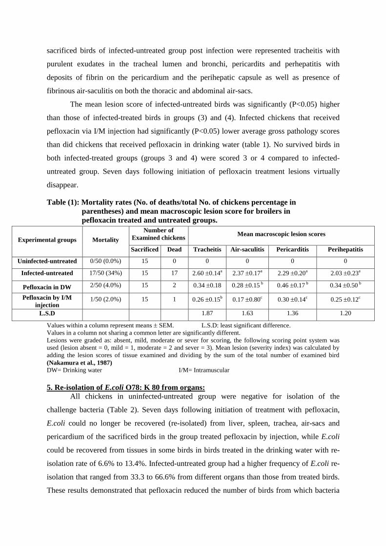
Data presented in table (1) show and summarize the effect of different pefloxacin
treatment regimen on mortality and mean lesions score over three weeks observation period
following initiation of treatment.
3. Mortalities:
Following medication, the total cumulative mortality percentages attributed to
generalized E.coli infection (septicaemia and serositis) was significantly (P<0.05) lower (2%)
and (4%) in birds administrated pefloxacin through I/M injection and those and received
pefloxacin in the drinking water; respectively when compared with uninfected-untreated
group (34%) that had a quite high cumulative mortality during the experiment. Mortalities
during 2 days post-infection were reduced by the 3rd
day of pefloxacin treatment and
completely stopped by the 7th
day of medication. These results indicated that pefloxacin
reduced the mortality rate when administered through I/M injection more than through
drinking water.
There were no mortalities recorded among chickens of uninfected-untreated group. Our
findings are partially constant with those reported by Adayel and Abdalla (2007) who found
that treatment of Salmonella enteritidis-infected chicks with pefloxacin at dose level of 5 mg
and 10 mg/kg b.wt for 3 successive days partially reduced clinical signs and decreased
mortality rate from (38%) to (6%) and (2%); respectively, while using of pefloxacin at dose
level 5 mg/kg b.wt but for 5 successive days ameliorate clinical symptoms and reduce
mortality rate to 2%.
4. Gross lesions and lesion scores:
Colibocillosis was confirmed in both dead and sacrificed birds on the basis of post-
mortem inspection for typical lesions of tracheitis, air-saculitis, pericarditis and perihepatits,
also the subsequent recovery of E.coli isolate. Macroscopic lesions observed in dead and
12
sacrificed birds of infected-untreated group post infection were represented tracheitis with
purulent exudates in the tracheal lumen and bronchi, pericardits and perhepatitis with
deposits of fibrin on the pericardium and the perihepatic capsule as well as presence of
fibrinous air-saculitis on both the thoracic and abdominal air-sacs.
The mean lesion score of infected-untreated birds was significantly (P<0.05) higher
than those of infected-treated birds in groups (3) and (4). Infected chickens that received
pefloxacin via I/M injection had significantly (P<0.05) lower average gross pathology scores
than did chickens that received pefloxacin in drinking water (table 1). No survived birds in
both infected-treated groups (groups 3 and 4) were scored 3 or 4 compared to infected-
untreated group. Seven days following initiation of pefloxacin treatment lesions virtually
disappear.
Table (1): Mortality rates (No. of deaths/total No. of chickens percentage in
parentheses) and mean macroscopic lesion score for broilers in
pefloxacin treated and untreated groups.
Values within a column represent means SEM. L.S.D: least significant difference.
Values in a column not sharing a common letter are significantly different.
Lesions were graded as: absent, mild, moderate or sever for scoring, the following scoring point system was
used (lesion absent = 0, mild = 1, moderate = 2 and sever = 3). Mean lesion (severity index) was calculated by
adding the lesion scores of tissue examined and dividing by the sum of the total number of examined bird
(Nakamura et al., 1987) DW= Drinking water I/M= Intramuscular
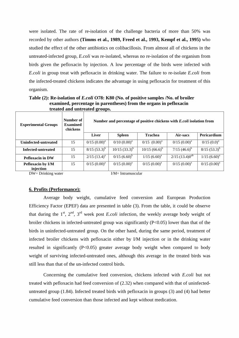
5. Re-isolation of E.coli O78: K 80 from organs:
All chickens in uninfected-untreated group were negative for isolation of the
challenge bacteria (Table 2). Seven days following initiation of treatment with pefloxacin,
E.coli could no longer be recovered (re-isolated) from liver, spleen, trachea, air-sacs and
pericardium of the sacrificed birds in the group treated pefloxacin by injection, while E.coli
could be recovered from tissues in some birds in birds treated in the drinking water with re-
isolation rate of 6.6% to 13.4%. Infected-untreated group had a higher frequency of E.coli re-
isolation that ranged from 33.3 to 66.6% from different organs than those from treated birds.
These results demonstrated that pefloxacin reduced the number of birds from which bacteria
Experimental groups Mortality
Number of
Examined chickens Mean macroscopic lesion scores
Sacrificed Dead Tracheitis Air-saculitis Pericarditis Perihepatitis
Uninfected-untreated 0/50 (0.0%) 15 0 0 0 0 0
Infected-untreated 17/50 (34%) 15 17 2.60 0.14a 2.37 0.17
a 2.29 0.20
a 2.03 0.23
a
Pefloxacin in DW 2/50 (4.0%) 15 2 0.34 0.18 0.28 0.15 b 0.46 0.17
b 0.34 0.50
b
Pefloxacin by I/M
injection 1/50 (2.0%) 15 1 0.26 0.15
b 0.17 0.80
c 0.30 0.14
c 0.25 0.12
c
L.S.D 1.87 1.63 1.36 1.20
13
were isolated. The rate of re-isolation of the challenge bacteria of more than 50% was
recorded by other authors (Timms et al., 1989, Freed et al., 1993, Kempf et al., 1995) who
studied the effect of the other antibiotics on colibacillosis. From almost all of chickens in the
untreated-infected group, E.coli was re-isolated, whereas no re-isolation of the organism from
birds given the pefloxacin by injection. A low percentage of the birds were infected with
E.coli in group treat with pefloxacin in drinking water. The failure to re-isolate E.coli from
the infected-treated chickens indicates the advantage in using pefloxacin for treatment of this
organism.
Table (2): Re-isolation of E.coli O78: K80 (No. of positive samples /No. of broiler
examined, percentage in parentheses) from the organs in pefloxacin
treated and untreated groups.
Experimental Groups
Number of
Examined
chickens
Number and percentage of positive chickens with E.coli isolation from
Liver Spleen Trachea Air-sacs Pericardium
Uninfected-untreated 15 0/15 (0.00)a 0/10 (0.00)
a 0/15 (0.00)
a 0/15 (0.00)
a 0/15 (0.0)
a
Infected-untreated 15 8/15 (53.3)b 10/15 (33.3)
b 10/15 (66.6)
b 7/15 (46.6)
b 8/15 (53.3)
b
Pefloxacin in DW 15 2/15 (13.4)a 0/15 (6.60)
b 1/15 (6.60)
a 2/15 (13.4)0
ab 1/15 (6.60)
a
Pefloxacin by I/M
injection
15 0/15 (0.00)a 0/15 (0.00)
a 0/15 (0.00)
a 0/15 (0.00)
a 0/15 (0.00)
a
DW= Drinking water I/M= Intramuscular
6. Profits (Performance):
Average body weight, cumulative feed conversion and European Production
Efficiency Factor (EPEF) data are presented in table (3). From the table, it could be observe
that during the 1st, 2
nd, 3
rd week post E.coli infection, the weekly average body weight of
broiler chickens in infected-untreated group was significantly (P<0.05) lower than that of the
birds in uninfected-untreated group. On the other hand, during the same period, treatment of
infected broiler chickens with pefloxacin either by I/M injection or in the drinking water
resulted in significantly (P<0.05) greater average body weight when compared to body
weight of surviving infected-untreated ones, although this average in the treated birds was
still less than that of the un-infected control birds.
Concerning the cumulative feed conversion, chickens infected with E.coli but not
treated with pefloxacin had feed conversion of (2.32) when compared with that of uninfected-
untreated group (1.84). Infected treated birds with pefloxacin in groups (3) and (4) had better
cumulative feed conversion than those infected and kept without medication.
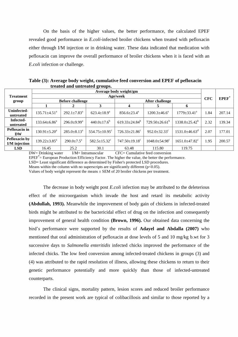
14
On the basis of the higher values, the better performance, the calculated EPEF
revealed good performance in E.coli-infected broiler chickens when treated with pefloxacin
either through I/M injection or in drinking water. These data indicated that medication with
pefloxacin can improve the overall performance of broiler chickens when it is faced with an
E.coli infection or challenge.
Table (3): Average body weight, cumulative feed conversion and EPEF of pefloxacin
treated and untreated groups.
Treatment
group
Average body weight/gm
CFC EPEF*
Age/week
Before challenge After challenge
1 2 3 4 5 6
Uninfected-
untreated 135.71±4.51
a 292.1±7.83
a 623.4±18.9
a 856.6±23.4
a 1200.3±46.6
a 1779±33.41
a 1.84 207.14
Infected-
untreated 133.64±6.86
a 296.0±9.99
a 440.0±17.6
b 619.33±24.84
b 729.50±26.61
b 1338.0±25.42
b 2.32 139.34
Pefloxacin in
DW 130.91±5.20
a 285.0±8.13
a 554.75±10.95
c 726.33±21.86
c 952.0±32.33
c 1531.0±46.63
d 2.07 177.01
Pefloxacin by
I/M injection 139.22±3.85
a 290.0±7.5
a 582.5±15.32
c 747.50±19.18
c 1048.0±54.98
c 1651.0±47.82
c 1.95 200.57
LSD 16.45 25.2 30.1 63.48 115.80 119.75
DW= Drinking water I/M= Intramuscular CFC= Cumulative feed conversion EPEF
*= European Production Efficiency Factor. The higher the value, the better the performance.
LSD= Least significant difference as determined by Fisher's protected LSD procedures.
Means within the column with no superscripts are significantly different (p<0.05).
Values of body weight represent the means ± SEM of 20 broiler chickens per treatment.
The decrease in body weight post E.coli infection may be attributed to the deleterious
effect of the microorganism which invade the host and retard its metabolic activity
(Abdullah, 1993). Meanwhile the improvement of body gain of chickens in infected-treated
birds might be attributed to the bactericidal effect of drug on the infection and consequently
improvement of general health condition (Brown, 1996). Our obtained data concerning the
bird’s performance were supported by the results of Adayel and Abdalla (2007) who
mentioned that oral administration of pefloxacin at dose levels of 5 and 10 mg/kg b.wt for 3
successive days to Salmonella enteritidis infected chicks improved the performance of the
infected chicks. The low feed conversion among infected-treated chickens in groups (3) and
(4) was attributed to the rapid resolution of illness, allowing these chickens to return to their
genetic performance potentially and more quickly than those of infected-untreated
counterparts.
The clinical signs, mortality pattern, lesion scores and reduced broiler performance
recorded in the present work are typical of colibacillosis and similar to those reported by a
15
number of authors (Piercy and West, 1976; Cheville and Arp, 1978; Smith et al., 1985;
Nakamura et al., 1992; Sasipreeyajan and Pakpinyo 1992; Raemdonk et al., 1993;
Mognet et al., 1997; Pourbakhshs et al., 1997; Fernandez et al., 2002 and Glisson et al.,
2004) and by using of these parameters as indices of E.coli, we evaluated the efficacy of
pefloxacin treatment.
The magnitude of suffering and losses among chickens infected with E.coli are
obvious and immense and can be attributed to high virulence of E.coli strain O78: K80 that
used in experimental infection in this study. Sekizaki et al., (1989) and Frenandez et al.,
(2002) mentioned that E.coli strain O78: K80 is highly virulent for poultry and induces high
mortality in a short time. Also cytotoxins of E.coli seem to be important factors in the
pathogenesis of the disease as they are the most potent bacterial toxins (Marks and Robert,
1993). These toxins in active host cell ribosoms disrupt protein synthesis and cause cell death
(Obrien et al., 1992).
Using of pefloxacin at dose level of 10 mg/kg b.wt/day for 3 successive days either in
the drinking water (continuous dosing) or by I/M injection for the treatment of E.coli-infected
broiler chickens resulted in rapid control of experimental colibacillosis as evidenced by rapid
resolution of clinical signs, significant reduction in mortalities, lesion score, rate of E.coli re-
isolation and improvement of performance (greater body weight and more efficient feed
conversion). All the aforementioned criteria indicated the efficacy of pefloxacin and provided
reasons for the efficacy in controlling E.coli infection in broilers.
The obtained results concerning the efficacy of pefloxacin can be explained as a result
of the following: 1- The potent bactericidal activity of pefloxacin as it inhibits and interferes
with the activity of DNA gyrase and Topoisomerase II enzymes which are needed for the
transcription and replication of bacterial DNA (Badet et al., 1982 Smith, 1986; Dudley,
1991; Glisson, 1994 and Chansiripornachai et al., 1995). As a result of inhibition of
transcription and replication of bacterial DNA, it exhibited broad spectrum, rapid and high
excellent bactericidal activity (Vanctsem et al., 1990 and Brown, 1996). 2- Favorable
pharmacokinetic profile of pefloxacin that documented in the present study and previously by
Isea et al., (2003) and Moutafchieva and Yarkove (2006) including rapid absorption, high
bioavailability, excellent tissue and body penetration, long elimination half-life time, low
protein binding and large volume of distribution concluded that administration of pefloxacin
at 10 mg/Kg in chickens might ensure serum and tissue concentrations therapeutically
effective for most pefloxacin-sensitive pathogens (Gram-negative aerobes and Gram–positive
16
aerobes and anaerobes). 3- High in-vitro sensitivity of E.coli to pefloxacin that observed in
this work and by Prasad et al., (1997).
Very little reports were published about the efficacy of pefloxacin for treatment of
colibacillosis in broiler chickens. On the other hand, numerous reports have indicated the
effectiveness of pefloxacin in the treatment of Salmonellae infections (Wille et al., 1988;
Nagah et al., 2004 and Adayel and Abdalla, 2007). They recorded that drinking water
administration of pefloxacin at dose level of 5 and 10 mg/ kg b.wt for 5 successive days was
highly effective in controlling experimental with Salmonellae spp infections in broiler
chickens as evident by reduction of clinical signs, mortality rate and re-isolation of the
organism as well as improving the body gain.
Our results are supported by the work of many authors who found that all new
quinolone derivatives including enrofloxacin, danofloxacin and sarafloxacin were very active
against E.coli infection in broiler chickens. Glisson et al., (2004) reported that chickens
infected with E.coli and medicated with enrofloxacin had significantly (P<0.05) less
mortality, lower average gross pathology scores, lower re-isolation rate of the bacteria, higher
average live weight and better feed conversion ratio than infected not treated chickens. In a
three field studies and in a disease model study, treatment of E.coli-infected broiler chickens
with danofloxacin at 5mg/kg b.wt/day for 3 consecutive days resulted in rapid resolution of
clinical signs, significant reduction in moralities and mean lesion scores and improving
performance of broilers (Raemdonck et al., 1993). In the same manner, obtained data here
were supported and in agreement with the findings of McCabe and Rippel (1993); Joong
Kim (1995); Chansiripornchai and Sasipreeyajan (2002) and Zhenling et al., (2002) who
found a significant increase in the average daily gain and feed conversion ratio and reduction
in mortalities of broilers treated with sarafloxacin (third generation of quinolones) than those
not received treatment after experimental infection with E.coli serotype O78.
7. Histopathology:
Septicaemic, serosal respiratory lesions and lymphocytic depletion of bursa of
fabricius and thymus were the main microscopical lesions demonstrated in broiler chickens
infected with E.coli and kept without treatment (group 2) (Fig. 1F, 2F, 3F, 4F, 5F, 6F and
7F). The septicaemic lesions consists of focal coagulative necrosis in diffuse heavy manner
all over the hepatic tissues with sever dilatation of portal, central vein and sinusoid of liver as
well as hyperemia of red pulps with focal necrosis in diffuse manner all over the splenic
tissues associated with thickening of the splenic capsule and depletion of lymphoid cells in
17
the white pulp. The serosal lesions consists of fibrinopuulant inflammation (fibrin exudates)
in the serosal system including pericardium, epicardium, air-sac with infiltration of fibrin
threads, dead heterophilis and other mononuclear leucocytes inflammatory cells in associated
with dilatation of blood vessels and capillaries. The respiratory microscopical lesions consist
of pneumonia and air-saculitis and showing dilatation in the interlobular blood vessels of the
lung and edema in the interstial stroma. The epithelium of air-sacs showed hyperplasia and
cellular infiltration associated with congestion and edema in addition to cellular infiltration in
sub-epithelial tissues. Marked lymphocytic depletion was noticed in the bursa and thymus of
chickens affected with fibrinopurulent serositis. There were no significant histopathological
lesions in any of the chickens in uninfected-untreated group (Fig. 1E, 2E, 3E, 4E, 5E, 6E and
7E)
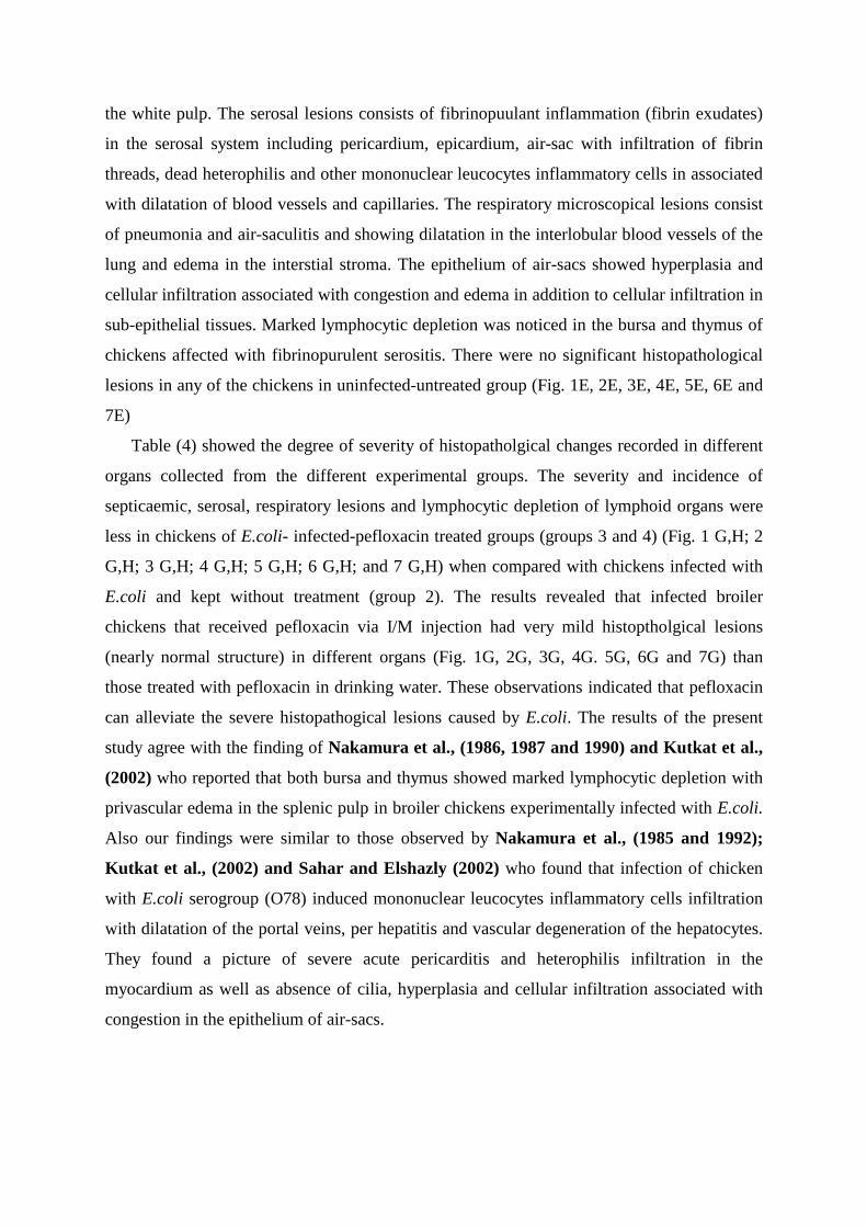
Table (4) showed the degree of severity of histopatholgical changes recorded in different
organs collected from the different experimental groups. The severity and incidence of
septicaemic, serosal, respiratory lesions and lymphocytic depletion of lymphoid organs were
less in chickens of E.coli- infected-pefloxacin treated groups (groups 3 and 4) (Fig. 1 G,H; 2
G,H; 3 G,H; 4 G,H; 5 G,H; 6 G,H; and 7 G,H) when compared with chickens infected with
E.coli and kept without treatment (group 2). The results revealed that infected broiler
chickens that received pefloxacin via I/M injection had very mild histoptholgical lesions
(nearly normal structure) in different organs (Fig. 1G, 2G, 3G, 4G. 5G, 6G and 7G) than
those treated with pefloxacin in drinking water. These observations indicated that pefloxacin
can alleviate the severe histopathogical lesions caused by E.coli. The results of the present
study agree with the finding of Nakamura et al., (1986, 1987 and 1990) and Kutkat et al.,
(2002) who reported that both bursa and thymus showed marked lymphocytic depletion with
privascular edema in the splenic pulp in broiler chickens experimentally infected with E.coli.
Also our findings were similar to those observed by Nakamura et al., (1985 and 1992);
Kutkat et al., (2002) and Sahar and Elshazly (2002) who found that infection of chicken
with E.coli serogroup (O78) induced mononuclear leucocytes inflammatory cells infiltration
with dilatation of the portal veins, per hepatitis and vascular degeneration of the hepatocytes.
They found a picture of severe acute pericarditis and heterophilis infiltration in the
myocardium as well as absence of cilia, hyperplasia and cellular infiltration associated with
congestion in the epithelium of air-sacs.
18
Table (4): Histopathological changes in the organs collected from in pefloxacin
treated and untreated groups.
Organ Lesion
Treatment regimen
Uninfected-
untreated
Infected-
untreated
Pefloxacin in
DW
Pefloxacin I/M
injection
Liver
Focal
necrosis - ++++ ++ -
Hyperemia - ++++ +++ ++
Perihepatitis - ++++ +++ ++
Heart Pericarditis - ++++ ++ +
Lung Hyperemia - +++ ++ +
Oedema - +++ ++ -
Air-sacs Airsacculitis - ++++ ++ -
Spleen
Necrosis - ++++ - -
Depletion - ++++ - -
Hyperemia - ++++ + +
Bursa of
fabricus Follicles - - - -
Thymus
glands
Depletion - ++ + -
Hyperemia - ++ + +
++++= Very severe +++= Severe ++= Moderate += Mild -= Nil DW= Drinking water
I/M= Intramuscular
8. Biochemical analysis:
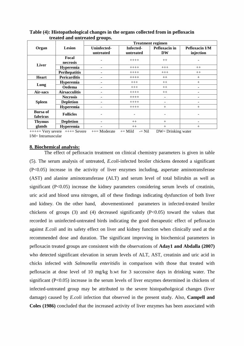
The effect of pefloxacin treatment on clinical chemistry parameters is given in table
(5). The serum analysis of untreated, E.coli-infected broiler chickens denoted a significant
(P<0.05) increase in the activity of liver enzymes including, aspertate aminotransferase
(AST) and alanine aminotransferase (ALT) and serum level of total bilirubin as well as
significant (P<0.05) increase the kidney parameters considering serum levels of creatinin,
uric acid and blood urea nitrogen, all of these findings indicating dysfunction of both liver
and kidney. On the other hand, abovementioned parameters in infected-treated broiler
chickens of groups (3) and (4) decreased significantly (P<0.05) toward the values that
recorded in uninfected-untreated birds indicating the good therapeutic effect of pefloxacin
against E.coli and its safety effect on liver and kidney function when clinically used at the
recommended dose and duration. The significant improving in biochemical parameters in
pefloxacin treated groups are consistent with the observations of Aday1 and Abdalla (2007)
who detected significant elevation in serum levels of ALT, AST, creatinin and uric acid in
chicks infected with Salmonella enteritidis in comparison with those that treated with
pefloxacin at dose level of 10 mg/kg b.wt for 3 successive days in drinking water. The
significant (P<0.05) increase in the serum levels of liver enzymes determined in chickens of
infected-untreated group may be attributed to the severe histopatholgoical changes (liver
damage) caused by E.coli infection that observed in the present study. Also, Campell and
Coles (1986) concluded that the increased activity of liver enzymes has been associated with
19
hepatocellular damage in birds infected with E.coli as well as increase in creatinin, blood urea
nitrogen and uric acid (kidney function parameters) may be attributed to septicemia caused
by E.coli and also due to effect of its toxin on kidney (Pai et al., 1984 and Tizipori et al.,
1987). Omaima (1987) and Mona and Osfor, (2002) observed significant increase in serum
levels of liver enzymes, creatinin, uric acid and blood urea nitrogen in chickens infected with
E.coli and referred that increase to liver and kidney damage associated with bacterial
infection. Moreover, Sachan et al., (2003) reported on the absence of possible hepatotoxicity
and nephrotoxicity potential of pefloxacin in broilers when administered daily at 10mg/kg
b.wt for 5 successive days in drinking water. Other investigations (Sachan et al., 2000 and
2002 and El- Boushy et al., 2006) recorded similar results as they concluded that pefloxacin
had no adverse effect on the liver enzymes at the clinically used dose and duration.
Table (5): Effect of pefloxacin administration on serum chemistry parameters in
pefloxacin treated and untreated groups.
Treatment
group
Serum biochemical values
Hepatic function parameters Renal function parameters
Total
bilirubin AST* ALT* Uric acid
Blood urea
nitrogen Creatinin
Uninfected-
untreated 0.24±0.026
a 193.5±7.94
a 69.6±5.90
a 8.78±0.23
a 0.63±0.039
a 0.39±0.020
a
Infected-
untreated 0.36±0.025
b 245.3±5.09
b 87.1±3.36
b 12.20±0.11
b 0.78±0.031
b 0.53±0.031
b
Pefloxacin in
DW 0.31±0.021
b 224.7±10.42
bc 80.5±3.60
bc 10.44±0.60
c 0.71±0.028
bc 0.48±0.025
b
Pefloxacin by
I/M injection 0.29±0.023
a 219.6±8.22
c 74.2±2.05
ac 9.32±0.31
a 0.65±0.028
ac 0.44±0.034
a
LSD 0.073 24.42 11.93 1.091 0.096 0.085
Values represent the mean ± SEM of 10 broiler chickens each per treatment (n=10).
Means in a column with no common superscripts differ significantly (P<0.05).
LSD= Least significant difference as determined by Fisher’s protected LSD procedure.
AST*= Aspertate aminotransferase. *ALT= Alanine aminotransferase. DW= Drinking water
I/M= Intramuscular
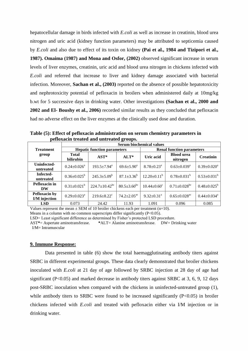
9. Immune Response:
Data presented in table (6) show the total haemagglutinating antibody titers against
SRBC in different experimental groups. These data clearly demonstrated that broiler chickens
inoculated with E.coli at 21 day of age followed by SRBC injection at 28 day of age had
significant (P<0.05) and marked decrease in antibody titers against SRBC at 3, 6, 9, 12 days
post-SRBC inoculation when compared with the chickens in uninfected-untreated group (1),
while antibody titers to SRBC were found to be increased significantly (P<0.05) in broiler
chickens infected with E.coli and treated with pefloxacin either via I/M injection or in
drinking water.
20
Table (6): Total haemagglutinating antibody titers (Log2) of pefloxacin treated
and untreated groups.
Treatment group
Agglutination to sheep red blood cells (SRBC)
Days after sensitization to SRBC
3 6 9 12
Uninfected-untreated 5.1±0.31a 5.9±0.23
a 5.5±0.34
a 4.8±0.41
a
Infected-untreated 3.2±0.20b 4.2±0.22
b 3.4±0.26
b 2.7±0.26
b
Pefloxacin in DW 4.3±0.21cd
5.7±0.26a 4.5±0.27
c 3.8±0.35
cd
Pefloxacin by I/M injection 4.6±0.22ad
5.9±0.37a 5.2±0.44
ac 4.4±0.30
ad
LSD 0.69 0.79 0.96 0.97
Values are Log2 of reciprocal of the highest serum dilution to cause agglutination.
LSD= Least significant difference as determined by Fisher's protected LSD procedures.
Values are geometric means ± SEM (n=10).
Means in the column accompanied by different superscripts differ significantly (P<0.05).
DW= Drinking water I/M= Intramuscular
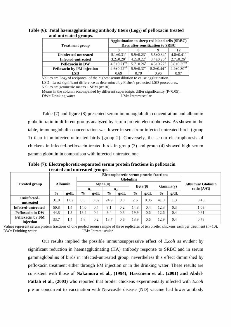
Table (7) and figure (8) presented serum immunoglobulin concentration and albumin/
globulin ratio in different groups analyzed by serum protein electrophoresis. As shown in the
table, immunoglobulin concentration was lower in sera from infected-untreated birds (group
1) than in uninfected-untreated birds (group 2). Conversely, the serum electrophoresis of
chickens in infected-pefloxacin treated birds in group (3) and group (4) showed high serum
gamma globulin in comparison with infected-untreated one.
Table (7): Electrophoretic-separated serum protein fractions in pefloxacin
treated and untreated groups.
Treated group
Electrophoretic serum protein fractions
Albumin
Globulins
Albumin/ Globulin
ratio (A/G)
Alpha(α) Beta(β) Gamma(γ)
α1 α2
% g/dL % g/dL % g/dL % g/dL % g/dL
Uninfected-
untreated 31.0 1.02 0.5 0.02 24.9 0.8 2.6 0.06 41.0 1.3 0.45
Infected-untreated 50.8 1.4 14.0 0.4 8.1 0.2 14.8 0.4 12.3 0.3 1.03
Pefloxacin in DW 44.8 1.3 13.4 0.4 9.4 0.3 19.9 0.6 12.6 0.4 0.81
Pefloxacin by I/M
injection 33.7 1.4 5.8 0.2 18.7 0.6 18.9 0.6 12.9 0.4 0.78
Values represent serum protein fractions of one pooled serum sample of three replicates of ten broiler chickens each per treatment (n=10).
DW= Drinking water I/M= Intramuscular
Our results implied the possible immunosuppressive effect of E.coli as evident by
significant reduction in haemagglutinating (HA) antibody response to SRBC and in serum
gammaglobulins of birds in infected-untreated group, nevertheless this effect diminished by
pefloxacin treatment either through I/M injection or in the drinking water. These results are
consistent with those of Nakamura et al., (1994); Hassanein et al., (2001) and Abdel-
Fattah et al., (2003) who reported that broiler chickens experimentally infected with E.coli
pre or concurrent to vaccination with Newcastle disease (ND) vaccine had lower antibody
21
titers as compared with non-infected vaccinated birds and they pointed-out on the possible
immunosuppressive role of E.coli and its adverse effect on the immune response to ND
vaccination.
The efficacy of immune system in chickens is dependent on the bursa of fabricus for
initiating humorally-related antibodies (Glick, 1970) and on thymus for antibody cellular-
related antibodies (Cooper et al., 1965). Modification of cellular integrity of these tissues by
infection and chemical or physical agents results in immunosuppression (Glick, 1967). Since
the potential of lymphoid tissues to produce antibodies is dependent on the bursa and thymus,
the marked bursal and thymic lymphocytic depletion (inhibition of specific immunological
tissues) induced by experimental infection with E.coli that observed in this study and
previously reported works (Nakomura et al., 1986, 1987, 1990 and 1992; Mona and
Hassanean, 1998; Hassan and Hassanein, 1999; Hassanein et al., 2001 and Kutkat et al.,
2002) is regarded as an attractive explanation for immunosuppressive ability of E.coli and
would be expected to resulted in impaired (HA) response to SRBC and reduction in serum
globulins levels of infected-untreated birds.
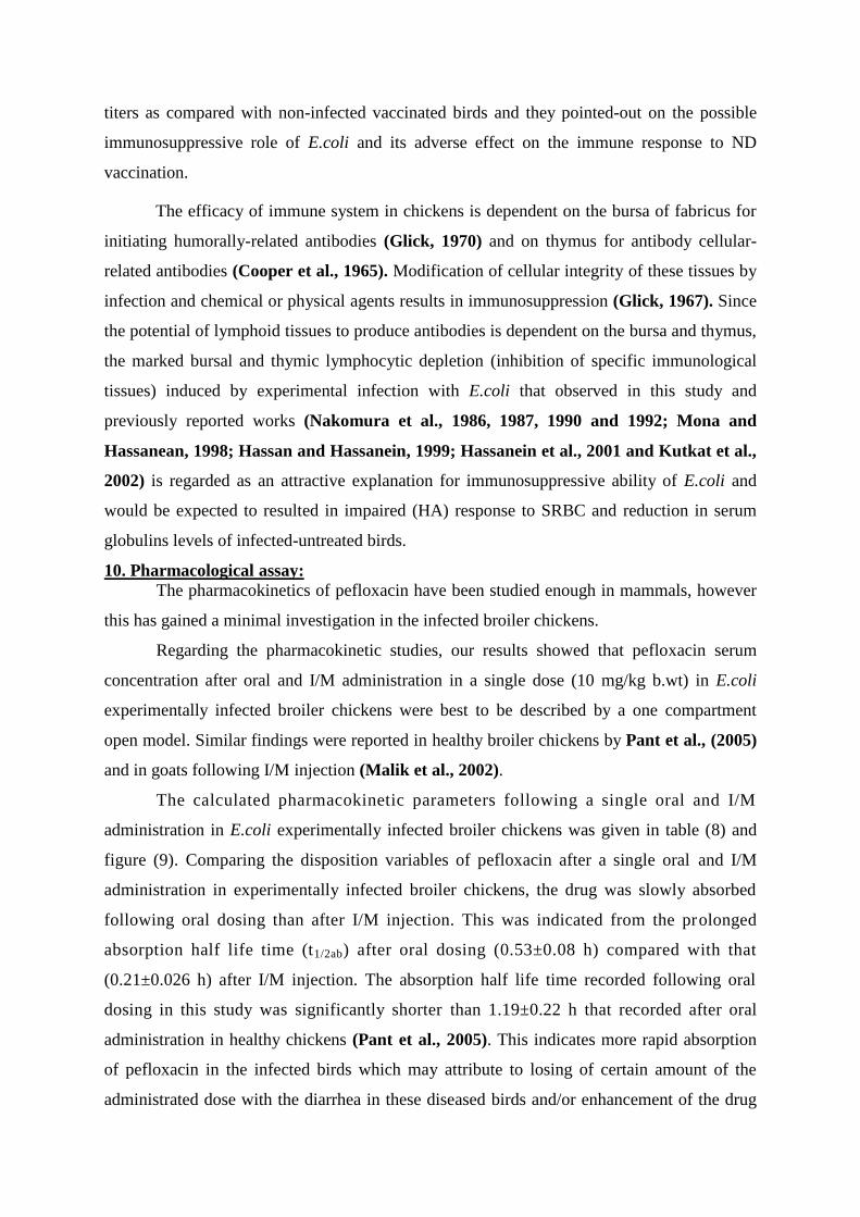
10. Pharmacological assay:
The pharmacokinetics of pefloxacin have been studied enough in mammals, however
this has gained a minimal investigation in the infected broiler chickens.
Regarding the pharmacokinetic studies, our results showed that pefloxacin serum
concentration after oral and I/M administration in a single dose (10 mg/kg b.wt) in E.coli
experimentally infected broiler chickens were best to be described by a one compartment
open model. Similar findings were reported in healthy broiler chickens by Pant et al., (2005)
and in goats following I/M injection (Malik et al., 2002).
The calculated pharmacokinetic parameters following a single oral and I/M
administration in E.coli experimentally infected broiler chickens was given in table (8) and
figure (9). Comparing the disposition variables of pefloxacin after a single oral and I/M
administration in experimentally infected broiler chickens, the drug was slowly absorbed
following oral dosing than after I/M injection. This was indicated from the prolonged
absorption half life time (t1/2ab) after oral dosing (0.53±0.08 h) compared with that
(0.21±0.026 h) after I/M injection. The absorption half life time recorded following oral
dosing in this study was significantly shorter than 1.19±0.22 h that recorded after oral
administration in healthy chickens (Pant et al., 2005). This indicates more rapid absorption
of pefloxacin in the infected birds which may attribute to losing of certain amount of the
administrated dose with the diarrhea in these diseased birds and/or enhancement of the drug
22
absorption as a result of its higher serum proteins binding affinity in the infected chickens
than healthy ones. The tested drug reached a maximum serum concentration (Cmax) of
2.71±0.14 µg.ml-1
following oral administration which is significantly lower than 11.68±1.15
µg.ml-1
after I/M injection. However, the time taken to reach this significant high
concentration (Tmax) after I/M route (1.17±0.103 h) was significantly shorter than the
calculated one after oral dosing (1.62±0.11 h). This could be explained by a rapid absorption
of pefloxacin from I/M injection site than oral administration and/or losing of great amount of
the orally administrated dose with the diarrhea affecting these diseased birds.
It’s really known that pefloxacin is metabolized to norfloxacin (Moutafchieva, 1997 and
Srivastava et al., 2000) and the bioassay method used in this study can’t differentiate
between the active metabolites and the parent drug. So the recorded Cmax values in this
study either following oral dosing (2.71±0.14 µg.ml-1
) or I/M injection (11.68±1.15 µg.ml-1
)
are represent the total concentration for pefloxacin and norfloxacin together. Otherwise, the
Cmax values recorded following oral administration in normal broiler chickens by Pant et
al., (2005) (3.78±0.23 µg.ml-1
) and Suresh Babu et al., (2006) (2.69±0.1.9 µg.ml-1
) represent
the pefloxacin concentration itself according to their assay method by HPLC.
The mean residence time (MRT) for pefloxacin after I/M injection (11.36±1.24 h) was
significantly longer than the recorded one (5.18±0.42 h) after oral dosing indicating more
continued absorption of pefloxacin after I/M injection than oral administration. Comparing
with the recorded values in healthy chickens, a prolonged MRT (14.32±1.94 h) was recorded
after oral dosing in healthy chickens the study of Pant et al., (2005) that revealed more
continued absorption of the tested drug in non infected birds than in E.coli infected ones.
Walker (2000) and Toutain et al., (2002) suggested that the potency of the antibacterial
efficacy of fluoroquinolones is determined by two main factors which are Cmax/MIC ≥ 10
and AUC/MIC ratio ≥ 100. These are the critical break points determine the efficacy of
fluoroquinolones (Forrest et al., 1993 and Meinen et al., 1995). According the MIC value
of pefloxacin for E.coli (0.06 µg.ml-1
) (Raemdonk et al., 1993 and Pant et al., 2005) and
the Cmax and AUC values obtained in this trial, the Cmax/MIC and AUC/MIC ratios
obtained following either oral or I/M administration were higher than 10 and 100,
respectively. So, from a clinical point of view the oral and I/M administration of pefloxacin
in a single dose of 10 mg/kg b.wt would sufficient enough to achieve a potent antibacterial
efficacy against E.coli infection in chickens.
23
Table (8): Mean ± S. E. of pefloxacin pharmacokinetic parameters after
oral and I/M administration in a single dose (10 mg/ kg b.wt) in E.coli
experimentally infected broiler chickens (n=10). Parameter Unit Oral I/M
A µg.ml-1
3.99±0.62 9.46±1.35**
Kab h-1
1.51±0.21 3.06±0.41**
t1/2ab h 0.53±0.08 0.21±0.026**
B µg.ml-1
4.26±0.73 13.62±1.08***
Kel h-1
0.163±0.01 0.091±0.02**
t1/2el h 4.03±0.52 7.41±0.85**
Tmax h 1.62±0.11 1.17±0.103**
Cmax µg.ml-1
2.71±0.14 11.68±1.15***
AUC µg.ml-1
h-1
30.08±3.75 155.29±10.68***
AUMC µg.ml-1
h-2
164.37±13.82 1645.7±38.91***
MRT h 5.18±0.42 11.36±1.24***
** Significant at p ≥ 0.01 *** Significant at p ≥ 0.001
I/M= Intramuscular
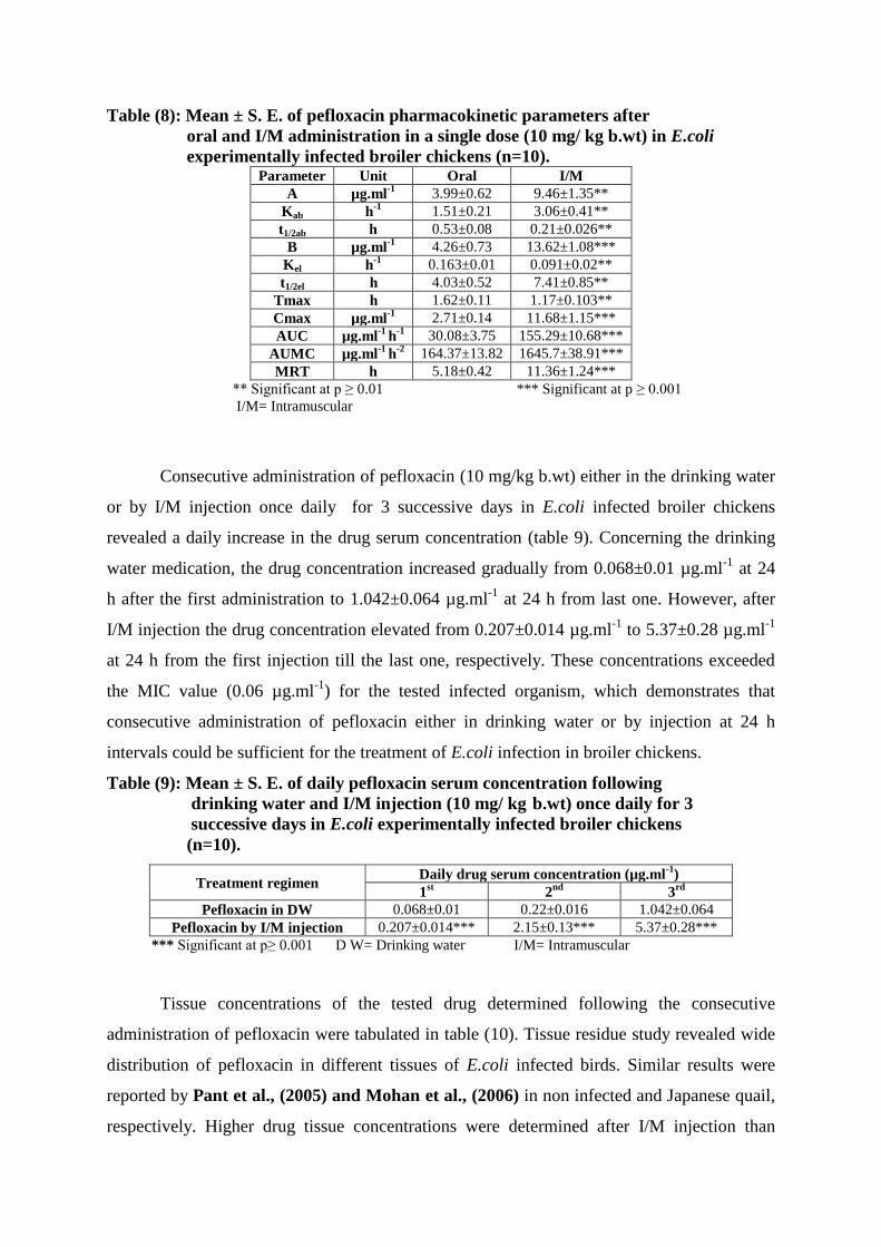
Consecutive administration of pefloxacin (10 mg/kg b.wt) either in the drinking water
or by I/M injection once daily for 3 successive days in E.coli infected broiler chickens
revealed a daily increase in the drug serum concentration (table 9). Concerning the drinking
water medication, the drug concentration increased gradually from 0.068±0.01 µg.ml-1
at 24
h after the first administration to 1.042±0.064 µg.ml-1
at 24 h from last one. However, after
I/M injection the drug concentration elevated from 0.207±0.014 µg.ml-1
to 5.37±0.28 µg.ml-1
at 24 h from the first injection till the last one, respectively. These concentrations exceeded
the MIC value (0.06 µg.ml-1
) for the tested infected organism, which demonstrates that
consecutive administration of pefloxacin either in drinking water or by injection at 24 h
intervals could be sufficient for the treatment of E.coli infection in broiler chickens.
Table (9): Mean ± S. E. of daily pefloxacin serum concentration following
drinking water and I/M injection (10 mg/ kg b.wt) once daily for 3
successive days in E.coli experimentally infected broiler chickens
(n=10).
Treatment regimen Daily drug serum concentration (µg.ml
-1)
1st 2
nd 3
rd
Pefloxacin in DW 0.068±0.01 0.22±0.016 1.042±0.064
Pefloxacin by I/M injection 0.207±0.014*** 2.15±0.13*** 5.37±0.28***
*** Significant at p≥ 0.001 D W= Drinking water I/M= Intramuscular
Tissue concentrations of the tested drug determined following the consecutive
administration of pefloxacin were tabulated in table (10). Tissue residue study revealed wide
distribution of pefloxacin in different tissues of E.coli infected birds. Similar results were
reported by Pant et al., (2005) and Mohan et al., (2006) in non infected and Japanese quail,
respectively. Higher drug tissue concentrations were determined after I/M injection than
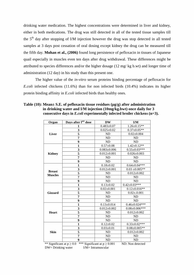
24
drinking water medication. The highest concentrations were determined in liver and kidney,
either in both medications. The drug was still detected in all of the tested tissue samples till
the 5th
day after stopping of I/M injection however the drug was stop detected in all tested
samples at 3 days post cessation of oral dosing except kidney the drug can be measured till
the fifth day. Mohan et al., (2006) found long persistence of pefloxacin in tissues of Japanese
quail especially in muscles even ten days after drug withdrawal. These differences might be
attributed to species differences and/or the higher dosage (12 mg/ kg b.wt) and longer time of
administration (12 day) in his study than this present one.
The higher value of the in-vitro serum proteins binding percentage of pefloxacin for
E.coli infected chickens (11.6%) than for non infected birds (10.4%) indicates its higher
protein binding affinity in E.coli infected birds than healthy ones.
Table (10): Mean± S.E. of pefloxacin tissue residues (µg/g) after administration
in drinking water and I/M injection (10mg/kg.bwt) once daily for 3
consecutive days in E.coli experimentally infected broiler chickens (n=3).
Organ Days after l
ast dose DW I/M
Liver
1 0.483±0.07 1.26±0.15**
3 0.025±0.02 0.37±0.05**
5 ND 0.02±0.004
7 ND ND
9 ND ND
Kidney
1 0.57±0.08 1.42±0.12**
3 0.083±0.006 0.55±0.03***
5 0.012±0.001 0.026±0.003
7 ND ND
9 ND ND
Breast
Muscles
1 0.18±0.02 0.64±0.04***
3 0.012±0.001 0.03 ±0.005**
5 ND 0.012±0.002
7 ND ND
9 ND ND
Gizzard
1 0.13±0.02 0.42±0.03***
3 0.02±0.001 0.12±0.016**
5 ND 0.02±.0.001
7 ND ND
9 ND ND
Heart
1 0.13±0.014 0.46±0.024***
3 0.012±0.002 0.08±0.006***
5 ND 0.012±0.002
7 ND ND
9 ND ND
Skin
1 0.12±0.02 0.33±0.02***
3 0.03±0.01 0.08±0.005**
5 ND 0.012±0.002
7 ND ND
9 ND ND
** Significant at p ≥ 0.0 *** Significant at p ≥ 0.001 ND: Non detected
DW= Drinking water I/M= Intramuscular
25
The results showed that there were no convincing differences in the clinical or
pathological features of E.coli infection by comparing the two methods of pefloxacin
administration indicating that administration of the drug in the drinking water is as effective
as I/M injection. Although, there were no persuasive differences between both methods, the
present study suggested that I/M injection be preferred and more efficacious than drinking
water administration (continuous dosing) in the treatment of avian colibacillosis because of
the drug ability to completely eliminate E.coli from its site of action and therefore it may be
minimize the risk of the disease reoccurring.
In conclusion; in the present study, pefloxacin therapy at 10 mg/kg b.wt/day has been
intensively evaluated in experimental infection in broiler chickens as shown to be highly
effective, useful and good choice in the treatment of E.coli infection regardless the method of
administration.
26
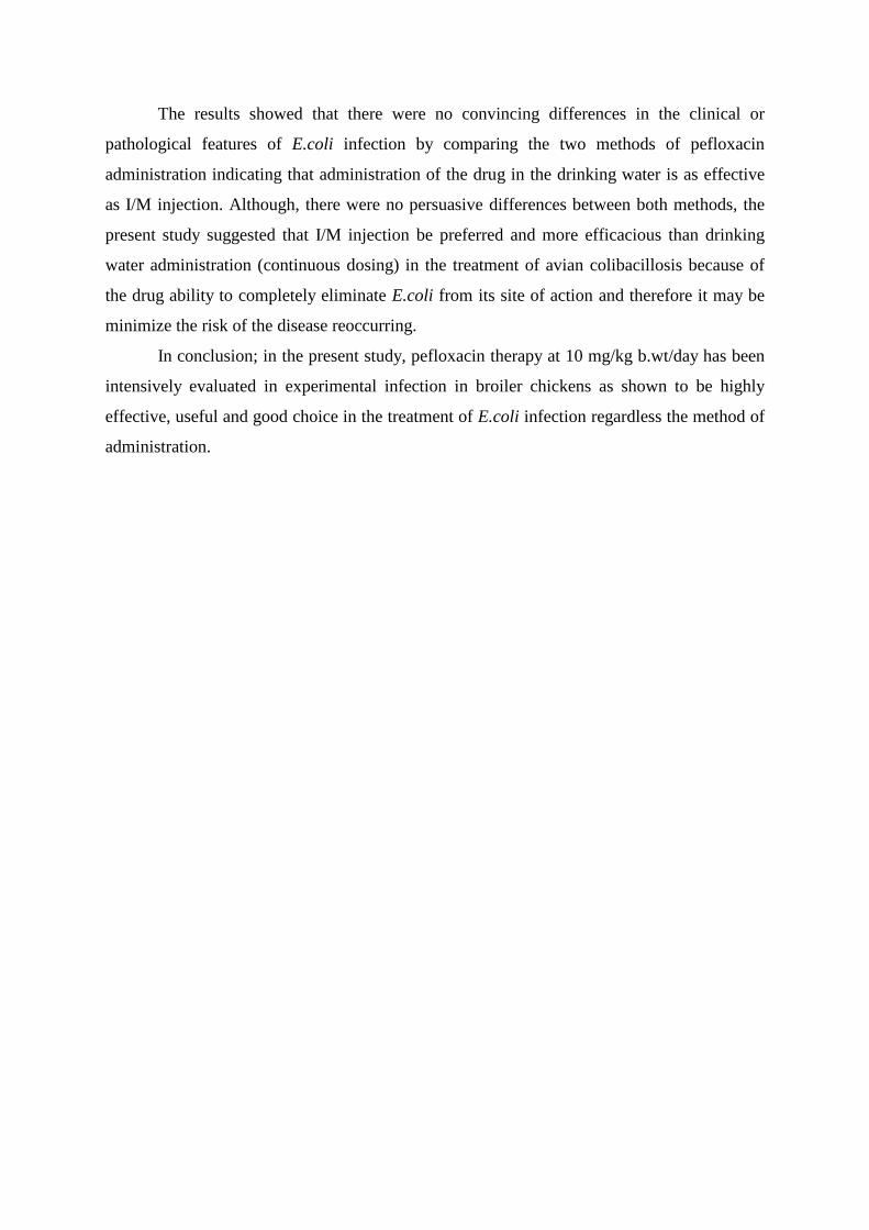
Fig. (1): E) Liver of the uninfected-untreated group showing normal histological structure without any histopathological
alterations (H&E X40). F) Liver of E.coli-infected untreated birds showing focal necrosis with severe dilatation of portal
vein, central vein and sinusoids (H&E X40). H) Liver of E.coli-infected and pefloxacin treated chickens in the drinking water
showing few focal necrosis with mononuclear leucocytes inflammatory cells infiltration in the portal area and dilated central
vein and siusoids (H&E X40). G) Liver of E.coli-infected and pefloxacin treated birds by I/M injection showing few focal
mononuclear leucocytes aggregation with dilatation of portal vein (H&E X40).
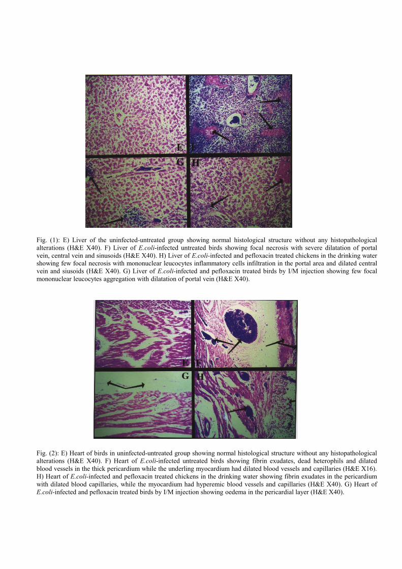
Fig. (2): E) Heart of birds in uninfected-untreated group showing normal histological structure without any histopathological
alterations (H&E X40). F) Heart of E.coli-infected untreated birds showing fibrin exudates, dead heterophils and dilated
blood vessels in the thick pericardium while the underling myocardium had dilated blood vessels and capillaries (H&E X16).
H) Heart of E.coli-infected and pefloxacin treated chickens in the drinking water showing fibrin exudates in the pericardium
with dilated blood capillaries, while the myocardium had hyperemic blood vessels and capillaries (H&E X40). G) Heart of
E.coli-infected and pefloxacin treated birds by I/M injection showing oedema in the pericardial layer (H&E X40).
27
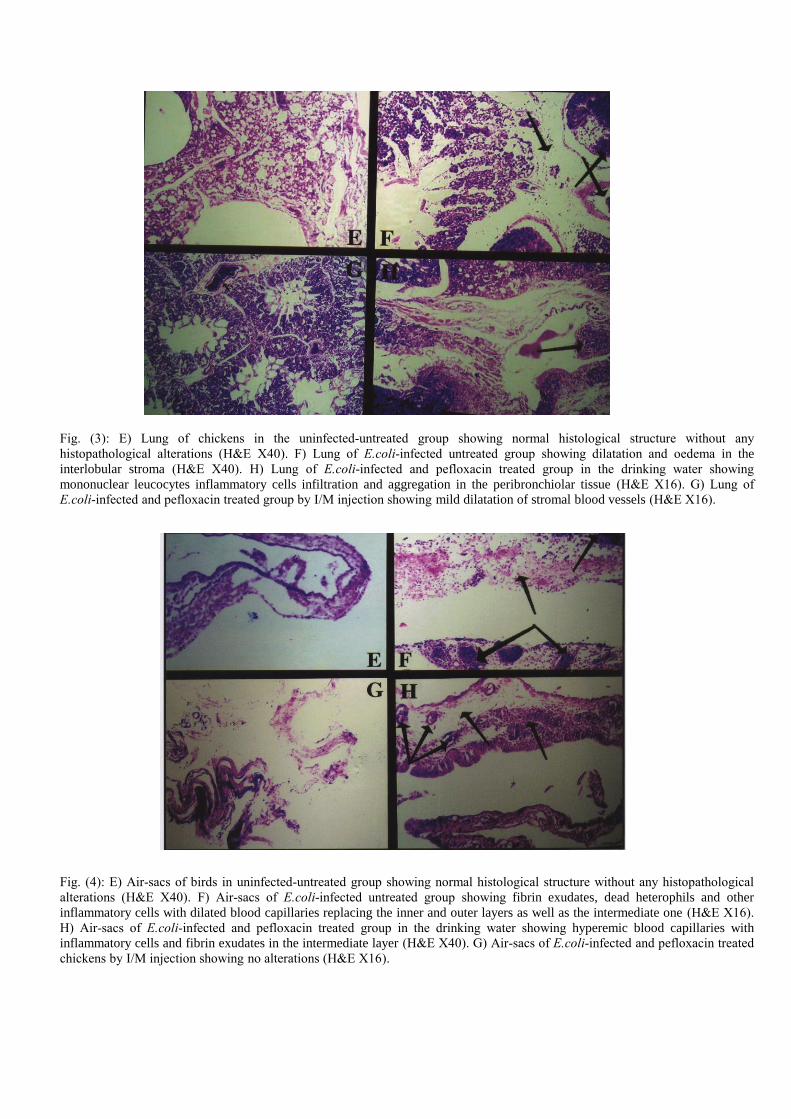
Fig. (3): E) Lung of chickens in the uninfected-untreated group showing normal histological structure without any
histopathological alterations (H&E X40). F) Lung of E.coli-infected untreated group showing dilatation and oedema in the
interlobular stroma (H&E X40). H) Lung of E.coli-infected and pefloxacin treated group in the drinking water showing
mononuclear leucocytes inflammatory cells infiltration and aggregation in the peribronchiolar tissue (H&E X16). G) Lung of
E.coli-infected and pefloxacin treated group by I/M injection showing mild dilatation of stromal blood vessels (H&E X16).
Fig. (4): E) Air-sacs of birds in uninfected-untreated group showing normal histological structure without any histopathological
alterations (H&E X40). F) Air-sacs of E.coli-infected untreated group showing fibrin exudates, dead heterophils and other
inflammatory cells with dilated blood capillaries replacing the inner and outer layers as well as the intermediate one (H&E X16).
H) Air-sacs of E.coli-infected and pefloxacin treated group in the drinking water showing hyperemic blood capillaries with
inflammatory cells and fibrin exudates in the intermediate layer (H&E X40). G) Air-sacs of E.coli-infected and pefloxacin treated
chickens by I/M injection showing no alterations (H&E X16).
28
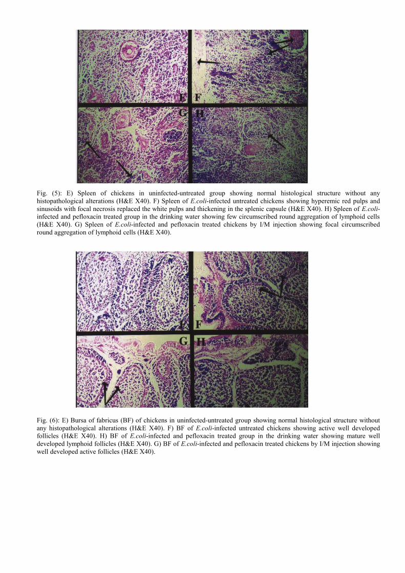
Fig. (5): E) Spleen of chickens in uninfected-untreated group showing normal histological structure without any
histopathological alterations (H&E X40). F) Spleen of E.coli-infected untreated chickens showing hyperemic red pulps and
sinusoids with focal necrosis replaced the white pulps and thickening in the splenic capsule (H&E X40). H) Spleen of E.coli-
infected and pefloxacin treated group in the drinking water showing few circumscribed round aggregation of lymphoid cells
(H&E X40). G) Spleen of E.coli-infected and pefloxacin treated chickens by I/M injection showing focal circumscribed
round aggregation of lymphoid cells (H&E X40).
Fig. (6): E) Bursa of fabricus (BF) of chickens in uninfected-untreated group showing normal histological structure without
any histopathological alterations (H&E X40). F) BF of E.coli-infected untreated chickens showing active well developed
follicles (H&E X40). H) BF of E.coli-infected and pefloxacin treated group in the drinking water showing mature well
developed lymphoid follicles (H&E X40). G) BF of E.coli-infected and pefloxacin treated chickens by I/M injection showing
well developed active follicles (H&E X40).
29
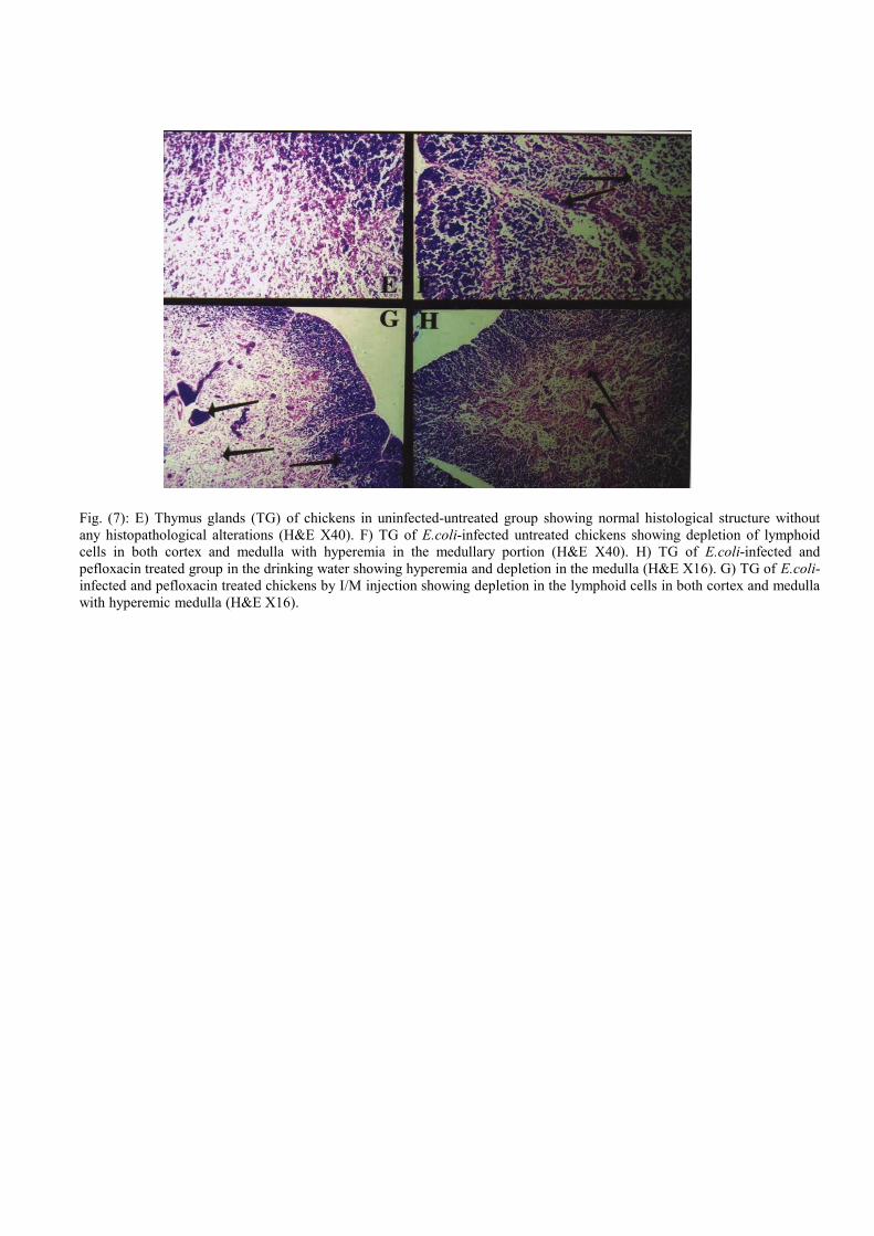
Fig. (7): E) Thymus glands (TG) of chickens in uninfected-untreated group showing normal histological structure without
any histopathological alterations (H&E X40). F) TG of E.coli-infected untreated chickens showing depletion of lymphoid
cells in both cortex and medulla with hyperemia in the medullary portion (H&E X40). H) TG of E.coli-infected and
pefloxacin treated group in the drinking water showing hyperemia and depletion in the medulla (H&E X16). G) TG of E.coli-
infected and pefloxacin treated chickens by I/M injection showing depletion in the lymphoid cells in both cortex and medulla
with hyperemic medulla (H&E X16).
30
I II
III IV
Serum protein fractions separated by electrophoresis
Fig. (8): Serum protein fractions separated by electrophoresis in uninfected-untreated group (I), in
infected-untreated group (II), in pefloxacin-treated group in the drinking water (III) and in pefloxacin
treated group by I/M injection (IV).
Fig. (9): Semi-logarithmic graph depicting serum concentration-time course of pefloxacin
following oral and I/M administration in a single dose (10 mg/kg b.wt) in E.coli
experimentally infected broiler chickens.
31
References Abdel-Fattah, M. M.; Hanan, M. F. A. and Mohamed, K. M. (2003): New
approach for stimulation of chickens immune response against E.coli infection and
Newcastle disease vaccine. Zagazig Vet. J., 31: 184-196
Abdullah, A. (1993): Clinicopathological studies on the effect of some
antibiotics used in chickens. Ph.D. Thesis (clinical pathology), Fac. Vet. Med.,
Zagazig Univ.
Adayel, S. A. and Abdalla, O. E. (2007): Efficacy of pefloxacin against
salmonellosis in balady chicks. Zagazig Vet. J., 35: 71-78.
Andon, A. (1992): Les fluoroquinolones: aspects pharmacologiques et
toxicologiques. Bulletin Academie Veterinaire de France, 65: 207-216.
Andriole, V. T. (1988): The quinolones. Academic Press, London.
Arret, B.; Johnson, D. P. and Kirshbaum, A. (1971): Outline of details for
microbiological assays of antibiotics: Second revision. J. Pharma. Sci., 60 (11): 1689-
1694.
Babu, N. S.; Malik, J. K.; Rao, G. S.; Aggarwal, M. and Ranganathan, V. (2006):
Interactive alterations of arsenic and malathion in the disposition kinetics of
pefloxacin. Arch. Environ. Contam. Toxicol., 50: 587-593.
Badet, B. Hugher, P.; Kohiyama, M. and Forterre, P. (1982): Inhibition of DNA
replication in vitro by pefloxacin. Federation of European Biochemical Societies
(Elsevier, Holland), 145: 355-359.
Bancroft, J. D.; Stevens, A. and Turner, D. R. (1996): Theory and practice of
histopathological techniques. 4th
Ed., Churchill Livingstone, New York.
Barnes, H. J. and Gross, W. B. (1997): Colibacillosis. In B.K. Calnek, H.J. Barnes, C. W.
Beard, L.R. McDougold and Y. M. Saif (eds) Diseases of poultry, 10th
edn, (Iowa state
University press, IA), 131-141.
Barre, J.; Houin, G. and Tillement, J. P. (1984): Dose-dependent pharmacokinetic
study of pefloxacin, a new antibacterial agent, in humans. J. Pharm. Sci., 73: 1379-
1382.
Berg, J. (1988): Clinical indications for enrofloxacin in domestic animal and poultry.
Proc. West. Vet. Conf. Las-Vegas, Nevada, 25-34.
Blanco, M.; Blanco, J. E.; Mora, A. And Blanco, J. (1996): Escherichia coli
septicemicos aviares: serolipos, factores de virulencia, resistencia a antibioticos y
desarrollo de vacunas. Medicina Veteriaria, 13: 525-535
Brown, S. A. (1996): Fluoroquinolones in animal health. J. Vet. Pharm. Therap., 19:
1-4.
Campell, T. and Coles, E. (1986): Avian clinical pathology, in veterinary clinical
pathology” 4th
Ed., W.B. Sounders Company. Philodelphia. London and Toronto.
Chansiripornchai, N. and Sasipreeyajan, J. (2002): Efficacy of sarafloxacin in
broilers after experimental infection with Escherichia coli. Vet. Res. Communication,
26: 255-262.
Chansiripornchai, N.; Sasipreeyajan, J. and Pakpinyo, S. (1995): The in vitro
antimicrobial sensitivity testing of Escherichia coli isolated from commercially reared
chickens. Thia. J. Vet. Med., 25: 275-283.
Cheville, N. F and Arp, L. H. (1978): Comparative pathologic findings of
Escherichia coli infection in birds. J. Amer. Vet. Med. Assoc., 173: 584-587.
Copper, M. D.; Peterson, R. D. A.; South, M. A. and Good, R. A. (1965): The functions of
the thymus system and bursa system in the chickens. J. Exp. Med., 123: 75-123
Craig, A. W. and Suh, B. (1980): Protein binding and the antibacterial effects.
32
Methods for determination of protein binding. In: Lorian V (ed.), Antibiotics in
Laboratory Medicine, (Williams and Wilkins, Baltimore, MD), 265-297.
Cruickshank, R.; Duguid, J. P.; Marmion, B. P. and Swain, R. H. A. (1975): Med. Microbiology Vol. II 12
th edn.
Debbia, E.; Schito, G. C. and Nicoletti, G. (1987): In vitro activity of pefloxacin
against Gram-negative and Gram-positive bacteria in comparison with other
antibiotics. Chemotherpia, 6: 313-323.
Dow, J.; Chazal, J.; Frydman, A. M.; Janny, P.; Woehrle, R.; Djebar, F. and
Gaillot, J. (1986): Transfer kinetics of pefloxacin into cerebro-spinal fluid after one
hour I.V. infusion of 400 mg in man. J. Antimicrob. Chemother., 17, Suppl. B., 81-87.
Dudley, M. N. (1991): Pharmacodynamics and pharmacokinetics of antibiotics with
special reference to fluoroquinolones. Am. J. Med., (6A): 45-50.
Edwards, P. R. and Ewing, W. H. (1972): Identification of enterbacteriaceae. 3rd
Ed., burgress Publishing Co., Minneapolis.
El- Boushy, M. E.; Sanaa, S. A. and Abeer, H. (2006): Immunological,
haematological and biochemical studies on pefloxacin in broilers infected with
E. coli. 8th
Sci. Vet. Med. Zagazig conference.
Epstein, E. and Karcher. R. F (1994): Electrophoresis: In Tietz Textbook of clinical
chemistry, 2nd
Ed, CA Burtis, E.R Ash wood, Philadelphia, W.B. Sanders,
1994.
Ershov, E.; Bellaiche, M.; Hanji, V.; Soback, S.; Gips, M. and Shlosberg, A.
(2001): Interaction of fluoroquinolones and certain ionophores in broilers: effect on
blood levels and hepatic cytochrome p-450 monooxygenase activity. Drug Metabol.
Drug Interact., 18: 209-219.
Fernandez, A.; Lara, C.; Loste, A. and Marca, M. C. (2002): Efficacy of calcium
fosfomycin for the treatment of experimental infection of broiler chickens with
Escherichia coli O78:K80. Vet. Res. Comunication, 26: 427-436.
Forrest, A.; Nix, D. E.; Ballow, C. H.; Gross, T. F.; Birminghan, M. C. and
Schentag, J. J. (1993): Pharmacodynamics of intravenous ciprofloxacin in
seriously ill patients. Antimicrobiol. Agents Chemother., 37: 1073-1081.
Freed, M.; Clarke J. P.; Bowersock, T. L.; Van Alstine, W. G.; Balog, J. M. and
J. M. and Haster, P. Y. (1993): Effect of spectinomycin on Escherichia coli
infection in 1-day old ducklings. Avian Dis., 37: 763-766.
Gibaldi, M. and Perrier, D. (1982): Pharmacokinetics, 2nd
edn, (Marcel Dekker,
New York), 409-417.
Glick, B., (1967): Antibody and gland studies in cortisone and ACTH- injected birds. J.
Immunol, 98: 1076-1084
Glick, B. (1970): The bursa of fabricius as a central issue. Bioscience, 20: 10-13.
Glisson, J. R. (1994): Fluoroquinolone use in the poultry industry. Broiler Industry,
July, 46-48.
Glisson, H. R.; Hafacere, C. I. and mathis, G. F (2004): Comparative efficacy of
enroflaxocin, oxytetrocycline and sulphadimethoxine for the control of morbidity and
mortality caused by Escherichia coli in broiler chickens. Avian Dis., 48: 856-662.
Gonzalez, J. P. and Henwood, J. M. (1989): Pefloxacin. A review of its antibacterial
activity, pharmacokinetic properties and therapeutic use. Drugs, 37: 628-668.
Gross, W. B. (1991): Colibacillosis. Pp. 138-144. In Calnek, W. B.; Barnes, H. J.;
Beard, C. W.; Reid, W. M. and Yoder, H. W. (ed.) Diseases of Poultry. Iowas State
University Press, Ames.
Hassan, M. K. and Hassanein, Z.A. (1999): Effect of Escherichia coli infection on
33
the immune response of chickens to Newcastle disease virus vaccine. J. Egypt. Vet. Med.
Ass., 59: 75-91.
Hassanein, Z. A.; Mona, M. A.; Hassan, M. K. and El-Mongey, F. A. (2001): Nature of
humoral immune suppression induced by Escherichia coli infection in vaccinated
chickens. J. Egyptian Vet. Med. Ass., 61: 79-89
Hui, A. K. and Das, R. (2000): Prevalence of E.coli in duck in two districts of
Western Bengal with their serotyping and antibiogram. Indian J. Anim. Health. 39:
61-64.
Isea, G.; Martinez, M. A.; Martinez-Larranaga, M. R.; Diaz, M. J. and Anadon,
A. (2003): Pharmacokinetic characteristics of pefloxacin and its metabolite N-
demethylpefloxacin in chickens. J. Vet. Pharmacol. Ther., 26 (Suppl.):113-114.
Jehan, R. Daoud. (2004): Determination of pefloxacin residues in serum and tissues
of broilers and the effect of heat treatments on its residues. J. Egypt. Vet. Med. Ass.,
64 (1): 179-189.
Jenkins, W. and Friedlander, L. (1988): The pharmacology of quinolone
antibacterial agents. Proc. West. Vet. Conf. Les-Vegas, Nevada, 5-16.
Joong Kim, S. (1995): Efficacy of sarafloxacin for the control of poultry bacterial
diseases. Samji Vet., 10: 2-23.
Kayser, F. M. (1985): The quinolones: mode of action and mechanisms of resistance.
Research and Clinical Forums, 7: 17-27.
Kempf, I.; Gesbert, F.; Guittet, M.; Froyman, R.; Deloporte J. and Bennejean, G.
(1995): Dose titration study of enrofloxacin (Baytril) against respiratory colibacillosis in
Muscovy ducks. Avian Dis., 39: 480-488.
Kutkat, M. A.; Nagwa, S. and Ebtehal, A. (2002): Effect of Lactobacillus
acidophilus on controlling of clostridium perfringens and E. coli infections in native
breed chickens. J. Egypt. Vet. Med. Ass., 62 (6): 89-101.
Leitnes, G. and Heller, E. D. (1992): Colonization of Escherichia coli in young
turkeys and chickens. Avian Dis., 36: 211-220.
Lublin, A.; Sara. Mechani; Malkinson, M. and Weisman, Y. (1993): Efficacy of
norofloxacin nicotinate treatment of broiler breeders against Haemophilus
paragallinarum. Avian Dis., 73: 673-679.
Malik, J. K.; Rao, G. S.; Ramesh, S.; Muruganandan, S.; Tripathi, H. C. and
Shukla, D. C. (2002): Pharmacokinetics of pefloxacin in goats after
intravenous and oral administration, Vet. Res. Com., 26: 141-149.
Mark, S. and Robert, T. (1993): E.coli O157:H7 ranks as the fourth most costly
food borne disease. Food Review USDA/ERS, 16: 51-59.
McCabe, M. W. and Rippel, R. E. (1993): Efficacy of sarafloxacin for the control of
mortality associated with E.coli in broiler chicken and turkey. Proc. 42nd
Wes.
Poultry Dis. Conf., (Sacramento, California), 75.
Meinen, J. B.; Mcclure, J. T. and Rosin, C. H. (1995): Pharmacokinetics of
enrofloxacin in clinically normal dogs and mice and drug pharmacodynamics
in neutropenic mice with Escherichia coli and staphylococcal infections.
American J. Vet. Res., 56: 1219-1224.
Mognet, L.; Bezille, P.; Guyonett, J. and Karembe, H. (1997): comparison de la
flumequine á l’amoxicilline dans deux modes d’adinistration par voie orale, en
traitement de la colibacillose du poulet: approche phamacodynamique et clinique.
Revue Medecine veterinaire, 148: 793-804.
Mohamed, E. A. and Dardeer, M. A. (2001): Influence of combination of BCG
vaccine and pefloxacin in chickens experimentally infected with Mycoplasma
gallisepticum. J. Egypt. Vet. Med. Ass., 61 (2): 281-292.
34
Mohan, J.; Sastry, K. V.; Singh, J. S. and Singh, D. K. (2004): Isolation of E.coli
from foam and effects of fluoroquinolones on E.coli and foam production in
male Japanese quail. Theriogenology, 62: 1383-1390.
Mohan, J.; Sastry, K. V.; Taygi, J. S.; Rao, G. S. and Singh, R. V. (2006): Residues of fluoroquinolone drugs in the cloacal gland and other tissues of Japanese
quail. Br. Poult. Sci., 47 (1): 83-87.
Mona, M. A. and Hassanean Z. A. (1998): Studies on the interaction between
infectious bursal disease virus and Escherichia coli in chickens. J. Egypt Vet. Med.
Ass., 58: 493-512
Mona, S. Z. and Osfor, M. H. (2002): Some body performance and
clinicopathological studies on the effect of Escherichia coli O157:H7 in chicken.
Beni-suef Vet. Med. J., XII: 35-44.
Moutafchieva, R. (1997): Pharmacokinetics of pefloxacin in pigeons (Columbia
livia). J. Vet. Pharmacol. Ther., 20 (suppl. 1): 208.
Moutafchieva, R. and Djouvinov, D. (1997): Pharmacokinetics of pefloxacin in
sheep. J. Vet. Pharmacol. Ther., 20: 405-407.
Moutafchieva, R.; Nouws, J. F. M.; Droumev, D. and Pandev, A. (1997): A
necessity for a change of the pefloxacin dosage regimen in birds, after addition of the
coccidiostatic compound maduramycin to their ration. Vet. Sci., 29: 219-225.
Moutafchieva, R. and Yarkove, D. (2006): Pharmacokinetics of pefloxacin in birds.
Trakia J. Sci., 4 (3): 28-33.
Nagah, E. E.; Gehan, W. A. and Wael, G. N. (2004): Efficacy and side effects of
pefloxacin in laying hens. Alexandria. J. Vet. Sci., 21: 366-383.
Nakamura, K.; Imada, Y. and Moeda, M. (1986): Lymphocytic depletion of bursa
of fabricus and thymus in chickens inoculated with Escherichia coli. Vet. Pathol., 23:
712-717.
Nakamura, K.; Imada, Y. and Nbc, H. (1987): Effect of cyclophoamide on
infections produced by Escherichia coli of high and law virulence in chickens. Avian
Pathol., 16: 237-252.
Nakamura, K.; Cook, K. A.; Frazier, A. and Narita, M. (1992): Escherichia coli
multiplication and lesions in the respiratory tract of chickens inoculated with
infectious bronchitis virus and / or E. coli. Avian Pathol., 36: 881-890.
Nakamura, K.; Ueda, H.; Tanimura, T. and Noguchi, K. (1994): Effect of mixed
live vaccine (Newcastle disease and infections bronchitis) and Mycoplasma
gallisepticum on the chicken respiratory tract and on Escherichia coli infection
J. Comp. Pathol., 111: 33-42.
Nakamura, K.; Youasa, N.; Abe, H. and Narita, M. (1990): Effect of infectious
bursal disease virus on infections produced by Escherichia coli of high and low
virulence in chickens. Avian pathol., 19: 713-712.
Nakamura, K.; Maeda, M.; Imada, Y.; Imada, T. and Sato, K. (1985): Pathology
of spontaneous colibacillosis in broiler flock. Vet. Pathol., 22: 592-597.
National Requirment Council (NRC) (1994): Nutrient requirement of poultry. 8th
rev. Ed National Academy Press.
Neer, T. M. (1988): Clinical and pharmacological features of fluoroquinolones
antimicrobial drugs. J. Am. Vet. Med. Ass., 193: 577-580.
Obrien, A.; Tesh, V.; Donohue – Rolf, A.; Jackson, M.; Olsnes, S.; Sandvig, K.;
Lindb, A. and Keusch, G. (1992): Shiga toxin: biochemistry genetics, mode of
action, and role in pathogenesis. Curr. Top. Microbiol. Immunol., 180: 65-94.
Omaima, A. R. (1987): Liver function tests in relation to some bacterial and viral
35
diseases in chickens. M. V. Sc., (Biochemistry Dept.), Fac.Vet. Med., Zagazig
University
Pai, C.; Gordon, R.; Sims, H. and Bryan, L. (1984): Sporadic cases of
haemorrhagic colitis associated with Escherichia coli O157:H7. Clinical,
epidemiologic and bacteriologic features. Ann. Intern. Med., 101: 738-742.
Pant, S.; Rao, G. S.; Sastry, K. V.; Tripathi, H. C. and Malik, J. K. (2005): Pharmacokinetics and tissue residues of pefloxacin and its metabolite norfloxacin in
broiler chickens. Br. Poult. Sci., 46 (5): 615-620.
Prasad, V.; Krishnamurlthy, K. and Janardhana Rao, T. V. (1997): In vitro
antibiogram studies of Escherichia coli in chickens. Indian Vet. J., 74: 616-617.
Piercy, D. W. T. and West, B. (1976): Experimental Escherichia coli infection in
broiler chickens: course of the disease induced by inoculation via the air-sac route. J.
Comp. Pathol., 86: 203-210.
Pourbakhshs, S. A.; Boulianne, M.; Nartineau – Doize, B.; Dozois, C. M.;
Desautels, C. and Fair brother, J. M. (1997): Dynamics of Escherichia coli
infection in experimentally inoculated chickens. Avian Dis., 41: 221-232.
Quinn, P. J.; Carte, M. E.; Markeryo, B. and Carter, G. R. (1994): Clinical Vet.
Microbiol. Year book-wolf publishing- Europe Limited.
Raemdonck, D. Y.; Hardda, Y. And Holck, T. (1993): Efficacy of danofloxacin
for treatment and control of colibacillosis in broilers using pulse dosing or
intermittent dosing regimens in “Broiler health in the 90” Pfizer symposium
XIII latin American poultry congress, Santo Domingo, Dominican Republic,
October. 13, 11-26.
Robson, R. A. (1992): Quinolone pharmacokinetics. Intern. J. Antimicro. Agents, 2:
3-10.
Rolinski. Z.; Kowalski, C.; Zan, R. and Sobol, M. (2002): Bacterial resistance to
quinolone antibiotics in Poland. J. Vet. Med. (B). Infect. Dis. Vet. Public health.
Ruiz, J.; Gomez, J.; Navia, M. M.; Ribera, A.; Sierra, J. M.; Marco, F. and Vila,
J. (2002): High prevalence of nalidixic acid resistant, ciprofloxacin susceptible
phenotype among clinical isolates of Escherichia coli and other enterobacteriaceae.
Diagn. Microbiol. Infect. Dis., 42 (4): 257-261.
Sachan, A.; Jayakumar, K.; Honnegowda Gowda, R. N. and Narayna, K. (2000):
Study in broiler chickens, the effect of pefloxacin on haemtological parameters in
clinical toxicity. Indian Vet. J., 77: 307-309.
Sachan, A., Jayakumar, K., Honnegowdag Gowda, R. N and Umesh, M.H.
(2002): Evaluation of hepatotoxicity potential of pefloxacin in chickens based on
biochemical parameters and histopathology. Indian J. Toxicol., 9: 43-45.
Sachan, A.; Jayakumar, K; Umesh, H; Udupa, V. and Narayana, K. (2003): Safety evaluation of pefloxacin in broiler chickens. Indian Vet. J., 80: 404-405.
Sahar, A. and Elshazly, M. (2002): Effect of probiotic and pribiotic combination
(Biomin IMBO and Biomin C-EX) on E.coli infection in broiler chickens. J. Egypt.
Vet. Med. Ass., 62 (6): 113-119.
Santurio, J. M.; Mallmann, C. A.; Rossa, A. P.; Apple, G.; Heer, A.; Dogeforde,
S. and Boltcher, A. (1999): Effect of sodium bentonite on the performance and blood
variable of broiler intoxicated with aflatoxin. Br. Poult. Sci., 40: 115-119.
Sarközy, G.; Semjen, G. and Laczay, P. (2004): Disposition of norfloxacin in
broiler chickens and turkeys after different methods of oral administration. Vet. J.,
168: 312-316.
Sasipreeyajan, J. and Pakpinyo, S. (1992): The result of using enrofloxacin in
broiler. Thai J. Vet. Med., 22: 95-104.
36
Sekizaki, T.; Nonomura, L. and Imada, Y. (1989): Loss of virulence associated
with plasmid curing of chickens pathogenic Escherichia coli. J. Japanese Vet. Sci.,
51: 659-661.
Sharma, A. K.; Khosla, R. K. and Metha, V. L. (1994): Fluoroquinolones:
Indian J. pharmacol., 249-261
Smith, H. W. (1970): The transfer of antibiotic resistance between strains of
enterobacteria in chickens, calves and pigs. J. Med. Microbiol., 3: 165-180.
Smith, J. T. (1986): The mode of action of 4 quinolone and possible mechanism of
resistance. J. Antimicrob. Chemother., 18 (suppl. d): 21-29.
Smith, H. W.; Cook, J. K. A. and Parsell, Z. E. (1985): The experimental infection
of chickens with mixtures of infectious bronchitis virus and Escherichia coli.
J. Gen. Virol., 66: 777-786.
Snedecor, G. W. and Cochran, W. G. (1980): Statistical Methods. 7th
Ed., (Iowa
State College Press, Ames, IA), 39-63.
Spoo, J. W. and Riviere, J. E. (1995): Chloramphenicol, macrolides, lincosamides,
fluoroquinolones and miscellaneous antibiotics, in: Richard, A. H. (Ed.) Veterinary
Pharmacology and Therapeutics, 7th
edn, pp. 832-842. (Ames, Iowa State, University
Press).
Srivastava, A. K.; Dunka, V. K. and Deol, S. S. (2000): Deposition kinetics and
urinary excretion of pefloxacin after intravenous injection in crossbred calves. Vet.
Res. Communication, 24: 189-196.
Stamm, J. M.; Hanson, C. W.; Chu, T. W.; Bailer, R; Vojtko, C. and Fernandes,
P. B. (1985): In vitro evaluation of ABBOTT-56619 (difloxacin) and ABBOTT-
56620: new aryl-fluoroquinolones. Antimicrob. Agents Chemother., 29: 193-200.
Stein, G. E. (1987): Review of the bioavailability and pharmacokinetics of oral
norfloxacin. The Amer. J. Med., 82 (Suppl.2):18-21.
Suresh Babu, N.; Malik, J. K.; Rao, G. S.; Manoj, A. and Ranganathan, V.
(2006): Interactive alterations of arsenic and malathion in the deposition
kinetics of pefloxacin. Archive Environ. Cont. Toxicol., 50: 587-593.
Tai, H. and Fang, S. C. (2000): Drug susceptibility of E.coli isolated from poultry.
J. Chin. Soci. Vet. Sci., 26: 36-42.
Tanner, A. C.; Fitz-Coy, S.; Wu, C. C. and Lautz, C. A. (1992): The efficacy of
danofloxacin in the treatment of Escherichia coli respiratory disease in commercial
broiler flocks. Zootechnico Intern., 60-62.
Timms, L. M.; Marchal, R. N. and Breslin, M. (1989): Evaluation of
chlorotetracyline for control of chronic respiratory disease caused by Escherichia coli
and Mycoplasma gallisepticum. Res. Vet. Sci., 47: 377-384.
Tizipori, S.; Wachsmutth, I.; Champan, C.; Birden, R.; Brittinghom, J. and
Jackson. C. (1987): The pathogenesis of haemorrhagic colitis caused by Escherichia
coli O157:H7 in gnotobiotic piglets. J. Infect. Dis., 154: 712-716.
Toutain, P. L.; Delcastillo, J. R. E. and Bousquet-Melon, A. (2002): The
Pharmacokinetic-pharmacodynamic approach to a rational dosage regimen for
antibiotics. Res. Vet. Sci., 73: 105-114.
Toyfour, M.; Yuce, A. and Yulug, N. (2001): Comparison of minimum inhibitory
concentration value of fluoroquinolones against Escherichia coli causing urinary
infection in both hospitalized patients and outpatients. Med. J., 22 (10): 845-851.
Vancutsem, P. M.; Babish, J. G. and Schwark, W. S. (1990): The fluoroquinolones
antimicrobials: structure, antimicrobial activity, pharmacokinetics, clinical use in
domestic animals and toxicity. Cornell Veterinarian, 80: 173-86.
Vandemaele, F.; Vereecken, M.; Derijcke, J. and Goddeeris, B. M. (2002):
37
Incidence and antibiotic resistance of pathogenic Escherichia coli among poultry in
Belgium. Vet. Rec., 151: 355-356.
Van der Zijpp, A. J. and Leenstra, F. R. (1980): Genetic analysis of the humoral
immune response of white leghorn chicks. Poult. Sci., 59: 1363-1369.
Walker, R. D. (2000): The use of flouroquinolones for companion animal
antimicrobial therapy. Austr. Vet. J., 78: 84-89.
Wille, A.; Altay, G. and Erdem, B. (1988): The susceptibility of Salmanella species
to various antibiotics. Microbiol. Bul., 22 (1): 17-24.
Xiapei, Y.; Feng, P.; Zhong, L.; Lxiao, J. and Lei, B. J. (2002): Accumulation of
ofloxacin and toxufloxacin in fluoroquinolone resistant Escherichia coli. Roum. Arch.
Microbiol. Immunol., 22 (3): 210-214.
Yamaoka, K.; Nakagawa, T. and Uno, T. (1978): Statistical moments in
pharmacokinetics. J. Pharmac. Biopharma., 6: 547-558.
Zhenling, Z.; Yongxue, S.; BingHu, F.; Zhangliu, C.; Zl, Z. and Fang, B. (2002): Therapeutic efficacy of sarafloxacin against mixed infection of Mycoplasma
gallisepticum and Escherichia coli in chickens. Chinese J. Vet. Med., 28: 17-19.