Embed Size (px)
Citation preview

The Pseudomonas syringae-derived
HrpA pilins – molecular characterization and
biotechnological application of the transcripts
Elina Hienonen
Department of Biological and Environmental Sciences
Division of General Microbiology
Faculty of Biosciences
and
Viikki Graduate School in Biosciences
University of Helsinki
ACADEMIC DISSERTATION
To be presented, with the permission of the Faculty of Biosciences of the
University of Helsinki, for public criticism in auditorium 1 at Viikki Info
Center (Viikinkaari 11, Helsinki) on October 18th, 2006, at 12 noon
Helsinki 2006
Supervisors: Docent Suvi Taira
Department of Biological and Environmental Sciences
Division of General Microbiology
Faculty of Biosciences
University of Helsinki
Professor Martin Romantschuk
Department of Ecological and Environmental Sciences
Faculty of Biosciences
University of Helsinki
Reviewers: Docent Benita Westerlund-Wikström
Department of Biological and Environmental Sciences
Division of General Microbiology
Faculty of Biosciences
University of Helsinki
Professor Thorsten Nürnberger
Zentrum fur Molekularbiologie der Pflanzen
Eberhard-Karls Universität
Opponent: Professor Per Saris
Department of Applied Chemistry and Microbiology
Division of Microbiology
Faculty of Agriculture and Forestry
University of Helsinki
ISSN 1795-7079 ISBN 952-10-3431-9 (printed) ISBN 952-10-3432-7 (PDF)
iii
Table of ContentsSummaryOriginal publicationsAbbreviations1 Introduction................................................................................................. 1
1.1 SECRETION SYSTEMS OF GRAM-NEGATIVE BACTERIA ............ 11.2 TYPE III SECRETION SYSTEMS........................................................ 2
1.2.1 Evolution and distribution................................................................ 31.2.2 Effects caused by the TTSSs of plant pathogenic bacteria ................ 41.2.3 The structures of the non-flagellar type III secretion apparatuses ..... 5
1.2.3.1 HrpA pilin of P. syringae........................................................... 71.2.4 Regulation of non-flagellar type III secretion ................................... 81.2.5 Type III secreted effectors and harpins........................................... 111.2.6 Type III secretion signals............................................................... 141.2.7 TTS chaperones............................................................................. 19
1.3 mRNA DEGRADATION..................................................................... 211.3.1 RNases .......................................................................................... 23
1.3.1.1 The degradosome .................................................................... 241.3.2 Protection against RNases.............................................................. 25
1.3.2.1 Ribosomes affect mRNA stability............................................. 251.3.2.2 Secondary structures of transcripts ......................................... 26
1.3.3 Transcript stability as a mechanism to control gene expression ...... 281.4 PROTEIN PRODUCTION................................................................... 30
1.4.1 Transcript stabilizing elements in recombinant protein production . 311.4.2 In vitro translation systems ............................................................ 311.4.3 Host organism for the production of foreign proteins ..................... 321.4.4 Vector design for the production of recombinant proteins .............. 321.4.5 Folding of recombinant proteins .................................................... 341.4.6 Purification of recombinant proteins .............................................. 35
2 Aims of the study....................................................................................... 373 Materials and methods.............................................................................. 374 Results and discussion............................................................................... 39
4.1 The secretion signal of hrpA ................................................................. 394.2 Translation of HrpA is not dependent on secretion................................ 394.3 Regions important for the accumulation of hrpA transcript ................... 394.4 Half-lives of hrpA transcripts................................................................ 404.5 Stabilization of heterologous transcripts by hrpA .................................. 414.6 Recombinant protein yields can be improved using the transcriptstabilizing elements from hrpA ................................................................... 42
5 Conclusions................................................................................................ 436 Acknowledgements.................................................................................... 457 References.................................................................................................. 47

iv
Summary
Many Gram-negative bacteria pathogenic to plants and animals possess type
III secretion systems that are used to cause disease. Effector proteins are
injected into host cells using the type III secretion machineries. Despite
vigorous studies, the nature of the secretion signal for type III secreted proteins
still remains elusive. Both mRNA and proteinaceous signals have been
proposed. Findings on coupling of translation to secretion by the type III
secretion systems are also still contradictory.
This study dealt with the secretion signal of HrpA from Pseudomonas
syringae pathovar tomato. HrpA is the major component of the type III
secretion system-associated Hrp pilus and a substrate for the type III secretion
systems. The secretion signal was shown to reside in the first 15 codons or
amino acids, a location typical for type III secretion signals. Translation of
HrpA in the absence of a functional type III secretion system was established,
but it does not exclude the possibility of coupling of translation to secretion
when the secretion apparatus is present.
The hrpA transcripts from various unrelated plant pathogenic bacteria were
shown to be extremely stable. The biological relevance of this observation is
unknown, but possible explanations include the high prevalence of HrpA
protein, an mRNA secretion signal or timing of secretion. The hrpA mRNAs are
stable over a wide range of temperatures, in the absence of translating
ribosomes and in the heterologous host Escherichia coli. The untranslated
regions (UTRs) of hrpA transcripts from at least 20 pathovars of Pseudomonas
syringae are highly homologous, whilst their coding regions exhibit low
similarity. The stable nature of hrpA messenger RNAs is likely to be due to the
folding of their 5’ and 3’ UTRs. In silico the UTRs seem to form stem-loop
structures, the hairpin structures in the 3’ UTRs being rich in guanidine and
cytosine residues.
v
The stable nature of the hrpA transcript directed the studies to the
stabilization of heterologous transcripts and to the use of stable messenger
RNAs in recombinant protein production. Fragments of the hrpA transcript can
be used to confer stability on heterologous transcripts from several sources of
bacterial and eukaryotic origin, and to elevate the levels of production of the
corresponding recombinant proteins several folds. hrpA transcript stabilizing
elements can be used for improving the yields of recombinant proteins also in
Escherichia coli, one of the commonly used hosts in industrial protein
production.

vi
Original publications
This thesis is based on the following articles and manuscript that in the text are
referred to by their Roman numerals.
I Hienonen E, Roine E, Romantschuk M, Taira S.
mRNA stability and the secretion signal of HrpA, a pilin secreted by the type
III system in Pseudomonas syringae. Mol. Genet. Genomics. 2002 266:973-8.
II Hienonen E, Rantakari A, Romantschuk M, Taira S.
The bacterial type III secretion system-associated pilin HrpA has an unusually
long mRNA half-life. FEBS Lett. 2004 571:217-20.
III Hienonen E, Romantschuk M, Fenel F, Taira S.
Transcript stabilization by mRNA sequences from hrpA of Pseudomonas
syringae. Manuscript (submitted to J. Biotechnol.).

vii
Abbreviations
ATP adenosine triphosphate
ATPase adenosine triphosphatase
CBD chaperone binding domain
C-terminus carboxy-terminus
EPEC enteropathogenic Escherichia coli
G/C guanidine/cytosine
HR hypersensitive reaction
Hrc hypersensitive reaction and pathogenesis conserved
Hrp hypersensitive reaction and pathogenesis
MLD membrane localization domain
mRNA messenger ribonucleic acid
NPT neomycin phosphotransferase
N-terminus amino-terminus
ORF open reading frame
PAPI Poly(A) polymerase
PNPase polynucleotide phosphorylase
PPK polyphosphate kinase
PR pathogenesis related
pv. pathovar
R resistance gene or protein
RBS ribosome binding site
RNA ribonucleic acid
RNase ribonuclease
rRNA ribosomal ribonucleic acid
rt-PCR reverse transcriptase polymerase chain reaction
SD Shine-Dalgarno
spp. subspecies
Tir translocated intimin receptor

viii
Abbreviations continued
tRNA transfer ribonucleic acid
TTSS type III secretion system
UTR untranslated region
Yop Yersinia outer protein
Ysc Yersinia secretion

1
1 Introduction
1.1 SECRETION SYSTEMS OF GRAM-NEGATIVE BACTERIA
Bacteria secrete various kinds of proteins into their extracellular
environment. The secreted proteins are needed outside the cell for nutritional,
defense or other purposes such as communication with other organisms. Gram-
negative bacteria use several different pathways to secrete proteins outside the
bacterial cell (reviewed by Stathopoulos et al. 2000, Pallen et al. 2003,
Henderson et al. 2004). Unlike Gram-positive bacteria that only have one cell
membrane and a thick cell wall consisting of peptidoglycan, Gram-negative
bacteria have two membranes interspaced with a thin peptidoglycan layer in the
periplasmic space. The secretion systems of Gram-negative bacteria are
categorized into type I to V secretion systems in a somewhat arbitrary manner
and the classification system is constantly changing with the discovery of novel
variants of secretion systems. The so called type III and IV secretion systems
differ from other secretion pathways by their ability to translocate their
substrates directly into eukaryotic cells.
Some secreted proteins cross both bacterial membranes in a single step as in
the type I and III secretion systems whereas others have a periplasmic
intermediate. Those with a two-step secretion process must first cross the inner
membrane using for example the general secretion pathway. The proteins are
selected for secretion through this system by their well characterized N-terminal
(amino-terminal), cleavable secretion signals with a short, positively charged N-
terminus, a central hydrophobic region and a more polar C-terminal (carboxy-
terminal) region (Paetzel et al. 1998). In the periplasmic space the proteins
partially fold and they are directed to different secretion systems by their
remaining secretion signals. The signals may lie in the primary polypeptide
sequence of the secreted proteins or be conformational ones as is suspected to

2
be the case for the so called type II secretion signals (reviewed by Sandkvist
2001). Signals might even appear in the transcript instead of the polypeptide
sequence as suggested for the type III secretion systems (Anderson and
Schneewind 1997, Anderson et al. 1999, Mudgett et al. 2000).
1.2 TYPE III SECRETION SYSTEMS
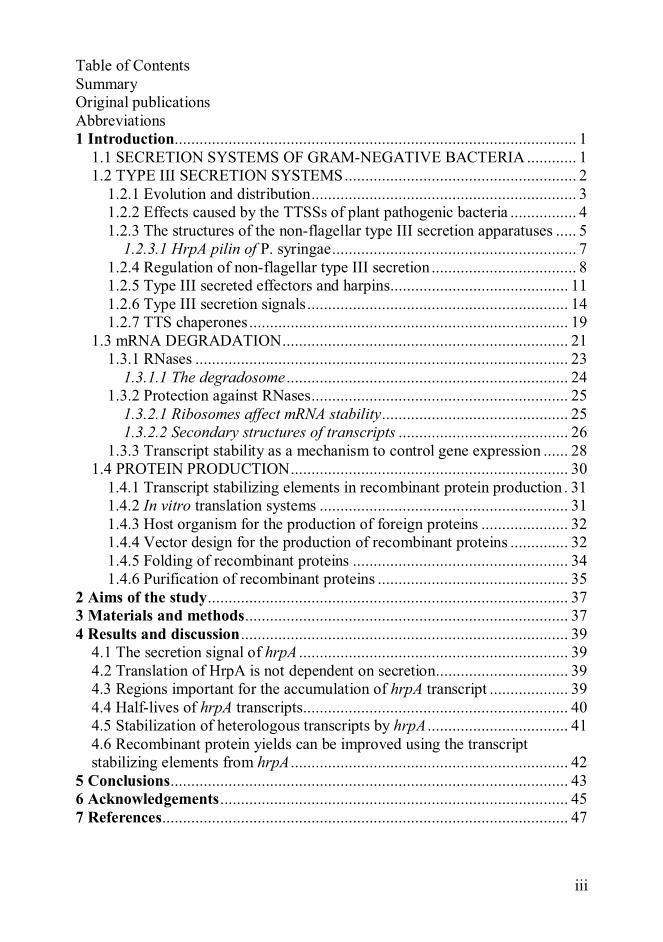
Type III secretion systems (TTSSs) can be divided into two categories, the
flagellar systems and the virulence or non-flagellar systems, the latter including
symbiosis-associated systems. The similarities and differencies between the two
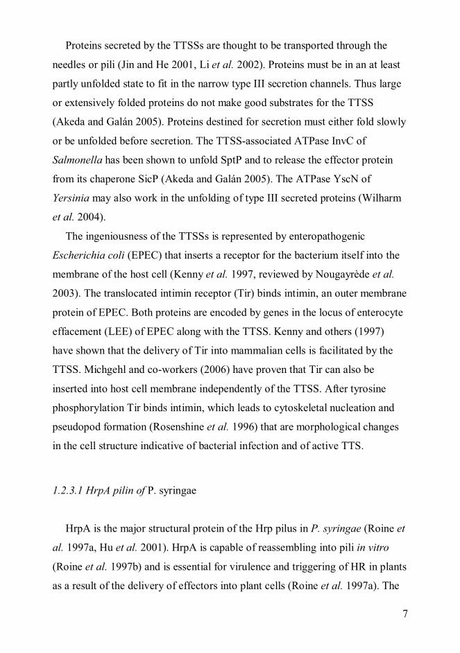
categories are schematically represented in Figure 1.
Figure 1. Schematic drawing of type III secretion systems. Hrp/Hrc denotesproteins of the virulence TTSS of Pseudomonas syringae and Flg/Flh/Fliflagellar proteins. Homologous proteins are separated by a slash and indicatedwith identical patterns. OM= outer membrane, CM= cytoplasmic membrane.Modified from the Masters thesis of Chun-Mei Li (2001) with permission.
Flagellum
OM
CM
FliI/HrcN
FliMN/HrcQ
FliG/HrpQFlgB, C
FliF/HrcJ HrpA
FliD
FlgH
FlgF
FlhA/HrcVFlhB/HrcUFliP/HrcRFliQ/HrcSFliR/HrcTFliH/HrpEFliO
HrcC
Hrp-pilus
FlgLFlgKFlgE
FlgG
FlgI
FliC

3
1.2.1 Evolution and distribution
The flagellar and the non-flagellar secretion systems share a common
ancestor (Foultier et al. 2002, Gophna et al. 2003). The divergence between the
systems may have occurred hundreds of millions of years ago. The order of
appearance of the virulent and the flagellar secretion systems is still unclear.
The flagellar secretion system is readily considered the more ancient one on the
basis that the eukaryotic hosts have evolved later, whereas the ability to move
by the use of flagella would be an older feature. Gophna and co-workers (2003)
did not find support for the claim by Macnab (1999) and Galán and Collmer
(1999) that the non-flagellar TTSS would have evolved from the flagellar
secretion system. They found the levels of diversity to be similar in the non-
flagellar TTSS and the flagellar systems, thus the systems have a similar degree
of antiquity. In addition, they noted the peculiarity of why a simpler non-
flagellar TTSS would have evolved from the more complex flagellar system.
The TTSSs have spread among bacteria by horizontal gene transfer and are
found in distantly related bacterial species (Foultier et al. 2002). TTSSs are
often encoded by genes on pathogenicity islands flanked by mobile elements or
on plasmids and they do not follow the G/C (guanidine/cytosine) content of
their hosts. Some species of bacteria have more than one TTSS. These systems
are a result of successive horizontal gene transfers, not of intragenomic gene
duplications (Troisfontaines and Cornelis 2005). After their acquisition, the
genes encoding the TTSSs have not undergone major reorganisation (Foultier et
al. 2002).
Non-flagellar TTSSs are found in many Gram-negative bacteria pathogenic
to plants, animals, including man and insects, and even amoebas, and in
symbionts. The bacteria include for example species of Aeromonas, Bordetella,
Burkholderia, Chlamydia, Chromobacterium, Citrobacterium, Desulfovibrio,
Edwardsiella, Erwinia, Escherichia, Pantoea, Photorhabdus, Pseudomonas,
Ralstonia, Rhizobium, Salmonella, Shigella, Sodalis, Vibrio, Xanthomonas and
4
Yersinia (reviewed by Galán and Collmer 1999, Pallen et al. 2005,
Troisfontaines and Cornelis 2005).
Type III secretion genes occur also in the symbiotic bacterium, Rhizobium
(Freiberg et al. 1997, Viprey et al. 1998). Viprey and others have shown (1998)
that the non-flagellar TTSS in Rhizobium is expressed later than the Nod genes.
TTSSs are probably needed for nodule initiation, but may not be vital for
nitrogen fixation. Viprey and others (1998) have also demonstrated that type III
secreted proteins affect the formation of nodules and act as host specificity
determinants. Some plant-Rhizobium interactions seem to benefit from the
proteins, whilst others exhibit the exact opposite phenotype.
The flagellar secretion systems have also been reported to function in the
secretion of virulence proteins in many bacteria (Young et al. 1999, Ghelardi et
al. 2002, Konkel et al. 2004). The functional conservation of the TTSS
apparatuses has been experimentally demonstrated by secretion of proteins
originating from one bacterial species by the secretion machinery of another
species. Examples range from the secretion of a virulence protein from Shigella
flexneri by Yersinia pseudotuberculosis, secretion and translocation of an
effector protein of Y. pseudotuberculosis by S. typhimurium (Rosqvist et al.
1995), to the secretion of effectors from Pseudomonas syringae (Ham et al.
1998) and Y. enterocolitica (Anderson et al. 1999) by the TTSS of Erwinia
chrysanthemi expressed in E. coli, and to the secretion of YlpA, a flagellar
TTSS secreted virulence factor of Y. enterocolitica through the two virulence
TTSSs of Yersinia and the flagellar secretion system (Young and Young 2002,
Warren and Young 2005).
1.2.2 Effects caused by the TTSSs of plant pathogenic bacteria
TTSSs are used by pathogenic bacteria to secrete proteins outside the
bacterial cell (e.g. harpins) and to translocate proteins directly into the host cells

5
(effector proteins, formerly known as avirulence proteins in plant pathogens). In
plant pathogenic bacteria the TTSSs are known as Hrp systems because of their
effects on the plant i.e. HR (hypersensitive reaction) and pathogenesis. HR is a
defensive plant reaction, a localized cell death that restricts the spread of the
pathogen. Plant resistance gene (R) products can recognise virulence factors
either directly or indirectly by their actions (the guard hypothesis). According to
the guard hypothesis, the guardian R gene products monitor their guardees that
may also be the targets of the (a)virulence proteins (reviewed by Van Der
Biezen and Jones 1998, Dangl and Jones 2001). After recognition of the
pathogen by the R gene products, several responses will follow. Reactive
oxygen intermediates (ROIs) are produced, HR is induced, systematic defence
signalling will follow and induce pathogenesis related (PR) genes in distant
parts of the plant and make the plant resistant to a wide variety of pathogens. In
experimental infections HR is used as a fast and easy assay for the presence of
bacteria with an active TTSS. Visible cell collapse typically appears within 24
hours of infection (Roine et al. 1997a). The pathogenic functions of effectors
are discussed in section 1.2.5.
1.2.3 The structures of the non-flagellar type III secretion apparatuses
TTSSs share homologous proteins that for example in the animal pathogenic
bacterium Yersinia are called Ysc for Yersinia secretion and in plant pathogenic
bacteria Hrc for hrp conserved. The conserved proteins form the core of the
secretion machinery. Ten of the 11 conserved genes encoding the TTSSs are
also conserved in the flagellar type III system, the exception being HrcC/YscC
(reviewed by Hueck 1998, Cornelis and Van Gijsegem 2000, Büttner and
Bonas 2002). The secretion is mediated by the secretion/translocation
apparatuses that comprise of two rings in the inner and outer membranes, of
long appendages called needles in animal pathogens or pili in the case of the
6
plant pathogenic bacteria and of a translocon that forms a pore in the host cell
membrane. The secretion apparatus is built of conserved proteins such as the
outer membrane protein HrcC, the inner membrane/membrane-spanning protein
HrcJ, the inner membrane proteins HrcR, HrcS, HrcT, HrcU and HrcV, the
cytoplasmic proteins HrpQ, HrcQ, HrpE and the ATPase (adenosine
triphosphatase) HrcN that provides energy for the secretion process in plant
pathogenic bacteria (see figure 1 for locations of the conserved proteins). Some
components of the TTSS are secreted through the general secretion system
(Sukhan et al. 2001, Kimbrough and Miller 2002, Gauthier et al. 2003). These
include the inner membrane and membrane-spanning components and at least
partly the outer membrane proteins of the secretion apparatus. The needle or
pilus proteins as well as components of the translocon are secreted by the
TTSS.
Needles of animal pathogenic bacteria are approximately 40-80 nm in length
and their external diameter is 6 to 13 nm, and internal diameter in the range of 2
nm (reviewed by Ghosh 2004). At the tips of the needles are structures needed
for the assembly of the translocation pore. These structures comprise of LcrV,
YopB and YopD in the case of Yersinia (reviewed by Cornelis 1998, Mueller et
al. 2005) and of EspA in E. coli (Ghosh 2004). The EspA filaments have an
outer diameter of about 12 nm and an inner diameter of 2.5 nm and are in
average 40 to 140 nm in length.
TTSS-associated pili of plant pathogenic bacteria have an external diameter
of ca. 8 nm, and can be several micrometers in length (reviewed by Ghosh
2004). J. Lee and colleagues (2001) demonstrated that the harpin HrpZ from P.
syringae binds lipid bilayers. They also demonstrated that HrpZ can form ion-
conducting pores. They hypothesised that HrpZ may either facilitate nutrient
release or the translocation of effector proteins into eukaryotic cells, thus being
functionally equal to the translocation structures of the animal pathogenic
bacteria. Harpin proteins are further discussed in section 1.2.5.
7
Proteins secreted by the TTSSs are thought to be transported through the
needles or pili (Jin and He 2001, Li et al. 2002). Proteins must be in an at least
partly unfolded state to fit in the narrow type III secretion channels. Thus large
or extensively folded proteins do not make good substrates for the TTSS
(Akeda and Galán 2005). Proteins destined for secretion must either fold slowly
or be unfolded before secretion. The TTSS-associated ATPase InvC of
Salmonella has been shown to unfold SptP and to release the effector protein
from its chaperone SicP (Akeda and Galán 2005). The ATPase YscN of
Yersinia may also work in the unfolding of type III secreted proteins (Wilharm
et al. 2004).
The ingeniousness of the TTSSs is represented by enteropathogenic
Escherichia coli (EPEC) that inserts a receptor for the bacterium itself into the
membrane of the host cell (Kenny et al. 1997, reviewed by Nougayrède et al.
2003). The translocated intimin receptor (Tir) binds intimin, an outer membrane
protein of EPEC. Both proteins are encoded by genes in the locus of enterocyte
effacement (LEE) of EPEC along with the TTSS. Kenny and others (1997)
have shown that the delivery of Tir into mammalian cells is facilitated by the
TTSS. Michgehl and co-workers (2006) have proven that Tir can also be
inserted into host cell membrane independently of the TTSS. After tyrosine
phosphorylation Tir binds intimin, which leads to cytoskeletal nucleation and
pseudopod formation (Rosenshine et al. 1996) that are morphological changes
in the cell structure indicative of bacterial infection and of active TTS.
1.2.3.1 HrpA pilin of P. syringae
HrpA is the major structural protein of the Hrp pilus in P. syringae (Roine et
al. 1997a, Hu et al. 2001). HrpA is capable of reassembling into pili in vitro
(Roine et al. 1997b) and is essential for virulence and triggering of HR in plants
as a result of the delivery of effectors into plant cells (Roine et al. 1997a). The
8
Hrp pilus has been shown to cross the plant cell wall (Brown et al. 2001). The
pili have been shown to both grow and secrete proteins from their tip (Jin and
He 2001, Li et al. 2002).
The pilus and needle subunit proteins are generally small (ca. 60-120 amino
acids) and form mainly -helical structures (Koebnik 2001, Weber et al. 2005,
Zhang et al. 2006). The C-terminus of HrpA is needed for the assembly of the
pili and for pathogenicity (Taira et al. 1999), whereas the N-terminus is not
needed for filament formation (Roine et al. 1997b, Taira et al. 1999). Similar
architecture exists in the Xanthomonas campestris major pilus protein called
HrpE (Weber and Koebnik 2005). The primary sequences of HrpA proteins,
however, are not very similar. The proteins from P. syringae pathovar (pv.)
tomato DC3000 and P. syringae pv. syringae 61, for example, are only 27%
identical and 43% similar (Deng et al. 1998).
1.2.4 Regulation of non-flagellar type III secretion
The TTSSs may be activated upon contact with the host cell or in the
apoplast of plants and thereafter deliver proteins from the bacterial cytoplasm
into the eukaryotic cells. Environmental signals, such as host cell contact, pH,
temperature, carbon source and host cell-derived molecules regulate the
transcription, translation and secretion of TTS-associated proteins in an
elaborate manner. The TTSSs can also be induced experimentally. In plant
pathogenic bacteria minimal medium with a low pH and fructose as the
preferred carbon source is used for the expression and secretion studies of the
Hrp-regulon (Huynh et al. 1989). Examples of regulatory cascades in TTS gene
expression have been found in several bacterial species, but the complete
networks remain to be solved.
The hrp clusters of plant pathogenic bacteria have been classified into two
groups that differ in gene organization and sequence as well as in their hrp gene
9
expression (reviewed by Alfano and Collmer 1996). Group I includes bacteria
such as E. amylovora and P. syringae and group II Ralstonia solanacearum and
X. campestris. The regulatory cascades of hrp gene expression in P. syringae
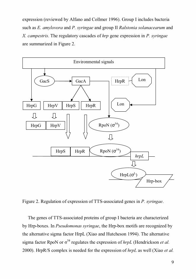
are summarized in Figure 2.
Figure 2. Regulation of expression of TTS-associated genes in P. syringae.
The genes of TTS-associated proteins of group I bacteria are characterized
by Hrp-boxes. In Pseudomonas syringae, the Hrp-box motifs are recognized by
the alternative sigma factor HrpL (Xiao and Hutcheson 1994). The alternative
sigma factor RpoN or 54 regulates the expression of hrpL (Hendrickson et al.
2000). HrpR/S complex is needed for the expression of hrpL as well (Xiao et al.
GacS
Environmental signals
Lon
HrpG HrpV HrpS
HrpRGacA
HrpVHrpG
HrpRHrpS RpoN ( 54)hrpL
LonHrpR
RpoN ( 54)
Hrp-boxHrpL( L)

10
1994). HrpV acts as a negative regulator of hrp gene expression (Preston et al.
1998). HrpG forms a complex with HrpV, which leads to the dissociation of
HrpV from HrpS and the activation of hrp gene expression (Wei et al. 2005). In
conditions mimicking the in planta environment, the HrpR/S complex is active
due to the lack of Lon-mediated degradation of HrpR (Bretz et al. 2002). The
expression of hrpR and hrpS as well as rpoN is controlled by GacA (Chatterjee
et al. 2003). GacA is a response regulator of a two-component system, GacS
being the sensor kinase. According to Chatterjee and colleagues (2003) GacA
regulates several genes in control of various systems in P. syringae pv. tomato.
Wei and colleagues (2000) have suggested that HrpA might also be involved in
the regulation of expression of TTSS. HrpA would act upstream of hrpRS in
regulating the expression of the operon.
Group II bacteria use AraC family transcriptional activators for induction of
hrp gene expression (reviewed by Alfano and Collmer 1996). A non-diffusible
molecule in the plant cell wall is recognised by PrhA and leads to induction of
hrp genes (Aldon et al. 2000).
At the temperature of +37°C an increase in the amount of extracellular
amino acids (glutamate, glutamine, aspartate or asparagine), in serum proteins,
such as albumin, or a decrease in calcium concentration triggers type III
secretion in animal pathogenic bacteria (Lee VT et al. 2001). The temperature
regulation of TTS in Shigella, enteroinvasive E. coli (Falconi et al. 1998) and Y.
enterocolitica (Rohde et al. 1999) involves conformational changes in the
virulence plasmid that encodes the genes of the TTSS.
In Yersinia three classes of genes controlling the expression and/or secretion
of Yersinia outer proteins (Yops) have been identified (reviewed by Anderson
et al. 2002). Mutations in class I genes, such as yopN result in the loss of
calcium regulation of synthesis and secretion, class II genes (yopD and lcrH) in
loss of calcium response of synthesis, and class III genes that are components of
the secretion apparatus, in loss of synthesis under low-calcium conditions.

11
Yersinia employs repressors and post-transcriptional control to regulate its
virulence gene activation. The complexity of regulation is exemplified by the
involvement of a number of proteins including YscM1, YscM2, SycH, YopD
and LcrH (Pettersson et al. 1996, Cambronne et al. 2000, Cambronne and
Schneewind 2002, Cambronne et al. 2004). Host cell contact leads to the TTSS-
dependent secretion of LcrQ/YscM, a negative regulator of Yop expression
(Pettersson et al. 1996, Cambronne et al. 2000). According to Cambronne and
colleagues (2004), secretion of the regulator per se is not required for the relief
of repression of Yops, but the binding of SycH chaperone to regulators of
expression is (Cambronne et al. 2000, Cambronne et al. 2004). The regulation
of Yops in Yersinia may involve degradation of yop messenger ribonucleic
acids (mRNAs) in the absence of secretion (Anderson et al. 2002). Anderson
and others (2002) suggest that the binding of YopD and LcrH to yop mRNA
may prevent the initiation of translation of the transcript. It remains to be seen
whether there is a more common connection between the stability of TTSS-
associated transcripts and type III secretion and if this connection involves an
mRNA secretion signal.
In P. aeruginosa secretion is coupled to transcription by a cascade of ExsE,
ExsC, ExsD and ExsA (Rietsch et al. 2005, Urbanowski et al. 2005). ExsE is
secreted from the cells as the secretion apparatus is opened and the secretion is
aided by ExsC, a chaperone and an anti-anti-activator. After the secretion of
ExsE, ExsC is free to bind ExsD, an anti-activator. The binding of ExsC to
ExsD releases ExsA, a DNA-binding protein, to activate the transcription of the
type III secreted proteins.
1.2.5 Type III secreted effectors and harpins
The components of the TTSS are conserved among bacteria, but the effector
proteins are not (Galán and Collmer 1999). Type III effectors of plant

12
pathogenic bacteria can determine the host range of the pathogen (Alfano and
Collmer 2004). Type III secreted effector proteins in plant pathogens were first
named avirulence proteins since they were discovered for their ability to elicit
plant defences, thus avirulence. Later it was noted that the proteins can act as
pathogenicity factors in some plants, whilst other plants have matching R genes
that recognize the effector proteins and launch defences. The effects caused on
plants by the TTSS-dependent proteins are discussed from the view of the plant
in section 1.2.2.
Type III secreted proteins help the pathogen to grow in host, to defeat its
defences and to cause disease symptoms (Alfano and Collmer 2004). Type III
secreted effectors have more sequence homology to eukaryotic than to bacterial
proteins (Cornelis 2002) and may have been acquired from eukaryotes (Galán
2001, Cornelis 2002, Troisfontaines and Cornelis 2005).
Little has been known about the cellular functions of the type III effectors in
plant pathogenic bacteria, but more information is been discovered at an
increasing rate. The effector proteins often act on the plasma membrane or the
nucleus of plant cells (Alfano and Colmer 2004). The effector proteins are
believed to stimulate or interfere with host cellular processes. They may affect
the host cell morphology or metabolisms and favour the pathogen by
suppressing the defences of the plant. The effectors may suppress programmed
cell death, jasmonic acid and salicylic acid signalling, the expression of defence
genes and cell wall-based defences, such as cell wall thickening papillae
(Hauck et al. 2003, Abramovitch and Martin 2004, Mudgett 2005, Li et al.
2005).
P. syringae pv. tomato DC3000, for example, has more than 30 effector
proteins (Guttman et al. 2002, Buell et al. 2003). Effector proteins of P.
syringae pv. tomato for example suppress the salicylic acid dependent callose
deposition (HopPtoM and AvrE) (DebRoy et al. 2004), target host immunity
associated proteins to the host proteasome/ubiquitination system to suppress the
extracellular cell wall-associated defenses (HopM1) (Nomura et al. 2006),

13
inhibit programmed cell death by mimicking host ubiquitin ligases (AvrPtoB)
(Janjusevic et al. 2006) and suppress early-defense signalling (AvrPto and
AvrPtoB) (He et al. 2006).
The genes induced by the TTSSs in plants are also associated with defence,
such as the salicylic acid dependent pathway (Hauck et al. 2003). These genes
are probably not induced to high enough levels to be effective against infection
in susceptible plants.
Effects similar to those executed by the effectors of plant pathogens occur
with the animal-targeting effectors: the inflammatory response is
downregulated, phagocytosis is inhibited, apoptosis is induced in macrophages
and lymphocyte activation is impaired (reviewed by Cornelis 2002).
Known and predicted functions of effector proteins include phosphatases,
kinases, ADP-ribosyltransferases, adenylate cyclases, proteases,
phosphodiesterases, syringolide synthases and transcription factors (reviewed
by Cornelis and Van Gijsegem 2000, Innes 2003, Grant et al. 2006). According
to Guttman and colleagues (2002) effectors have a high overall serine and
asparagine content, and low leucine, isoleucine and valine content. They also
have a low aspartate and lysine content in their N-termini. These observations
are in line with those of Petnicki-Ocwieja and others (2002) who analysed by
computer the N-termini of type III secreted proteins from P. syringae (see also
section 1.2.7 on type III secretion signals). According to Guttman and
colleagues (2002) the N-termini of type III secreted effectors resemble
chloroplast and mitochondrial targeting sequences. Many P. syringae effectors
are believed to localize in the chloroplasts, and some animal pathogens target
their effectors to mitochondria. The significance of this finding, whether it
speaks for the common evolutionary origin for the secretion and targeting
mechanisms or for analogous functional requirements or for something else,
remains elusive (Guttman et al. 2002).
Some effectors are modified inside host cells by host enzymes.
Modifications may be needed for the virulence and avirulence functions of the

14
effectors. Effectors may have evolved to mimic host cell proteins that are
modified. AvrPto of P. syringae is both myristoylated (Shan et al. 2000,
Anderson et al. 2006) and phosphorylated (Anderson et al. 2006) in tobacco
and tomato leaves, and these modifications affect the outcome of the interaction
between plants and bacteria.
Harpins are glycine-rich proteins that lack cystein residues, are heat-stable
and can elicit HR when injected into non-host plants (He et al. 1993, Alfano
and Collmer 1996). They may facilitate the delivery of effector proteins into the
plant cell cytoplasm (J. Lee et al. 2001) as noted in section 1.2.3 on the
structures of the non-flagellar type III secretion apparatuses.
1.2.6 Type III secretion signals
Despite extensive research, the signal for type III secretion is still enigmatic.
In the non-flagellar secretion systems, the signals found so far lie in the region
covering the first 7 to 28 codons (for examples see Sory et al. 1995, Anderson
and Schneewind 1999, Mudgett et al. 2000, Rüssmann et al. 2002, Ramamurthi
and Schneewind 2005). Both N-terminal amino acid signals, perhaps
amphipathic ones (Lloyd et al. 2001), and 5’ terminal mRNA signals (Anderson
and Schneewind 1997, Anderson et al. 1999, Mudgett et al. 2000) have been
proposed.
Aldridge and Hughes (2001) have proposed several models for the secretion
of TTSS substrates. The secretion signal could be an mRNA signal, leading to
co-translational secretion, an N-terminal signal, or assisting secretion
chaperones could be used. Their models are mostly based on flagellar TTSS,
but may be applicable to the non-flagellar TTSS as well. They propose that the
secretion of late-substrates, such as effectors for the non-flagellar TTSS, could
be hindered by ribosomes translating the structural components of the
machinery in a co-translational manner, until the secretion machinery is ready
15
(the closed gate model). In this model secretion could still be assisted by
chaperones associated with the growing polypeptide near the secretion
machinery. After completion of the TTS machinery, the ribosome gate would
open and allow the secretion of the effector proteins. In the open gate model, on
completion of the TTS machinery, the secretion chaperones sense the secretion
substrates flowing to the cytoplasm from the full secretion channel, thus
inducing the shutdown of secretion or a switch between substrates.
Evidence for the mRNA secretion signal has come mostly from experiments
illustrating that point mutations or some frame-shift mutations that completely
alter the amino acid sequence of the secretion signals of YopE, YopN or YopQ
of Y. enterocolitica do not prevent secretion of reporter proteins (Anderson and
Schneewind 1997, Anderson and Schneewind 1999). Goss and colleagues
(2004) demonstrate that some synonymous mutations in YopN abolish the
secretion of hybrid proteins. Ramamurthi and Schneewind (2005) could show
that a single synonymous mutation almost completely abolished the secretion of
a reporter protein fused to the minimal secretion signal of YopE from Y.
enterocolitica, whereas changing the reading frame of the secretion signal being
comprised of the first 15 codons (minimal secretion signal and the suppressor
region) did not abolish reporter protein secretion. Synonymous mutations were
also made to the secretion signal of YopQ by Ramamurthi and Schneewind
(2002). Some of the mutations abolished secretion of the reporter protein,
whilst others did not. Frameshifts in the secretion signal of InvJ in Salmonella
did not abolish secretion (Rüssmann et al. 2002). However, synonymous
mutations changing the mRNA sequence of invJ did not hinder secretion either.
Lee and Schneewind (2002) opted for the mRNA signal based on the notion
that the type III pathway of Y. enterocolitica cannot be occluded by folded
proteins with TTS signals. They reasoned that the completed polypeptides
rejected by the TTSS cannot re-enter the pathway because they no longer are
attached to mRNA signals. The rejection of folded polypeptides from TTSS
was also demonstrated by Sorg and colleagues (2005). They also proved that

16
some impassable substrates can inhibit the expression of other substrates of
TTSSs.
The N-terminal secretion signal is supported by Warren and Young (2005)
who made a frameshift mutation in the secretion signal of YlpA of Y.
enterocolitica. The frameshift resulted in poor secretion of YlpA. Lloyd and
colleagues (2001) made frame-shift mutations to the first 11 codons of yopE of
Y. pseudotubercolis. Their mutations that changed the amino acid sequence of
the protein drastically reduced the secretion of YopE in a chaperone-deficient
yerA- background, whereas mutations altering the mRNA sequence while
leaving the amino acid sequence intact allowed secretion. Ramamurthi and
Schneewind (2003a) noted that the construct made by Lloyd and others (2001)
mutated codons 12 and 13 that are a part of the suppressor region sensitive to
mutations.
Some of the differences in the results of several groups may be attributed to
the discrepancy in the behaviour of different TTS substrates, whilst others may
be the result of the use of differing constructs. Ramamurthi and Schneewind
(2002, 2003a, 2003b, 2005) studied the effect of the length of the secretion
signal to tolerance of frame-shift mutations. Their observations illustrate that
even though the minimal secretion signal of YopQ of Y. enterocolitica is in the
first 10 codons, codons 11-15 help in tolerance for mutations. They call this
tolerance region the suppressor region, and show it to be sensitive to
mutagenesis (2003a). The same was proven to be true for YopE (2005). The
first 7 codons are sufficient for secretion, but codons 8-15 can suppress
mutations in the minimal secretion signal.
Rüssmann and colleagues (2002) hypothesised that instead of an mRNA
secretion signal, secretion of TTSS-dependent proteins could be accomplished
by the use of polypeptide sequences that do not acquire structures rapidly.
Lloyd and colleagues (2001) noted the amphipathic nature of the N-termini of
Yops, and created a functional synthetic, amphipathic serine/isoleucine
secretion signal for YopE. Petnicki-Ocwieja and others (2002) analysed by

17
computer the N-termini of type III secreted proteins from P. syringae. They
defined the following rules: the first five amino acids include solvent exposed,
equivalent amino acids, no acidic amino acids (aspartate or glutamate) reside in
the first 12 amino acids and the first 50 amino acids are in general rich in polar
amino acids (especially serine and glutamine) and amphipathic.
Some TTS substrates only seem to have one secretion signal. These proteins
include parts of the secretion machinery (Anderson and Schneewind 1999).
Other secreted proteins that are often translocated into host cells have a second
secretion signal located further downstream of the first signal (Sory et al. 1995,
Schesser et al. 1996, Cheng et al. 1997, Mudgett et al. 2000, Chiu and Syu
2005). The second signal comprises of a binding site for small, cytoplasmic
proteins known as TTS chaperones (Anderson and Schneewind 1999). The TTS
chaperones are described in more detail in the following section (1.2.7). The N-
terminal secretion signal has been referred to as the primary secretion signal
and the chaperone binding domain (CBD) as the translocation domain. Some
experiments argue against the CBD acting as a translocation signal. A Y.
enterocolitica strain lacking most effectors delivered YopE with its CBD
deleted into eukaryotic cells (Boyd et al. 2000). No secretion or translocation
could be detected without the primary 5’ secretion signal. Conflicting evidence
has been published by Cheng and colleagues (1997), who have demonstrated
that a fusion protein of NPT with YopE in Y. enterocolitica was secreted
without the N-terminal secretion signal.
Co-translational secretion has been proposed for some of the TTS substrates
of both the flagellar and non-flagellar types (Karlinsey et al. 2000, Anderson
and Schneewind 1999). As an extreme example, Anderson and Schneewind
(1999) showed that YopQ is only translated when a functional TTSS is present.
The results of Anderson and Schneewind (1999) showing that YopQ was only
present in the culture medium and not found in the cytosol of Y. enterocolitica
are in conflict with the results of Tr ek and others (2002) who could detect
YopQ in the cytosol of Y. enterocolitica and show post-translational secretion.

18
Coupling of translation to secretion might be mediated by type III chaperones
(Karlinsey et al. 2000). In contrast, some TTSS-dependent proteins have been
shown to be secreted from a pre-made pool. For example, translation of YopE
is not coupled to its secretion in Y. pseudotuberculosis (Lloyd et al. 2001). Post-
translational secretion in this case is dependent on chaperones. The same seems
true for YopE of Y. enterocolitica (Cheng et al. 1997).
No conclusive proof on the secretion signal for TTS has been provided that
would account for the secretion of all the known type III secreted proteins. Both
the nucleotide and polypeptide sequences of TTS-dependent proteins vary
greatly. Sequence or secondary structure data unambiguously proving the
existence of a general mRNA signal is lacking. No unequivocal evidence exist
that would establish how an amphipathic, unstructured polypeptide signal that
seems to exist only in a portion of TTS proteins could exclude the secretion of
all non-TTS proteins, whilst promoting the secretion of all TTS proteins either.
The secretion signal for the flagellar TTSS also needs further investigations.
Flagellar export chaperones interact with the C-termini of their substrates,
unlike their virulence system counterparts (Evdokimov et al. 2003). FliS, the
chaperone of the flagellar filament protein FliC seems to prevent the premature
polymerization of FliC in the cytosol. According to Evdokimov and others
(2003) the FliS-related flagellar type III chaperones share no common
evolutionary ancestry with the non-flagellar type III secretion chaperones.
Majander and co-workers (2005) used the 173 bp untranslated region upstream
of the fliC gene of E. coli to secrete heterologous proteins through a modified
flagellar TTSS. They were also able to secrete heterologous proteins fused to
FliC without its 5’ UTR. They concluded that either the 5’ UTR of fliC or other
regions, perhaps with the help of chaperones are needed for the secretion
through the flagellar TTSS. In S. typhimurium the TTS signal of the flagellin is
in amino acids 26 to 47 that are sufficient for the export of polypeptides fused
to them (Végh et al. 2006). These residues are among the most conserved of the

19
disordered N-terminal region of flagellins. They are hypothesised to form
amphipathic helical structures.
1.2.7 TTS chaperones
TTS chaperones are small (ca. 15 kDa) proteins that have an acidic pI and an
amphipathic alpha-helix in their C-termini (Alfano and Collmer 2004). They
often act as dimers and are encoded adjacent to their cognate effector proteins
(reviewed by Feldman and Cornelis 2003). Chaperones are likely to have
evolved from common ancestral proteins (Birtalan et al. 2002).
TTS chaperones have been mostly studied in animal pathogenic species but
exist also in plant pathogens. Many roles have been assigned to type III
chaperones (reviewed by Feldman and Cornelis 2003, Ghosh 2004). They may
act as anti-aggregation and -folding factors. In complex with effector proteins
type III chaperones may, at least in some cases, form three-dimensional signals
recognized by the TTSS (Birtalan et al. 2002). A hierarchy of secretion could
be introduced by chaperones to the effectors (Boyd et al. 2000, Birtalan et al.
2002). Protein fusions containing only the N-terminal secretion signal are
unable to compete with effector proteins harboring both the N-terminal
secretion signal and the chaperone binding site (Boyd et al. 2000). Hierarchy is
not conferred on the stage of transcription, at least in the case of P. syringae pv.
phaseolicola (Thwaites et al. 2004).
TTS chaperones have been shown to act only on the domains they bind to
(Birtalan et al. 2002). Results of Birtalan and co-workers (2002) on Y.
pseudotuberculosis SycE-YopE complex and of Luo and colleagues (2001) on
Salmonella SigD-SigE and E. coli Tir-CesT complexes exhibit that chaperones
do not promote global unfolding of these effectors. According to the results of
Birtalan and colleagues (2002) chaperones do not protect effectors against
proteolysis. On the contrary, Losada and Hutcheson (2005) have demonstrated

20
that chaperones of P. syringae do protect their cognate effectors against Lon-
mediated degradation. The chaperones may keep their cognate effectors in an
unfolded or non-globular state that is competent for secretion through the TTSS
(Stebbins and Galán 2001).
Boyd and co-workers (2000) have studied the CBD of YopE of Y.
enterocolitica. They showed that the removal of YopE residues binding to SycE
downstream of the minimal CBD (amino acids 15 to 50) leads to the mutant
protein being secreted by Y. enterocolitica independently of SycE. They
concluded that amino acids 50 to 77 inhibit secretion of YopE in the absence of
its chaperone. Ehrbar and colleagues (2006) also hypothesise that an inhibitory
factor would bind the CBD of newly synthesised proteins and prohibit transport
via the TTSSs. The binding of the cognate chaperone would release this
inhibitory factor. The work of Letzelter and colleagues (2006) on the effector
protein YopE of Y. enterocolitica has verified that a deletion of the CBD
(residues 20 to 77) of YopE does abolish the need for its cognate chaperone for
secretion and translocation. They have, however, concluded that the CBD
creates the need for the chaperone by reducing the solubility of the effector
protein.The CBDs may act as the membrane localization domains (MLD) of
effectors and the chaperones can prevent their insolubility in the bacterial
cytoplasm (Letzelter et al. 2006). SycO of Y. enterocolitica binds the MLD of
YopO and SycE covers the MLD of YopE. Letzelter and colleagues (2006)
hypothesize that the primary function of type III chaperones could be to cover
the MLDs of membrane-associated effector proteins inside the bacteria and that
the function of targeting the proteins to the secretion machinery would have
evolved later.
The role of the CBD in secretion pathway specificity of type III secreted
proteins has been studied by Lee and Galán (2004) and Ehrbar and colleagues
(2006). They both share the view of the importance of the CBD, but the details
vary. Lee and Galán (2004) believe that the chaperones confer secretion-
pathway specificity, whereas according to Ehrbar and others (2006) the CBD

21
prevents secretion in the absence of the chaperone. Birtalan and colleagues
(2002) believe that the CBDs may not act as inhibitors of secretion in the
absence of their cognate chaperones, but the inhibition may be a by-product of
being aggregation-prone regions. The results of Letzelter and colleagues (2006)
on effectors of Y. enterocolitica show that the chaperones may indeed mask the
aggregation-prone MLDs. Lee and Galán (2004) demonstrated that SopE was
only secreted through the flagellar TTSS in the absence of the CBD. This was
interpreted as a sign of an ancestral flagellar secretion signal, actions of which
can be masked by the CBD and its chaperone. Changing the CBD of SopE from
Salmonella led to its secretion via the flagellar TTSS as well as the SPI-1 TTSS
in the experiments performed by Ehrbar and colleagues (2006). Without its
cognate chaperone wild type SopE is not secreted at all, whilst the chaperone
binding site mutant is secreted by both TTSSs (Ehrbar et al. 2006).
Type III chaperones may regulate the expression of some TTS-associated
genes. An interaction between the SicA TTS chaperone and the transcriptional
activator InvF was shown by Darwin and Miller (2001). They demonstrated that
both proteins are needed for the activation of some TTS promoters of proteins
needed for invasion in S. typhimurium. They also suggest a model in which
SicA could dock the transcription and translation machineries near the TTSS
and thus couple translation to secretion.
1.3 mRNA DEGRADATION
Typical mRNA half-lives in E. coli range from 3 to 8 min. (Bernstein et al.
2002). In Bacillus subtilis, the half-lives of 80 % of mRNAs are less than 7 min.
(Hambraeus et al. 2003). According to Bernstein and colleagues (2002) factors
such as UTR length, G/C content or codon composition, predicted secondary
structure stabilities, degree of single-strandedness, or the frequency of RNase E
cleavage sites could not be used to predict the stability of transcripts. The

22
transcripts of genes with similar functions were, however, found to have similar
stabilities. No correlation was found between the stable mRNAs in E. coli
versus their counterparts in B. subtilis (Hambraeus et al. 2003). Similar to the
results of Bernstein and colleagues (2002), Hambraeus and colleagues (2003)
found no structures predominant in the 5’ UTRs of stable or unstable Bacillus
transcripts. The stability of the interaction between the ribosome binding site
(RBS) and the ribosome was not different between the stable and unstable
transcripts either. Similar results have also been obtained from a eukaryote,
Saccharomyces cerevisiae (Wang et al. 2002). Transcripts encoding subunits of
multicomponent, stoichiometric complexes had similar decay rates. Transcript
half-lives did not correlate with ribosome density, or with ORF (open reading
frame) size or codon bias.
Three main theories exist on the functional degradation of mRNAs. They
have been tested experimentally and using mechanistic modelling (Carrier and
Kealing 1997b, c). Carrier and Keasling (1997b) used a model that takes into
account the binding of RNA polymerase at the promoter, transcription
elongation and termination, ribosome binding at the RBS, translation elongation
and termination, as well as protein degradation. According to the so-called 5’
binding theory, the ribonuclease (Rnase) will bind to the free 5’ end of the
transcript and move along the mRNA behind the ribosomes until it reaches the
cleavage site. This theory failed to predict the effects of ribosome loading and
translational rate on transcript stability. The ribosome binding theory assumes
that the RNase can bind to any site in the transcript that is not covered by a
ribosome. This theory was not able to predict the 5’ to 3’ direction of
degradation of mRNAs. Carrier and Keasling (1997b) found the hybrid 5’
binding/ribosome protection theory of mRNA degradation to be superior to the
other two theories. In the hybrid theory the nuclease binds to the 5’ end of the
transcript and loops to a cleavage site. The RNase will cleave at the site if a
protecting ribosome is not present.
23
1.3.1 RNases
E. coli has at least five endoribonucleases: RNase III, RNase E, RNase G
and RNase I/M (reviewed by Kushner 2002). There may be some functional
overlap between RNases E and G. RNase I/M is found in the periplasm.
Exonucleases include RNase II, RNase R, RNase BN, RNase PH, RNase D,
RNase T and polynucleotide phosphorylase (PNPase) (reviewed by Kushner
2002). PNPase and RNase II as well as PNPase and RNase R exhibit some
functional redundancy. In addition, there is an oligoribonuclease that degrades
the short 4- to 7-mers created by PNPase and RNase II. Other proteins involved
in mRNA decay include RNA helicases, poly(A) binding proteins and auxiliary
proteins, such as Hfq (reviewed by Kushner 2004).
The site of the first endonucleolytic cleavage of a transcript is not random
(Belasco et al. 1986). mRNA degradation is initiated by a cleavage by RNase E
(Carrier and Keasling 1997b). The structure of the catalytic domain of RNase E
has been solved and it resembles partly a deoxyribonuclease (DNase)
(Callaghan et al. 2005). It has a 5’ sensing site in addition to the catalytic
cleavage site. The binding of the 5’ end of the mRNA to the enzyme changes its
conformation, and induces the cleavage reaction (Callaghan et al. 2005).
RNase III cleaves double-stranded ribonucleic acid (RNA) molecules either
with a single-stranded or a double-stranded break (Ehretsmann et al. 1992,
Grunberg-Manago 1999). In bacteria, mRNA degradation is 5’ to 3’ directional
(von Gabain et al. 1983, Selinger et al. 2003). Exonucleases RNase II and
PNPase degrade mRNA in a 3’ to 5’ direction. Degradation by RNase II ends in
5’ monophosphates and by PNPase in nucleoside diphosphates (Ehretsmann et
al. 1992, Grunberg-Manago 1999). PNPase can also synthetise polynucleotide
tails to the ends of mRNAs (Mohanty et al. 2004). Relatively strong secondary
structures can act as barriers to exonucleases but can be overcome by
oligoadenylation (Coburn and Mackie 1996). Poly-adenylation may help in the
degradation of mRNAs that have Rho-independent termination creating stem-

24
loop structures that inhibit RNase II and PNPase (Kushner 2002, 2004).
Poly(A) polymerase (PAPI) adds about 10 to 60 nucleotides to the 3’ end of the
mRNA and provides single-stranded tails for PNPase (O’Hara et al. 1995,
Grunberg-Manago 1999). Efficient polyadenylation by PAPI of mRNAs with
Rho-independent transcription terminators requires Hfq, an RNA-binding
protein (Mohanty et al. 2004). The partial elimination of polyadenylation
stabilizes some mRNAs and alters their degradation patterns (O’Hara et al.
1995).
1.3.1.1 The degradosome
The degradosome has endo- (RNase E) and exonuclease (PNPase) as well as
helicase activities (RhlB helicase) (reviewed by Grunberg-Manago 1999,
Rauhut and Klug 1999). The activity of the helicase is ATP (adenosine
triphospate)-dependent (Py et al. 1996). The role of enolase that is a glycolytic
enzyme in the degradosome is not clear yet, but it might be structural not
functional (Py et al. 1996, Grunberg-Manago 1999). Other proteins found to be
associated with the degradosome include DnaK, a chaperone, and
polyphosphate kinase (PPK) that removes inhibitory polyphosphate and
nucleotide diphosphates (NDPs) and regenerates ATP (Blum et al. 1997). Blum
and others (1997) have also shown that PPK binds RNA. GroEL, a chaperone
and a member of the heat shock protein family, may also be associated with the
degradosome, although this association is still elusive (Sohlberg et al. 1993).
The degradosome is formed on an RNase E scaffold and it interacts with
PAPI. Removal of the C-terminus of RNase E does not impair cell growth, but
does affect mRNA degradation and degradosome foundation (Lopez et al.
1999). Lopez and others (1999) speculate that RNase E and PNPase act for the
most part independently and that mRNA degradation does not need to proceed
fast. The advantages of having a ribonuclease complex might include the
25
elimination of the need for a free PNPase molecule to find the newly formed 3’
end of the mRNA (Grunberg-Manago 1999).
1.3.2 Protection against RNases
1.3.2.1 Ribosomes affect mRNA stability
Experimental data exist on the protecting/stabilizing effect of ribosomes on
mRNAs. Ribosomes have been proven to interfere with RNase E nuclease
activity (Braun et al. 1998, Vytvytska et al. 2000). However, it has been noted
that not all untranslated regions are unstable (von Gabain et al. 1983).
Puromycin strips transcripts of ribosomes (Odom et al. 1990), whereas
aminoglycosides, such as kanamycin (Hirokawa et al. 2002) and
chloramphenicol (Pato et al. 1973) are inhibitory to the release of mRNA from
ribosomes. According to Pato and co-workers (1973) 80-90 % of mRNAs are
stabilized by chloramphenicol, and puromycin destabilizes the transcripts to
half their original stability. They also demonstrate that the portions of mRNA
chains that are synthesised after the addition of chloramphenicol degrade faster
than the portions that are already protected by ribosomes.
The stabilizing effect of some antibiotics can be due to the titration of
RNases by the increase of ribosomal ribonucleic acid (rRNA) synthesis
following the translational block (Lopez et al. 1998). lacZ mRNA lacking a
RBS, for example, was stabilized by all tested translation inhibitors. The
changes of these antibiotics on the stability of the translated mRNAs may be
due to additive or antagonistic effects of ribosome stalling or stripping,
respectively, and titration of RNases (Lopez et al. 1998). The trans-effects
(titration) seem to overcome the cis effects (absence of stabilizing ribosomes) in
long-term. It is also possible that the degradosome is inhibited by translation
inhibitors. One of its components may be unstable, thus requiring ongoing

26
synthesis and replacement or the antibiotics may change the structure of the
complex to inhibit its actions or hamper its access to mRNAs (Lopez et al.
1998).
1.3.2.2 Secondary structures of transcripts
Some structures, notably hairpins, in the UTRs of transcripts can stabilize
mRNAs. 5’ hairpins are believed to protect the mRNAs against RNases,
especially RNase E that requires a free 5’ end for beginning the degradation of
the transcript. The 5’ stabilizing hairpins prevent the binding and thus action of
RNase E. Hairpins and other paired regions that can protect transcripts against
RNases are conserved through evolution (James et al. 1989, Chen et al. 1991).
Thus a hypothetical secondary structure that can only be found in the transcript
from one species is probably not existent in vivo (Chen et al. 1991). The
sequences per se may not be conserved, but changes in one side of the paired
region result in complementary changes in the binding bases. As an example,
the 5’ UTRs of ompA transcripts from several species are sequencially
divergent, but they fold in a similar fashion into two imperfect stem-loops
(Chen et al. 1991).
Heterologous transcripts can be stabilized by the use of naturally existing or
man-made elements. Often hairpin structures or stem-loops are used in the ends
of the transcript. Typically, 3- to 5-fold elevations in half-lives are seen with the
use of stabilizing elements (Wong and Chang 1986, Chen et al. 1991, Carrier
and Keasling 1997a, Carrier et al. 1998). The 5’ stabilizing region of ompA and
the 3’ region of B. thuringiensis cry gene, for example, elevated the half-lives
of a fragment of the bla transcript from approximately three to 15 min (Belasco
et al. 1986, Chen et al. 1991) and of the penicillinase gene from B.
licheniformis from two to six min. (Wong and Chang 1986), respectively.
According to the experiments of Belasco and co-workers (1986), the stabilizing

27
effect of ompA was only seen when using translational fusion constructs.
Inserting a stop codon between ompA and the gene coding for the recombinant
partner destabilized the transcript.
Although 5’ hairpins can stabilize mRNAs there is no uniform correlation
between secondary structure folding energy and mRNA half-lives (Carrier and
Keasling 1999). RNase III is known to degrade double-stranded regions, and is
probably responsible for the poor stabilizing effect of some hairpin structures.
Carrier and Keasling (1999) have also confirmed that unpaired bases in the 5’
end of the transcript destabilize it, even if a hairpin structure is present.
Also 3’ elements can be used to stabilize heterologous mRNAs. The cry
terminator fragment containing an inverted repeat forming a hypothetical stem-
loop structure was used to stabilize transcripts fused to it both in E. coli and in
B. subtilis (Wong and Chang 1986). Engineered 3’ hairpins have also been
demonstrated to stabilize transcripts (Smolke et al. 2000, Smolke and Keasling
2002).
Although RNA secondary structures in the 3’ regions of transcripts are
sufficient to protect the mRNAs against PNPase and RNase II in vitro,
additional factors may be needed for the stabilizing effect they confer in vivo
(McLaren et al. 1991). In the experiments of McLaren and colleagues (1991),
the exoribonuclease stalling effect of different stem-loop structures was only a
few minutes in vitro.
In some cases, also the sequence of the loop in a hairpin structure has been
proven to be of importance (Tuerk et al. 1988). The sequence UUCG stabilized
hairpin structures. Hairpin loop sequences may be recognized by some proteins.
Sometimes the elements used do not form stable secondary structures
themselves, but need trans-acting elements for the creation of the stabilizing
structures (Agaisse and Lereclus 1996). The STAB-SD elements that are Shine-
Dalgarno (SD) sequences found in the 5’ UTRs of many Gram-positive bacteria
can confer stability to sequences downstream of them (Agaisse and Lereclus
1996). These SD sequences do not act in translation initiation. No secondary

28
structures form in these regions according to computer analysis, but the 3’ end
of the 16S rRNA binds to these sequences. This interaction is likely to block
access of RNases to these sequences.
1.3.3 Transcript stability as a mechanism to control gene expression
Transcript stability is also dependent upon growth conditions, oxygen
availability and temperature changes, and the efficiency of translation
(reviewed by Grunberg-Manago 1999). Ribosomes and polymerases may
protect transcripts against RNases. Transcript stability is also influenced by
growth rate and the occurrence of rare codons. The removal of rare codons can
have either a positive or a negative effect on mRNA stability. It may uncover
regions containing cleavage sites, or help the ribosomes cover the mRNA
faster, thus masking it from RNases (Carrier and Keasling 1997c).
The labile nature of most mRNAs reflects the fact that transcript instability is
an effective way to adapt to rapid changes in the environment (reviewed by
Ehretsmann et al. 1992). In the case of polycistronic mRNAs the expression of
proteins can be controlled on the level of stability of the different mRNA
segments.
Transcript stability can be a mechanism for the cells to control the
expression of a gene. By alternating the degradation speed of a transcript, the
cell can respond to specific conditions. In some cases the half-life of a transcript
varies according to the growth-rate of the cells (Nilsson et al. 1984). The least
stable of the ompA mRNA fragments has a half-life of 15 min. at a doubling
rate of one per 40 min., but only 4 min. when the cells are dividing once in 200
min. This ensures that equal amounts of protein are present in the cells at all
growth rates. The same kind of reduction in half-life was seen with the cat
transcripts, from 2 min. to 0.4 min (Nilsson et al. 1984). The stability of the
ompA transcript is controlled by a complicated system, parts of which still
29
remain unsolved. The dependence of the half-life of the ompA transcript on the
growth-rate of the cell is mediated by host factor I, Hfq (Vytvytska et al. 1998).
Hfq has been shown to bind small RNAs around its central pore (Schumacher et
al. 2002). Hfq can unwind secondary structures of RNAs hence destabilizing
surrounding RNA structures and permitting new RNA-RNA interactions
(Schumacher et al. 2002). Hfq binds to the 5’ UTR of ompA. According to
Vytvytska and colleagues (1998), the amount of Hfq is dependent on growth-
rate, being the highest in slowly growing cells. The binding-site of Hfq
coincides with one of the RNase E cleavage sites in the 5’ UTR of ompA
transcript (Moll et al. 2003). Still, binding of Hfq destabilizes ompA mRNA
since it hinders 30S ribosomal subunits from binding to the 5’ UTR of ompA
and stabilizing it (Vytvytska et al. 2000, Moll et al. 2003). The growth-rate
dependent regulation of Hfq may be further mediated by some small RNA or
component (Rasmussen et al. 2005).
A small regulatory RNA also binds to the translational initiation region of
ompA mRNA (Rasmussen et al. 2005, Udekwu et al. 2005). According to
Rasmussen and colleagues (2005) the growth-phase regulation of ompA mRNA
is different from the growth-rate regulation. They state that the antisense
regulator RNA MicA (SraD) accumulates in the stationary phase and is a
growth-phase dependent regulator. Hfq facilitates the binding of MicA to ompA
mRNA (Rasmussen et al. 2005, Udekwu et al. 2005) and strains lacking Hfq
have less stable MicA RNA (Rasmussen et al. 2005). MicA interferes with
ribosome binding (Udekwu et al. 2005). MicA has been found in several
bacteria, and the differences in their sequences are often located in single-
stranded regions or have compensatory changes in the stem-regions (Udekwu et
al. 2005). The regions complementary to ompA are highly conserved.

30
1.4 PROTEIN PRODUCTION
Industrially important proteins are produced in large quantities in
microorganisms. Problems of protein production in vivo include plasmid loss,
especially with high copy number plasmids and toxic or growth rate reducing
proteins (reviewed by Baneyx 1999). Figure 3 illustrates the critical points in
the design of recombinant protein production from vector design to production
and purification of the heterologous protein product.
Figure 3. Steps to be considered in recombinant protein production: vectordesign, scale-up and purification in an active form. R= regulator element, P=promoter, SD= Shine-Dalgarno sequence, SE= stabilizing element, Tag=polypeptide motif that may help in the purification or improve the solubility ofthe recombinant protein (optional), ORF= open reading frame (protein to beproduced), T= transcriptional terminator/stabilizing element, A= antibioticresistance marker and Ori= origin of replication.
Fermentation
R SDP ORF T A OriSE Tag Tag
Small scale production
Secretion
Re-folding
Inclusion bodies Soluble form

31
1.4.1 Transcript stabilizing elements in recombinant protein production
Transcript stability can play a role in recombinant protein production if the
amount of mRNA is the limiting factor. The use of transcript stabilizing
elements may lead to corresponding elevations in both mRNA stability and
protein synthesis (Wong and Chang 1986, Carrier and Keasling 1997a, Carrier
et al. 1998). The strains with plasmids coding for stabilized transcripts
produced in these experiments 2- to 5.3-fold higher amounts of recombinant
proteins than the control strains. The effect of the stabilizing elements on
recombinant protein yields was greater with low inducer concentrations
(Smolke et al. 2000). mRNA stabilizing elements burden the cells metabolism,
since precursors or machinery may become limiting in the synthesis of cellular
components (Carrier et al. 1998). mRNA stabilizing elements for recombinant
protein production are on their best in low copy plasmids, since the stability
plays an important part in the amount of protein produced in a wide range of
inducer concentrations, whereas other factors become limiting at high induction
concentrations with high copy number plasmids (Carrier et al. 1998). Also, the
amounts of protein produced from a low copy plasmid were greater than those
from a high-copy plasmid at relatively low induction conditions. Thus low copy
plasmids with mRNA stabilizing elements could be very useful in continuous
cultures (Carrier et al. 1998).
1.4.2 In vitro translation systems
The advantages of in vitro translation compared to the in vivo protein
production include the ability to add unnatural amino acids and produce toxic,
poorly expressed and unstable polypeptides (reviewed by Spirin 2004). These
systems consist of cell-free extracts of E. coli, wheat germ cells or rabbit
reticulocytes, or of pure bacterial translation system components with some

32
necessary additives, such as ions. The proteins produced in cell-free in vitro
translation systems need less purification steps than proteins produced using
whole cells since fewer contaminating proteins are present in the in vitro
translation systems. The continuous-action cell-free translation systems can
function for weeks with the addition of consumable substrates and mRNA and
the removal of reaction products. Folding modulators and the removal of
reducing activity of the cell extracts are needed with certain proteins to catalyse
the production of disulfide bonds in the polypeptide chain and otherwise hinder
incorrect folding of the polypeptide (Spirin 2004, Baneyx and Mujacic 2004).
1.4.3 Host organism for the production of foreign proteins
The choice of the host organism depends on the protein to be produced.
Some eukaryotic proteins may not be efficiently modified in prokaryotes.
Commonly used production hosts include E. coli, lactic acid bacteria, Bacillus,
molds, yeasts, insect cells, mammalian and plant cell cultures and transgenic
animals and plants (reviewed by Jana and Deb 2005, Hunt 2005). Expression
strains deficient in the production of certain proteases (Park et al. 1999) or with
a C-terminal deletion of RNase E (Lopez et al. 1999) may be especially helpful
in the production of highly degradable recombinant proteins or those with labile
mRNAs (reviewed by Sørensen and Mortensen 2005), respectively. E. coli
strains have also been created for the production of membrane proteins and
inclusion body prone proteins (reviewed by Sørensen and Mortensen 2005).
1.4.4 Vector design for the production of recombinant proteins
The amount of the recombinant protein needed together with the possible
negative effects of overproduction determine the vector to be used. The gene
coding for the desired protein product can be inserted into the chromosome or

33
in plasmids. Copy number of the plasmid, i.e. origin of replication, and the
choice of promoter as well as the antibiotic selection marker affect the
expression of the recombinant protein (Jana and Deb 2005).
High copy number often equals high productivity, but also high metabolic
burden on the cells. Low copy number plasmids are more stable and less of a
burden to the cell. The production of recombinant proteins requires energy and
may be stressful for the production host (reviewed by Sørensen and Mortensen
2005). Due to the stress, components of the protein production machinery may
be down-regulated and proteolysis may increase. Some expression vectors have
so called dual regulation, i.e. both the promoter and the copy number of the
plasmid are under the control of the same inducer (reviewed by Jana and Deb
2005).
Promoters and upstream elements must be chosen on the basis of the
application used. In most cases the ideal promoter is highly controlled but
efficient, can be induced to varying degrees, is easily transferable between
strains to be tested, and easily inducible with an inexpensive inducer or by
thermal induction (reviewed by Jana and Deb 2005). Promoter leakage can be a
problem especially when protein production is wanted only at a specific stage,
such as after the induction of another protein or after a sufficient biomass has
been formed. This is the case when a chaperone or a component of an export
pathway is needed before the production of the actual end product, when a
multicomponent protein is being produced or in the case of a metabolic
pathway.
High accumulation of the transcript and efficient translation are key elements
in the production of large quantities of proteins. In high amounts, mRNA may
cause ribosome destruction and cell death (Baneyx 1999, Hunt 2005). An
optimal RBS (Ma et al. 2002) and codon usage resembling that of the
production host, or the addition of genes coding for minor transfer ribonucleic
acids (tRNAs) (Brinkmann et al. 1989), are essential for high-level translation
(reviewed by Jana and Deb 2005). Translation can be hindered by unusual
34
codons usage, especially in the case of eukaryotic proteins produced in
prokaryotes. The effect of overproducing genes encoding minor tRNAs may be
more complex than enhanced translation. Brinkmann and colleagues (1989)
noted that tRNAArg had an effect on cell viability and plasmid stability as well
as on yields of recombinant proteins abundant in rare arginine codons.
Co-expression of all components of multi-component protein complexes in a
single cell can be achieved in various ways: several plasmids each carrying one
gene coding for one component of the complex, one plasmid with multiple
promoters each promoting the expression of one polypeptide or polycistronic
plasmids. The advantage of polycistronic plasmids is the high number of
components that can be expressed simultaneously (Tan 2001). The number of
different plasmids that can be maintained in a cell is limited due to
incompability issues.
1.4.5 Folding of recombinant proteins
Incorrectly folded proteins form inclusion bodies or they are degraded
(reviewed by Baneyx and Mujacic 2004). Using low growth temperature
reduces both proteolysis (Baneyx et al. 1991) and protein misfolding by
reducing the strength of hydrophobic interactions (reviewed by Baneyex and
Mujacic 2004).
Misfolding increases when folding modulators are titrated due to the vast
over-production of the heterologous protein. This especially happens with the
use of strong promoters and high inducer concentrations. Chaperones can
sometimes disintegrate aggregates and refold misfolded proteins. Simultaneous
induction of chaperones may help to hinder wrong folding of recombinant
proteins (Jana and Deb 2005, Tolia and Joshua-Tor 2006). Simultaneous
expression of partners of a multi-component protein complex may facilitate
their recovery in active form (Li et al. 1997). Tags, such as thioredoxin, maltose

35
binding protein (MBP) and N-utilizing substance A (NusA) may also be used to
enhance the solubility of proteins fused to them (Hammarström et al. 2002,
reviewed by Hunt 2005, Sørensen and Mortensen 2005).
Occasionally, inclusion bodies can be useful, such as in the case of toxic or
unstable proteins. In inclusion bodies they will not cause toxic effects or be
degraded, and can sometimes be refolded easily after purification (reviewed by
Tsumoto et al. 2003, Baneyex and Mujacic 2004). Tsumoto and colleagues
(2003) have reviewed the steps needed for the refolding of proteins from
inclusion bodies. Proteins are first solubilised by adding detergents, urea or
guanidine HCl, and then refolded by gradually decreasing the concentration of
the solubilising agent. This decrease can be achieved by dialysis, gel filtration,
solid resin phase refolding or dilution. Small molecule additives such as urea or
guanidine HCl along with co-solutes may be used to facilitate the correct
folding of recombinant proteins. Co-solutes include certain amino acids, sugars
and salts and act as aggregation suppressors that reduce side chain interactions
or folding enhancers that enhance protein-protein interactions.
Some proteins naturally contain disulphide bonds that are normally not
produced in the cytosol of bacteria. In the periplasm disulfide bonds may form
(Schlapschy et al. 2006, reviewed by Baneyx and Mujacic 2004). Disulphide
bond formation may also be achieved in the cytoplasm by the use of certain
commercially available mutant host strains (reviewed by Sørensen and
Mortensen 2005). Over-expression of enzymes needed for the formation of
disulphide and other native bonds may enhance the production of native-state
proteins in the periplasm (Schlapschy et al. 2006).
1.4.6 Purification of recombinant proteins
Protein purification may be facilitated by the use of tags (reviewed by Hunt
2005). These include glutathione S-transferase (GST), polyhistidine (His6),

36
maltose binding protein, and many more. Purification may be further facilitated
by the secretion of the heterologous protein product to the periplasm of bacteria
or outside the cells. Advantages of periplasmic localization or secretion in
addition to disulphide bond formation in the periplasm include easier
purification since fewer proteins are present in the periplasm and the cell
culture medium than inside cell cytoplasm, decreased proteolysis with fewer
proteases present, and correctly processed termini of the polypeptide (Talmadge
and Gilbert 1982, reviewed by Mergulhão et al. 2005, Sørensen and Mortensen
2005). Care must be taken on the translational level of recombinant proteins,
since the amount most suitable for secretion may be narrow and must be
determined experimentally (Simmons and Yansura 1996).
Periplasmic proteolysis may be further diminished by the use of strains with
mutations in the genes coding for proteases, by mildly acidic growth medium
and by the use of zinc that inhibits protease activity (Baneyx et al. 1991).
Known protease recognition and degradation sites may also be mutated in
recombinant proteins (Bielli et al. 2001). Holin and lysozyme have been
employed for release of proteins from E. coli after the production of the
recombinant protein (Morita et al. 2001). In addition to periplasmic leakage that
may be enhanced by mechanical, chemical or enzymatic treatments or by the
use of leaky strains, type I and II secretion systems are most commonly used for
recombinant protein secretion (reviewed by Mergulhão et al. 2005). The use of
the flagellar TTSS of E. coli for the purpose of producing heterologous proteins
in the supernatant was recently described (Majander et al. 2005). A mutant
strain lacking the fliC and fliD genes coding for the flagellar filament protein
and the capping protein, respectively, was created for this purpose. Up to 15 mg
of protein was secreted per litre of culture medium, which corresponded to over
50 % of the total secreted protein.
Bacillus species (reviewed in Simonen and Palva 1993) and fungi (reviewed
in Conesa et al. 2001) naturally secrete high amounts of proteins. This
phenomenon has been used for the production and purification of recombinant

37
proteins. Fungi, including yeasts, are able to perform many of the post-
translational modifications needed for the proper function of most recombinant
proteins of eukaryotic origin (Cereghino and Cregg 2000, Conesa et al. 2001).
Problems may arise from the differencies in modification patterns of the
production host and the organism from which the heterologous gene originated
(Cereghino and Cregg 2000). In addition, some fungal and Bacillus strains have
been ascertained the generally recognized as safe (GRAS) status and are thus a
tempting choice for the production of proteins for human use.
2 Aims of the study
The TTSSs have been widely studied, but basic knowledge of some crucial
aspects of the system is still lacking. The hypothesis of a novel kind of a
secretion signal, an mRNA signal, led to research on the mRNA of hrpA, a type
III secreted protein and a component of the TTSS. The aims of this study were
to map the regions needed for the secretion of HrpA of P. syringae and factors
affecting the accumulation of the hrpA transcript. The discovery of the
extremely long half-life of hrpA transcripts directed the studies from a major
virulence system of a multitude of pathogenic bacteria, the TTSS, to a more
applied direction i.e. to the studies of mRNA stabilizing elements of hrpA. The
aims of this biotechnological part of the project were to narrow down the
regions needed for the stabilizing effect of hrpA, to demonstrate the scope of
the application with diverse transcripts and finally, to demonstrate the usability
of the stabilizing system in recombinant protein production.
3 Materials and methods
The materials, methods, strains and plasmids used in this thesis are described
in more detail in the articles and manuscript and are listed in Tables 1 and 2.
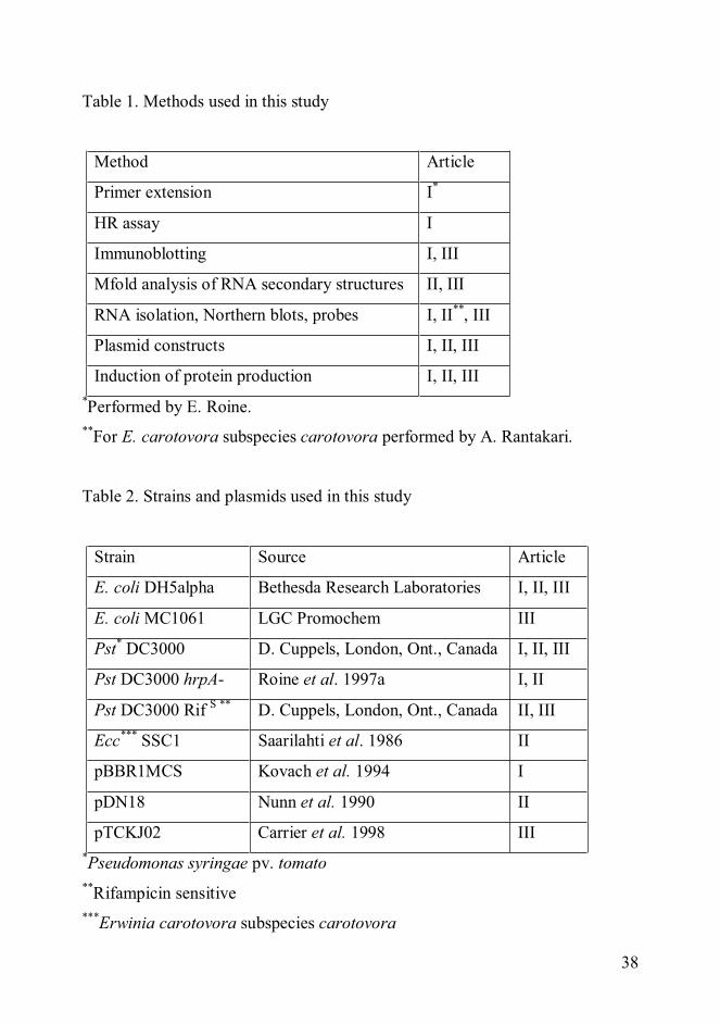
38
Table 1. Methods used in this study
Method Article
Primer extension I*
HR assay I
Immunoblotting I, III
Mfold analysis of RNA secondary structures II, III
RNA isolation, Northern blots, probes I, II**, III
Plasmid constructs I, II, III
Induction of protein production I, II, III*Performed by E. Roine.**For E. carotovora subspecies carotovora performed by A. Rantakari.
Table 2. Strains and plasmids used in this study
Strain Source Article
E. coli DH5alpha Bethesda Research Laboratories I, II, III
E. coli MC1061 LGC Promochem III
Pst* DC3000 D. Cuppels, London, Ont., Canada I, II, III
Pst DC3000 hrpA- Roine et al. 1997a I, II
Pst DC3000 Rif S ** D. Cuppels, London, Ont., Canada II, III
Ecc*** SSC1 Saarilahti et al. 1986 II
pBBR1MCS Kovach et al. 1994 I
pDN18 Nunn et al. 1990 II
pTCKJ02 Carrier et al. 1998 III*Pseudomonas syringae pv. tomato**Rifampicin sensitive***Erwinia carotovora subspecies carotovora
39
4 Results and discussion
4.1 The secretion signal of hrpA
The secretion signals of the TTSS-dependent proteins have been studied
extensively, but the data is somewhat contradictory (see section 1.2.6 on the
secretion signals for TTS). The transcription start site of the hrpA transcript was
located to a site 42 nucleotides upstream of the translation start codon using
primer extension analysis. Promoter and protein fusion constructs revealed that
the secretion signal of HrpA of P. syringae pv. tomato resides in the first 15
codons of hrpA mRNA or amino acids of HrpA protein (I). The location of the
signal is typical for type III secreted proteins. The nature of the secretion signal,
whether it resides in the mRNA or in the amino acid sequence, remains elusive.
4.2 Translation of HrpA is not dependent on secretion
Some TTSS-associated proteins are secreted in a co-translational way with
no intracellular pool detected (Anderson and Schneewind 1999). HrpA protein
can be expressed without a functional secretion system (I). HrpA transcribed
from a lacZ promoter was translated in P. syringae under Hrp-non-inducing
conditions. Thus translation and secretion are not coupled for HrpA, even
though the secretion compatibility of pre-made HrpA has not been established.
4.3 Regions important for the accumulation of hrpA transcript
Whilst studying the secretion signal of hrpA from P. syringae, the
importance of specific regions for transcript accumulation became apparent.
The first 15 codons needed for the secretion of HrpA, as well as the 5’ UTR of
the transcript, are important for the accumulation of the hrpA mRNA (I).

40
Additional regions involved in the accumulation of the transcript reside in the
3’ UTR of hrpA (II). Replacing the 5’ UTR of hrpA with that of lacZ or
insertions made in the 3’ UTR substantially reduced the levels of hrpA
transcripts. Computer analysis revealed that the 3’ UTR of hrpA mRNA forms a
hypothetical GC-rich stem-loop structure. The insertions in the 3’ UTR that
reduced the hrpA mRNA levels were predicted to change the hairpin structure
in this region (II).
The regions in the 5’ and the 3’ UTRs of hrpA transcripts of P. syringae
pathovars form, according to computer analysis, extremely similar stem-loop
structures (III). The sequences of the elements forming these structures are
almost identical even though the regions between these conserved sequences,
including the coding regions, do not exhibit high homology. This suggests that
the stem-loops are functionally important (James et al. 1989, Chen et al. 1991).
The GC-rich hairpins in the 3’ UTRs are likely to serve as barriers for the
attacks of RNases and might also serve as transcriptional terminators. The role
of the stem-loop structures formed in the 5’ UTRs of hrpA transcripts, if other
than stabilization of the transcripts, remains elusive.
4.4 Half-lives of hrpA transcripts
In P. syringae pv. tomato hrpA is transcribed as two mRNAs; hrpA that is
0.4 kb and hrpAZ that is 1.7 kb. The hrpA transcript is extremely stable in
different species as analysed by Northern blots (II). Its half-life was measured
as 20-40 min. in P. syringae pv. tomato. The transcript degraded slowly in the
first time points after inhibiting de novo RNA synthesis with rifampicin, and
faster after 40 min. when the hrpAZ signal had disappeared. The half-life of the
P. syringae pv. phaseolicola race 4 hrpA transcript was measured in P. syringae
pv. tomato DC3000, since a rifampicin sensitive strain was unavailable, and
was calculated as 34 min. The half-life of hrpA of E. carotovora subspecies

41
(spp.) carotovora (recently renamed as Pectobacterium carotovorum) was
calculated to be 47 min.
According to reverse transcriptase polymerase chain reaction (rt-PCR)
measurements (Thwaites et al. 2004) the half-life of hrpA from P. syringae pv.
phaseolicola race 7 was 8 min. Results obtained by Northern blot analysis of
hrpA transcripts and contradictory to those achieved by rt-PCR and demonstrate
that the hrpA mRNA is stable in P. syringae pv. phaseolicola race 7 (Hienonen
E, unpublished results). The differences between the results (Thwaites et al.
2004, II and Hienonen E, unpublished data) cannot be attributed to the strains
used, but might be explained by the use of different methods. Northern blots
measure all the different folding forms of the mRNA without any preference,
whereas rt-PCR employed by Thwaites and colleagues (2004) might give a
more biased view if one form of the transcript is more readily amplified by the
polymerase than others.
Ribosomes may protect transcripts from degradation by RNases (Braun et al.
1998, Vytvytska et al. 2000). The high stability of hrpA mRNA is, however, not
dependent on translation or on the presence of ribosomes, as the transcript is
equally stable after the addition of antibiotics that release the ribosomes from
the mRNA (III). The stability of the hrpA transcript is not dependent on
additional species-specific factors either, since the transcript is stable also in E.
coli (II). The stability of the hrpA transcript does not control the temperature
dependence of the TTSS induction (van Dijk et al. 1999) as the transcript is
stable under temperatures ranging from 18 to 37 °C (II).
4.5 Stabilization of heterologous transcripts by hrpA
The hrpA transcript can be used to stabilize heterologous transcripts
originating from Gram-negative and Gram-positive bacteria and from
eukaryotic species (III). These heterologous transcripts include neo, xln2, gfp

42
and fbp54. Their half-lives were elevated from a few minutes to the range of 17
min in E. coli and 25 min in P. syringae. Both translational fusion and out-of-
frame constructs were stabilized to the same extent. Some transcript stabilizing
elements only work as translational fusions (Belasco et al. 1986), and if non-
fusion recombinant protein is required, a cleavage site must be inserted between
the coding regions of the two proteins. The hrpA transcript from P. syringae pv.
phaseolicola has the same stabilizing effect on heterologous transcripts as that
of P. syringae pv. tomato.
The regions in the 5’ and 3’ elements of the hrpA mRNA needed for the
accumulation of the transcript (I and II) are also necessary and sufficient for the
stabilization of heterologous transcripts as shown by deletion analysis (III). The
3’ UTR of hrpA has a major effect on transcript stability, whereas more minor
influence on stability was assigned to the 5’ regions. This is in accordance with
the result that the transcript hrpAZ is less stable than hrpA, having a half-life of
11 min. (II), which is still above the average 3-8 min. of bacterial transcripts.
The sequences of the transcripts hrpA and hrpAZ are identical in the 5’ region,
but differ in the 3’ regions.
4.6 Recombinant protein yields can be improved using the transcript stabilizing
elements from hrpA
The stabilizing effect of hrpA elements on heterologous transcripts was also
shown to have an application in the production of heterologous proteins (III).
Transcript stabilizing elements of hrpA fused to the gene encoding neomycin
phosphotransferase (NPT) resulted in higher yields of NPT as compared with
the control lacking transcript stabilizing elements. The strain of E. coli with
hrpA mRNA stabilizing elements produced up to 5.5 times more NPT than the
control as analysed by Coomassie blue staining of poly acryl amide gels.
43
Further optimization, such as a less leaky promoter, would be needed for more
extensive and quantitative studies.
5 Conclusions
This study addressed questions relating to the type III secretion system-
associated proteins of plant pathogenic bacteria. In addition to the information
on the major virulence system of various Gram-negative pathogens, new
knowledge was generated on the production of recombinant proteins in bacteria.
As was in the beginning of this project, it still remains unclear whether the
secretion signal for type III secretion is an amino acid signal or an mRNA
signal. The search for the secretion signal of the type III secretion-dependent
HrpA protein from the plant pathogenic bacterium P. syringae did reveal that
the signal resides in the first few codons of the mRNA or amino acids of the
protein, as is typical for type III secreted proteins. The fact that HrpA can be
translated independently of a functional secretion machinery was not
unexpected since coupling of translation to secretion has been shown for only a
few type III secreted proteins (Karlinsey et al. 2000, Anderson and Schneewind
1999). HrpA may not be among the co-translationally secreted type III proteins
since no chaperone has been found for HrpA thus far and type III chaperones
may contribute to the secretion of co-translationally secreted proteins (Darwin
and Miller 2001). The secretion competence of pre-made HrpA still remains to
be shown.
The studies on the secretion signal of HrpA that potentially is RNA in nature
led to the discovery of the extreme stability of the hrpA transcript. The half-life
of the transcript is roughly five- to tenfold that of typical bacterial mRNAs
(Bernstein et al. 2002). Conservation of the sequence of the untranslated
regions of otherwise dissimilar hrpAs from several P. syringae pathovars, the
hypothetical stem-loop structures forming in those regions and the stable nature

44
of the transcript also in E. carotovora indicate a biological relevance of the long
half-life. The conservation of the presumably stabilizing secondary structures
and the relevance of the stability of the transcripts in nature are intriguing issues
and require further investigation. The relative amounts of TTS-dependent
proteins may be at least in part controlled by the stabilities of their transcripts,
HrpA being one of the most abundant TTSS-associated proteins (Roine et al.
1997a). Additional research is needed to establish whether stability of mRNAs
and conservation of secondary structures are features commonly shared
between hrpA and analogous transcripts. No published data exists on the
stabilities of transcripts coding for pilus or needle components from species
other than P. syringae and E. carotovora.
Stability is an endogenous feature of the hrpA mRNA since the transcript is
stable also in E. coli. The conserved untranslated regions of the transcript are
likely to protect the hrpA mRNA against the attacks of RNases. Secondary
structures may cover the RNase cleavage sites or hinder the attachment and
cleavage by RNases that require free ends or cleave only single-stranded RNA.
Data from this study on the start site of transcription of hrpA and the mapping
of the regions involved in transcript accumulation and stability were used to
investigate the stabilizing effect of the 5’ and 3’ regions of hrpA mRNA on
heterologous transcripts. The half-lives of transcripts originating from diverse
organisms were elevated substantially when fused with stabilizing elements of
hrpA from pathovars of P. syringae. The transcript stabilizing system based on
the hrpA mRNA is at least as potent as those previously described in the
literature and summarised in the introduction section. Compared with other
stabilizing mRNA structures used for elongating the half-lives of heterologous
transcripts, the half-lives achieved with this system are equal or even longer.
The stabilizing effect of hrpA mRNA on heterologous transcripts was proven
to be of use in the production of recombinant proteins in bacteria, including the
widely-used industrial production host E. coli. Recombinant protein yields were
elevated manifold with the addition of the transcript stabilizing elements despite

45
the teething problems associated with this novel production system. Compared
with the previously published results that are reviewed in the introduction
section on the use of transcript stabilizing elements in the production of
recombinant proteins in bacteria, the findings using this system are among the
best published. Prospective development in the form of better control of
induction and more defined growth conditions will presumably further improve
the yields of recombinant proteins using this transcript stabilization system
based on the hrpA transcripts from pathovars of P. syringae.
6 Acknowledgements
This work was carried out at the Department of Biosciences, Division of
General Microbiology with financial support from the Viikki Graduate School
in Biosciences and University of Helsinki Research Funds.
I thank professors Kielo Haahtela and Timo Korhonen for giving me the
opportunity to work and do my PhD thesis in the division. Great thanks go to
my reviewers, docent Benita Westerlund-Wikström and professor Thorsten
Nürnberger for their valuable comments. I also wish to thank Benita and docent
Hannu Saarilahti for being my follow up group members and finding the time
for the meetings. I thank professor Per Saris, for accepting the invitation to be
my opponent and in advance for the hopefully fruitful and enjoyable debate we
will have. I’m obliged to my supervisors docent Suvi Taira and professor
Martin Romantschuk for all their help both in the lab and with writing and
applying for funds.
My thanks also go to Antti Nyyssölä, Fred Fenel, Heikki Ojamo, Katariina
Majander and Lena Anton for their collaboration. Cheers, John Mansfield and
Richard Thwaites, for always finding the time to answer my questions and for
reading my manuscripts.

46
Thanks to all of those whom I have shared lab or office space with. Minna,
I’m grateful for your vital help in the lab and for all those coffee table
conversations over Hesari. Chun Mei and Anssi, thanks for your help and
support. I thank all the past and present members of YMBO for being such nice
co-workers.
Heli, thanks for the lunch and coffee breaks. I will be waiting for invitations
for your and Eijas defenses in 2008! Thanks, Marjo for all those entertaining e-
mails. I wish to thank Asta for long-time friendship and patience with my
endless computer-related questions. Pirkko, thanks for being you.
For my parents, Inkeri and Juhani, a huge thanks for all your support, both
financial and mental. I appreciate you letting me have my way.
Last but not least Tommi, you are the sunshine of my life. Thanks for help
with pictures and for saving my thesis. I’ll beat you to it!

47
7 References
Abramovitch RB, Martin GB. (2004) Strategies used by bacterial pathogens tosuppress plant defenses. Curr. Opin. Plant Biol. 7, 356-64
Agaisse H, Lereclus D. (1996) STAB-SD: a Shine-Dalgarno sequence in the 5'untranslated region is a determinant of mRNA stability. Mol. Microbiol. 20,633-43
Akeda Y, Galán JE. (2005) Chaperone release and unfolding of substrates intype III secretion. Nature. 437, 911-5
Aldon D, Brito B, Boucher C, Genin S. (2000) A bacterial sensor of plant cellcontact controls the transcriptional induction of Ralstonia solanacearumpathogenicity genes. EMBO J. 19, 2304-14
Aldridge P, Hughes KT. (2001) How and when are substrates selected for typeIII secretion? Trends Microbiol. 9, 209-14
Alfano JR, Collmer A. (1996) Bacterial Pathogens in Plants: Life up against theWall. Plant Cell. 8, 1683-1698
Alfano JR, Collmer A. (2004) Type III secretion system effector proteins:double agents in bacterial disease and plant defense. Annu. Rev.Phytopathol. 42, 385-414
Anderson DM, Schneewind O. (1997) An mRNA signal for the type IIIsecretion of Yop proteins by Yersinia enterocolitica. Science. 278, 1140-3
Anderson DM, Schneewind O. (1999) Yersinia enterocolitica type III secretion:an mRNA signal that couples translation and secretion of YopQ. Mol.Microbiol. 31, 1139-48
Anderson DM, Fouts DE, Collmer A and Schneewind O (1999) Reciprocalsecretion of proteins by the bacterial type III machines of plant and animalpathogens suggests universal recognition of mRNA targeting signals. Proc.Natl. Acad. Sci. U. S. A. 96, 12839–12843
Anderson DM, Ramamurthi KS, Tam C, Schneewind O. (2002) YopD andLcrH regulate expression of Yersinia enterocolitica YopQ by aposttranscriptional mechanism and bind to yopQ RNA. J. Bacteriol. 184,1287-95
Anderson JC, Pascuzzi PE, Xiao F, Sessa G, Martin GB. (2006) Host-mediatedphosphorylation of type III effector AvrPto promotes Pseudomonasvirulence and avirulence in tomato. Plant Cell. 18, 502-14
Baneyx F, Ayling A, Palumbo T, Thomas D, Georgiou G. (1991) Optimizationof growth conditions for the production of proteolytically-sensitive proteinsin the periplasmic space of Escherichia coli. Appl. Microbiol. Biotechnol.36, 14-20
Baneyx F. (1999) Recombinant protein expression in Escherichia coli. Curr.Opin. Biotechnol. 10, 411-21
Baneyx F, Mujacic M. (2004) Recombinant protein folding and misfolding inEscherichia coli. Nat. Biotechnol. 22, 1399-408

48
Belasco JG, Nilsson G, von Gabain A, Cohen SN. (1986) The stability of E.coli gene transcripts is dependent on determinants localized to specificmRNA segments. Cell. 46, 245-51
Bernstein JA, Khodursky AB, Lin PH, Lin-Chao S, Cohen SN. (2002) Globalanalysis of mRNA decay and abundance in Escherichia coli at single-generesolution using two-color fluorescent DNA microarrays. Proc. Natl. Acad.Sci. U. S. A. 99, 9697-702
Bielli P, Bellenchi GC, Calabrese L. (2001) Site-directed mutagenesis of humanceruloplasmin: production of a proteolytically stable protein and structure-activity relationships of type 1 sites. J. Biol. Chem. 276, 2678-85
Birtalan SC, Phillips RM, Ghosh P. (2002) Three-dimensional secretion signalsin chaperone-effector complexes of bacterial pathogens. Mol. Cell. 9, 971-80
Blum E, Py B, Carpousis AJ, Higgins CF. (1997) Polyphosphate kinase is acomponent of the Escherichia coli RNA degradosome. Mol. Microbiol. 26,387-98
Boyd AP, Lambermont I, Cornelis GR. (2000) Competition between the Yopsof Yersinia enterocolitica for delivery into eukaryotic cells: role of the SycEchaperone binding domain of YopE. J. Bacteriol. 182, 4811-21
Braun F, Le Derout J, Régnier P. (1998) Ribosomes inhibit an RNase Ecleavage which induces the decay of the rpsO mRNA of Escherichia coli.EMBO J. 17, 4790-7
Bretz J, Losada L, Lisboa K, Hutcheson SW. (2002) Lon protease functions asa negative regulator of type III protein secretion in Pseudomonas syringae.Mol. Microbiol. 45, 397-409
Brinkmann U, Mattes RE, Buckel P. (1989) High-level expression ofrecombinant genes in Escherichia coli is dependent on the availability of thednaY gene product. Gene. 85, 109-14
Brown IR, Mansfield JW, Taira S, Roine E, Romantschuk M. (2001)Immunocytochemical localization of HrpA and HrpZ supports a role for theHrp pilus in the transfer of effector proteins from Pseudomonas syringae pv.tomato across the host plant cell wall. Mol. Plant Microbe Interact. 14,394-404
Buell CR, Joardar V, Lindeberg M, Selengut J, Paulsen IT, Gwinn ML, DodsonRJ, Deboy RT, Durkin AS, Kolonay JF, Madupu R, Daugherty S, Brinkac L,Beanan MJ, Haft DH, Nelson WC, Davidsen T, Zafar N, Zhou L, Liu J,Yuan Q, Khouri H, Fedorova N, Tran B, Russell D, Berry K, Utterback T,Van Aken SE, Feldblyum TV, D'Ascenzo M, Deng WL, Ramos AR, AlfanoJR, Cartinhour S, Chatterjee AK, Delaney TP, Lazarowitz SG, Martin GB,Schneider DJ, Tang X, Bender CL, White O, Fraser CM, Collmer A. (2003)The complete genome sequence of the Arabidopsis and tomato pathogenPseudomonas syringae pv. tomato DC3000. Proc. Natl. Acad. Sci. U. S. A.100, 10181-6
Büttner D, Bonas U. (2002) Getting across--bacterial type III effector proteinson their way to the plant cell. EMBO J. 21, 5313-22

49
Callaghan AJ, Marcaida MJ, Stead JA, McDowall KJ, Scott WG, Luisi BF.(2005) Structure of Escherichia coli RNase E catalytic domain andimplications for RNA turnover. Nature. 437, 1187-91
Cambronne ED, Cheng LW, Schneewind O. (2000) LcrQ/YscM1, regulators ofthe Yersinia yop virulon, are injected into host cells by a chaperone-dependent mechanism. Mol. Microbiol. 37, 263-73
Cambronne ED, Schneewind O. (2002) Yersinia enterocolitica type IIIsecretion: yscM1 and yscM2 regulate yop gene expression by aposttranscriptional mechanism that targets the 5' untranslated region of yopmRNA. J. Bacteriol. 184, 5880-93
Cambronne ED, Sorg JA, Schneewind O. (2004) Binding of SycH chaperone toYscM1 and YscM2 activates effector yop expression in Yersiniaenterocolitica. J. Bacteriol. 186, 829-41
Carrier TA, Keasling JD (1997a) Engineering mRNA stability in E. coli by theaddition of synthetic hairpins using a 5 cassette system Biotechnol. Bioeng.55, 577-580
Carrier TA, Keasling JD. (1997b) Mechanistic modeling of prokaryotic mRNAdecay. J. Theor. Biol. 189, 195-209
Carrier TA, Keasling JD. (1997c) Controlling messenger RNA stability inbacteria: Strategies for engineering gene expression. Biotechnol. Prog. 13,699-708
Carrier T, Jones KL, Keasling JD. (1998) mRNA stability and plasmid copynumber effects on gene expression from an inducible promoter system.Biotechnol. Bioeng. 59, 666-72
Carrier TA, Keasling JD. (1999) Library of synthetic 5' secondary structures tomanipulate mRNA stability in Escherichia coli. Biotechnol. Prog. 15, 58-64
Cereghino JL, Cregg JM. (2000) Heterologous protein expression in themethylotrophic yeast Pichia pastoris. FEMS Microbiol. Rev. 24, 45-66
Chatterjee A, Cui Y, Yang H, Collmer A, Alfano JR, Chatterjee AK. (2001)GacA, the response regulator of a two-component system, acts as a masterregulator in Pseudomonas syringae pv. tomato DC3000 by controllingregulatory RNA, transcriptional activators, and alternate sigma factors. Mol.Plant Microbe Interact. 16, 1106-17
Chen LH, Emory SA, Bricker AL, Bouvet P, Belasco JG. (1991) Structure andfunction of a bacterial mRNA stabilizer: analysis of the 5' untranslatedregion of ompA mRNA. J. Bacteriol. 173, 4578-86
Cheng LW, Anderson DM, Schneewind O. (1997) Two independent type IIIsecretion mechanisms for YopE in Yersinia enterocolitica. Mol. Microbiol.24, 757-65
Chiu HJ, Syu WJ. (2005) Functional analysis of EspB from enterohaemorrhagicEscherichia coli. Microbiology. 151, 3277-86
Coburn GA, Mackie GA. (1996) Differential sensitivities of portions of themRNA for ribosomal protein S20 to 3'-exonucleases dependent on
50
oligoadenylation and RNA secondary structure. J. Biol. Chem. 271, 15776-81
Conesa A, Punt PJ, van Luijk N, van den Hondel CA. (2001) The secretionpathway in filamentous fungi: a biotechnological view. Fungal Genet. Biol.33, 155-71
Cornelis GR (1998) The Yersinia deadly kiss. J. Bacteriol. 180, 5495-504Cornelis GR, Van Gijsegem F. (2000) Assembly and function of type III
secretory systems. Annu. Rev. Microbiol. 54, 735-74Cornelis GR. (2002) The Yersinia Ysc-Yop 'type III' weaponry. Nat. Rev. Mol.
Cell Biol. 3, 742-52Dangl JL, Jones JD. (2001) Plant pathogens and integrated defence responses to
infection. Nature. 411, 826-33Darwin KH, Miller VL. (2001) Type III secretion chaperone-dependent
regulation: activation of virulence genes by SicA and InvF in Salmonellatyphimurium. EMBO J. 20, 1850-62
DebRoy S, Thilmony R, Kwack YB, Nomura K, He SY. (2004) A family ofconserved bacterial effectors inhibits salicylic acid-mediated basal immunityand promotes disease necrosis in plants. Proc. Natl. Acad. Sci. U. S. A. 101,9927-32
Deng WL, Preston G, Collmer A, Chang CJ, Huang HC. (1998)Characterization of the hrpC and hrpRS operons of Pseudomonas syringaepathovars syringae, tomato, and glycinea and analysis of the ability of hrpF,hrpG, hrcC, hrpT, and hrpV mutants to elicit the hypersensitive response anddisease in plants. J. Bacteriol. 180, 4523-31
Ehrbar K, Winnen B, Hardt WD. (2006) The chaperone binding domain ofSopE inhibits transport via flagellar and SPI-1 TTSS in the absence of InvB.Mol. Microbiol. 59, 248-64
Ehretsmann CP, Carpousis AJ, Krisch HM. (1992) mRNA degradation inprocaryotes. FASEB J. 6, 3186-92
Evdokimov AG, Phan J, Tropea JE, Routzahn KM, Peters HK 3rd, Pokross M,Waugh DS. (2003) Similar modes of polypeptide recognition by exportchaperones in flagellar biosynthesis and type III secretion. Nat. Struct. Biol.10, 789-93
Falconi M, Colonna B, Prosseda G, Micheli G, Gualerzi CO. (1998)Thermoregulation of Shigella and Escherichia coli EIEC pathogenicity. Atemperature-dependent structural transition of DNA modulates accessibilityof virF promoter to transcriptional repressor H-NS. EMBO J. 17, 7033-43
Feldman MF, Cornelis GR. (2003) The multitalented type III chaperones: allyou can do with 15 kDa. FEMS Microbiol. Lett. 219, 151-8
Foultier B, Troisfontaines P, Muller S, Opperdoes FR, Cornelis GR. (2002)Characterization of the ysa pathogenicity locus in the chromosome ofYersinia enterocolitica and phylogeny analysis of type III secretion systems.J. Mol. Evol. 55, 37-51

51
Freiberg C, Fellay R, Bairoch A, Broughton WJ, Rosenthal A, Perret X. (1997)Molecular basis of symbiosis between Rhizobium and legumes. Nature. 387,394-401
Galán JE, Collmer A. (1999) Type III secretion machines: bacterial devices forprotein delivery into host cells. Science. 284, 1322-8
Galán JE. (2001) Salmonella interactions with host cells: type III secretion atwork. Annu. Rev. Cell Dev. Biol. 17, 53-86
Gauthier A, Puente JL, Finlay BB. (2003) Secretin of the enteropathogenicEscherichia coli type III secretion system requires components of the typeIII apparatus for assembly and localization. Infect. Immun. 71, 3310-9
Ghelardi E, Celandroni F, Salvetti S, Beecher DJ, Gominet M, Lereclus D,Wong AC, Senesi S. (2002) Requirement of flhA for swarmingdifferentiation, flagellin export, and secretion of virulence-associatedproteins in Bacillus thuringiensis. J. Bacteriol. 184, 6424-33
Ghosh P. (2004) Process of protein transport by the type III secretion system.Microbiol. Mol. Biol. Rev. 68, 771-95
Gophna U, Ron EZ, Graur D. (2003) Bacterial type III secretion systems areancient and evolved by multiple horizontal-transfer events. Gene. 312, 151-63
Goss JW, Sorg JA, Ramamurthi KS, Ton-That H, Schneewind O. (2004) Thesecretion signal of YopN, a regulatory protein of the Yersinia enterocoliticatype III secretion pathway. J. Bacteriol. 186, 6320-4
Grant SR, Fisher EJ, Chang JH, Mole BM, Dangl JL. (2006) Subterfuge andManipulation: Type III Effector Proteins of Phytopathogenic Bacteria. Annu.Rev. Microbiol. 60, 425-49
Grunberg-Manago M. (1999) Messenger RNA stability and its role in control ofgene expression in bacteria and phages. Annu. Rev. Genet. 33, 193-227
Guttman DS, Vinatzer BA, Sarkar SF, Ranall MV, Kettler G, Greenberg JT.(2002) A functional screen for the type III (Hrp) secretome of the plantpathogen Pseudomonas syringae. Science. 295, 1722-6
Ham JH, Bauer DW, Fouts DE, Collmer A. (1998) A cloned Erwiniachrysanthemi Hrp (type III protein secretion) system functions inEscherichia coli to deliver Pseudomonas syringae Avr signals to plant cellsand to secrete Avr proteins in culture. Proc. Natl. Acad. Sci. U. S. A. 95,10206-11
Hambraeus G, von Wachenfeldt C, Hederstedt L. (2003) Genome-wide surveyof mRNA half-lives in Bacillus subtilis identifies extremely stable mRNAs.Mol. Genet. Genomics. 269, 706-14
Hammarström M, Hellgren N, van Den Berg S, Berglund H, Härd T. (2002)Rapid screening for improved solubility of small human proteins producedas fusion proteins in Escherichia coli. Protein Sci. 11, 313-21
Hauck P, Thilmony R, He SY. (2003) A Pseudomonas syringae type IIIeffector suppresses cell wall-based extracellular defense in susceptibleArabidopsis plants. Proc. Natl. Acad. Sci. U. S. A. 100, 8577-82

52
He P, Shan L, Lin NC, Martin GB, Kemmerling B, Nürnberger T, Sheen J.(2006) Specific bacterial suppressors of MAMP signaling upstream ofMAPKKK in Arabidopsis innate immunity. Cell. 125, 563-75
He SY, Huang HC, Collmer A. (1993) Pseudomonas syringae pv. syringaeharpinPss: a protein that is secreted via the Hrp pathway and elicits thehypersensitive response in plants. Cell. 73, 1255-66
Henderson IR, Navarro-Garcia F, Desvaux M, Fernandez RC, Ala'Aldeen D.(2004) Type V protein secretion pathway: the autotransporter story.Microbiol. Mol. Biol. Rev. 68, 692-744
Hendrickson EL, Guevera P, Ausubel FM. (2000) The alternative sigma factorRpoN is required for hrp activity in Pseudomonas syringae pv. maculicolaand acts at the level of hrpL transcription. J. Bacteriol. 182, 3508-16
Hirokawa G, Kiel MC, Muto A, Selmer M, Raj VS, Liljas A, Igarashi K, KajiH, Kaji A. (2002) Post-termination complex disassembly by ribosomerecycling factor, a functional tRNA mimic. EMBO J. 21, 2272-81
Hu W, Yuan J, Jin QL, Hart P, He SY. (2001) Immunogold labeling of Hrp piliof Pseudomonas syringae pv. tomato assembled in minimal medium and inplanta. Mol. Plant Microbe Interact. 14, 234-41
Hueck CJ. (1998) Type III protein secretion systems in bacterial pathogens ofanimals and plants. Microbiol. Mol. Biol. Rev. 62, 379-433
Hunt I. (2005) From gene to protein: a review of new and enabling technologiesfor multi-parallel protein expression. Protein Expr. Purif. 40, 1-22
Huynh TV, Dahlbeck D, Staskawicz BJ. (1989) Bacterial blight of soybean:regulation of a pathogen gene determining host cultivar specificity. Science.245, 1374-7
Innes R. (2003) New effects of type III effectors. Mol Microbiol. 50, 363-5James BD, Olsen GJ, Pace NR. (1989) Phylogenetic comparative analysis of
RNA secondary structure. Methods Enzymol. 180, 227-39Jana S, Deb JK. (2005) Strategies for efficient production of heterologous
proteins in Escherichia coli. Appl. Microbiol. Biotechnol. 67, 289-98Janjusevic R, Abramovitch RB, Martin GB, Stebbins CE. (2006) A bacterial
inhibitor of host programmed cell death defenses is an E3 ubiquitin ligase.Science. 311, 222-6
Jin Q, He SY. (2001) Role of the Hrp pilus in type III protein secretion inPseudomonas syringae. Science. 294, 2556-8
Karlinsey JE, Lonner J, Brown KL, Hughes KT. (2000) Translation/secretioncoupling by type III secretion systems. Cell. 102, 487-97
Kenny B, DeVinney R, Stein M, Reinscheid DJ, Frey EA, Finlay BB. (1997)Enteropathogenic E. coli (EPEC) transfers its receptor for intimate adherenceinto mammalian cells. Cell. 91, 511-20
Kimbrough TG, Miller SI. (2002) Assembly of the type III secretion needlecomplex of Salmonella typhimurium. Microbes Infect. 4, 75-8
Koebnik R. (2001) The role of bacterial pili in protein and DNA translocation.Trends Microbiol. 9, 586-90
53
Konkel ME, Klena JD, Rivera-Amill V, Monteville MR, Biswas D, Raphael B,Mickelson J. (2004) Secretion of virulence proteins from Campylobacterjejuni is dependent on a functional flagellar export apparatus. J. Bacteriol.186, 3296-303
KovachME, Phillips RW, Elzer PH, Roop RM 2nd, Peterson KM (1994)pBBR1MCS: a broad-host-range cloning vector. Biotechniques 16:800–2
Kushner SR. (2002) mRNA decay in Escherichia coli comes of age. J.Bacteriol. 184, 4658-65
Kushner SR. (2004) mRNA decay in prokaryotes and eukaryotes: differentapproaches to a similar problem. IUBMB Life. 56, 585-94
Lee J, Klüsener B, Tsiamis G, Stevens C, Neyt C, Tampakaki AP, PanopoulosNJ, Nöller J, Weiler EW, Cornelis GR, Mansfield JW, Nürnberger T. (2001)HrpZPsph from the plant pathogen Pseudomonas syringae pv. phaseolicolabinds to lipid bilayers and forms an ion-conducting pore in vitro. Proc. Natl.Acad. Sci. U. S. A. 98, 289-94
Lee SH, Galán JE. (2004) Salmonella type III secretion-associated chaperonesconfer secretion-pathway specificity. Mol. Microbiol. 51, 483-95
Lee VT, Mazmanian SK, Schneewind O. (2001) A program of Yersiniaenterocolitica type III secretion reactions is activated by specific signals. J.Bacteriol. 183, 4970-8
Lee VT, Schneewind O. (2002) Yop fusions to tightly folded protein domainsand their effects on Yersinia enterocolitica type III secretion. J. Bacteriol.184, 3740-5
Letzelter M, Sorg I, Mota LJ, Meyer S, Stalder J, Feldman M, Kuhn M,Callebaut I, Cornelis GR. (2006) The discovery of SycO highlights a newfunction for type III secretion effector chaperones. EMBO J. 25, 3223-33
Li C, Schwabe JW, Banayo E, Evans RM. (1997) Coexpression of nuclearreceptor partners increases their solubility and biological activities. Proc.Natl. Acad. Sci. U. S. A. 94, 2278-83
Li CM. (2001) Masters thesis. University of Helsinki.Li CM, Brown I, Mansfield J, Stevens C, Boureau T, Romantschuk M, Taira S.
(2002) The Hrp pilus of Pseudomonas syringae elongates from its tip andacts as a conduit for translocation of the effector protein HrpZ. EMBO J. 21,1909-15
Li X, Lin H, Zhang W, Zou Y, Zhang J, Tang X, Zhou JM. (2005) Flagellininduces innate immunity in nonhost interactions that is suppressed byPseudomonas syringae effectors. Proc. Natl. Acad. Sci. U. S. A. 102, 12990-5
Lloyd SA, Norman M, Rosqvist R, Wolf-Watz H. (2001) Yersinia YopE istargeted for type III secretion by N-terminal, not mRNA, signals. Mol.Microbiol. 39, 520-31
Lopez PJ, Marchand I, Yarchuk O, Dreyfus M. (1998) Translation inhibitorsstabilize Escherichia coli mRNAs independently of ribosome protection.Proc. Natl. Acad. Sci. U. S. A. 95, 6067-72

54
Lopez PJ, Marchand I, Joyce SA, Dreyfus M. (1999) The C-terminal half ofRNase E, which organizes the Escherichia coli degradosome, participates inmRNA degradation but not rRNA processing in vivo. Mol. Microbiol. 33,188-99
Losada LC, Hutcheson SW. (2005) Type III secretion chaperones ofPseudomonas syringae protect effectors from Lon-associated degradation.Mol. Microbiol. 55, 941-53
Luo Y, Bertero MG, Frey EA, Pfuetzner RA, Wenk MR, Creagh L, Marcus SL,Lim D, Sicheri F, Kay C, Haynes C, Finlay BB, Strynadka NC. (2001)Structural and biochemical characterization of the type III secretionchaperones CesT and SigE. Nat. Struct. Biol. 8, 1031-6
Ma J, Campbell A, Karlin S. (2002) Correlations between Shine-Dalgarnosequences and gene features such as predicted expression levels and operonstructures. J. Bacteriol. 184, 5733-45
Macnab RM. (1999) The bacterial flagellum: reversible rotary propellor andtype III export apparatus. J. Bacteriol. 181, 7149-53
Majander K, Anton L, Antikainen J, Lång H, Brummer M, Korhonen TK,Westerlund-Wikström B. (2005) Extracellular secretion of polypeptidesusing a modified Escherichia coli flagellar secretion apparatus. Nat.Biotechnol. 23, 475-81
McLaren RS, Newbury SF, Dance GS, Causton HC, Higgins CF. (1991)mRNA degradation by processive 3'-5' exoribonucleases in vitro and theimplications for prokaryotic mRNA decay in vivo. J. Mol. Biol. 221, 81-95
Mergulhão FJ, Summers DK, Monteiro GA. (2005) Recombinant proteinsecretion in Escherichia coli. Biotechnol. Adv. 23, 177-202
Michgehl S, Heusipp G, Greune L, Ruter C, Schmidt MA. (2006) Esp-independent functional integration of the translocated intimin receptor (Tir)of enteropathogenic Escherichia coli (EPEC) into host cell membranes. Cell.Microbiol. 8, 625-33
Mohanty BK, Maples VF, Kushner SR. (2004) The Sm-like protein Hfqregulates polyadenylation dependent mRNA decay in Escherichia coli. Mol.Microbiol. 54, 905-20
Moll I, Afonyushkin T, Vytvytska O, Kaberdin VR, Bläsi U. (2003) CoincidentHfq binding and RNase E cleavage sites on mRNA and small regulatoryRNAs. RNA. 9, 1308-14
Morita M, Asami K, Tanji Y, Unno H. (2001) Programmed Escherichia colicell lysis by expression of cloned T4 phage lysis genes. Biotechnol. Prog. 17,573-6
Mudgett MB, Chesnokova O, Dahlbeck D, Clark ET, Rossier O, Bonas U andStaskawicz B (2000) Molecular signals required for type III secretion andtranslocation of the Xanthomonas campestris AvrBs2 protein to pepperplants. Proc. Natl. Acad. Sci. U. S. A. 97, 13324–13329
Mudgett MB. (2005) New insights to the function of phytopathogenic bacterialtype III effectors in plants. Annu. Rev. Plant Biol. 56, 509-31

55
Mueller CA, Broz P, Muller SA, Ringler P, Erne-Brand F, Sorg I, Kuhn M,Engel A, Cornelis GR. (2005) The V-antigen of Yersinia forms a distinctstructure at the tip of injectisome needles. Science. 310, 674-6
Nilsson G, Belasco JG, Cohen SN, von Gabain A. (1984) Growth-ratedependent regulation of mRNA stability in Escherichia coli. Nature. 312,75-7
Nomura K, Debroy S, Lee YH, Pumplin N, Jones J, He SY. (2006) A bacterialvirulence protein suppresses host innate immunity to cause plant disease.Science. 313, 220-3
Nougayrède JP, Fernandes PJ, Donnenberg MS. (2003) Adhesion ofenteropathogenic Escherichia coli to host cells. Cell. Microbiol. 5, 359-72
Nunn D, Bergman S, Lory S. (1990) Products of three accessory genes, pilB,pilC, and pilD, are required for biogenesis of Pseudomonas aeruginosa pili.J. Bacteriol. 172, 2911-9
Odom OW, Picking WD, Hardesty B. (1990) Movement of tRNA but not thenascent peptide during peptide bond formation on ribosomes. Biochemistry.29, 10734-44
O'Hara EB, Chekanova JA, Ingle CA, Kushner ZR, Peters E, Kushner SR.(1995) Polyadenylylation helps regulate mRNA decay in Escherichia coli.Proc. Natl. Acad. Sci. U. S. A. 92, 1807-11
Paetzel M, Dalbey RE, Strynadka NC. (1998) Crystal structure of a bacterialsignal peptidase in complex with a beta-lactam inhibitor. Nature. 396, 186-90
Pallen MJ, Chaudhuri RR, Henderson IR. (2003) Genomic analysis of secretionsystems. Curr. Opin. Microbiol. 6, 519-27
Pallen MJ, Beatson SA, Bailey CM. (2005) Bioinformatics, genomics andevolution of non-flagellar type-III secretion systems: a Darwinianperspective. FEMS Microbiol. Rev. 29, 201-29
Park SJ, Georgiou G, Lee SY. (1999) Secretory production of recombinantprotein by a high cell density culture of a protease negative mutantEscherichia coli strain. Biotechnol. Prog. 15, 164-7
Pato ML, Bennett PM, von Meyenburg K. (1973) Messenger ribonucleic acidsynthesis and degradation in Escherichia coli during inhibition of translation.J. Bacteriol. 116, 710-8
Petnicki-Ocwieja T, Schneider DJ, Tam VC, Chancey ST, Shan L, Jamir Y,Schechter LM, Janes MD, Buell CR, Tang X, Collmer A, Alfano JR. (2002)Genomewide identification of proteins secreted by the Hrp type III proteinsecretion system of Pseudomonas syringae pv. tomato DC3000. Proc. Natl.Acad. Sci. U. S. A. 99, 7652-7
Pettersson J, Nordfelth R, Dubinina E, Bergman T, Gustafsson M, MagnussonKE, Wolf-Watz H. (1996) Modulation of virulence factor expression bypathogen target cell contact. Science. 273, 1231-3
Preston G, Deng WL, Huang HC, Collmer A. (1998) Negative regulation of hrpgenes in Pseudomonas syringae by HrpV. J. Bacteriol. 180, 4532-7

56
Py B, Higgins CF, Krisch HM, Carpousis AJ. (1996) A DEAD-box RNAhelicase in the Escherichia coli RNA degradosome. Nature. 381, 169-72
Ramamurthi KS, Schneewind O. (2002) Yersinia enterocolitica type IIIsecretion: mutational analysis of the yopQ secretion signal. J. Bacteriol. 184,3321-8
Ramamurthi KS, Schneewind O. (2003a) Substrate recognition by the Yersiniatype III protein secretion machinery. Mol. Microbiol. 50, 1095-102
Ramamurthi KS, Schneewind O. (2003b) Yersinia yopQ mRNA encodes abipartite type III secretion signal in the first 15 codons. Mol. Microbiol. 50,1189-98
Ramamurthi KS, Schneewind O. (2005) A synonymous mutation in Yersiniaenterocolitica yopE affects the function of the YopE type III secretion signal.J. Bacteriol. 187, 707-15
Rasmussen AA, Eriksen M, Gilany K, Udesen C, Franch T, Petersen C,Valentin-Hansen P. (2005) Regulation of ompA mRNA stability: the role ofa small regulatory RNA in growth phase-dependent control. Mol. Microbiol.58, 1421-9
Rauhut R, Klug G. (1999) mRNA degradation in bacteria. FEMS Microbiol.Rev. 23, 353-70
Rietsch A, Vallet-Gely I, Dove SL, Mekalanos JJ. (2005) ExsE, a secretedregulator of type III secretion genes in Pseudomonas aeruginosa. Proc. Natl.Acad. Sci. U. S. A. 102, 8006-11
Rohde JR, Luan XS, Rohde H, Fox JM, Minnich SA. (1999) The Yersiniaenterocolitica pYV virulence plasmid contains multiple intrinsic DNA bendswhich melt at 37 degrees C. J. Bacteriol. 181, 4198-204
Roine E, Wei W, Yuan J, Nurmiaho-Lassila EL, Kalkkinen N, RomantschukM, He SY. (1997a) Hrp pilus: an hrp-dependent bacterial surface appendageproduced by Pseudomonas syringae pv. tomato DC3000. Proc. Natl. Acad.Sci. U. S. A. 94, 3459-64
Roine E, Saarinen J, Kalkkinen N, Romantschuk M. (1997b) Purified HrpA ofPseudomonas syringae pv. tomato DC3000 reassembles into pili. FEBS Lett.417, 168-72
Rosenshine I, Ruschkowski S, Stein M, Reinscheid DJ, Mills SD, Finlay BB.(1996) A pathogenic bacterium triggers epithelial signals to form afunctional bacterial receptor that mediates actin pseudopod formation.EMBO J. 15, 2613-24
Rosqvist R, Håkansson S, Forsberg Å, Wolf-Watz H. (1995) Functionalconservation of the secretion and translocation machinery for virulenceproteins of yersiniae, salmonellae and shigellae. EMBO J. 14, 4187-95
Rüssmann H, Kubori T, Sauer J, Galán JE. (2002) Molecular and functionalanalysis of the type III secretion signal of the Salmonella enterica InvJprotein. Mol. Microbiol. 46, 769-79

57
Saarilahti HT and Palva ET. (1986) Major outer membrane proteins in thephytopathogenic bacteria Erwinia carotovora subsp. carotovora and subsp.atroseptica. FEMS Microbiol. Lett. 35, 267-70
Sandkvist M. (2001) Biology of type II secretion. Mol Microbiol. 40, 271-283Schesser K, Frithz-Lindsten E, Wolf-Watz H. (1996) Delineation and
mutational analysis of the Yersinia pseudotuberculosis YopE domains whichmediate translocation across bacterial and eukaryotic cellular membranes. J.Bacteriol. 178, 7227-33
Schlapschy M, Grimm S, Skerra A. (2006) A system for concomitantoverexpression of four periplasmic folding catalysts to improve secretoryprotein production in Escherichia coli. Protein Eng. Des. Sel. 19, 385-90
Schumacher MA, Pearson RF, Møller T, Valentin-Hansen P, Brennan RG.(2002) Structures of the pleiotropic translational regulator Hfq and an Hfq-RNA complex: a bacterial Sm-like protein. EMBO J. 21, 3546-56
Selinger DW, Saxena RM, Cheung KJ, Church GM, Rosenow C. (2003) GlobalRNA half-life analysis in Escherichia coli reveals positional patterns oftranscript degradation. Genome Res. 13, 216-23
Shan L, Thara VK, Martin GB, Zhou JM, Tang X. (2000) The PseudomonasAvrPto protein is differentially recognized by tomato and tobacco and islocalized to the plant plasma membrane. Plant Cell. 12, 2323-2338
Simmons LC, Yansura DG. (1996) Translational level is a critical factor for thesecretion of heterologous proteins in Escherichia coli. Nat. Biotechnol. 14,629-34
Simonen M, Palva I. (1993) Protein secretion in Bacillus species. Microbiol.Rev. 57, 109-37
Smolke CD, Carrier TA, Keasling JD. (2000) Coordinated, differentialexpression of two genes through directed mRNA cleavage and stabilizationby secondary structures. Appl. Environ. Microbiol. 66, 5399-405
Smolke CD, Keasling JD. (2002) Effect of gene location, mRNA secondarystructures, and RNase sites on expression of two genes in an engineeredoperon. Biotechnol. Bioeng. 80, 762-76
Sohlberg B, Lundberg U, Hartl FU, von Gabain A. (1993) Functionalinteraction of heat shock protein GroEL with an RNase E-like activity inEscherichia coli. Proc. Natl. Acad. Sci. U. S. A. 90, 277-81
Sørensen HP, Mortensen KK. (2005) Advanced genetic strategies forrecombinant protein expression in Escherichia coli. J. Biotechnol. 115, 113-28
Sorg JA, Miller NC, Marketon MM, Schneewind O. (2005) Rejection ofimpassable substrates by Yersinia type III secretion machines. J. Bacteriol.187, 7090-102
Sory MP, Boland A, Lambermont I, Cornelis GR. (1995) Identification of theYopE and YopH domains required for secretion and internalization into thecytosol of macrophages, using the cyaA gene fusion approach. Proc. Natl.Acad. Sci. U. S. A. 92, 11998-2002

58
Spirin AS. (2004) High-throughput cell-free systems for synthesis offunctionally active proteins. Trends Biotechnol. 2004 22, 538-45
Stathopoulos C, Hendrixson DR, Thanassi DG, Hultgren SJ, St Geme JW 3rd,Curtiss R 3rd. (2000) Secretion of virulence determinants by the generalsecretory pathway in Gram-negative pathogens: an evolving story. MicrobesInfect. 2, 1061-72
Stebbins CE, Galán JE. (2001) Maintenance of an unfolded polypeptide by acognate chaperone in bacterial type III secretion. Nature. 414, 77-81
Sukhan A, Kubori T, Wilson J, Galán JE. (2001) Genetic analysis of assemblyof the Salmonella enterica serovar Typhimurium type III secretion-associated needle complex. J. Bacteriol. 183, 1159-67
Taira S, Tuimala J, Roine E, Nurmiaho-Lassila EL, Savilahti H, RomantschukM. (1999) Mutational analysis of the Pseudomonas syringae pv. tomatohrpA gene encoding Hrp pilus subunit. Mol. Microbiol. 34, 737-44
Talmadge K, Gilbert W. (1982) Cellular location affects protein stability inEscherichia coli. Proc. Natl. Acad. Sci. U. S. A. 79, 1830-3
Tan S. (2001) A modular polycistronic expression system for overexpressingprotein complexes in Escherichia coli. Protein Expr. Purif. 21, 224-34
Thwaites R, Spanu PD, Panopoulos NJ, Stevens C, Mansfield JW. (2004)Transcriptional regulation of components of the type III secretion system andeffectors in Pseudomonas syringae pv. phaseolicola. Mol. Plant MicrobeInteract. 17, 1250-8
Tolia NH, Joshua-Tor L. (2006) Strategies for protein coexpression inEscherichia coli. Nat. Methods. 3, 55-64
Tr ek J, Wilharm G, Jacobi CA, Heesemann J. (2002) Yersinia enterocoliticaYopQ: strain-dependent cytosolic accumulation and post-translationalsecretion. Microbiology. 148, 1457-65
Troisfontaines P, Cornelis GR. (2005) Type III secretion: more systems thanyou think. Physiology (Bethesda). 20, 326-39
Tsumoto K, Ejima D, Kumagai I, Arakawa T. (2003) Practical considerations inrefolding proteins from inclusion bodies. Protein Expr. Purif. 28, 1-8
Tuerk C, Gauss P, Thermes C, Groebe DR, Gayle M, Guild N, Stormo G,d'Aubenton-Carafa Y, Uhlenbeck OC, Tinoco I Jr, Brody EN, Gold L.(1988) CUUCGG hairpins: extraordinarily stable RNA secondary structuresassociated with various biochemical processes. Proc. Natl. Acad. Sci. U. S.A. 85, 1364-8
Udekwu KI, Darfeuille F, Vogel J, Reimegard J, Holmqvist E, Wagner EG.(2005) Hfq-dependent regulation of OmpA synthesis is mediated by anantisense RNA. Genes Dev. 19, 2355-66
Urbanowski ML, Lykken GL, Yahr TL. (2005) A secreted regulatory proteincouples transcription to the secretory activity of the Pseudomonasaeruginosa type III secretion system. Proc. Natl. Acad. Sci. U. S. A. 102,9930-5

59
Van der Biezen EA, Jones JD. (1998) Plant disease-resistance proteins and thegene-for-gene concept. Trends Biochem. Sci. 23, 454-6
van Dijk K, Fouts DE, Rehm AH, Hill AR, Collmer A, Alfano JR. (1999) TheAvr (effector) proteins HrmA (HopPsyA) and AvrPto are secreted in culturefrom Pseudomonas syringae pathovars via the Hrp (type III) proteinsecretion system in a temperature- and pH-sensitive manner. J. Bacteriol.181, 4790-7
Végh BM, Gál P, Dobó J, Závodszky P, Vonderviszt F. (2006) Localization ofthe flagellum-specific secretion signal in Salmonella flagellin. Biochem.Biophys. Res. Commun. 345, 93-8
Viprey V, Del Greco A, Golinowski W, Broughton WJ, Perret X. (1998)Symbiotic implications of type III protein secretion machinery in Rhizobium.Mol. Microbiol. 28, 1381-9
von Gabain A, Belasco JG, Schottel JL, Chang AC, Cohen SN. (1983) Decay ofmRNA in Escherichia coli: investigation of the fate of specific segments oftranscripts. Proc. Natl. Acad. Sci. U. S. A. 80, 653-7
Vytvytska O, Jakobsen JS, Balcunaite G, Andersen JS, Baccarini M, vonGabain A. (1998) Host factor I, Hfq, binds to Escherichia coli ompA mRNAin a growth rate-dependent fashion and regulates its stability. Proc. Natl.Acad. Sci. U. S. A. 95, 14118-23
Vytvytska O, Moll I, Kaberdin VR, von Gabain A, Bläsi U. (2000) Hfq (HF1)stimulates ompA mRNA decay by interfering with ribosome binding. GenesDev. 14, 1109-18
Wang Y, Liu CL, Storey JD, Tibshirani RJ, Herschlag D, Brown PO. (2002)Precision and functional specificity in mRNA decay. Proc. Natl. Acad. Sci.U. S. A. 99, 5860-5
Warren SM, Young GM. (2005) An amino-terminal secretion signal is requiredfor YplA export by the Ysa, Ysc, and flagellar type III secretion systems ofYersinia enterocolitica biovar 1B. J. Bacteriol. 187, 6075-83
Weber E, Koebnik R. (2005) Domain structure of HrpE, the Hrp pilus subunitof Xanthomonas campestris pv. vesicatoria. J. Bacteriol. 187, 6175-86
Weber E, Ojanen-Reuhs T, Huguet E, Hause G, Romantschuk M, KorhonenTK, Bonas U, Koebnik R. (2005) The type III-dependent Hrp pilus isrequired for productive interaction of Xanthomonas campestris pv.vesicatoria with pepper host plants.J. Bacteriol. 187, 2458-68
Wei CF, Deng WL, Huang HC. (2005) A chaperone-like HrpG protein acts as asuppressor of HrpV in regulation of the Pseudomonas syringae pv. syringaetype III secretion system. Mol. Microbiol. 57, 520-36
Wei W, Plovanich-Jones A, Deng WL, Jin QL, Collmer A, Huang HC, He SY.(2000) The gene coding for the Hrp pilus structural protein is required fortype III secretion of Hrp and Avr proteins in Pseudomonas syringae pv.tomato. Proc. Natl. Acad. Sci. U. S. A. 97, 2247-52

60
Wilharm G, Lehmann V, Neumayer W, Tr ek J, Heesemann J. (2004) Yersiniaenterocolitica type III secretion: evidence for the ability to transport proteinsthat are folded prior to secretion. BMC Microbiol. 4:27
Wong HC, Chang S. (1986) Identification of a positive retroregulator thatstabilizes mRNAs in bacteria. Proc. Natl. Acad. Sci. U. S. A. 83, 3233-7
Xiao Y, Hutcheson SW. (1994) A single promoter sequence recognized by anewly identified alternate sigma factor directs expression of pathogenicityand host range determinants in Pseudomonas syringae. J. Bacteriol. 176,3089-91
Xiao Y, Heu S, Yi J, Lu Y, Hutcheson SW. (1994) Identification of a putativealternate sigma factor and characterization of a multicomponent regulatorycascade controlling the expression of Pseudomonas syringae pv. syringaePss61 hrp and hrmA genes. J. Bacteriol. 176, 1025-36
Young GM, Schmiel DH, Miller VL. (1999) A new pathway for the secretionof virulence factors by bacteria: the flagellar export apparatus functions as aprotein-secretion system. Proc. Natl. Acad. Sci. U. S. A. 96, 6456-61
Young BM, Young GM. (2002) YplA is exported by the Ysc, Ysa, and flagellartype III secretion systems of Yersinia enterocolitica. J. Bacteriol. 184, 1324-34
Zhang L, Wang Y, Picking WL, Picking WD, De Guzman RN. (2006) Solutionstructure of monomeric BsaL, the type III secretion needle protein ofBurkholderia pseudomallei. J. Mol. Biol. 359, 322-30