Embed Size (px)
Citation preview

The protists include a weird
and wonderful potpourri of
eukaryotic organisms that few people
ever see. Most protists are single-celled organisms (unicellular) and
live in aquatic habitats. There are at least 100 000 species and new
ones are being discovered continually. Photosynthetic protists are
major primary producers in lakes, rivers and oceans, and during
photosynthesis they release into the atmosphere at least 30% of the
planet’s oxygen. Herbivorous protists are the link in food chains
between algal primary producers and larger animal consumers,
such as fishes and invertebrates. Parasitic protists are responsible for
serious human diseases, such as malaria, sleeping sickness and certain
types of dysentery. Protists also parasitise other animals and plants,
causing agricultural losses.
The classification of protists is undergoing major changes as
their relationships are still being discovered. Some groups that were
traditionally classified as ‘orders’ are now treated at a higher level—as
new ‘phyla’. The protists are polyphyletic, including a number
of major lines of evolution; various types that were once classified
together (such as the ‘algae’) are now known to be only distantly
related. Thus, in this chapter we will not use formal taxonomic
names for the different groups until protistologists agree on a new
system of classification.
C H A P T E R 3 5
The protistsProtists are a diverse group of eukaryotes 000
Where did eukaryotic cells come from? 000Origin of the nucleus 000The endomembrane system: extension of the nuclear envelope 000Mitochondria and plastids arose by endosymbiosis 000Cilia and flagella: extensions of the cytoskeleton 000
Are simple protists ancient eukaryotes? 000
Sponge-like protists 000‘Collar’ flagellates: choanoflagellates 000
Slime moulds 000Cellular slime moulds 000Acellular slime moulds: myxomycetes 000
Parasitic flagellates that contaminate water supplies: diplomonads 000
Symbionts and parasites: parabasalids 000
Amoebae 000Rhizopods are amoebae that can alter their shape 000Actinopods are radially symmetrical unicells 000
Protists with plastids 000
Protists with primary plastids: the ‘green lineage’ 000Missing links in endosymbiosis: glaucophytes 000Red algae: rhodophytes 000Green algae: chlorophytes 000Applications Green algae and biotechnology 000
Protistan pirates with second-hand plastids 000Chromist protists: the ‘brown lineage’ 000Flagellates with second-hand plastids: cryptomonads 000Golden flagellates: chrysophytes 000Chalk comes from dead algae: haptophytes 000Algae in glass houses: diatoms 000Brown algae: phaeophytes 000Water moulds and downy mildews: oomycetes 000Applications Dieback disease 000
Alveolates: dinoflagellates, ciliates and parasites 000Dinoflagellates: whirling algae 000Small but deadly: apicocomplexans 000Research Malaria 000
Ciliates: eukaryotes with two different nuclei 000
Euglenoids and kinetoplasts 000Euglenoid flagellates 000Flagellate parasites: kinetoplasts 000
Cercozoa and forams 000Amoebae with second-hand chloroplasts: chlorarachniophytes 000More chalky protists: forams 000
KNOX CH35.indd 835KNOX CH35.indd 835 2/4/09 1:14:57 PM2/4/09 1:14:57 PM

836 Part 5 Evolution and biodiversity For fu r ther read ing and rev i s ion
cryp
tom
onad
s
fossils,450 mya
The ‘primary lineage’
FUN
GI
choa
nofla
gella
tes
AN
IMA
LS
dipl
omon
ads
para
basil
ids
amoe
bae
actin
opod
s, rh
izop
ods
myc
etoz
oa, s
lime
mou
lds
glau
coph
ytes
red
alga
e
gree
n al
gae
LAN
D P
LAN
TS
fossils,600 mya
Precambrian fossils,700 mya
Precambrian fossils,900 mya
PrecambrianEukarya 1.5 billion years
fossils, 590 mya
Fig. 35.1 Current view of the phylogeny of eukaryotes (super kingdom Eukarya). Everything other than the three kingdoms fungi, animals and plants are protists. Several protist lineages are nearest relatives to these more familiar eukaryotic kingdoms. Other protist lineages form large groups, such as the chromists and the alveolates. How these protist groups relate to one another is not yet clear so the tree has a comb-like appearance to reflect this lack of understanding of the branching orders. Some groups are recorded in the fossil record and their ages are shown. The lineages that have plastids (e.g. photosynthetic chloroplasts or remnant non-photosynthetic organelles) are indicated on the tree.
Protists are a diverse group of eukaryotesProtists are diverse. Comparing two protistan phyla is like comparing elephants with mushrooms or eels with tomatoes. In the past, protists were grouped together based on their form of nutrition—whether they were autotrophic (able to pro-duce food by photosynthesis) or heterotrophic (consumers of organic substances or other organisms). Photosynthetic protists were known as algae, protists that ate smaller organisms were known as protozoa (simple animals), and some protists that absorbed small food molecules from the environment were considered to be fungi.
It is now obvious that this system was far too simplistic. Numerous photosynthetic protists, for example, swim about like animals and even capture smaller cells and eat them. These organisms are both animal-like and plant-like and can-not be classified on the basis of nutrition. A more natural clas-sification based on morphological, biochemical and molecular features, particularly gene sequences, is now emerging. Most of the newly recognised natural groups include organisms with various modes of nutrition. Alveolates (p. 000), for example, have photosynthetic, parasitic and predatory members, but all are close relatives based on comparison of the fine structural details of their cells and their DNA sequences.
From the phylogenetic tree in Figure 35.1, you can see that protists are not a monophyletic group (Chapter 31). For a long time all protists have been collectively grouped
into kingdom Protista. However, it is patently obvious that there is no such kingdom, and many of its members are more closely related to other kingdoms than to each other. Green algae, for example, are the closest relatives of land plants (Chapter 36), and choanoflagellates are an early offshoot on the way to animals (Chapter 38). So why do we still put most protists together in one chapter as though they were one evo-lutionary lineage? The answer is partly historical and partly practical. There are still groups of unicellular eukaryotes of unknown evolutionary relationships, some not even named. For convenience, these organisms are temporarily grouped together under the banner of protists. The study of protists is at a very exciting stage; new insights are being made daily and revolutionary changes are sweeping through the discipline of protist research.
Protists may be photosynthetic, parasitic, predatory or absorb • small food molecules from the environment.Relationships among them are still unclear but they are a diverse • range of eukaryotic cell types, and the kingdom Protista is polyphyletic.
Where did eukaryotic cells come from?The oldest fossils of eukaryotic organisms do not appear until about 1.4 billion years ago. Since fossils of prokaryotes are older
KNOX CH35.indd 836KNOX CH35.indd 836 2/4/09 1:14:57 PM2/4/09 1:14:57 PM

www.mhhe.com/au/knox4e Th e p r o t i s t s C h a p t e r 3 5 837
CH
AP
TE
R 3
5
(3.5 billion years ago), it is generally thought that eukaryotes evolved from prokaryotic organisms.
As we have seen in earlier chapters, prokaryotic and eukaryotic cells share many cellular processes but the internal layout of their cells is different. Prokaryotic cells are essentially one single compartment, whereas eukaryotic cells contain several membrane-bound subcompartments. How did these subcompartments originate? The answer turns out to be quite a surprise.
Origin of the nucleusThe eukaryotic nucleus differs from the prokaryotic nucleoid in numerous respects. Two major distinctions are the nuclear envelope and the multiple linear chromosomes of eukaryotes (Chapter 8). Prokaryotes lack a nuclear envelope and usually have a single circular chromosome. Transformation from a circular chromosome to linear chromosomes might have arisen from a break in the circle and duplication of the linear chromo-some to give multiple copies.
The origin of the nuclear envelope can be explained by the accumulation of vesicles resulting from the infolding (invagination) of the cell membrane around the prokaryotic nucleoid. If the vesicles flatten around the nucleoid, as shown in Figure 35.2, they form a rudimentary double envelope complete with gaps or nuclear pores. Such accumulations of membrane vesicles around the nucleoid are known to occur in certain cyanobacteria (Chapter 34).
The endomembrane system: extension of the nuclear envelopeThe endomembrane system of eukaryotes forms a conduit from the nuclear envelope to various subcellular compartments and also to the exterior of the cell via the plasma membrane.
cryp
tom
onad
s
chry
soph
ytes
phae
ophy
tes
diat
oms
oom
ycet
es
eugl
enoi
ds
kine
topl
asts
cilia
tes
dino
flage
llate
s
apic
ompl
exan
s
cerc
ozoa
ns
fora
ms
Precambrian fossils,700 mya
fossils,540 mya
Cambrianfossils,
540 mya
Chromists—the ‘brown lineage’ Alveolates
chromalveolates
primary plastid
secondary plastid
mya = million years ago
Key
hapt
ophy
tes
nucleoid (DNA)
prokaryote
endoplasmic reticulumnuclear envelope
No extantdescendantsknown
aerobicbacterialendosymbiont
Fungi
Animals
mitochondrion
photosyntheticbacterialendosymbiont
chloroplast
Green algae
Plants
nucleated cell
Fig. 35.2 The evolution of eukaryotic cells. The origin of the nucleus and endomembrane system. Mitochondria originated from an aerobic bacterial endosymbiont. Chloroplasts originated from a photosynthetic bacterial endosymbiont.
KNOX CH35.indd 837KNOX CH35.indd 837 2/4/09 1:14:58 PM2/4/09 1:14:58 PM

838 Part 5 Evolution and biodiversity For fu r ther read ing and rev i s ion
It probably evolved as a means of sorting and transporting proteins and glycoproteins in large eukaryotic cells. Indeed, the evolution of the endomembrane system may have allowed the enlargement of cell size so characteristic of eukaryotes. The endoplasmic reticulum probably developed from protrusions of the nuclear envelope, to which it still remains attached (Fig. 35.2). Interestingly, the plasma membrane of prokaryotes bears ribosomes for secretion of proteins. Internalisation of a ribosome-bearing membrane, such as this, could form a rudi-mentary rough endoplasmic reticulum that could secrete pro-teins into its lumen. These protrusions could then have become elaborated into the Golgi apparatus and other components of the endomembrane network characteristic of eukaryotic cells.
The nuclear membrane and endomembrane system of eukaryotes • probably evolved from a prokaryote where invaginations of the bacterial cell membrane enveloped the nucleoid.
Mitochondria and plastids arose by endosymbiosis
Mitochondria and plastids of eukaryotes arose by an extraor-dinary process known as endosymbiosis, which refers to an organism living inside another (‘endo’, inside, ‘symbiosis’, liv-ing together). Plastids are sometimes referred to as chloroplasts, but chloroplast actually means ‘green plastid’ and the term should really be reserved for plastids occurring in plants and green algae. In this chapter you will be introduced to a range of plastids that are red, brown, gold and even colourless, so we use the generic term plastid unless we are talking about a green plastid.
Plastids and mitochondria have long been recognised as having a degree of autonomy within the cell. They divide before the rest of the cell by fission, just like bacteria (Chapter 8). This led nineteenth century microscopists to remark that plastids were reminiscent of cyanobacterial cells living inside plant cells. The organelles also have membranes separating them from the main cell compartment. These ideas of endo-symbiosis did not achieve much acceptance, though, until researchers in the 1960s discovered that plastids and mito-chondria contain DNA. With the revelation that the DNA in plastids and mitochondria are circular chromosomes (Chapter 10) and that the organelle genes were typically prokaryotic, the endosymbiotic theory of the origin of these organelles gained almost universal acceptance.
In fact, the more we look at plastids and mitochondria, the more convincing is the argument. Plastids and mito-chondria have 70 S ribosomes that contain ribosomal RNAs (rRNAs; Chapter 11) with nucleotide sequences most similar to bacteria. Like bacterial ribosomes, ribosomes of plastids and mitochondria are sensitive to antibacterial compounds such as chloramphenicol but insensitive to cycloheximide, which stops RNA translation, and thus protein synthesis, in
eukaryotic cytoplasmic ribosomes. Phylogenetic trees based on nucleotide sequences of rRNAs actually group mitochondria and plastids with bacteria, not with eukaryotes. Plastids derive from cyanobacteria and mitochondria are descended from alpha purple bacteria.
Interestingly, the circular chromosomes of plastids and mito-chondria are considerably smaller than those of their bacterial counterparts. In fact, they are so small that their DNA can only encode a minor fraction of the proteins needed in the organelle. The remaining proteins (which number in the hundreds) are encoded by nuclear genes. Messenger RNAs (mRNAs) from these nuclear genes are translated on 80 S ribosomes in the cytoplasm and the proteins are translocated into the plastid or mitochondrion. This was initially rather puzzling but it is now believed that many of the endosymbiont’s genes moved from the organelle’s chromosome into the nucleus of the host. Exactly why this should have occurred remains a matter of vigorous debate but it certainly serves to ‘hobble’ the endosymbiont by making it absolutely dependent on the host for its survival. We can think of this in terms of the host confiscating some of the endosymbiont’s genes as a means of enslaving it.
One common feature of plastids and mitochondria is the presence of a double membrane. The two membranes almost certainly derive from the two membranes that sur-round Gram-negative bacteria (Chapter 34). The host plasma membrane (food vacuole) that surrounded the endosymbiont during engulfment has apparently been lost.
An endosymbiotic origin of eukaryotic organelles means that the evolutionary tree (Fig. 35.3) has two grafts joining the prokaryotic line of descent to the eukaryotic line: one for the mitochondrion of all eukaryotes and a second for the plastid of plants.
Plastids and mitochondria are derived from endosymbiotic bacteria • that have become organelles in eukaryotic cells.
Bacteria
Archaeaoriginofmitochondrion
origin of greenchloroplast
Green algaeand plants
Fungi
Animals
purple bacterialendosymbiont
cyanobacterialendosymbiont
Eukarya
Fig. 35.3 Evolutionary tree showing the descent of the Bacteria and Archaea, animals, fungi and plants. Grafts joining lines of descent are formed by eukaryotic cells engulfing Bacteria (see Fig. 35.2), once for the origin of mitochondria, a second time for the origin of chloroplasts. Animal and fungal cells are chimaeras (derived from cells of two different organisms) of two evolutionary lineages and plant cells are chimaeras of three lineages.
KNOX CH35.indd 838KNOX CH35.indd 838 2/4/09 1:14:59 PM2/4/09 1:14:59 PM

www.mhhe.com/au/knox4e Th e p r o t i s t s C h a p t e r 3 5 839
CH
AP
TE
R 3
5
Cilia and flagella: extensions of the cytoskeletonCilia or flagella, fine projections of cells for motility, occur in most eukaryotic organisms. Although they are referred to by two names (cilia in animals and certain protists; flagella in sponges, plants and sperm—including plant, animal and protist sperm, algae and flagellates), the two organelles are homologous, derived from a common ancestral structure (Fig. 35.4). However, bacterial flagella should not be confused with eukaryotic flagella as they are fundamentally different in both chemical composition and structure (Chapter 4) and are not homologous structures. They are a case of convergent evolution: two similar solutions to the one problem—how to get around in a liquid medium.
So where did eukaryotic cilia and flagella come from? This is presently one of the most contentious questions in evolutionary cell biology. One school of biologists suggests that cilia or flagella arose as extensions of the cytoskeleton. A second school suggests that flagella or cilia are derived by endosymbiosis, one organism living inside another, in this case a spirochaete bacterium living within a eukaryotic cell. Some controversial experimental work suggests that cilia and flagella contain DNA just as chloroplasts and mitochondria do. This work needs to be substantiated because it would support the notion that cilia and flagella were originally organisms in their own right. A major component of cilia and flagella is tubulin protein. Recent studies of protein structure demonstrate that tubulin probably evolved from a bacterial protein known as FtsZ, which has a key role in bacterial cell division. This exciting insight tells us that, contrary to previous dogma, prokaryotes do indeed have the rudiments of a cytoskeleton. A filament-forming protein similar to actin (Chapter 4) and a motor protein known as dyanmin have also been discovered in prokaryotes very recently.
Are simple protists ancient eukaryotes?To understand our own origins we would like to know what the first eukaryotic cell was like. This cell, which existed more than one billion years ago, would presumably have been rather simple, fairly small, and might have lacked most of the struc-tures currently recognised as hallmarks of eukaryotes. Do such cells still exist today? Probably not, but if they do we’d call them protists. Although a number of protists that fit the above description have been regarded as potentially primitive exam-ples of eukaryotes, it has recently emerged that these organisms, which were often known as the Archaezoa (oldest animals), have, in fact, undergone reversion from a complex state to a more simple cell organisation. For example, several protists that lack typical mitochondria, such as microsporidia, diplo-monads and trichomonads, were proposed to have diverged from the eukaryotic lineage prior to mitochondrial acquisition by endosymbiosis. However, detective work by protistologists has shown that most of these organisms have a cryptic mito-chondrion. Some may have lost the mitochondrion entirely but some molecular footprints, in the form of mitochondrial genes transferred to the nucleus, assure us that these are second-ary losses rather than signs of a pre-mitochondrial existence. These discoveries have caused biologists to revise their models of early eukaryotes and the phylogenetic relationships of vari-ous protistan groups (Fig. 35.1). Thus, the earliest lineages of eukaryotes are probably extinct, and it is unlikely that small, ephemeral organisms will have left much trace in the fossil record. We may never know exactly how eukaryotes arose.
Primitive eukaryotes may no longer exist. Simple protists lacking • major eukaryotic organelles are now recognised as having lost organelles and reverted to a simpler structure.
Sponge-like protistsProtists are classified as 10 major groups (Table 35.1), repre-senting the main evolutionary lineages shown in Figure 35.1.
‘Collar’ flagellates: choanoflagellatesChoanoflagellates are free-living, usually unicellular hetero-trophs found in marine, brackish-water and freshwater envi-ronments. Although tiny, they are of immense importance as major grazers of phytoplankton and thus a key link in aquatic food chains. The cell has a single flagellum, which is surround-ed by a ring of microvilli, tiny finger-like extensions that form a collar (Fig. 35.5). If the choanoflagellate is sessile (attached to a substrate by a stalk), the flagellar beat draws water through the collar, where any small bacterial cells or detritus particles are captured and ingested. Some choanoflagellates swim freely, using the flagellum to push them through the water. Cells are
anchorin cell
mobile shaft
Fig. 35.4 Flagella or cilia? With the invention of the electron microscope it was discovered that cilia and flagella are essentially identical and differ only in length.
KNOX CH35.indd 839KNOX CH35.indd 839 2/4/09 1:14:59 PM2/4/09 1:14:59 PM

840 Part 5 Evolution and biodiversity For fu r ther read ing and rev i s ion
TABLE 35.1 Key characteristics of protists
Protist group Key characteristicsPlastid No. of
speciesExample
organismsChoanoflagellate Free-living uniflagellates, ring of tentacles, lorica;
related to the animalsNo 120 Collar flagellates
Mycetozoa Decomposers with complex life cycles No 900
A. Plasmodial slime moulds
Net-like, single-celled plasmodium No Physarum
B. Cellular slime moulds
Plasmodium formed by aggregated cells No Dictyostelium
Diplomonads Free-living and parasitic flagellate protozoa, mitochondria may be lost
No 100 Giardia
Parabasalids Symbionts/parasites, parabasal body, mitochondrion can produce H2 and lacks DNA
No 400 Trichomonas vaginalis, Mixotricha, Triconympha
Amoebae Pseudopodia No
A. Rhizopods Lobe-like pseudopodia 5000 Amoeba proteus
B. Actinopods Axopods 11 650 Dictyacantha, Trizona
The ‘green lineage’ Primary endosymbiosis plastids, photosynthetic algae related to land plants
Primary Glaucophytes, red algae, green algae
A. Glaucophytes Phycobilin and chlorophyll a, plastid (cyanelle) has peptidoglycan wall
Primary ~20 Cyanophora
B. Red algae No flagella, pit connections, phycobilin and chlorophyll a, starch stored in cytoplasm
Primary 4500 Porphyra (nori)
C. Green algae Chlorophylls a and b, starch stored in plastid Primary 16 000 Ulva, Cauterpa, Chlamydomonas
Chromists: the ‘brown lineage’
Diverse algae and saprobes, most having plastid of secondary origin with chlorophylls a and c, most store !-(1"3)-glucan in cytoplasm
Secondary
A. Cryptomonads Phycobilins, nucleomorph, store starch Secondary ~60
B. Chrysophytes Heterokont flagellates, fucoxanthin, scales Secondary 1000 Golden flagellates, Synura
C. Phaeophytes Fucoxanthin, heterokont flagella, multicellularity Secondary 900 Brown algae, Hormosira, Durvillea
D. Haptophytes Isokont flagella plus haptonema, fucoxanthin, scales and coccoliths
Secondary 500 Chrysochromulina, Pontosphaera, Discosphaera
E. Diatoms Fucoxanthin, silica frustules Secondary >100 000 Navicula, Arachnoidiscus, Triceratium
F. Oomycetes Heterokont flagellates, lost secondary plastid, saprobes, hyphae like fungi, heterokont zoospores
Lost 800 Phytophthora, water moulds
Alveolates Alveoli, parasites, algae, free-living
A. Dinoflagellates Secondary red plastids (3 membranes), chlorophylls a and c plus peridinin, cellulose plates in alveoli, transverse flagellum in girdle
Secondary 1900 Noctiluca, zooxanthellae
B. Apicomplexa Apical complex, secondary plastids (4 membranes, non-photosynthetic), intracellular parasites
Secondary 5000 Plasmodium (malaria parasite), Toxoplasma
C. Ciliates Surface covered in cilia, macro and micronuclei, plastids not known
None (lost?) 8000
Euglenozoa Flagellates, algae, parasites, free-living Secondary 1600
A. Euglenoids Chlorophyll a and b, paramylon stored in cytoplasm, protein pellicle
Secondary 600 Euglena
B. Kinetoplastids Kinetoplast-type mitochondria Lost (?) 1000 Leishmania, trypanosomes
Cercozoa and forams Flagellates and amoebae
A. Cercozoans Secondary green plastids (not all), reticulopodia Secondary Chlorarachnion
B. Forams Pseudopodia, tests No 45 000 Globigerina
KNOX CH35.indd 840KNOX CH35.indd 840 2/4/09 1:14:59 PM2/4/09 1:14:59 PM

www.mhhe.com/au/knox4e Th e p r o t i s t s C h a p t e r 3 5 841
CH
AP
TE
R 3
5
small (less than 10 µm) but they are often surrounded by a basket-shaped structure, the lorica. The choanoflagellate lorica is composed of several silica strips cemented together and sur-rounded by a membranous web. Reproduction is asexual and the parent cell releases a smaller juvenile cell. In some forms, the juvenile cell inherits the silica strips from the parent lorica and uses them to commence construction of its own lorica.
Collar cells (choanocytes) of sponges (see Fig. 38.6) bear a striking resemblance to choanoflagellates, and DNA data show that they are related. Thus, these protists share a com-mon ancestor with sponges. Studies of signal transduction genes of choanoflagellates confirm that they are an early line of evolution leading to animals.
Choanoflagellates are marine protists that eat bacteria and detrital • particles.They resemble sponge collar cells, and choanoflagellates and • sponges are close relatives.Animals evolved from a choanoflagellate-like ancestor.•
Slime mouldsSlime moulds are amoeboid protists that produce fruiting bodies, sorocarps, as part of their life history. They were often classified with fungi because they absorb nutrients directly from the environment, but this is their only similarity to fungi. The term slime mould refers to the habit of the most conspicu-ous part of the life cycle, which is a small slimy mass.
Cellular slime moulds
You could perhaps mistake a cellular slime mould for a minute slug if you found one creeping across the forest floor. The ‘slug’, or pseudoplasmodium, is a mass of amoe-bae that have aggregated to form a single travelling colony. The amoebae, which are normally free-living individuals that prey on bacteria, congregate when their food supply runs short and move off collectively as a ‘slug’. Having found a suitable location, the slug differentiates into a fruiting body that produces numerous spores (Fig. 35.6). Spores are
Fig. 35.5 Choanoflagellate cell showing the collar of microvilli around the flagellum. These cells closely resemble sponge cells (see Chapter 38).
fruiting bodies
spores
spore
germination
amoebae
aggregation
pseudoplasmodium
migration ofpseudoplasmodium
slugs
0.5 mm
0.5 mm
Fig. 35.6 Life cycle of the cellular slime mould Dictyostelium discoideum. Amoebae aggregate to form a pseudoplasmodium. They move off together as a slug, eventually forming a fruiting body in which spores are produced.
KNOX CH35.indd 841KNOX CH35.indd 841 2/4/09 1:14:59 PM2/4/09 1:14:59 PM

842 Part 5 Evolution and biodiversity For fu r ther read ing and rev i s ion
released and eventually produce amoebae, completing the life cycle.
Cellular slime moulds inhabit damp places in forests and gardens, where they are usually found on rotting plant mate-rial or animal dung. Slime mould amoebae are often referred to as myxamoebae (slime amoebae) to distinguish them from normal amoebae. Most cellular slime moulds do not have flagella.
Acellular slime moulds: myxomycetesMyxomycetes are another group of slime moulds that are acellular. Whereas the pseudoplasmodium of cellular slime moulds consists of numerous individual cells aggregated together, the plasmodium of a myxomycete is one large (up to 10 cm) multinucleate cell. The plasmodium resembles a slimy scum, sometimes vivid yellow or orange in colour (Fig. 35.7), and is the major feeding stage, absorbing organic matter and ingesting bacteria and other microorganisms. Should the plasmodium encounter a nutrient-poor region or other adverse environmental conditions, it differentiates into
a fruiting body or sporangium (Fig. 35.8), with cells dividing by meiosis to produce haploid spores. Spores germinate to pro-duce haploid myxamoebae, which are the gamete stage. In the presence of sufficient water, they convert to biflagellate forms. Two amoebae (or two biflagellates) fuse to form a zygote. The diploid nucleus of the zygote divides mitotically but no cell membranes separate the daughter nuclei, resulting in a multi-nucleate plasmodium.
Slime moulds are amoeboid protists that aggregate to form • colonies, either cellular or acellular, with fruiting bodies that
produce spores.
Fig. 35.7 Streaming masses of the acellular slime mould, Physarum, can move.
Fig. 35.8 Slime moulds. (a) The sporangia of the slime mould Stemonitis fusca take the form of tufts of brown threads on a log of wood. (b) Fruiting bodies of Arcyria are brilliant orange and of (c) Trichia look like little cups.
(a)
(c)
(b)
KNOX CH35.indd 842KNOX CH35.indd 842 2/4/09 1:15:00 PM2/4/09 1:15:00 PM

www.mhhe.com/au/knox4e Th e p r o t i s t s C h a p t e r 3 5 843
CH
AP
TE
R 3
5
Parasitic flagellates that contaminate water supplies: diplomonadsDiplomonads are unicellular, heterotrophic flagellates. The name diplomonad refers to the presence of two nuclei, each of which is associated with a pair of flagella. Diplomonads inhabit the gut of various animals, where they attach by a sucker-like, ventral disc. They lack obvious mitochondria and are restricted to an anaerobic environment.
Giardia, an intestinal parasite causing severe dysentery, is the best known diplomonad (Fig. 35.9). It is one of the first protists on record, accurately described by van Leeuwenhoek in 1681 from his own diarrhoeic stools. Giardia caused a major health scare in Australia in 1998 when it was discovered in Sydney’s water supply.
Symbionts and parasites: parabasalidsParabasalids are flagellates normally involved in commensal or parasitic relationships with animals. They typically have a sin-gle nucleus, a parabasal body and a large Golgi-type membrane complex beside the basal body. An axostyle, a stiff rod-like bunch of microtubules, runs the length of the cell. Trichomonas vaginalis is a parabasalid that infects the human genital tract. This parasite causes a relatively benign sexually transmitted infection, affecting about 3.5% of the world’s population.
Many parabasalids have unusual mitochondria, called hydrogenosomes, which emit hydrogen gas by anaerobic oxidation of glucose. Whereas aerobic respiration results in the reduction of O2 to produce water, hydrogenosomes can transfer electrons onto protons and produce H2. In addition to their unusual anaerobic respiration, these extraordinary mitochondria lack any mitochondrial DNA. Two types of parabasalids (Triconympha and Mixotricha) are symbionts in termite guts, where they are responsible for the digestion of wood. Trichonympha has several thousand flagella. Mixotricha has only four eukaryotic flagella but also has thousands of filamentous spirochaete bacteria (Chapter 34) attached to its surface that propel the flagellate through the soup of wood in the termite gut (Fig. 35.10).
nuclei
ventraldisc flagella
Fig. 35.9 Giardia is a simple eukaryote (a diplomonad). (a) Giardia parasitises humans and other animals. (b) Cells have two nuclei (n), each of which is associated with a set of flagella. On the ventral side of the cell is a disc through which the cell attaches to the host’s gut lining. Infection is spread by cysts excreted in faeces, either animal or human. The cysts, which remain viable in water for several months, can infect the gut of animals drinking from the contaminated water source. Giardia is not restricted to polluted waters and can occur in metropolitan water supplies or even in wilderness streams. The most effective means of purification is to boil the water; cysts are resistant to iodine and chlorine.
(a)
(b)
wood being ingested
internal bacteria
surface bacteria
large spirochaetes
small spirochaetes
flagella
axostyle
parabasalbody
hydrogenosomes
Fig. 35.10 The parabasalid Mixotricha paradoxa is a symbiont par excellence. The cell is actually a co-operative, involving as many as 500 000 individual organisms. The host cell is a quadriflagellate eukaryote. On the surface, two forms of spirochaete bacteria propel the cell. The spirochaetes attach to the cell surface via anchor bacteria embedded in the host cell membrane. Numerous internal bacteria within the host cell aid metabolism. This parabasalid is an endosymbiont within the gut of Australian termites and is responsible for the digestion of wood.
KNOX CH35.indd 843KNOX CH35.indd 843 2/4/09 1:15:01 PM2/4/09 1:15:01 PM

844 Part 5 Evolution and biodiversity For fu r ther read ing and rev i s ion
Amoebae
Rhizopods are amoebae that can alter their shapeRhizopods are amoebae that are able to transiently produce extensions of the cell surface, pseudopodia (‘false feet’), involved in locomotion or feeding (Chapter 29). One of the first amoe-bae to be named was Amoeba proteus (Fig. 35.11) after the sea god Proteus of Greek mythology, who could change his shape at will (Gr. amoeba, change). Many rhizopods are naked but some produce internal or external skeletons. Most species are unicellular and have a single nucleus. Rhizopods are common in aquatic habitats, where they prey on bacteria and other protists.
Actinopods are radially symmetrical unicellsActinopods are single-celled, radially symmetrical organisms, characterised by axopods, long slender radial projections. Axopods contain a thin layer of cytoplasm bounded by plasma membrane and are reinforced with a highly ordered bundle of microtubules. Axopod microtubules collectively form an axoneme, which should not be confused with the microtubules of flagella and cilia given the same name. Axopod microtubules do not inter-slide to create bending.
The main function of axopods is prey capture. Food par-ticles stick to their surface and are transported to the cell for ingestion. In one group (Sticholonche), axopods are modified to function as oars and ‘row’ the cell through the water. The axoneme microtubules of these oar-like axopods are attached to the nucleus by ball-and-socket articulations (like our hip joint) and the axopod is moved by co-ordinated contraction/relaxation of non-actin fibres that interconnect the axopods.
The cells of actinopods are highly variable in organisation and are often partitioned into inner and outer zones. The
outer zone can harbour zooxanthellae (dinoflagellate endo-symbionts). Some actinopods are amoeboid and others pro-duce flagellate stages that are able to swim rather than crawl like an amoeba. Skeletons can be composed of organic mate-rial, accreted sand particles and diatom valves, celestite (stron-tium sulfate) or silica with traces of magnesium, copper and calcium, depending on the class of actinopod. Skeletons form fossils and huge deposits of ‘radiolarian ooze’, a sludge found on the ocean floor. Like diatom valves, actinopod skeletons also form chert (rock containing silica) and no extremely old fossils are known. The best known actinopods are radiolarians (Fig. 35.12), which are called sun animalcules because they resemble a minuscule sun with radiating rays.
Rhizopods are amoebae that can alter their shape. Most are • heterotrophs.Actinopods have radial skeletons and projections known as axopods • with which they capture food.
Fig. 35.11 A characteristic trait of amoebae is their ability to alter cell shape transiently to produce pseudopodia (false feet). Amoeba proteus has several pseudopodia projecting from the cell in different directions. Here A. proteus is consuming Euglena, another protist (small green cell at arrow).
Fig. 35.12 Actinopods include radiolarians such as (a) Dictyacantha and (b) Trizona, which produce spectacular siliceous skeletons that accumulate on the sea-floor, forming a radiolarian ooze.
(a)
(b)
KNOX CH35.indd 844KNOX CH35.indd 844 2/4/09 1:15:01 PM2/4/09 1:15:01 PM

www.mhhe.com/au/knox4e Th e p r o t i s t s C h a p t e r 3 5 845
CH
AP
TE
R 3
5
Protists with plastidsIt appears likely that a single endosymbiosis produced the many different coloured plastids observed in modern protists. From this you might expect that all plastid-containing protists are closely related (descendants of the original host cell that acquired an endosymbiont) but the story is not that simple.
The original plastid has apparently been faithfully handed down through hundreds of millions of years of evolution to the modern green algae and their descendants, land plants. However, other protist groups are now recognised to have stolen this plastid. They did this by simply engulfing algal cells and retaining them within their cells, much like the endo-symbiosis of a cyanobacterium but this time with a eukaryotic endosymbiont. This means that heterotrophic eukaryotes can convert to autotrophy by taking the photosynthetic organelle from a distant relative. From this you can see that it is not valid to unite all plastid-containing protists into one group, traditionally labelled algae, because they do not share a com-mon ancestor, only an acquired organelle.
The groups discussed from hereon mostly have plastids, but some large groups lacking plastids occur within these groups. It is not yet clear whether these groups, ciliates for instance,
have lost their plastid or never had one. To further confuse the issue, it is emerging that many protists, the malaria parasite for instance, retain a vestigial plastid that has no pigments and does not photosynthesise. These remnant plastids are tricky to recognise (the plastid DNA and the multiple bounding membranes are the key give-aways) so some protist groups currently thought to lack plastids may yet harbour them secre-tively. Exactly why these non-photosynthetic relicts persist is one of the enduring mysteries of protistology. We can only assume that the organelle provides something to the cell other than food.
Protists with primary plastids: the ‘primary lineage’Many algae have plastids bound by two membranes, and all are descendants of what is termed a primary endosymbiosis (Fig. 35.13). These include the ‘primary lineage’ of protists (see Fig. 35.1): glaucophytes, red algae and green algae (related to land plants). Precambrian fossils that are 1.2 billion years old are evidence that this lineage is very ancient. Unicellular and filamentous green-algal fossils have been found in acid rocks
in Central Australia (Bitter Springs forma-tion) that are 900 million years old. Fossils identified as calcified red algae are recorded from the early Cambrian (590 million years ago), but there are no known fossils of glau-cophytes.
Missing links in endosymbiosis: glaucophytesGlaucophytes (Fig. 35.14) are living exam-ples of an intermediate stage in the evolution of a plastid from a photosynthetic prokaryo-tic endosymbiont. Plastids of glaucophytes are known as cyanelles. Cyanelles are unique in that they have a peptidoglycan wall the same as bacteria. The presence of the wall is evidence that the cyanelle (plastid) was once a bacterium before it took up residence in the host cell. Cyanelles contain chloro-phyll a and phycobilin pigments identical to cyanobacteria, and cyanelles have a circu-lar chromosome similar to bacterial DNA.
N
eukaryotichost
Primary endosymbiosis(plastid with 2 membranes)
cyanobacterium
nucleus
outer membrane disappearsnucleomorph as in cryptomonads
N
another eukaryotic host
N N N
N N
N
genetransfer
Secondary endosymbiosis(plastid with 3 or 4 membranes)
gene transfer
Fig. 35.13 Primary endosymbiosis between a eukaryote and a cyanobacterium produces a plastid with two membranes (glaucophytes, red algae and green algae/plants). Subsequent secondary endosymbiosis involves different hosts and any of the algae that contain a primary endosymbiont. Secondary endosymbioses have produced a wide range of protist lineages, such as the chromists, euglenoids and alveolates.
KNOX CH35.indd 845KNOX CH35.indd 845 2/4/09 1:15:02 PM2/4/09 1:15:02 PM

846 Part 5 Evolution and biodiversity For fu r ther read ing and rev i s ion
As with other plastids they are no longer fully independent, having lost genes to the nucleus during the endosymbiotic relationship. Some genes for producing peptidoglycan have been found on the cyanelle chromosome, which is otherwise the same as a plastid chromosome. Cyanelles are thus partially dependent on the host cell and cannot survive independently. Host cells are typically flagellates with two smooth flagella.
Glaucophytes are photosynthetic flagellates with unusual plastids • (cyanelles) that still retain the ancestral peptidoglycan wall of the cyanobacterial endosymbiont.
Red algae: rhodophytesRed algae (rhodophytes) are common seaweeds on rocky sea-shores around the world. There are some 4000 species, many of which are endemic to Australia (Chapter 41). Red seaweeds are of commercial importance in the production of agar for micro-biology and as food in the Orient, North America and Ireland. Sushi is prepared with the red alga Porphyra, dried as Japanese nori. About 60 000 hectares of nori are grown by mariculture around the Japanese coast. Carrageenan from red algae is used also as a stabilising agent in confectionery, ice-cream, cosmetics and pet foods.
Most red algae are multicellular, adjacent cells often being attached by pit connections (Fig. 35.15), and a few are uni-cellular. Multicellular seaweeds have a thallus (plant body) with branches and blades, plus extensions attaching it to the substrate. Red algae have complex life histories with alternat-ing stages that are often markedly different in morphology. Some red algae are calcified, hardened with calcium carbonate, and are known as coralline red algae because they were mistak-enly thought to be coral animals.
Plastids of red algae contain chlorophyll a and phycobi-lin pigments—phycocyanin and phycoerythrin (the latter producing the typical red colouration). Red algae absorb short-wavelength blue and green light that penetrates deepest into the ocean, allowing them to photosynthesise at depths
central zone
dividingcyanelle(plastid)
peptidoglycanwall betweenmembranes
flagella
Fig. 35.14 Glaucophytes are an ‘intermediate stage’ in the evolution of a plastid from a photosynthetic prokaryotic endosymbiont. (a) The biflagellate glaucophyte Cyanophora has a plastid known as a cyanelle resembling a cyanobacterium. (b) A peptidoglycan wall between the two plastid membranes gives these algae away as a ‘missing link’ in the origin of plastids.
(a) (b)
Fig. 35.15 Red algae. These algae range from (a) fine feathery structures to (b) crusty calcified plants resembling corals. (c) Adjacent cells are often attached by pit plugs.
cell 1
pitconnection
chloroplast
cell 2
cell wall
mitochondrion
(a)
(b)
(c)
KNOX CH35.indd 846KNOX CH35.indd 846 2/4/09 1:15:02 PM2/4/09 1:15:02 PM

www.mhhe.com/au/knox4e Th e p r o t i s t s C h a p t e r 3 5 847
CH
AP
TE
R 3
5
of 250 m below the surface. The product of photosynthesis is stored in the cytoplasm as #-(1"4)-glucan.
Red algal cells completely lack flagella and basal bodies. Because their sperm cannot swim, for sexual reproduction they rely on the randomness of ocean currents to bring sperm to the female part of the thallus containing the egg. When a sperm does contact an egg to form a zygote, the alga capitalises on the event by distributing copies of the diploid nucleus to other female parts of the thallus. Thus, from a single ferti-lisation event, multiple spores can be produced for the next generation.
The lack of flagella and basal bodies was originally inter-preted as a primitive character, suggesting that red algae are ancient. Molecular analysis has failed to confirm this view, instead showing that red algae are advanced organisms that have lost the ability to produce flagella.
Red algae are familiar seaweeds. Most are multicellular and • macroscopic and they lack flagella.Red algae contain chlorophyll • a and phycobilin pigments.
Green algae: chlorophytesGreen algae are a large group (about 16 000 species), includ-ing unicellular, colonial and multicellular forms found in marine or freshwater habitats (Figs 35.16, 35.17). Green algal
Fig. 35.16 Green algae. (a) The sea lettuce Ulva lactuca is used as a garnish in Japanese miso soup but (b) its relative, the sea-cactus Caulerpa, can be poisonous. Both are common green algae found on rocky shores around the south-eastern coast of Australia.
(a) (b)
Fig. 35.17 Unicellular green algae. In addition to bird life, lilies and crocodiles, the water holes of Kakadu National Park in the Northern Territory contain this splendid selection of unicellular green algae.
KNOX CH35.indd 847KNOX CH35.indd 847 2/4/09 1:15:05 PM2/4/09 1:15:05 PM

848 Part 5 Evolution and biodiversity For fu r ther read ing and rev i s ion
chloroplasts (green plastids) contain the same pigments as land plant chloroplasts—chlorophylls a and b, !-carotene and other carotenoid derivatives. Like land plants, the product of photo-synthesis of green algae is stored as starch (an #-(1"4)-glucan) within the chloroplast, and the cell walls are primarily cellulose (!-(1"4)-glucan).
These and other similarities leave us in no doubt that green algae are related to land plants (Chapter 36). In particular, the charophyte green algae are the closest relatives of the land plants. Charophytes are essentially restricted to freshwater hab-itats. They are delicate and typically small (2–30 cm in length) with some, the stoneworts, encrusted with CaCO3 (calcite). Gametes are asymmetrical and mitosis involves a phragmo-plast—characteristics shared with land plants (Chapter 36).
Green algae fix an estimated 1 billion tonnes of carbon from the atmosphere per annum. They are used as food (Spirogyra, Fig. 35.18, as vitamin supplement tablets) and are being tested in biotechnological applications (Box 35.1).
cell 1
cell wall
chloroplast
Fig. 35.18 The edible filamentous green alga Spirogyra. This alga is named for the spiral chloroplast that winds its way around the periphery of the elongate cells.
Not only do green algae grow in a wide range of habitats, such as fresh water, oceans, salt lakes and snow, but they also show a
great diversity in their chemistry. It is this chemical diversity, combined with the ability of some species to grow in extreme environments, that makes green algae attractive to biotechnologists.
The single-celled green alga Chlamydomonas is a model organism for research. It is readily grown in the laboratory, reproduces sexually, and produces a range of mutants able to be mapped by classic and molecular genetic techniques. Sequencing of the genome of Chlamydomonas is near completion and research on this protist has been a valuable aid to our understanding of the workings of photosynthetic cells, knowledge that can be applied for human uses.
Since the early 1980s, the focus of algal biotechnology has been the commercial production of high-value chemicals, such as carotenoids, lipids, fatty acids and pharmaceuticals.
An important alga is Dunaliella salina. When grown at high salinity (about 10 times the concentration of sea water) and with high light intensity, D. salina accumulates large amounts of an orange-red carotenoid, !,!-carotene. This pigment compound is used to colour products, such as margarine, noodles and soft drinks, and as a vitamin supplement because it is readily converted to vitamin A. There is also evidence that !,!-carotene may help prevent lung cancer. Production of !,!-carotene from D. salina means growing and harvesting vast quantities of algae in ‘farms’. Some of the world’s largest algal farms are at Hutt Lagoon in Western Australia (see Fig. B35.1) and Whyalla in South Australia.
Another alga under study is the freshwater chlorophyte Haematococcus pluvialis, which is the best natural source of the carotenoid astaxanthin. Astaxanthin is used in aquaculture as a fish
food additive to give trout and salmon flesh the natural pink colour. Fish food currently contains synthetic carotenoids and astaxanthin is a desirable natural alternative.
Green algae may also be a future source of alternative fuels. Botryococcus braunii produces long-chain hydrocarbons similar to crude oils, and these can be cracked in a refinery to produce petrol and other useful fractions. Tetraselmis species accumulate fats and oils, and, once extracted, the lipids can be used as a diesel fuel substitute.
BOX 35.1 Green algae and biotechnologyAPPLICATIONS
Fig. B35.1 Ponds of Dunaliella salina at Hutt Lagoon, Western Australia. With its wide flat spaces and intense sunshine, Australia is the perfect place for algal farms producing food, fuel and pharmaceuticals. These ponds range in colour from green to brick red depending on how much of the valuable !,!-carotene cells have accumulated.
KNOX CH35.indd 848KNOX CH35.indd 848 2/4/09 1:15:06 PM2/4/09 1:15:06 PM

www.mhhe.com/au/knox4e Th e p r o t i s t s C h a p t e r 3 5 849
CH
AP
TE
R 3
5
Green algae were classified traditionally on the basis of their form—unicellular, colonial, filamentous, coenocytic (technically unicellular but multinucleate and greatly enlarged to form a macroscopic thallus) and multicellular three-dimensional forms. Closer investigation with the electron microscope revealed that these categories were artificial, with several cases of convergent evolution (Chapter 31). Studies of mitosis, for example, have shown that two species originally grouped together in the filamentous genus Klebsormidium actually belong in different classes. Although superficially similar, the two species of Klebsormidium have different types of mitosis (the phragmoplast and phycoplast types described in Chapter 36) and fundamentally different motile cells, so one species had to be taken out of the genus. A filamentous thallus, therefore, seems to have evolved more than once in the green algae.
The scope for using green algae in biotechnology to pro-duce pharmaceuticals, antibiotics, fuels and foods, and in waste treatment is enormous (Box 35.1). Manipulation of strains by genetic engineering will contribute to the produc-tion of useful natural substances.
Green algae (Chlorophytes) are unicellular, colonial or • multicellular, and one group is the closest relative of land plants.Chloroplasts of green algae contain chlorophyll • a and b, the product of photosynthesis is stored as starch, and cell walls are composed of cellulose.
Protistan pirates with second-hand plastidsRecent work has demonstrated that a number of groups of protists have stolen the ability to photosynthesise from chloro-plast-bearing cells. The groups in the next sections all acquired plastids by cannibalising parts from photosynthetic prey. We refer to this type of acquisition as secondary endosymbiosis because it follows the earlier primary endosymbiosis (see Fig. 35.13). Organisms with these so-called second-hand plastids usually have multiple (three or four) membranes surrounding the plastids. No one knows exactly how many secondary endo-symbioses have occurred, which makes it hard to define which groups are related. For instance, several groups might have grabbed the same kind of endosymbiont, making their plastids appear similar despite the fact that the host lineage might be different. In any event, secondary endosymbiosis has been a driving force for eukaryotic diversity spawning an enormous range of protists.
Cryptomonads, heterokonts, haptophytes, euglenoids, dinoflagellates and apicomplexans almost certainly acquired their plastids through secondary endosymbioses. Interestingly, it is now emerging that many non-photosynthetic protists—some of which are important pathogens, such as Plasmodium, which causes malaria (p. 000), and trypanosomes, which cause
sleeping sickness—belong to groups of protistan pirates with second-hand plastids. In an extraordinary turn of evolutionary events they have converted from heterotrophy to autotrophy by a secondary endosymbiosis only to revert to heterotrophy again at a later point. Two groups, cryptomonads and chlora-rachniophytes, are key models for understanding secondary endosymbiosis. This is because they retain a remnant, known as the nucleomorph, of the secondary endosymbiont’s nucle-us. Cryptomonads and chlorarachniophyte nucleomorphs are proof that secondary endosymbiosis occurred. In all other secondary endosymbionts the nucleomorph has been lost, and only the multiple membranes remain as a telltale trace of previous engulfment events (see Fig. 35.13).
Chromist protists: the ‘brown lineage’Chromists are a diverse group embracing a wide range of lifestyles. They clearly acquired plastids by secondary endo-symbiosis of a red alga but chromist plastids, such as in brown algae, are characterised by chlorophyll c, whereas red algal plastids lack chlorophyll c. Nevertheless, gene sequence data identify the endosymbiont as a red alga. Numerous chromists (e.g. oomycetes) appear to have subsequently lost the plastid and reverted to heterotrophy.
Chromists typically have one smooth flagellum directed posteriorly and one hairy flagellum directed anteriorly, typical of protists called heterokonts (Fig. 35.19). The hairy flagel-lum has numerous thin, tubular appendages that alter the direction of thrust produced by the flagellar beat. The beat of the hairy flagellum thus drags the cell through the water. If the cell happens to be fixed in place, the flagellar beat draws the water down and over the cell.
Fig. 35.19 Most chromists are heterokont flagellates, having an anterior hairy flagellum and a posterior smooth flagellum.
KNOX CH35.indd 849KNOX CH35.indd 849 2/4/09 1:15:07 PM2/4/09 1:15:07 PM

850 Part 5 Evolution and biodiversity For fu r ther read ing and rev i s ion
Chromists acquired plastids by secondary endosymbiosis and • probably include the photosynthetic cryptomonads, chrysophytes,
haptophytes, diatoms and brown algae, and the non-photosynthetic oomycetes, but not all biologists accept it as a group.
Flagellates with second-hand plastids: cryptomonadsCryptomonads have a small anterior invagination (the ‘crypt’) into which their two flagella are inserted. They are unicel-lular and usually reproduce asexually. All genera, except Goniomonas, which is heterotrophic, possess a secondary plastid. Cryptomonad plastids have chlorophylls a and c plus a phycobilin pigment, either phycocyanin or phycoerythrin. The product of photosynthesis is stored outside the plastid as starch.
Cryptomonads have a second small nucleus (a nucleo-morph) associated with the plastid. The nucleomorph is proof that the endosymbiont was not a prokaryote but a photo-synthetic eukaryote. Cryptomonads have thus acquired the capacity to photosynthesise second-hand by cannibalising a eukaryote (a red alga) that had already formed a permanent association (a primary endosymbiosis) with a prokaryote (Fig. 35.20). The much-reduced second nucleus associated with the cryptomonad plastid is the remnant of the eukaryotic endosymbiont’s nucleus (see Fig. 35.13). Cryptomonads are important as a kind of ‘missing link’ in the process of plastid
acquisition by secondary endosymbiosis. They are considered by some to be the earliest diverging lineage of the chromists, but their plastids are different in that they have phycobilins and store starch. The presence of both phycobilins and chlo-rophyll c make cryptomonads similar to both chromists and red algae, further implicating them as a likely early offshoot from the chromists.
Golden flagellates: chrysophytesChrysophytes are golden-brown flagellates of marine and fresh-water habitats. Cells are unicellular or colonial (Fig. 35.21) and have heterokont flagellation (p. 000).
Plastids contain chlorophylls a and c plus fucoxanthin, an accessory pigment giving the golden colour. Numerous heterotrophic forms have a colourless plastid or no plastid whatsoever, and even coloured photosynthetic forms can ingest food particles. The product of photosynthesis is stored in the vacuole as chrysolaminarin (!-(1"3)-glucan). Various cell coverings, including spines and scales composed of silica or a lorica (external vase-shaped shell) made of either cellulose or chitin, adorn the cells. Silicoflagellates contain spectacular, star-shaped silica skeletons (Fig. 35.22).
Chrysophytes (golden-brown flagellates) are heterokonts, cells with • one smooth and one hairy flagellum.Fig. 35.20 Cryptomonads. These are single-celled protists with two flagella
inserted in a crypt. They contain secondary red algal endosymbionts as plastids.
flagella
connectedposteriorextension cells
Fig. 35.21 Synura is a colonial chrysophyte common in fresh water.
KNOX CH35.indd 850KNOX CH35.indd 850 2/4/09 1:15:07 PM2/4/09 1:15:07 PM

www.mhhe.com/au/knox4e Th e p r o t i s t s C h a p t e r 3 5 851
CH
AP
TE
R 3
5
Chalk comes from dead algae: haptophytesHaptophytes are extremely abundant in oceans. Emiliana huxleyi (named after T. H. Huxley) occurs in massive blooms visible in satellite photographs. A global correlation between these satellite photographs and surface truthing (water samples taken from oceanographic vessels at the same time) indicates that E. huxleyi may have the largest biomass of any single spe-cies on earth. Several haptophytes are toxic to fish and shellfish, and blooms of these algae can totally decimate marine life across great areas.
Haptophytes are thought to be close relatives of the chryso-phytes because they have similar plastids and mitochondria. Their flagella are, however, quite different. The two flagella of haptophytes are both smooth and lack hairs (isokont), which means that haptophytes are not true heterokonts (Fig. 35.23). Nevertheless, molecular trees indicate a close relationship between haptophytes and true heterokonts.
The name haptophytes refers to the curious haptonema, a thread-like (filiform) extension situated between the two flagella (Fig. 35.23). The haptonema can move, either bend-ing or coiling, and can capture prey, drawing them down to a ‘mouth’ on the posterior of the cell for ingestion. It resembles a flagellum but has a different structure and is driven by a hydraulic system employing endomembranes.
A major group of haptophytes is the coccolithophorids (Fig. 35.24), which are covered with intricately sculptured calcite plates, coccoliths. Coccoliths form by crystallisa-tion of CaCO3 within the cell and are extruded onto the cell surface in overlapping arrays. The function of these elaborate structures is unknown. Enormous chalk deposits, such as the white cliffs of Dover, were formed from cocco-liths and other protist skeletons accumulated over millions of years.
Fig. 35.22 Chrysophytes have various cell coverings. The beautiful star-shaped skeleton of a silicoflagellate is made from silica.
Fig. 35.23 Haptophytes have two equal flagella and a unique flagellum-like organelle known as the haptonema. Despite the unusual flagella, they are related to other heterokonts in the chromist group.
Fig. 35.24 Examples of haptophytes. (a) The filmy scales of Chrysocromulina are only visible by high-resolution electron microscopy. The calcite (calcium carbonate) armour plating of coccolithophorids can vary in shape from (b) flat discs, as in Pontosphaera, to (c) the elaborate trumpet-shaped structures of Discosphaera tubifera.(a)
(b) (c)
KNOX CH35.indd 851KNOX CH35.indd 851 2/4/09 1:15:08 PM2/4/09 1:15:08 PM

852 Part 5 Evolution and biodiversity For fu r ther read ing and rev i s ion
Haptophytes are unicellular and have plastids similar to • chrysophytes.
Although classified with heterokonts, haptophytes have two • identical, smooth flagella.Between the flagella is a haptonema for capturing prey.•
Algae in glass houses: diatomsDiatoms are unicellular, golden-brown algae (Fig. 35.25). They are common in aquatic environments and are important producers. Plastids and storage products of diatoms are the same as their close relatives, the chrysophytes.
Diatoms have a unique cell wall composed of silica. Each cell has two silica dishes, valves, that are interconnected by silica hoops, girdle bands, to create frustules. The valves are highly ornamented with pores and spines, creating some remarkable patterns (Fig. 35.26). The valves and bands are perhaps derived from silica scales of an ancestor resembling modern-day chrysophytes. The silica valves form some of the best-preserved fossils of any protists but, in older deposits, they have been converted to chert, destroying early diatom fossils. Massive recent deposits of diatom valves (diatomaceous earth) are mined for use as a very fine, high-grade filtration material or as an abrasive in toothpaste and metal polishes.
Diatoms are classified into two groups—centrics, radi-ally symmetrical, and pennates, bilaterally symmetrical (Fig. 35.25). Many pennate forms have a longitudinal slit, a
raphe, in the valve, which enables them to move by gliding along the substrate. The only life stage with flagella are the wall-less sperm, which swim up to and enter the ‘glass house’ surrounding the egg to achieve fertilisation. Although diatom sperm only have one flagellum, they are technically heter-okont. The second flagellum simply fails to develop fully.
Diatoms are unicellular golden-brown algae with a unique silica • wall that forms two valves.
Brown algae: phaeophytes
There are about 900 species of brown algae, nearly all of which are marine and multicellular. They include the giant kelps, such as Macrocystis pyrifera, growing off the coast of California, which are as long as a blue whale and as tall as the biggest mountain ash trees in south-west Tasmania. Durvillea potatorum (Fig. 35.27) is a massive southern Australian brown seaweed that forms underwater forests that are home to a variety of temper-ate ocean marine life. Kelps are a source of alginic acid, a gelling agent used in foods, adhesives, paint and explosives.
The large thallus of kelps and related brown algae is differen-tiated into a holdfast, which attaches to the substrate, a stipe and blades (Fig. 35.28). This organisation parallels that of ter-restrial plants and kelps were once regarded as ‘underwater trees’ but we now know that they are not the marine ancestors of land plants—that honour belongs to green algae.
Although kelps are large and highly visible, many other brown algae are small inconspicuous tufts or simple filaments barely visible to the naked eye. Even some of the larger kelps have a microscopic filamentous life form as one of their alter-nating generations.
raphe
Fig. 35.25 Diatoms. Forms are typically either (a) pennate, such as Navicula lyra, or centric (radially symmetrical), such as (b) Arachnoidiscus and (c) Triceratium. The silica valves have an opalescent appearance in the light microscope.
(a)
(b) (c)
Fig. 35.26 Diatom valves. Seen in detail under the scanning electron microscope, the markings observed on diatom valves by light microscopy are revealed to be small, regularly shaped pores in the silica. The pores allow transfer of materials through the cell’s otherwise impervious, glass-like case.
KNOX CH35.indd 852KNOX CH35.indd 852 2/4/09 1:15:09 PM2/4/09 1:15:09 PM

www.mhhe.com/au/knox4e Th e p r o t i s t s C h a p t e r 3 5 853
CH
AP
TE
R 3
5
Brown algae have plastids with the same pigments as chryso-phytes, haptophytes and diatoms. The storage product of brown algae is laminarin, a !-(1"3)-glucan (Chapter 2), similar to chrysolaminarin. The heterokont motile cells released as gametes or zoospores clearly place brown algae within the chromists.
Brown algae (Phaeophytes) include the largest protists with a • differentiated, multicellular thallus.Pigments and the storage product, laminarin, of brown algae are • similar to chrysophytes.
Neptune’s necklaceIf you poke around in the tide pools on the south-eastern coast of Australia, you will almost certainly find short strings of drab, olive-coloured beads splayed over the rocks. These beads are the brown alga Neptune’s necklace, Hormosira banksii (Fig. 35.29). Like other intertidal life forms, H. banksii must withstand exposure to the air twice daily, and the leathery, fluid-filled beads, termed receptacles, are resistant to drying out (desiccation). Supported by sea water on the flood tide, the floppy strings of beads fan up and out to sway back and forth in the surging waves.
Hormosira banksii is dioecious, meaning it has male and female reproductive structures on separate thalli. Reproductive structures are found within small warty growths, conceptacles, which stud the surface of the receptacles (Fig. 35.30). Within the conceptacles on the male thallus are two types of hairs: long, unbranched paraphyses and shorter, branching anth-eridial hairs on which sperm-producing antheridia develop. Each antheridium undergoes meiosis and several subsequent rounds of mitosis to produce 64 sperm cells. Motile sperm are biflagellate heterokonts (having one smooth and one hairy flagellum) and bear an orange eyespot. At low tide, an orange ooze of antheridia exudes from the conceptacles on the male thallus. Sperm are released on the flood tide.
Eggs are produced by oogonia on a female thallus. Like antheridia, oogonia develop in conceptacles. Four eggs (ova, sing. ovum) are released from each oogonium. Ova have no flagella and drift motionless on the incoming tide. Sperm are attracted to a secretion produced by the ovum and cluster around the ovum until one successfully fertilises it. The zygote
Fig. 35.27 Phaeophytes (brown algae). Durvillea potatorum is a massive brown alga that occurs on southern Australian rocky shores subject to high wave action. The disc-shaped holdfast adheres tenaciously to rocks, preventing the thallus from being ripped away by waves.
blades
stipeholdfast
Fig. 35.28 Diagram of the thallus of a kelp (brown alga). The stipe contains a vascular system that translocates material down from the photosynthetic blades to the holdfast, which may be many metres below the surface.
Fig. 35.29 The brown alga Hormosira banksii (Neptune’s necklace) is common on the Victorian coast.
KNOX CH35.indd 853KNOX CH35.indd 853 2/4/09 1:15:10 PM2/4/09 1:15:10 PM

854 Part 5 Evolution and biodiversity For fu r ther read ing and rev i s ion
cross-sectionsof receptacles
receptacle
conceptacles
heterokont sperm (n)
conceptacle
plant
rock rock
ovum nucleus (n)
plant
antheridiaparaphyses
antheridialhairs
oogonium(4 ova)
oogonium
ovum (n)
paraphyses
Fig. 35.30 Reproductive structures of Hormosira banksii—conceptacles and receptacles.
KNOX CH35.indd 854KNOX CH35.indd 854 2/4/09 1:15:12 PM2/4/09 1:15:12 PM

www.mhhe.com/au/knox4e Th e p r o t i s t s C h a p t e r 3 5 855
CH
AP
TE
R 3
5
settles and, if it finds a suitable location, immediately develops into a new, diploid, male or female thallus. The gametes are the only haploid stage of the life cycle.
Water moulds and downy mildews: oomycetesWater moulds and downy mildews, oomycetes, have a super-ficial resemblance to fungi (Chapter 37) since they produce a network of filaments (hyphae) that penetrate their food substrate. The hyphae are coenocytic, having no septa (cross-walls). Oomycetes are different from fungi, however, in that cell walls are cellulosic rather than chitinous.
Oomycetes are so-named for their distinctive oogonium, the female reproductive structure containing ova. Male gametes are produced in nearby antheridia and non-motile ‘sperm’ are
brought to the oogonium through a fertilisation tube. Fusion of gametes (syngamy) produces a diploid oospore within which meiosis usually occurs to produce zoospores with heterokont flagella. These zoospores are remarkably similar to chrysophytes (golden algae) and the sperm of brown algae and comparison of gene sequences from oomycetes and other chromists confirms that they are related. Although they seem to lack a plastid, there is molecular evidence that oomycetes were once photosynthetic but reverted to being heterotrophic. The development of the hyphal morphology in oomycetes and fungi (see Chapter 37) is an example of parallel evolution. Functionally, hyphae represent a good way to absorb food from the environment. It is a success-ful way of life for both oomycetes and fungi.
Oomycetes are of considerable commercial and environ-mental importance, causing dieback disease in Australian forests (Box 35.2). Phytophthora infestans, which causes
In the 1920s there were a number of reports of mysterious deaths of jarrah trees, Eucalyptus marginata, in Western Australian forests
(Fig. B35.2a). Tree deaths appeared to follow bush tracks and logging sites and were at first attributed to soil disturbance. When sand and gravel from these cleared areas was transported to other regions, trees at these sites also died.
It was not until the late 1960s that the cause of the forest dieback was identified as the oomycete, Phytophthora cinnamomi. This pathogen attacks the roots of susceptible plants, causing problems in water uptake and translocation. Infected trees show symptoms of water stress, with leaf yellowing and dieback of upper branches. Spread of the disease occurs underground by movement of flagellated zoospores, which are able to swim through moist soil. Zoospores seek a host rootlet, attach themselves and produce hyphae that invade the plant’s root system (Fig. B35.2b). This
mechanism of disease transfer explains how transport of contaminated soil or flushing of floodwater spreads the disease.
Phytophthora cinnamomi is thought to originate from cinnamon trees in Sumatra and was probably introduced to Australia by European colonists. Many endemic plants have no apparent resistance to dieback and some highly susceptible Banksia species are threatened with extinction. The massive scale of the problem prevents the use of fungicide and outbreaks of the disease must usually run their course before natural antagonistic soil microbes bring the epidemic under control.
BOX 35.2 Dieback diseaseAPPLICATIONS
Fig. B35.2(a) Dieback of jarrah trees in Western Australia caused by the oomycete Phytophthora cinnamomi.
cystgerminatinghypha
Fig. B35.2 (b) Cysts of Phytophthora germinating on a plant rootlet. Phytophthora zoospores swim through soil water and encyst when they contact a plant root. The cyst then germinates to produce hyphae that penetrate the root and invade the vascular system of the host, eventually causing dieback.
KNOX CH35.indd 855KNOX CH35.indd 855 2/4/09 1:15:12 PM2/4/09 1:15:12 PM

856 Part 5 Evolution and biodiversity For fu r ther read ing and rev i s ion
late blight of potatoes, destroyed potato crops in the 1840s in Ireland. Potatoes, which were introduced from South America, had become the staple food of workers in Europe. The average Irish farm worker ate 5 kg of potatoes—boiled, mashed, roasted or fried—every day. However, due to cool, damp summer weather, the stored potatoes became infected with P. infestans and all rotted. During the resultant famine, one million people perished, prompting many Irish to seek a new life in the United States and Australia. Also in the nineteenth century, another oomycete, Plasmopara viticola, attacked French grapevines and almost obliterated the French wine industry in a single season.
Oomycetes have coenocytic hyphae with cellulosic walls.• Gametes are non-motile and sperm are brought to the female • reproductive organ, an oogonium, through a fertilisation tube.Oomycetes are related to the other chromists.•
Alveolates: dinoflagellates, ciliates and parasitesMembers of the alveolates all have distinctive vesicles, cortical alveoli, just beneath the plasma membrane. The alveoli are flat sacs of endoplasmic reticulum. In some species, the cortical alveoli are involved in the formation of the cell’s covering, such as plates and scales. Although they are a diverse group, includ-ing photosynthetic, parasitic and predatory organisms, gene sequence data confirm that they are monophyletic. Moreover, the latest genetic data indicate that alveolates and chromists are related (the two groups are known together as the supergroup chromalveolates) and that they share an ancestor that acquired a plastid secondarily by engulfing a red alga.
Dinoflagellates: whirling algaeDinoflagellates are an extremely diverse group. About half the species are photosynthetic and major primary producers in tropical seas. Their name refers to the characteristic spinning motion of the cells as they swim through the water. By protist standards, dinoflagellates are quite vigorous swimmers and can swim at speeds of 1 m per hour. Cells have one posteriorly directed flagellum that steers the cell, plus a unique transverse flagellum positioned in a girdle encircling the cell (Fig. 35.31). This transverse flagellum is corkscrew-shaped and its beat causes the cell to spin as well as generating forward movement.
Plastids of photosynthetic dinoflagellates contain chloro-phylls a and c, plus a xanthophyll, peridinin. Three membranes surround the plastid, which tells us that it is of secondary ori-gin. Starch is stored in the cytoplasm. Some dinoflagellates are naked, some have scales, and some are covered with an armour of cellulosic plates (Fig. 35.32). Dinoflagellates such as Noctiluca (night light) are bioluminescent and congregate in the surf, creating phosphorescence. The luminescence is perhaps
a mechanism to startle would-be predators. Dinoflagellates known as zooxanthellae are endosymbionts in the tissues of corals, sea anemones and molluscs, supplying the host animal with nutrition in return for protection and a supply of nitro-gen from the animal’s excretory products.
Many dinoflagellate species lacking plastids are predatory, capturing other cells. Several predatory species have feed-ing tentacles that pierce prey and suck out the contents. An extraordinary feature of certain dinoflagellates is their ‘eye’. The eye-like structure has a lens that changes shape, seeming to focus images onto a light-sensitive retinoid. Dinoflagellates may be able to ‘see’ their prey.
Several characteristics distinguish dinoflagellates from all other protists. Dinoflagellate DNA appears to be permanently condensed and is complexed with proteins that are different from typical eukaryotic histones (Chapter 10). Originally thought to be a primitive feature described as mesokaryotic (intermediate between prokaryotic and eukaryotic), it is now thought that dinoflagellates lost their histones secondarily. Molecular studies of gene sequences clearly show that dinoflagellates are closely related to ciliates and Apicomplexa (see below).
Dinoflagellates are alveolates with two flagella, one of which • encircles the cell.Many dinoflagellates are photosynthetic, containing chlorophylls • a and c, and some are predatory.
Toxic dinoflagellatesSome dinoflagellates cause red tides, which may be toxic. Red tides occur when the concentration of dinoflagellates in sea water becomes so high that they discolour the surface of the sea
transverse flagellumin girdle
posterior flagellum
3 m
Fig. 35.31 Dinoflagellates have two flagella. One flagellum is corkscrew shaped, encircling the cell at the transverse girdle, and the other is situated posteriorly.
KNOX CH35.indd 856KNOX CH35.indd 856 2/4/09 1:15:13 PM2/4/09 1:15:13 PM

www.mhhe.com/au/knox4e Th e p r o t i s t s C h a p t e r 3 5 857
CH
AP
TE
R 3
5
(Fig. 35.33). The explosive burst of growth results in millions of cells per litre and is induced by a particular set of environmental conditions, such as high temperatures, excess nutrients, and a stratified, stable water column. Most red tides, such as those caused by the bioluminescent dinoflagellate Noctiluca scintil-lans, appear to be harmless events. However, under exceptional conditions, blooms of dinoflagellates can cause severe problems. Sometimes the algae become so densely concentrated that they generate anoxic conditions, suffocating fish and invertebrates in sheltered bays. Other dinoflagellates, such as Gymnodinium mikimotoi, cause serious damage to fish in intensive aquacul-ture systems, either by the production of mucus, which causes mechanical damage to fish gills, or by the production of haemo-lytic substances that destroy red blood cells in gill tissues.
About 30 species of dinoflagellates produce potent toxins that move through food chains via fish or shellfish to humans.
Fig. 35.32 Dinoflagellates. (a) The cellulose armour plating of dinoflagellates glows an eerie blue when stained with a fluorescent dye and viewed with an ultraviolet microscope. (b) This dinoflagellate from the Coral Sea has wing-like extensions of its plates that are believed to act like sails and catch water currents, moving the cell through the ocean. (c) The distinctive girdle formed by a constriction in the mid-region of dinoflagellates is where the spiral transverse flagellum is normally located. In this cell, prepared for scanning electron microscopy, the delicate flagellum is lost.
girdle
(a)
(b)
(c)
Fig. 35.33 Red tides occur when explosive plankton growth produces so many algal cells that they discolour the water. This bloom of the harmless dinoflagellate Noctiluca scintillans occurred in Lake Macquarie, New South Wales. Blooms of other species can be toxic.
KNOX CH35.indd 857KNOX CH35.indd 857 2/4/09 1:15:13 PM2/4/09 1:15:13 PM

858 Part 5 Evolution and biodiversity For fu r ther read ing and rev i s ion
Dinoflagellate toxins are so potent that a pinhead-size quan-tity (about 500 mg), an amount easily accumulated in just one 100 g serving of shellfish, could be fatal to humans. The toxins involved rarely affect the nervous systems of fish or shellfish but they evoke a variety of gastrointestinal and neurological symptoms in humans. The resulting illnesses are known as paralytic shellfish poisoning (PSP), diarrhoetic shellfish poisoning (DSP) and ciguatera food poisoning.
Tasmania was the first Australian state to suffer problems with toxic dinoflagellates contaminating the shellfish indus-try. In 1986, dense blooms of the chain-forming species Gymnodinium catenatum, a species causing PSP, resulted in the temporary closure of 15 Tasmanian shellfish farms. In 1988, the dinoflagellate Alexandrium catenella, which causes PSP, caused limited toxicity in wild mussels from Port Phillip Bay but fortunately no commercial shellfish farms were affected. Ciguatera poisoning caused by the coral reef dinoflagellate Gambierdiscus toxicus poses an increasing danger in the Great Barrier Reef region.
Small but deadly: apicomplexansThere are at least 5000 species of apicomplexans, most of which are intracellular parasites of animals. Apicomplexa are named for their apical complex, a structure involved in the penetration of host cells (Fig. 35.34). The apical complex is a conical arrangement of microtubules and secretory structures. The parasite attaches to the host at the apical complex and then forces its way into the host cell.
Some apicomplexans alternate between a vertebrate host and an invertebrate host whereas others, such as Toxoplasma, move from vertebrate to vertebrate. Toxoplasma can leave one host as spores in the faeces and remain in the open environ-ment until they can infect the second host. Humans can con-tract toxoplasmosis by ingesting spores of Toxoplasma present on the fur of cats carrying the infection. Toxoplasmosis is a common infection with flu-like symptoms but is usually only a problem during pregnancy (when it can affect the develop-ing fetus) or for immunocompromised individuals (such as HIV sufferers) who cannot combat infection and often die. Some apicomplexa, such as Plasmodium, the causal agent of malaria (Box 35.3), invade blood cells of vertebrates, where they feed on haemoglobin. Plasmodium species alternate between vertebrate and invertebrate hosts and have efficient ways of effecting cross-transfer between host species.
The protistology world was recently stunned by the discov-ery that apicomplexan parasites have a plastid similar to those of plants and algae. The plastid (Fig. 35.34) of these parasites is small and lacks chlorophyll but it contains a circular DNA genome similar to that of all other plastids. This discovery tells us that these organisms were once photosynthetic but convert-ed to a parasitic lifestyle. Why they kept the plastid remains a mystery but parasitologists are hopeful that the plastid might be the Achilles’ heel they have long searched for. Many proc-esses in plastids can be blocked with herbicide-like drugs and antibacterials. Because humans lack a plastid, these types of drugs often have no side-effects on the patient. Targeting the plastid will kill the parasite but not the human host.
Apicomplexans are endoparasites of animals. They often parasitise • two hosts and cause diseases such as malaria.An apical complex is used to penetrate host cells.•
Ciliates: eukaryotes with two different nucleiCiliates are unicellular alveolates, ranging from 10 µm to 3 mm in length, with numerous cilia on the surface (Fig. 35.35).
nucleus
plastidGolgi apparatus
alveoli
apicalcomplex
Fig. 35.34 Apicomplexa. These protists have an apical complex to penetrate the host, alveoli beneath the plasma membrane and a non-photosynthetic plastid.
Fig. 35.35 Scanning electron micrograph of a ciliate from the Pacific Ocean. The cilia are distributed uniformly across the surface.
KNOX CH35.indd 858KNOX CH35.indd 858 2/4/09 1:15:14 PM2/4/09 1:15:14 PM
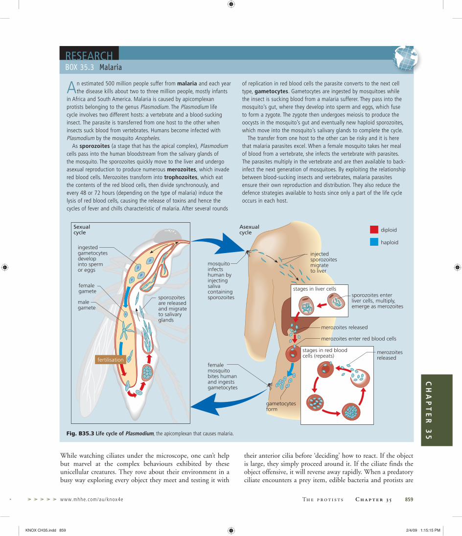
www.mhhe.com/au/knox4e Th e p r o t i s t s C h a p t e r 3 5 859
CH
AP
TE
R 3
5
While watching ciliates under the microscope, one can’t help but marvel at the complex behaviours exhibited by these unicellular creatures. They rove about their environment in a busy way exploring every object they meet and testing it with
their anterior cilia before ‘deciding’ how to react. If the object is large, they simply proceed around it. If the ciliate finds the object offensive, it will reverse away rapidly. When a predatory ciliate encounters a prey item, edible bacteria and protists are
An estimated 500 million people suffer from malaria and each year the disease kills about two to three million people, mostly infants
in Africa and South America. Malaria is caused by apicomplexan protists belonging to the genus Plasmodium. The Plasmodium life cycle involves two different hosts: a vertebrate and a blood-sucking insect. The parasite is transferred from one host to the other when insects suck blood from vertebrates. Humans become infected with Plasmodium by the mosquito Anopheles.
As sporozoites (a stage that has the apical complex), Plasmodium cells pass into the human bloodstream from the salivary glands of the mosquito. The sporozoites quickly move to the liver and undergo asexual reproduction to produce numerous merozoites, which invade red blood cells. Merozoites transform into trophozoites, which eat the contents of the red blood cells, then divide synchronously, and every 48 or 72 hours (depending on the type of malaria) induce the lysis of red blood cells, causing the release of toxins and hence the cycles of fever and chills characteristic of malaria. After several rounds
of replication in red blood cells the parasite converts to the next cell type, gametocytes. Gametocytes are ingested by mosquitoes while the insect is sucking blood from a malaria sufferer. They pass into the mosquito’s gut, where they develop into sperm and eggs, which fuse to form a zygote. The zygote then undergoes meiosis to produce the oocysts in the mosquito’s gut and eventually new haploid sporozoites, which move into the mosquito’s salivary glands to complete the cycle.
The transfer from one host to the other can be risky and it is here that malaria parasites excel. When a female mosquito takes her meal of blood from a vertebrate, she infects the vertebrate with parasites. The parasites multiply in the vertebrate and are then available to back-infect the next generation of mosquitoes. By exploiting the relationship between blood-sucking insects and vertebrates, malaria parasites ensure their own reproduction and distribution. They also reduce the defence strategies available to hosts since only a part of the life cycle occurs in each host.
BOX 35.3 MalariaRESEARCH
injectedsporozoitesmigrateto liver
sporozoites enterliver cells, multiply,emerge as merozoites
merozoites released
merozoitesreleased
merozoites enter red blood cells
stages in liver cells
stages in red bloodcells (repeats)
femalemosquitobites humanand ingestsgametocytes
gametocytesform
sporozoitesare releasedand migrateto salivaryglands
mosquitoinfectshuman byinjectingsalivacontainingsporozoites
ingestedgametocytesdevelopinto spermor eggs
femalegamete
malegamete
diploidAsexualcycle
haploid
Sexualcycle
fertilisation
Fig. B35.3 Life cycle of Plasmodium, the apicomplexan that causes malaria.
KNOX CH35.indd 859KNOX CH35.indd 859 2/4/09 1:15:15 PM2/4/09 1:15:15 PM

860 Part 5 Evolution and biodiversity For fu r ther read ing and rev i s ion
rapidly swallowed, sometimes after hot pursuit. Some preda-tory ciliates eat other ciliates.
Cilia are often arranged in clusters, cirri, that beat synchro-nously and work as paddles or feet for the cell. Sometimes cilia are arranged evenly over the surface and beat in rhythmic waves to propel cells. Cilia are all embedded in a layer of pro-tein fibres that are cross-linked. Elements of this layer are con-tractile, like miniature muscles. In certain species they allow the ciliate to change its shape. No plastids are found in ciliates. If it is true that a single secondary plastid acquisition created the chromalveolates, then ciliates must have lost the plastid.
Most ciliates are predatory, bacteria being the favoured prey. Prey are driven into an invagination, the buccal cav-ity, through which they are ingested. Undigested material is excreted through the cytoproct (Fig. 35.36).
Ciliates have two types of nuclei: a micronucleus and a macronucleus. Cells usually have one of each type but some cells have several macronuclei. The micronucleus is diploid, contains normal chromosomes with all the ciliate’s genes, and divides mitotically. The macronucleus develops from the micronucleus and contains multiple copies of genes on relatively short pieces of DNA, some of which are circular. Macronuclei divide simply by pinching into two approximate-ly equal halves. Because the macronucleus contains multiple copies of the ciliate’s genes (sometimes several thousand cop-
ies), it is not essential that the genetic material be accurately and equally divided, as occurs in mitosis. Messenger RNAs are transcribed from the multiple gene copies in the macro-nucleus, whereas the master genes in the micronucleus are not transcribed. The macronucleus seems to be the working copy of the DNA blueprint. The micronucleus is retained as a master copy to be passed on during the sexual process. This is analogous to making several working copies of a computer program for daily use, while keeping the original program safely stored away.
Ciliates reproduce asexually by binary fission (Chapter 8) along the short axis of the cell. Sexual reproduction is by con-jugation, when two individual cells of opposite mating types become attached to one another (Fig. 35.37). The two cells remain attached over a period of several hours and exchange haploid versions of their micronuclei produced by meiosis. The haploid micronuclei fuse to form diploid micronuclei. After conjugation, the macronuclei degenerate and new macronuclei are produced from the recombinant micronuclei. Conjugation in ciliates is essentially a form of sex in which haploid nuclei from two individuals are brought together without ever needing to form gamete cells.
Ciliates are predatory unicells with numerous cilia and two types • of nuclei.Ciliates reproduce sexually by conjugation.•
Euglenoids and kinetoplastsThis group includes flagellated unicells that are photosynthetic or heterotrophic, some being parasitic, others free-living. They all have an anterior depression (gullet) from which the flagella emerge. Some of the heterotrophic forms ingest food particles through this anterior gullet.
Euglenoid flagellatesEuglenoids (Fig. 35.38) are flagellates of both marine and freshwater habitats. About one-third of the species are photo-synthetic but the others lack chloroplasts and are heterotrophic. Even some of the chloroplast-containing forms are occasion-ally heterotrophic. When kept in darkness, their chloroplasts shrivel and they revert to heterotrophy, engulfing prey through the gullet.
The euglenoid chloroplast is similar to that of green algae and land plants in that it contains chlorophylls a and b and !-carotene. However, the euglenoid chloroplast is bounded by three membranes, and the organisation of genes on the chlo-roplast chromosome is unique. Unlike green algae, euglenoids do not store any starch in the chloroplast. It is not exactly clear how photosynthetic euglenoids came by their chloro-plasts. Most biologists think euglenoids stole their chloroplasts from green algae by secondary endosymbiosis. Products of
cytoproct
posteriorcontractilevacuole
buccal cavitycortex
cytostome
macronucleus
micronucleus
foodvacuole
cilia
anteriorcontractilevacuole
Fig. 35.36 A ciliate is a single cell that moves, responds to stimuli and captures prey. Food particles are wafted into the buccal cavity by the cilia and ingested through the cytostome into food vacuoles. Undigested material is ejected via the cytoproct.
KNOX CH35.indd 860KNOX CH35.indd 860 2/4/09 1:15:15 PM2/4/09 1:15:15 PM

www.mhhe.com/au/knox4e Th e p r o t i s t s C h a p t e r 3 5 861
CH
AP
TE
R 3
5
photosynthesis are stored as paramylon, a !-(1"3)-glucan, which forms solid granules in the cytoplasm. At the anterior end of the cell is a small red organ, the stigma, which is involved in the detection of light. Euglenoids usually swim with one long flagellum (a second short flagellum does not usually emerge from the gullet) and many species perform a sinuous gyration or crawling motion known as metaboly. Euglenoids are technically naked, having no cell wall or ornamentation outside the plasma membrane, but many species have an elaborate proteinaceous pellicle, comprising overlocking helical strips that inter-slide as the cell moves. Reproduction is principally by asexual division.
Flagellate parasites: kinetoplastsKinetoplasts are flagellate parasites known as trypanosomes and leishmanias and include disease-causing organisms of major medical and veterinary significance. Species of Phytomonas infect plants and are a major problem in coconut palms, oil palms, coffee trees and various fruit crops in Latin America.
Kinetoplasts are unicellular with two flagella. The two flagella are inserted into an apical depression. The name kinetoplast refers to the large mass of DNA present in the single mitochondrion near the base of the flagella. A kineto-plast is composed of thousands of tiny DNA circles, linked together like a chain, often forming an elongated rod-shaped structure in the mitochondrion (Fig. 35.39). The kineto-plast DNA also contains normal circular mitochondrial chromosomes.
Two Parameciumindividuals of differentmating types comeinto contact.
The diploidmicronucleus in eachdivides by meiosis toproduce four haploidmicronuclei.
Three of the haploidmicronucleidegenerate. Theremainingmicronucleus in eachdivides by mitosis.
Mates exchangemicronuclei.
In each individual, thenew micronucleusfuses with themicronucleus alreadypresent, forming adiploid micronucleus.
The macronucleusdisintegrates, and thediploid micronucleusdivides by mitosis toproduce two identicaldiploid micronucleiwithin each individual.
One of thesemicronuclei is theprecursor of themicronucleus for thatcell, and the othereventually gives rise tothe macronucleus.
macronucleusmicronucleus (2n)
haploidmicronucleus (n)
dipliodmicronucleus (2n)
Fig. 35.37 Sexual reproduction in the ciliate Paramecium. Two mature cells of opposite mating strains fuse in a process called conjugation.
flagellum
stigma
secondflagellum
gullet
basal body
contractilevacuole
pelliclestrip
nucleus
chloroplast
paramylongranule
Fig. 35.38 Euglenoids. (a) Diagram of a euglenoid. These flagellates can possess chloroplasts but are often heterotrophic. The helical proteinaceous strip forms a spiral pellicle, giving the cell its shape. Several paramylon granules are distributed throughout the cell. (b) This species of Euglena is photosynthetic but can also live heterotrophically.
(a)
(b)
KNOX CH35.indd 861KNOX CH35.indd 861 2/4/09 1:15:15 PM2/4/09 1:15:15 PM

862 Part 5 Evolution and biodiversity For fu r ther read ing and rev i s ion
Trypanosomes cause African sleeping sickness and nagana. These parasitic flagellates are free-swimming in the blood of humans and other vertebrates. Infection is usually transmit-ted by the blood-sucking tsetse fly. Occasional cross-infection occurs through bites from vampire bats. Another trypanosome causes Chagas’ disease (Fig. 35.40), which infects about 10–12 million people in South and Central America. Again the dis-ease is transmitted by blood-sucking insects, but in the case of Chagas’ disease the parasite is conveyed by bites of the so-called assassin bug (Fig. 35.40a). Charles Darwin is thought to have contracted Chagas’ disease while deliberately allowing assassin bugs to suck his blood in South America.
Leishmaniasis is an infection of macrophage cells (white blood cells that normally ingest foreign particles in the bloodstream) caused by the kinetoplast parasite Leishmania. Disease transmission is by sandflies and the parasite occurs in South and Central America, Africa, the Middle East, the Mediterranean and Asia. Relatively benign forms cause skin lesions (Fig. 35.41), but visceral leishmaniasis attacks macro-phages of the liver, spleen and bone marrow, often resulting in fatal anaemia.
A remarkable feature of trypanosomes, such as the kine-toplast parasite Leishmania, is their ability to survive in the host’s bloodstream and avoid elimination by the immune system. Trypanosomes do this by constantly changing the molecules on their surface. Thus, no sooner does the host mount an immune response (Chapter 26) to the invader, than
the trypanosomes slip into another ‘jacket’ that the immune system cannot yet ‘see’. The parasite has up to 1000 different versions of surface molecules that it produces by sequentially rearranging the genes that code for surface glycoproteins. In this way, the parasite can stay one step ahead of the host’s
mitochondrial cristae
chain of DNA minicircles
kinetoplastinner and outer membranesof mitochondrion Golgi apparatus
flagellum
Fig. 35.39 Kinetoplasts are parasitic flagellates. The name kinetoplast refers to a specialised mitochondrion containing thousands of DNA minicircles, which are visible here as a tangled mass of threads forming an elongate body. The kinetoplast lies at the base of the flagellum.
adhesionzone of cellwhere flagellumattaches
attached flagellum
mitochondrion
nucleus
basal body offlagellumkinetoplast
kinetoplasts
nucleus
Fig. 35.40 Flagellate parasites: kinetoplasts. (a) Chagas’ disease, caused by Trypanasoma cruzi, is transmitted by bites of the so-called assassin bug. (b) Diagram of a trypanosome cell. (c) Numerous kinetoplastid parasites showing the nuclei and kinetoplasts.
(a)
(b)
(c)
KNOX CH35.indd 862KNOX CH35.indd 862 2/4/09 1:15:16 PM2/4/09 1:15:16 PM

www.mhhe.com/au/knox4e Th e p r o t i s t s C h a p t e r 3 5 863
CH
AP
TE
R 3
5
immune system. Morphologically, kinetoplasts are similar to euglenoids and studies of gene sequences confirm that these groups are related. Euglenoids differ in that they are free-living, can have chloroplasts and never have kinetoplast DNA. Although kinetoplastids seem to lack a plastid, there is molecular evidence that they once were photosynthetic, possibly suggesting a reversion to heterotrophy.
Euglenoids and kinetoplasts are related flagellated cells.• Euglenoids are free-living, some of which have chloroplasts and • some of which engulf prey through an anterior gullet.Kinetoplasts are parasitic with a unique mitochondrion.•
Cercozoa and foramsThe Cercozoa and forams are a new group recently recognised on the basis of genetic data. They are diverse but many mem-bers form amoeboid cells. Cercozoa and forams often form pseudopodia that are reticulated (one pseudopod connects to others). One group has secondary green plastids, but most are heterotrophic.
Amoebae with second-hand chloroplasts: chlorarachniophytesChlorarachniophytes are a small group within the Cercozoa that includes only a handful of species restricted to marine environments. They are important, however, as they are the only members known to possess plastids. The best-known genus is Chlorarachnion (Fig. 35.42), which exists principally as a plasmodium—a mass of amoeboid cells. Individual cells are linked by a network of cytoplasmic strands called reticu-lopodia. The plasmodial network (which resembles a spider web, hence the name Chlor, green, plus arachnion, spider-like)
captures small prey, which are ingested. When starved, the plasmodium separates and forms individual walled cysts that subsequently release uniflagellate swarmers. These swarmers regenerate a plasmodium.
Chlorarachnion is also photosynthetic and each amoeba has several grass-green chloroplasts containing chlorophylls a and b, like green algae. Chlorarachnion chloroplasts are different from green algal chloroplasts, however, in that they are bound-ed by four membranes rather than two, and no starch is stored within the chloroplast. Associated with the Chlorarachnion chloroplast is a nucleus-like structure (a nucleomorph), prov-ing that Chlorarachnion acquired its chloroplast secondarily (see Fig. 35.13) by engulfing a photosynthetic eukaryote, pre-sumably a green alga.
More chalky protists: foramsForams are mostly marine heterotrophs that produce calcare-ous (CaCO3) shells. Sometimes the shell (also known as a test) can incorporate sand grains. Tests can be quite elaborate and have multiple chambers (Gr. forum, opening) for flotation. The chambers are often arranged in a spiral. Some forams, although unicellular, are particularly large (up to 12 cm in diameter in the case of Nummulites) and contain many symbiotic algae. The shells are mini-greenhouses with algal endosymbionts housed in thin-windowed chambers around the surface to capture light. The spiny foram Globigerina (Fig. 35.43) acts as ‘shepherd’ to a ‘flock’ of dinoflagellate symbionts. At night, algae are harboured safely inside the foram’s shell but each morning they venture out along the spines into the sunlight to photosynthesise.
About 45 000 species have been described, of which 90% are extinct and known only from fossil shells up to
Fig. 35.41 Leishmaniasis. In Costa Rica, the small scars produced by cutaneous leishmaniasis, caused by the kinetoplast parasite Leishmania, are known as the ‘seal of the forest’. More severe forms of leishmaniasis often result in death.
reticulopodia
cell
Fig. 35.42 Chlorarachnion has secondary chloroplasts obtained from engulfing a green algae. Cells produce reticulopodia that link to other cells to form a plasmodium capable of catching prey cells.
KNOX CH35.indd 863KNOX CH35.indd 863 2/4/09 1:15:17 PM2/4/09 1:15:17 PM

864 Part 5 Evolution and biodiversity For fu r ther read ing and rev i s ion
600 million years old. Forams were once so numerous that deposition of their skeletons produced large chalk depos-its. When building the great pyramids, Egyptian engineers noticed that the limestone blocks contained numerous num-mulites (Fig. 35.44), fossil remnants of the large foraminiferan Nummulites gizehensis. Foram fossils are indicators of geologic strata and are used extensively by the petroleum industry to characterise sediments in the search for fossil fuels. Forams
occur in great abundance both in plankton and on the seabed down to 10 000 m deep.
Not all eukaryotes with chloroplasts are close relatives. • Photosynthetic Chlorarachnion have captured and enslaved an algal prey cell, which they now use for photosynthesis, but their close relatives are non-photosynthetic.Forams are marine amoebae with shells, often housing • photosynthetic symbionts.
shell pseudopods
Fig. 35.43 The foraminiferan Globigerina has a calcareous shell through which pseudopods extend.
Fig. 35.44 Nummulites are coin-shaped foraminiferans. These examples with holes in the middle are from Great Keppel Island beach and measure approximately 1 cm in diameter, but much larger forms are known.
Protists are eukaryotes not belonging to the plant, animal or fungal kingdoms and have a variety of ways of gaining nutrition
! Protists can be unicellular, colonial or multicellular and have many cellular forms, including flagellates, amoebae, cysts, plasmodia, multicellular filaments and parenchymatous tissue.
! The majority of protists are aquatic and have flagella or cilia. They gain nutrition by photosynthesis, parasitism, predation and absorption.
The first eukaryotic organisms were probably similar to modern-day protists
! The nuclear membrane and endomembrane system probably evolved from invaginations (infolding) of the bacterial cell membrane that enveloped the nucleoid. Plastids and mitochondria are almost certainly derived from endosymbiotic bacteria that have become organelles in eukaryotic cells.
Major eukaryotic lineages, such as animals, plants and fungi, arose from different protist lineages, but many more independent protist lineages exist
! Choanoflagellates are marine protists that eat bacteria and detrital particles. Their resemblance to sponge collar cells suggests that choanoflagellates and sponges are close relatives and early offshoots of the animal lineage.
! Slime moulds are amoeboid and heterotrophic, aggregating to form colonies (cellular or acellular) with fruiting bodies that produce spores. They are distantly related to the fungi and animal lines of evolution.
! Other heterotrophic protists include amoeboid forms, such as actinopods and rhizopods. Actinopods are cells with radial skeletons and projections (axopods) with which they capture food. Rhizopods are amoebae that form pseudopodia.
K E Y C O N C E P T S A N D S U M M A R Y
KNOX CH35.indd 864KNOX CH35.indd 864 2/4/09 1:15:18 PM2/4/09 1:15:18 PM

www.mhhe.com/au/knox4e Th e p r o t i s t s C h a p t e r 3 5 865
CH
AP
TE
R 3
5
actinopods (p. 000)
agar (p. 000)
amoeba (p. 000)
antheridium (p. 000)
apical complex (p. 000)
apicomplexans (p. 000)
autotrophic (p. 000)
axoneme (p. 000)
axopod (p. 000)
axostyle (p. 000)
biotechnology (p. 000)
blade (p. 000)
brown alga (p. 000)
buccal cavity (p. 000)
carrageenan (p. 000)
centric, diatom (p. 000)
Cercozoa (p. 000)
chlorarachniophyte (p. 000)
choanoflagellate (p. 000)
chromist (p. 000)
chrysolaminarin (p. 000)
chrysophyte (p. 000)
ciguatera (p. 000)
ciliate (p. 000)
coccolith (p. 000)
coccolithophorid (p. 000)
coenocytic (p. 000)
conceptacle (p. 000)
conjugation (p. 000)
cortical alveoli (p. 000)
cryptomonad (p. 000)
cyanelle (p. 000)
cytoproct (p. 000)
diarrhoetic shellfish poisoning (DSP) (p. 000)
diatom (p. 000)
dinoflagellate (p. 000)
diplomonad (p. 000)
endosymbiosis, primary and secondary (p. 000)
euglenoid (p. 000)
foram (p. 000)
frustule (p. 000)
fucoxanthin (p. 000)
gametocyte (p. 000)
girdle (p. 000)
glaucophyte (p. 000)
green alga (p. 000)
gullet (p. 000)
haptonema (p. 000)
haptophyte (p. 000)
heterokont (p. 000)
heterotrophic (p. 000)
holdfast (p. 000)
kinetoplast (p. 000)
laminarin (p. 000)
Leishmania (p. 000)
Leishmaniasis (p. 000)
lorica (p. 000)
macronucleus (p. 000)
malaria (p. 000)
merozoite (p. 000)
metaboly (p. 000)
micronucleus (p. 000)
myxamoeba (p. 000)
nucleomorph (p. 000)
oogonium (p. 000)
oomycete (p. 000)
ovum (p. 000)
parabasalid (p. 000)
paralytic shellfish poisoning (PSP) (p. 000)
paramylon (p. 000)
pellicle (p. 000)
pennate, diatom (p. 000)
peptidoglycan (p. 000)
peridinin (p. 000)
phragmoplast (p. 000)
phycobilin (p. 000)
phycocyanin (p. 000)
phycoerythrin (p. 000)
phycoplast (p. 000)
pit connection (p. 000)
plasmodium (p. 000)
polyphyletic (p. 000)
protist (p. 000)
Protista (p. 000)
pseudoplasmodium (p. 000)
raphe (p. 000)
receptacle (p. 000)
red alga (p. 000)
K E Y T E R M S
Photosynthetic protists, commonly called algae, are diverse and are not all related. Primary plastids arose once in the ‘primary lineage’, including three groups—glaucophytes, red algae and green algae, which led to land plants
! Glaucophytes are photosynthetic flagellates with apparently primitive chloroplasts (cyanelles) that have a peptidoglycan wall like bacteria.
! Red algae (rhodophytes) are familiar red seaweeds. Most are multicellular and macroscopic and they lack flagella. They contain chlorophyll a and phycobilin pigments.
• Green algae (chlorophytes) include unicellular, colonial and multicellular forms, one group of which is the closest relative of land plants. Chloroplasts contain chlorophyll a and b, the product of photosynthesis is stored as starch and the cell walls are cellulosic.
Secondary plastid acquisitions created an enormously diverse new array of protists, some of which subsequently abandoned photosynthesis to become heterotrophic or even parasitic
! Chromists, or the ‘brown lineage’, are characterised by flagellar architecture and secondary plastids. They typically have one smooth flagellum directed posteriorly and one hairy flagellum directed anteriorly.
! Chromists include cryptomonads, chrysophytes (golden flagellates), haptophytes (such as coccolithophorids), diatoms, phaeophytes (brown algae, the largest protistans) and oomycetes (water moulds and downy
mildews). Most are photosynthetic, with chlorophylls a and c, but oomycetes absorb food through filamentous hyphae.
! Cryptomonads (biflagellates) clearly stole their plastids from eukaryotic prey cells that they ingested. Their nucleomorph is the relict nucleus of the engulfed algal cell.
Alveolates are unicells with distinctive vesicles, cortical alveoli, beneath the cell membrane. They include dinoflagellates, apicomplexans and ciliates
! Dinoflagellates have two flagella, one of which encircles the cell. Many are photosynthetic, containing chlorophylls a and c, and some are predatory.
! Apicomplexans are intracellular parasites of animals, causing diseases such as malaria. An apical complex is used to penetrate host cells.
! Ciliates are predatory unicells characterised by two types of nuclei and a covering of cilia.
Euglenoids and kinetoplasts are closely related flagellates that all have an anterior depression (gullet) from which the flagella emerge
! Euglenoids are free-living, some of which have chloroplasts and some of which engulf prey through an anterior gullet. Kinetoplasts, such as trypanosomes, are parasitic with unique mitochondria.
KNOX CH35.indd 865KNOX CH35.indd 865 2/4/09 1:15:18 PM2/4/09 1:15:18 PM

866 Part 5 Evolution and biodiversity For fu r ther read ing and rev i s ion
1. Choanoflagellate protists are morphologically similar to:
(a) red blood cells(b) sponge cells(c) sperm cells(d) macrophages
2. Chloroplasts of green algae contain:
(a) chlorophylls a and b(b) chlorophylls a and c(c) chlorophyll a and phycobilins(d) chlorophylls a, b and c
3. Parabasalid mitochondria are unique in that they:
(a) lack DNA(b) reduce protons to produce H2 gas(c) do not put electrons onto O2(d) all of the above
4. Flagellates with one smooth flagellum directed posteriorly and one hairy flagellum directed anteriorly are known as:
(a) trypanosomes(b) dinoflagellates(c) heterokonts(d) euglenoids
5. The storage product in euglenoids is:
(a) paramylon(b) starch(c) chrysolaminarin(d) glycogen
6. The flattened sacs of membrane, known as alveoli, beneath the plasma membrane, are characteristic of which protists?
(a) ciliates(b) dinoflagellates(c) apicomplexans(d) all of the above
7. A pseudoplasmodium is:
(a) an aggregation of individual cells(b) a large multinucleate cell(c) a false form of malaria(d) a reproductive structure of red algae
8. Fucoxanthin is an accessory pigment found in:
(a) protostelids(b) red algae
(c) green algae(d) haptophytes
9. The parasite that caused a health scare in Sydney water reservoirs in 1998 was:
(a) an amoeba(b) the diplomonad Giardia(c) a pseudoplasmodium(d) a trypanosome
10. Which type of alga has plastids from a secondary endosymbiosis?
(a) cryptomonads(b) chlorophytes(c) rhodophytes(d) charophytes
1. What major technological advances have allowed biologists to discover great diversity and previously unrecognised evolutionary relationships in the protists? Why is the kingdom Protista considered to be an artificial taxon? (p. 000)
2. Make a table comparing the main distinguishing features of red, green and brown algae. What useful substances are obtained from red, green and brown algae? (p. 000)
3. (a) Why are brown algae called heterokonts and what are their closest relatives? (p. 000)
(b) What are the closest relatives of the green algae? (p. 000)
4. What features would enable you to identify a protist as (a) a euglenoid or (b) a dinoflagellate? Describe how each of these protists moves and how they gain their nutrition. (pp. 000, 000)
5. What are red tides? Why have they become a problem in Australia? (p. 000)
6. What is unusual about the genetic material in ciliates? How do ciliates reproduce sexually? (p. 000)
7. What type of protist is a trypanosome? How do these parasites avoid being eliminated by the immune system when they are in the bloodstream of their host? (p. 000)
8. What groups of protists belong to the alveolates and what characteristics do they have in common? (p. 000)
9. What is the apical complex? Which protists possess one? What does it do? (p. 000)
10. What is the difference between cellular slime moulds and plasmodial slime moulds? (p. 000)
S E L F - A S S E S S M E N T Q U E S T I O N S
red tide (p. 000)
reticulopodium (p. 000)
rhizopod (p. 000)
rhodophyte (p. 000)
silicoflagellate (p. 000)
slime mould (p. 000)
sorocarp (p. 000)
sporozoite (p. 000)
stigma, of Euglena (p. 000)
stipe (p. 000)
stonewort (p. 000)
test (p. 000)
thallus (p. 000)
transverse flagellum (p. 000)
trophozoite (p. 000)
trypanosome (p. 000)
valve (p. 000)
zooxanthella (p. 000)
R E V I E W Q U E S T I O N S
KNOX CH35.indd 866KNOX CH35.indd 866 2/4/09 1:15:19 PM2/4/09 1:15:19 PM

www.mhhe.com/au/knox4e Th e p r o t i s t s C h a p t e r 3 5 867
CH
AP
TE
R 3
5
The protists
1. Which protist causes jarrah dieback? What features of this protist explain the extensive spread of the disease through Australian forests? How would you go about controlling the spread of this disease in a logging area or national park?
2. It has been determined that not all protists that have a simple cellular structure, such as those apparently lacking mitochondria, are early primitive lineages among the eukaryotes. Discuss why this is the case.
3. Describe the life cycle of the protist that causes malaria and suggest why parasitising two different hosts may have been a selective advantage.
4. What do we mean by primary endosymbiosis and secondary endosymbiosis? What is the impact of these processes on eukaryotic
diversity? (Hint: Add up the number of species in groups with secondary plastids.)
Science research
5. What has been the impact of technologies such as electron microscopy, biochemistry and gene sequencing on our understanding of protist evolution?
Applying science
6. How could we exploit the existence of a non-photosynthetic plastid in malaria parasites to combat the disease?
7. Algae grown by aquaculture are used in generating important food additives, biofuels and gelling agents. Can you think of a way to employ algae to help with the climate damaging CO2 emissions caused by Australia’s heavy use of coal for energy generation? (Hint: Think about what goes on in photosynthesis.)
E X T E N S I O N Q U E S T I O N S
SUGGESTED FURTHER READINGCanter-Lund, H. and Lund, J. W. G. (1995). Freshwater Algae. Bristol: Biopress.Encyclopedia of Life Sciences. (2006). Chichester: John Wiley & Sons Ltd. Online. Available: http://www.els.net, viewed
15 December 2008 (requires subscription).Entwisle, T. J., Sonneman, J. A., Lewis, S. H. (1997). Freshwater Algae in Australia. Sydney: Sainty & Associates.Margulis, L., Corliss, J. O., Melkonian, M., Chapman, D. J. (1989). Handbook of Protoctista. Boston: Jones & Bartlett.O’Kelly, C. J. and Littlejohn, T. (2004). Protist Image Data. Online. Available: http://www.bch.umontreal.ca/protists/, viewed
15 December 2008.Patterson, D. J. (1996). Free-living Freshwater Protozoa. New York: John Wiley & Sons.Triemer, R. E. and Farmer, M. A. (1999). The Euglenid Project. Online. Available: http://euglena.msu.edu, viewed
15 December 2008.
ANSWERS TO SELF-ASSESSMENT
QUESTIONS1. (b), 2. (a), 3. (b), 4. (c), 5. (a), 6. (d), 7. (a), 8. (d),
9. (b), 10. (a)
KNOX CH35.indd 867KNOX CH35.indd 867 2/4/09 1:15:19 PM2/4/09 1:15:19 PM