Embed Size (px)
Citation preview
THE PREVALENCE OF PARVOVIRUS B19 INFECTION IN A COHORT OF
HIV INFECTED PATIENTS WITH SEVERE ANAEMIA
Dr Nadia Glatt
A Research Report submitted to the Faculty of Health Sciences, University of Witwatersrand,
Johannesburg in part fulfilment of the requirements for the degree of Masters of Medicine in the
branch of Haematology
Johannesburg, 2017
i
DECLARATION
I, Nadia Glatt, declare that this Research Report is my own work. It is being submitted for the degree
of Master of Medicine (in the branch of Haematology) to the University of the Witwatersrand,
Johannesburg. It has not been submitted before for any degree or examination at this or any other
university.
........................................
Nadia Glatt
...01. day of ........03......... 2017
ii
ABSTRACT
Parvovirus B19, a single stranded deoxyribose nucleic acid (DNA) virus, is known to cause anaemia in
the setting of immune suppression such as Human Immunodeficiency Virus (HIV) infection. It is
typically associated with a severe, isolated, normochromic normocytic anaemia and
reticulocytopenia. The bone marrow classically shows a pure red cell aplasia (PRCA) with absence of
maturing erythropoiesis, giant pronormoblasts and a variable presence of erythroid viral inclusions.
Parvovirus B19 infection is a treatable cause of anaemia using red cell transfusions, intravenous
immunoglobulin (Ig) therapy and in the setting of HIV, antiretroviral therapy. In the setting of HIV
infection, testing for Parvovirus B19 infection using molecular techniques such as polymerase chain
reaction (PCR) are preferred over serological methods, as antibodies are either not made or are
dysfunctional. In South Africa, the prevalence of Parvovirus B19 infection in the HIV infected
population with severe anaemia is not known. The aim of this study was to assess the prevalence of
Parvovirus B19 in a cohort of HIV infected patients with severe anaemia.
The Inclusion criteria for specimens into the study included all specimens submitted for a bone
marrow examination submitted for routine diagnostic workup between January 2012 and November
2013 at two academic hospitals in Johannesburg. The study population included HIV infected
patients with severe anaemia, defined as haemoglobin levels <8 g/dl for men and non-pregnant
women. Real-time PCR using the PrimerDesign™ genesig® Kit for Human Parvovirus B19
(Southampton, United Kingdom) was performed on DNA extracted from bone marrow aspirate
slides of these patients. The Parvovirus B19 results (qualitative and semi-quantitative values) were
assessed in conjunction with various Parvovirus B19-related clinical and laboratory parameters
obtained from the laboratory information system (LIS).
The prevalence of Parvovirus B19 in this cohort of patients was 13.3% (19/143). PCR testing was
possible even in samples that were suboptimal for morphological assessment, with 36.8% (7/19) of
the Parvovirus B19 infection being observed in these samples. Of note, 31.6% (6/19) of the positive
iii
samples were not requested for Parvovirus B19 testing by the clinician or pathologist, indicating that
it is being under diagnosed in this population. PRCA was not observed in all Parvovirus B19 positive
samples, with a sensitivity and specificity of 60.0% and 85.1% respectively. Alternate causes of
anaemia were present in 42.1% (8/19) of the Parvovirus B19 positive samples, including 21.1%
(4/19) of cases which showed Mycobacterium Tuberculosis infection, 5.3% (1/19) with iron
deficiency and 15.8% (3/19) of cases with marrow infiltration by malignancy. This highlights the
importance of excluding Parvovirus B19 infection even in the setting of alternate causes of anaemia.
In patients with severe anaemia and both HIV infection and Parvovirus B19-positivity, there was no
statistically significant correlation between Parvovirus B19 viral load and HIV viral load, haemoglobin
(Hb) level or CD4 count. Parvovirus B19 positivity was higher than expected in HIV virally suppressed
patients, with a prevalence of 18.5% (5/27). However the CD4 counts in these samples were low
(<350 cells/µl), suggesting that although viral suppression had been achieved, there was inadequate
immune reconstitution to mount an effective humoral response to control the Parvovirus B19
infection.
Serology for IgM as a method for diagnosing Parvovirus B19 infection showed poor sensitivity (60%)
but good specificity (100%) suggesting that this is an inadequate screening test in the setting of HIV
infection.
The Parvovirus B19 positive samples had statistically significant lower reticulocyte production index
(RPI) than the Parvovirus B19 negative samples. The negative predictive value of an RPI was 100%.
Although this is a retrospective pilot study, notable findings were observed. In the setting of HIV
infection and severe anaemia, Parvovirus B19 infection may be diagnosed by PCR even in the
following scenarios: a negative IgM serology result, no morphological evidence of a PRCA, presence
of other causes to explain the anaemia and confirmed HIV viral suppression.
Parvovirus B19 is a treatable cause of anaemia and therefore an important entity to exclude. The

iv
cost of molecular diagnosis of parvovirus B19 is relatively higher than using serological methods,
therefore should only be performed in the correct clinical setting. In HIV infected patients with grade
four anaemia (Hb <6g/dl) and a reduced RPI, these findings support the use of molecular diagnosis
for Parvovirus B19 infection regardless of other clinical and laboratory findings.
ACKNOWLEDGEMENTS
I would like to express my sincere appreciation to the following people who assisted me with this
research report:
Dr Robyn Marshall, Dr Michelle Bronze and Dr Sergio Carmona for their continued support, advice
and encouragement.
The PCR laboratory staff, in particular Vibha Kana, for their assistance.
Dr Jenifer Vaughan for her assistance in data collection.
v
TABLE OF CONTENTS
DECLARATION .......................................................................................................................................... i
ABSTRACT ................................................................................................................................................ ii
ACKNOWLEDGEMENTS .......................................................................................................................... iv
TABLE OF CONTENTS ............................................................................................................................... v
LIST OF TABLES ..................................................................................................................................... viii
LIST OF FIGURES ..................................................................................................................................... ix
NOMENCLATURE ..................................................................................................................................... x
ETHICS .................................................................................................................................................... xi
1 INTRODUCTION ............................................................................................................................... 1
1.1 HIV infection and Anaemia ..................................................................................................... 1
1.2 Parvovirus B19 infection ......................................................................................................... 6
1.2.1 Definition of Parvovirus B19 ........................................................................................... 6
1.2.2 Pathogenesis of Parvovirus B19 ...................................................................................... 6
1.2.3 Prevalence of Parvovirus B19 ......................................................................................... 8
1.2.4 Clinical presentation of Parvovirus B19 in the HIV infected host ................................... 9
1.2.5 Investigations .................................................................................................................. 9
1.2.5.1 Bone marrow investigation ........................................................................................... 10
1.2.5.2 Serological and Molecular Testing for Parvovirus B19 testing ................................. 12
1.2.6 Treatment ..................................................................................................................... 14
1.2.7 Monitoring response to therapy ................................................................................... 14
1.2.8 Prognosis ....................................................................................................................... 15
1.3 Aim of the study .................................................................................................................... 15
2 RESEARCH QUESTIONS .................................................................................................................. 17
3 MATERIALS AND METHODS .......................................................................................................... 18
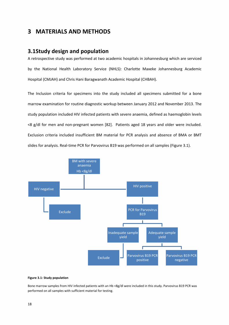
3.1 Study design and population ................................................................................................ 18
3.2 Data collection ...................................................................................................................... 19
3.3 Diagnostic testing .................................................................................................................. 19
3.3.1 FBC, Differential count and RPI ..................................................................................... 19
3.3.2 Haematinic studies ........................................................................................................ 20
3.3.3 Serology Parvovirus B19 ............................................................................................... 20
3.3.4 CD4 counts .................................................................................................................... 21
3.3.5 Morphologic analysis .................................................................................................... 22
vi
3.3.5.1 Bone marrow slides preparation .............................................................................. 22
3.3.5.2 Ziehl-Neelsen stain .................................................................................................... 23
3.3.5.3 Immunohistochemical stain for Parvovirus B19 ....................................................... 23
3.4 PCR for Parvovirus B19 ......................................................................................................... 24
3.4.1 The TaqMan principle ................................................................................................... 24
3.4.2 DNA extraction .............................................................................................................. 25
3.4.3 PCR set-up ..................................................................................................................... 25
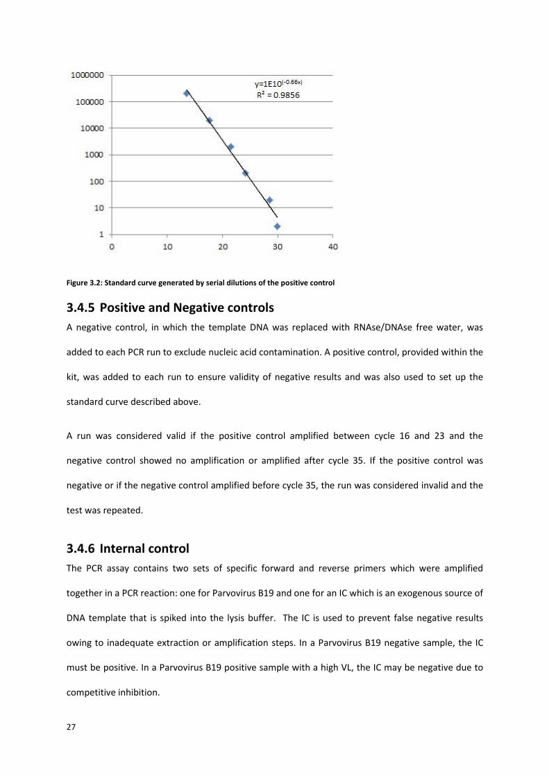
3.4.4 Standard curve .............................................................................................................. 26
3.4.5 Positive and Negative controls...................................................................................... 27
3.4.6 Internal control ............................................................................................................. 27
3.4.7 PCR amplification and detection ................................................................................... 28
3.4.8 PCR for the house keeping gene Beta Actin (ACTB) ...................................................... 28
3.5 Statistical analysis ................................................................................................................. 29
4 RESULTS......................................................................................................................................... 30
4.1 Study population ................................................................................................................... 30
4.2 Patient demographics and haematology findings ................................................................ 30
4.2.1 Study Population Demographic .................................................................................... 31
4.2.2 Clinical information ....................................................................................................... 32
4.2.3 Laboratory investigations.............................................................................................. 32
4.2.3.1 Full blood count ........................................................................................................ 32
4.2.3.2 CD4 count and HIV viral load .................................................................................... 32
4.2.3.3 Reticulocyte production index .................................................................................. 33
4.2.3.4 Haematinics ............................................................................................................... 33
4.2.3.5 Parvovirus B19 serology ............................................................................................ 34
4.2.3.6 Correlation between viral load of Parvovirus B19 eluted from the bone marrow and
haemoglobin level ..................................................................................................................... 34
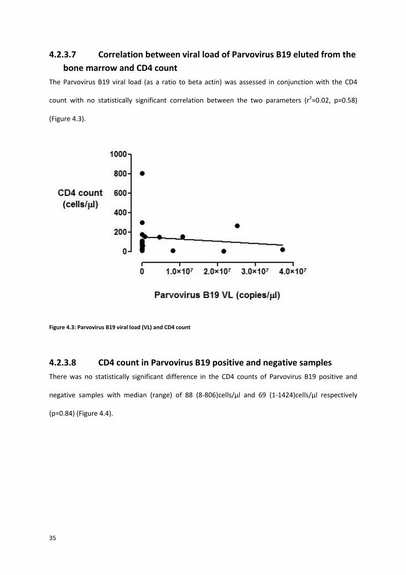
4.2.3.7 Correlation between viral load of Parvovirus B19 eluted from the bone marrow and
CD4 count .................................................................................................................................. 35
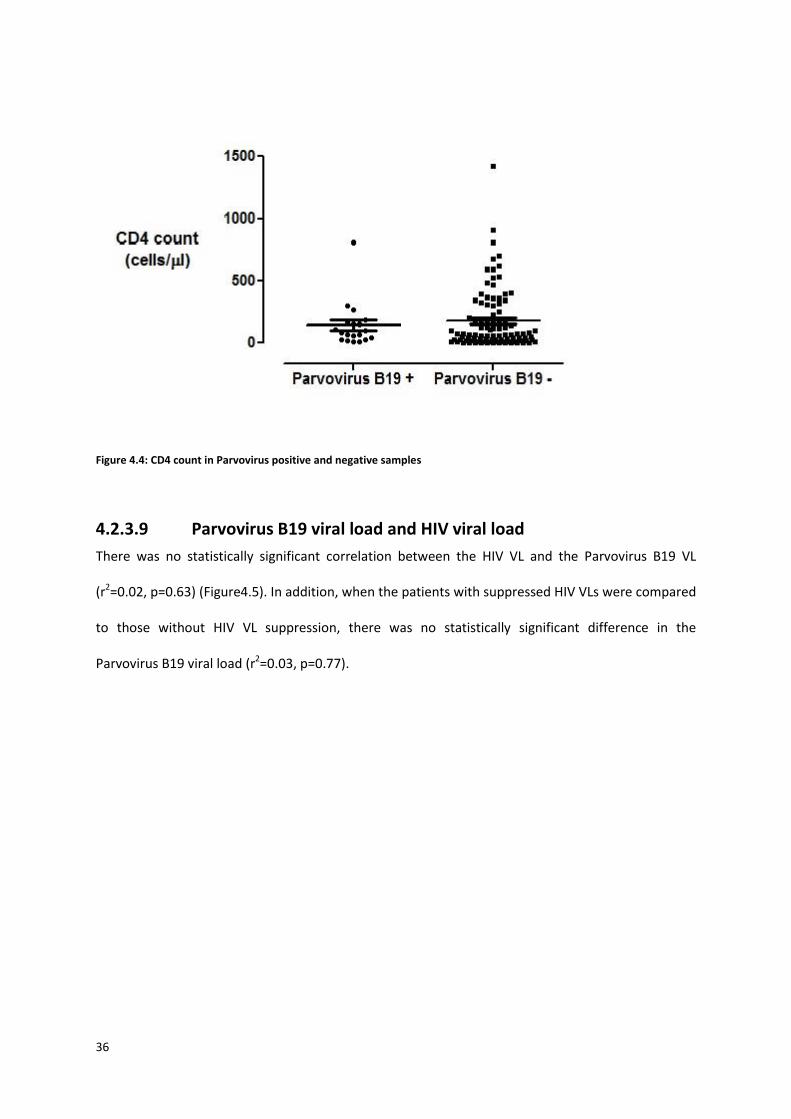
4.2.3.8 CD4 count in Parvovirus B19 positive and negative samples ................................... 35
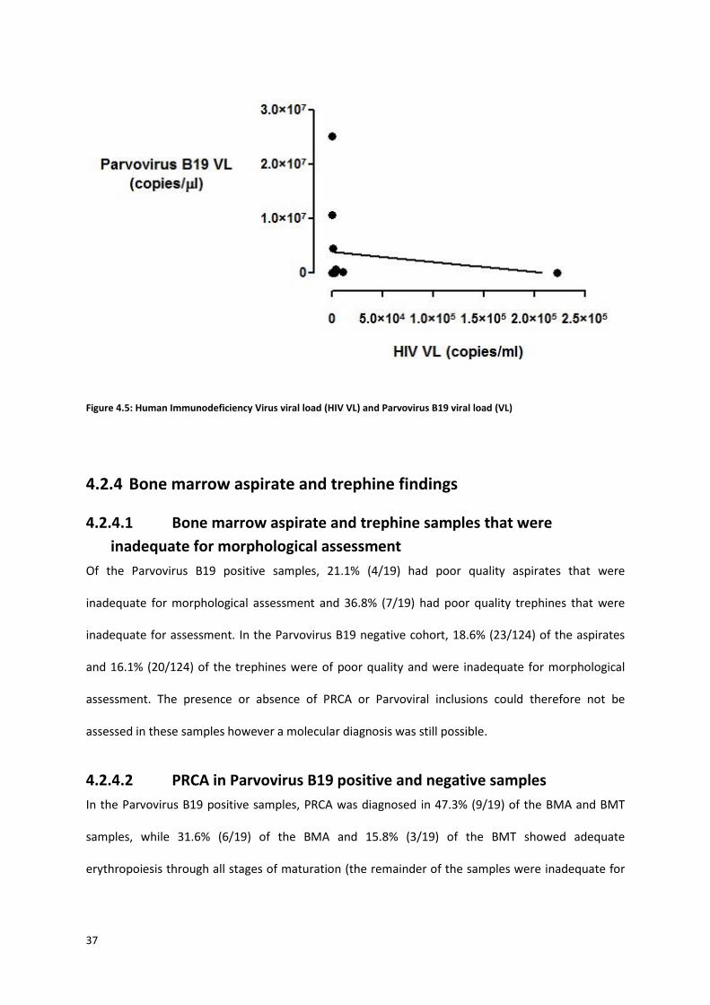
4.2.3.9 Parvovirus B19 viral load and HIV viral load ............................................................. 36
4.2.4 Bone marrow aspirate and trephine findings ............................................................... 37
4.2.4.1 Bone marrow aspirate and trephine samples that were inadequate for
morphological assessment ........................................................................................................ 37
4.2.4.2 PRCA in Parvovirus B19 positive and negative samples ........................................... 37
4.2.4.3 Parvovirus B19 inclusions .......................................................................................... 38
vii
4.2.4.4 Immunohistochemical stains for Parvovirus B19 ...................................................... 38
4.2.4.5 Morphologic evidence of other pathology ............................................................... 38
4.2.5 Parvovirus B19 PCR requested as routine investigation by clinician/pathologist ........ 39
5 DISCUSSION ................................................................................................................................... 40
5.1 Prevalence of Parvovirus B19 in this cohort ......................................................................... 40
5.2 The association between Parvovirus B19 VL and the degree of anaemia and HIV immune
suppression ....................................................................................................................................... 42
5.2.1 Association between Parvovirus B19 VL and anaemia ................................................. 42
5.2.2 Association between Parvovirus B19 VL and CD4 count .............................................. 42
5.2.3 Association between Parvovirus B19 VL and HIV VL .................................................... 43
5.3 Clinical utility of laboratory parameters to distinguish Parvovirus B19 positivity ................ 44
5.3.1 Parvovirus quantification .............................................................................................. 44
5.3.2 Contributing factors to anaemia in this cohort ............................................................. 45
5.3.2.1 Parvovirus B19 positivity and co-existing causes of anaemia ................................... 45
5.3.2.2 Anaemia and associated cytopenias ......................................................................... 45
5.3.3 Reticulocyte production index ...................................................................................... 46
5.3.4 Parvovirus B19 PCR and serology (IgM) testing ............................................................ 46
5.4 Bone marrow morphology as a diagnostic tool for Parvovirus B19 infection ...................... 47
5.4.1 Bone marrow aspirate and trephine findings ............................................................... 47
5.4.2 Viral inclusions on trephine biopsy and Parvovirus B19 positivity ............................... 48
5.4.3 Diagnosis of Parvovirus B19 infection in suboptimal bone marrow specimens ........... 48
5.5 Limitations............................................................................................................................. 49
5.6 Recommendations ................................................................................................................ 50
6 CONCLUSION ................................................................................................................................. 52
7 APPENDIX A ................................................................................................................................... 54
REFERENCES .......................................................................................................................................... 59
viii
LIST OF TABLES
Table Page
1.1 Anaemia and reticulocyte production index (RPI) 2
1.2 Causes of anaemia in HIV infection 4
3.1 Reagent preparation for Parvovirus B19 24
3.2 Amplification protocols 26
3.3 Reagent preparation for B actin (ACTB) 26
4.1 Patient demographics and haematology findings- comparison of pertinent demographic, clinical and laboratory findings of Parvovirus B19 positive and negative groups
29
4.2 Alternative causes of cytopenias in Parvovirus B19 positive patients 37
A1 Data information sheet containing data collected for Parvovirus B19 positive and
negative samples
50
A2 Reference ranges used for parameters assessed in this study 51
A3 Definitions of terms used in the text 52
A4 Parvovirus B19 positive data information 53
A5 Turn it in report 54
ix
LIST OF FIGURES
Figure Page
1.1 Hepcidin and anaemia of chronic inflammation 5
1.2 Schematic representation of Parvovirus B19 genome 6
1.3 Bone marrow aspirate in a patient with Parvovirus B19 infection 11
1.4 Bone marrow trephine biopsy in a patient with Parvovirus B19 infection 12
1.5 Bone marrow trephine biopsy with immunohistochemical staining in a patient
with Parvovirus B19 infection
12
3.1 Study population 17
3.2 Standard curve generated by serial dilutions of the positive control 25
4.1 Flow diagram of sample selection 28
4.2 Parvovirus B19 viral load versus haemoglobin level 32
4.3 Parvovirus B19 viral load and CD4 count 33
4.4 CD4 count in Parvovirus B19 positive and negative samples 34
4.5 HIV viral load and Parvovirus B19 viral load 35
x
NOMENCLATURE
3TC lamivudine
ACTB beta actin
ARV anti retroviral therapy
AZT zidovudine
BM bone marrow
BMA bone marrow aspirate
BMT bone marrow trephine
CHBAH Chris Hani Baragwanath Academic Hospital
CMJAH Charlotte Maxeke Johannesburg Academic Hospital
Ct cycle threshold
DLBCL diffuse large B cell lymphoma
DNA deoxyribose nucleic acid
EBV Epstein-Barr virus
ELISA enzyme linked immunosorbent assay
FBC full blood count
FNR false negative rate
FRET fluorescent resonance energy transfer
Hb haemoglobin
HIV human immunodeficiency virus
IC internal control
Ig immunoglobulin
IL interleukin
INH isoniazide
K2EDTA dipotassium ethylenediaminetetraacetic acid
KS Kaposi sarcoma
LIS laboratory information system
MCHC mean cell haemoglobin concentration
MCV mean cell volume
NHLS National Health Laboratory Service
NPV negative predictive value
ORF open reading frame
PCR polymerase chain reaction
PLT platelet
PPV positive predictive value
PRCA pure red cell aplasia
RNA ribonucleic acid
RPI reticulocyte production index
RQ PCR real time polymerase chain reaction
SD standard deviation
TGF- tumour growth factor beta
VL viral load
VP viral protein
WCC white cell count

xi
ETHICS
This study was approved by the Medical Ethics Committee of the University of Witwatersrand,
Johannesburg, South Africa. Ethics clearance number: M121143.
1
1 INTRODUCTION
Parvovirus B19, a single stranded deoxyribose nucleic acid (DNA) virus belonging to the genus
Erythrovirus of the family Parvoviridae [1], infects humans and causes various health sequela
depending on the person’s age and general health condition. In the setting of immunsuppression
such as human immunodeficiency virus (HIV) infection, it is known to cause anaemia [2]. The cause
of anaemia in HIV infected persons is most often multifactorial. As South Africa has the highest
prevalence of HIV infection in the world [3] and since cytopenias, in particular anaemia [4], are
known to be an independent prognostic indicator of outcome in HIV infected patients [5], it is
especially important to assess in detail all the causes of anaemia in this setting.
1.1 HIV infection and Anaemia HIV is a single-stranded, enveloped ribonucleic acid (RNA) virus and a member of the genus
Lentivirus of the family Retroviridae. It is transmitted via blood and other body fluids and targets
host immune cells, specifically CD4+ T cells, macrophages and dendritic cells. The virus evades
immune detection by incorporating itself into the target cell genome: viral RNA is reverse
transcribed into double-stranded DNA by a virally encoded reverse transcriptase and is then
integrated into the host cell DNA by a virally encoded integrase and host co-factors. The virus then
becomes latent or is transcribed, producing new RNA genomes and viral proteins that are packaged
and released from the cell as new virus particles that are capable of continued replication [6].
Infection with HIV leads to immune dysfunction through various mechanisms of CD4+ T-cell
depletion and abnormal immune activation. The result is a paralysed immune system that is
susceptible to opportunistic infections and malignancies. As immune dysregulation progresses, all
organs are affected directly or indirectly by the virus [6]. This immune dysregulation has a number of
2
consequences which include the common finding of persistent and often multiple cytopenias as
demonstrated by the full blood count (FBC) parameters.
Cytopenias are well described in HIV infection [7-9], with the degree of cytopenia correlating with
the degree of infection [8]. Anaemia, the most common cytopenia, is also the most common
haematological complication of HIV infection [4, 5] and, affects at least 10-20% of people at
presentation and 70-95% during the course of infection [4, 7]. Studies have demonstrated that
anaemia is an independent prognostic indicator of outcome [5] and is associated with increased
morbidity and mortality independent of CD4 count and HIV viral load (VL) [4, 8, 9]. A large
observational cohort study including 19,213 HIV infected people throughout the United States
showed that median survival was significantly shortened (21.5 months compared to 30.3 months
respectively, P <0.05) in anaemic (Haemoglobin (Hb) <10g/dl) versus non-anaemic controls when
matched for other parameters regardless of the underlying cause [10].
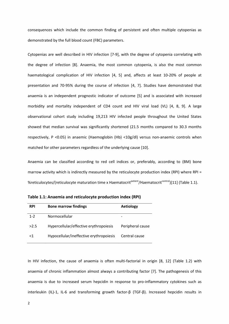
Anaemia can be classified according to red cell indices or, preferably, according to (BM) bone
marrow activity which is indirectly measured by the reticulocyte production index (RPI) where RPI =
%reticulocytes/(reticulocyte maturation time x Haematocritpatient/Haematocritcontrol)[11] (Table 1.1).
Table 1.1: Anaemia and reticulocyte production index (RPI)
RPI Bone marrow findings Aetiology
1-2 Normocellular -
>2.5 Hypercellular/effective erythropoiesis Peripheral cause
<1 Hypocellular/ineffective erythropoiesis Central cause
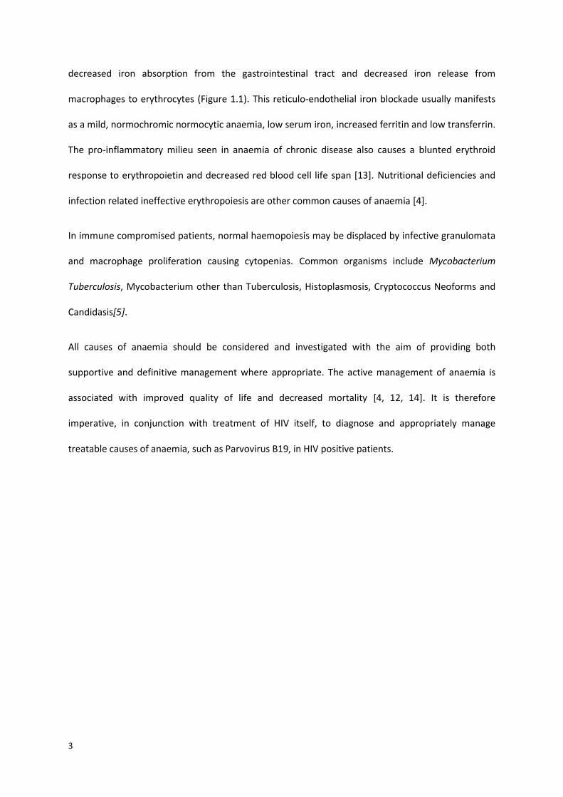
In HIV infection, the cause of anaemia is often multi-factorial in origin [8, 12] (Table 1.2) with
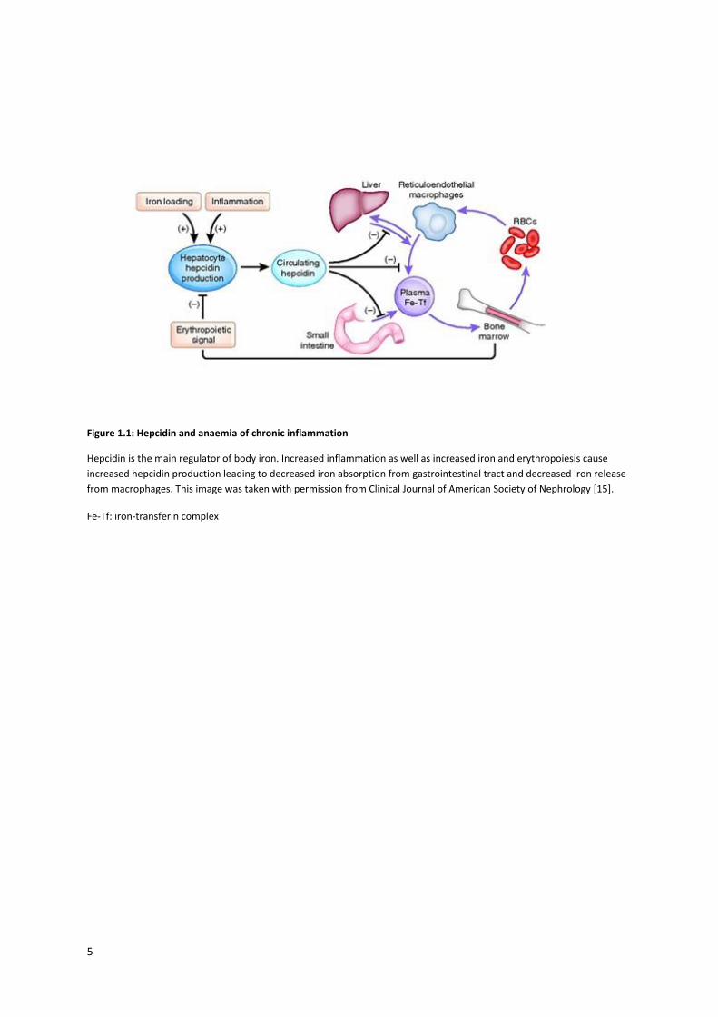
anaemia of chronic inflammation almost always a contributing factor [7]. The pathogenesis of this
anaemia is due to increased serum hepcidin in response to pro-inflammatory cytokines such as
interleukin (IL)-1, IL-6 and transforming growth factor-β (TGF-β). Increased hepcidin results in
3
decreased iron absorption from the gastrointestinal tract and decreased iron release from
macrophages to erythrocytes (Figure 1.1). This reticulo-endothelial iron blockade usually manifests
as a mild, normochromic normocytic anaemia, low serum iron, increased ferritin and low transferrin.
The pro-inflammatory milieu seen in anaemia of chronic disease also causes a blunted erythroid
response to erythropoietin and decreased red blood cell life span [13]. Nutritional deficiencies and
infection related ineffective erythropoiesis are other common causes of anaemia [4].
In immune compromised patients, normal haemopoiesis may be displaced by infective granulomata
and macrophage proliferation causing cytopenias. Common organisms include Mycobacterium
Tuberculosis, Mycobacterium other than Tuberculosis, Histoplasmosis, Cryptococcus Neoforms and
Candidasis[5].
All causes of anaemia should be considered and investigated with the aim of providing both
supportive and definitive management where appropriate. The active management of anaemia is
associated with improved quality of life and decreased mortality [4, 12, 14]. It is therefore
imperative, in conjunction with treatment of HIV itself, to diagnose and appropriately manage
treatable causes of anaemia, such as Parvovirus B19, in HIV positive patients.
4
Table 1.2: Causes of anaemia in HIV infection
Decreased production Increased production
Drugs Haemolysis
Zidovudine Thrombotic thrombocytopenic purpura
Trimethoprin-sulfamethoxale G6PD deficiency
Amphotericin B Autoimmune haemolytic anaemia
Ganciclovir Bleeding
Dapsone Drugs
Deficiencies Infection
Erythropoietin Cytomegalo virus
Iron Candida
Folate Hypersplenism
Vitamin B12 Infection
Infection Lymphoma
HIV Haemophagocytic lymphohistiocytosis
Parvovirus B19 Cirrhosis
Mycobacterium avium
Mycobacterium tuberculosis
Cytomegalo virus
Epstein-Barr virus
Cryptococcus neoformans
Pneumocystis carinii pneumonia
Histoplasma capsulatum
Neoplasms
Non-Hodgkin lymphoma
Multiple myeloma
Castleman's disease
Hodgkin lymphoma
Other
Anaemia of chronic disease
Haemophagocytic lymphohistiocytosis
Pre-exisiting condition
5
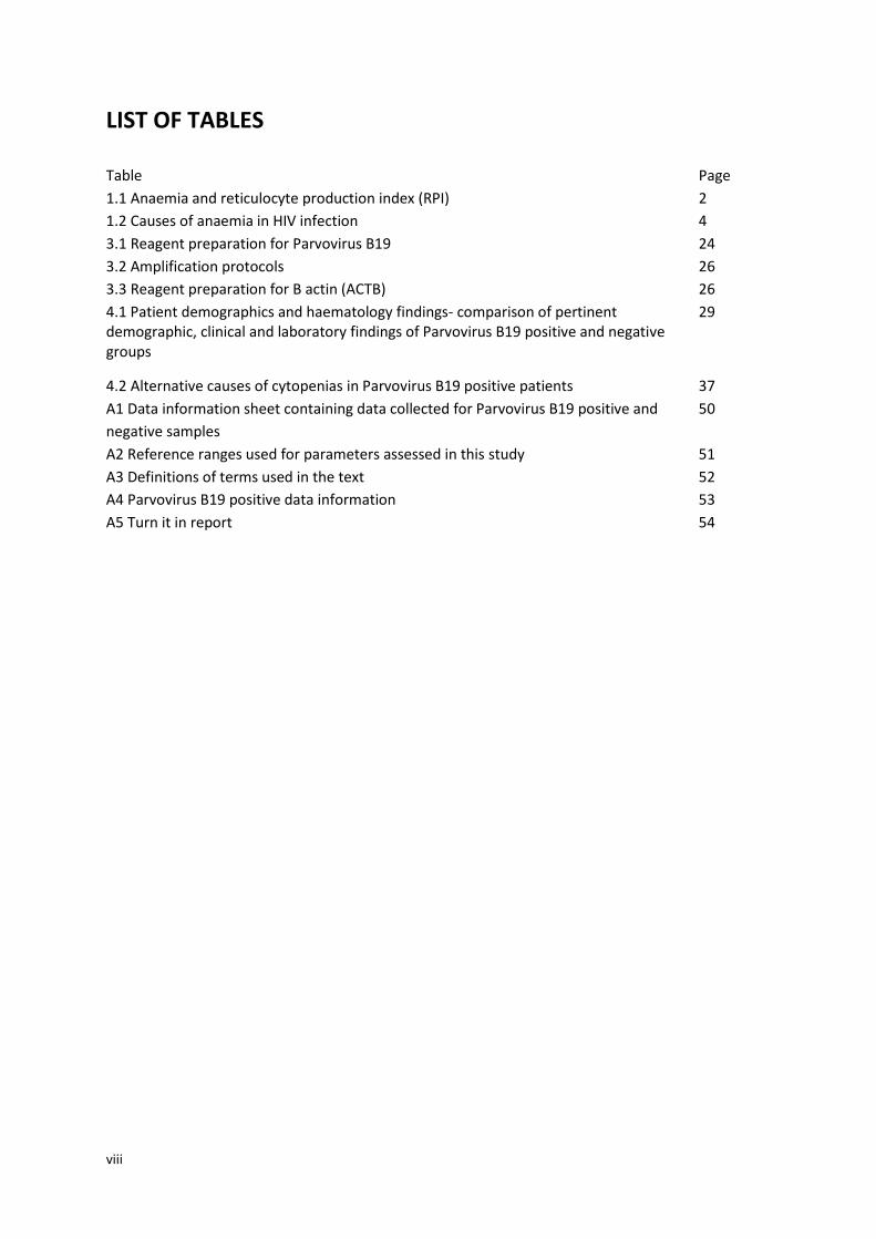
Figure 1.1: Hepcidin and anaemia of chronic inflammation
Hepcidin is the main regulator of body iron. Increased inflammation as well as increased iron and erythropoiesis cause
increased hepcidin production leading to decreased iron absorption from gastrointestinal tract and decreased iron release
from macrophages. This image was taken with permission from Clinical Journal of American Society of Nephrology [15].
Fe-Tf: iron-transferin complex
6
1.2 Parvovirus B19 infection
1.2.1 Definition of Parvovirus B19
Parvovirus B19 is a small non-enveloped, single stranded DNA virus with a genome size of 5.6kb
belonging to the genus Erythrovirus of the family Parvoviridae [1]. There are three genotypes of
Parvovirus B19 that are known to cause anaemia in humans [16, 17]. While genotype 1 is the
prototype [18, 19] and the most common genotype tested for in the United States and European
countries [20], all three genotypes are present in varying levels in South Africa [21].
1.2.2 Pathogenesis of Parvovirus B19
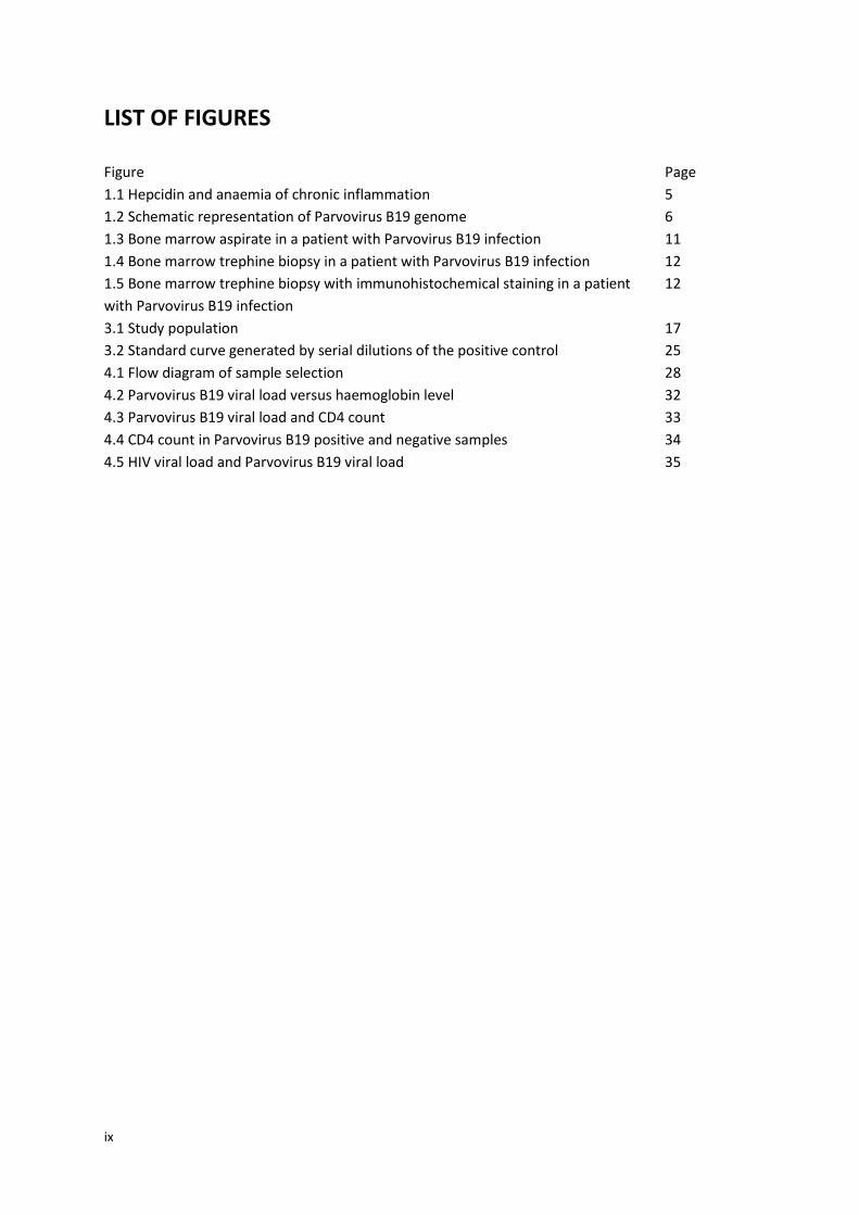
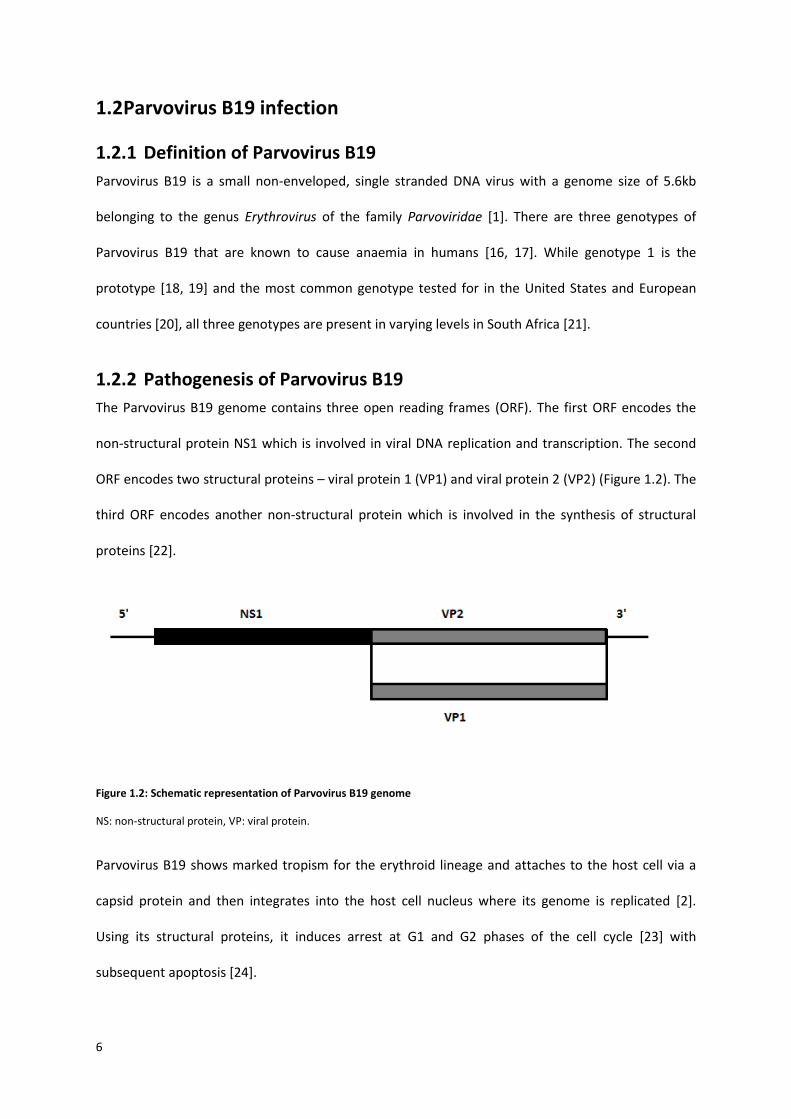
The Parvovirus B19 genome contains three open reading frames (ORF). The first ORF encodes the
non-structural protein NS1 which is involved in viral DNA replication and transcription. The second
ORF encodes two structural proteins – viral protein 1 (VP1) and viral protein 2 (VP2) (Figure 1.2). The
third ORF encodes another non-structural protein which is involved in the synthesis of structural
proteins [22].
Figure 1.2: Schematic representation of Parvovirus B19 genome
NS: non-structural protein, VP: viral protein.
Parvovirus B19 shows marked tropism for the erythroid lineage and attaches to the host cell via a
capsid protein and then integrates into the host cell nucleus where its genome is replicated [2].
Using its structural proteins, it induces arrest at G1 and G2 phases of the cell cycle [23] with
subsequent apoptosis [24].
7
Although any host cell may be targeted by Parvovirus B19, as evidenced by the presence of viral DNA
found in various tissues [25], viral replication only occurs in the erythroid lineage in the BM.
In Parvovirus B19 infection in immune competent hosts, erythropoiesis is transiently suppressed and
the RPI falls to zero. However, the Hb levels remain stable because erythrocytes have a long lifespan
of approximately 120 days and therefore there are adequate circulating erythrocytes to compensate
for the transient absence of production [26]. Thus in most cases, individuals are asymptomatic with
normal Hb levels. The immune competent host is able to mount an effective humoral response to
this primary infection and repeated infection of erythrocytes does not occur on subsequent
exposure as lifelong immunity is achieved [2].
In contrast, Parvovirus B19 infection in patients with a reduced erythroid lifespan and those with an
impaired immune system is more severe. The pathophysiology and degree of severity are however
different in these two categories.
A transient aplastic crisis may occur in patients whose erythrocytes have a reduced lifespan for
example in sickle cell anaemia and hereditary spherocytosis. In this situation the shortened
erythrocyte lifespan is unable to compensate for the transient absence of erythrocyte production,
with a resultant severe anaemia. While this is often severe, it is self-limiting and confers life-long
immunity to Parvovirus B19 infection [2].
In the case of the immune deficient host, Parvovirus B19 infection causes a persistent infection as
protective antibodies are either not sufficiently produced or are non-functional. In HIV infected
individuals and other immunocompromised states, erythroid-lineage cells are continuously
destroyed by the active Parvovirus B19 infection and a chronic and persistent pure red cell aplasia
(PRCA) develops [27].
8
Other causes of PRCA include HIV itself, Epstein-Barr virus (EBV), viral hepatitis, drugs including
zidovudine (AZT), lamivudine (3TC), isoniazide (INH) and sulphonamides, autoimmune diseases,
pregnancy and haematological malignancies.
1.2.3 Prevalence of Parvovirus B19
The prevalence of Parvovirus B19 infection in non-HIV populations has been widely published in local
and international studies. Locally the prevalence of Parvovirus B19 infection in pregnant women
[28, 29] and young haemophiliacs [30] has been reported as 3.4% and 5.1% respectively.
International studies have described prevalence data in blood donors [31-40] ranging from 0.12% to
7.53%. Retrospective record reviews of anaemic patients performed in Taiwan [41] and Kenya [42]
reported a prevalence of Parvovirus B19 infection ranging from 0 to 3% using serology for
Immunoglobulin (Ig) M or molecular methods. HIV status however was not assessed in these studies.
Further, it is difficult to compare the results of these published studies because of the different
diagnostic tests used to diagnose Parvovirus B19 infection.
While there are no published studies on the prevalence of Parvovirus B19 infection in the South
African HIV infected population with severe anaemia, there are numerous case reports of HIV
infected patients with severe anaemia and Parvovirus B19 infection which have been reported
internationally [43-56]. In addition, two large multi-centre studies have been performed: The study
conducted in Brazil in 2012 looked at 88 HIV infected patients with and without anaemia and found
the prevalence of concomitant anaemia and Parvovirus B19 infection to be 3.4% [57]. A study
conducted in France in 2010 of 428 HIV positive anaemic patients, reported a similar prevalence of
Parvovirus B19 infection of 3.73% [58].
Studies performed in the developing world in HIV infected patients in whom the Hb level was not
assessed, reported a wide prevalence range of 0 to 13.1% [57, 59-64]. This variability may be
attributed to the different diagnostic tests used (IgM or molecular methods), the patient populations
(studies were conducted in Iran, Ghana, China and Brazil) and their HIV prevalence as well as the
9
study designs ( single-centre, multi-centre studies with and with-out case-controls, retrospective
and prospective study designs).
The above studies thus have several limitations precluding extrapolation of their results to the SA
HIV infected patient cohort
1.2.4 Clinical presentation of Parvovirus B19 in the HIV infected host
Parvovirus B19 infection in HIV infected adults may be asymptomatic such as seen in HIV immune
competent persons. It may also present with mild exanthematous disease associated with a viral
prodrome and erythematous, reticular rash or with a PRCA which traditionally presents as a severe
normocytic anaemia associated with reticulocytopenia [27]. Patients may also present with
symptoms and signs of anaemia such as fatigue, dizziness, weakness, shortness of breath,
palpations, tachycardia and high output cardiac failure. A bicytopenia or pancytopenia may also be
noted. This is seen less commonly and although Parvovirus B19 specifically targets the erythroid
lineage in the bone marrow, resulting in an isolated anaemia, immune mediated destruction of
neutrophils and platelets (PLT) by Parvovirus B19, resulting in a neutropenia (neutrophil count of less
than 1.6 x10⁹/l [65]) and thrombocytopenia (platelet count of less than 171x109/l [65]) have also
been described [48, 66, 67].
1.2.5 Investigations
Preliminary investigation of the cause of anaemia includes an FBC with a differential count and
peripheral blood smear assessment, RPI, substrate deficiency testing which includes iron studies,
vitamin B12 and red cell folate levels and possibly a haemolytic work up which includes haptoglobin,
lactate dehydrogenase and a Coombs test if haemolysis is suspected as an alternative cause for the
severe anaemia. More specific investigations into the cause of the anaemia would include bone
marrow investigations and testing for Parvovirus B19 itself.
10
1.2.5.1 Bone marrow investigation
A bone marrow aspirate (BMA) and bone marrow trephine biopsy (BMT) are often performed as
part of the diagnostic work-up of anaemia to provide information regarding the haemopoietic
activity of the bone marrow in general, degree and nature of erythropoiesis and presence or
absence of malignant or infective infiltrates.
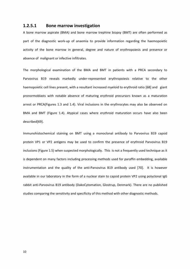
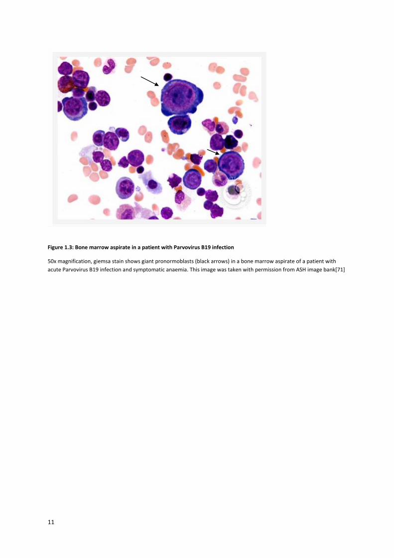
The morphological examination of the BMA and BMT in patients with a PRCA secondary to
Parvovirus B19 reveals markedly under-represented erythropoiesis relative to the other
haemopoietic cell lines present, with a resultant increased myeloid to erythroid ratio [68] and giant
pronormoblasts with notable absence of maturing erythroid precursors known as a maturation
arrest or PRCA(Figures 1.3 and 1.4). Viral inclusions in the erythrocytes may also be observed on
BMA and BMT (Figure 1.4). Atypical cases where erythroid maturation occurs have also been
described[69].
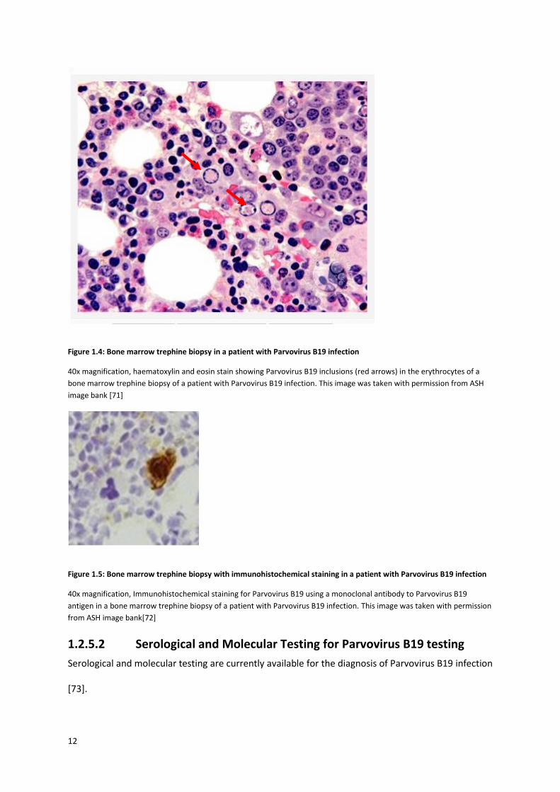
Immunohistochemical staining on BMT using a monoclonal antibody to Parvovirus B19 capsid
protein VP1 or VP2 antigens may be used to confirm the presence of erythroid Parvovirus B19
inclusions (Figure 1.5) when suspected morphologically. This is not a frequently used technique as it
is dependent on many factors including processing methods used for paraffin embedding, available
instrumentation and the quality of the anti-Parvovirus B19 antibody used [70]. It is however
available in our laboratory in the form of a nuclear stain to capsid protein VP2 using polyclonal IgG
rabbit anti-Parvovirus B19 antibody (DakoCytomation, Glostrup, Denmark). There are no published
studies comparing the sensitivity and specificity of this method with other diagnostic methods.
11
Figure 1.3: Bone marrow aspirate in a patient with Parvovirus B19 infection
50x magnification, giemsa stain shows giant pronormoblasts (black arrows) in a bone marrow aspirate of a patient with
acute Parvovirus B19 infection and symptomatic anaemia. This image was taken with permission from ASH image bank[71]
12
Figure 1.4: Bone marrow trephine biopsy in a patient with Parvovirus B19 infection
40x magnification, haematoxylin and eosin stain showing Parvovirus B19 inclusions (red arrows) in the erythrocytes of a
bone marrow trephine biopsy of a patient with Parvovirus B19 infection. This image was taken with permission from ASH
image bank [71]
Figure 1.5: Bone marrow trephine biopsy with immunohistochemical staining in a patient with Parvovirus B19 infection
40x magnification, Immunohistochemical staining for Parvovirus B19 using a monoclonal antibody to Parvovirus B19
antigen in a bone marrow trephine biopsy of a patient with Parvovirus B19 infection. This image was taken with permission
from ASH image bank[72]
1.2.5.2 Serological and Molecular Testing for Parvovirus B19 testing
Serological and molecular testing are currently available for the diagnosis of Parvovirus B19 infection
[73].
13
Serological results report both IgM and IgG. IgM positivity suggests the presence of neutralizing
antibodies to a current infection whereas IgG positivity suggests antibodies present due to previous
exposure to a now cleared infection.
Molecular methods detect the presence or absence of the viral genome.
In general, serology is more widely available, less technically challenging, less labour intensive and
lower cost than molecular methods and is therefore used more often. However, antibodies to
Parvovirus B19 are usually non-functional or absent in the setting of HIV infection and therefore
serology is an unreliable diagnostic tool in this setting [43]. Prevalence data based on serological
diagnostic tests therefore does not accurately reflect the prevalence reported. Rather, viral DNA
detection using molecular testing, namely PCR, is indicated. It is imperative to establish accurate
cut-off values for positive and negative samples to distinguish between a low viral load which
indicates previous exposure and absence of acute current infection, known as ‘viral dust’, and a high
viral load indicating current disease [58]. Previous studies using molecular methods reported a
positive PCR at a lower than recommended cut-off level. Therefore some samples reported as
positive may in fact represent viraemia but not infectivity or active disease [58].
Molecular testing may be performed on peripheral blood, BMA and BMT samples at the request of
the clinician or as a recommendation by the pathologist reviewing the BM. BM is the preferred
specimen because it is more likely to yield a positive result in the setting of Parvovirus B19 and HIV
infection as compared to peripheral blood testing.
BM erythroid progenitors are the target cells of the Parvovirus B19 infection which are present on
BMA and BMT sections. Further, the BMA and BMT sections assist with the diagnostic interpretation
and exclusion of an associated infiltrate. Classic BM findings in Parvovirus B19 infection of a PRCA
with absent/markedly reduced erythropoiesis and giant pronormoblasts with occasional erythroid
viral inclusions have been well described. A BM, however, is an invasive procedure which requires a
14
specialist diagnostic service for interpretation. This is an important consideration in the developing
world with limited access to resources.
1.2.6 Treatment
Persistent infection with Parvovirus B19 is clinically relevant because it is a potentially treatable
cause of anaemia [27, 52].
Treatment of Parvovirus B19 induced PRCA in HIV infected patients includes specific and supportive
measures. Improvement of the immunosuppression caused by HIV with anti-retroviral (ARV) therapy
is a cornerstone of treatment, as reported in various studies [44-48, 51, 74]. Intravenous
immunoglobulin administration is used as a form of passive immunity against Parvovirus B19 [43, 75,
76].
Supportive measures include steroid therapy and transfusion of packed red cells with iron chelation
therapy to prevent or treat iatrogenic iron overload secondary to the chronic transfusions that are
often required.
1.2.7 Monitoring response to therapy
Monitoring of response to therapy includes clinical and laboratory features: improvement of the
symptoms of anaemia, Hb level and RPI. Currently, monitoring of Parvovirus B19 VL by quantitative
PCR is not routinely performed. PCR testing is expensive and does not offer more information than
the routine surrogate markers used to monitor response to therapy (Hb, RPI). In addition, when
multiple pathologies contribute to the anaemia, Hb level and RPI are a better reflection of clinical
status than Parvovirus B19 PCR. Perhaps for specific cases who fail to respond to therapy,
monitoring Parvovirus B19 VL by PCR testing would be of value [77] to determine if the persistent
anaemia is due to the Parvovirus B19 itself or another pathology.
15
1.2.8 Prognosis
Prognosis varies in different clinical settings. Excellent results have been described, with 90% of
cases of PRCA in HIV positive patients fully recovering in less than three months [44-47, 51].
Interestingly, reports have shown that in some cases, being on ARV therapy alone may result in
adequate immunological recovery and therefore in clearance of the Parvovirus B19 and
improvement of anaemia [46, 47, 51, 78]. This has considerable clinical and cost implications as
blood transfusions are costly and associated with significant risks (immune suppression, iron
overload, alloimmunity and infections) [79]. In addition, intravenous immunoglobin therapy is not
always readily available in our setting and is expensive [80].
However, in the South African setting results have not been as promising, with many patients
requiring repeated administration of intravenous immunoglobin often accompanied by multiple
blood transfusions [81]. The reason for this poor response is uncertain, but may be due to patients
late presentation, that is severe HIV infection and very low haemoglobin levels due to delayed access
to medical care as well as co-existing diseases contributing to the anaemia [81].
The CD4 count in an HIV infected person is used as a marker of immune status. A lower CD4 count is
associated with more severe immune suppression. PRCA secondary to Parvovirus B19 in HIV positive
patients has been shown to be associated with very low CD4 levels, ranging from a median of 24 –
42 cells/µL [43, 59, 76]. It has been suggested that the CD4 count may be used as a prognostic
marker to predict the development of a chronic PRCA in an HIV infected patient exposed to
Parvovirus B19, with very low counts associated with PRCA and anaemia and high counts associated
with adequate humoral response and absence of severe anaemia.
1.3 Aim of the study The aim of this study was to assess the prevalence of Parvovirus B19 infection in the HIV infected
population group in South Africa with severe anaemia in order to assess the role of routine

16
Parvovirus B19 PCR screening. This has implications for the testing and management of this subset of
patients and treatment guidelines.
17
2 RESEARCH QUESTIONS
To determine the prevalence of Parvovirus B19 infection in HIV infected patients with severe
anaemia at two academic hospitals in Johannesburg who had a bone marrow examination
submitted for routine diagnostic workup between January 2012 and November 2013.
To determine the association between Parvovirus B19 VL and 1) the degree of anaemia and
2) HIV immune suppression as indicated by high HIV VL and low CD4 count
To evaluate the clinical utility of laboratory parameters in anaemic patients to determine the
most suitable markers for distinguishing Parvovirus B19 positive and Parvovirus B19 negative
patients
To assess the role of bone marrow morphology as a diagnostic tool for Parvovirus B19
infection
18
3 MATERIALS AND METHODS
3.1 Study design and population A retrospective study was performed at two academic hospitals in Johannesburg which are serviced
by the National Health Laboratory Service (NHLS): Charlotte Maxeke Johannesburg Academic
Hospital (CMJAH) and Chris Hani Baragwanath Academic Hospital (CHBAH).
The Inclusion criteria for specimens into the study included all specimens submitted for a bone
marrow examination for routine diagnostic workup between January 2012 and November 2013. The
study population included HIV infected patients with severe anaemia, defined as haemoglobin levels
<8 g/dl for men and non-pregnant women [82]. Patients aged 18 years and older were included.
Exclusion criteria included insufficient BM material for PCR analysis and absence of BMA or BMT
slides for analysis. Real-time PCR for Parvovirus B19 was performed on all samples (Figure 3.1).
Figure 3.1: Study population
Bone marrow samples from HIV infected patients with an Hb <8g/dl were included in this study. Parvovirus B19 PCR was
performed on all samples with sufficient material for testing.
BM with severe anaemia
Hb <8g/dl
HIV negative
Exclude
HIV positive
PCR for Parvovirus B19
Inadequate sample yield
Exclude
Adequate sample yield
Parvovirus B19 PCR positive
Parvovirus B19 PCR negative
19
BM: bone marrow, Hb: haemoglobin, HIV: Human Immunodeficiency Virus, PCR: polymerase chain reaction
3.2 Data collection Demographic information (gender and age), clinical information (ARV regimen, known medical
conditions, infections and malignancies) and laboratory information (HIV VL, CD4 count,
haemoglobin, mean cell haemoglobin concentration (MCHC), mean cell volume (MCV), white cell
count (WCC), neutrophil count, PLT, RPI, haematinics (iron, vitamin B12, red cell folate) and
Parvovirus B19 serology), where available, were obtained from the laboratory information system
(LIS) and recorded on data collection sheets (Appendix A, Table A1)
Parameters were assessed according to reference ranges and definitions presented in Appendix A
(Tables A2 and A3).
3.3 Diagnostic testing
3.3.1 FBC, Differential count and RPI
Blood samples were collected indipotassium ethylenediaminetetraacetic acid (K2EDTA) tubes which
contain between 1.5–2.2 mg of K2EDTA dihydrate per millilitre of blood.
The ADVIA 2120 Hematology System (Siemens Diagnostics, Deerfield, IL, USA) and SLS XT-series
(Sysmex, Kobe, Japan) were used to measure the FBC, differential count and RPI parameters. The
samples analysed on the ADVIA 2120 had cells counted and sized by light scatter (flow cytometry)
technology using white light for white cells and laser light for red cells and platelets. Hb was
measured by the conventional cyanmethaemoglobin method. The six-part differential analysis was
performed in two channels. Cells in the peroxidase channel were measured by peroxidase staining
intensity and light scatter. Cells in the basophil/lobularity channel were measured by dual laser light
scatter, nuclear density and lobulation index. Reticulocytes were stained with oxazine 750. The SLS
XT-series (Sysmex, Kobe, Japan) uses impedance, light scatter (flow cytometry) and fluorescent dye
technology. The WCC, erythrocytes and platelets were counted by ‘impedance technology’ in which
20
cells pass between two sensing electrodes and cause impedance to the current which is proportional
to the volume of the cell passing through. Flow cytometry with appropriate fluorescent dyes was
used for the white cell -5 part differential count, nucleated erythrocytes and reticulocyte count. Hb
was measured with cyanide-free sodium lauryl sulphate reagent.
The films were spread manually or on the Advia autoslide slide maker and stained on the Hema-Tek
slide stainer.
3.3.2 Haematinic studies
Iron studies, Vitamin B12 and red cell folate assays were analysed using the Advia 1800 chemistry
analyser (Siemens Diagnostics, Deerfield, IL, USA) at CMJAH and the cobas-8000 (Roche, Basel,
Switzerland) at CHBAH.
3.3.3 Serology Parvovirus B19
Serological testing for Parvovirus B19 IgM and IgG was performed on samples at the request of the
treating clinician. Serology was performed manually using an enzyme linked immunosorbent assay
(ELISA) method (IBL international, GMBH, Hamburg, Germany). Diluted serum samples were added
to Microtiter strip wells precoated with Parvovirus B19 antigens. After washing the wells to remove
all unbound sample material horseradish peroxidase labelled anti-human IgG conjugate was added.
This conjugate binds to the captured Parvovirus B19 specific antibodies in the sample serum. The
immune complex formed by the bound conjugate was visualized by adding tetramethylbenzidine
substrate which gives a blue reaction product. The intensity of this product is proportional to the
amount of Parvovirus B19 specific IgG antibodies in the specimen. Sulphuric acid was then added to
stop the reaction. This produces a yellow endpoint colour. Absorbance at 450 nm was read using an
ELISA microwell plate reader. A positive, negative and cut-off control, supplied by the manufacturer,
were used in each run. A run was considered valid if the cut-off control absorbance value was
between 0.150 and 1.30, the negative control absorbance value was less than the cut-off control
value and the positive control absorbance value was greater than the cut off control value. A result
21
was considered positive if its absorbance value was more than 10% higher than the cut-off
absorbance value. All results were reported as either positive or negative. No indeterminate results
were reported. Testing was performed in one central location.
3.3.4 CD4 counts
Single platform PanLeucogate[83] CD4 counts were prepared using the ImmunoprepTM lyse-no-
wash method (Beckman Coulter, USA). Post preparation, Flow Count TM beads were added and
samples acquired and analysed on a FC 500 MPL flow cytometer (Beckman Coulter, USA). Briefly,
10µl of Beckman Coulter FlowCareTM CD45/CD4 monoclonal antibody was added to 100 µl of whole
blood. The samples were incubated for 10 minutes during which the sample reacted with
fluorochrome labelled CD4 and CD45 monoclonal antibodies (contained in the Beckman Coulter
Flow Care PLG CD4 kit) that are specific for the cell surface antigens CD4 and CD45. Post incubation,
ImmunoprepTM reagents (lysing agent, stabiliser and fixative) were added to haemolyse red blood
cells while maintaining the integrity of the white blood cells and the white blood cell surface
antigens. Prior to acquisition and analysis on the flow cytometer, 100µl of beads was added to each
sample. Cells that had bound the fluorochrome labelled antibody were identified on the basis of
their specific fluorescence emission related to the specific fluorochrome attached to either the CD45
or the CD4 antibody. A sequential automated gating strategy was used to include all CD45+ cells and
isolate the bright CD4+ lymphocytes from dimly expressing CD4+monocytes. The CD4+ lymphocytes
were further separated from the rest of the white cells on the basis of their complexity (side scatter)
and their specific CD45 and CD4 expression. The CD4 absolute count was subsequently calculated by
comparing the CD4+ lymphocyte events in the CD4 region to the bead events in the bead region and
referencing the bead calibration factor supplied with each bottle of Flow Count TM beads using the
formula: CD4 events/ bead events x calibration factor. CD4 count was reported as the number of
CD4 cells per µl.
22
3.3.5 Morphologic analysis
3.3.5.1 Bone marrow slides preparation
Preparation of BM slides for morphologic assessment was performed in the same way at both
CMJAH and CHBAH. Bone marrow aspirate slides were fixed for ten minutes in methanol, air dried
and stained twice using the Hematek stainer and Hematek Stain Pak (Siemens Diagnostics, Deerfield,
IL, USA) at a pH of 6.8. The Hematek Stain Pak comprises three solutions: stain solution (methanol
>99%, polychrome methylene blue-eosin stain), buffer solution (phosphate buffers, surfactant,
preservative) and rinse solution (methanol 10%, phosphate buffers, surfactant, preservative).
Haemoglobin molecules and eosinophilic granules are basic and take up the eosin dye (acidic dye).
Nucleic acid, proteins of cell nuclei and cytoplasm of primitive cells are acidic and take up methylene
blue (basic dye).
Bone marrow trephine biopsies were prepared for morphologic assessment at the Department of
Anatomical Pathology for samples received from both CMJAH and CHBAH. These samples were
received in 10% buffered formal saline and fixed in this solution for at least 24 hours, after which
they were placed on a labelled cassette which offers a support structure and is optimised to obtain
maximum fluid exchange during processing. Cores were measured and those of 1cm or longer were
cut in half or quarters depending on the length. Samples in their cassettes were decalcified in EDTA
for a minimum of 48 hours and then processed in an automated tissue processor. Samples were
inserted into the processing chamber and processed overnight with formalin at different
concentrations, dehydrated in alcohol, xylene cleared and wax embedded. They were then
embedded in small moulds and ribbon cut at 1µ by the microtome. Three levels were cut for each
block with 6-8 sections per slide. Slides were manually stained with haematoxylin for nuclear
staining and eosin phyloxine for cytoplasmic staining and were then dehydrated in absolute alcohol,
mounted on coverslip machine and labelled.
All BMA and BMT samples were reviewed as part of the routine assessment. They were considered
23
inadequate for morphological assessment if they were not representative of BM i.e. if the BMA
sample comprised no marrow elements such as particles and background stromal cells and if the
BMT samples did not have bony spicules and intact marrow expanses present or were too
traumatised for accurate assessment. All BMA and BMT of the Parvovirus B19 positive samples were
reviewed again during this study.
3.3.5.2 Ziehl-Neelsen stain
Ziehl-Neelsen staining for acid fast bacilli on trephine biopsy slides was performed as part of routine
workflow, briefly, as follows: slides were stained with filtered Carbol Fuchsin for ten minutes,
washed with tap water, flooded with 95% alcohol to remove excess carbol fuchsin without altering
the bacilli staining, washed in tap water, differentiated in 1% acid alcohol, washed in tap water and
counterstained with 1% methylene blue for less than 10 seconds, following which they were washed
again in tap water, air dried and cover slipped. The mycolic acid present in the cell wall of
Mycobacterium tuberculosis is resistant to penetration by the acid alcohol so that the background
appears pale blue and the acid fast bacilli appear red (due to the carbol fuchsin) when viewed under
the microscope. A positive control, obtained from a tissue with known Mycobacterium tuberculosis
infection, was performed with each sample.
3.3.5.3 Immunohistochemical stain for Parvovirus B19
Immunohistochemical staining using polyclonal rabbit anti-Parvovirus B19 IgG (Code: B-0091)
(DakoCytomation, Glostrup, Denmark) was performed as part of routine workflow, briefly, as
follows: slides were washed for five minutes in hydrogen peroxide and then rinsed with buffer. The
primary antibody to the Parvovirus B19 capsid protein VP2 was added and left to incubate at room
temperature for 20 minutes and then rinsed with buffer. The envision kit was applied and incubated
for 20 minutes at room temperature and then rinsed with buffer. For visualization, the substrate and
chromogen (3,3'-Diaminobenzidine) were added and incubated at room temperature for ten
minutes. Slides were then rinsed thoroughly and counterstained in haematoxin. They were then
24
dehydrated in alcohol and zylene and cover slipped. A positive control from a known Parvovirus B19
positive sample was performed with each sample.
3.4 PCR for Parvovirus B19 All samples in this study were tested for Parvovirus B19 infection with the PrimerDesign™ genesig®
Kit for Human Parvovirus B19 (Southampton, United Kingdom) [84] in the PCR laboratory at the
NHLS, CMJAH.
The primers and probe in this kit have 100% homology with all reference sequences currently in the
NCBI database including the three genotypes known to be present in South Africa ((AY083234,
DQ333428, DQ225151). The assay results were measured using both qualitative (positive or
negative) and semi-quantitative (Parvovirus B19 viral load) outputs. The amount of house-keeping
gene, beta actin, in each sample was used as a reference value to give a semi-quantitative Parvovirus
B19 viral load. Under optimal PCR conditions genesig® HPVB19 detection kit has a sensitivity of >95%
and can detect less than 100 copies of target template in 5µl of sample.
3.4.1 The TaqMan principle
The PCR reaction used in this study exploits the TaqMan principle which uses primers and
fluorescently labelled probes. During PCR amplification, forward and reverse primers hybridise to
the Parvovirus B19 DNA. A fluorogenic DNA probe labelled with a 5’-fluorescent dye and 3’-non-
fluorescent quencher is included in the same reaction mixture. In the intact probe, the non-
fluorescent quencher and the fluorescent dye are in close proximity and as such the quencher
quenches the fluorescence energy emitted by the dye through the fluorescent resonance energy
transfer (FRET) effect.
During PCR amplification, the 5’ exonuclease activity of the Taq polymerase cleaves the probe in a 5’
to 3’ direction, separating the reporter dye and quencher. The resulting increase in fluorescence is
measured in real-time since it is read after each PCR cycle, with the amount of fluorescence directly
proportional to the amount of DNA at the start of that cycle.
25
3.4.2 DNA extraction
A total of 250µl of phosphate buffered saline was used to wash the bone marrow aspirate from their
slides. DNA Extraction was performed using the NucliSENS® EasyMag® (bioMérieux) system, (Boxtel,
Netherlands) with an input volume of 196µl and an elution volume of 25µl. Four micro-litres (4µl) of
internal control (IC) was spiked into the sample prior to extraction. Briefly, buffer containing
guanidine thiocyanate and Triton X-100 was added to each sample in an extraction cartridge vessel.
This disrupts all cellular matter or viral particles and inactivates RNAses and DNAses in the sample.
Fifty microlitres of magnetic silica was then manually added to the sample. Nucleic acids present in
the sample bound to the magnetic silica dioxide particles under high salt conditions. The magnetic
silica was washed several times and subsequently the nucleic acids were eluted in a volume of 25µl
and were available for the downstream PCR reaction.
3.4.3 PCR set-up
Real time PCR (RQ-PCR) using the PrimerDesign™ genesig® Kit for Human Parvovirus B19
(Southampton, United Kingdom)[84] was used to detect Parvovirus B19 DNA.
The commercial assay includes:
Parvovirus B19 specific primer/probe (proprietary information)
Parvovirus B19 positive control with a viral copy number of 2000000 copies/5μl
Internal extraction control DNA
Internal extraction control primer/probe mix
Endogenous beta actin (ACTB) primer/probe mix
RNAse/DNAse free water
Two separate PCR reactions were performed – the first to detect the presence of Parvovirus B19
DNA and the second to detect the presence of a house-keeping gene ACTB.
26
The pathogen detection reagent mix was prepared as per standard procedure (Table 3.1). Each well
was filled with 15µl of this mix before 10µl of extracted DNA template was added with a final volume
of 25µl.
Table 3.1: Reagent preparation for Parvovirus B19
Component Volume (µl)
2 x precision master mix 10
Pathogen primer/probe mix 1
Internal control primer/probe mix 1
RNAse/DNAse free water 3
Final volume 15
3.4.4 Standard curve
A standard curve (Figure 3.2) was generated by serial dilutions of the positive control (from 2x10⁵
copies/µl to 2x100 copies/µl) followed by a PCR reaction. These VL copies were plotted on the y-axis,
against the cycle threshold (Ct) value on the x-axis using Microsoft Excel 2013 from which a
logarithmic equation was then generated y=1E10(-0.66x) . Based on the results of the standard curve a
Ct of 30 (2x100 copies/µl) was derived as the lower limit of detection. This equation was then used
for test samples to calculate Parvovirus B19 VL from Ct values.
27
Figure 3.2: Standard curve generated by serial dilutions of the positive control
3.4.5 Positive and Negative controls
A negative control, in which the template DNA was replaced with RNAse/DNAse free water, was
added to each PCR run to exclude nucleic acid contamination. A positive control, provided within the
kit, was added to each run to ensure validity of negative results and was also used to set up the
standard curve described above.
A run was considered valid if the positive control amplified between cycle 16 and 23 and the
negative control showed no amplification or amplified after cycle 35. If the positive control was
negative or if the negative control amplified before cycle 35, the run was considered invalid and the
test was repeated.
3.4.6 Internal control
The PCR assay contains two sets of specific forward and reverse primers which were amplified
together in a PCR reaction: one for Parvovirus B19 and one for an IC which is an exogenous source of
DNA template that is spiked into the lysis buffer. The IC is used to prevent false negative results
owing to inadequate extraction or amplification steps. In a Parvovirus B19 negative sample, the IC
must be positive. In a Parvovirus B19 positive sample with a high VL, the IC may be negative due to
competitive inhibition.
28
The Parvovirus B19 fluorescence is detected in the FAM channel (excitation wavelength 452-488nm,
emission wavelength 505-545nm) and the IC fluorescence is detected in the VIC channel (excitation
wavelength 542-582nm, emission wavelength 665-705nm).
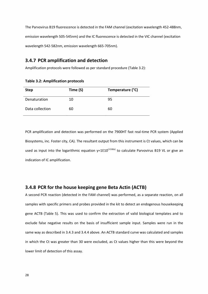
3.4.7 PCR amplification and detection
Amplification protocols were followed as per standard procedure (Table 3.2):
Table 3.2: Amplification protocols
Step Time (S) Temperature (°C)
Denaturation 10 95
Data collection 60 60
PCR amplification and detection was performed on the 7900HT fast real-time PCR system (Applied
Biosystems, inc. Foster city, CA). The resultant output from this instrument is Ct values, which can be
used as input into the logarithmic equation y=1E10(-0.66x) to calculate Parvovirus B19 VL or give an
indication of IC amplification.
3.4.8 PCR for the house keeping gene Beta Actin (ACTB)
A second PCR reaction (detected in the FAM channel) was performed, as a separate reaction, on all
samples with specific primers and probes provided in the kit to detect an endogenous housekeeping
gene ACTB (Table 5). This was used to confirm the extraction of valid biological templates and to
exclude false negative results on the basis of insufficient sample input. Samples were run in the
same way as described in 3.4.3 and 3.4.4 above. An ACTB standard curve was calculated and samples
in which the Ct was greater than 30 were excluded, as Ct values higher than this were beyond the
lower limit of detection of this assay.
29
Table 3.3: Reagent preparation for Beta Actin
Component Volume (µl)
2 x precision master mix 10
Endogenous ACTB primer/probe mix 1
RNAse/DNAse free water 4
Final volume 15
3.5 Statistical analysis Inorder to accurately estimate the prevalence of Parvovirus B19 positivity in this cohort, sample size
was calculated using a confidence level of 95% and a confidence interval of 5-15%. This yielded a
suggested sample size of 96. A larger sample size of 170 was used to account for any technical errors
or inadequate sample yield.
Data were captured in Microsoft Excel 2013 and analysed using GraphPad Prism version 5.00 for
Windows (GraphPad Software, San Diago, CA). Statistical comparisons were performed using the
Chi-squared and Fisher’s exact tests as appropriate for categorical variables and the parametric
paired t-test or non-parametric Wilcoxon Matched Pairs test for continuous variables depending
upon the normal distribution. The degree of linearity between the Parvovirus B19 VL levels and the
laboratory tests (CD4 count, HIV VL and haemoglobin) were illustrated in a scatter plot with an
appropriately fitted regression line. Sensitivity, specificity, positive predictive value (PPV), negative
predictive value (NPV) and false negative rate (FNR) were calculated as necessary for serological
results, presence of PRCA, presence of viral inclusions and RPI. Statistical significance was set at a P <
0.05.
30
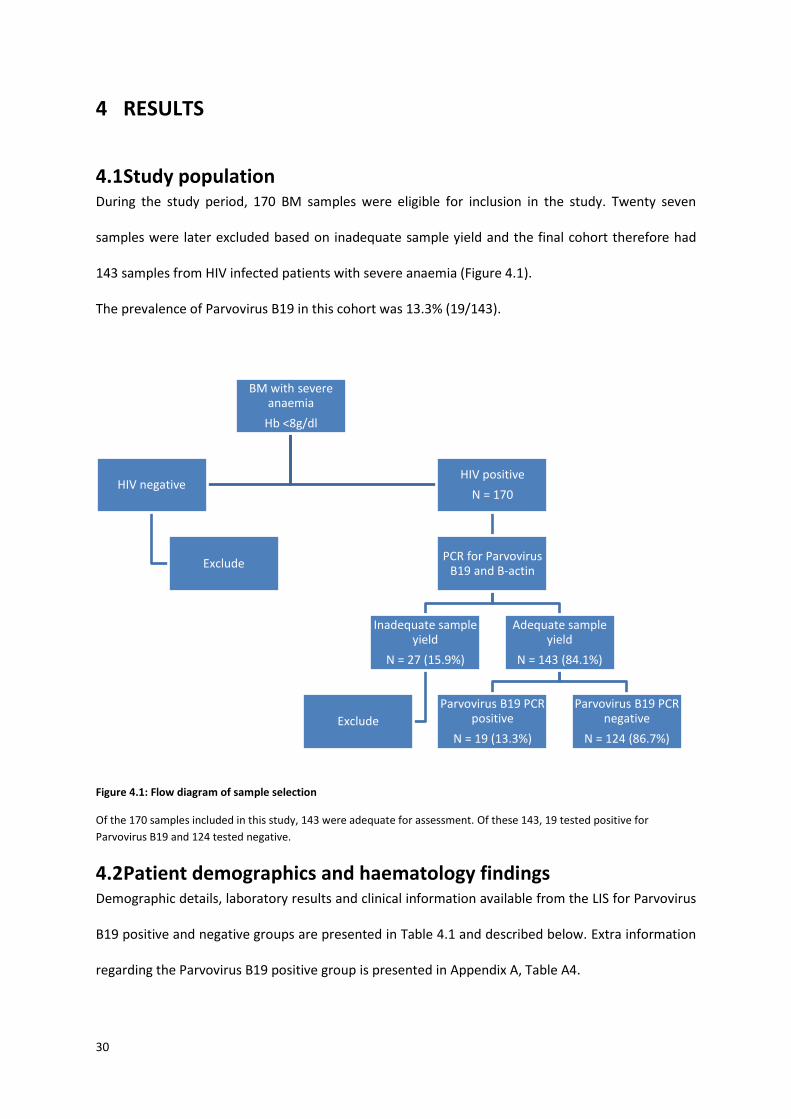
4 RESULTS
4.1 Study population During the study period, 170 BM samples were eligible for inclusion in the study. Twenty seven
samples were later excluded based on inadequate sample yield and the final cohort therefore had
143 samples from HIV infected patients with severe anaemia (Figure 4.1).
The prevalence of Parvovirus B19 in this cohort was 13.3% (19/143).
Figure 4.1: Flow diagram of sample selection
Of the 170 samples included in this study, 143 were adequate for assessment. Of these 143, 19 tested positive for
Parvovirus B19 and 124 tested negative.
4.2 Patient demographics and haematology findings Demographic details, laboratory results and clinical information available from the LIS for Parvovirus
B19 positive and negative groups are presented in Table 4.1 and described below. Extra information
regarding the Parvovirus B19 positive group is presented in Appendix A, Table A4.
BM with severe anaemia
Hb <8g/dl
HIV negative
Exclude
HIV positive
N = 170
PCR for Parvovirus B19 and B-actin
Inadequate sample yield
N = 27 (15.9%)
Exclude
Adequate sample yield
N = 143 (84.1%)
Parvovirus B19 PCR positive
N = 19 (13.3%)
Parvovirus B19 PCR negative
N = 124 (86.7%)
31
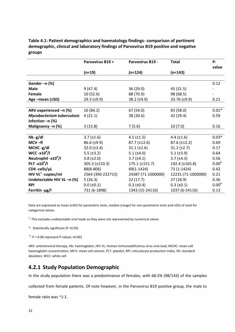
Table 4.1: Patient demographics and haematology findings- comparison of pertinent demographic, clinical and laboratory findings of Parvovirus B19 positive and negative groups
Parvovirus B19 + Parvovirus B19 - Total P-value
(n=19) (n=124) (n=143)
Gender –n (%) 0.12 Male 9 (47.4) 36 (29.0) 45 (31.5) - Female 10 (52.6) 88 (70.9) 98 (68.5) - Age –mean (±SD) 29.3 (±9.9) 38.2 (±9.9) 33.76 (±9.9) 0.21
ARV experienced –n (%) 16 (84.2) 67 (54.0) 83 (58.0) 0.01* Mycobacterium tuberculosis infection –n (%)
4 (21.1) 38 (30.6) 42 (29.4) 0.59
Malignancy –n (%) 3 (15.8) 7 (5.6) 10 (7.0) 0.16
Hb -g/dl 3.7 (±1.6) 4.5 (±1.5) 4.4 (±1.6) 0.03* MCV –fl 86.6 (±9.9) 87.7 (±12.6) 87.6 (±12.2) 0.69 MCHC -g/dl 32.0 (±3.4) 31.1 (±2.6) 31.2 (±2.7) 0.17 WCC -x109/l 5.5 (±3.2) 5.1 (±4.0) 5.1 (±3.9) 0.64 Neutrophil -x109/l 3.8 (±2.0) 3.7 (±4.1) 3.7 (±4.3) 0.56 PLT -x109/l 305.3 (±210.3) 175.1 (±151.7) 192.4 (±165.8) 0.00# CD4 -cells/µL 88(8-806) 69(1-1424) 73 (1-1424) 0.42 HIV VL1 -copies/ml 2564 (390-222715) 24387 (71-1000000) 12231 (71-1000000) 0.21 Undetectable HIV VL –n (%) 5 (26.3) 22 (17.7) 27 (18.9) 0.36 RPI 0.0 (±0.1) 0.3 (±0.4) 0.3 (±0.1) 0.00# Ferritin -µg/l 731 (6-1898) 1343 (15-24116) 1037 (6-24116) 0.13
Data are expressed as mean (±SD) for parametric tests, median (range) for non-parametric tests and n(%) of total for
categorical values.
1: This excludes undetectable viral loads as they were not represented by numerical values
*: Statistically significant (P <0.05)
#: P = 0.00 represent P-values <0.001
ARV: antiretroviral therapy, Hb: haemoglobin, HIV VL: Human Immunodeficiency virus viral load, MCHC: mean cell
haemoglobin concentration, MCV: mean cell volume, PLT: platelet, RPI: reticulocyte production index, SD: standard
deviation, WCC: white cell
4.2.1 Study Population Demographic
In the study population there was a predominance of females, with 68.5% (98/143) of the samples
collected from female patients. Of note however, in the Parvovirus B19 positive group, the male to
female ratio was ~1:1.
32
The mean (±standard deviation (SD)) age in the Parvovirus B19 positive and negative groups was
29.3 (±9.9) and 38.2 (±9.9) respectively (p=0.21).
4.2.2 Clinical information
A total of 58.0% (83/143) of the study population were on ARV therapy with 84.2% (16/19) of the
subjects in the Parvovirus B19 positive group on ARV therapy, of which 31.3% (5/16) were virally
suppressed (HIV VL <40copies/mL).
In the Parvovirus B19 positive group who were on ARV therapy, the ARV regimen was documented
in 62.5% (10/16) of the patients, of which 50.0% (5/10) were on AZT and 100% (10/10) were on 3TC
as part of a three-drug regimen.
4.2.3 Laboratory investigations
4.2.3.1 Full blood count
All patients in the Parvovirus B19 positive group had a grade four anaemia (Hb <6g/dl) [82].
The mean Hb was significantly lower in the Parvovirus B19 positive group compared to the
Parvovirus B19 negative group (p<0.05).
There was no difference for MCV, MCHC, WCC and neutrophil count in the two groups.
The mean platelet count was significantly lower in the Parvovirus B19 negative group (p<0.05).
In the Parvovirus B19 positive group, 26.3% (5/19) had associated thrombocytopenia and 31.6%
(6/19) had associated neutropenia of which 40.0% (2/5) and 66.7% (4/6) had another documented
cause for the cytopenia respectively.
4.2.3.2 CD4 count and HIV viral load
The median (range) for CD4 count was 88 (8-806)cells/µl and 69 (1-1424)cells/µl in the Parvovirus
B19 positive and negative groups respectively. There was no significant difference in CD4 or HIV VL
in the Parvovirus B19 positive and negative groups (p= 0.42 and 0.21 respectively).
33
Of the Parvovirus B19 positive group, 26.3% (5/19) had HIV viral suppression and had a mean (±SD)
Hb of 3.7 (±1.6) g/dl. There was no statistically significant difference between the Hb, CD4 count and
Parvovirus B19 viral load in this group compared to those not virally suppressed. In addition, all of
the CD4 counts in this group were less than 350 cells/µl and all had classical PRCA findings on BM
analysis.
4.2.3.3 Reticulocyte production index
This test was requested on 95 of the 143 samples in this study, 15 of which were in the Parvovirus
B19 positive group (defined according to the Parvovirus B19 PCR diagnostic test). The mean RPI was
significantly lower in the Parvovirus B19 positive group (p<0.05) as compared to the Parvovirus B19
negative group. The diagnostic performance of the RPI was compared with the Parvovirus B19 PCR
test by ROC analysis. The area under the curve was 0.81 with a 95% confidence interval of 0.72 to
0.89 for the RPI. The best RPI cut-off was ≤0.1, which corresponds to a sensitivity of 93.3% and a
specificity of 57.5%. A single patient in the Parvovirus B19 positive group and 45 patients in the
Parvovirus B19 negative group had an RPI of >0.1.
4.2.3.4 Haematinics
The median(range) ferritin level was 731 (6-1898)µg/l and 1343 (15-24116)µg/l in the Parvovirus B19
positive and negative groups respectively. This was not found to be statistically different (p=0.13).
Anaemia of chronic inflammation (as defined in Appendix A, Table A3) was a contributing factor in
85.3% (122/143) of the specimens, with no statistically significant difference in the Parvovirus B19
positive and negative groups (p=0.64). Iron deficiency anaemia was present in 5.3% (1/19) and 5.7%
(7/124) of the Parvovirus B19 positive and negative samples respectively. Megaloblastic anaemia
secondary to deficiencies of Vitamin B12 and folate were documented in 3.7% (4/107) and 3.7%
(4/109) respectively of the parvovirus B19 negative group. There was no documented megaloblastic
anaemia in the Parvovirus B19 positive group.
34
4.2.3.5 Parvovirus B19 serology
Parvovirus B19 serology (IgM and IgG) was available in 25.9% (37/143) subjects (5 in the Parvovirus
B19 positive group and 32 in the Parvovirus B19 negative group).
IgM was positive in 60% (3/5) and negative in 40% (2/5) of the Parvovirus B19 positive group with
sensitivity and specificity (when using Parvovirus B19 PCR as the gold standard) of 60.0% and 100%
respectively and positive and negative predictive value of 100% and 94.1% respectively. In the
Parvovirus B19 negative group all IgM results were negative (32/32).
In the Parvovirus B19 positive group all IgG results were negative (5/5), while in the Parvovirus B19
negative group 31.3 % (10/32) were IgG negative and 68.8% (22/32) were IgG positive.
4.2.3.6 Correlation between viral load of Parvovirus B19 eluted from the
bone marrow and haemoglobin level
There was no statistically significant correlation between the viral load of Parvovirus B19 (as a ratio
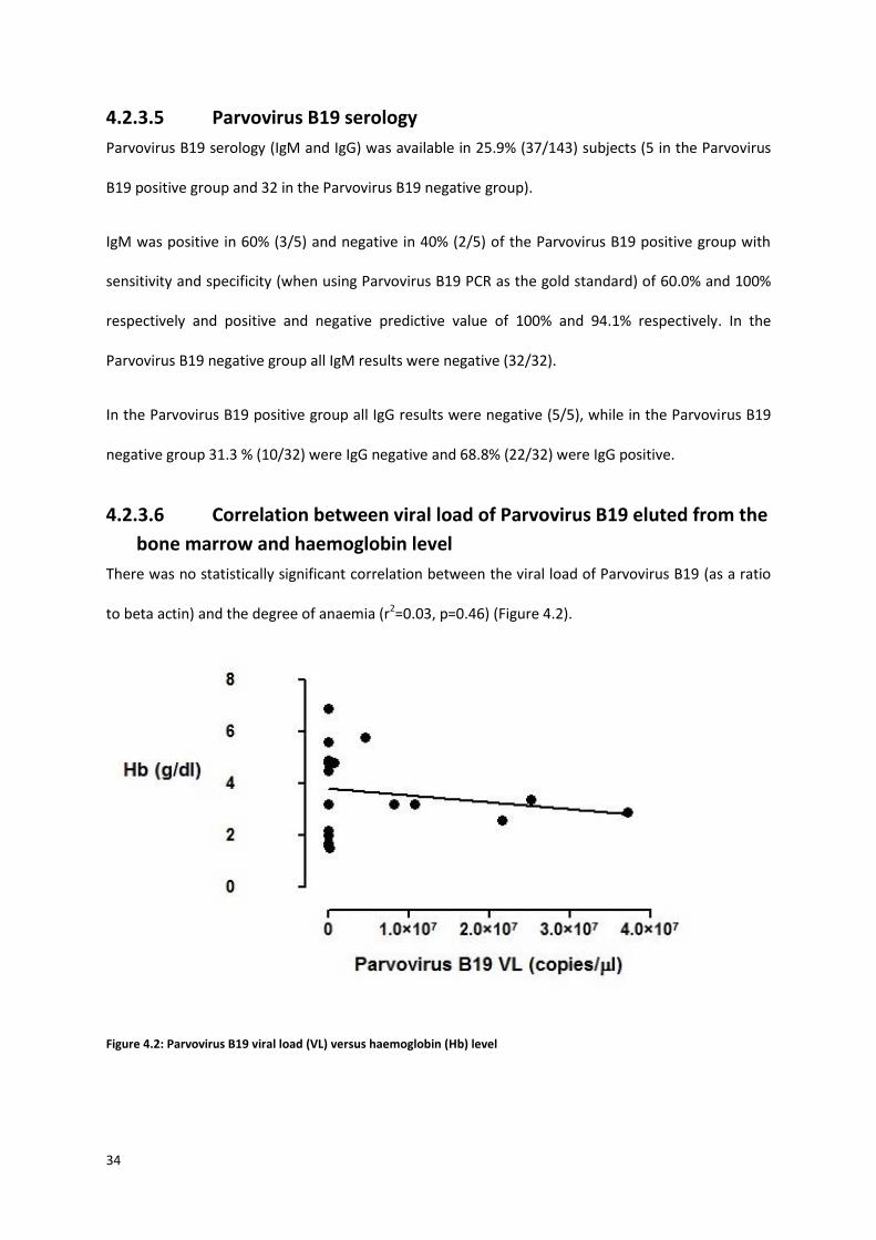
to beta actin) and the degree of anaemia (r2=0.03, p=0.46) (Figure 4.2).
Figure 4.2: Parvovirus B19 viral load (VL) versus haemoglobin (Hb) level
35
4.2.3.7 Correlation between viral load of Parvovirus B19 eluted from the
bone marrow and CD4 count
The Parvovirus B19 viral load (as a ratio to beta actin) was assessed in conjunction with the CD4
count with no statistically significant correlation between the two parameters (r2=0.02, p=0.58)
(Figure 4.3).
Figure 4.3: Parvovirus B19 viral load (VL) and CD4 count
4.2.3.8 CD4 count in Parvovirus B19 positive and negative samples
There was no statistically significant difference in the CD4 counts of Parvovirus B19 positive and
negative samples with median (range) of 88 (8-806)cells/µl and 69 (1-1424)cells/µl respectively
(p=0.84) (Figure 4.4).
36
Figure 4.4: CD4 count in Parvovirus positive and negative samples
4.2.3.9 Parvovirus B19 viral load and HIV viral load
There was no statistically significant correlation between the HIV VL and the Parvovirus B19 VL
(r2=0.02, p=0.63) (Figure4.5). In addition, when the patients with suppressed HIV VLs were compared
to those without HIV VL suppression, there was no statistically significant difference in the
Parvovirus B19 viral load (r2=0.03, p=0.77).
37
Figure 4.5: Human Immunodeficiency Virus viral load (HIV VL) and Parvovirus B19 viral load (VL)
4.2.4 Bone marrow aspirate and trephine findings
4.2.4.1 Bone marrow aspirate and trephine samples that were
inadequate for morphological assessment
Of the Parvovirus B19 positive samples, 21.1% (4/19) had poor quality aspirates that were
inadequate for morphological assessment and 36.8% (7/19) had poor quality trephines that were
inadequate for assessment. In the Parvovirus B19 negative cohort, 18.6% (23/124) of the aspirates
and 16.1% (20/124) of the trephines were of poor quality and were inadequate for morphological
assessment. The presence or absence of PRCA or Parvoviral inclusions could therefore not be
assessed in these samples however a molecular diagnosis was still possible.
4.2.4.2 PRCA in Parvovirus B19 positive and negative samples
In the Parvovirus B19 positive samples, PRCA was diagnosed in 47.3% (9/19) of the BMA and BMT
samples, while 31.6% (6/19) of the BMA and 15.8% (3/19) of the BMT showed adequate
erythropoiesis through all stages of maturation (the remainder of the samples were inadequate for
38
assessment). There was no statistically significant difference in the Hb (p=0.26), HIV VL (p=0.87) and
Parvovirus B19 VL (p=0.77) in cases with and without PRCA.
In the Parvovirus B19 negative samples, PRCA was diagnosed in 12.1% (15/124) of the BMA and
16.1% (20/124) of the BMT samples.
In summary, a total of 20.3% (29/143) of the BM samples examined had evidence of a PRCA on
either the aspirate or trephine or both, 31.0% (9/19) of which were Parvovirus B19 positive.
BMA as a diagnostic test for PRCA yielded a sensitivity and specificity of 60.0% and 85.1%
respectively and a PPV and NPV of 37.5% and 93.5% respectively when using Parvovirus B19 PCR as
the gold standard. Similarly, BMT as a diagnostic test for PRCA yielded a sensitivity and specificity of
75.0% and 80.8% respectively and a PPV and NPV of 31.0% and 96.6% respectively when using
Parvovirus B19 PCR as the gold standard.
4.2.4.3 Parvovirus B19 inclusions
Parvovirus B19 inclusions were observed in 10.5% (2/19) of BMT of Parvovirus B19 positive samples
and in none of the Parvovirus B19 negative samples, with a sensitivity and specificity (when using
Parvovirus B19 PCR as the gold standard) of 15.38% and 100% respectively.
4.2.4.4 Immunohistochemical stains for Parvovirus B19
Two Parvovirus B19 positive samples also had a Parvovirus immunohistochemical stain performed
on the BMT (as a routine test), both of which were positive. Both of these samples had evidence of
PRCA on BMT.
4.2.4.5 Morphologic evidence of other pathology
Additional causes of anaemia were found in a subset of Parvovirus B19 positive patients (Table 4.2).
A total of 29.4% (42/143) had documented evidence of Mycobacterium Tuberculosis infection
(granulomata and acid fast bacilli on Ziehl-Neelsen stain). Of the Parvovirus B19 positive samples,
21.1% (4/19) had documented evidence of co-existing Mycobacterium Tuberculosis infection. There
39
was no statistical difference in the prevalence of Mycobacterium Tuberculosis infection between the
Parvovirus B19 positive and negative samples (p=0.58).
In the Parvovirus B19 positive group, 15.8% (3/19) had bone marrow involvement by a malignancy
including Kaposi Sarcoma (KS) (2/19) and Diffuse large B cell lymphoma(DLBCL) (1/19). In the
Parvovirus B19 negative group, 5.6% (7/124) had evidence of a malignancy including KS (3/124),
Hodgkin Lymphoma (2/124) and carcinoma of the breast (2/124). There was no statistical difference
in the prevalence of malignancies in the Parvovirus B19 positive and negative groups (p=0.16).
Chemotherapy treatment status of both groups of patients was not known.
Table 4.2: Alternate causes of cytopenias in Parvovirus B19 positive patients
Alternate cause of anaemia Number (%) of samples
Mycobacterium Tuberculosis 4 (21.05)
Kaposi sarcoma 2 (10.53)
Diffuse large B-cell lymphoma 1 (5.26)
Iron deficiency 1 (5.26)
4.2.5 Parvovirus B19 PCR requested as routine investigation by
clinician/pathologist
Of the 143 samples assessed a total of 69.2% (99/143) did not have routine Parvovirus B19 PCR
testing, these samples had Parvovirus B19 PCR as part of this study. Of note, 31.6% (6/19) of the
samples that were found to be Parvovirus B19 positive in this study, were not routine requests.
These six samples have a mean (±SD) Hb of 4.2 ±(1.80)g/dl and median CD4 count of 74 (14-806)
cells/µl which were not statistically different from the Parvovirus B19 positive samples that were
routinely requested (p=0.21 and p=0.66 respectively). The 13 Parvovirus B19 positive samples that
were requested as a routine investigation by either the clinician (30.8%) or the pathologist (69.2%)
were prompted by clinical or morphological/histological findings respectively.
40
5 DISCUSSION
5.1 Prevalence of Parvovirus B19 in this cohort This is the first documented study, to our knowledge, showing the prevalence of Parvovirus B19 in
HIV infected patients with severe anaemia in South Africa.
The prevalence of Parvovirus B19 in this study, at 13.3% is much higher than what was expected
compared to a study with similar inclusion criteria reporting a prevalence of 3.4% [57, 78]. This may
be because in the South African setting patients often present later to health care institutions and
have more immune suppression predisposing them to opportunistic infections such as Parvovirus
B19.
In the Parvovirus B19 positive group, the male to female ratio was equal, however in the total study
population there was a predominance of females. This is in keeping with findings seen in health care
services in South Africa [85] and perhaps reflects different health seeking behaviour of the different
genders.
31.6% (6/19) of the samples positive for Parvovirus B19 were not performed as a routine request by
the attending clinician. These six patients who were not diagnosed with Parvovirus B19 at the time
of their hospital admission were tested as part of the study only. On review, the aspirate and
trephine findings of these cases revealed atypical findings, namely adequate erythroid maturation in
contrast to an erythroid maturation arrest. Giant pronormoblasts and viral inclusions were not
reported. These six cases highlight the fact that Parvovirus B19 is not exclusively associated with a
classical PRCA. Parvovirus B19 specifically targets the erythroid lineage in the bone marrow, resulting
in an isolated anaemia. Parvovirus however also causes immune mediated destruction of neutrophils
and platelets and can present with neutropenia or thrombocytopenia. Although this is less
commonly seen, parvovirus testing should also be considered in this clinical setting. Parvovirus B19

41
infection in this cohort is therefore being under-diagnosed. This diagnosis is extremely important as
it may cause severe, life threatening cytopenias which are treatable and curable.
42
5.2 The association between Parvovirus B19 VL and the degree of
anaemia and HIV immune suppression
5.2.1 Association between Parvovirus B19 VL and anaemia
Although the Parvovirus B19 positive group had a statistically significant lower Hb than the
Parvovirus B19 negative group, there was no correlation between Parvovirus B19 viral load and
degree of anaemia suggesting that Hb level cannot be used to assess the severity of the Parvovirus
B19 infection. This may be due to the effect of other contributors to the Hb level, such as baseline
Hb level, nutritional status, co-existent causes of anaemia and when in the course of the Parvovirus
B19 infection the test was performed.
5.2.2 Association between Parvovirus B19 VL and CD4 count
After exposure to Parvovirus B19, an adequate humoral immune response is required to prevent
chronic infection [43]. Persistent infection occurs when protective antibodies are either not
produced or are non-functional such as in HIV infection and other immunocompromised states [27] .
In this setting the CD4 count, as a measure of immune response, is likely to be suppressed.
PRCA secondary to parvovirus B19 has been described to be associated with very low CD4 levels,
ranging from 24 – 42 cells/µL [19, 20], however this study noted a higher median CD4 count of 88(8-
806) cells/µL in the Parvovirus B19 positive patients. In agreement with the findings in previous
studies [20, 21], this study did not show a statistically significant difference in the CD4 counts in the
Parvovirus B19 positive and negative groups.
Possible reasons for the difference in median CD4 counts in the different study groups as well as lack
of statistical significance between the Parvovirus B19 positive and negative groups include: CD4
count not being an accurate reflection of immune status, in that a high CD4 count may still be
associated with immune dysregulation and a low CD4 count may be found in the setting of adequate
HIV viral suppression.
This study shows that there is no association between CD4 count and Parvovirus B19 status.
43
5.2.3 Association between Parvovirus B19 VL and HIV VL
The HIV VL in an HIV infected person is a quantitative measure of the viraemia and is used clinically
as a surrogate marker of immune status [14]. A high HIV VL is associated with severe immune
suppression and an increased risk of development of, or persistence of, diseases that use immune
dysregulation as part of their pathogenesis for example opportunistic infections including
Mycobacterium Tuberculosis and pneumocystis jerovici as well as malignancies such as DLBCL and
KS. An undetectable HIV VL is generally associated with immune reconstitution [14].
This is the basis of using ARV therapy in the treatment of many illnesses associated with HIV
including Parvovirus B19. Elsewhere it has been shown a 70% response rate from ARV therapy alone
and a 90% response rate with the addition of blood transfusion [46, 47, 51, 78]. In South Africa,
results are not as promising with patients requiring not only ARV therapy but also other supportive
and specific therapy [81]. Speculative reasons for this include the fact that this patient population
often present later in the course of their disease with more severe immune suppression and more
co-morbidities [77].
The proportion of total patients on ARV therapy in this study (58.0%) is comparable to national
findings, as is the proportion on treatment who are virally suppressed (31.3%) [85].
In this study, 26.3% (5/19) of the patients with positive Parvovirus B19 PCR and a documented HIV
VL were on ARVs and had undetectable HIV VLs, however they still had severe anaemia with a Hb
mean (±SD) of 3.72(±1.55) and persistent Parvovirus B19 infection. This suggests that the HIV VL is
not a reliable marker for prediction of development of PRCA after exposure to Parvovirus B19
infection in HIV positive patients and therefore there is likely no association between Parvovirus B19
VL and HIV VL. It is important to note, however, that the length of time of HIV viral suppression (and
therefore the level of immune reconstitution) was not known in these patients. These findings may
differ in patients with long-term HIV viral suppression who, in theory, are able to mount a sufficient
immune response to destroy the Parvovirus B19 infection.
44
There was no statistically significant difference between the Hb level and Parvovirus B19 viral load in
the group with HIV viral suppression compared to those not virally suppressed and all showed classic
features of PRCA on BM. Of note however, all of the CD4 counts were less than 350 cells/µl, possibly
indicating that although viral suppression had been achieved, this has not yet translated into
immune reconstitution. This highlights the importance of achieving adequate immune reconstitution
to prevent and manage Parvovirus B19 infection in this setting.
5.3 Clinical utility of laboratory parameters to distinguish Parvovirus
B19 positivity
5.3.1 Parvovirus quantification
This study reported both a qualitative and quantitative Parvovirus B19 result. The qualitative result
reflected the prevalence of Parvovirus B19 infection in this cohort. The quantitative result was
analysed in conjunction with the CD4 count, HIV VL and Hb level and there was no correlation found
between VL of Parvovirus B19 infection and degree of anaemia, CD4 count or HIV VL suggesting
quantification of Parvovirus B19 VL is of no clinical value.
Routine testing in our laboratory, and in other laboratories internationally using the same assay, only
report a qualitative result. For accurate semi-quantitative results, a housekeeping gene must be
added to each sample. This increases cost, labour and turn-around time. In addition, primers and
probes for this gene are not routinely supplied in the kit.
Perhaps a semi-quantitative reporting may have a role in monitoring of response therapy in a
patient with persistent anaemia, with a decrease in Parvovirus B19 VL suggesting disease eradication
and an alternate cause of the persistent anaemia. A negative result on a qualitative assay however,
would likely be just as valuable. In summary, this study suggests that for routine testing and
management, a qualitative Parvovirus B19 result will suffice.
45
5.3.2 Contributing factors to anaemia in this cohort
Anaemia is most often multifactorial in the setting of HIV infection as was found in this study where
anaemia of chronic inflammation, iron deficiency and megaloblastic anaemia contributed in 85.3%,
5.26% and 3.7% respectively.
5.3.2.1 Parvovirus B19 positivity and co-existing causes of anaemia
There was a statistically significant difference in the Hb levels of the Parvovirus B19 positive and
negative groups, with all Parvovirus B19 positive samples having grade 4 anaemia (Hb <6g/dl) [82]. In
addition, an important finding of this study was the presence of more than one pathology in a
significant proportion of Parvovirus B19 positive samples 42.1% (8/19).
In the Parvovirus B19 positive group, 21.1% (4/19) had Mycobacterium Tuberculosis infection, 5.3%
(1/19) had iron deficiency and 21.1% (3/19) had a malignancy which included DLBCL and KS, all of
which could have contributed to the anaemia. Although this study lacks comparison with a control
group without severe anaemia, the above findings suggest that in the correct clinical setting, testing
for Parvovirus B19 infection should be considered even when the severe anaemia may be explained
by another pathology.
5.3.2.2 Anaemia and associated cytopenias
Classically, Parvovirus B19 infection is associated with an isolated severe anaemia. In this study the
mean PLT and neutrophil counts (3.85±2.03) fell within the laboratory’s normal reference ranges in
the Parvovirus B19 positive group, indicating that the majority of these patients had an isolated
severe anaemia. In contrast, the Parvovirus B19 negative group had a significantly lower PLT count
albeit that the mean cell count was still within the laboratory’s normal reference range. This is
perhaps a reflection on the different underlying causes of anaemia in the two groups. While
Parvovirus B19 most often results in an isolated anaemia [27], some of the other causes of anaemia
in HIV infection are more likely to affect other haemopoietic cell lines too, with more patients in
Parvovirus B19 negative group presenting with a thrombocytopenia.
46
Concurrent autoimmune mediated destruction of neutrophils and platelets by Parvovirus B19,
resulting in a neutropenia and thrombocytopenia have also been described [48, 66, 67].
Thrombocytopenia and neutropenia were seen in a proportion of the Parvovirus B19 positive
samples (26.3% and 31.6% respectively) however 40.0% and 66.7% respectively were in the setting
of additional causes of cytopenias. In this setting the anaemia may be multifactorial and be caused
by both pathologies present while the other cytopenias may be unrelated to the Parvovirus B19
infection i.e. secondary to marrow infiltration by Mycobacterium Tuberculosis or chemotherapy for
malignancy. It is difficult however to confirm with any certainty which is the cause of these
cytopenias.
5.3.3 Reticulocyte production index
Perhaps the most consistent finding in all Parvovirus B19 positive samples was the significantly low
RPI level, with a mean (±SD): 0.04 (±0.08) (p<0.05), in keeping with the pathogenesis of Parvovirus
B19 infection. Importantly, the RPI level was independent of the morphology findings of the absence
or presence of PRCA.
The RPI had NPV of 97.8% in this study. This suggests that an RPI should be performed as a first line
investigation for anaemia secondary to Parvovirus B19. Parvovirus B19 PCR should be performed as
a second line investigation if the RPI result is low in view of the PPV of 22.01%. Currently in this
clinical setting the RPI is not routinely being performed as a screening test. In this study a RPI was
requested in 66.4% (95/143).An RPI should be requested as part of the initial work up of all patients
with severe anaemia. It is important to educate clinicians about the predictive value of this
inexpensive test. In contrast to a Parvovirus B19 PCR which is performed on BM, an RPI is performed
on peripheral blood. Further it gives important information about the aetiology of anaemia.
5.3.4 Parvovirus B19 PCR and serology (IgM) testing
Parvovirus B19 serology (IgM) tests the presence of a humoral response to Parvovirus B19 infection,
while Parvovirus B19 PCR tests for the presence of viral DNA.
47
In immunodeficient states such as HIV infection, antibodies to Parvovirus B19 may be non-functional
or absent [43] which may explain the poor sensitivity of Parvovirus B19 serology (IgM) in this cohort
(60.0%). Serology (IgM) should therefore not be used as a screening tool due to its high false
negative rate.
It is important for clinicians to understand the pathogenesis of Parvovirus B19 infection in HIV
infected persons so that the correct testing will be requested. Unlike in the immunologically
competent state in which IgM negativity correlates with absence of current infection, serological
testing for acute infection of Parvovirus B19 has limited usefulness in the presence of HIV infection
when the humoral response may be suppressed.
5.4 Bone marrow morphology as a diagnostic tool for Parvovirus
B19 infection
5.4.1 Bone marrow aspirate and trephine findings
Classically the bone marrow aspirate and trephine findings in Parvovirus B19 infection are described
as a PRCA with absent/markedly reduced erythropoiesis and giant pronormoblasts [43]. Occasionally
erythroid viral inclusions can be appreciated on the trephine biopsy [26].
In this study the presence of a PRCA on BMA and BMT showed a sensitivity of 60.0% and 75.0%
respectively and specificity of 85.1% and 80.8% respectively for the molecular confirmation of
Parvovirus B19 infection. These findings are comparable to a previous study which found 45.5% of
the PRCA BM samples to be associated with Parvovirus B19 infection [75]. The low PPV (37.5% and
31.0% respectively for BMA and BMT) shows that there are other causes of a PRCA which need to be
considered such as HIV itself, EBV, viral hepatitis, drugs (including AZT, 3TC, INH and
sulphonamides), autoimmune diseases, pregnancy and haematological malignancies.
In addition, while not all cases with PRCA were Parvovirus B19 positive, not all Parvovirus B19
positive samples showed evidence of PRCA on BM. As previously mentioned, 31.6% (6/19) were
48
confirmed positive despite showing adequate erythropoiesis through all stages of maturation. These
are important findings because morphologic analysis is currently used as a screening tool for
Parvovirus B19, however if Parvovirus B19 PCR testing is only requested by the pathologist when
classical features of PRCA are seen, up to one third of cases will be missed.
In the Parvovirus B19 positive group, the morphological findings of the presence or absence of PRCA
were independent of the severity of the Parvovirus B19 infection (as assessed by Parvovirus B19 VL),
severity of the HIV infection (as assessed by HIV VL) and degree of the anaemia.
This study shows that in HIV infected patients with severe anaemia, PRCA on BM is not sensitive
enough as a screening tool for Parvovirus B19 infection, nor does the absence of PRCA in the setting
of Parvovirus B19 infection confer a lower viral burden.
5.4.2 Viral inclusions on trephine biopsy and Parvovirus B19 positivity
The presence of Parvovirus B19 inclusions in the erythrocytes on trephine biopsy may be
appreciated in Parvovirus B19 infection. This was observed in 10.5% (2/19) of the Parvovirus B19
positive samples, with a high FNR of 89.5%. However, the presence of viral inclusions was highly
specific (100%) for the diagnosis of Parvovirus B19 infection on PCR testing. It is important then for
the pathologist to know that the presence of these may help to confirm a diagnosis but that their
absence should not prevent further Parvovirus B19 testing in the correct clinical and laboratory
context.
5.4.3 Diagnosis of Parvovirus B19 infection in suboptimal bone marrow
specimens
The PrimerDesign™ genesig® assay for Human Parvovirus B19 (Southampton, United Kingdom) used
in this study may be used on both peripheral blood and bone marrow aspirate samples. This is
beneficial even in aparticulate and haemodilute bone marrow samples in which morphological
examination is not able to contribute to assessing the cause of the anaemia.
49
Of the samples in which the diagnosis of Parvovirus B19 infection was made, 21.0% (4/19) of the
bone marrow aspirates and 36.8% (7/19) of the bone marrow trephines were inadequate for
assessment. This highlights the importance of Parvovirus B19 molecular diagnosis in this cohort of
patients even when the bone marrow aspirate and trephine morphology/histology is not
contributory.
5.5 Limitations This is a retrospective study and some data were therefore missing from the LIS, as not every
parameter assessed had been requested by the clinician for each sample. There was no ethical
clearance for access to hospital and clinic notes and there was no contact with treating clinicians.
These factors limited the interpretation of the data produced.
As this was a retrospective study, there was no opportunity to repeat a suboptimal BM to assess for
evidence of PRCA or Parvovirus B19 inclusions nor was it possible to test follow up samples to assess
the trend in Parvovirus B19 VL. Although immunohistochemical staining for Parvovirus B19 had
excellent PPV, it was only performed on two samples.
A proportion of samples that should have been included had to be excluded due to lack of adequate
BM tissue for PCR testing.
There is possible sample selection bias, as only samples from Johannesburg were used, and only
from academic hospitals. In addition, if patients fulfilled criteria for study inclusion but their clinician
did not perform a BM, they were not included.
This study only looked at the prevalence of Parvovirus B19 infection in HIV infected patients with
severe anaemia, with no testing of HIV infected patients without severe anaemia. The overall
prevalence of Parvovirus B19 infection in the setting of HIV infection was therefore not established.
HIV infection was an inclusion criteria for this study. As such, the prevalence of Parvovirus B19 in HIV
uninfected patients with severe anaemia was not established.
50
This study looks specifically at molecular testing for Parvovirus B19 and therefore comparison to
other available tests, such as serology, is very valuable. Due to the retrospective nature of this study,
not all samples had serology testing performed, hindering diagnostic interpretation.
5.6 Recommendations Further improvements on this study may include:
Assessing a cohort of patients on a prospective basis which will allow for obtaining all
laboratory tests which will be of value upfront, as well as ensuring quality BM samples are
obtained. Full clinical details will be available and patients can be followed longitudinally to
assess response to treatment and patient outcome. In particular serology (IgM and IgG)
testing could be included on all patients to guide diagnostic algorithms.
Although the sample size was sufficient for a pilot study and many interesting observations
were made, a larger study would be of value in increasing the sample size of specific
subgroups, such as those patients with undetectable HIV VL, those without PRCA on BM
morphology and those patients with malignancy or Mycobacterium Tuberculosis infection.
A study comparing the Parvovirus B19 VL of BM and peripheral blood samples taken at the
same time will be useful to understand if these yield similar enough results to be used
interchangeably.
Parvovirus B19 PCR testing of HIV infected patients regardless of their Hb level would be of
value to establish the overall prevalence in this group, as well as comparing the Parvovirus
B19 VL in the anaemic versus non-anaemic groups.
The prevalence of Parvovirus B19 infection in the setting of severe anaemia regardless of
HIV status will be of value. Comparison of the prevalence of Parvovirus B19 infection in HIV
infected compared to HIV uninfected patients with severe anaemia would be useful in
confirming the proposed pathogenesis of anaemia due to Parvovirus B19 in the setting of
immune suppression.

51
52
6 CONCLUSION
This is the first documented study showing the prevalence of Parvovirus B19 in HIV infected patients
with severe anaemia in South Africa. The prevalence of Parvovirus B19 infection in this cohort of
adult patients with HIV infection and severe anaemia was 13.3%. This is higher than reported in
other cohorts in the developing world. This highlights the value of diagnostic testing for Parvovirus
B19 infection.
An important finding of this study is the diagnostic sensitivity and specificity of the assays for the
diagnosis of Parvovirus B19. In the setting of HIV infection, it is imperative that correct testing is
performed in order to avoid false negative results. This studied confirmed the poor sensitivity of
serology (IgM) owing to the absence or ineffective production of antibodies in the setting of immune
suppression associated with HIV infection. This study also showed that an undetectable HIV viral
load, at which time one would assume sufficient immune reconstitution, may still be associated with
acute Parvovirus B19 infection and testing should still be considered in this subgroup of patients. For
accurate diagnosis of Parvovirus B19 infection, PCR testing is required which is expensive and not
widely available. Clinical acumen is therefore required to offer the best service to the patient in
conjunction with the best use of available resources.
In developing countries with limited resources, the RPI is a useful screening test for HIV infected
patients with anaemia. In this study, the RPI yielded an excellent NPV. In contrast to PCR, this test is
cost-effective and widely available. Based on the findings of this study a diagnostic algorithm has
been proposed. In patients with grade four anaemia and a reduced RPI, PCR testing for Parvovirus
B19 infection is recommended. This is regardless of the clinical setting and morphology findings of
BMA and BMT.
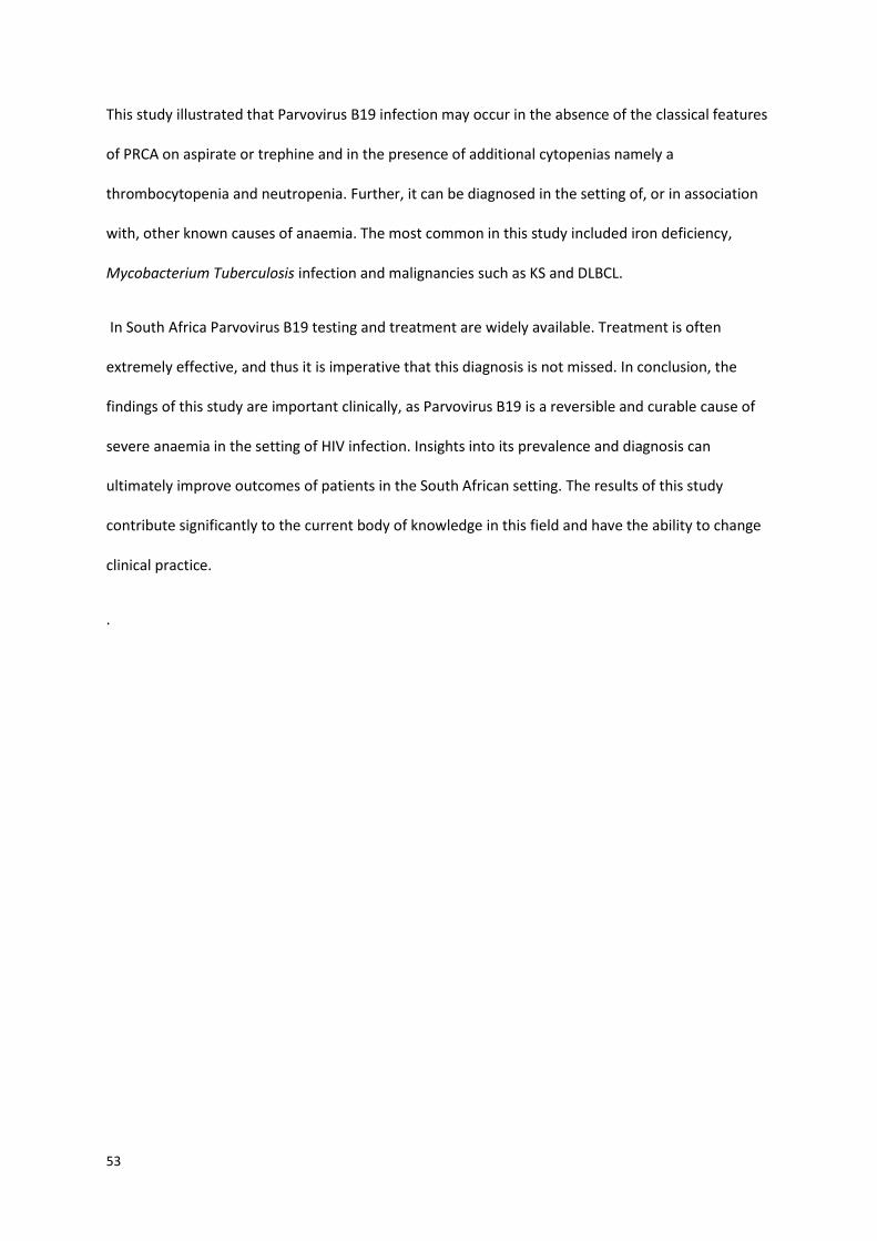
53
This study illustrated that Parvovirus B19 infection may occur in the absence of the classical features
of PRCA on aspirate or trephine and in the presence of additional cytopenias namely a
thrombocytopenia and neutropenia. Further, it can be diagnosed in the setting of, or in association
with, other known causes of anaemia. The most common in this study included iron deficiency,
Mycobacterium Tuberculosis infection and malignancies such as KS and DLBCL.
In South Africa Parvovirus B19 testing and treatment are widely available. Treatment is often
extremely effective, and thus it is imperative that this diagnosis is not missed. In conclusion, the
findings of this study are important clinically, as Parvovirus B19 is a reversible and curable cause of
severe anaemia in the setting of HIV infection. Insights into its prevalence and diagnosis can
ultimately improve outcomes of patients in the South African setting. The results of this study
contribute significantly to the current body of knowledge in this field and have the ability to change
clinical practice.
.
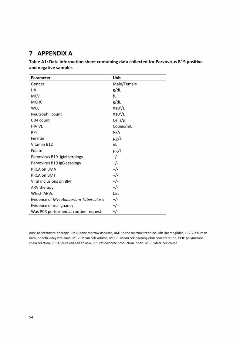
54
7 APPENDIX A Table A1: Data information sheet containing data collected for Parvovirus B19 positive and negative samples
Parameter Unit
Gender Male/Female
Hb g/dL
MCV fL
MCHC g/dL
WCC X109/L
Neutrophil count X109/L
CD4 count Cells/µl
HIV VL Copies/mL
RPI N/A
Ferritin µg/L Vitamin B12 nL
Folate µg/L Parvovirus B19 IgM serology +/-
Parvovirus B19 IgG serology +/-
PRCA on BMA +/-
PRCA on BMT +/-
Viral inclusions on BMT +/-
ARV therapy +/-
Which ARVs List
Evidence of Mycobacterium Tuberculosis +/-
Evidence of malignancy +/-
Was PCR performed as routine request +/-
ARV: antiretroviral therapy, BMA: bone marrow aspirate, BMT: bone marrow trephine, Hb: Haemoglobin, HIV VL: human
immunodeficiency viral load, MCV: Mean cell volume, MCHC: Mean cell haemoglobin concentration, PCR: polymerase
chain reaction, PRCA: pure red cell aplasia, RPI: reticulocyte production index, WCC: white cell count
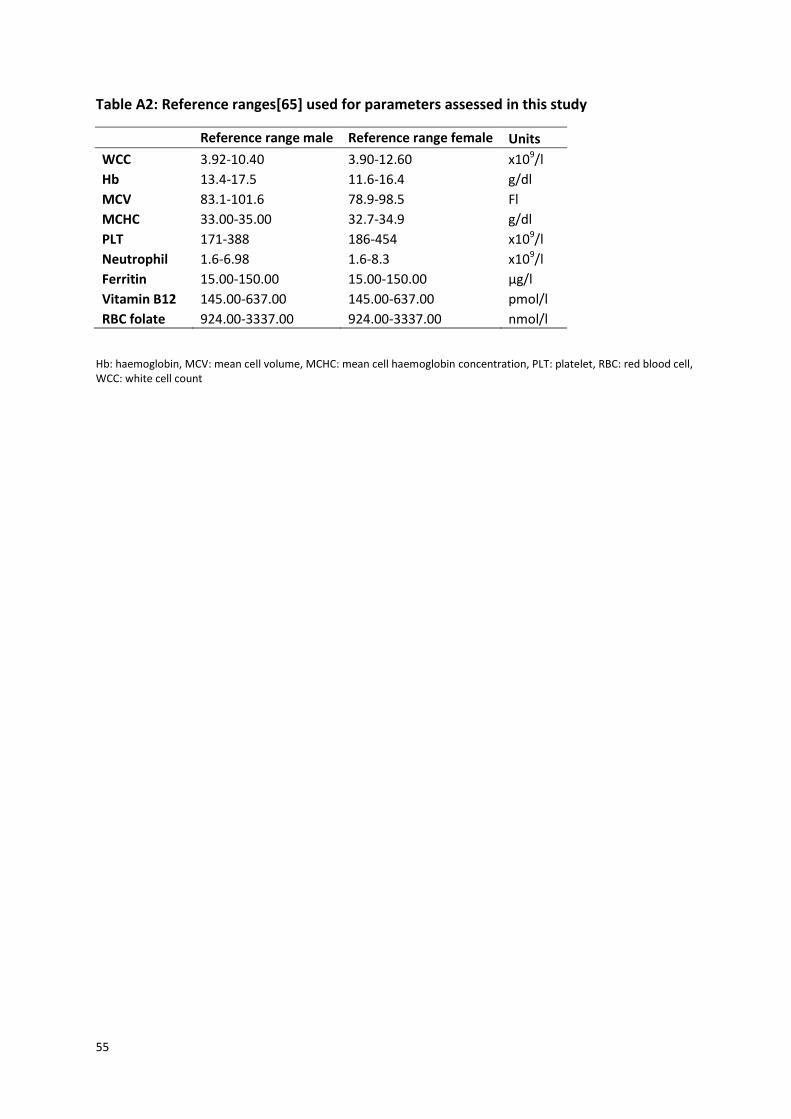
55
Table A2: Reference ranges[65] used for parameters assessed in this study
Reference range male Reference range female Units
WCC 3.92-10.40 3.90-12.60 x10⁹/l
Hb 13.4-17.5 11.6-16.4 g/dl
MCV 83.1-101.6 78.9-98.5 Fl
MCHC 33.00-35.00 32.7-34.9 g/dl
PLT 171-388 186-454 x109/l
Neutrophil 1.6-6.98 1.6-8.3 x10⁹/l
Ferritin 15.00-150.00 15.00-150.00 µg/l
Vitamin B12 145.00-637.00 145.00-637.00 pmol/l
RBC folate 924.00-3337.00 924.00-3337.00 nmol/l
Hb: haemoglobin, MCV: mean cell volume, MCHC: mean cell haemoglobin concentration, PLT: platelet, RBC: red blood cell, WCC: white cell count
56
Table A3: Definitions of terms used in the text
Term Definition HIV infected Positive ELISA or PCR[86]
Severe anaemia Hb< or = 8 g/dl[82]
Thrombocytopenia Platelet count <171 x109/l[65]
Neutropenia Neutrophil count <1.6 x10⁹/l[65]
PRCA as seen on morphologic assessment Absent or markedly reduced erythropoiesis and a lack of serial maturation of erythrocytes on BMA/BMT[68]
Megaloblastic anaemia Vitamin B12 or red blood cell folate level below the given reference range in conjunction with an anaemia[82]
Iron deficiency Ferritin level below the given reference range or absent iron stores on a Perl’s stain (in an adequate quality bone marrow aspirate)[68]
Anaemia of chronic disease Increased ferritin, usually in association with decreased transferrin and mild normocytic or microcytic anaemia[7]
HIV viral suppression HIV VL <40 copies/ml
Sensitivity True positives/(true positives + false negatives)
Specificity True negatives/(true negatives + false positives)
Negative predictive value True negatives/(true negatives + false negatives)
Positive predictive value True positives/(true positives + false positives)
False negative rate False negative/(false negative + true positive)
BMA: bone marrow aspirate, BMT: bone marrow trephine, ELISA: enzyme linked immunosorbent assay, Hb: haemoglobin,
HIV VL: Human Immunodeficiency Virus VL, PCR: polymerase chain reaction, PRCA: pure red cell aplasia
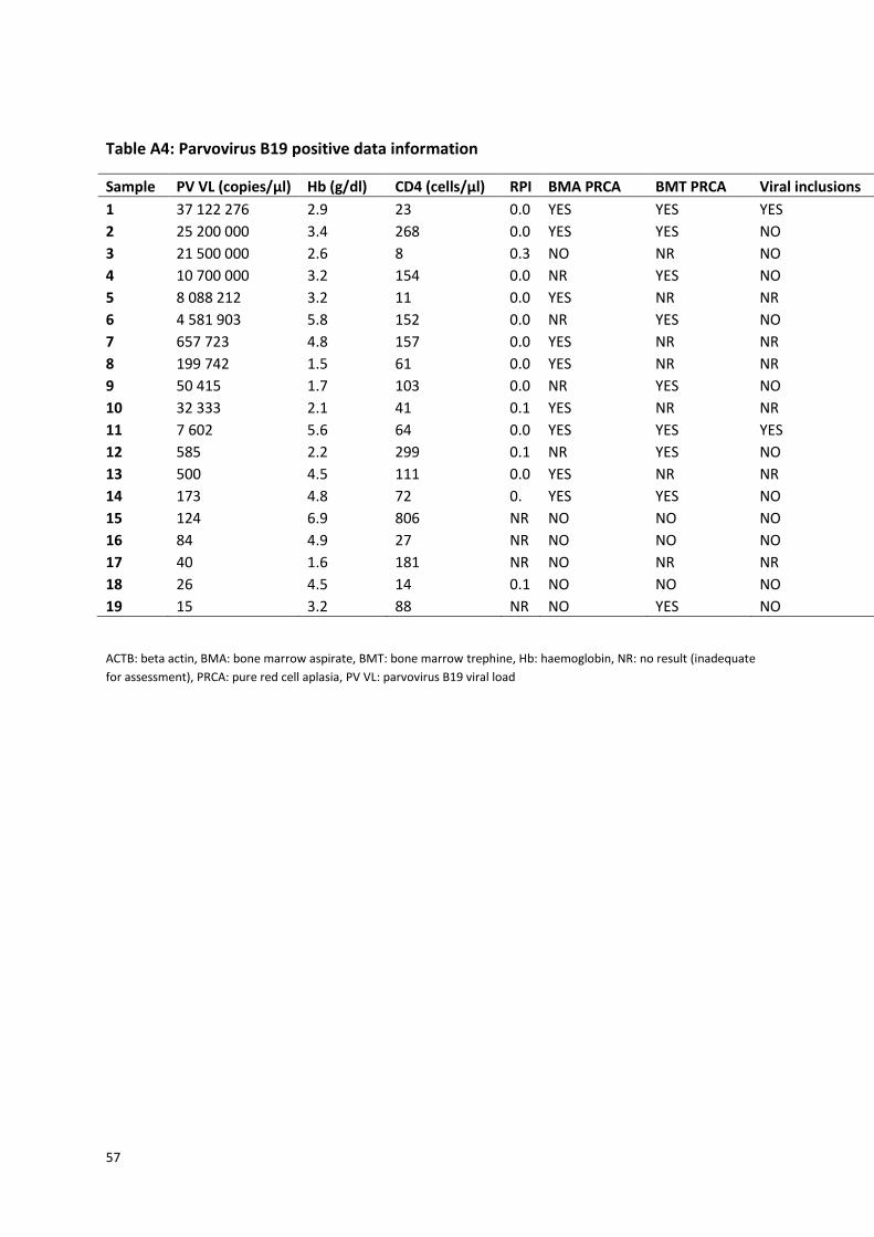
57
Table A4: Parvovirus B19 positive data information
Sample PV VL (copies/µl) Hb (g/dl) CD4 (cells/µl) RPI BMA PRCA BMT PRCA Viral inclusions ACTB (copies/ µl)
1 37 122 276 2.9 23 0.0 YES YES YES 2320
2 25 200 000 3.4 268 0.0 YES YES NO 542
3 21 500 000 2.6 8 0.3 NO NR NO 7080
4 10 700 000 3.2 154 0.0 NR YES NO 550
5 8 088 212 3.2 11 0.0 YES NR NR 2908
6 4 581 903 5.8 152 0.0 NR YES NO 2078
7 657 723 4.8 157 0.0 YES NR NR 4190
8 199 742 1.5 61 0.0 YES NR NR 1695
9 50 415 1.7 103 0.0 NR YES NO 14175
10 32 333 2.1 41 0.1 YES NR NR 343
11 7 602 5.6 64 0.0 YES YES YES 408
12 585 2.2 299 0.1 NR YES NO 672
13 500 4.5 111 0.0 YES NR NR 101
14 173 4.8 72 0. YES YES NO 14.9
15 124 6.9 806 NR NO NO NO 185
16 84 4.9 27 NR NO NO NO 32
17 40 1.6 181 NR NO NR NR 122
18 26 4.5 14 0.1 NO NO NO 108
19 15 3.2 88 NR NO YES NO 320
ACTB: beta actin, BMA: bone marrow aspirate, BMT: bone marrow trephine, Hb: haemoglobin, NR: no result (inadequate
for assessment), PRCA: pure red cell aplasia, PV VL: parvovirus B19 viral load
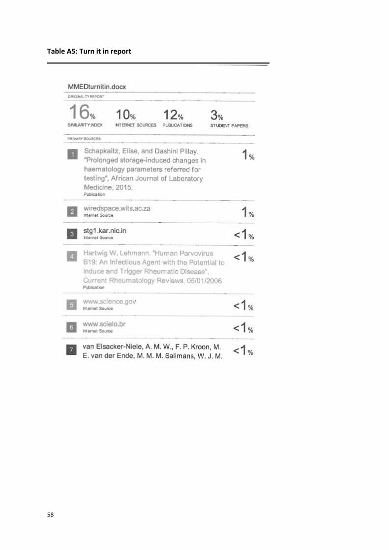
58
Table A5: Turn it in report
59
REFERENCES
1. Corcoran, A. and S. Doyle, Advances in the biology, diagnosis and host-pathogen interactions of parvovirus B19. J Med Microbiol, 2004. 53(Pt 6): p. 459-75.
2. Chisaka, H., et al., Parvovirus B19 and the pathogenesis of anaemia. Rev Med Virol, 2003. 13(6): p. 347-59.
3. Hallett, T.B., et al., Embracing different approaches to estimating HIV incidence, prevalence and mortality. AIDS, 2014. 28 Suppl 4: p. S523-32.
4. Belperio, P.S. and D.C. Rhew, Prevalence and outcomes of anemia in individuals with human immunodeficiency virus: a systematic review of the literature. Am J Med, 2004. 116 Suppl 7A: p. 27S-43S.
5. Redig, A.J. and N. Berliner, Pathogenesis and clinical implications of HIV-related anemia in 2013. Hematology Am Soc Hematol Educ Program, 2013. 2013: p. 377-81.
6. Sompayrac, L., How the immune system works. 4th ed. 2012, Chichester, West Sussex ; Hoboken, NJ: Wiley-Blackwell.
7. Bain, B.J., Pathogenesis and pathophysiology of anemia in HIV infection. Curr Opin Hematol, 1999. 6(2): p. 89-93.
8. Claster, S., Biology of anemia, differential diagnosis, and treatment options in human immunodeficiency virus infection. J Infect Dis, 2002. 185 Suppl 2: p. S105-9.
9. Ellaurie, M., E.R. Burns, and A. Rubinstein, Hematologic manifestations in pediatric HIV infection: severe anemia as a prognostic factor. Am J Pediatr Hematol Oncol, 1990. 12(4): p. 449-53.
10. Sullivan, P.S., et al., Epidemiology of anemia in human immunodeficiency virus (HIV)-infected persons: results from the multistate adult and adolescent spectrum of HIV disease surveillance project. Blood, 1998. 91(1): p. 301-8.
11. Bain, B.J., J.V. Dacie, and S.M. Lewis, Dacie and Lewis practical haematology. 11th ed. 2012, [Edinburgh]: Churchill Livingstone. xi, 653 p.
12. Egloff, N., M. Rossi, and H. Furrer, [Anemia in HIV infection]. Praxis (Bern 1994), 2000. 89(23): p. 1007-13.
13. Hoffbrand, A.V. and P.A.H. Moss, Essential haematology. 6th ed. 2011, Chichester: Wiley-Blackwell. xi, 454 p.
14. Moore, R.D., Anemia and human immunodeficiency virus disease in the era of highly active antiretroviral therapy. Semin Hematol, 2000. 37(4 Suppl 6): p. 18-23.
15. Young, B. and J. Zaritsky, Hepcidin for clinicians. Clin J Am Soc Nephrol, 2009. 4(8): p. 1384-7. 16. Sharada Raju, R., et al., Acute human parvovirus b19 infection: cytologic diagnosis. Indian J
Hematol Blood Transfus, 2014. 30(Suppl 1): p. 133-4. 17. Trosemeier, J.H., et al., Genome sequences of parvovirus b19 reference strains. Genome
Announc, 2014. 2(5). 18. Servant, A., et al., Genetic diversity within human erythroviruses: identification of three
genotypes. J Virol, 2002. 76(18): p. 9124-34. 19. Candotti, D., et al., Identification and characterization of persistent human erythrovirus
infection in blood donor samples. J Virol, 2004. 78(22): p. 12169-78. 20. Norja, P., et al., Rapid sequence change and geographical spread of human parvovirus B19:
comparison of B19 virus evolution in acute and persistent infections. J Virol, 2008. 82(13): p. 6427-33.
60
21. Corcoran, C., et al., Genetic variants of human parvovirus B19 in South Africa: cocirculation of three genotypes and identification of a novel subtype of genotype 1. J Clin Microbiol, 2010. 48(1): p. 137-42.
22. Zhi, N., et al., Molecular and functional analyses of a human parvovirus B19 infectious clone demonstrates essential roles for NS1, VP1, and the 11-kilodalton protein in virus replication and infectivity. J Virol, 2006. 80(12): p. 5941-50.
23. Chen, A.Y. and J. Qiu, Parvovirus infection-induced cell death and cell cycle arrest. Future Virol, 2010. 5(6): p. 731-743.
24. Kidane, B., et al., Worse than a slap in the face. Am J Med, 2010. 123(2): p. 122-4. 25. Norja, P., et al., Bioportfolio: lifelong persistence of variant and prototypic erythrovirus DNA
genomes in human tissue. Proc Natl Acad Sci U S A, 2006. 103(19): p. 7450-3. 26. Morinet, F., et al., Concise review: Anemia caused by viruses. Stem Cells, 2011. 29(11): p.
1656-60. 27. Volberding, P.A., K.R. Baker, and A.M. Levine, Human immunodeficiency virus hematology.
Hematology Am Soc Hematol Educ Program, 2003: p. 294-313. 28. Schoub, B.D., et al., Primary and secondary infection with human parvovirus B19 in pregnant
women in South Africa. S Afr Med J, 1993. 83(7): p. 505-6. 29. Guidozzi, F., D. Ballot, and A.D. Rothberg, Human B19 parvovirus infection in an obstetric
population. A prospective study determining fetal outcome. J Reprod Med, 1994. 39(1): p. 36-8.
30. Rubinstein, R., et al., Prevalence of human parvovirus B19 and TT virus in a group of young haemophiliacs in South Africa. Haemophilia, 2000. 6(2): p. 93-7.
31. Ihara, T., et al., A population-based epidemiological survey of human parvovirus B19 infection: a project of the Kyushu and Okinawa Population Study (KOPS). Arch Virol, 2013. 158(12): p. 2465-72.
32. Levican, J., et al., [Parvovirus B19 among blood donors from three hospitals in Santiago, Chile]. Rev Med Chil, 2011. 139(2): p. 143-9.
33. Kooistra, K., et al., Epidemiology of high-level parvovirus B19 viraemia among Dutch blood donors, 2003-2009. Vox Sang, 2011. 100(3): p. 261-6.
34. Nabae, K., et al., Estimating the risk of parvovirus B19 infection in blood donors and pregnant women in Japan. PLoS One, 2014. 9(3): p. e92519.
35. Kumar, S., et al., Seroprevalence of human parvovirus B19 in healthy blood donors. Med J Armed Forces India, 2013. 69(3): p. 268-72.
36. Zhang, W., et al., Parvovirus B19V DNA contamination in Chinese plasma and plasma derivatives. J Transl Med, 2012. 10: p. 194.
37. Slavov, S.N., et al., Molecular and phylogenetic analyses of human Parvovirus B19 isolated from Brazilian patients with sickle cell disease and beta-thalassemia major and healthy blood donors. J Med Virol, 2012. 84(10): p. 1652-65.
38. Juhl, D., et al., Parvovirus b19 infections and blood counts in blood donors. Transfus Med Hemother, 2014. 41(1): p. 52-9.
39. Sakata, H., et al., Impact of chemiluminescent enzyme immunoassay screening for human parvovirus B19 antigen in Japanese blood donors. Transfusion, 2013. 53(10 Pt 2): p. 2556-66.
40. Grabarczyk, P., et al., [Parvovirus B19 DNA testing in Polish blood donors, 2004-2010]. Przegl Epidemiol, 2012. 66(1): p. 7-12.
41. Hsu, H.C., et al., Bone marrow samples from patients with aplastic anemia are not infected with parvovirus B19 and Mycobacterium tuberculosis. Am J Clin Pathol, 2002. 117(1): p. 36-40.
42. Wildig, J., et al., Parvovirus B19 infection and severe anaemia in Kenyan children: a retrospective case control study. BMC Infect Dis, 2010. 10: p. 88.
43. Koduri, P.R., Parvovirus B19-related anemia in HIV-infected patients. AIDS Patient Care STDS, 2000. 14(1): p. 7-11.
61
44. Mylonakis, E., et al., Persistent parvovirus B19 related anemia of seven years' duration in an HIV-infected patient: complete remission associated with highly active antiretroviral therapy. Am J Hematol, 1999. 60(2): p. 164-6.
45. Scapellato, P.G., A.M. Palumbo, and S. Del Valle, Improvement of anemia induced by parvovirus B19 in a patient with AIDS after combined antiretroviral therapy. Mayo Clin Proc, 2000. 75(2): p. 215-6.
46. Arribas, J.R., J.M. Pena, and J.E. Echevarria, Parvovirus B19-related anemia in an HIV-infected patient: rapid control after production of neutralizing antibodies during highly active antiretroviral therapy. Ann Intern Med, 2000. 132(12): p. 1011.
47. Ware, A.J. and T. Moore, Resolution of chronic parvovirus b19-induced anemia, by use of highly active antiretroviral therapy, in a patient with acquired immunodeficiency syndrome. Clin Infect Dis, 2001. 32(7): p. E122-3.
48. Taguchi, H., et al., Acute parvovirus B19 infection during anti-retroviral therapy. J Infect Chemother, 2001. 7(2): p. 110-2.
49. Intalapaporn, P., Y. Poovorawan, and C. Suankratay, Immune reconstitution syndrome associated with parvovirus B19-induced pure red cell aplasia during highly active antiretroviral therapy. J Infect, 2006. 53(2): p. e79-82.
50. Nolan, R.C., G. Chidlow, and M.A. French, Parvovirus B19 encephalitis presenting as immune restoration disease after highly active antiretroviral therapy for human immunodeficiency virus infection. Clin Infect Dis, 2003. 36(9): p. 1191-4.
51. Chen, M.Y., et al., Reconstituted immunity against persistent parvovirus B19 infection in a patient with acquired immunodeficiency syndrome after highly active antiretroviral therapy. Clin Infect Dis, 2001. 32(9): p. 1361-5.
52. Sanphasitvong, W., et al., Parvovirus b19 infection in HIV patient with pure red cell aplasia. Southeast Asian J Trop Med Public Health, 2005. 36(5): p. 1216-20.
53. Clarke, J. and J.D. Lee, Primary human parvovirus B19 infection in an HIV infected patient on highly active antiretroviral therapy. Sex Transm Infect, 2003. 79(4): p. 336.
54. Christensen, L.S., T.V. Madsen, and T. Barfod, Persistent erythrovirus B19 urinary tract infection in an HIV-positive patient. Clin Microbiol Infect, 2001. 7(9): p. 507-9.
55. Naval-Srinivas, R.M. and L.W. Church, Significant decrease in hemoglobin and hematocrit levels in a virologically controlled HIV-infected patient. Pure red cell aplasia (PRCA) caused by human parvovirus B19. AIDS Read, 2003. 13(4): p. 189.
56. Lau, S.M., et al., Parvovirus B19 infection in a human immunodeficiency virus-infected patient with anemia. J Formos Med Assoc, 2000. 99(2): p. 162-5.
57. Azevedo, K.M., et al., Parvovirus B19 seroconversion in a cohort of human immunodeficiency virus-infected patients. Mem Inst Oswaldo Cruz, 2012. 107(3): p. 356-61.
58. Ferry, T., et al., Infrequent replication of parvovirus B19 and erythrovirus genotypes 2 and 3 among HIV-infected patients with chronic anemia. Clin Infect Dis, 2010. 50(1): p. 115-8.
59. Abdollahi, A., et al., Status of immunity against PVB19 in HIV-infected patients according to CD4(+) cell count, and antiretroviral therapy regimen groups. Niger Med J, 2014. 55(1): p. 20-3.
60. Azadmanesh, K., et al., Frequency and genotype of human parvovirus B19 among Iranian patients infected with HIV. J Med Virol, 2015. 87(7): p. 1124-9.
61. Compston, L.I., et al., Prevalence of persistent and latent viruses in untreated patients infected with HIV-1 from Ghana, West Africa. J Med Virol, 2009. 81(11): p. 1860-8.
62. He, M., et al., Human immunodeficiency virus/human parvovirus B19 co-infection in blood donors and AIDS patients in Sichuan, China. Blood Transfus, 2012. 10(4): p. 502-14.
63. Aguiar, F.S., et al., Human parvovirus B19 infection in HIV-positive patients. Rev Soc Bras Med Trop, 2001. 34(3): p. 239-42.
62
64. Azevedo, K.M., et al., Seroepidemiological study of human parvovirus B19 among human immunodeficiency virus-infected patients in a medium-sized city in Rio de Janeiro, Brazil. Mem Inst Oswaldo Cruz, 2009. 104(6): p. 901-4.
65. Lawrie, D., et al., Local reference ranges for full blood count and CD4 lymphocyte count testing. S Afr Med J, 2009. 99(4): p. 243-8.
66. Seo, J.Y., H.J. Kim, and S.H. Kim, Neutropenia in parvovirus B19-associated pure red cell aplasia. Ann Hematol, 2011. 90(8): p. 975-8.
67. Alliot, C., et al., Parovirus B19 infection in an HIV-infected patient with febrile pancytopenia and acute hepatitis. Eur J Clin Microbiol Infect Dis, 2001. 20(1): p. 43-5.
68. Bain, B.J., D.M. Clark, and B. Wilkins, Bone marrow pathology. 4th ed. 2010, Oxford: Wiley-Blackwell. x, 630 p.
69. Koduri, P.R., R. Kumapley, and N.D. Khokha, Nondiagnostic bone marrow in parvovirus B19-related anemia in AIDS: confounding effect of prior transfusion? J Infect Dis, 1998. 177(6): p. 1773.
70. Stephenson, J.R. and A. Warnes, Diagnostic virology protocols. 2nd ed. Methods in molecular biology. 2011, [Totowa, N.J.]: Humana Press. xiii, 470 p.
71. Parvovirus inclusions. Image available: http://imagebank.hematology.org/SetDetail.aspx?SetID=338&AssetType=AssetSet [Accessed 15.07.2015].
72. Parvovirus B19-induced pure red cell aplasia in a heart transplant recipient. Image available: http://imagebank.hematology.org/AssetDetail.aspx?AssetID=14466&AssetType=Asset [Accessed 15.07.2015].
73. Mendelson, E., et al., Laboratory assessment and diagnosis of congenital viral infections: Rubella, cytomegalovirus (CMV), varicella-zoster virus (VZV), herpes simplex virus (HSV), parvovirus B19 and human immunodeficiency virus (HIV). Reprod Toxicol, 2006. 21(4): p. 350-82.
74. Saah, A.J., et al., Factors influencing survival after AIDS: report from the Multicenter AIDS Cohort Study (MACS). J Acquir Immune Defic Syndr, 1994. 7(3): p. 287-95.
75. Crabol, Y., et al., Intravenous immunoglobulin therapy for pure red cell aplasia related to human parvovirus b19 infection: a retrospective study of 10 patients and review of the literature. Clin Infect Dis, 2013. 56(7): p. 968-77.
76. Morelli, P., et al., Persistent parvovirus B19-induced anemia in an HIV-infected patient under HAART. Case report and review of literature. Eur J Clin Microbiol Infect Dis, 2007. 26(11): p. 833-7.
77. Patel, M., Haematologist, personal communication. August 2015 78. Pereira, R.F., et al., Clinical features and laboratory findings of human parvovirus B19 in
human immunodeficiency virus-infected patients. Mem Inst Oswaldo Cruz, 2014. 109(2): p. 168-73.
79. Sahu, S., Hemlata, and A. Verma, Adverse events related to blood transfusion. Indian J Anaesth, 2014. 58(5): p. 543-51.
80. Kallenberg, C.G., A 10% ready-to-use intravenous human immunoglobulin offers potential economic advantages over a lyophilized product in the treatment of primary immunodeficiency. Clin Exp Immunol, 2007. 150(3): p. 437-41.
81. Patel, M., Haematologist, personal communication. January 2012. 82. WHO. Haemoglobin concentrations for the diagnosis of anaemia and assessment of severity.
Vitamin and Mineral Nutrition Information System. Geneva, World Health Organization,2011(WHO/NMH/NHD/MNM/11.1).
83. Glencross, D., et al., CD45-assisted PanLeucogating for accurate, cost-effective dual-platform CD4+ T-cell enumeration. Cytometry, 2002. 50(2): p. 69-77.
84. PrimerDesignTM ingene kit for Human Parvovirus B19 package insert available at: http://www.genesig.com/assets/files/hpvb19_std.pdf [accessed 06/01/2017].

63
85. MacLeod, W.B., et al. Analysis of age- and sex-specific HIV care cascades in South Africa suggests unequal progress towards UNAIDS 90-90-90 treatment targets [Abstract TUPDC0102]. in 21st International AIDS Conference. 18-22 July 2016. Durban, South Africa.
86. Branson, B., et al., Laboratory testing for the diagnosis of HIV infection : updated recommendations. 2014.