Embed Size (px)
Citation preview

THE PREVALENCE OF BIRTH ASPHYXIA AND IMMEDIATE OUTCOMES AMONG
PREGNANT WOMEN ATTENDING MATERNAL HEALTH SERVICES AT ISHAKA
ADVENTIST HOSPITAL.
BY
MUHUMUZA AMOS
REG NO: BMS/ 0100/ 113 /DU
A DISSERTATION SUBMITTED TO THE FACULTY OF CLINICAL MEDICINE AND
DENTISTRY IN PARTIAL FULFILLMENT OF THE REQUIREMENTS FOR THE
AWARD OF THE BACHELORS DEGREE IN MEDICINE AND SURGERY OF
KAMPALA INTERNATIONAL UNIVERSITY
DECEMBER, 2014

i
DECLARATION
I, Muhumuza Amos of Registration Number BMS/0100/113/DU declare that this dissertation,
“The prevalence of birth asphyxia and immediate outcomes among pregnant women attending
maternal health services at Ishaka Adventist hospital.” is my own original work under
supervision of Dr.Saima Husnain and has never been submitted in part or fully to any other
University or Institution of higher learning for scrutiny for the purpose of an academic award
unless stated otherwise.
Signature …………………………… Date………………………………
ii
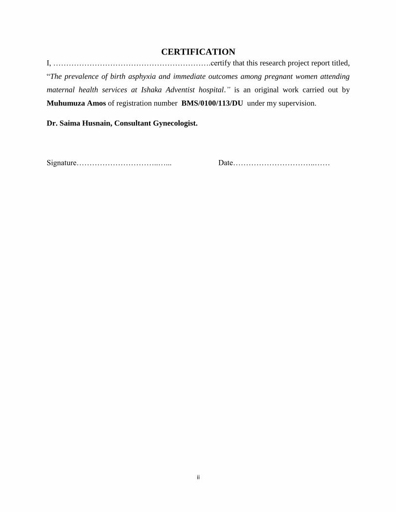
CERTIFICATION
I, …………………………………………………….certify that this research project report titled,
“The prevalence of birth asphyxia and immediate outcomes among pregnant women attending
maternal health services at Ishaka Adventist hospital.” is an original work carried out by
Muhumuza Amos of registration number BMS/0100/113/DU under my supervision.
Dr. Saima Husnain, Consultant Gynecologist.
Signature…………………………..…... Date…………………………..……

iii
DEDICATION
I dedicate this work to the Almighty God who has given me life, faithfully lead and still guiding
me through my course.
To my dear parents, Mr. & Mrs. Tindiwensi Charles who have continuously and tirelessly
supported me in my studies and played a great role in my life. Thank you for being there for me.
And finally to all my brothers, sister, cousins, uncles and aunts especially my only sister
Mrs.Banya confidence, thanks for your support and know that I love you all.

iv
ACKNOWLEDGEMENT
I would like to acknowledge Kampala international university for granting me an opportunity to
pursue my dream in life.
I acknowledge, Mr.Nyendde Jimmy (CEO) Ishaka Adventist hospital and his staff for their help
in offering me with the necessary resources for my research work.
I acknowledge my dear parents, Mr. & Mrs. Tindiwensi Charles, who have continuously and
tirelessly supported me in my studies and played a great role in my life. Thank you a lot, you
have given me a bright future. God bless you all.
I acknowledge my supervisor Dr.Saima Husnain, who kindly and tirelessly guided me
throughout this work despite her tight programmes and responsibilities.
And finally, I acknowledge all my friends for the support and advice given during my research
work in several way. God reward you abundantly.
v
TABLE OF CONTENTS
DECLARATION…..…………………………….…………………………………………………………………… I
CERTIFICATION………………………...................................…………………………………………………… II
DEDICATION…………….......……………………………………….…………………………………………… III
ACKNOWLEDGEMENT……..………………………………………………….……………………………….. IV
TABLE OF CONTENTS………………………......………………………………………….……………………..V
LIST OF FIGURES…………....…………………………………………………………….…………….……… VII
LIST OF TABLES………….......…………………………………………………………..….………………….. VII
ACRONYM AND ABBREVIATIONS LISTS…………………………….......................…………………….. VIII
DEFINITIONS……………….......………………………………………………………………….……………… IX
ABSTRACT…………………..........……………………………………………….………...……………………… X
CHAPTER ONE: INTRODUCTION…………………………..…………………………….…………………….. 1
1.0 INTRODUCTION ................................................................................................................................................... 1
1.1 BACKGROUND ..................................................................................................................................................... 1
1.2 PROBLEM STATEMENT ...................................................................................................................................... 3
1.3 PURPOSE OF THE STUDY ................................................................................................................................... 4
1.4 OBJECTIVES .......................................................................................................................................................... 4
1.4.1 General objective .................................................................................................................................................. 4
1.4.2 Specific objectives ................................................................................................................................................. 4
1.5 RESEARCH QUESTIONS ...................................................................................................................................... 5
1.6 JUSTIFICATION OF THE STUDY ........................................................................................................................ 5
1.7 SCOPE OF STUDY ................................................................................................................................................. 6
1.7.1 Geographical scope .............................................................................................................................................. 6
1.7.2 Content scope ....................................................................................................................................................... 6
1.7.3 Time scope ............................................................................................................................................................ 6
1.8 CONCEPTUAL FRAME WORK ............................................................................................................................ 7
1.8.1 Explanation of the conceptual frame work. .......................................................................................................... 8
CHAPTER TWO: LITERATURE REVIEW……………………………………..……………………….………. 9
2.0 INTRODUCTION ................................................................................................................................................... 9
2.1 GLOBAL PREVALENCE OF BIRTH ASPHYXIA ............................................................................................... 9
2.1.1 Birth asphyxia in Uganda. .................................................................................................................................. 11
2.1.2 Babies with low Apgar score in Mulago Hospital, Uganda. .............................................................................. 11
2.1.3 Birth Asphyxia in Ishaka –Bushenyi district ....................................................................................................... 11
2.2 BIRTHS IN UGANDA ANNUALLY ................................................................................................................... 11
2.2.1 Births in Bushenyi district annually .................................................................................................................... 12
2.3 NEWBORN DEATHS IN UGANDA .................................................................................................................... 12
2.4 GLOBAL OUTCOMES OF BIRTH ASPHYXIA ................................................................................................. 13
2.5 FACTORS ACCOUNTING FOR BIRTH ASPHYXIA CASES ........................................................................... 14
2.5.1 Maternal diseases ............................................................................................................................................... 14
2.5.2 Labor and deliveries ........................................................................................................................................... 14
2.5.3 Fetal factors ........................................................................................................................................................ 14

vi
CHAPTER THREE: STUDY METHODOLOGY…………....………………………………………….………. 15
3.0 INTRODUCTION ................................................................................................................................................. 15
3.1 STUDY DESIGN ................................................................................................................................................... 15
3.2 STUDY AREA ...................................................................................................................................................... 15
3.3 STUDY POPULATION ........................................................................................................................................ 15
3.4 SAMPLE SIZE DETERMINATION ..................................................................................................................... 16
3.5 SAMPLE TECHNIQUE ........................................................................................................................................ 16
3.6 INCLUSION AND EXCLUSION CRITERIA ...................................................................................................... 17
3.6.1 Inclusion Criteria ............................................................................................................................................... 17
3.6.2 Exclusion Criteria ............................................................................................................................................... 17
3.7 DATA COLLECTION .......................................................................................................................................... 17
3.8 DATA ANALYSIS ................................................................................................................................................ 17
3.9 ETHICAL CONSIDERATIONS ........................................................................................................................... 18
3.10 STUDY LIMITATIONS AND THEIR SOLUTIONS ......................................................................................... 18
3.11 DISSEMINATION OF RESULTS ...................................................................................................................... 18
CHAPTER FOUR: STUDY FINDINGS AND RESULTS……………………………………………….……… 19
4.0 INTRODUCTION ................................................................................................................................................. 19
4.1 PRESENTATION OF SPECIFIC OBJECTIVE ONE ........................................................................................... 19
4.2 PRESENTATION OF SPECIFIC OBJECTIVE TWO .......................................................................................... 21
4.3 PRESENTATION OF SPECIFIC OBJECTIVE THREE ...................................................................................... 23
4.4 PRESENTATION OF SPECIFIC OBJECTIVE FOUR ......................................................................................... 28
CHAPTER FIVE: DISCUSSION, CONCLUSIONS AND RECOMMENDATIONS…………………….…… 33
5.0 INTRODUCTION ................................................................................................................................................. 33
5.1 DISCUSSIONS ...................................................................................................................................................... 33
5.1.1 Discussion on the number of births and proportion of asphyxiated neonates. ................................................... 33
5.1.2 Discussion of the Immediate Outcomes .............................................................................................................. 34
5.1.3 Discussion of Risk Factors for Birth Asphyxia ................................................................................................... 36
5.2. CONCLUSIONS .................................................................................................................................................. 37
5.3 RECOMMENDATIONS ....................................................................................................................................... 37
5.4 STUDY CHALLENGES ....................................................................................................................................... 38
BIBLIOGRAPHY…………………….........……………………………………………………………….………. 39
APPENDICES………………………....…………………………………………………………………….……… 42
APPENDIX 1: DESIGNED DATA COLLECTION TOOL ........................................................................................ 42
APPENDIX 2: PROPOSED BUDGET FOR THE STUDY ........................................................................................ 43
APPENDIX 3: WORK PLAN ..................................................................................................................................... 44
APPENDIX 4: APPROVAL LETTER TO ACCESS RESEARCH RESOURCES AT IAH ....................................... 45
APPENDIX 5: A MAP OF BUSHENYI SHOWING THE LOCATION OF ISHAKA ............................................... 46
APPENDIX 6: A MAP OF UGANDA SHOWING THE LOCATION OF BUSHENYI DISTRICT .......................... 47

vii
LIST OF FIGURES FIGURE 1: SHOWS TOTAL NUMBER OF BIRTHS AT IAH FROM JAN. 2012 TO DEC. 2013 ............................. 19
FIGURE 2: SHOWS TOTAL NUMBER OF LIVE BIRTHS AT IAH FROM JAN. 2012 TO DEC. 2013 ..................... 20
FIGURE 3: SHOWS TOTAL NUMBER OF ASPHYXIATED BIRTHS FROM JAN. 2012 TO DEC. 2013 .................. 21
FIGURE 4: SHOWS NUMBER OF STILLBIRTHS AT IAH IN 2012 AND 2013 .................................................... 22
FIGURE 5: SHOWS IMMEDIATE OUTCOMES OF BIRTH ASPHYXIA AT IAH FROM JAN. TO DEC. 2012 ........... 23
FIGURE 6: SHOWS IMMEDIATE OUTCOMES OF BIRTH ASPHYXIA IN IAH FROM JAN. TO DEC. 2013 ........... 23
FIGURE 7: SHOWS NUMBER OF REFERRAL IN CASES AND IMMEDIATE OUTCOMES ..................................... 24
FIGURE 8: SHOWS RESUSCITATED NEONATES AND IMMEDIATE OUTCOMES IN 2012 AND 2013 ................. 25
FIGURE 9: SHOWS FACTORS ACCOUNTING FOR BA DURING DELIVERY IN 2012 AND 2013 AT IAH ........... 28
FIGURE 10: SHOWS THE WEIGHTS OF THE ASPHYXIATED NEONATES IN 2012 AND 2013 AT IAH .............. 29
FIGURE 11: SHOWS MATERNAL AGE OF THE ASPHYXIATED NEONATES IN 2012 AND 2013 AT IAH ........... 30
FIGURE 12: SHOWS THE PARITY OF THE MOTHERS WITH ASPHYXIATED NEONATES IN 2012 AND 2013 ..... 32
LIST OF TABLES
TABLE 1: SHOWS APGAR SCORE OF NEONATES AT BIRTH IN RELATION TO IMMEDIATE OUTCOMES ......... 25
TABLE 2: SHOWS APGAR SCORE OF NEONATES AT BIRTH IN RELATION TO IMMEDIATE OUTCOMES .......... 26
TABLE 3: SHOWS IMMEDIATE OUTCOMES IN RELATION TO MODE OF DELIVERY IN 2012 ........................... 27
TABLE 4: SHOWS IMMEDIATE OUTCOMES IN RELATION TO MODE OF DELIVERY IN 2013 ........................ 27
TABLE 5: SHOWS GENDER OF THE ASPHYXIATED NEONATES IN 2012 AND 2013 ....................................... 29
TABLE 6: SHOWS THE PARITY OF THE MOTHERS WITH ASPHYXIATED NEONATES IN 2012 AND 2013 ........ 31

viii
ACRONYM AND ABBREVIATIONS LISTS
HIE: Hypoxic Ischemic Encephalopathy
IUGR: Intrauterine Growth Retardation
ADHD: Attention deficit hyperactivity disorder
AIDS: Acquired Immunodeficiency syndrome
CVS: Cardiovascular system
DALYs: Disability Adjusted Life Years
DIC: Disseminated intravascular coagulation
HIV: Human Immune Virus
ICU: Intensive Care Unit
IPPV: Intrapulmonary Positive Pressure Ventilation
KIUTH: Kampala International University Teaching Hospital
IAH: Ishaka Adventist Hospital
MDG 4: Millennium Development Goal 4
MPDR: Maternal and Perinatal death review
PaCO2: Partial Pressure of carbon dioxide
PNFP: Private not-for-profit
SIADH: Syndrome of inappropriate antidiuretic hormone secretion
WHO: World Health Organization
SVD: Spontaneous Vertex Delivery
EMC/S: Emergency Caesarean Section
C/S: Caesarean Section
MSB: Macerated Still Births
CPD: Cephalo-Pelvic Disproportion
PROM: Pre labor Rupture of Membrane
MoH: Ministry of Health
BA: Birth asphyxia

ix
DEFINITIONS Birth asphyxia is a hypoxic insult severe enough to cause metabolic acidosis, neonatal
encephalopathy and multiorgan system dysfunction. (Olga Golubnitschaja et al, 2011) WHO
defines birth asphyxia simply as “the failure to initiate and sustain breathing at birth.”
Newborn or neonate refers to an infant in the first 28 days after birth. The term "newborn"
includes premature infants, post mature infants and full term newborns. (MoH Uganda, 2008)
Infant is typically applied to children between the ages of 1 month and 12 months; however,
definitions vary between birth and 3 years of age. (Stedman’s medical dictionary)
Stillbirths refers to Pregnancy losses occurring after seven completed months of gestation and
are expressed per 1,000 total births. (Dr. Gelasius Mukasa et al, 2008)
Hypoxic ischemic encephalopathy is a cellular damage that occurs within the central nervous
system (the brain and spinal cord) from inadequate oxygen due to intrauterine hypoxia and
birth asphyxia. (Oxford concise Medical dictionary)
Perinatal mortality is defined as the "number of stillbirths and deaths in the first week of life
per 1,000 live births"(WHO, 2012)
Neonatal Mortality Rate (NMR): The number of deaths during the first 28 days of life per
1,000 live births. Early neonatal mortality refers to a death in the first week of life while late
neonatal mortality refers to deaths between 7 and 28 days of life. (Dr. Gelasius Mukasa et al,
2008)
Live birth is the complete expulsion or extraction from its mother of a fetus/baby of 1000 grams
or 28 weeks gestation; which, after such separation, breathes or shows any other evidence of
life, such as beating of the heart, pulsation of the umbilical cord, or definite movement
of voluntary muscles, whether or not the umbilical cord has been cut or the placenta is
attached.(MoH Uganda, 2008)

x
ABSTRACT
Background: Birth Asphyxia is defined by WHO as “the failure to initiate and sustain breathing
at birth.” Millions of child deaths and stillbirths are attributable to birth asphyxia, yet limited
information is available to high light the burden of the condition to guide policy and practice,
particularly at the community level in developing countries like Uganda. This study will compile
insight on the prevalence and immediate outcomes of birth asphyxia in pregnant mothers
attending maternal health services at Ishaka Adventist hospital.
Method: A descriptive cross sectional study involving a sample size of 310 deliveries obtained
from Fisher’s et al, 1990 formula was undertaken to review HMIS records from January 2012 to
December 2013 in IAH Theatre, Maternity and Pediatric Ward to determine the prevalence and
immediate outcomes of birth asphyxia as determined by Apgar Score < 7/10 in 5 minutes, birth
weight >1500grams and gestational age >34 weeks.
Results: An overall prevalence of 4.8% was obtained from this study, out of which more males
were asphyxiated with 56.9% in 2012 and 58.3% in 2013 compared to females with 43.1% in
2012 and 41.7% in 2013, giving a male to female ratio of 1.4:1 in the study. Generally, statistics
showed that on average 267 deliveries were made from 2012 to 2013 at IAH and an incidence of
48/1000 births and an incidence of 52 deaths per 1000 live births was obtained from the study.
Majority of the neonates improved well upon resuscitation with 17cases (38.6%) and 23cases
(50%) in 2012 and 2013 respectively. In both years, low Apgar score between 0-3 resulted in
poor prognosis and no patient improved following resuscitation. Most cases with Apgar score of
4-7 improved 15 cases in 2012 and 20 cases in 2013, a few cases in the same range thus 12 cases
died shortly and 25 cases died later in both years. Both MSB and FSB contributed 60 cases
(71.8%) in both years. Important risk factors included; obstructed labor 34 cases (26.2%) in 2012
and 40 cases (22.2%) in 2013, followed by the fetal distress 27 cases (20.8%) in 2012 due to
idiopathic intrapartum conditions and prolonged labor, 27 cases (15.0%) in 2013.
Conclusion: There is urgent need to institute appropriate measures to prevent and manage
asphyxiated newborns in the hospital and region at large. Lastly, Without more attention to
improve obstetric care and advance birth asphyxia research, the millions of deaths related to
asphyxia, will remain out of reach of effective care, either by skilled or at community level, for
many years to come in Uganda.

1
CHAPTER ONE
1.0 INTRODUCTION
Over 200,000 children under the age of five years still die in Uganda annually, with more than
half of these dying in their first year and 45,000 within the first month of life – the newborn
period making Uganda the country with the fifth highest number of newborn deaths in sub-
Saharan Africa. Despite the struggles by the government of Uganda, in collaboration with
development partners to meet a reduction in childhood morbidity and mortality to achieve the
Poverty Eradication Action Plan and the Millennium Development Goals, Uganda’s neonatal
mortality rate possibly an under-estimate, is still very high at 29 deaths per 1,000 live births and
has not declined over a period of 15 years. This study will determine the prevalence of birth
asphyxia and its immediate out comes in pregnant women attending maternal health services at
Ishaka Adventist hospital, as one of the leading causes of neonatal deaths in Uganda and
information will be available for health policy makers to make informed decisions on child
health. (MoH, 2008)
1.1 BACKGROUND
World Health Organization defines birth asphyxia as “the failure to initiate and sustain breathing
at birth.” It is a hypoxic insult severe enough to cause metabolic acidosis, neonatal
encephalopathy and multiorgan system dysfunction. There have been a main challenge of
operationalizing the criteria of definition at community level in developing country settings by
using available information according to: feasibility thus the variables should be easily observed
by the health workers and easily recollected from the mothers, biological and clinical relevance
(defined by review of scientific evidence and expert consensus) thus accurate estimation of the
proportion of neonatal mortalities attributable to birth asphyxia is limited. However, definitions
of birth asphyxia designed for use in hospital- based settings require evaluation of umbilical cord
blood pH, Apgar scores, neurologic clinical status, and markers of multisystem organ function
and are not feasible for community settings (Anne CC. Lee et al, 2008).
Regarding the definition according to American College of Obstetricians and Gynecologists and
the American Academy of Pediatrics, a neonate is labeled to be asphyxiated if the following
conditions are fulfilled: (1) Umbilical artery metabolic or mixed respiratory-metabolic academia
2
with pH less than 7.00; (2) Apgar score of 0 to 3 for longer than 5 minutes; (3) Neurological
manifestations (e.g., seizures, coma, or hypotonic); and (4) Multisystem organ dysfunction, e.g.,
cardiovascular, gastrointestinal, hematological, pulmonary, or renal system(Dr Ornella Lincetto,
2007, Shazia et al, 2012)
Birth asphyxia causes in a global perspective differ between industrialized and non-industrialized
countries but the events have their roots in the antepartum, the intrapartum, or the postpartum
periods or combinations thereof. A recent review suggests that asphyxia is probably primarily
caused by antepartal factors in 50%-70% of cases, intrapartal factors in 20%-40% of cases and
postpartal factors in the remaining 10% of cases in industrialized countries. The concept that
difficult birth is the main factor of birth asphyxia and subsequent sequelae has in industrialized
countries been challenged in the recent years although in developing countries intrapartum
events still are the most important cause of birth asphyxia. (Ellis et al BMJ, 2000)
Clinical manifestation of asphyxia in a baby at the time of birth may include: baby is not
breathing or breathing is very weak, skin color is bluish or pale, heart rate is low, muscle tone is
poor or reflexes are weak, too much acid is in the blood (acidosis), the amniotic fluid is stained
with meconium (first stool) and the baby is experiencing seizures.
The clinical diagnosis of perinatal asphyxia is based on several criteria, the two main ones being
evidence of cardiorespiratory and neurological depression (defined as an Apgar score remaining
less than 7 at 5 minutes after birth) and evidence of acute hypoxic compromise with acidaemia
(defined as an arterial blood pH of less than 7 or base excess greater than 12 mmol/L). In many
settings, especially resource-poor countries, it may be impossible to assess fetal or neonatal
acidaemia. (William McGuire, 2007).
Dr. Virginia Apgar developed the Apgar score system in the middle of 20th century and since
then, it is still the worldwide practiced grading of the severity of perinatal asphyxia. The score
uses five criteria: Appearance, Pulse, Grimace, Activity, and Respiration, shortly APGAR.
Ranging from 0 to 10, the scores below 3 are considered as critically low for cases of the highest
emergency, 4 to 6 as fairly low, and the scores equal to or above 7 correspond to generally
normal states of the newborn’s health. In regions with a traditionally high neonatal mortality, the
Apgar score is frequently calculated as less than 7. Hence, in Saudi Arabia, the Apgar scores
below 7 were registered for 22% of newborns; 7.6% of them represented cases of neonatal
morbidity. In Tanzania, Apgar scores below 7 registered for 79% of the neonatal deaths. These

3
are clear indications for perinatal asphyxia as the major cause of neonatal morbidity. (Olga
Golubnitschaja et al, 2011)
In management of birth asphyxia anticipation is the key to preventing asphyxia neonatorum.
Others include; Good intrapartum care, Adequate and effective resuscitation, ICU monitoring for
complications. Regular BP, respiratory, urine output, acidosis among others, General measures;
that include, Nurse in thermo-neutral environment thus avoid high environmental temperature as
fever is associated with adverse outcome, Avoid hypo or hyperglycemia, Adequate ventilation
and avoid hypoxemia and hypercarbia or hypocarbia, Review infection risk and treatment with
antibiotics, Maintain adequate hydration but do not dehydrate or overhydrate, Treat jaundice as
necessary.
1.2 PROBLEM STATEMENT
According to the statistical data by the Global Burden of Disease Study, worldwide 10.5 million
children aged below 5 years die annually and of these 4 million are neonates, representing 38 %
of all deaths of children under 5 years of age.(Olga Golubnitschaja et al, 2011)Despite the
struggles by WHO to achieve the 4th MDG, Birth asphyxia still accounts for the death of
approximately 1 million (23%) infants annually and another 1.1 million(25% to 30% )
intrapartum stillbirths are also associated with Birth Asphyxia as well as unknown burden of
long term neurological disability and impairment in many sub-Saharan countries like Uganda.
Such a health emergency is unfortunately under estimated and often even unrecognized in the
developing countries where over half of neonatal deaths occur at home in the absence of skilled
care and just three major causes account for over three quarters of these deaths – serious
infections including tetanus (36%), complications of preterm births (27%) and birth asphyxia
(23%), (Seyal T and Hanif A, 2009).
To note is that, in Uganda more than half of the 200,000 children under five years that die each
year, die in their first year and 45,000 within the first month of life – the newborn period, making
Uganda the country with the fifth highest number of newborn deaths in sub-Saharan Africa
where birth asphyxia is one of the leading cause of neonatal deaths.(Dr.Gelasius Mukasa et al,
2008) However, the exact prevalence of low Apgar score and attendant risk factors for birth
asphyxia remains unknown despite use of the score system for over 50 years especially in
developing countries like Uganda where the socioeconomic status is not up to expected standards
4
and as a result, a large proportion of births and deaths occur in non-hospital settings limiting the
availability of data, hence under estimation of the burden of mortality and morbidity due to birth
asphyxia and thus the need for an epidemiological research in community based settings together
with hospital settings to determine the true prevalence of birth asphyxia in developing countries
like Uganda.
1.3 PURPOSE OF THE STUDY
At least 45,000 newborn deaths occur each year and an equal number are stillborn. Uganda’s
neonatal mortality rate (NMR), possibly an under-estimate, is very high at 29 deaths per 1,000
live births, has not declined over a period of 15 years.(MoH,2008) The common causes of
neonatal deaths in Uganda are similar to the rest of Africa and include birth asphyxia, infections
and complications of preterm birth. This makes Uganda the country with the fifth highest
number of newborn deaths in sub-Saharan Africa. Thus this study will help to contribute to the
limited information on the prevalence of birth asphyxia in Bushenyi district enabling district
policy makers able to make decisions aimed at reducing newborn morbidity and mortality with
particular attention to birth asphyxia, as the country looks forward to achieving the 4th MDG.
1.4 OBJECTIVES
1.4.1 General objective
To determine the prevalence of birth asphyxia and immediate outcomes in pregnant mothers
attending maternal health services at Ishaka Adventist Hospital (IAH).
1.4.2 Specific objectives
To determine the total number of births in IAH from Jan2012 toDec2013.
To find out the proportion of asphyxiated births in IAH from Jan.2012 to Dec. 2013.
To assess the immediate outcomes of the asphyxiated neonates in the hospital.
To find out the various factors that lead to birth asphyxia.
5
1.5 RESEARCH QUESTIONS
What was the total number of births in Ishaka Adventist hospital from Jan. 2012 to Dec. 2013?
What is the proportion of asphyxiated births in pregnant women attending maternal health
services in IAH from Jan. 2012 to Dec. 2013?
What are the immediate outcomes of birth asphyxia in Ishaka Adventist hospital?
What are the various factors leading to birth asphyxia?
1.6 JUSTIFICATION OF THE STUDY
Globally, perinatal asphyxia is responsible for 42 million disability life adjusted years, doubling
that due to diabetes and three quarters of that due to HIV/AIDS. Almost one quarter of the
world’s 4 million annual neonatal deaths are caused by perinatal asphyxia and 99% of these
deaths occur in low and mid-resource settings (Kinoti SN, PubMed).
To note is that, Uganda is ranked 5th African country among those that contribute 50% of the
newborn deaths annually in Africa with stillbirth and birth asphyxia being some of the leading
causes of these deaths in the country though there is only a handful of published studies from the
developing countries and none from Uganda, to shed light on the magnitude of the problem of
birth asphyxia. (Tunde Adegboyega et al, 2010)
With little information about the condition in Uganda, it’s on this background that I deeply feel
that information from this research will bring the problem to light with documented evidence in
order to attract the attention and concerns it deserves from the hospital administration and
ministry of health in Uganda.

6
1.7 SCOPE OF STUDY
1.7.1 Geographical scope
This study will be conducted at Ishaka Adventist hospital in Bushenyi district, Western Uganda.
1.7.2 Content scope
This study is restricted to the prevalence of birth asphyxia and immediate outcomes in pregnant
women attending maternal health services at Ishaka Adventist hospital. The proportion of
asphyxiated births in pregnant women attending maternal health services in the hospital will also
be determined in this study. Also factors accounting for these cases in the patients attending
Ishaka Adventist Hospital will be determined from the study.
1.7.3 Time scope
A retrospective study will be conducted to find out the prevalence of birth asphyxia and
immediate outcomes in pregnant women attending maternal health services at Ishaka Adventist
hospital from January 2012 to December 2013.

7
1.8 CONCEPTUAL FRAME WORK
Source: Muhumuza Amos, 2014
ANTEPARTUM
FACTORS
Prolonged/obstructed
labor
Cord accidents
Hemorrhages
Nutrition of mother
Health status
Increased Newborn deaths
Decline in development
Government support
Health Workers
Good Infrastructure
Skilled attendance
INTERVENTION
Anticipation of patients at risk of
birth asphyxia
Good intrapartum care
Adequate and effective resuscitation
ICU monitoring for complications
General measures
Review infection risks
SHORT TERM OUTCOMES
Death
HIE
Shock
Increased hypoxemia
LONG TERM OUTCOMES
Multiorgan dysfunction
Neuropsychiatric disorders
Cerebral palsy
Epilepsy
ADHD
↑ BIRTH
ASPHYXIA CASES
↑NEWBORN
SURVIVAL RATES
↓ BIRTH
ASPHYXIA CASES
OTHER FACTORS
Infections
Maternal diseases-DM,
HTN, Renal diseases
Place of birth
Understaffing
Lack of awareness to
mothers
SOCIOECONOMIC
FACTORS
Poverty
Poor infrastructures
Cultural behaviors
POSTPARTUM
FACTORS
Ineffective
resuscitation at birth
Severe respiratory
diseases
Low birth weight
INTRAPARTUM
FACTORS
Placental insufficiency
Prolapse or occlusion of the umbilical cord
Maternal Smoking.
Intrauterine growth
restriction
Intrauterine hypoxia

8
1.8.1 Explanation of the conceptual frame work.
According to the conceptual frame work above, it is postulated that birth asphyxia is as a result
of many factors that include: Antepartum, Intrapartum, postpartum and socioeconomic factors
which lead to increase in cases of birth asphyxia if no intervention done during pregnancy and
delivery period. While use of several intervention measures that include; Anticipation of patients
at risk of birth asphyxia, good intrapartum care, adequate and effective resuscitation, ICU
monitoring for complications, general measures and review infection risks have shown to lower
greatly cases of birth asphyxia hence increase in child survival rates.
If birth asphyxia is not dealt with it leads to both short and long term outcomes in infants that
include; increase in newborn morbidity and mortality rates, metabolic particulars, impairment
and tissue damage, brain injury- hypoxic ischemic encephalopathy, development of severe
disabilities in newborns and in return leads to increased deaths of newborn and decline in
country’s development.
9
CHAPTER TWO
2.0 INTRODUCTION
This chapter will focus on the burden of birth asphyxia globally, in Africa, Uganda and Ishaka-
Bushenyi district.it will involve literature review about birth asphyxia from past researchers as
per the set four specific objectives in chapter one.
2.1 GLOBAL PREVALENCE OF BIRTH ASPHYXIA
Birth asphyxia is one of the leading causes of early neonatal mortality especially in developing
countries accounting for an estimated 900, 000 deaths each year (Dr.Ornella Lincetto, 2007). Of
the 130 million infants born every year globally, about 4 million die in the first 28 days of their
life- the newborn period (Iawn, Cousens, & Zupan, 2005).
Globally, the neonatal mortality rate (NMR) is 36 per 1000 live births. Most of the 5
million deaths annually occur in developing countries, for which the NMR is 39 per 1000
compared with 7 per 1000 for more developed countries. MDG4, to reduce child mortality, sets a
target of reducing the under-5 mortality rate (U5MR, the probability of dying by age 5 per 1,000
live births) by two-thirds by the year 2015 from a base line in 1990. The World Health
Organization estimates that 38 percent of all under-5 deaths occur in the neonatal period and
three-quarters of neonatal deaths occur in the first week, and more than one-quarter occur in the
first 24 hours. Neonatal deaths now account for more than 2/3 of all deaths in the first year of life
and for about 1/2 of all deaths in <5 children (Reshma Parvin S. et al, 2012)
According to WHO, 2001 birth asphyxia has been estimated to account for approximately one-
third of the estimated 4 million neonatal deaths annually, resulting in a total of over 1 million
neonatal deaths, and an unknown number with long-term neurological disability. In-addition,
40% of the 3.9 million estimated stillbirths (WHO, 2000) are thought to be due to intrapartum
hypoxia.
Furthermore, National Center for Health Statistics (NCHS), 2002 reported that infant mortality
caused by asphyxia neonatorum amounted to 14.4 deaths per 100,000 live births in the United
States, representing the tenth leading cause of infant mortality.
In developing countries, asphyxia neonatorum is one of the leading causes of newborn deaths in
which 4 to 9 million cases of newborn asphyxia occur each year, accounting for about 20 percent
of the infant mortality rate. Of these, an estimated 1.2 million die and at least same number

10
develop severe consequences such as cerebral palsy, epilepsy and developmental delay. (World
Health Report, 2003; WHO, 2006)
Africa accounts for 11% of the world’s population but more than 25% of the world’s newborn
deaths are from the continent. Of the 20 countries in the world with the highest risk of neonatal
death, 15 (75%) are in Africa. Each year in Africa, 30 million women become pregnant,
approximately 1 million babies are stillborn, at least 1 million babies die in their first month of
life, about half a million die on their first day and4 million low birth weight babies are born and
others with neonatal complications may live, but not reach their full potential. (Tunde
Adegboyega et al, 2010)
The highest risk countries include; Somalia has the world’s highest first-day death rate (18 per
1,000 live births). First-day death rates are almost as high in DRC, Mali and Sierra Leone (17 per
1,000). Liberia which has the highest risk of newborn death in Africa, with 6.6% of babies dying
in the first month of life. Five countries account for almost 600,000 deaths, over half the total
newborn deaths in Africa ,Ten countries account for over 790,000 deaths, two thirds of the total,
Fifteen countries account for over 910,000 deaths, over three quarters of the total deaths
worldwide. (Nikki Gillette et al, 2013)
Therefore, Birth asphyxia remains an important problem in developing country communities,
accounting for more deaths than measles or malaria, yet receiving much less policy and
programmatic attention. If the MDG 4 for child survival is to be achieved, a concerted and
coordinated effort is required to reduce birth asphyxia deaths by all involved along the pathway
to survival, including women, families, the community, community health workers, health
professionals and policy makers – this would also benefit MDG 5 and maternal health as well as
stillbirths. (Joy E Lawn et al, 2007)
To note is that, perinatal morbidity and mortality can be reduced if high risk infants can be
identified and adequately resuscitated. Several authors have established the association between
very low Apgar scores and increased perinatal morbidity and mortality.(C. Ondoa-Onama
et al, 2003)
In addition to the above, following improvements in primary and obstetric care in most
industrialized countries, the incidence of birth asphyxia has reduced significantly and less than
0.1% newborn infants die from birth asphyxia whereas in developing countries, rates of birth

11
asphyxia are several folds higher, ranging from 4.6 per 1000 in Cape Town to 26 per 1000 in
Nigeria, and case fatality rates may be 40% or higher. (WHO, 2006)
2.1.1 Birth asphyxia in Uganda.
Available estimates indicate that over one quarter (26%) of neonatal deaths in Uganda are due to
birth asphyxia. These deaths occur in the early neonatal period and often result from obstructed
labor or hemorrhage, and are closely linked to the delay of mothers accessing proper care. A
woman with obstructed labor has a 7 to 85 times higher risk of her baby dying compared to a
normal birth. Birth asphyxia takes an additional toll, as babies may be stillborn. If they survive,
they may suffer permanent disability (MoH Uganda, 2008).
According to Tunde Adegboyega et al, 2010report indicates that 1,155,800newborn deaths occur
in Africa annually and Uganda contributing 44,500 newborn deaths each year is ranked 5th
among countries that contribute 50% of the newborn deaths in Africa and 19th country globally
with the highest under-5 deaths. Despite the available information, there is only a handful of
published studies from the developing countries and none from Uganda, to shed light on the
magnitude of the problem of birth asphyxia.
2.1.2 Babies with low Apgar score in Mulago Hospital, Uganda.
According to Ondoa-Onama C et al, 2003 report, the prevalence of low Apgar score at one and
five minutes was 8.4% and 2.8% respectively. Adverse outcome was seen in 57.3% of cases:
death in 12.1% and clinical complications in 45.2%. HIE occurred in 21.8%, hypoxemia in
12.9%, hypoglycemia in 16.9% and aspiration pneumonia in 4.8%.
2.1.3 Birth Asphyxia in Ishaka –Bushenyi district
There is so far no epidemiological data available concerning birth asphyxia in Western Uganda,
specifically Bushenyi district thus this study will provide a data base of birth asphyxia in the
district.
2.2 BIRTHS IN UGANDA ANNUALLY
In Uganda, annual number of births is 1,177,431 births with a birth rate of 6300 births/1,000
population as per the Demographic Health Survey (2011). The fertility rate is 6.0%, NMR per
1000 live births is 23 while PMR is 40 in Uganda.

12
2.2.1 Births in Bushenyi district annually
According to the 2009 statistical abstract published by the Uganda Bureau of Statistics (UBOS),
the 2009 mid-year population projection for Bushenyi district is 841,600 with a sex ratio of 92
males per 100 females and a growth rate of 2% per annum. Bushenyi district has a very young
population with children below 5 years constituting about 20% of the total district population.
(Michael Oturu et al, 2010)
2.3 NEWBORN DEATHS IN UGANDA
Babies born in sub-Saharan Africa have a very high risk of birth asphyxia and of intrapartum
stillbirth whereby the condition still accounts for 280,000 deaths a year in sub-Saharan Africa
including Uganda. The best intervention is prevention through improved antenatal care and,
particularly, skilled attendance and emergency obstetric care.
Recent Demographic and Health Survey data for Uganda reports a national neonatal mortality
rate (NMR) of 29 deaths per 1,000 live births for the period 2000 to 2005, compared to 33 and
27 for 1995-2000 and 1990-1995 respectively. Newborn mortality contributes to more than a
third of infant mortality (deaths during the first year of life), and at least one-fifth of under-five
mortality. (MoH Uganda, 2008)
According to Dr. Sarah Byakika et al, 2012/13 report indicates that Uganda has registered
notable reduction in infant and under five mortality in the past two decades.
Unfortunately the same does not apply to newborn mortality rates. Maternal mortality ratio
has slowly declined from 506 to 438 maternal deaths per 100,000 live births while neonatal
mortality rate has almost stagnated at 29 per 1000 live births (UDHS, 2011).
In addition to the above, WHO 2005 report indicates that preterm birth accounts for 30% of
global neonatal deaths, sepsis or pneumonia for 27%, birth asphyxia for 23%, congenital
abnormality for 6%, neonatal tetanus for 4%, diarrhea for 3%, and other causes for 7% of all
neonatal deaths. An estimated 1 million children who survive birth asphyxia live with
chronic neuro- developmental morbidities, including cerebral palsy, mental retardation, and
learning disabilities.( Reshma Parvin S. et al, 2012)
Furthermore, neonatal death reports in Uganda indicate that; Preterm 25%, Asphyxia 26%,
Sepsis/ Pneumonia 31%, other 7%, Congenital 7%, Diarrhea 2%, Tetanus 2%. Infections (mainly

13
sepsis and pneumonia), birth asphyxia and complications of preterm birth account for 82 percent
of all newborn deaths in Uganda. (MoH Uganda, 2008)
Therefore, recognizing that neonatal deaths (deaths in the first 28 days of life) account for almost
40% of under-five deaths, it is clear that MDG4 (aiming for a two-thirds reduction of under-five
mortality), cannot be met without substantially reducing neonatal deaths with three major causes
accounting for over three quarters of these deaths, being serious infections that include tetanus
(36%), complications of preterm birth (27%) and birth asphyxia (23%).( Joy E Lawn et al,2007)
2.4 GLOBAL OUTCOMES OF BIRTH ASPHYXIA
Depending on the grade of oxygen deficiency that a newborn suffered at delivery and individual
reactions developed under asphyxic event, the corresponding perinatal asphyxia is graduated
either as mild or severe one. The latter is the most frequent cause of perinatal and neonatal death
as well as of severe injury of central nervous system and damage to other organs resulting in
hypoxic-ischemic encephalopathy, nephropathy, and cardiomyopathy as the most usual long-
term outcomes. (Olga Golubnitschaja et al, 2011)
Furthermore, the outcomes depend on Apgar score at 5 minutes, heart rate at 90 seconds, time to
first breath, duration of resuscitation arterial blood gases and acid -base status at 10 and 30
minutes of age. It is measured as short-term (early) and long-term outcome. The early outcome is
either death/or presence of hypoxic ischemic encephalopathy (HIE) grade I, II or III, according
to Sarnat’s staging. (Shazia et al, 2012)
According to WHO, 2006 an estimated 1.2 million infants die and at least the same number
develop severe consequences, such as epilepsy, cerebral palsy, and developmental delay annually
from over four million newborns that develop birth asphyxia globally. The numbers of disability-
adjusted life years (DALYs) for birth asphyxia estimated by WHO exceed those due to all
childhood conditions preventable by immunization. However, community-based data on
disability in less developed settings are rare and studies reliably assessing the cause are virtually
nonexistent, making the estimates intrinsically uncertain.

14
2.5 FACTORS ACCOUNTING FOR BIRTH ASPHYXIA CASES
2.5.1 Maternal diseases
Health condition of the mother, hypertension in pregnancy accounts for some of the cases, age of
the mother, and maternal pyrexia during the perinatal period and anemia in pregnancy. (William
McGuire, 2012) Other diseases that can affect the health and nutritional status of the fetus during
pregnancy and include; Diabetes Mellitus, PIH, Heart, liver and renal diseases and Post term
pregnancies (longer than 42 weeks)
2.5.2 Labor and deliveries
Many of the babies delivered outside the specialized centers for the treatment of these babies in
the developing countries do not reach the centers on time as a result of poverty and poor
infrastructural development, so that by the time they arrive at these centers, complications have
already set in. Religious beliefs and ignorance also play a major role in the occurrence of birth
asphyxia and its complications (Hashima-E-Nasreen, 2009).
Other factors in labor and deliveries that cause asphyxiated babies include; inadequate relaxation
of the uterus, early separation of the placenta from the uterus, called placental abruption
compression of the umbilical cord, analgesics and opioid analgesics, prolonged labor,
Antepartum hemorrhage, premature, multiple gestations and abnormal presentations including
cord prolapse and fetal distress. (William McGuire, 2012)
Furthermore, high-risk pregnancies for asphyxia neonatorum include: maternal age of less than
16 years old or over 40 years old, low socioeconomic status ,maternal illnesses, such as diabetes,
hypertension, Rh-sensitization, severe anemia ,mothers with previous abortions, stillbirths, early
neonatal deaths, or preterm birth ,lack of prenatal care ,abnormal fetal presentation or position,
alcohol abuse and smoking by the mother ,severe fetal growth retardation ,preterm labor.(
Children's Health )
2.5.3 Fetal factors
These are due to fetal state and include; prematurity, multiple births (usually LBW or
premature), IUGR- inability to compensate for stress, fetal anomalies, Severe anemia limits the
oxygen-carrying ability of the blood, heart or lung disease, failure to execute newborn
resuscitation.

15
CHAPTER THREE
3.0 INTRODUCTION
This chapter includes: The Study Design, Study Area, Study Population, Sample Size
determination, Sampling technique, Study Criteria, Data Collection Methods, Data Analysis,
Data analysis, Ethical Consideration, study limitations, delimitations, References, and Appendix.
3.1 STUDY DESIGN
A descriptive retrospective study was carried out at Ishaka Adventist hospital in Bushenyi
district. The researcher reviewed post-natal records of mothers attending maternal health services
in the hospital. The study focused on all post-natal records that were included in the sample size
from January 2012 to December 2014.
3.2 STUDY AREA
The above study was conducted at Ishaka Adventist hospital in Bushenyi District, Western
Uganda. Bushenyi District is bordered by Kasese district to the North, Kamwenge district and
Ibanda district to the Northeast, Ntungamo district to the South, Rukungiri to the Southwest and
DRC to the West. The main language spoken in Bushenyi district is Runyankole.
Ishaka Adventist Hospital is located in the Ishaka town, Bushenyi District, Western Uganda. Its
location is approximately 77 kilometers (48 miles) by road, West of Mbarara, the largest city in
the sub-region, immediately North of the junction of the Ntungamo-Kasese Road with the
Mbarara-Ishaka Road. This location lies approximately360 kilometers (224 miles) by road,
Southwest of Kampala, the capital of Uganda. It is a 110-bed community hospital that is owned
and administered by the Seventh-day Adventist Church in Uganda. It primarily caters to the
health needs of the rural subsistence farmers who live in the community where the hospital is
located. As of 2010 the hospital's professional staff included 3 doctors, 6 clinical assistants and
about 60 nurses, midwives and nurse's aides. (wikipedia.org)
3.3 STUDY POPULATION
The study involved all births both caesarean sections and spontaneous vaginal deliveries from
pregnant women attending maternal health services at Ishaka Adventist Hospital and babies
referred to the hospital’s Pediatric wards with birth asphyxia from January 2012 to Dec. 2013.
16
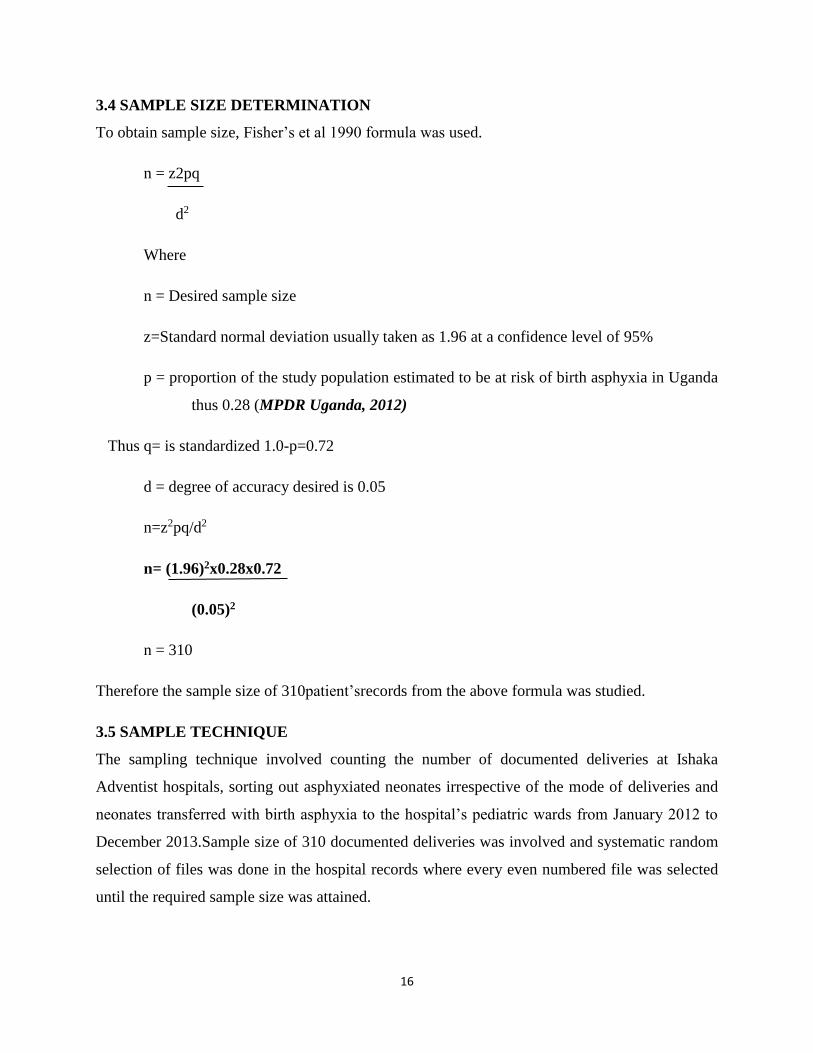
3.4 SAMPLE SIZE DETERMINATION
To obtain sample size, Fisher’s et al 1990 formula was used.
n = z2pq
d2
Where
n = Desired sample size
z=Standard normal deviation usually taken as 1.96 at a confidence level of 95%
p = proportion of the study population estimated to be at risk of birth asphyxia in Uganda
thus 0.28 (MPDR Uganda, 2012)
Thus q= is standardized 1.0-p=0.72
d = degree of accuracy desired is 0.05
n=z2pq/d2
n= (1.96)2x0.28x0.72
(0.05)2
n = 310
Therefore the sample size of 310patient’srecords from the above formula was studied.
3.5 SAMPLE TECHNIQUE
The sampling technique involved counting the number of documented deliveries at Ishaka
Adventist hospitals, sorting out asphyxiated neonates irrespective of the mode of deliveries and
neonates transferred with birth asphyxia to the hospital’s pediatric wards from January 2012 to
December 2013.Sample size of 310 documented deliveries was involved and systematic random
selection of files was done in the hospital records where every even numbered file was selected
until the required sample size was attained.
17
3.6 INCLUSION AND EXCLUSION CRITERIA
3.6.1 Inclusion Criteria
History of delayed cry.
History of fetal distress and diagnosis made by the doctor.
Apgar score <7 at 5 minutes of life.
3.6.2 Exclusion Criteria
Preterms with < 34 weeks of gestation.
Preterms with < 1500 grams birth weight.
Neonates with major congenital malformations of cardiovascular, central nervous system,
respiratory system or dysmorphic babies.
Severe hyperbilirubinemia bordering on kernicterus.
Cases with hypoglycemia or meningitis as a cause of encephalopathy
3.7 DATA COLLECTION
Information about deliveries in Ishaka Adventist hospitals was got from the records from theatre,
Maternity wards and paediatric wards of the hospital using a designed data collection tool (check
list). One research assistant trained on how to use the data collection tool was used in the study.
The check list used in data collection was first pretested prior to commencing data collection
process in the hospital.
3.8 DATA ANALYSIS
Using, Epi-info 7 and Statistical packaging for social scientists (SPSS) version 16.0 and Microsoft
Excel 2013, Data was entered and analyzed statistically in different presentations thus cross-
tabulations, bivariate and multivariate variables for events during pregnancy and delivery that
lead to birth asphyxia in pregnant mothers attending maternal health services at Ishaka Adventist
hospital.
18
3.9 ETHICAL CONSIDERATIONS
The research proposal was submitted to IREC for approval and a written permit from IREC and
hospital authorities was obtained first before commencing the study.
All the legal protocols were followed in accessing, collection and processing data and no
administrative persons were asked for undesirable favour in any form.
The information obtained was presented in an aggregate and de-identified form by using codes
instead of names on the records.
All results from this research were treated with utmost confidentiality by ensuring that only
authorized people gained access to them.
The community Advisory Committee (CAB) was fully involved in the whole process until the
completion of the study. Continuous feedback was given to CAB and any advice from them was
put into consideration.
3.10 STUDY LIMITATIONS AND THEIR SOLUTIONS
Financial predicaments narrowed the research tools and presentation even though the researcher
obtained money from the parents and other relatives to solve the problem.
Gaining access to the hospital’s data base and logistics was difficult but the researcher thought
permission from the necessary officials.
Some documents were unclearly filled thus the researcher opted for soft copy of hospital records
in some instances.
Lastly, there was a problem of finding the hospital authorities for the permission and information
access but the research contacted and made arrangements with hospital authorities on when to
meet them.
3.11 DISSEMINATION OF RESULTS
Information from this study was disseminated to IREC, a copy to Ishaka Adventist hospital and
another was sent to the district health officer in order to create awareness of the need to fight
newborn mortality and late complications secondary to birth asphyxia in Bushenyi district.

19
CHAPTER FOUR
4.0 INTRODUCTION
During this study on birth asphyxia from Jan. 2012 to Dec. 2013 at IAH, 345 cases on
asphyxiated births were enlisted and a sample size of 310 was obtained using systematic random
sampling technique as described in chapter 3. Some cases were excluded due to incomplete
information.
4.1 PRESENTATION OF SPECIFIC OBJECTIVE ONE
Figure 1: Shows total number of births at IAH from Jan. 2012 to Dec. 2013
Generally from the data obtained at IAH, statistics showed that the number of deliveries
corresponded for the various months of the year 2012 and 2013.
In both years, the highest number of deliveries were in the middle of the year thus around May
with 327 deliveries (10.1%) in 2013 and July with 302 deliveries (9.5%) in 2012. The lowest
monthly delivery 230 cases (7.3%) was in October 2012. On average 267 deliveries were made
in 2012 and 2013 at Ishaka Adventist Hospital (IAH).
0
50
100
150
200
250
300
350
J F M A M J J A S O N D
Number
ofbirths
Months
DELIVERIES (2012) DELIVERIES (2013)
20
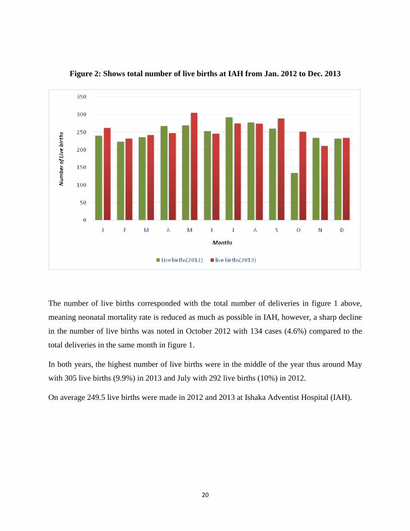
Figure 2: Shows total number of live births at IAH from Jan. 2012 to Dec. 2013
The number of live births corresponded with the total number of deliveries in figure 1 above,
meaning neonatal mortality rate is reduced as much as possible in IAH, however, a sharp decline
in the number of live births was noted in October 2012 with 134 cases (4.6%) compared to the
total deliveries in the same month in figure 1.
In both years, the highest number of live births were in the middle of the year thus around May
with 305 live births (9.9%) in 2013 and July with 292 live births (10%) in 2012.
On average 249.5 live births were made in 2012 and 2013 at Ishaka Adventist Hospital (IAH).

21
4.2 PRESENTATION OF SPECIFIC OBJECTIVE TWO
Figure 3: Shows total number of asphyxiated births from Jan. 2012 to Dec. 2013
Birth asphyxia, a condition of fetal hypoxia has been shown to be a variable case; this is
observed from the variation of the values for the various months. 2013 had the greatest number
of birth asphyxia except in June were the highest number was in 2012.
There was also inverse proportionality between birth asphyxia and live birth; this shown birth
asphyxia as one of the leading causes of neonatal mortality.
On average 13 cases of asphyxiated births occurred in 2012 and 2013 at IAH.
22
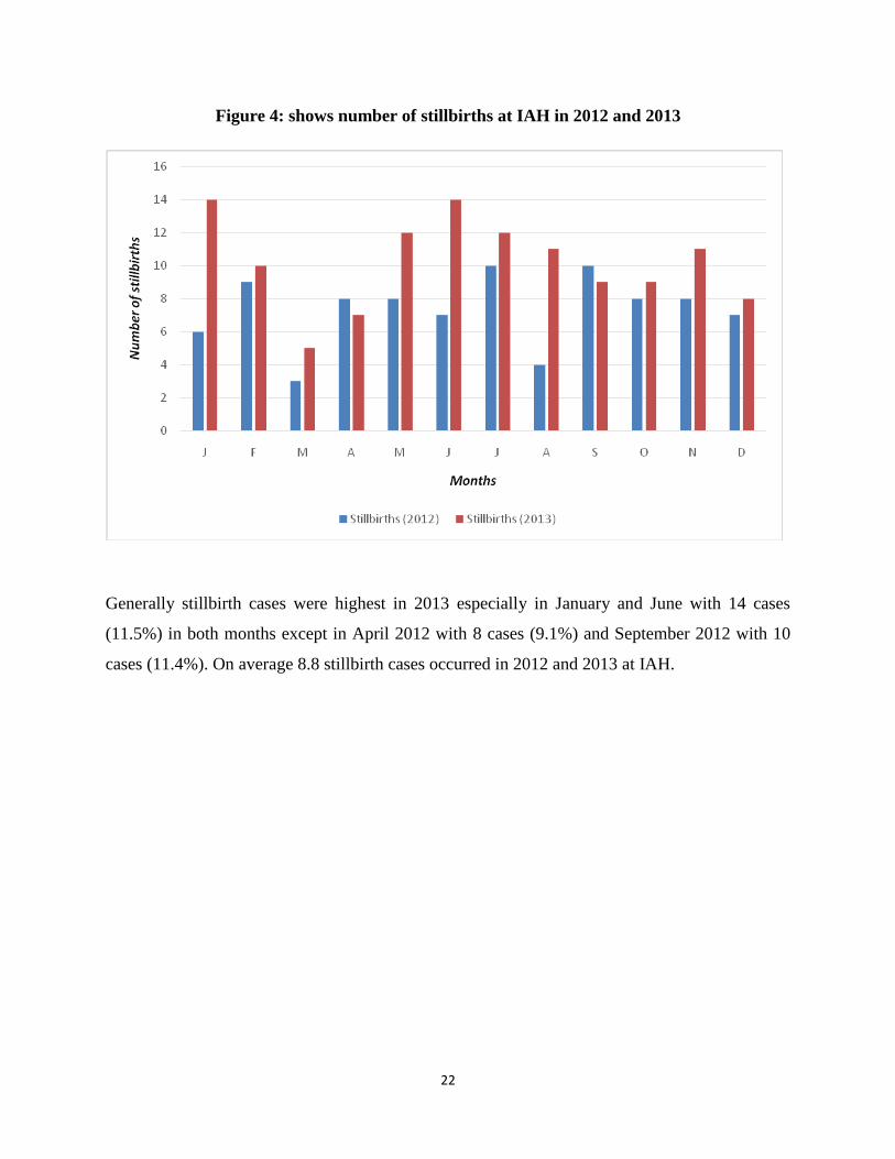
Figure 4: shows number of stillbirths at IAH in 2012 and 2013
Generally stillbirth cases were highest in 2013 especially in January and June with 14 cases
(11.5%) in both months except in April 2012 with 8 cases (9.1%) and September 2012 with 10
cases (11.4%). On average 8.8 stillbirth cases occurred in 2012 and 2013 at IAH.

23
4.3 PRESENTATION OF SPECIFIC OBJECTIVE THREE
Figure 5: Shows immediate outcomes of birth asphyxia at IAH from Jan. to Dec. 2012
The figure above shows that majority of the asphyxiated birth at IAH, 28% had FSB, 25% died
shortly,21% died later , 15 % of them improved and then 11% had MSB as immediate outcomes
of birth asphyxia in the year 2012.
Figure 6: Shows immediate outcomes of birth asphyxia in IAH from Jan. to Dec. 2013
In 2013, however, the results pattern of immediate outcomes changed in the deliveries, in that the
majority of the neonates died shortly after delivery 42%, followed FSB at 24%, 17 % of the

24
cases died later, while 9% had MSB and lastly 8 % improved despite their low Apgar score after
delivery.
Figure 7: Shows number of referral in cases and immediate outcomes
Majority of the Referral in cases were highest 41(43.2%) in 2013 compared to 24 cases (32.4%)
highest in 2012 and these died shortly due to birth asphyxia at IAH, followed by FSH with
25(26.3%) cases in 2013. In both years a few of the referral in cases improved 5(6.8%) in 2012
and 9 (9.5%) in 2013. 20 (27.0%) and 15(15.8%) of the cases in 2012 and 2013 respectively died
later. 8 cases (10.8%) in 2012 and 5 cases (5.2%) in 2013 were MSB.
25
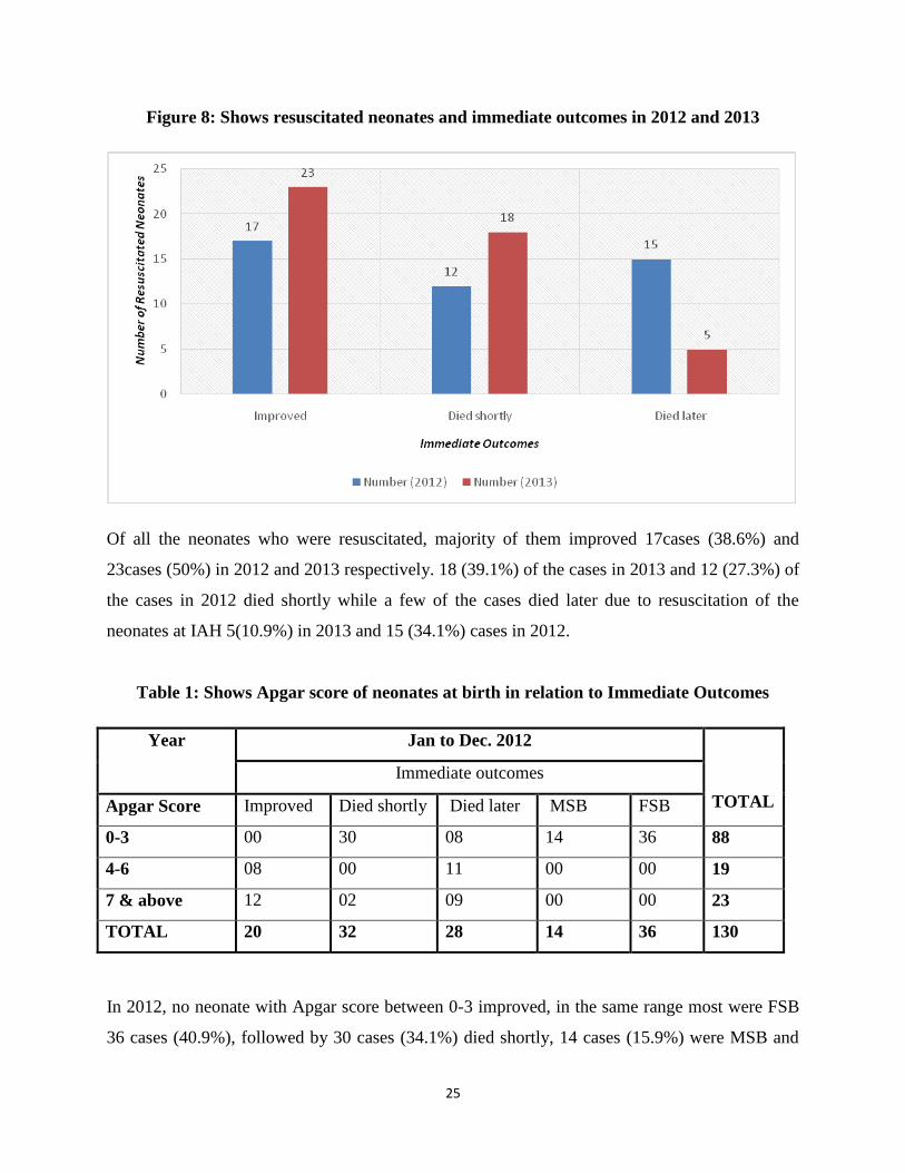
Figure 8: Shows resuscitated neonates and immediate outcomes in 2012 and 2013
Of all the neonates who were resuscitated, majority of them improved 17cases (38.6%) and
23cases (50%) in 2012 and 2013 respectively. 18 (39.1%) of the cases in 2013 and 12 (27.3%) of
the cases in 2012 died shortly while a few of the cases died later due to resuscitation of the
neonates at IAH 5(10.9%) in 2013 and 15 (34.1%) cases in 2012.
Table 1: Shows Apgar score of neonates at birth in relation to Immediate Outcomes
Year Jan to Dec. 2012
TOTAL
Immediate outcomes
Apgar Score Improved Died shortly Died later MSB FSB
0-3 00 30 08 14 36 88
4-6 08 00 11 00 00 19
7 & above 12 02 09 00 00 23
TOTAL 20 32 28 14 36 130
In 2012, no neonate with Apgar score between 0-3 improved, in the same range most were FSB
36 cases (40.9%), followed by 30 cases (34.1%) died shortly, 14 cases (15.9%) were MSB and

26
08 cases (9.1%) died later. Most cases with Apgar score of 7 and above improved 12 cases
(52.2%), a few cases thus 2 (8.7%) died shortly and 09 cases (39.1%) in the same range died
later .08 cases (42.1%) in the range of 4-6 Apgar score improved and 11 cases(57.9%) died later.
Both MSB and FSB contributed 60 cases (56.8%) in 2012.
Table 2: Shows Apgar score of neonates at birth in relation to immediate outcomes
Year Jan to Dec. 2013
TOTAL
Immediate outcomes
Apgar Score Improved Died shortly Died later MSB FSB
0-3 00 65 25 17 43 150
4-6 03 10 02 00 00 15
7 & above 12 00 03 00 00 15
TOTAL 15 75 30 17 43 180
In 2013 still, no neonate with Apgar score between 0-3 improved, in the same range the pattern
of immediate outcomes changed with 65 cases (43.3%) dying shortly being the highest, followed
by 43 cases (28.7%) FSB, 25(16.7%) died later and 17cases (11.3%) MSB. Most cases with
Apgar score of 7 and above improved, 12 cases (80%) and a few cases, 03 (20%) died later. A
total of 15 cases improved in 2013 compared to 20 cases in 2012. Both MSB & FSB contributed
60 cases (33.3%) in 2013.

27
Table 3: Shows immediate outcomes in relation to mode of delivery in 2012
Year Jan to Dec. 2012
TOTAL
Immediate outcomes
Mode of delivery Improved Died shortly Died later MSB FSB
SVD 03 19 13 00 17 52
C/S 10 08 10 05 15 48
EMC/S 07 05 05 09 04 30
TOTAL 20 32 28 14 36 130
In most cases of death due to birth asphyxia, the mother had undergone SVD 19 cases (36.5%)
died shortly and 13 cases (25%) died later of the 52 SVD cases, followed by the C/S 08 cases
(16.7%) and 10 cases (20.8%) of the 48 cases died shortly and later respectively. The least
number of deaths was in the EMC/S group with 05 cases (16.7%) in both those who died shortly
and later out of 30 cases. Most cases that improved were of C/S with 10 cases (50%) of the 20
cases that improved in relation to mode of delivery.
Table 4: Shows Immediate Outcomes in Relation to Mode of Delivery in 2013
Year Jan to Dec. 2013
TOTAL
Immediate outcomes
Mode of delivery Improved Died shortly Died later MSB FSB
SVD 02 44 15 03 19 83
C/S 12 26 07 09 15 69
EMC/S 01 05 08 05 09 28
TOTAL 15 75 30 17 43 180
In 2013, most birth asphyxia related deaths occurred following mothers undergoing SVD, 44
cases (53%) died shortly and 15 cases(18.1%) died later of the 83 SVD cases thus an increase in
cases compared to 2012, followed by the C/S 26 cases(37.7%) and 07 cases(10.1%) of the 69
cases. The least number of deaths was in the EMC/S group with 05 cases (17.9%) died shortly,
08 cases (28.6%) died later and 14 cases (50%) out of 28 cases was due to MSB & FSB. Still

28
fewer cases, 15 improved in 2013 compared to the 20 cases in 2012 in relation to mode of
delivery hence the need to address the condition.
4.4 PRESENTATION OF SPECIFIC OBJECTIVE FOUR
Figure 9: Shows factors accounting for BA during delivery in 2012 and 2013 at IAH
Generally, factor during delivery that account for birth asphyxia accounted for more neonatal
deaths in 2013 than in 2012 except for fetal distress and no fetal heart sound. Obstructed labor
was the most significant factor associated with asphyxia among the neonates, 34 cases (26.2%)
in 2012 and 40 cases (22.2%) in 2013, followed by the fetal distress 27 cases (20.8%) in 2012
and prolonged labor, 27 cases (15.0%) in 2013, others which included factors like severe pre-
eclampsia, contracted pelvis, among others contributed 12 cases (9.2%) in 2012 and 28 cases
(15.6%) in 2013.

29
Table 5: Shows gender of the asphyxiated neonates in 2012 and 2013
Year Jan to Dec. 2012 Jan to Dec. 2013
Gender NUMBER PERCENT NUMBER PERCENT
MALE 74 56.9 105 58.3
FEMALE 56 43.1 75 41.7
More males were asphyxiated with 56.9% in 2012 and 58.3% in 2013 compared to females with
43.1% in 2012 and 41.7% in 2013 as shown in the table above.
Figure 10: Shows the weights of the asphyxiated neonates in 2012 and 2013 at IAH
Majority of asphyxiated neonates weighed 2.0-2.4 kg 69 cases (38.3%) in 2013 and 2.5-2.9 kg 53
cases (40.8%) in 2012. Fewer cases of asphyxiated neonates weighed 3.5-4.0kg 28 (15.6%) in
2013 and 16 cases (12.3%) in 2012.
30
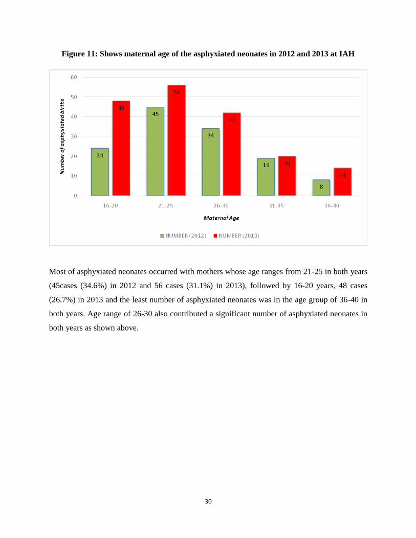
Figure 11: Shows maternal age of the asphyxiated neonates in 2012 and 2013 at IAH
Most of asphyxiated neonates occurred with mothers whose age ranges from 21-25 in both years
(45cases (34.6%) in 2012 and 56 cases (31.1%) in 2013), followed by 16-20 years, 48 cases
(26.7%) in 2013 and the least number of asphyxiated neonates was in the age group of 36-40 in
both years. Age range of 26-30 also contributed a significant number of asphyxiated neonates in
both years as shown above.

31
Table 6: Shows the parity of the mothers with asphyxiated neonates in 2012 and 2013
Year Jan to Dec. 2012 Jan to Dec. 2013
Parity NUMBER PERCENT NUMBER PERCENT
G1 32 24.6 42 23.3
G2 23 17.7 30 16.6
G3 21 16.2 26 14.4
G4 15 11.5 17 9.4
G5 16 12.3 13 7.2
G6 08 6.2 10 5.5
G7 05 3.8 12 6.6
G8 00 00 11 6.1
G9 00 00 03 1.6
G10& above 03 2.3 05 2.7
Elderly primigravida 07 5.4 12 6.6
Total 130 100 180 100
Majority of the asphyxiated neonates (47.9%) were delivered by Primiparous mothers in both
years with the highest in 2013, 42 cases (23.3%) and 32 cases (24.6%) in 2012, followed by the
gravid 2s (34.3%) then gravid 3s (30.6%) in both years while the rest had minimal contributions
that declined further with increase in parity of the mother.

32
Figure 12: Shows the parity of the mothers with asphyxiated neonates in 2012 and 2013
From the figure above, majority of the asphyxiated neonates (47.9%) were delivered by
Primiparous mothers in both years with the highest in 2013, 42 cases (23.3%) and 32 cases
(24.6%) in 2012, followed by the gravid 2s (34.3%) then gravid 3s (30.6%) in both years while
the rest had minimal contributions that declined further with increase in parity of the mother.

33
CHAPTER FIVE
5.0 INTRODUCTION
This chapter presents discussion of the findings in chapter four according to the research
questions of the study. It also contains the conclusion and recommendations based on the
discussion of the findings from the study.
5.1 DISCUSSIONS
5.1.1 Discussion on the number of births and proportion of asphyxiated neonates.
The finding from this study showed that the total number of deliveries from Jan. 2012 to Dec.
2013 at Ishaka Adventist hospital was 6,402 deliveries, out of which 5,989 were live births and
on average 266.75 deliveries were made in 2012 and 2013 in the hospital.
To note is that, 310 cases (4.8%) were asphyxiated births giving an incidence of 48/1000 births
and 5.2% of the live births, giving an incidence of 52 deaths per 1000 live births. This is fairly
high as compared to the study carried out in Warri-Nigeria with incidence of 28/1000 births(G.I
Mcgil et al, 2012) and 29 deaths per 1,000 live births as Uganda’s neonatal mortality rate
(NMR) that has not declined over a period of 15 years.(MoH,2008).
With an incidence of 52 deaths per 1000 live births, this shows that birth asphyxia is one of the
leading contributor to the 1,155,800 newborn deaths reported in Africa annually to which
Uganda contributes 44,500 newborn deaths though there is only a handful of published studies
from the developing countries and none from Uganda, to shed light on the magnitude of the
problem of birth asphyxia. (Tunde Adegboyega et al, 2010)
In another study according to William McGuire, 2007 showed that estimates of the incidence of
perinatal asphyxia vary. In resource-rich countries, severe perinatal asphyxia (causing death or
severe neurological impairment) is 1/1000 live births; in resource-poor countries, studies suggest
an incidence of 5–10/1000 live births, however, this probably represents an underestimate of the
true community incidence of perinatal asphyxia in resource-poor countries. Thus my findings at
52/ 1000 live births, suggest a serious burden of birth asphyxia in relation to neonatal mortality.
From my study, it shows that Uganda’s neonatal mortality rate is an under-estimate, at 29 deaths
per 1,000 live births. This is backed up by information according to MoH, Uganda 2008 that
34
stated Uganda’s neonatal mortality rate as a possibly an under-estimate, that is still very high at
29 deaths per 1,000 live births and that has not declined over a period of 15 years.
Also a Recent Demographic and Health Survey data for Uganda reported a national neonatal
mortality rate (NMR) of 29 deaths per 1,000 live births for the period 2000 to 2005, compared to
33 and 27 for 1995-2000 and 1990-1995 respectively which was an under estimate according to
my study. (MoH Uganda, 2008)
Furthermore, according to Dr. Sarah Byakika et al, 2012/13 reported that while Maternal
mortality ratio has slowly declined from 506 to 438 maternal deaths per 100,000 live births,
neonatal mortality rate has almost stagnated at 29 per 1000 live births.(UDHS, 2011).This
is lower than my findings at 52 per 1000 live births at IAH from my study hence the need for
more studies at national level to study the burden of birth asphyxia in the country and intervene
accordingly in order to attain the MDG4.
5.1.2 Discussion of the Immediate Outcomes
Resuscitation is done at birth to ensure adequate ventilation, when withheld many of these
babies die or survive with severe neurological damage. Findings on immediate outcomes
showed that, the majority of asphyxiated neonates improved well upon resuscitation at the
hospital with 17cases (38.6%) and 23cases (50%) in 2012 and 2013 respectively (figure.6). The
majority of the deaths were fresh still births followed by those who died shortly (42%) during the
resuscitation period in 2013. The results indicates that resuscitation through various means of
newly born can increase survival rates and some studies state that although most babies
breathe spontaneously at birth, up to 10 percent require some assistance to initiate
breathing while less than one percent need extensive resuscitation. (Dr.Gelasius Mukasa et al,
2010)
In both years, low Apgar score between 0-3 resulted in poor prognosis and no patient improved
following resuscitation. Most cases with Apgar score of 4-7 improved 15 cases in 2012 and 20
cases in 2013, a few cases in the same range thus 12 cases died shortly and 25 cases died later in
both years. Both MSB and FSB contributed 60 cases in both years (Table1 &2). Thus use of the
Apgar score can help predict the outcomes in neonates with birth asphyxia, early intervention
necessity and it’s still the most feasible and practical to perform. Therefore, 5 minute Apgar

35
score is still the valid index for assessing the effectiveness of resuscitation and vitality of
newborn. Outcome of birth asphyxia depends on Apgar score at5 minutes, heart rate at 90
seconds, time to first breath, duration of resuscitation arterial blood gases and acid –base
status at 10, and 30 minutes of age. According to a study done in Nepal incidence was 2.9
per 1000 live born of whom 20% had severe (Apgar score:1-3) and 80% moderate birth asphyxia
(Apgar score: 4-6). (Shazia Memon et al., 2012)
Furthermore, in Tanzania, Apgar scores below 7 registered 79% of the neonatal deaths. These
are clear indications for perinatal asphyxia as the major cause of neonatal morbidity (Olga
Golubnitschaja et al, 2011)
In relation to mode of delivery, most cases of birth asphyxia with poor prognosis occurred in
mother who had undergone SVD accounting for a total of 135 cases (43.5%) in both years, this is
probably due to factors like obstructed labor, prolonged labor among others that are related with
SVD while C/S and EMC/S showed improvement in most of the resuscitated cases in both years.
Hence the need to improved obstetric care to reduce the incidence of birth asphyxia in developed
countries.
Intrapartum still births (FSB & MSB), accounted for 110 cases (71.8%) of the total immediate
outcomes in both years and this gives an incidence of 17 still births per 1000 births.(Table 3& 4)
A study done in South Africa shows an incidence of 18 still births per 1000 births (Calverton
MD, 2008) while a study carried out in Nepal and Australia shows, a prevalence of fresh still
births to be 10/1000 births and 1/1000 births respectively as compared to my research results of
12 fresh still births per 1000 births (Ellis et al, BMJ 2000 & Badawi et al, BMJ 1998). This
implies that the incidence varies markedly from less than 1/1000 in developed (rich) countries to
more than 12- fold in developing (poor) countries and thus the need for intervention to reduce
this incidence in developing countries like Uganda.

36
5.1.3 Discussion of Risk Factors for Birth Asphyxia
Generally, risk factors during delivery that account for birth asphyxia resulted into more neonatal
deaths in 2013 than in 2012 except for fetal distress and no fetal heart sound.
According to my study, obstructed labor was the most significant factor associated with asphyxia
among the neonates, 34 cases (26.2%) in 2012 and 40 cases (22.2%) in 2013, followed by the
fetal distress 27 cases (20.8%) in 2012 due to idiopathic intrapartum conditions and prolonged
labor, 27 cases (15.0%) in 2013, others which included factors like severe pre-eclampsia,
contracted pelvis, Big babies among others contributed 12 cases (9.2%) in 2012 and 28 cases
(15.6%) in 2013. These findings were in contrast to the study conducted by Ugwul et al, 2012 in
which prolonged labor was reported as the leading risk factor of birth asphyxia.
According to gender, my research findings show that more males were asphyxiated with 56.9%
in 2012 and 58.3% in 2013 compared to females with 43.1% in 2012 and 41.7% in 2013. (Table
5) This gives a male to female ratio of 1.4:1 and compared with a study conducted in Warri-
Niger Delta of Nigeria, a male to female ratio of 3:2 thus twice my finding was obtained. (G.I
Mcgil Ugwu1 at el, 2012).The incidence is higher in males probably because females are more
resistance to diseases as a result of their XX chromosomes, X being the site of immunoglobulin
production, giving them double protection (Susan & Clark, 2009). This implies that the males
are at a higher risks of developing birth asphyxia than the females.
According to my findings, weight also is in important factor to consider as birth asphyxia cases
showed to reduce with increase in birth weights thus highest at 69 cases (38.3%) for 2.0-2.4 kg in
2013 and 53 cases (40.8%) for 2.5-2.9 kg in 2012 while fewer cases weighed between 3.5-4.0kg
28 cases (15.6%) in 2013 and 16 cases (12.3%) in 2012.
To note is that, maternal age could be considered as a risk factor because from my study results,
most of asphyxiated neonates occurred in mothers whose age ranged from 21-25 in both years
(45cases (34.6%) in 2012 and 56 cases (31.1%) in 2013), followed by 16-20 years and the least
number of asphyxiated neonates was in the age group of 36-40 in both years thus it shows that
the younger the age group, the more risk for birth asphyxia. This is in agreement to the study
carried out by MoH, Uganda in conjunction with UNICEF, Save the Children and WHO (2008)

37
Also in another study, results showed that majority of the mothers that had perinatal deaths
were aged between 18 and 30 years 66(75.4%), followed by age category 31-35 (8%).
(National MPDR, Uganda 2012/13)
In addition to the above, Parity of mothers with asphyxiated neonates showed that primiparous
most probably is a major predisposing factor for birth asphyxia with 47.9% of the cases being
from primiparous mothers in both years with the highest in 2013, 42 cases (23.3%) and 32 cases
(24.6%) in 2012, followed by the gravid 2s (34.3%) then gravid 3s (30.6%) in both years while
the rest had minimal contributions that declined further with increase in parity of the mother.
5.2. CONCLUSIONS
This study found out that Birth asphyxia still remains a serious challenge in developing countries
as one of the leading cause of neonatal deaths as well as under-fives mortality though there is
only a handful of published studies from these countries and none from Uganda, to shed light on
the magnitude of the problem of birth asphyxia. Because of this, the incidence of birth asphyxia
is greatly under estimated in developing countries and has been given little attention therefore
compromising the achievements of the 4th MDG in many of the developing countries including
Uganda. Most of the identified maternal risk factors that were significantly related to birth
asphyxia included obstructive labor and prolonged labor among others that are preventable if
continuous sensitization of the public, negligence, poverty and improvement in obstetric care
among other interventions are addressed in this hospital and Uganda at large.
5.3 RECOMMENDATIONS
Increased attention to the early recognition and management of these factors in community-based
maternal and child health programs should be implemented.
Team work should be encouraged among the mid wives, obstetricians and pediatricians in the
hospital for proper health service provision.
A well-equipped neonatal unit should be put in place to aid in the management of patients with
poor prognosis and emergencies.
A special ward to act as waiting place for pregnant mothers that stay far away from the hospital
to reside in as they are being monitored by health workers for early intervention incase need
arises.
38
Prospective study of the incidence and complications of birth asphyxia should be undertaken in
several health Centre’s, so as to compare with this study in other hospitals in Bushenyi and
Uganda.
5.4 STUDY CHALLENGES
There were some unfilled information about neonatal deaths in record, which needs to be
improved for future purposes.
Limited information about the condition in Uganda to compare my finding too.

39
BIBLIOGRAPHY
1. Dr. Gelasius Mukasa et al, Situation analysis of newborn health in Uganda: current status and
opportunities to improve care and survival. Kampala, Ministry of Health Uganda. Save the
Children, UNICEF, WHO; 2008.pg. 5-8
2. Anne CC.Lee et al, Risk Factors for Neonatal Mortality Due to Birth Asphyxia in Southern
Nepal. Official Journal of the American Academy of Pediatrics, at Uganda: AAP Sponsored
on May 21, 2013: 1-6 (pediatrics.aappublications.org)
3. Dr.Ornella Lincetto, Birth asphyxia –summary of the previous meeting and protocol
overview, 11 June 2007, WHO Genève Milano. Pdf (www.curoservice.com/health.../pdf/10-
09-2007_birth_asphyxia02.pdf)
4. Shazia Memon et al, December 2012, Department of Paediatric, Department of Gynae and
Obstetrics,Liaquat University of Medical & Health Sciences, Jamshoro, Hyderabad.
(http://www.jpma.org.pk/full_article_text.php?article_id=3855)
5. Ola Didrik Saugstad, Birth asphyxia, Department of pediatric research Rikshospitalet,
university of Oslo, Norway, Sept. 2007. (www.curoservice.com/health.../pdf/10-09-
2007_birth_asphyxia01.pdf)
6. William McGuire, Associate Professor. Clinical Evidence of Perinatal asphyxia, March 2007
(Pub Med)
7. Olga Golubnitschaja et al, Birth asphyxia as the major complication in newborns: moving
towards improved individual outcomes by prediction, targeted prevention and tailored
medical care, April 2011, EPMA Journal (2011) 2:197–210 pdf.
8. Seyal T and Hanif A. Factors Related to Adverse Outcome in Asphyxiated Babies, 2009
(PubMed)
9. Ministry of Health Uganda: Situation analysis of newborn health in Uganda, current status
and opportunities to improve care and survival. Kampala, Government of Uganda, Save the
Children, UNICEF and WHO 2008. P 20-26.
10. Kinoti SN: Asphyxia of the newborn in east, central and southern Africa, East African
Medical Journal, July 1993: 422-33.
11. Tunde Adegboyega et al,(2010) Opportunities for Africa’s newborn, Practical data, policy
and programmatic support for newborn care in Africa, Mills Litho, Capetown, South Africa.
40
12. Iawn, J. E., Cousens, S., & Zupan, J. (2005). 4 Million Deaths. When? Where? Why? Lancet,
365(9462), 891-900. (http://dx.doi.org/10.1016/S0140-6736(05)71048-5)
13. Reshma Parvin S.et al, Observation of Birth Asphyxia and Its Impact on Neonatal Mortality
in Khulna Urban Slum Bangladesh, International Journal of Advanced Nutritional and Health
Science, 13thDecember 2012, Volume1.
14. WHO. Neonatal and perinatal mortality: regional, country and global estimates. (2006).
Geneva, Switzerland: World Health Organization
15. Susan Furdon, & Clark, D. A. The World Health Report (2003), Shaping the Future, Geneva
(http://emedicine.com/article/975909-overview.June2009)
16. Nikki Gillette, Beryl Levinger et al, (2013) Surviving the First Day, State of the World’s
Mothers report, Spirals, Inc. (www.savethechildren.net)
17. Leuthner, S. R., Ug. D. (2004). Low Apgar score and the Definition of Birth Asphyxia.
Pediatr Clin N Am; 51:737-745. (http://dx.doi.org/10.1016/j.pcl.2004.01.016)
18. Ondoa-Onama C and Tumwine JK, “Immediate outcomes of babies with low Apgar score in
Mulago Hospital, Uganda”. East African Medical Journal. January 2003, 80(1): 22-9 and
July 1993 70(7):422-33.
19. UBOS 2011. Uganda Demographic and health survey 2011. Kampala, Uganda .Calverton,
Maryland USA: Uganda Bureau of Statistics and ICF International Inc. 2012.
20. http://www.indexmundi.com/uganda/demographics_profile.html
21. Michael Oturu et al, Child survival end of program knowledge, practice and coverage and
health facility assessment bushenyi district, June, 2010, pg.2
22. Dr. Sarah Byakika, Ms. Emily Atuheire et al, 2ndNational Maternal and Perinatal death
review (MPDR) report, Ministry of Health Uganda, 2012/2013.
23. William McGuire, Associate Professor. Clinical Evidence of Perinatal asphyxia, March 2012
(Pub Med)

41
24. Hashima-E-Nasreen. (2009). Maternal, Neonatal and Child Health in Selected Northern
Bangladesh. Bangladesh Research and Evaluation Commission. Retrieved from
(http://www.bracresearch.org/reports/MNCH_baseline_2008_new.pdf)
25. Calverton, MD: National Department of Health, Medical Research Council, Demographic &
Health Survey, 1998- 2002, South Africa: 1-2. (www.childpip.org.za/everydeathcounts)
26. G.I Mcgil, H. A. (2012). Incidence of Birth Asphyxia as Seen in Central Hospital and GN
Children’s Clinic both in Warri Niger Delta of Nigeria, August 2012. Global Journal of
Health Science vol.4, 1(7):140- 144.
27. http://www.health of children.com/A/Asphyxia-Neonatorum.html
28. http://en.wikipedia.org/wiki/Bushenyi

42
APPENDICES
APPENDIX 1: DESIGNED DATA COLLECTION TOOL
G/A SEX MOD AANC POB RFDP RFDD PARITY DOL A/S MGT IOC
G/A= gestational age
MOD= mode of delivery
AANC=attended antenatal care
POB= place of birth
RFDP=Risk factors during pregnancy
RFDD=Risk factors during delivery
DOL=duration of labor
A/S=Apgar score
MGT=management
IOC=immediate outcome

43
APPENDIX 2: PROPOSED BUDGET FOR THE STUDY
ITEM COST PER UNIT NO. OF UNITS AMOUNT
Data analysis 100,000/= 1 100,000/=
Printing 10,000/= 4 copies 40,000/=
Data collection tool 100/= 350 copies 35,000/=
Binding 2500/= 4 10,000/=
Research assistants 10,000/= 4 40,000/=
TOTAL COST 225,000/=
Funds were provided by the researcher to carry out the study.

44
APPENDIX 3: WORK PLAN
Month
Oct.
2014
Oct.
2014
Oct.
2014
Oct.
2014
Nov.
2014
Nov
2014
Dec.
2014
Proposal
Approval
Data
collection
Data
analysis
Report
writing
Submission

45
APPENDIX 4: APPROVAL LETTER TO ACCESS RESEARCH RESOURCES AT IAH

46
APPENDIX 5: A MAP OF BUSHENYI SHOWING THE LOCATION OF ISHAKA

47
APPENDIX 6: A MAP OF UGANDA SHOWING THE LOCATION OF BUSHENYI
DISTRICT