Embed Size (px)
Citation preview
THE PRETERM INFANT
Any infant born prior to completing 37 weeks’ gestation is identified as premature. Thus, the level ofdevelopment and maturity, and often the degree of complications, varies within this group, dependent on the length ofgestation.
NEONATAL ASSESSMENT DATA BASE
CirculationApical pulse may be rapid and/or irregular within a normal range (120–160 bpm).Audible heart murmur may indicate PDA.
Food/FluidWeight less than 2500 g (5 lb 8 oz).Body long, thin, limp with a slight potbelly.Suck/swallow reflex may be absent/uncoordinated (impacts feeding choices).
NeurosensoryHead size large in relation to body; sutures may be easily movable; fontanels may be large or wide open.May demonstrate twitching or eye rolling.Edema of eyelids common; eyes may be fused shut (depends on gestational age).Reflexes depend on gestational age; rooting well established by 32 weeks’ gestation; coordinated reflexes for sucking,
swallowing, and breathing usually established by 32 wk; first component of Moro’s reflex (lateral extension ofupper extremities with opening of hands) appears at 28 wk; second two components (anterior flexion and audiblecry) appear at 32 wk.
Dubowitz examination indicates gestational age between 24 and 37 wk.
RespirationApgar scores may be low.Respirations may be shallow, irregular; diaphragmatic with intermittent or periodic breathing (40–60/min).Grunting, nasal flaring, suprasternal or substernal retractions, or varying degrees of cyanosis may be present.Auscultatory presence of “sandpaper” sound indicates RDS.
SafetyTemperature fluctuates easily.Cry may be weak.Face may be bruised; caput succedaneum may be present; labor or delivery may have been precipitous.Skin reddened or translucent; color may be pink/ruddy, acrocyanotic, or cyanotic/pale.Lanugo widely distributed over entire body.Extremities may appear edematous.Sole creases may or may not be present on all or part of the foot.Nails may be short.
SexualityFemale labia minora may be larger than labia majora, with prominent clitoris.Male testes may not be descended; rugae may be scant or absent on scrotum.
Teaching/LearningMaternal history may reveal factors that contributed to preterm labor, such as young age; low socioeconomic
background; closely spaced pregnancies; multiple gestation; poor nutrition; previous preterm birth; obstetric
complication such as abruptio placentae, premature rupture of membranes (PROM), premature dilation of cervix,presence of infection; blood incompatibility associated with erythroblastosis fetalis; or use of prescription, over-the-counter, or street drugs.
DIAGNOSTIC STUDIESChoice of tests and the expected results depend on presenting problems and secondary complications.Amniotic Fluid Studies: For lecithin-to-sphingomyelin (L/S) ratio, fetal lung profile, and phosphatidyl
glycerol/phosphatidyl inositol may have been performed during pregnancy to assess fetal maturity.CBC: Decreases in Hb/Hct may be associated with anemia or blood loss. WBC count may be <10,000/mm3 with a
shift to the left (excess early neutrophils and bands), which is usually associated with severe bacterial disease.Dextrostix: Reveals hypoglycemia. Serum glucose test may be required if Dextrostix result is 45 mg/ml.Serum Calcium: May be low.Electrolytes (Na+, K+, Cl-): Usually within normal limits initially, but susceptible to critical fluctuations.Blood Type: May reveal potential for ABO incompatibility.Rh and Direct Coombs’ Determination (if mother is Rh-negative and father is Rh-positive): Determines
incompatibilities.ABGs: PO2 may be low; PCO2 may be elevated and reflect mild/moderate acidosis, sepsis, or prolonged respiratory
difficulties.Erythrocyte Sedimentation Rate (ESR): Elevation indicates an acute inflammatory response. Diminishing ESR
indicates resolution of inflammation.C-reactive Protein (a beta globulin): Present in serum in proportion to severity of infectious or noninfectious
inflammatory process.Platelet Count: Thrombocytopenia may accompany sepsis.Fibrinogen Levels: May decrease during disseminated intravascular coagulation (DIC) or become elevated during
injury or inflammation.Fibrin Split Products: Present with DIC.Blood Cultures: Identify causative organisms associated with sepsis.Urinalysis (on second voided specimen): Detects abnormalities, renal injury.Urine-Specific Gravity: Ranges between 1.006 and 1.013; elevated with dehydration.Clinitest/Clinistix: Identifies presence of sugar in urine.Hematest: Examines stools for blood; positive results suggest necrotizing enterocolitis.Shake Test on Gastric Aspirate: Determines presence or absence of surfactant. (Intermediate results if blood or
meconium is present.)Chest X-Ray (posteroanterior [PA] and lateral) with Air Bronchogram: May have ground-glass appearance
(RDS).Serial Cranial Ultrasonography: Detects presence and severity of intraventricular hemorrhage (IVH).Lumbar Puncture: May be performed to rule out meningitis.Maternal GBS Status: Determines potential for meningitis/infections.
NURSING PRIORITIES1. Promote optimal respiratory functioning.2. Maintain neutral thermal environment.3. Prevent or reduce risk of potential complications.4. Maintain homeostasis.5. Foster development of healthy family unit.DISCHARGE GOALS1. Maintaining physiological and behavioral homeostasis with minimal external support.2. Weight 41/2 lb or greater appropriate to age/condition.3. Complications prevented/resolving or independently managed.4. Family identifying and using resources appropriately.5. Family demonstrates ability to manage infant care.6. Plan in place to meet needs after discharge.
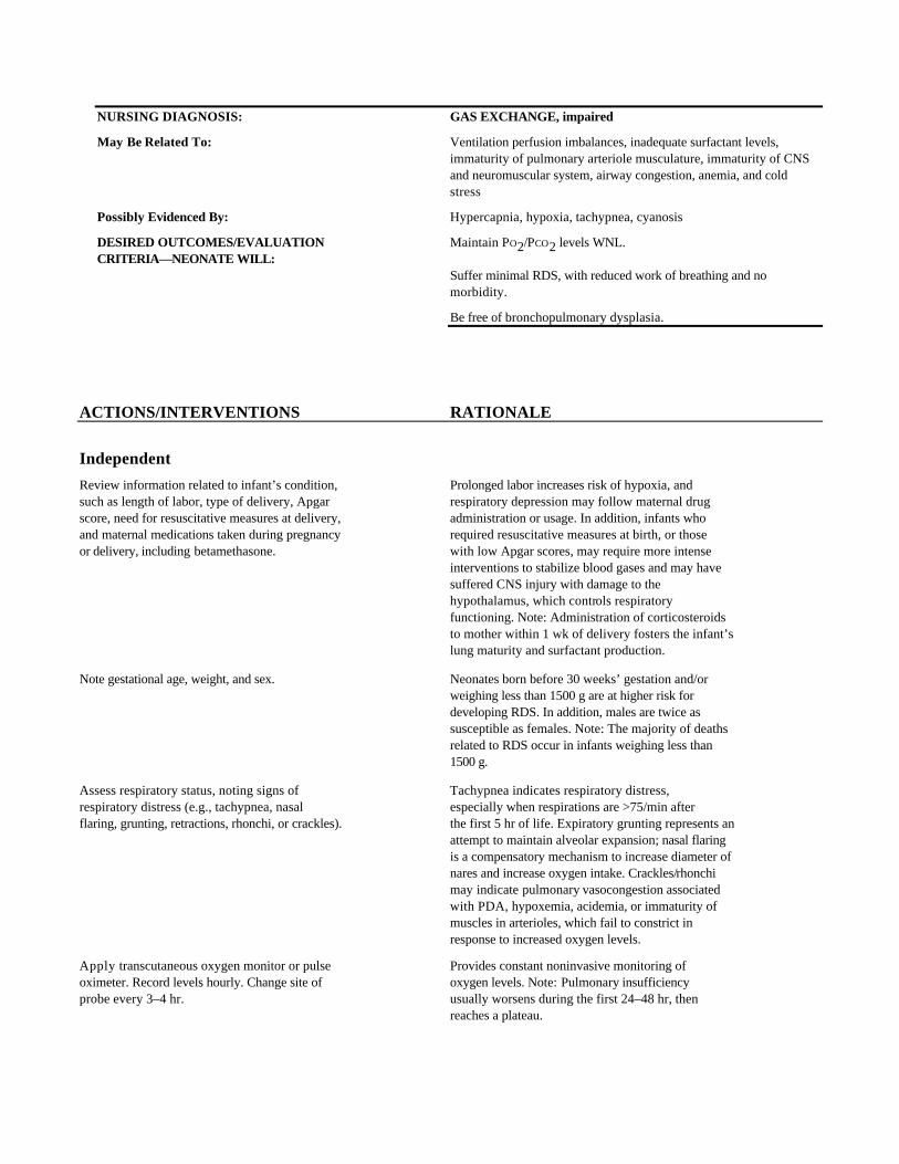
NURSING DIAGNOSIS: GAS EXCHANGE, impaired
May Be Related To: Ventilation perfusion imbalances, inadequate surfactant levels,immaturity of pulmonary arteriole musculature, immaturity of CNSand neuromuscular system, airway congestion, anemia, and coldstress
Possibly Evidenced By: Hypercapnia, hypoxia, tachypnea, cyanosis
DESIRED OUTCOMES/EVALUATION Maintain PO2/PCO2 levels WNL.CRITERIA—NEONATE WILL:
Suffer minimal RDS, with reduced work of breathing and nomorbidity.
Be free of bronchopulmonary dysplasia.
ACTIONS/INTERVENTIONS RATIONALE
Independent
Review information related to infant’s condition, Prolonged labor increases risk of hypoxia, andsuch as length of labor, type of delivery, Apgar respiratory depression may follow maternal drugscore, need for resuscitative measures at delivery, administration or usage. In addition, infants whoand maternal medications taken during pregnancy required resuscitative measures at birth, or thoseor delivery, including betamethasone. with low Apgar scores, may require more intense
interventions to stabilize blood gases and may havesuffered CNS injury with damage to thehypothalamus, which controls respiratoryfunctioning. Note: Administration of corticosteroidsto mother within 1 wk of delivery fosters the infant’slung maturity and surfactant production.
Note gestational age, weight, and sex. Neonates born before 30 weeks’ gestation and/orweighing less than 1500 g are at higher risk fordeveloping RDS. In addition, males are twice assusceptible as females. Note: The majority of deathsrelated to RDS occur in infants weighing less than1500 g.
Assess respiratory status, noting signs of Tachypnea indicates respiratory distress,respiratory distress (e.g., tachypnea, nasal especially when respirations are >75/min afterflaring, grunting, retractions, rhonchi, or crackles). the first 5 hr of life. Expiratory grunting represents an
attempt to maintain alveolar expansion; nasal flaringis a compensatory mechanism to increase diameter ofnares and increase oxygen intake. Crackles/rhonchimay indicate pulmonary vasocongestion associatedwith PDA, hypoxemia, acidemia, or immaturity ofmuscles in arterioles, which fail to constrict inresponse to increased oxygen levels.
Apply transcutaneous oxygen monitor or pulse Provides constant noninvasive monitoring ofoximeter. Record levels hourly. Change site of oxygen levels. Note: Pulmonary insufficiencyprobe every 3–4 hr. usually worsens during the first 24–48 hr, then
reaches a plateau.
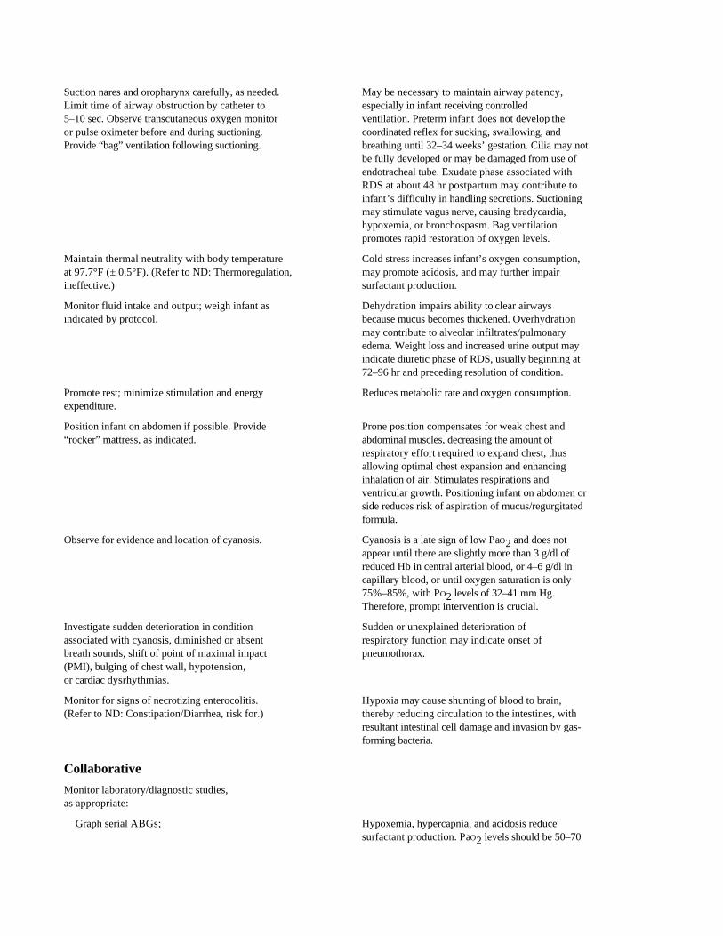
Suction nares and oropharynx carefully, as needed. May be necessary to maintain airway patency,Limit time of airway obstruction by catheter to especially in infant receiving controlled5–10 sec. Observe transcutaneous oxygen monitor ventilation. Preterm infant does not develop theor pulse oximeter before and during suctioning. coordinated reflex for sucking, swallowing, andProvide “bag” ventilation following suctioning. breathing until 32–34 weeks’ gestation. Cilia may not
be fully developed or may be damaged from use ofendotracheal tube. Exudate phase associated withRDS at about 48 hr postpartum may contribute toinfant’s difficulty in handling secretions. Suctioningmay stimulate vagus nerve, causing bradycardia,hypoxemia, or bronchospasm. Bag ventilationpromotes rapid restoration of oxygen levels.
Maintain thermal neutrality with body temperature Cold stress increases infant’s oxygen consumption,at 97.7°F (± 0.5°F). (Refer to ND: Thermoregulation, may promote acidosis, and may further impairineffective.) surfactant production.
Monitor fluid intake and output; weigh infant as Dehydration impairs ability to clear airwaysindicated by protocol. because mucus becomes thickened. Overhydration
may contribute to alveolar infiltrates/pulmonaryedema. Weight loss and increased urine output mayindicate diuretic phase of RDS, usually beginning at72–96 hr and preceding resolution of condition.
Promote rest; minimize stimulation and energy Reduces metabolic rate and oxygen consumption.expenditure.
Position infant on abdomen if possible. Provide Prone position compensates for weak chest and“rocker” mattress, as indicated. abdominal muscles, decreasing the amount of
respiratory effort required to expand chest, thusallowing optimal chest expansion and enhancinginhalation of air. Stimulates respirations andventricular growth. Positioning infant on abdomen orside reduces risk of aspiration of mucus/regurgitatedformula.
Observe for evidence and location of cyanosis. Cyanosis is a late sign of low PaO2 and does notappear until there are slightly more than 3 g/dl ofreduced Hb in central arterial blood, or 4–6 g/dl incapillary blood, or until oxygen saturation is only75%–85%, with PO2 levels of 32–41 mm Hg.Therefore, prompt intervention is crucial.
Investigate sudden deterioration in condition Sudden or unexplained deterioration ofassociated with cyanosis, diminished or absent respiratory function may indicate onset ofbreath sounds, shift of point of maximal impact pneumothorax.(PMI), bulging of chest wall, hypotension,or cardiac dysrhythmias.
Monitor for signs of necrotizing enterocolitis. Hypoxia may cause shunting of blood to brain,(Refer to ND: Constipation/Diarrhea, risk for.) thereby reducing circulation to the intestines, with
resultant intestinal cell damage and invasion by gas-forming bacteria.
Collaborative
Monitor laboratory/diagnostic studies,as appropriate:
Graph serial ABGs; Hypoxemia, hypercapnia, and acidosis reducesurfactant production. PaO2 levels should be 50–70
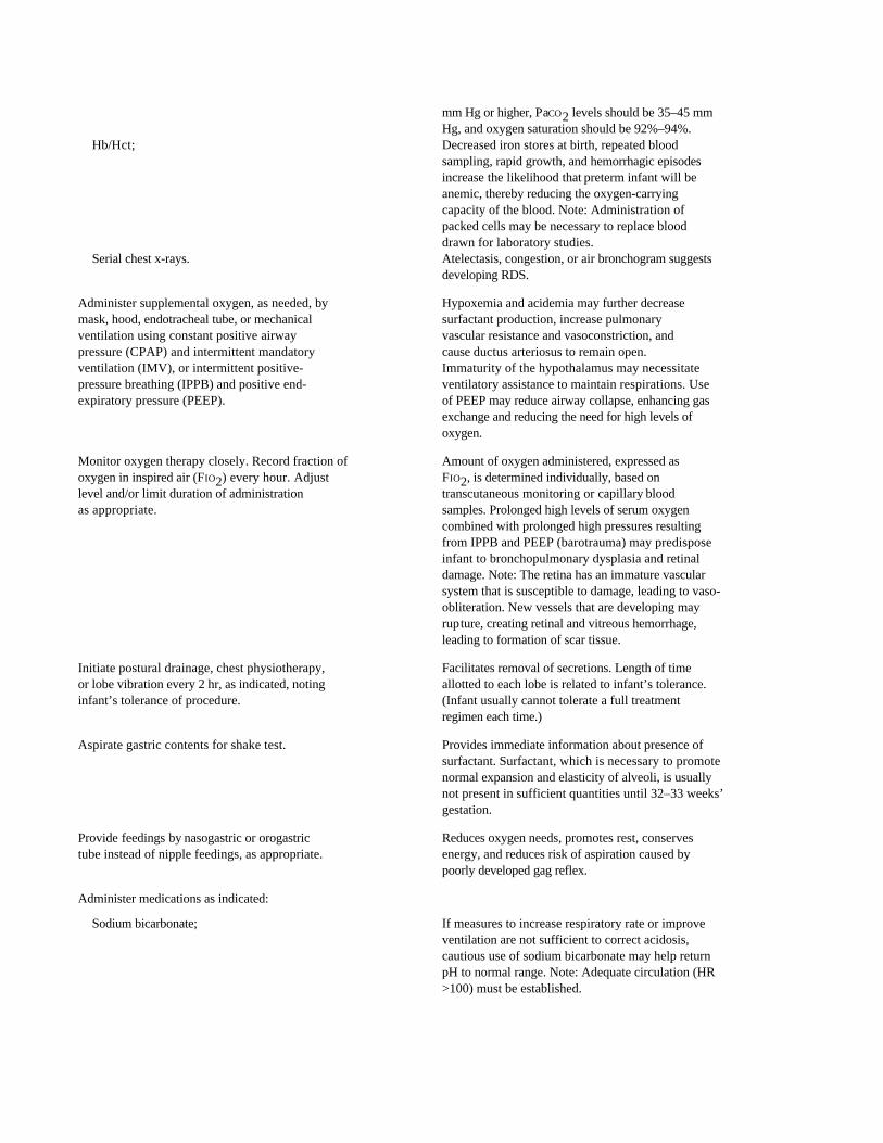
mm Hg or higher, PaCO2 levels should be 35–45 mmHg, and oxygen saturation should be 92%–94%.
Hb/Hct; Decreased iron stores at birth, repeated bloodsampling, rapid growth, and hemorrhagic episodesincrease the likelihood that preterm infant will beanemic, thereby reducing the oxygen-carryingcapacity of the blood. Note: Administration ofpacked cells may be necessary to replace blooddrawn for laboratory studies.
Serial chest x-rays. Atelectasis, congestion, or air bronchogram suggestsdeveloping RDS.
Administer supplemental oxygen, as needed, by Hypoxemia and acidemia may further decreasemask, hood, endotracheal tube, or mechanical surfactant production, increase pulmonaryventilation using constant positive airway vascular resistance and vasoconstriction, andpressure (CPAP) and intermittent mandatory cause ductus arteriosus to remain open.ventilation (IMV), or intermittent positive- Immaturity of the hypothalamus may necessitatepressure breathing (IPPB) and positive end- ventilatory assistance to maintain respirations. Useexpiratory pressure (PEEP). of PEEP may reduce airway collapse, enhancing gas
exchange and reducing the need for high levels ofoxygen.
Monitor oxygen therapy closely. Record fraction of Amount of oxygen administered, expressed asoxygen in inspired air (FIO2) every hour. Adjust FIO2, is determined individually, based onlevel and/or limit duration of administration transcutaneous monitoring or capillary bloodas appropriate. samples. Prolonged high levels of serum oxygen
combined with prolonged high pressures resultingfrom IPPB and PEEP (barotrauma) may predisposeinfant to bronchopulmonary dysplasia and retinaldamage. Note: The retina has an immature vascularsystem that is susceptible to damage, leading to vaso-obliteration. New vessels that are developing mayrupture, creating retinal and vitreous hemorrhage,leading to formation of scar tissue.
Initiate postural drainage, chest physiotherapy, Facilitates removal of secretions. Length of timeor lobe vibration every 2 hr, as indicated, noting allotted to each lobe is related to infant’s tolerance.infant’s tolerance of procedure. (Infant usually cannot tolerate a full treatment
regimen each time.)
Aspirate gastric contents for shake test. Provides immediate information about presence ofsurfactant. Surfactant, which is necessary to promotenormal expansion and elasticity of alveoli, is usuallynot present in sufficient quantities until 32–33 weeks’gestation.
Provide feedings by nasogastric or orogastric Reduces oxygen needs, promotes rest, conservestube instead of nipple feedings, as appropriate. energy, and reduces risk of aspiration caused by
poorly developed gag reflex.
Administer medications as indicated:
Sodium bicarbonate; If measures to increase respiratory rate or improveventilation are not sufficient to correct acidosis,cautious use of sodium bicarbonate may help returnpH to normal range. Note: Adequate circulation (HR>100) must be established.
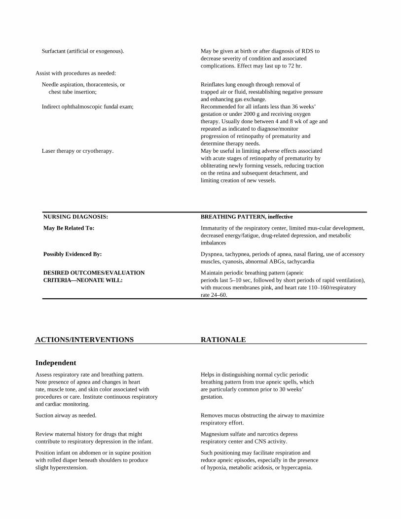
Surfactant (artificial or exogenous). May be given at birth or after diagnosis of RDS todecrease severity of condition and associatedcomplications. Effect may last up to 72 hr.
Assist with procedures as needed:
Needle aspiration, thoracentesis, or Reinflates lung enough through removal ofchest tube insertion; trapped air or fluid, reestablishing negative pressure
and enhancing gas exchange.Indirect ophthalmoscopic fundal exam; Recommended for all infants less than 36 weeks’
gestation or under 2000 g and receiving oxygentherapy. Usually done between 4 and 8 wk of age andrepeated as indicated to diagnose/monitorprogression of retinopathy of prematurity anddetermine therapy needs.
Laser therapy or cryotherapy. May be useful in limiting adverse effects associatedwith acute stages of retinopathy of prematurity byobliterating newly forming vessels, reducing tractionon the retina and subsequent detachment, andlimiting creation of new vessels.
NURSING DIAGNOSIS: BREATHING PATTERN, ineffective
May Be Related To: Immaturity of the respiratory center, limited mus-cular development,decreased energy/fatigue, drug-related depression, and metabolicimbalances
Possibly Evidenced By: Dyspnea, tachypnea, periods of apnea, nasal flaring, use of accessorymuscles, cyanosis, abnormal ABGs, tachycardia
DESIRED OUTCOMES/EVALUATION Maintain periodic breathing pattern (apneicCRITERIA—NEONATE WILL: periods last 5–10 sec, followed by short periods of rapid ventilation),
with mucous membranes pink, and heart rate 110–160/respiratoryrate 24–60.
ACTIONS/INTERVENTIONS RATIONALE
Independent
Assess respiratory rate and breathing pattern. Helps in distinguishing normal cyclic periodicNote presence of apnea and changes in heart breathing pattern from true apneic spells, whichrate, muscle tone, and skin color associated with are particularly common prior to 30 weeks’procedures or care. Institute continuous respiratory gestation.and cardiac monitoring.
Suction airway as needed. Removes mucus obstructing the airway to maximizerespiratory effort.
Review maternal history for drugs that might Magnesium sulfate and narcotics depresscontribute to respiratory depression in the infant. respiratory center and CNS activity.
Position infant on abdomen or in supine position Such positioning may facilitate respiration andwith rolled diaper beneath shoulders to produce reduce apneic episodes, especially in the presenceslight hyperextension. of hypoxia, metabolic acidosis, or hypercapnia.
Maintain optimal body temperature. (Refer to ND: Even a slight increase or decrease inThermoregulation, risk for ineffective.) environmental temperature can lead to apnea.
Provide prompt tactile stimulation (e.g., rub Stimulates CNS to promote body movement andinfant’s back) if apnea occurs. Note presence of spontaneous return of respirations. Sometimes,cyanosis, bradycardia, or hypotonia. Encourage infants experience fewer or no episodes of apneaparental contact. or bradycardia if parents touch and talk to them.
Place infant on neowave mattress. Movement provides stimulation, which may reduceapneic episodes.
Collaborative
Monitor laboratory studies (e.g., ABGs, serum Hypoxia, metabolic acidosis, hypercapnia,glucose, electrolytes, cultures, and drug levels) hypoglycemia, hypocalcemia, and sepsis mayas indicated. contribute to apneic spells. Drug toxicity, which
depresses respiratory function, may occur because oflimited excretion and prolonged drug half-life.
Administer supplemental oxygen, as indicated. Correction of oxygen and carbon dioxide levels(Refer to ND: Gas Exchange, impaired.) may improve respiratory function.
Administer medications, as indicated:
Sodium bicarbonate; Judicious use may be required to help correctacidosis, especially after hypoxic episodes, becausebicarbonate is excreted at lower serum levels andimmature kidneys are slower to excrete lactic acid.
Antibiotics; Treat respiratory infection or sepsis.Calcium gluconate; Hypocalcemia predisposes infant to apnea.Aminophylline; May increase activity of respiratory center and lower
sensitivity to carbon dioxide, reducing frequency ofapnea.
Pancuronium bromide (Pavulon); Induces skeletal muscle relaxation, which may benecessary if infant is to be mechanically ventilated.
Glucose solutions. Prevent hypoglycemia. (Refer to ND: Nutrition:altered, risk for less than body requirements.)
NURSING DIAGNOSIS: THERMOREGULATION, ineffective
May Be Related To: Immature CNS development (temperature regulation center),decreased ratio of body mass to surface area, decreased subcutaneousfat, limited brown fat stores, an inability to shiver or sweat,limited/inability to flex extremities, poor metabolic reserves (limitedglycogen stores), thinner skin, muted response to hypothermia, andfrequent medical/nursing manipulations and interventions
Possibly Evidenced By: Fluctuation of body temperature below/above normal range
Tachypnea/apnea, generalized cyanosis, bradycardia, lethargy (coldstress)
Tachycardia, flushed color, lethargy, apnea (hyperthermia)
DESIRED OUTCOMES/EVALUATION Maintain skin/axillary temperature withinCRITERIA—NEONATE WILL: 97.7°F–99.1°F (36.5°C–37.3°C).
Be free of signs of cold stress.
ACTIONS/INTERVENTIONS RATIONALE
Independent
Assess temperature frequently. Check rectal Hypothermia predisposes infant to cold stress,temperature initially; thereafter, check axillary utilization of nonrenewable brown fat stores iftemperature or use thermostat probe with open present, and reduced sensitivity to increased levelsbed and radiant warmer. Repeat every 15 min of carbon dioxide (hypercapnia) or decreasedduring rewarming. oxygen levels (hypoxia). Note: Too rapid rewarming
is associated with apneic states. This causes furtherrespiratory depression instead of increasedrespiratory rate, leading to apnea and reducedoxygen uptake.
Ascertain medications mother received during Fetal hypoxia or maternal use of meperidineprenatal and intrapartal periods. Note presence (Demerol) alters fetal metabolism of brown fat,of fetal distress or hypoxia. often causing significant drop in neonate’s
temperature. Magnesium sulfate can causevasodilation and interfere with infant’s ability toretain heat.
Place infant in warmer, Isolette, incubator, open Maintains thermoneutral environment, helpsbed with radiant warmer, or open crib with prevent cold stress.appropriate clothing for larger or older infants.Use heating pad under infant as necessary, inconjunction with Isolette or open bed.
Use heat lamps during procedures. Warm objects Decreases loss of heat to the cooler environment ofcoming in contact with infant’s body, such as the room/treatment surfaces, or by infusion ofstethoscopes, linens, and clothing. Use blanket/ chilled blood.diaper to pad cold surfaces such as scale,examination table, x-ray plate, and hands. Surroundinfant with warmed receiving blankets. Coverradiant warmers with plastic wrap, if appropriate.Warm blood products, if administered.
Reduce exposure to drafts; avoid unnecessary Reduces heat losses due to convection/opening of portholes in Isolette. conduction. Limits heat losses from radiation.
Change clothing or bed linens when wet. Keep Decreases evaporative losses.infant’s head covered.
Note environmental temperature/monitor Hyperthermia with resultant increases intemperature-regulating system, radiant warmers, metabolic rate, oxygen and glucose needs, andor incubators. (Maintain upper limit at 98.6°F insensible water losses can occur when controlled[37°C], depending on infant’s size or age.) environmental temperatures are too high.
Conversely, a decrease in the environmentaltemperature of 3.6°F (2°C) also results in significantincrease of oxygen consumption and glucose needs.
Maintain relative humidity of 50%–80%. Warm Prevents excessive evaporation, reducinghumidified oxygen to 88°F–93°F (31°C–34°C). insensible fluid losses.
Note presence of tachypnea or apnea; generalized These signs indicate cold stress, which increasescyanosis, acrocyanosis, or mottled skin; oxygen and caloric consumption and predisposesbradycardia, poor cry, or lethargy. Evaluate infant to acidosis associated with anaerobicdegree and location of jaundice. (Refer to metabolism. Hypothermia increases risk ofCP: Newborn: Hyperbilirubinemia.) kernicterus, as fatty acids released with brown fat
metabolism compete with bilirubin for binding siteson albumin. Note: Skin color may be bright redperipherally, with cyanosis noted centrally as a resultof failure of dissociation of oxyhemoglobin.
Provide gradual warming for infant with cold stress. Rapid increase in body temperature may causeexcessive oxygen consumption and apnea.
Measure urine output and specific gravity. Reduced urine output and increased specific gravityof urine are related to reduced kidney perfusionduring periods of cold stress. Note: Use of radiantheat for warming, for longer than a few hours,potentiates water loss decreasing circulating volumeand renal perfusion.
Monitor serial weight gain. If weight gain is Inadequate weight gain despite sufficient caloricinadequate, increase environmental temperature intake may indicate that calories are being used toas indicated. maintain body temperature, necessitating increased
environmental temperature. Note: Excessivewarming also increases metabolic demands as infantattempts to cool down.
Note frequency and amount of food intake. Poor feeding is common in infants with thermalMonitor Dextrostix. Assess infant for vomiting, instability; in addition, cold stress increasesabdominal distension, or apathy. consumption of metabolic reserves. Dextrostix levels
<45 mg/dl indicate hypoglycemia, necessitatingprompt intervention.
Assess infant’s progressive ability to adapt to Bassinet may be used when infant can maintainlowered temperatures in incubator or Isolette, stable body temperature of 97.7°F (36.5°C) in roomor to room temperature, while demonstrating air and still gain weight.appropriate weight gains.
Monitor infant’s temperature when out of warmed Out-of-bed contact, especially with parents, mayenvironment. Provide parents with information need to be brief, if allowed at all, to prevent coldabout thermoregulation. stress. Note: Hyperthermia can also occur when
infant is held by parents.
Note development of tachycardia, flushed color, These signs of hyperthermia (body temperaturediaphoresis, lethargy, apnea, coma, or seizure 99°F [37.2°C]) can progress to brain damage,activity. if untreated.
Evaluate external sources of heat (e.g., These measures are generally successful inphototherapy, heat lamp, or sunlight), limit correcting hyperthermia. Note: If hyperthermiaclothing, and provide tepid sponge bath, persists after assuring proper position andas appropriate. Verify proper positioning of functioning of temperature probe, the possibilitytemperature probe, if used. of a hypermetabolic state such as sepsis or narcotic
withdrawal should be considered.
Collaborative
Monitor pulse oximetry, laboratory studies, as Cold stress increases the needs for glucose andindicated (e.g., ABGs, serum glucose, electrolytes, oxygen and may result in acid-base problems ifand bilirubin levels). (Refer to ND: Gas Exchange, infant resorts to anaerobic metabolism whenimpaired.) sufficient oxygen levels are not available. Elevated
indirect bilirubin levels may occur because of therelease of fatty acids from brown fat metabolism,with fatty acids competing with bilirubin for bindingsites on albumin. Metabolic acidosis may also occurwith hyperthermia.
Administer D10W and volume expanders IV, Administration of dextrose may be necessary toas needed. correct hypoglycemia. Hypotension caused by
peripheral vasodilation may require treatment inheat-stressed infant. Hyperthermia may cause athreefold to fourfold increase in dehydration.
Provide supplemental oxygen as indicated. If oxygen is not readily available to meet increasedmetabolic needs associated with efforts to increasebody temperature, the infant will use anaerobicmetabolism, resulting in acidosis caused by lacticacid buildup. Hypothermia reduces the preterminfant’s response to hypoxia and hypercapnia, whichcauses further respiratory depression instead ofincreased respiratory rate, leading to apnea andreduced oxygen uptake. Hyperthermia caused by toorapid/excessive warming is associated with apneicstates, increased insensible water losses, andincreased metabolic rates with increased demands foroxygen and glucose.
Administer medications, as indicated:
Phenobarbital; Helps prevent seizures associated with CNS irritationcaused by hyperthermia.
Sodium bicarbonate. Corrects acidosis, which may occur with bothhypothermia and hyperthermia.
NURSING DIAGNOSIS: FLUID VOLUME, risk for deficit
Risk Factors May Include: Extremes of age and weight (premature, under 2500 g), excessive fluidlosses (thin skin, lack of insulating fat, increased environmentaltemperature, immature kidney/failure to concentrate urine)
Possibly Evidenced By: [Not applicable; presence of signs/symptoms establishes an actualdiagnosis]
DESIRED OUTCOMES/EVALUATION Be free of signs of dehydration or glycosuria withCRITERIA—NEONATE WILL: fluid intake approximating output and pH, Hct, and urine specific
gravity WNL.
Display weight gain of 20–30 g/day.
ACTIONS/INTERVENTIONS RATIONALE
Independent
Obtain daily serial weights using same scale at Weight is the most sensitive indicator of fluidsame time of day. balance. Weight loss should not exceed 15% of total
body weight or 1%–2% of total body weight per day.Inadequate weight gain may be related to waterimbalance or inadequate caloric intake.
Calculate fluid balance (total intake minus total Output should be 1–3 ml/kg/hr, while fluidoutput) each shift and cumulative balance each therapy needs are approximately 80–100 ml/kg/24-hr period. Maintain hourly records of infusing day on the 1st day of life, increasing to 120–140IV fluids/feedings. Determine output through ml/kg/day by the 3rd day after delivery. Lowermeasuring urine from collecting bag or through gestational age has a negative impact on theweighing/counting diapers. Also record amount glomerular filtration rate (GFR) and is furtherof blood taken for laboratory testing. limited by conditions that impair renal blood flow or
oxygen content (e.g., dehydration, respiratorydistress), often resulting in oliguria/anuria. Positivefluid balance and corresponding weight gain inexcess of 20–30 g/day suggest fluid excess.
Measure urine specific gravity after each voiding, Although renal immaturity and inability toor every 2–4 hr, by aspirating urine from diaper concentrate urine usually result in low specificif infant cannot tolerate adhesive or urine gravity in the preterm infant (normal range iscollecting bag. 1.006–1.013), urine specific gravity may vary,
providing an indication of the level of hydration.Low levels indicate excessive fluid volume; levels>1.013 indicate insufficient fluid intake anddehydration.
Test urine with Dextrostix per protocol. Even in cases of hypoglycemia, glycosuria occurs asimmature kidneys begin excreting glucose, whichmay lead to osmotic diuresis, increasing risk ofdehydration.
Minimize insensible fluid losses through use of Preterm infant loses large amounts of waterclothing, thermoneutral temperatures, and warm through skin, because blood vessels are close toor humidified oxygen. surface and insulating fat levels are decreased or
absent. Phototherapy or use of radiant warmer mayincrease insensible losses by 50%, necessitatingincreased intake to as much as 200 ml/kg/day. Note:Infants weighing <1500 g (3 lb 5 oz) are mostsusceptible to insensible fluid losses.
Monitor BP, pulse, and mean arterial pressure A loss of 25% of blood volume results in shock, with(MAP). MAP of less than 25 mm Hg indicating hypotension.
Note: BP is related to weight, that is, the smaller thebaby, the lower the MAP.
Evaluate skin turgor, mucous membranes, and Fluid reserves are limited in the preterm infant.status of anterior fontanel. Minimal fluid losses/shifts can quickly lead to
dehydration, as noted by poor skin turgor, drymucous membranes, and depressed (sunken)fontanels.
Note lethargy, high-pitched cry, abdominal These signs reflect hypocalcemia, which is mostdistension, increased apnea, twitching, hypotonia, likely to occur during the first 10 days of life.or seizure activity.
Assess IV site every hour. Note edema or failure of Swelling may indicate that infiltration of fluid isfluid infusion. Do not check needle position by occurring or that tape is too tight. Back-up oflowering fluid below needle level. blood caused by lowering fluid may clog needle.
Monitor infusion rate closely and use pumps to Verifies accuracy of infusion rate to meetadminister fluids. individual needs of infant and to prevent excess
intake. Note: Limited ability of kidneys to excreteexcess fluid increases risks of overhydration withcardiac or respiratory involvement as noted bypresence of crackles, rhonchi, dyspnea, tachypnea,and/or peripheral edema.
Institute measures to prevent infection. (Refer to Infection places increased demands on an already-ND: Infection, risk for.) compromised renal system.
Collaborative
Monitor laboratory studies as indicated:
Hct; Dehydration increases Hct level beyond the normalreading of 45%–53%. Removal of blood for testingcauses reduction in Hb/Hct levels.
Serum calcium and serum magnesium; Preterm infant is susceptible to hypocalcemia(calcium level <7 mg/dl) because of low stores,depressed parathyroid stimulation, and stress causedby hypoxia, sepsis, or hypoglycemia.Hypomagnesemia often accompanies hypocalcemia.
Serum potassium; Hypokalemia may occur because of losses throughnasogastric tube, diarrhea, or vomiting. Excessivelevels of potassium (hyperkalemia) can result fromreplacement errors, potassium shifts fromintracellular to extracellular compartments, acidosis,or renal failure.
Blood urea nitrogen (BUN), creatinine, and Assesses adequacy of kidney function.uric acid levels.
Administer parenteral infusions in amounts Fluid replacement expands blood volume; helps 180 ml/kg, especially in PDA, reverse vasoconstriction associated with hypoxia,bronchopulmonary dysplasia (BPD), or acidosis, and right-to-left shunting through PDA;necrotizing enterocolitis (NEC). and has been helpful in reducing complications of
NEC and BPD.
Administer potassium chloride, 10% calcium Correction of electrolyte imbalances is necessary togluconate, and 50% magnesium sulfate, as maintain or achieve homeostasis. Calciumindicated. Monitor infant for potential bradycardia administered through umbilical venous cathetervia cardiac monitor; observe infusion site for signs may cause liver necrosis; if administered throughof irritation or edema. umbilical artery, it may contribute to NEC. Early
recognition and prompt intervention may limituntoward effects of infiltration of medication, such assloughing, calcification, and necrosis. Note: Calciumreplacement is ineffective in presence of magnesiumdeficit.
Administer blood transfusions. May be necessary to maintain optimal Hb/Hct levelsand replace blood losses.
Administer dopamine hydrochloride, as indicated. May be used to counteract drops in blood pressure,especially when related to administration ofpancuronium (Pavulon).
NURSING DIAGNOSIS: NUTRITION: altered, risk for less than body requirements
Risk Factors May Include: Immaturity of enzymatic production; reduced production ofhydrochloric acid (reduces absorption of fat and fat-soluble vitamins);immaturity of the cardiac sphincter; lax abdominal muscles; smallstomach capacity; weak, absent, or unsynchronized reflexesassociated with feeding; inadequate levels of stored nutrients
Possibly Evidenced By: [Not applicable; presence of signs/symptoms establishes an actualdiagnosis]
DESIRED OUTCOMES/EVALUATION Maintain growth and weight gain in a normalCRITERIA—NEONATE WILL: curve, with steady weight gain of at least 20–30 g/day.
Maintain serum glucose WNL and positive nitrogen balance.
ACTIONS/INTERVENTIONS RATIONALE
Independent
Assess maturity of reflexes associated with feeding Determines appropriate feeding method for infant.
(ie., sucking, swallowing, gag, andcoughing).
Auscultate for presence of bowel sounds. Assess The first feeding in a stable infant with peristalsisphysical state and respiratory status. can begin 3–6 hr following birth. If respiratory
distress is present, parenteral fluids are indicated,and oral fluids should be withheld.
Initiate intermittent or tube feedings, as indicated. Gavage feedings may be necessary to provideadequate nutrition in infant who has a poorlycoordinated suck-and-swallow reflex or whobecomes fatigued during oral feedings. Note:Transition to oral feeding is significantly delayed ifapneic episodes are experienced.
Assess infant for proper placement of feeding Improper placement of tube in trachea cantube; use appropriate clamping procedures to compromise respiratory function. When 1 ml orprevent entry of air into stomach. less is aspirated from the stomach, this sum should
be subtracted from the feeding and reinstilled intube. When more than 2 ml is aspirated, feedingschedule may need to be altered.
Instill breast milk/formula slowly over 20 min at Too-rapid entry of feeding into stomach may causea rate of 1 ml/min. rapid rebound response with regurgitation, increased
risk of aspiration, and abdominal distension, all ofwhich compromise respiratory status.
Assess energy level and expenditure, degree of Excessive expenditure of energy during feedingsfatigability, respiratory rate, and length of time reduces calories available for normal growth andneeded for feedings. development. Total or intermittent use of tube feedings
may be necessary to reduce fatigue. Oral feedings arenot appropriate if respiratory rate is >70/min.
Minimize social interaction stimuli other than Excessive stimulation may interfere with feeding,those directly related to feeding if infant displays so that the necessary stimuli must be providedsigns of sensory overload. Reduce stimuli prior between feedings. Overstimulation prior toto feedings. feedings may negatively affect sucking and GI
motility and may cause vomiting or regurgitation.
Provide pacifier during tube feedings. (If baby is Accommodates infant’s need for sucking. Providesgoing to be breastfed eventually, mother may rub oral satisfaction so that infant associates self-pacifier on breast, moistening it with a dab of gratification in sucking with comfort of fillingbreast milk to scent it. She may also hold the stomach.baby during feedings.)
Position infant on right side or prone, with head Facilitates gastric emptying and prevents reflux.of mattress elevated 30 degrees.
Postpone postural drainage for at least 1 hr Allows optimal ingestion and absorption ofafter feeding. feeding; helps prevent regurgitation associated with
increased handling.
Note presence of diarrhea, vomiting, regurgitation, Indicates impaired gastric function. Gastricexcessive gastric residual, or positive result of residual greater than 2 ml (aspirated via gavageguaiac test. (Refer to ND: Constipation/Diarrhea, tube before feedings) suggests a need to reducerisk for.) amount of feedings and may indicate poor
absorption or NEC.
Monitor Dextrostix levels and urine Clinitest Because the immature liver does not store orper protocol. release glycogen well, risk of hypoglycemia is
increased. Hypoglycemia can be diagnosed by aDextrostix level <45 mg/dl. Note: Infant may beasymptomatic, even when Dextrostix results are aslow as 20 mg/dl.
Maintain thermoneutral environment and Cold stress, hypoxia, and excessive handlingappropriate oxygenation of tissues. Disturb increase infant’s metabolic rate and caloric needs,infant as little as possible. possibly sacrificing growth and weight gains.
Monitor infant for local or systemic reactions to About 50% of complications associated with totalparenteral feeding (e.g., increased temperature, parenteral nutrition (TPN) are caused by sepsis,dyspnea, vomiting, cyanosis, or blood vessel usually Candida septicemia. Other complicationsthrombosis). include fluid overload and obstruction or
dislodgement of catheter.
Record growth by plotting daily weight and Growth and weight gain are criteria forweekly measurements of body length and establishing caloric requirements, for adjustinghead circumference. formula, and for determining frequency of feedings.
Growth spurts increase caloric requirements andprotein needs.
Encourage/support mother’s efforts to pump Breast milk is easy to digest (reduces potential forand collect own breast milk. Identify NEC), and research suggests it boosts infantadditional resources. immunities and is associated with reduced
occurrence of allergic food reactions, fewer earinfections, lower rate of childhood cancer. Note:There are seven breast-milk banks that can becontacted through the Human Milk BankingAssociation of North America (Fax 1-860-232-0113) toobtain supplements for infant, educational material,or to refer mother as potential donor if infant is notable to use breast milk presently and mother wishesto maintain lactation.
Collaborative
Start feedings of sterile water, glucose, and breast Early feedings prevent depletion of reserves.milk or formula, as appropriate.
Feed as frequently as indicated based on infant’s Infants <1250 g (2 lb 12 oz) are usually fed everyweight and estimated stomach capacity. 2 hr; infants between 1500 and 1800 g (3 lb 8 oz to
4 lb) are fed every 3 hr.
Use concentrated formula to provide 120–150 cal/ Calorie intake must be sufficient to preventkg/day or more, with 3–4 g/kg/day of protein. catabolism. Concentrated formulas/fortified breastAdd human milk fortifiers (HMF) to breast milk milk supply more calories in less volume, which isfor gavage feeding, as needed. necessary because of reduced gastric capacity and
emptying, and the danger of stressing immaturekidneys. Note: Sick infants may require half-strengthformula initially with volume/concentrationadvanced over 1–10 days, as infant tolerates.
Administer supplemental vitamins and minerals, Replaces low nutrient stores to promote adequateespecially vitamins A, C, D, and E, and iron, nutrition and reduce risk of infection. Vitamin C mayas indicated. reduce susceptibility to hemolytic anemia and
alleviate BPD and retrolental fibroplasia. Vitamin Dfacilitates retention of calcium and increased bonedensity. Vitamin E helps prevent RBC hemolysis andcorrects deficiencies associated with use of iron-fortified formulas and diet high in polyunsaturatedfats.
Maintain patency; assist with change of indwelling Provides continuous infusion of formula in veryfeeding tube (transpyloric, nasojejunal, small preterm infants who meet specific criteria;nasoduodenal tubes). e.g., tachypnea, chronic lung disease, respirator
dependence, recurrent aspiration, or repeatedelevated gastric residuals with other feedingapproaches. Note: Potential risks accompanying useof these indwelling tubes (e.g., gastric/intestinalperforation) must be weighed against benefits.
Administer TPN feedings via infusion pump TPN infusion of protein hydrolysate, glucose,using indwelling catheter into vena cava or electrolytes, minerals, and vitamins may beperipheral line. Infuse fat emulsions (Intralipid) necessary for infant with chronic diarrhea;through peripheral line. malabsorption syndrome; surgical repair of GI
anomalies, obstruction, or NEC; or extremeprematurity. Intralipid infusion provides essentialfatty acids to infant receiving TPN. Note: Benefits ofusing Intralipid must be weighed against possiblerisk of fat accumulation in the lungs.
Monitor laboratory studies, e.g., serum glucose, Measures necessity for and effectiveness of TPNelectrolytes, total protein, prealbumin. administration.
NURSING DIAGNOSIS: INFANT BEHAVIOR, risk for disorganized
Risk Factors May Include: Prematurity (immaturity of CNS system, hypoxia), environmentaloverstimulation, invasive/painful procedures and therapies, separationfrom parent(s)
Possibly Evidenced By: [Not applicable; presence of signs/symptoms establishes an actualdiagnosis]
DESIRED OUTCOMES/EVALUATION Exhibit organized behaviors that allow theCRITERIA—NEONATE WILL: achievement of optimal potential for growth and development as
evidenced by modulation of physiological, motor, state, andattentional-interactive functioning.
ACTIONS/INTERVENTIONS RATIONALE
Independent
Determine infant’s chronological and develop- Useful in choosing interventions to meet specificmental age; note length of gestation. Assess needs of infant and reduce detrimentalindividual behaviors using appropriate tool environmental stimuli. The APIB scale measures five(e.g., assessment of Preterm Infant Behavior Scale). areas of developmental behaviors, autonomic, motor
control, state differentiation, attention maintenanceand social interaction, self-regulation.
Provide a primary nurse for each shift. (Assign Promotes continuity of care and follow-throughone primary nurse per baby to provide information with developmental program. Enhancesto parents.) recognition of subtle changes in infant’s behavior and
condition. Consistent and predictable care enablesinfant to develop trust in caregiver, environment, andself and facilitates coping. Multiple caretakersconfuse the infant, increase distress during feeding,cause irritability, and upset usual attention. Note:Having one nurse responsible for giving informationhelps to reduce instances of parents’ beinguninformed or misunderstanding.
Create womblike atmosphere whenever possible Providing dark, quiet environment reduces stress,by covering Isolette for extended periods, playing promotes adaptation, and has been found torecorded placental or maternal heart sounds, and correlate positively with weight gain, earlysurrounding infant with rolled blankets or weaning from oxygen or ventilators, and earliermanufactured “nesting” device. discharge. Recorded maternal heart sounds tend to
reduce or eliminate infant’s perception of noise fromthe Isolette. Nesting position facilitates hand-to-mouth behavior for self-consoling.
Cover top of radiant warmer with plastic wrap, Reduces environmental stress from air currents,if appropriate. which startle the infant as personnel move past the
warmer.
Reposition infant using rolled diapers placed at Neuromuscular immaturity can impair infant’sthe back and front, if infant is in lateral position, ability to seek a position of comfort or to relieveor at sides, if infant can tolerate a prone position. stress through repositioning. Rolled diapers
surrounding baby provide a sense of security andhave a calming effect. Prone position promotes sleepand optimal relaxation.
Change infant’s position periodically (especially Provides kinesthetic stimulation. Neuromuscularlyif infant has nasal CPAP or endotracheal tube). immature infant is unable to reposition self or move
about in the Isolette.
Use containment measures when handling/moving Using hands to hold infant’s arms and legs ininfant and avoid sudden postural changes. flexed position close to midline of body helps
stabilize infant’s motor and physiologicalsubsystems.
Provide gentle stroking and caressing, especially at Provides tactile stimulation, which is associatedfeeding time, introducing textures (tongue blade, with weight gains and is especially critical whenwashcloth), as appropriate. infant is 40 weeks after conception or more. Note:
Slow, sure movements provide stimulation whilereducing motor disorganization.
Provide pacifier/enable hand-to-mouth activity Nonnutritive sucking provides calming effect,after feedings. decreases body movement, enhances sleep, and
increases weight gain.
Talk or quietly sing to infant, call infant by name, Provides auditory stimulation. Playing tape ofplay soft music in nursery, or play a tape of parents’ voices may enhance infant’s recognitionparent(s) voice. of them.
Interact with infant at face level (en face Visual stimulation is best provided by objectsinteraction), allowing eye contact. Provide colorful placed 7–9 in from face. Black and white faces andlinens and changing designs or pictures on side a checkerboard design promote visual attention.of incubator, and encourage parents to make Infant may become habituated to stimuli that domobiles of construction paper and string once not change. Involving parents in creating stimuliinfant reaches postconception age of 40 wk. for infant helps ensure that the process continues
after discharge.
Hold infant in ventral position (e.g., baby held to Enhances visual stimulation/orientation.shoulder to burp) when possible, uncover eyesperiodically if infant is receiving phototherapy.
Encourage periodic skin-to-skin contact, as Research suggest kangaroo-care technique notappropriate (i.e., mother holds diaper-clad only provides closeness, strengthening mother-infant upright between her bare breasts). infant attachment, but also reduces periodic
breathing and promotes deep sleep.
Assess infant for physiological signs/behavioral Disorganization of the autonomic system is oftencues indicating stress (e.g., apnea, color change, associated with prematurity, resulting in somebradycardia, sneezing, yawning), irritability or infants lacking the developmental capability ofapathy, change in muscle tension, disorganized dealing with more than one sensory input at amotor activity and sleep-wake cycles, measured time. Familiarity with the infant’s usual behavioralchange in sensory acuity), noting causative responses and personality traits is necessary forfactors and eliminating or reducing stressors identifying subtle changes that indicate stresswhen possible. and the need for intervention to modify causative
factors.
Plan activities to allow periods of sleep. Prevent Helps protect infant from overstimulation, whichloud noises, limit conversation near bedside, can negatively affect growth and physiologicalrespond to alarms quickly; and reduce light status; promotes infant’s sense of the day-nightintermittently by covering incubator with towel, cycle. Note: Research reveals cycled lightingshielding infant’s eyes, or lowering room lighting. lowers infant’s heart rate and motor activity,
promoting longer periods of quiet inactivityresembling quiet sleep, and conserving energy.
Weigh infant daily. Note feeding frequency and Vagal stimulation produced by appropriate tactileintake and frequency of stools. and kinesthetic stimulation promotes weight gain,
increases peristalsis and expulsion of waste products,reduces gastric retention, and increases feedingactivity.
Note risk factors of birth weight, coexisting Retinopathy of prematurity is no longer believedconditions, and associated therapies. to be exclusively the result of prolonged/high levels
of oxygen therapy. Immaturity, presence of somecongenital anomalies, and various therapies place theinfant at risk. Note: Infants with birth weight <1000 ghave an 88% incidence of retinopathy.
Provide parents with information about infant’s Parents must gain skill in recognizing subtle infantbehavioral cues and responses to stressors. (Refer cues indicating stress so that they can effectivelyto CP: The Parents of a Child With Special Needs; intervene to minimize stress and facilitate theND: Parent/Infant Attachment, risk for altered.) infant’s positive adaptation to extrauterine life.
Awareness that visually impaired infant may notshow recognition or feelings by changes in facialexpression encourages parent(s) to observe bodylanguage/other cues reflecting self-expression,thereby strengthening the attachment bond.
Collaborative
Provide rocking or water beds, if indicated. Kinesthetic stimulation in preterm infants of 34weeks’ gestation has been shown to improve sleep,decrease heart rate, reduce frequency of statechanges, and increase head size and biparietaldiameter.
NURSING DIAGNOSIS: INJURY, risk for CNS damage
Risk Factors May Include: Tissue hypoxia, altered clotting factors, metabolic imbalances(hypoglycemia, electrolyte shifts, elevated bilirubin)
Possibly Evidenced By: [Not applicable; presence of signs/symptoms establishes an actualdiagnosis]
DESIRED OUTCOMES/EVALUATION Be free of seizures and signs of CNS impairment.CRITERIA—NEONATE WILL:
Maintain homeostasis, as evidenced by ABGs; serum glucose,electrolytes, and bilirubin levels WNL.
ACTIONS/INTERVENTIONS RATIONALE
Independent
Assess respiratory effort. Note presence of Respiratory distress and hypoxia affect cerebralpallor or cyanosis. function and may damage or weaken walls of
cerebral blood vessels, increasing risk of rupture. Ifuntreated, hypoxia may result in permanent damage.(Refer to ND: Gas Exchange, impaired.)
Monitor Dextrostix levels, and observe infant Because of its demands for glucose, the brain mayfor behaviors indicating hypocalcemia or suffer irreparable damage when serum glucosehypoglycemia (such as convulsions, twitching, levels are lower than 30–40 mg/dl. Hypocalcemiamyoclonic jerks, or eye rolling.) (Refer to ND: (serum calcium levels 7 mg/dl) oftenNutrition: altered, risk for less than body accompanies hypoglycemia and may result inrequirements.) apnea and seizures.
Observe infant for alterations in CNS function, Birth trauma, fragile capillaries, and impairedas manifested by behavior changes, lethargy, coagulation processes place preterm infant at riskhypotonia, bulging or tense fontanel, eye rolling, for IVH, especially those infants weighingor seizure activity. Investigate deteriorating 1500 g or under 34 weeks’ gestation. Tense orstatus indicated by high-pitched cry, labored bulging anterior fontanel may be first sign of IVH,respirations, and cyanosis, followed by apnea, hemorrhagic shock, or increased intracranialflaccid quadriparesis, unresponsiveness, pressure (IICP), which can easily lead to deathhypotension, tonic posturing, and areflexia. from circulatory collapse. Infant of 32 weeks’
gestation may become lethargic or hypotonic andmay manifest uncontrolled “roving-eye” movementsand lack of visual tracking. Note: Clinical signs ofdeveloping IVH may be absent, very subtle, orsudden and life-threatening.
Measure head circumference, as indicated. Helps detect possible IICP or hydrocephalus, whichmay be a sequela of subdural hemorrhage. Only35%–50% of infants with hydrocephalus developnormally.
Assess skin color, noting evidence of increasing Preterm infant is more susceptible to kernicterus atjaundice associated with behavior changes such lower serum bilirubin levels than full-term infantas lethargy, hyperreflexia, convulsions, and because of increased levels of unconjugatedopisthotonos. (Refer to CP: Newborn: circulating bilirubin crossing the blood-brainHyperbilirubinemia.) barrier.
Collaborative
Monitor laboratory studies, as indicated:
Hb/Hct; ABGs; Lowered Hb levels or anemia reduce oxygen-carrying capacity, increasing risk of permanent CNSdamage associated with hypoxemia. Abrupt fall inHct may be first indicator of IVH. Note: Pulseoximetry may be used to monitor O2 level routinelywith periodic ABGs to monitor other parameters ofacid/base balance.
Bilirubin levels. Rapidly rising levels may result in kernicterus if nottreated promptly.
Provide supplemental oxygen. Hypoxemia increases the risk of impairment orpermanent CNS damage.
Assist with diagnostic or therapeutic procedures,as indicated:
Computerized tomography (CT) scanning, Identifies presence/extent of hemorrhage, which iscranial ultrasonography; useful in predicting likelihood of long-term
complications and in choice of treatment.Lumbar puncture; A bloody CSF specimen confirms IVH. Some
hospitals carry out serial daily lumbar punctures toreduce ICP and prevent deleterious effects ofhydrocephalus.
Exchange transfusion; Elevated or rapidly rising bilirubin levels indicate theneed for a double-volume exchange transfusion withO-negative blood to remove bilirubin and to preventfurther hemolysis of RBCs.
Ventriculopuncture or taps; May be used to remove excess blood from theventricles, although studies have not indicated anycorresponding improvement in outcome.
Placement of ventriculoperitoneal shunt. Progressive ventricular dilation unresponsive toother measures may require surgical intervention tocorrect or prevent hydrocephalus.
Administer medications, as indicated:
Calcium, magnesium, sodium bicarbonate, Correction of imbalances helps prevent neonataland/or glucose; seizure activity, which may occur in response to
transient metabolic imbalances.Phenobarbital; Helps to control acute convulsions and status
epilepticus in newborn.Phenytoin or diazepam (Valium); May be used if other antiepileptic drugs are not
successful in controlling seizure activity. Note:Dosage should be based on blood levels.
Furosemide (Lasix), acetazolamide (Diamox), Helps reduce intracranial pressure and treatsor steroids; secondary effects of bleeding.
Vitamin E; Antioxidant property protects RBC membranesagainst hemolysis.
Indomethacin (Indacin). IV administration may correct hemodynamicimbalances through closure of PDA.
Assist with fluid replacement or maintain Cerebral perfusion depends on adequaterestrictions, as appropriate. circulatory volume. Note: Fluids may need to be
restricted in cases of hypertonicity, CNS damage withbleeding, or cerebral palsy.
Perform/obtain results of car-seat test prior Car-seat test is performed by nursing staff orto discharge. occupational therapist (OT) on infant <2500 g or 37
weeks’ gestation. Infant is placed in car seat for 2 hr.During that time, infant is monitored for possibleapnea, bradycardia, or oxygen desaturation. Infant issurrounded by precut foam forms or rolled receivingblankets to keep the head in a neutral, uprightposition.
NURSING DIAGNOSIS: INFECTION, risk for
Risk Factors May Include: Immature immune response, fragile skin, trauma-tized tissues,invasive procedures, environmental exposure (PROM, transplacentalexposure)
Possibly Evidenced By: [Not applicable; presence of signs/symptoms establishes an actualdiagnosis]
DESIRED OUTCOMES/EVALUATION Be free of signs of infection, for example,CRITERIA—NEONATE WILL: temperature instability, lethargy, respiratory
distress, purulent drainage/secretions.
Maintain negative serum, CSF, urine, and nasopharyngeal cultureswith CBC, platelets, and pH level WNL.
ACTIONS/INTERVENTIONS RATIONALE
Independent
Review record of delivery to determine whether Infant who has been resuscitated and has requiredresuscitative measures were required, length of invasive interventions is especially prone torupture of membranes, and presence of introduction of pathogens and infection. Maternalchorioamnionitis. Note maternal GBS status and/ factors such as PROM with preterm labor andor other sexually transmitted diseases delivery possibly caused by an infectious process(STDs) present. predispose the preterm infant to ascending infection.
Early-onset sepsis (occurring within the first 2 daysof life) is affected by host defenses and durationof antepartal rupture of membranes. Transplacentallyacquired infections (which affect two-thirds of allinfected infants) are also a threat.
Determine gestational age of fetus, using Delivery prior to 28–30 weeks’ gestation increasesDubowitz criteria. infant’s susceptibility to infection, because of reduced
ability of WBCs to destroy bacteria, reduced transferof IgG (IgG is transported across the placentaprimarily in the third trimester), lack of IgA if infantdoes not receive breast milk, and poorly keratinizedskin with ineffective barrier qualities. Note: Infantwho suffers from intrauterine growthretardation/restriction is at greater risk for infection.
Promote meticulous hand washing by staff, Hand washing is the most important practice forparents, and ancillary workers per protocol. preventing cross-contamination and controllingUse antiseptic before assisting with surgical or infection in the nursery.invasive procedure.
Monitor staff and visitors for presence of skin Transmission of disease to neonate by employeeslesions, draining wounds, acute respiratory or visitors can occur directly or indirectly.infections, fever, gastroenteritis, activeherpes simplex (oral, genital, or paronychial),and herpes zoster.
Provide adequate space between infants or between Providing 4–6 ft of space between infants helpsIsolettes or individual units. Use separate isolation prevent spread of droplet or airborne infections.rooms and isolation technique, as indicated.
Assess infant for signs of infection, such as Useful in the diagnosis of infection; bodytemperature instability (hypothermia or temperature alone is an unreliable means ofhyperthermia), lethargy or behavior changes, assessing infection in the preterm infant withrespiratory distress (apnea, cyanosis, or impaired inflammatory response and WBCtachypnea), jaundice, petechiae, nasal mobilization.congestion, or drainage from eyes or umbilicus.
Establish a cohort of infants, when possible, and Infants who are born within the same time frameensure that same nurse cares for the infants (usually 24–48 hr), or who are colonized/infectedgrouped together. with the same pathogen, may be grouped together
until discharge. Such grouping is an importantmeasure in infection control in that it limits theamount of contact of one infant with othersusceptible infants or personnel.
Perform care of umbilical cord according to Local application of alcohol, triple dye, or varioushospital protocol. antimicrobials helps prevent colonization.
Prepare site(s) of invasive procedures with alcohol Reduces incidence of possible phlebitis or(70%), tincture of iodine, or iodophor. Monitor IV bacteremia.infusion site(s) and sites of invasive monitoringlines per protocol.
Use aseptic technique during suctioning. Date the Reduces opportunity for introduction of bacteriaopened solution for humidification, irrigation, or that could result in respiratory infection.nebulization, and discard after 24 hr. Ensureroutine cleaning or replacement of respiratoryequipment.
Treat arterial line, stopcocks, and catheter as sterile Helps prevent bacteriemia associated with arterialfields; draw all blood specimens at the same time, line and its direct access to blood and deep tissues.when possible.
Monitor infant for signs of late-onset disease Late-onset disease may occur as early as the 5thor infection. day, but usually occurs after the 1st wk of life. Signs
of late-onset infection are likely to be caused bybacteria acquired from the maternal genital tract, orfrom human contact or contaminatedequipment/supplies after birth.
Observe for signs of shock or DIC, such as DIC may occur with gram-negative septicemia.bradycardia, decreasing BP, temperatureinstability, listlessness, edema, or erythema ofabdominal wall.
Provide breast milk for feeding, if available. Breast milk contains IgA, macrophages, lymphocytes,and neutrophils, which provide some protectionfrom infection.
CollaborativeObtain specimens as indicated (e.g., urine through Cultures/sensitivity tests are necessary tosuprapubic aspiration, blood, CSF, visible skin diagnose pathogens and identify appropriatelesions, nasopharynx, or sputum, if infant is therapy.intubated).
Monitor laboratory studies, as indicated:
Serial WBC count and differential; Prematurity reduces the immune response toinfection. WBC count in preterm infant varies from6000 to 225,000/mm3 and may change from day today, limiting diagnostic reliability. A marked andsudden increase or decrease in WBCs or band cellsmay suggest infection.
Platelet count; Sepsis causes platelet count to drop, but in thepreterm infant, the normal platelet range may be60,000 (in the first 3 days) to 100,000/mm3.
Serum glucose and pH levels. Hypoglycemia, hyperglycemia, or metabolic acidosis(with bicarbonate levels <21 mEq/L) suggestsinfection.
Administer antibiotics IV based on results of Broad-spectrum antibiotic coverage withculture and sensitivity. ampicillin and an aminoglycoside is usually initiated,
pending results of culture and sensitivity tests.Indiscriminate or inappropriate use of systemicantibiotics may cause undesirable side effects, fosteremergence of resistant bacterial strains, and alter thenewborn’s normal flora.
Monitor drug levels, especially if infant is receiving Kidney immaturity inhibits or retards druggentamicin or nafcillin. excretion, so that in the preterm infant, toxicity can
occur more quickly and at lower levels than in thefull-term infant.
Assure proper amount and concentration of Human milk contains less renal solute than doessupplemental formula. cow’s milk. Kidney may be unable to handle formula
with excess concentration of solute.
Assist with lumbar puncture, as needed. Helps identify organism and site of infection whenmeningitis is suspected.
Assist with treatment for possible conditions Associated physiological events and sequelae mayassociated with infection, for example, hypoxemia, be as life-threatening to the infant as the infectionthermal abnormalities, electrolyte and acid-base itself.imbalances, anemia, or shock.
Administer IV Ig as appropriate. Research suggests IV administration of Ig mayincrease survival rates in septic infants. In addition,prophylactic therapy for infants weighing <1500 gmay reduce incidence of late-onset nosocomialinfections.
NURSING DIAGNOSIS: CONSTIPATION/DIARRHEA, risk for
Risk Factors May Include: Dietary/fluid intake, physical inactivity, weak abdominalmusculature, altered gastric motility
Possibly Evidenced By: [Not applicable; presence of signs/symptoms establishes an actualdiagnosis]
DESIRED OUTCOMES/EVALUATION Establish customary bowel habits, dependent onCRITERIA—NEONATE WILL: type of feeding, with abdomen soft and nondistended.
Be free of signs of NEC and Clostridium difficile organism.
ACTIONS/INTERVENTIONS RATIONALE
Independent
Consider frequency and characteristics of stool Decreased bowel functioning and GI motilityin relation to infant’s age and type of feeding. result in infrequent stools and abdominalAuscultate bowel sounds. Measure abdominal distension.girth, reporting any increase of 1 cm or morefrom previous measurement.
Note presence of risk factors, such as hypoxia, These conditions can contribute to development ofsepsis, or circulatory problems associated with NEC. Recent findings suggest that the developmentPDA. (Refer to NDs: Gas Exchange, impaired; of NEC is related to developmental andInfection, risk for.) gestational age.
Assess hydration status and fluid intake and Inadequate hydration may contribute to dry oroutput. (Refer to NDs: Fluid Volume risk for constipated stool.deficit; Nutrition: altered, risk for less thanbody requirements.)
Monitor for signs of NEC, such as abdominal NEC is a potentially life threatening complicationdistension, rigidity, or tenderness; shiny or that affects 3%–8% of preterm infants, usuallytaut abdominal skin; visible bowel loops; presenting within the first 2 wk of life.excessive spitting up, bile-stained emesis; failureof gavage feedings to be absorbed or excessivegastric residual; and absence of bowel sounds.Test stools (unless bloody diarrhea is present)using Hematest or guaiac. Test gastric residual.
Minimize handling of infant; provide stroking of Avoids further abdominal trauma. Emotional andface, hands, and feet. Talk to infant. stroking needs can be met through touching
extremities and head and through quiet conversation.
Monitor for signs of C. difficile. Initiate/maintain Increased foul-smelling stools, thick green to tancontact precautions if detected by culture (use of color and texture suggest the presence of C. difficilegloves/gown). organism. Associated cramping is manifested by
infant’s bringing knees to abdomen and crying.Discomfort may be noted by scowl on face and mild,continuous, moaning cry.
Avoid use of diapers and rectal thermometers. Diapers increase lower abdominal pressure andprevent or restrict observation of abdomen. Rectalthermometers may cause trauma to rectal mucosa.
Monitor infant for signs of sepsis, shock, or DIC NEC can progress to bowel perforation with(e.g., bradycardia, decreasing BP, temperature peritonitis, resulting in sepsis, shock, and DIC.instability, listlessness, and edema or erythemaof abdominal wall).
Maintain strict policy of hand washing before/ Helps prevent an epidemic of NEC from occurringafter handling each infant. in the nursery.
Collaborative
Use breast milk for feedings whenever possible. Breast milk is more easily digested, produces softerstool, and may reduce risk of enteric infections ordevelopment of NEC.
Increase dilution of supplemental formula Diarrhea may indicate intolerance to formulaas indicated. concentration.
Monitor laboratory studies, as indicated, e.g., Increased or decreased WBC count or a shift to theWBC count and differential, platelet count, left suggests sepsis. Thrombocytopenia or prolongedPT, and PTT. clotting times may indicate developing DIC.
Review abdominal x-rays. Presence of distended loops of bowel, thickenedwalls, and ascites reflects NEC.
Send initial bloody or positive Hematest stool to Alum-precipitated toxoid test is required tothe laboratory. differentiate infant from maternal blood.
Discontinue oral or gavage feedings for 7–10 days, Allows the bowel to rest, promoting tissue healingas indicated. Provide TPN feedings. while meeting fluid and nutritional needs.
Insert orogastric or NG tube, and connect to May be necessary for gastric decompression incontinuous, low suction, as needed. cases of suspected NEC or following surgical
intervention.
Administer antibiotics, such as vancomycin, May be given IV or PO to combat enteric infection.as indicated. May promote healing of bowel.
Prepare for surgery, as appropriate. Operative procedure may be necessary to removesegments of inflamed bowel.
NURSING DIAGNOSIS: SKIN INTEGRITY, risk for impaired
Risk Factors May Include: Thin skin, fragile capillaries near the skin surface, absence ofsubcutaneous fat over bony prominences, inability to changepositions to relieve pressure points, use of restraints (protectinginvasive lines/tubes), alterations in nutritional state
Possibly Evidenced By: [Not applicable; presence of signs/symptoms establishes an actualdiagnosis]
DESIRED OUTCOMES/EVALUATION Maintain intact skin.CRITERIA—NEONATE WILL:
Be free of dermal injury.
ACTIONS/INTERVENTIONS RATIONALE
Independent
Inspect skin, noting areas of redness or pressure. Identifies areas of potential dermal breakdown,which can result in sepsis. (Refer to ND: Infection,risk for.)
Provide mouth care using saline or glycerin swabs. Helps prevent drying and cracking of lipsApply petroleum jelly to lips. associated with absence of oral intake or the drying
effects of oxygen therapy.
Avoid application of harsh topical agents; Helps prevent skin breakdown and loss ofcarefully wash off povidone-iodine solutions protective epidermal barrier.after procedures.
Provide range-of-motion exercises, routine Helps prevent possible necrosis related to edemaposition changes, and fleece or flotation pad. of dermis or lack of subcutaneous fat over bony
prominences.
Minimize use of tape to secure tubes, electrodes, Removal of tape may accidentally removeurine bags, IV lines, and so forth. epidermal layer, because cohesion is stronger
between tape and stratum corneum than betweendermis and epidermis.
Bathe infant using sterile water and mild soap. After 4 days, skin develops some bactericidalWash only grossly soiled body parts. Minimize properties because of acid pH. Frequent bathingmanipulation of infant’s skin. using alkaline soaps or moisturizers may raise skin
pH, compromising normal flora and natural defensemechanisms that protect against invading pathogens.
Change electrodes only when necessary. Frequent changing may contribute to skinirritation/dermal injury.

Collaborative
Apply antibiotic ointment to nares, mouth, and lips Promotes healing of lesions associated withif they are cracked or irritated. administration of oxygen; reduces risk of infection.