Embed Size (px)
Citation preview

THE JOURNAL OF BIOLOGICAL CHEMISTRY (0 1990 by The American Society for Biochemistry and Molecular Biology, Inc.
Vol. 265, No. 30, issue of October 25, pp. 18650-18655,199O Printed in U.S. A.
The Polymerization and Thrombin-binding Properties of Des-(BPl-42)-fibrin*
(Received for publication, April 13, 1990)
Kevin R. Siebenlist$§, James P. DiOrio.$, Andrei Z. Budzynskin, and Michael W. MosessonS From the $Sinai Samaritan Medical Center, University of Wisconsin Medical School, Milwaukee Clinical Campus, Milwaukee, Wisconsin 53233 and the IlDepartment of Biochemistry, Temple University School of Medicine, Philadelphia, Pennsylvania 19140
Multiple factors affect the thrombin-catalyzed con- version of fibrinogen to fibrin, including: fibrinopep- tide (FPA and FPB) release leading to exposure of two types of polymerization domains (“A” and “B,” respec- tively) in the central portion of the molecule, and ex- posure of a noncatalytic “secondary” thrombin-binding site in fibrin. Fibrinogen containing the FPA sequence but lacking the Bfil-42 sequence (“des-(Bfil-42)-fi- brinogen”), was compared to native fibrinogen (con- taining toth FPA and FPB) to investigate the role played by Bfll-42 in the polymerization of a-fibrin (i.e. fibrin lacking FPA), to compare reptilase and thrombin cleavage of FPA from fibrinogen, and to explore the location and function of the secondary thrombin-binding site. Electron microscopy of evolv- ing polymer structures (w, 0.14; pH 7.4) plus turbidity measurements, showed that early thin fibril formation as well as subsequent lateral fibril associations were impaired in des-(B/31-42)-a-fibrin, thus indicating that the Bj31-42 sequence contributes to the A polym- erization site. Reptilase-activated des-(B@l-42)-a-fi- brin polymerized even more slowly than thrombin- activated des-(B@l-42)-a-fibrin, differences that dis- appeared when repolymerization of preformed fibrin monomers was carried out. Since existing data indicate that thrombin releases FPA in a concerted manner, resulting in relatively rapid evolution of fully func- tional divalent a-fibrin monomers, it can be inferred that delayed fibrin assembly of reptilase fibrin is due to slower formation of divalent a-fibrin monomers. Thrombin-activated des-(BBl-42)-a-fibrin polymer- ized more rapidly at low ionic strength (p, 0.04) than did native &l-fibrin, a reversal of their behavior at physiological ionic strength (~.r, 0.14). Concomitant measurement of FPA release revealed modest slowing of release at low ionic strength from des-(Bj31-42)- fibrinogen (tIh, 36.5 versus 21.5 min) and markedslow- ing from native fibrinogen (tnh, 138 versus 22.2 min). This behavior correlated with increased thrombin binding to native c&fibrin at low ionic strength, cou- pled with weak thrombin binding to des-(B/31-42)-a- fibrin, and indicates that secondary thrombin binding plays an important role in regulating thrombin diffu-
* This investigation was supported by National Heart, Lung, and Blood Institute Grants HL-28444 and HL-36221. An abstract of this work was presented at the XII Congress of the International Society on Thrombosis and Haemostasis (l), August, 1989 and at the Ninth International Workshop on Fibrinogen, Kyoto, Japan, August, 1989. The costs of publication of this article were defrayed in part by the payment of page charges. This article must therefore be hereby marked “advertisement” in accordance with 18 U.S.C. Section 1734 solely to indicate this fact.
$j To whom reprint requests should be addressed: Sinai Samaritan Medical Center, 836 North 12th St., Milwaukee, WI 53233.
sion and catalytic activity. Des-(BBl-42)-fibrinogen lacks or has a markedly defective secondary thrombin- binding site, from which we conclude that the B/315- 42 sequence in fibrin plays a major role in forming or providing this site.
Under physiological conditions, a&fibrin assembly com- mences with thrombin-catalyzed release of FPA’ from the amino termini of fibrinogen Aa chains accompanied by slower release of FPB from BP chains (2,3). Selective release of FPA by reptilase, which is capable of releasing only this fibrino- peptide (4), or release of FPB by copperhead venom proco- agulant enzyme (5), is sufficient to initiate fibrin polymeri- zation, forming (Y- or P-fibrin, respectively. Mature cr- and a&fibrin clots evidently assemble from a network of thin fibrils which undergo lateral aggregation resulting in in- creased fiber thickness and branching (6-11). The P-fibrin clot matrix shares many structural features with (Y- and ol,p- fibrin, including formation of twisting fibrils, trimolecular branch points (ll), and ultimately, thick striated fibers (10). However, the &fibrin matrix structure is weaker than LY- or a&fibrin and is readily dissociable at 37 “C (5).
Release of FPA and FPB exposes two types of polymeriza- tion sites in the amino-terminal regions of fibrinogen mole- cules (12) which appear to function cooperatively in fibrin self-assembly (5). FPA release exposes an “A” polymerization site in its central region (E domain) which subsequently aligns with a complementary “a” site in the outer region (D domain) of another molecule (3,13) to form staggered overlapping two- stranded fibrils. FPB release uncovers an independent “B” polymerization site (5). The complementary “b” site is appar- ently formed through cooperative interactions resulting from participation and/or proper alignment of two D domains in the a&fibrin polymer (5, 13).
Existing evidence suggests that the A polymerization site is comprised of more than a single peptide sequence, one portion of which is located at the amino-terminal region of the fibrin a-chain, beginning at Aa 17, the site of thrombin cleavage. Peptide sequences that are homologous with A~17- 20 (e.g. Gly-Pro-Arg-Val) inhibit fibrin polymerization (14, 15). Another portion of the A site is probably in the amino- terminal region of the fibrinogen BP chain. Photo-oxidation of BP-His-16 impaired binding of the E domain of fibrin to fibrinogen (A to a polymerization) but did not impair its
’ The abbreviations used are: FPA, fibrinopeptide A; FPB, fibri- nopeptide B; n-fibrin, fibrinogen lacking FPA; B-fibrin, fibrinogen lacking FPB; n&fibrin, fibrinogen lacking FPA and FPB; Acr, BP, fibrinogen chains containing FPA and FPB, respectively; des-(Bpl- 42).fibrinogen, fibrinogen lacking B/3-42; des-AB-NDSK, amino- terminal disulfide knot fragment lacking FPA and FPB.
18650
by guest on April 10, 2018
http://ww
w.jbc.org/
Dow
nloaded from
Des-(B/31-42)-fibrin 18651
binding to D dimer-Sepharose, which presumably contains the b site (16). Deletion of B&9-72, as occurs in Fibrinogen New York I, results in impaired fibrin polymerization (17, 18). Furthermore, selective removal of Bfll-42 by protease III from Crotalus atrox venom, resulted in a molecule (des-(Bpl- 42)-fibrinogen) lacking a cleavable B peptide whose rate of polymerization as a-fibrin, as assessed by clot turbidity, was MO-fold slower than that of native a&fibrin (19). Since development of clot turbidity is an exponential function of fiber width (20), these results suggest that thick fiber forma- tion (i.e. lateral fibril association) is markedly impaired.
Fibrinogen contains two types of sites having demonstrable thrombin affinity. The first type, termed the “substrate site,” involves regions concerned with thrombin-mediated release of FPA and FPB (21-23). Following fibrinopeptide release, a “secondary” anionic binding site on fibrin becomes measura- ble (24-27). There is less than one (0.4 mol/mol) high affinity site (K,, 5 to 6 x 10” M-‘) per fibrin molecule (25, 28). It can be increased at low ionic strength, abolished at high ionic strength, and is located in the fibrin E domain (27, 29, 30). Studies by Liu et al. (17, 18) with fibrinogen New York I have suggested that at least a part of this site is located in the amino-termimal region of the fibrin P-chain, since this ab- normal molecule lacks the Bog-72 sequence, and after con- version to fibrin, does not bind thrombin.
The present studies were initiated to assess the function of the Bpl-42 segment of the molecule in fibrin polymerization. In order to do this, we compared the polymerization of native- and des-(@I-42)-fibrin formed from thrombin- or reptilase- treated fibrinogen, evaluated the ultrastructure of evolving fibrin matrices, and measured binding of thrombin to native and des-(Bpl-42)-fibrin. Our findings offer new insights into the role played by this region of the molecule in the fibrinogen to fibrin conversion, both with respect to its thrombin-binding properties and its role in the fibrin polymerization process.
MATERIALS AND METHODS
Tris was obtained from Aldrich. Reptilase (Atroxin), phenylmeth- ylsulfonyl fluoride, and Coomassie Brilliant Blue R-250 were pur- chased from Sigma. Trasylol (aprotinin) was obtained from Mobay Chemical Corp. Human thrombin (7100 units/ml, 2334 units/mg) was generously supplied by Dr. John W. Fenton II, New York De- partment of Health, Albany, NY. Common chemicals were reagent- grade or better.
Native human fibrinogen and des-(B/j’-42)-fibrinogen were pre- pared as described (31) and stored freeze-dried at -20 “C. The lyo- phylized fibrinogens were redissolved with gentle stirring in 150 mM Tris-HCI buffer, pH 7.5-8.0, containing 0.1 mM phenylmethylsulfonyl fluoride and Trasylol, 10 kallikrein-inactivating unit/ml, and dialyzed against 50 mM Tris-HCI, 100 mM NaCl, pH 7.4, buffer, containing Trasylol, 5 kallikrein-inactivating unit/ml. The subunit structure of these materials was determined by sodium dodecyl sulfate-polyacryl- amide gel electrophoresis (32) on 1.5-mm polyacrylamide slab gels (5% gels, nonreduced samples; 9% gels, reduced samples).
To express enzyme activity in standard units, thrombin or reptilase clotting activity was calibrated in a Fibrometer Precision Coagulation Timer (BBL) at room temperature by adding 100 ~1 of enzyme diluted in 50 mM Tris, 100 mM NaCl, pH 7.4, buffer to 300 ~1 of titrated plasma. One unit of activity was defined as the amount of enzyme that clotted pooled titrated human plasma within 15 + 0.5 s.
The rate of fibrinopeptide release from fibrinogen was determined by incubating fibrinogen (3-5 mg/ml) in 0.15 M ammonium acetate buffer, pH 7.5, or in 0.05 M ammonium acetate buffer, pH 7.5, with thrombin (0.05 to 0.2 unit/ml) or reptilase (0.075-0.2 unit/ml) at room temperature for periods of up to 360 min. The reaction was terminated by heating the mixture in a boiling water bath for 3 min, the supernatant solution clarified by filtration, and FPA quantified by high performance liquid chromatography (33). Over the range of enzyme studied, the rate of FPA release (tu, min) was proportional to the amount of enzyme added to the reaction mixture. Under physiological buffer conditions (pH 7.4; p = 0.14), thrombin that had
a 0.4
Time ( min )
20 40
Time ( min )
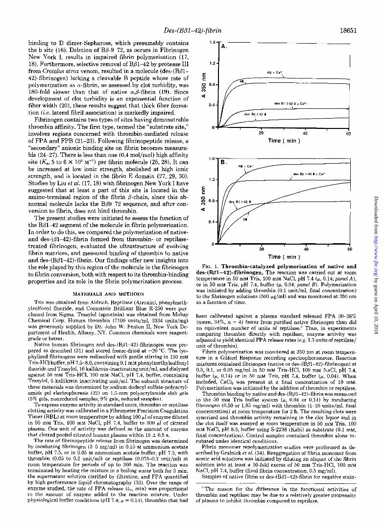
FIG. 1. Thrombin-catalyzed polymerization of native and des-(Bfll-42)-fibrinogen. The reaction was carried out at room temperature in 50 mM Tris, 100 mM NaCl, pH 7.4 (F, 0.14; panel A), or in 50 mM Tris, pH 7.4, buffer (p, 0.04; panel B). Polymerization was initiated by adding thrombin (0.1 unit/ml, final concentration) to the fibrinogen solutions (500 Kg/ml) and was monitored at 350 nm as a function of time.
been calibrated against a plasma standard released FPA 30-38% (mean, 34%, n = 4) faster from purified native fibrinogen than did an equivalent number of units of reptilase.’ Thus, in experiments comparing thrombin directly with reptilase, enzyme activity was adjusted to yield identical FPA release rates (e.g. 1.3 units of reptilase/ unit of thrombin).
Fibrin polymerization was monitored at 350 nm at room tempera- ture in a Gilford Response recording spectrophotometer. Reaction mixtures contained fibrinogen (native or des-(B/31-42)-fibrinogen) at 0.5, 0.1, or 0.05 mg/ml in 50 mM Tris-HCl, 100 mM NaCl, pH 7.4, buffer (p, 0.14) or in 50 mM Tris, pH 7.4, buffer (II, 0.04). When included, CaCIZ was present at a final concentration of 10 mM. Polymerization was initiated by the addition of thrombin or reptilase.
Thrombin binding by native and des-(B@l-42)-fibrin was measured in the 50 mM Tris buffer system (CL, 0.04 or 0.14) by incubating fibrinogen (0.50 or 1.85 mg/ml) with thrombin (l-25 units/ml, final concentration) at room temperature for 2 h. The resulting clots were synerized and thrombin activity remaining in the clot liquor and in the clot itself was assayed at room temperature in 50 mM Tris, 100 mM NaCl, pH 8.3, buffer using S-2238 (Kabi) as substrate (0.1 mM, final concentration). Control samples contained thrombin alone in- cubated under identical conditions.
Fibrin monomer repolymerization studies were performed as de- scribed by Gralnick et al. (34). Reaggregation of fibrin monomer from acetic acid solutions was initiated by diluting an aliquot of the fibrin solution into at least a lo-fold excess of 50 mM Tris-HCl, 100 mM NaCl, pH 7.4, buffer (final fibrin concentration, 0.5 mg/ml).
Samples of native fibrin or des-(Bpl-42)-fibrin for negative stain-
’ The reason for the difference in the functional activities of thrombin and reptilase may be due to a relatively greater propensity of plasma to inhibit thrombin compared to reptilase.
by guest on April 10, 2018
http://ww
w.jbc.org/
Dow
nloaded from
Des-(BOl-42)-fibrin
ing electron microscopy were formed at room temperature (p, 0.14) as 50.~1 drops (0.1 mg/ml fibrinogen) in a Petri dish, incubated for O-20 min with thrombin or reptilase (0.1 unit/ml, final), picked up on glow discharged carbon/formvar-coated grids, and then negatively contrasted with 2% (w/v) uranyl acetate. For processing by the critical point drying technique, the clots were formed overnight and critical point-dried as previously described (10).
Electron microscopy was performed in a Philips Model 400 trans- mission electron microscope at 80 or 120 kV. Fibrin fiber diameters were measured from 8 x 10 enlargements of micrographs. All fibers within randomly selected areas of each photograph were measured and averaged.
RESULTS AND DISCUSSION
Thrombin-catalyzed Polymerization of Native and Des- (BB1-42,J-fibrin-Des-(B/31-42)-fibrinogen (0.5 mg/ml) at physiological pH and ionic strength (p, 0.14) polymerized at a much reduced rate when compared to native fibrinogen (AA3dmin, des-(Bpl-42)-fibrin, O.Ol/min; native fibrin, 0.28/min) (Fig. lA), even though the rate of FPA release did not differ significantly (tth, des-(B@l-42)-fibrinogen, 20.2 min; native fibrinogen, 21.4 min at G.2 unit/ml, thrombin). Addi- tion of calcium to these mixtures resulted in a small increase in the polymerization rate of native fibrin and a more marked increase in that of des-(B@l-42)-fibrin (1.3- versus 6-fold). In each case, the lag period before the onset of turbidity was
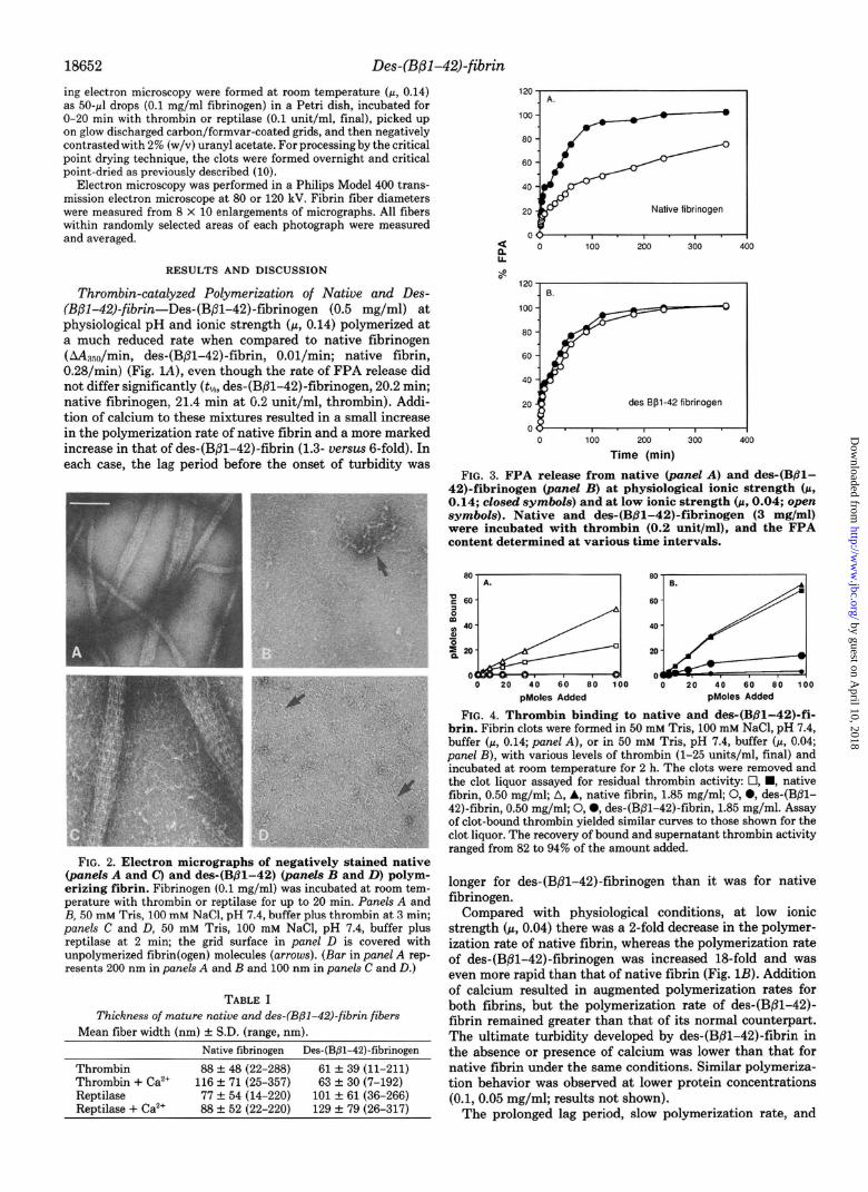
FIG. 2. Electron micrographs of negatively stained native (panels A and C) and des-(Bpl-42) (panels B and D) polym- erizing fibrin. Fibrinogen (0.1 mg/ml) was incubated at room tem- perature with thrombin or reptilase for up to 20 min. PaneLs A and B, 50 mM Tris, 100 mM NaCl, pH 7.4, buffer plus thrombin at 3 min; panels C and D, 50 mM Tris, 100 mM NaCl, pH 7.4, buffer plus reptilase at 2 min; the grid surface in panel D is covered with unpolymerized fibrin(ogen) molecules (arrows). (Bar in panel A rep- resents 200 nm in panels A and I3 and 100 nm in panels C and D.)
TABLE I Thickness of mature native and des-(B@l-42)-fibrin fibers
Mean fiber width (nm) & S.D. (range, nm). Native fibrinogen Des-(B/3-42)-fibrinogen
Thrombin 88 + 48 (22-288) 61 f 39 (11-211) Thrombin + Ca*+ 116 f 71 (25-357) 63 + 30 (7-192) Reptilase 77 + 54 (14-220) 101 + 61 (36-266) Reptilase + Ca*+ 88 + 52 (22-220) 129 Z!I 79 (26-317)
Native fibrinogen
60
des Bpl-42 flbmogen
0 100 200 300 400
Time (min)
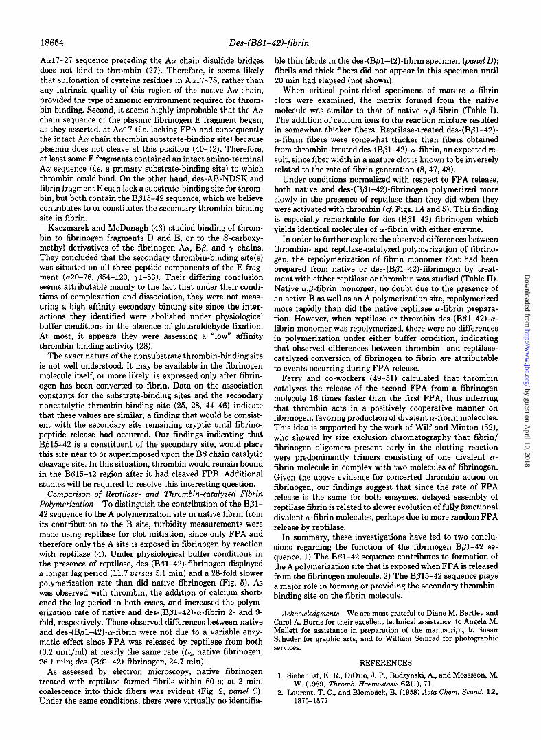
FIG. 3. FPA release from native (panel A) and des-(Bpl- 42)-fibrinogen (panel B) at physiological ionic strength (p, 0.14; closed symbols) and at low ionic strength (p, 0.04; open symbols). Native and des-(BBl-42)-fibrinogen (3 mg/ml) were incubated with thrombin (0.2 unit/ml), and the FPA content determined at various time intervals.
0 60 E 0 0 20 40 60 80 100 0 20 40 60 80 100
phloles Added pMoles Added
FIG. 4. Thrombin binding to native and des-(Bfil-42)-fi- brin. Fibrin clots were formed in 50 mM Tris, 100 mM NaCI, pH 7.4, buffer (p, 0.14; panel A), or in 50 mM Tris, pH 7.4, buffer (II, 0.04; panel B), with various levels of thrombin (l-25 units/ml, final) and incubated at room temperature for 2 h. The clots were removed and the clot liquor assayed for residual thrombin activity: 0, n , native fibrin, 0.50 mg/ml; A, A, native fibrin, 1.85 mg/ml; 0, 0, des-(B/31- 42)-fibrin, 0.50 mg/ml; 0, 0, des-(Bpl-42)-fibrin, 1.85 mg/ml. Assay of clot-bound thrombin yielded similar curves to those shown for the clot liquor. The recovery of bound and supernatant thrombin activity ranged from 82 to 94% of the amount added.
longer for des-(B@l-42)-fibrinogen than it was for native fibrinogen.
Compared with physiological conditions, at low ionic strength (CL, 0.04) there was a P-fold decrease in the polymer- ization rate of native fibrin, whereas the polymerization rate of des-(B/31-42)-fibrinogen was increased 18-fold and was even more rapid than that of native fibrin (Fig. 1B). Addition of calcium resulted in augmented polymerization rates for both fibrins, but the polymerization rate of des-(Bpl-42)- fibrin remained greater than that of its normal counterpart. The ultimate turbidity developed by des-(Bpl-42)-fibrin in the absence or presence of calcium was lower than that for native fibrin under the same conditions. Similar polymeriza- tion behavior was observed at. lower protein concentrations (0.1,0.05 mg/ml; results not shown).
The prolonged lag period, slow polymerization rate, and
by guest on April 10, 2018
http://ww
w.jbc.org/
Dow
nloaded from

Des-(Bpl-42)-fibrin 18653
1.6
Time ( min ) FIG. 5. Reptilase-induced polymerization of native and des-
(BBl-42)-fibrinogen. The reaction was carried out at room tem- perature in 50 mM Tris. 100 mM NaCl. DH 7.4. buffer. ReDtiIase (0.13 unit/ml, final concentration) was add&I to fidrinogen s&ions ‘(500 pg/ml) to initiate the reaction and polymerization was monitored at 350 nm as a function of time.
TABLE II
Lag time, polymerization rate, and turbidity developed during repolymerization of native and des-(BPl-42)-fibrin monomer
Fibrin monc~mer preparation Lag time” Maximum ASO at rate 120 min
mill AAAnsolmin Des-(Bol-42) (IIa) 2.6 0.017 0.262 Des-(Bpl-42) (R) 2.3 0.014 0.251 Des-(Bpl-42) (IIa) + Ca’+ 0.1 0.236 0.448 Des-(Bpl-42) (R) + Ca’+ 0 0.254 0.471 Native (IIa) 2.1 0.354 1.066 Native (R) 3.8 0.192 0.759 Native (IIa) + Ca’+ 0 0.709 0.641 Native (R) + Ca” 0 0.624 0.725
“The lag time was measured from the time of fibrin monomer dilution to the onset of turbidity.
decreased final turbidity of des-(B/31-42)-fibrin could result from delayed fibril formation, retarded lateral association of fibrils into thicker cables, or both. To explore these possibil- ities, evolving fibrin clots were examined by electron micros- copy to assess the rate of fibril and thick fiber formation (Fig. 2). In native fibrin (cc, 0.14), thin fibrils were plentiful after 2-min incubation with thrombin, either in the absence or presence of calcium ions. At 3 min the sample contained thick fibers as well as fibrils (Fig. 2, panel A). In contrast, the des- (B/3-42)-fibrin sample (panel B) contained only a few thin fibrils. Thus, the slow polymerization rate of des-(Bpl-42)- fibrin is attributable to delayed formation of primary fibrillar structures, which may in turn contribute to delayed lateral fibril association.
Ultrastructural examination of critical point-dried mature native a&fibrin formed at p 0.14, pH 7.4, in the presence or absence of calcium, yielded a typical clot matrix composed predominantly of thick branching striated fibers (Table I). The matrix formed from mature thrombin-treated des-(BBl- 42)-fibrin displayed a similar structure, although under either condition the mean fiber width was less than that of the native fibrin fibers and the augmenting effect of calcium ions on fiber thickness was minimal. These foregoing results, demonstrating that thin fibril as well as ihick fiber formation is impaired in the des-(B/31-42)-fibrin assembly process, in- dicate that the B@l-42 segment contributes significantly to the A polymerization domain in the native molecule.
Thrombin Binding to Des-(Bpl-42)-fibrin-Previous stud-
ies with native fibrinogen showed that the accelerating effect of low ionic strength on polymerization is evidently offset by a decreased rate of FPA cleavage due to increased binding of thrombin to the secondary site on fibrin (35-37). In contrast to the slowed polymerization of native fibrin at low ionic strength (Fig. lB), we observed an acceleratory effect of low ionic strength on des-(B/3-42)-fibrin polymerization. To in- vestigate the possibility that this effect was attributable to decreased thrombin binding by des-(Bpl-42)-fibrin, relative to that of native fibrin, we measured both the rate of throm- bin-mediated FPA release as well as the binding of thrombin to these fibrin molecules. At physiological ionic strength, thrombin (0.2 unit/ml) released FPA from native fibrinogen with a tlh of 22.2 min and from des-(Bpl-42)-fibrinogen with a tlh of 21.5 min (Fig. 3). At ionic strength fi 0.04, there was more than a g-fold decrease in the FPA release rate from native fibrinogen (tlj2, 138 min), but only a modest decrease in the release rate from des-(Bpl-42)-fibrinogen (tlls, 36.5 min).
Under physiological buffer conditions, native fibrin (0.50 mg/ml) bound 20-60% of the added thrombin (Fig. 4), whereas at a higher fibrin concentration (1.85 mg/ml), 50- 75% of the added thrombin was bound. At low ionic strength, 70-90% of the added thrombin bound to native fibrin. In contrast, thrombin binding to des-(Bpl-42)-fibrin was unde- tectable under physiological buffer conditions at any fibrin concentration, and was very low (2-20%) at low ionic strength.
These findings suggest an explanation for the more rapid polymerization of des-(Bpl-42)-fibrin at low ionic strength compared with native a&fibrin. Since secondary thrombin binding is only minimally expressed in des-(Bpl-42)fibrin, the acceleratory effect of low ionic strength,per se, on polym- erization is augmented by an FPA cleavage rate that is only slightly less than that observed at physiological ionic strength. The addition of calcium, which further reduces the extent of secondary thrombin binding to fibrin (25, 29), enhances po- lymerization by increasing the thrombin effectively available for fibrinopeptide cleavage as well as by augmenting lateral fiber association (38, 39). Furthermore, the existence of a deficient A polymerization site and the possible absence or modification of the B polymerization site in des-(Bpl-42)- fibrinogen, probably account for the finding that thrombin- activated des-(Bfll-42)-fibrin forms thinner fibers than the corresponding native fibrin despite a greatly reduced polym- erization rate, which by itself would favor thick fiber forma- tion.
These observations on thrombin binding to des-(B/3-42)- fibrin clearly indicate that the BP-15-42 sequence in fibrin plays a major role in forming or providing at least a portion of the high affinity secondary thrombin-binding site, and are supported by the finding that Fibrinogen New York I, which lacks B@9-72 (18), fails to bind thrombin (17). Our conclusion as to the location of the secondary thrombin-binding site does not agree with that of Vali and Scheraga (30) who positioned this site within AcuI7-51. Their conclusion was based on the finding that this sequence was common to all fragments or peptides derived from fibrinogen that bound to active site- inhibited thrombin (i.e. des-AB-NDSK, fibrin, or fibrinogen fragment E, and S-sulfonated Aa17-78). This interpretation is open to question, since as summarized below, it appears that these fragments may each have bound to thrombin at a site in their structure that did not involve the native Aa17- 51 sequence. First, their S-sulfonated Aa17-78 peptide con- tained four negative charges inserted on cysteine residues at positions 28, 36, 45, and 49 (40) that are not present in the native Acu sequence. Furthermore, it has been shown that the
by guest on April 10, 2018
http://ww
w.jbc.org/
Dow
nloaded from
18654 Des-(Bpl-42)-fibrin
AcY~~-27 sequence preceding the Aa chain disultide bridges does not bind to thrombin (27). Therefore, it seems likely that sulfonation of cysteine residues in Acu17-78, rather than any intrinsic quality of this region of the native Aa chain, provided the type of anionic environment required for throm- bin binding. Second, it seems highly improbable that the Aa chain sequence of the plasmic fibrinogen E fragment began, as they asserted, at Aa (i.e. lacking FPA and consequently the intact ACY chain thrombin substrate-binding site) because plasmin does not cleave at this position (40-42). Therefore, at least some E fragments contained an intact amino-terminal Aa sequence (i.e. a primary substrate-binding site) to which thrombin could bind. On the other hand, des-AB-NDSK and fibrin fragment E each lack a substrate-binding site for throm- bin, but both contain the B@15-42 sequence, which we believe contributes to or constitutes the secondary thrombin-binding site in fibrin.
Kaczmarek and McDonagh (43) studied binding of throm- bin to fibrinogen fragments D and E, or to the S-carboxy- methyl derivatives of the fibrinogen Acu, B/3, and y chains. They concluded that the secondary thrombin-binding site(s) was situated on all three peptide components of the E frag- ment (~~20-78, 654-120, ~1-53). Their differing conclusion seems attributable mainly to the fact that under their condi- tions of complexation and dissociation, they were not meas- uring a high affinity secondary binding site since the inter- actions they identified were abolished under physiological buffer conditions in the absence of glutaraldehyde fixation. At most, it appears they were assessing a “low” affinity thrombin binding activity (28).
The exact nature of the nonsubstrate thrombin-binding site is not well understood. It may be available in the fibrinogen molecule itself, or more likely, is expressed only after fibrin- ogen has been converted to fibrin. Data on the association constants for the substrate-binding sites and the secondary noncatalytic thrombin-binding site (25, 28, 44-46) indicate that these values are similar, a finding that would be consist- ent with the secondary site remaining cryptic until fibrino- peptide release had occurred. Our findings indicating that BP1542 is a constituent of the secondary site, would place this site near to or superimposed upon the B/l chain catalytic cleavage site. In this situation, thrombin would remain bound in the Bfi15-42 region after it had cleaved FPB. Additional studies will be required to resolve this interesting question.
Comparison of Reptilase- and Thrombin-catalyzed Fibrin Polymerization-To distinguish the contribution of the Bfil- 42 sequence to the A polymerization site in native fibrin from its contribution to the B site, turbidity measurements were made using reptilase for clot initiation, since only FPA and therefore only the A site is exposed in fibrinogen by reaction with reptilase (4). Under physiological buffer conditions in the presence of reptilase, des-(B/31-42)-fibrinogen displayed a longer lag period (11.7 uersus 5.1 min) and a 28-fold slower polymerization rate than did native fibrinogen (Fig. 5). As was observed with thrombin, the addition of calcium short- ened the lag period in both cases, and increased the polym- erization rate of native and des-(Bpl-42)~a-fibrin 2- and 9- fold, respectively. These observed differences between native and des-(B/31-42)-a-fibrin were not due to a variable enzy- matic effect since FPA was released by reptilase from both (0.2 unit/ml) at nearly the same rate (tlh, native fibrinogen, 26.1 min; des-(B@l-42)-fibrinogen, 24.7 min).
As assessed by electron microscopy, native fibrinogen treated with reptilase formed fibrils within 60 s; at 2 min, coalescence into thick fibers was evident (Fig. 2, panel C). Under the same conditions, there were virtually no identifia-
ble thin fibrils in the des-(B@l-42)-fibrin specimen (panel D); fibrils and thick fibers did not appear in this specimen until 20 min had elapsed (not shown).
When critical point-dried specimens of mature a-fibrin clots were examined, the matrix formed from the native molecule was similar to that of native a&fibrin (Table I). The addition of calcium ions to the reaction mixture resulted in somewhat thicker fibers. Reptilase-treated des-(Bpl-42)- a-fibrin fibers were somewhat thicker than fibers obtained from thrombin-treated des-(B/?l-42)-a-fibrin, an expected re- sult, since fiber width in a mature clot is known to be inversely related to the rate of fibrin generation (8,47,48).
Under conditions normalized with respect to FPA release, both native and des-(B@l-42)-fibrinogen polymerized more slowly in the presence of reptilase than they did when they were activated with thrombin (cf. Figs. lA and 5). This finding is especially remarkable for des-(Bpl-42)-fibrinogen which yields identical molecules of a-fibrin with either enzyme.
In order to further explore the observed differences between thrombin- and reptilase-catalyzed polymerization of fibrino- gen, the repolymerization of fibrin monomer that had been prepared from native or des-(BPl-42)-fibrinogen by treat- ment with either reptilase or thrombin was studied (Table II). Native c&fibrin monomer, no doubt due to the presence of an active B as well as an A polymerization site, repolymerized more rapidly than did the native reptilase a-fibrin prepara- tion. However, when reptilase or thrombin des-(B@l-42)-a- fibrin monomer was repolymerized, there were no differences in polymerization under either buffer condition, indicating that observed differences between thrombin- and reptilase- catalyzed conversion of fibrinogen to fibrin are attributable to events occurring during FPA release.
Ferry and co-workers (49-51) calculated that thrombin catalyzes the release of the second FPA from a fibrinogen molecule 16 times faster than the first FPA, thus inferring that thrombin acts in a positively cooperative manner on fibrinogen, favoring production of divalent a-fibrin molecules. This idea is supported by the work of Wilf and Minton (52), who showed by size exclusion chromatography that fibrin/ fibrinogen oligomers present early in the clotting reaction were predominantly trimers consisting of one divalent CX- fibrin molecule in complex with two molecules of fibrinogen. Given the above evidence for concerted thrombin action on fibrinogen, our findings suggest that since the rate of FPA release is the same for both enzymes, delayed assembly of reptilase fibrin is related to slower evolution of fully functional divalent a-fibrin molecules, perhaps due to more random FPA release by reptilase.
In summary, these investigations have led to two conclu- sions regarding the function of the fibrinogen B/31-42 se- quence. 1) The Bpl-42 sequence contributes to formation of the A polymerization site that is exposed when FPA is released from the fibrinogen molecule. 2) The Bb15-42 sequence plays a major role in forming or providing the secondary thrombin- binding site on the fibrin molecule.
Acknowledgments-We are most grateful to Diane M. Bartley and Carol A. Burns for their excellent technical assistance, to Angela M. Mallett for assistance in preparation of the manuscript, to Susan Schuder for graphic arts, and to William Semrad for photographic services.
REFERENCES
1. Siebenlist, K. R., DiOrio, J. P., Budzynski, A., and Mosesson, M. W. (1989) Thromb. Haemostask 62(l), 71
2. Laurent, T. C., and Blomback, B. (1958) Acta Chem. &and. 12, 1875-1877
by guest on April 10, 2018
http://ww
w.jbc.org/
Dow
nloaded from
Des-(BP1 -42)-fibrin
3. Blomback, B., Hessel, B., Hogg, D., and Therkildsen, L. (1978) Nature 275, 501-505
D. (1988) Biochemistry 27, 7106-7112 28. Liu, C. Y., Nossel, H. L., and Kaplan, K. L. (1979) J. Biol. Chem.
254, 10421-10425 4. Blomback, B.,.and Laurent, T. C. (1958) Ark. Kemi. 12,137-146 5. Shainoff, J. R., and Dardik, B. N. (1983) Ann. N. Y. Acad. Sci.
408,254-267 6. Shen, L. L., Hermans, J. McDonagh, J., and McDonagh, R. P.
(1977) Am. J. Physiol. 232, H629-H633 7. Hantgan, R., McDonagh, J., and Hermans, J. (1983) Ann. N. Y.
Acad. Sci. 408, 344-366 8. Wolfe, J. K., and Waugh, D. F. (1981) Arch. Biochem. Biophys.
211,125-142 9. Miiller, M. F., Ris, H., and Ferry, J. D. (1984) J. Mol. Biol. 174,
369-384
J. Viol Chem. 249,3322-3325 13. Olexa. S. A.. and Budzvnski. A. Z. (1980) Proc. Natl. Acad. Sci.
10. Mosesson, M. W., DiOrio, J. P., Miiller, M. F., Shainoff, J. R., Siebenlist, K. R., Amrani, D. L., Homandberg, G. A., Soria, J., Soria, S., and Samama, M. (1987) Blood 69, 1073-1081
11. Mosesson, M. W., Siebenlist, K. R., Amrani, D. L., and DiOrio, J. P. (1989) Proc. Natl. Acad. Sci. U. S. A. 86,1113-1117
12. Kudrvk, B., Collen, D.. Woods, K. R., and Blomback, B. (1974)
U. i. A. 7i, 1374-1378 14. Laudano, A. P., and Doolittle, R. F. (1978) Proc. Natl. Acad. Sci.
U. S. A. 75,3085-3089 15. Laudano, A. P., and Doolittle, R. F. (1980) Biochemistry 19,
1013-1019 16. Shim& A., Saito, Y., and Inada, Y. (1986) Proc. Natl. Acad. Sci.
U. S. A. 83,591-593 17. Liu, C. Y., Nossel, H. L., and Kaplan, K. L. (1979) Thromb.
Haemostasis 42, 79 18. Liu, C. Y., Koehn, J. A., and Morgan, F. J. (1985) J. Biol. Chem.
260.4390-4396 19. Pandya, B. V., Cierniewski, C. S., and Budzynski, A. Z. (1985) J.
Biol. Chem. 260,2994-3000 20. Carr, M. E., Jr., and Hermans, J. (1978) Macromolecules 11,46-
50 21. Nagy, J. A., Meinwald, Y. C., and Scheraga, H. A. (1985) Bio-
chemistry 24,882-887 22. Ni, F., Konishi, Y., Bullock, L. D., Rivetna, M. N., and Scheraga,
H. A. (1989) Biochemistry 28.3106-3119 23. Ni, F., Meinwald, Y. C., Vasquez, M., and Scheraga, H. A. (1989)
Biochemistry 28,3094-3105 24. Wilner, G. D., Danitz, M. P., Mudd, M. S., Hsieh, K.-H., and
Fenton, J. W., II (1981) J. Lab. Clin. Med. 97.403-411 25. Kaminski, M., and McDonagh, J. (1983) J. Biol. Chem. 258,
10530-10535 26. Berliner, L. J., Sugawara, Y., and Fenton, J. W., II (1985) Bio-
chemistry 24, 7005-7009 27. Fenton, J. W., II, Olson, T. A., Zabinski, M. P., and Wilmer, G.
18655
29. Kaminski, M., and McDonagh, J. (1987) Biochem. J. 242, 881- 887
30. Vali, Z., and Scheraga, H. A. (1988) Biochemistry 27, 1956-1963 31. Pandva. B. V.. and Budzvnski. A. Z. (1984) Biochemistry 23,
460-470 I
32. Laemmli, U. K. (1970) Nature 227,680-685 33. Kehl, M., and Henschen, A. (1981) in HPLC in Protein and
Peptide Chemistry (Lottspeich, F., Henschen, A., and Hupe, K.-P., eds) p. 339, Walter de Gruyter, New York
34. Gralnick, H. R., Givelber. H. M. Shainoff, J. R., and Finlavson, J. S. (1971) J; Clin. Znuest. 50, 1819-1830
_
35. Carr. M. E.. Jr.. Kaminski. M.. McDonaeh. J.. and Gabriel. D. A. (1985) Thromb. Haemosta& 54, 159 - ’
36. Kaminski, M., and McDonagh, J. (1985) Thromb. Haemostasis 54,293
(1986) Thromb. Haemostasis 55, 352-356 38. Bover. M. H.. Shainoff. J. R.. and Ratnoff. 0. D. (1972) Blood
37. Nilsen, D. W. T., Brosstad, F., Kierulf, P., and Godal, H. C.
i9,‘382-387 39. Furlan, M., Rupp, C., Beck, E. A., and Svendsen, L. (1982)
Thromb. Haemostasis 47,118-121 40. Watt, K. W. K., Cottrell, B. A., Strong, D. D., and Doolittle, R.
F. (1979) Biochemistry 18,5410-5416 41. Budzynski, A. Z., Marder, V. J., and Shainoff, J. R. (1974) J. Biol.
Chem. 249,2294-2302 42. Olexa, S. A., Budzynski, A. Z., Doolittle, R. F., Cottrell, B. A.,
and Greene, T. C. (1981) Biochemistry 21,6139-6145 43. Kaczmarek, E., and McDonagh, J. (1988) J. Biol. Chem. 263,
13896-13900 44. Higgins, D. L., Lewis, S. D., and Shafer, J. A. (1983) J. Biol.
Chem. 258,9276-9282 45. Hofsteenge, J., Taguchi, H., and Stone, S. R. (1986) Biochem. J.
237,243-251 46. Naski, M. C., and Schafer, J. A. (1990) J. Biol. Chem. 265,1401-
1407 47. Rosser, R. W., Roberts, W. W., and Ferry, J. D. (1977) Biophys.
Chem. 7.153-157 48. Shah, G. A., Nair, C. H., and Dhall, D. P. (1985) Thromb. Res.
40.181-188 49. Bale, M. D., Janmey, P. A., and Ferry, J. D. (1982) Biopolymers
21,2265-2277 50. Janmey, P. A., Bale, M. D., and Ferry, J. D. (1983) Biopolymers
22,2017-2019 51. Janmey, P. A., Erdile, L., Bale, M. D., and Ferry, J. D. (1983)
Biochemistry 22,4336-4340 52. Wilf, J., and Minton, A. P. (1986) Biochemistry 25, 3124-3133
by guest on April 10, 2018
http://ww
w.jbc.org/
Dow
nloaded from
K R Siebenlist, J P DiOrio, A Z Budzynski and M W MosessonThe polymerization and thrombin-binding properties of des-(B beta 1-42)-fibrin.
1990, 265:18650-18655.J. Biol. Chem.
http://www.jbc.org/content/265/30/18650Access the most updated version of this article at
Alerts:
When a correction for this article is posted•
When this article is cited•
to choose from all of JBC's e-mail alertsClick here
http://www.jbc.org/content/265/30/18650.full.html#ref-list-1
This article cites 0 references, 0 of which can be accessed free at
by guest on April 10, 2018
http://ww
w.jbc.org/
Dow
nloaded from