Embed Size (px)
Citation preview

1
50S PRPms-su-revised
JBC (protein synthesis and post-translational modifications)
The Plastid Ribosomal Proteins (2): Identification of all the Proteins in the
50S Subunit of an Organelle Ribosome (Chloroplast)
Running title: Plastid ribosomal 50S subunit proteins
Kenichi Yamaguchi and Alap R. Subramanian*
From the Department of Biochemistry, The University of Arizona, Tucson, USA
and the Max-Planck-Institut für molekulare Genetik, Berlin-Dahlem, Germany
*Corresponding Author:
Dr. A. R. Subramanian, Department of Biochemistry, BioSciences West, The University
of Arizona, Tucson, AZ 85721-0088
Fax: +1 520 621 9288
E-mail: [email protected]
The protein and nucleic acid sequences reported in this paper have been deposited in
SWISS-PROT and GenBank databases, respectively. The accession numbers (total of
over 40) are listed in Table I and in text.
Copyright 2000 by The American Society for Biochemistry and Molecular Biology, Inc.
JBC Papers in Press. Published on June 28, 2000 as Manuscript M005012200 by guest on July 26, 2018
http://ww
w.jbc.org/
Dow
nloaded from

2
ABBREVIATIONS
The abbreviations used are: RP, ribosomal protein; PRP, plastid ribosomal protein; PSRP,
plastid-specific ribosomal protein; RRF, ribosome recycling factor; TP50, total protein
from 50S subunit; TP70, total protein from 70S ribosome; 2D-PAGE, two-dimensional
polyacrylamide gel electrophoresis; HPLC, high-performance liquid chromatography;
MS, mass spectrometry; LC/MS, reversed-phase HPLC coupled to electrospray
ionization mass spectrometry; ESI MS, electrospray ionization mass spectrometry; EST,
expressed sequence tag; AA, amino acid(s); NTE, N-terminal extension; CTE, C-terminal
extension.
by guest on July 26, 2018http://w
ww
.jbc.org/D
ownloaded from

3
SUMMARY
We have completed identification of all the ribosomal proteins (RP's) in spinach plastid
(chloroplast) ribosomal 50S subunit via a proteomic approach using 2-dimensional
electrophoresis, electroblotting/protein sequencing, HPLC purification, PCR-based
screening of cDNA library/nucleotide sequencing and mass spectrometry (LC/MS and ESI
MS). Spinach plastid 50S subunit comprises 33 proteins, of which 31 are orthologues of
E. coli RP's and 2 are plastid-specific RP's (PSRP-5 and PSRP-6) having no homologues
in other types of ribosomes. Orthologues of E. coli L25 and L30 are absent in spinach
plastid ribosome. Twenty-five of the plastid 50S RP's (PRP's) are encoded in the nuclear
genome and synthesized on cytosolic ribosomes while 8 of the PRP's are encoded in the
plastid organelle genome and synthesized on plastid ribosomes. Sites for transit peptide
cleavages in the cytosolic RP precursors and formyl Met processing in the plastid-
synthesized RP's were established. Post-translational modifications were observed in
several mature plastid RP's, including multiple forms of L10, L18, L31 and PSRP-5, and
N-terminal/internal modifications in L2, L11 and L16. Comparison of the RP's in gradient-
purified 70S ribosome with those in the 30S and 50S subunits revealed an additional
protein, in approximately stoichiometric amount, specific to the 70S ribosome. It was
identified to be plastid ribosome recycling factor (P-RRF). Combining with our recent
study of the proteins in plastid 30S subunit (ms. submitted), we show that spinach
plastid ribosome comprises 59 proteins (33 in 50S subunit, 25 in 30S subunit and RRF in
70S), of which 53 are E. coli orthologues and 6 are plastid-specific proteins (PSRP-1 to -
6). We propose the hypothesis that PSRP's were evolved to perform functions unique to
plastid translation and its regulation, including protein targeting/translocation to thylakoid
membrane via plastid 50S subunit.
by guest on July 26, 2018http://w
ww
.jbc.org/D
ownloaded from

4
INTRODUCTION
The plastid (chloroplast) ribosome is a plant-specific, organelle ribosome that produces
proteins encoded by the plastid genome. Plastid ribosomes are responsible for the
synthesis of huge amounts of biomass, since the large subunit of ribulose 1,5-bisphophate
carboxylase/oxygenase (a most abundant protein in the biosphere) is synthesized in
plastids. Plastid ribosomes are very similar to the eubacterial 70S-type ribosome, in
composition and general mode of function (1-4). The rRNA's and most of the
characterized ribosomal proteins (RP's) in plastid ribosomes also bear close resemblance
to the corresponding components so far identified in cyanobacteria, a correlation
supporting the importance of endosymbiotic theory in plastid evolution (5).
The E. coli ribosome, the most well-studied of the eubacterial ribosomes (6), is
composed of 21 RP's in the 30S subunit and 33 RP's in the 50S subunit. Two more
possible E. coli RP's have been suggested: protein Y, the product of E. coli yfia gene (7),
bearing a distant sequence homology to a chloroplast-specific RP (PSRP-1), and a protein
designated S22 (8). Post-translational modifications are found in many E. coli RP's,
although a modification in L16 (Arg-81) remains yet to be characterized (see Results for
plastid L16). We have recently identified all the RP's in spinach plastid 30S ribosomal
subunit, including all its PSRP's and many post-translational modifications (Yamaguchi,
K., von Knoblauch, K., and Subramanian, A. R., ms submitted for publication). The
number of RP's in plastid 50S subunits has so far only been estimated (35~39, reviewed
in ref. 2), but has not been determined.
Although the constituents of plastid translational machinery in general are similar to
those of E. coli, the genes are distributed in two genome compartments: the plastid and
the nucleus. The rRNA and tRNA genes are located in the plastid genome, whereas the
genes for processing/modification enzymes, aminoacyl tRNA synthetases and 60% of the
RP's are located in the nuclear genome (1-4). The plastid translation system also differs
from the eubacterial system in other significant ways, e.g. chloroplast mRNA is often
edited (9), about 60% of chloroplast mRNAs lack canonical ribosome binding sites (RBS)
by guest on July 26, 2018http://w
ww
.jbc.org/D
ownloaded from

5
found in E. coli mRNAs (10), mRNA levels in chloroplasts remain relatively unchanged
through dark/light transitions whereas protein synthesis rates increase dramatically upon
illumination (11, 12), nuclear-coded factors mediate light-regulated translation (13), and
nuclear mutants occur with defects in chloroplast polysome assembly (14). Gene
expression in chloroplasts depends overwhelmingly on nuclear gene products which
mediate both transcription/post-transcriptional processing (15, 16) and translation. We
speculated (ms. submitted to JBC) that some of the nuclear factors exert their roles
through evolutionary alterations in plastid ribosome. Since few evolutionary changes are
observed in the plastid rRNA, but many in plastid RP (e.g. PSRP's), a key to
understanding light-dependent translational regulation might involve chloroplast RP's.
We have applied a proteomic approach (2D-PAGE, HPLC separation, protein
sequencing, PCR-based screening/DNA sequencing, and mass spectrometry: LC/MS and
ESI MS) to the plastid ribosomal 50S subunit to establish a complete identification of all
its protein components. Proteins in both 50S subunits and 70S ribosomes were
identified, yielding an unexpected result that plastid ribosomal recycling factor (P-RRF) is
present in the approximate stoichiometry of one in the 70S ribosome. Transit peptide
cleavage sites in all 25 cytosolically synthesized plastid RP precursors, post-translational
modifications in many of the mature PRP's, the absence of the orthologues of two E. coli
RP's, and possible function of a 70S ribosome-bound P-RRF are discussed.
EXPERIMENTAL PROCEDURES
Preparation of Spinach Chloroplast 70S Ribosome, 50S Subunit, TP50 and TP70 -
Spinach (Spinacia oleracea, cv. Alwaro) ribosomes were prepared as described
previously (17). Purification of 70S ribosomes and the 50S subunits were done by zonal
centrifugation (5000 A260 units of ribosome/zonal rotor for 70S purification and 3000
A260 units of purified 70S/ zonal rotor for 50S isolation) as described previously (18).
The approximate conversion factor used for the estimation of protein amount was: 1 A260
unit of 70S or 50S = 20 µg protein. TP50 and TP70 (total protein of 50S subunit and 70S
by guest on July 26, 2018http://w
ww
.jbc.org/D
ownloaded from

6
ribosome, respectively) were extracted basically according to Hardy et al. (19). Sample
preparations for 2D-PAGE analysis, HPLC resolution (1 mg TP50/run), and LC/MS
analysis were as described earlier (30S ms. submitted to JBC).
Reversed-phase HPLC - The HPLC system was assembled with a WellChrom Maxi-
Star K-1000 HPLC pump (KNAUER, Germany), an injection valve, No.7125
(Rheodyne, USA), and an UV detector, UV-VIS S-3702 (Soma, Japan). Separation of
TP50 was performed basically according to Kamp et al. (20) using Vydac C18 column
(4.6 x 250 mm) and a gradient with 0.1% trifluoroacetic acid (TFA) and 0.1% TFA in
isopropanol at a flow rate of 0.5 ml/min.
Protein Electrophoresis and Electroblotting - SDS-PAGE was done by the method of
Laemmli et al. (21). Tricine SDS-PAGE was performed according to the method of
Schägger and von Jagow (22). 2D-PAGE (basic and acidic) were done as described
previously (23, 24). Electroblotting was done according to Walsh et al. (25) using tank-
blotting chamber and performed in 25 mM Tris-HCl, pH 8.4, 0.5 mM dithioerythritol,
0.02% SDS at 500 mA for 16 h at 4 ˚C. PVDF-membrane (Sequi-Blot, BIO-RAD) was
used for transfer membrane. After electroblotting, the resulting blot was rinsed three times
in water for 5 sec and stained with 0.1% Amido Black 10B (Sigma) in 50% methanol for 5
min, destained with 50% methanol until the background disappeared, rinsed 3 times in
water, then dried on a Whatman 3MM filter paper, and stored in the dark at -20 ˚C until
used.
Protein sequencing- Protein sequencing was carried out at the Laboratory for Protein
Sequencing and Analyze, the University of Arizona, using an Applied Biosystem 477A
Protein/Peptide sequencer interfaced with a 120A HPLC analyzer to determine
phenylthiohydantoin (PTH) amino acids. After the conversion step, 50 µl of the PTH-
derivative (out of 135 µl or 37%) is injected into an ABI PTH-Narrowbore C18 column
(2.1 x 250 mm) for detection of PTH-AA (remaining sample going to fraction collector).
PCR Screening for cDNA's of PrpL19, PrpL34, Psrp-5 and Psrp-6 - A lambda gt11
spinach cDNA library prepared in our laboratory previously (26) was screened by
by guest on July 26, 2018http://w
ww
.jbc.org/D
ownloaded from

7
thermal gradient PCR using a Mastercycler gradient PCR apparatus (Eppendorf
Scientific, Inc., USA). Thermal gradient PCR was performed for 3 min at 94 ˚C, 35 cycles
of 1 min at 94 ˚C, 1 min at 43-60 ˚C, 1 min 30 sec at 72 ˚C with 0.25U of Taq DNA
polymerase (Gibco-BRL) in a 20 µl reaction volume containing 1 µl of spinach cDNA
library (~108 plaque forming units), 5 µM gene specific primer or 10 µM degenerate
primer, 5 µM lambda arm primer (PF or PR), 20 µM each dNTP, 1.5 mM MgCl2, and 50
mM KCl in 20 mM Tris-HCl (pH 8.4).
Plaque Screening of cDNA Library/Cloning of Spinach PrpL5 and PrpL34 - The
lambda gt11 spinach cDNA library (26) was screened using random-primed, [32P]-labeled
Arabidopsis EST clone (E10B7T7) and a 5’-PRPL34 cDNA portion (PF/PL34R1),
respectively, as probes for PrpL5 and PrpL34. Clone E10B7T7 was from Arabidopsis
Biological Resource Center (Ohio State University). Radio-labeling was carried out as
described by the supplier of Random Primed DNA Labeling Kit (Boehringer Mannheim).
150,000 plaque-forming units were plated on four 132 mm plates, and plaques were lifted
onto Nylon filters. Prehybridization was performed in 500 mM sodium phosphate, pH
7.0, at 50 ˚C (for PrpL5) or 65 ˚C (for PrpL34) for 2 h and hybridization in 500 mM
sodium phosphate, pH 7.0, 7% SDS at 50 ˚C for 4 h (for PrpL5) or at 65 ˚C for 16 h (for
PrpL34). The filters were washed twice in 100 mM sodium phosphate, pH 7.0, 1% SDS
at 37 ˚C for 10 min followed by a 10-min wash in 40 mM sodium phosphate, pH 7.0, 1%
SDS at 37 ˚C and autoradiographed. Plaques giving positive signals were purified two
further rounds of screening (27).
Oligonucleotide primers and DNA sequencing - The oligonucleotide primers used in
this study were: PF, 5’-CGG-GAT-CCG-GTG-GCG-ACG-ACT-CCT-GGA-GCC-CG-
3’; PR, 5’-CGG-GAT-CCC-AAC-TGG-TAA-TGG-TAG-CGA-CCG-GC-3’; PL5F1,
5’-TGG-CAC-TGA-TTA-CTG-GGC-AAA-GGC-3’; PL5R1, 5’-GTG-TTT-TGT-
TAC-ACG-GAA-TGC-3’; PL5R2, 5’-TAC-CTT-CTC-TGA-CCT-TAA-ACC-CTG-
3’; PL19F1, 5’-AAR-GAR-ATH-AAR-GTI-GTI-GCI-CAY-MG-3’; PL19F2, 5’-CAA-
TGA-CTT-GAA-TTT-CCC-TG-3’; PL19F3, 5’-GCC-ATT-GAA-GAA-GCA-ATT-
by guest on July 26, 2018http://w
ww
.jbc.org/D
ownloaded from

8
AG-3’; PL19F4, 5’-GGA-GAC-ATT-GTG-CAA-ATC-AG-3’; PL19R1, 5’-CTT-
AGA-TAG-TAT-AGC-CTT-GCC-3’; PL19R2, 5’-CTG-ATT-TGC-ACA-ATG-TCT-
CC-3’; PL19R3, 5’-GCT-ATC-CTC-CGC-TTC-CGA-CC-3’; PL19R4, 5’-CTA-ATT-
GCT-TCT-TCA-ATG-GC-3’; PL34F1, 5’-GGI-AAR-GCI-GCI-YTI-ISI-YTI-ACI-
AAR-MG-3’; PL34F2, 5’-GTC-ATT-GGC-TCG-GAC-ACA-TG-3’; PL34R1, 5’-
GCT-CAT-TCG-CAG-ACG-AAA-ACC-3’; PL34R2, 5’-AGA-GCA-ATG-GAG-
TGA-CCC-GG-3’; PPSRP-5F1, 5’-GGA-ATT-CTA-GAT-ATC-GTC-GAC-GAG-
AGA-TGG-CAC-TCC-TTT-C-3’; PPSRP-5F2, 5’-GAA-GCT-AAC-ATC-TCA-GTT-
CAG-3’; PPSRP-5R1, 5’-GGA-ATT-CGT-CGA-CGC-GTT-TTT-GAG-AAA-AAG-
ATT-TAC-ACT-G-3’; PPSRP-5R2, 5’-GCC-TGT-TCC-TTC-GGA-GTC-TG-3’;
PPSRP-6F1, 5’-GGA-ATT-CTA-GAT-ATC-GTC-GAC-CCT-TCC-AKA-GCA-
AAA-TAG-AAA-AAA-AAG-AGG-3’; PPSRP-6R1, 5’-CAT-RTG-RTG-IGC-IGT-
ICC-YTT-YTT-YTG-3’; PPSRP-6R2, 5’-TCA-TCA-AAC-AGT-TCA-TAT-GC-3’;
PTAG1, 5’-GGA-ATT-CTA-GAT-ATC-GTC-G-3’; PTAG2, 5’-GGA-ATT-CGT-
CGA-CGC-G-3’; PT7, 5’-TAA-TAC-GAC-TCA-CTA-TAG-GG-3’; PT3, 5’-AAT-
TAA-CCC-TCA-CTA-AAG-GG-3’.
PF (forward primer) and PR (reverse primer) are complementary to the cloning site of
lambda gt11. Degenerate oligonucleotide primers, PL19F1, PL34F1, and PPSRP-6R1
were designed from PRPL19 peptide 1 (sequence region: KEIKVVSHR), N-terminal
sequence of PRPL34 (sequence region: GKAALXLTKR), and N-terminal sequence of
PSRP-6 (sequence region: QKKGTAHHM), respectively (see also Table I). Gene-
specific PCR primers for PrpL19 and PrpL34 (PL19R1 and PL34R1) were designed from
the nucleotide sequences of PCR products PL19F1/PR and PL34F1/ PR, respectively,
which were amplified using primer sets (PL19F1 and PR) and (PL34F1 and PR). Gene-
specific PCR primer for Psrp-6 (PPSRP-6F1) was designed from the nucleotide sequence
of PF/PPSRP-6R1 and tagged with PTAG1 sequence. Gene-specific PCR primers for
Psrp-5 (PPSRP-5F1 and PPSRP-5R1) was designed from the nucleotide sequence of
spinach chloroplast L40 (28), and tagged with PTAG1 and PTAG2 sequences,
by guest on July 26, 2018http://w
ww
.jbc.org/D
ownloaded from

9
respectively. Other sequencing primers shown above were designed from obtained DNA
sequences during primer walking. PCR products were analyzed by agarose gel
electrophoresis using 1% agarose gel and visualized by ethidium bromide staining. PrpL5
insert DNA in the phage clone (L5F2-1) was amplified by PCR using primer sets PF and
PR, and sequenced using primers PF, PL5F1, PL5R1, and PL5R2. The nucleotide
sequence of PrpL19 was obtained by sequencing PCR products PF/PL19R1 and
PL19F1/PR using sequencing primers: PL19F2, PL19F3, PL19F3, PL19F4, PL19R2,
PL19R3, and PL19R4. For PrpL34, insert DNA in the phage clone (L34D2-1-1) was
amplified by PCR using primer sets PF and PR, then cleaved by EcoRI digestion and
subcloned into the plasmid vector pBluescript SK- (Stratagene). The insert DNA in
PrpL34 plasmid clone was sequenced using primers, PT3, PL34F2, PT7, and PL34R2.
The nucleotide sequence of Psrp-5 was obtained by sequencing PPSRP-5F1/PPSRP-5R1
using sequencing primers: PTAG1, PPSRP-5F2, PTAG2, and PPSRP-5R2. The
nucleotide sequence of Psrp-6 was obtained by sequencing PPSRP-6F1/PR using
sequencing primers: PTAG1 and PPSRP-6R2. Nucleotide sequences were determined at
the DNA Sequencing Facility, University of Arizona, using an Applied Biosytems model
377 sequencer.
Mass Spectrometry - Mass spectrometry was done at the Mass Spectrometry Facility,
Chemistry Department, University of Arizona. Electrospray ionization mass
spectrometry (ESI-MS) was carried out on a Finnigan LCQ system. Liquid
chromatography - mass spectrometry (LC/MS) was done on the same LCQ system
interfaced with a Michrom HPLC system (Magic 2002) using a Microbore C18 column (1
x 150 mm). The solvent system was: 0.1% TFA in 2% acetonitrile (solvent #1) and 0.1%
TFA in 90% acetonitrile (solvent #2). 50 pmol purified PRP in 10 µl 4% acetic acid was
subjected to ESI-MS. For LC/MS, 20 µl TP50 (100 pmol) in solvent A was injected to
the Microbore C18 column.
Internal peptide preparation- ‘In-gel’ digestion of PRPL19 was performed basically
according to Hellman et al. (29). Five spots were excised from Coomassie Blue stained
by guest on July 26, 2018http://w
ww
.jbc.org/D
ownloaded from
10
2D-gels of TP50 (200 pmol/gel) and equilibrated with mixing for 40 min at 30 ˚C in 2 ml
of 50 mM Tris-HCl, pH 8.5 in 50% acetonitrile. The gel pieces were completely dried in
a Speed-Vac, then rehyderated with 150 µl of 50 mM Tris-HCl, pH 8.5, 0.02%
polyoxyethylenesorbitan monolaurate (Tween 20), 10% acetonitrile, containing 0.4 µg
(ca. 1/40 enzyme/substrate ratio by weight) endoproteinase Lys-C (Sigma), and incubated
at 30 ˚C for 16 h. The enzyme reaction was stopped by adding 1/10 reaction volume of
10% TFA. Gel pieces were transferred into 500 µl of 0.1% TFA in 60% acetonitrile and
peptides were extracted by shaking at 30 ˚C for 80 min. The extract was dried in a Speed-
Vac, dissolved in 50 µl of 5% acetic acid, and peptides were purified by reversed-phase
HPLC using a Vydac C8 (4.6 x 50 mm) column in TFA-acetonitrile solvent system. The
purified peptides (PRPL19 peptides 1 and peptide 2, see Table I) were dried, dissolved in
25 µl of 30% acetic acid and subjected to a protein sequencing.
In order to obtain internal peptides of RRF (the 70S ribosome-specific protein,
identified later as plastid ribosome recycling factor), it was purified from pool 37 from a
previous work (30). Pool 37 showed two components, identified ~60% RRF and ~40%
PSRP-2, by 2D-PAGE analysis. RRF was purified using a Vydac C4 (4.6 x 150 mm)
column in TFA-acetonitrile solvent system. The purified protein (ca. 20 µg) was digested
using 0.2 µg endoproteinase Asp-N (Sigma) in 50 mM Tris-HCl, pH 8.0, 2 M urea, at 37
˚C for 16 h. The digest was dried in a Speed-Vac, then subjected to Tricine SDS-PAGE.
Peptides separated on the gel were electroblotted onto a PVDF-membrane and stained as
described above. A 3.5 kDa peptide band was excised from the blot and sequenced (RRF
peptide 1 shown in Table I).
Small subunit of RuBisCo- Spinach SSU (small subunit of ribulose 1, 5-bisphosphate
carboxylase/oxygenase) was present in a pool from previous work, pool 40 (30). It was
purified by reversed-phase HPLC using a Vydac C8 (4.6 x 50 mm) column in TFA-
acetonitrile solvent system, and also by 2D-PAGE/electroblotting. The N-terminal
sequence of the purified protein (12 cycles), Methyl-MKVWPTQNMKRY, confirmed
its identity as spinach SSU.
by guest on July 26, 2018http://w
ww
.jbc.org/D
ownloaded from

11
Computer analyses - The program BLAST from the National Center for
Biotechnology Information (NCBI) was used for sequence searches. Protein sequence
searches were performed using ‘blastp’ program versus nr (non-redundant Database of
GenBank CDS translations (PDB, Swiss-Prot, PIR, and PRF). EST searches were done
using ‘tblastn’ program versus dbEST (non-redundant Database of GenBank, EMBL,
DDBJ EST Divisions). ORF’s (open reading frame) from cDNA sequences were
analyzed using the ‘map’ program from GCG software (31). Isoelectric points and
sequence masses were calculated using ‘peptidesort’ program (31). Sequence alignments
and comparisons were performed using ‘pileup’ and ‘gap’ programs (31). LC/MS data
was analyzed using the ThermoQuest Finnigan Xcalibur data system. Masses were
deconvoluted from resulted m/z using ‘BIOMASS Deconvolution’ program and charge
states were convoluted using ‘BIOMASS Calculation’ program.
RESULTS
Proteins of Chloroplast Ribosomal 50S Subunit - In order to identify all the PRP’s in
pure spinach chloroplast 50S ribosomal subunit, spinach chloroplast 70S ribosomes were
first purified on a zonal gradient and then run through a second dissociating zonal gradient
to obtain 30S and 50S subunits. Efficient dissociation of chloroplast ribosomes required
the development of a phosphate-containing dissociation buffer (see Ref. 18 for details and
typical gradient profiles). TP50 was extracted from 50S subunits and the proteins were
separated by 2D-PAGE and transferred onto PVDF membrane. Fig. 1A shows such a
membrane, stained with Amido Black. The individual spots have a slightly different
staining pattern with Amido Black as compared to staining with Coomassie Blue, e.g.
acidic proteins like L12 are stained poorly by Amido Black (compare Fig. 1A with
Fig.8A). Each of the spots was excised and was subjected to N-terminal sequencing. Since
several of the protein spots were composite-looking, indicating overlapping separations
(Fig. 1A), the experiment was repeated, with the spots excised into 2 or 3 segments for
by guest on July 26, 2018http://w
ww
.jbc.org/D
ownloaded from

12
the N-terminal analysis. The sequence data from all these experiments are summarized in
Table I.
Most of the N-terminal sequences were sufficient to allow protein identification from
similarity using BLAST search and, in several cases, from the identity to reported spinach
PRP sequences in databases. In a few instances, however, the N-terminal data was
insufficient to provide a positive identification. In those cases, additional experiments
were done to obtain internal peptide sequences and/or to isolate cDNA's from a spinach
cDNA library for nucleotide sequencing. Plastid (chloroplast) ribosomal proteins (PRP’s)
are designated in Table I as L1, L2,…to L36, as per their sequence similarity to the
corresponding E. coli ribosomal proteins. The 2D-PAGE pattern is diagramatically
represented in Fig. 1B, depicting the positions of all the identified proteins. The
designations alpha, beta, .., PSRP-5, PSRP-6, etc. are discussed later.
To confirm whether all of spinach chloroplast 50S subunit PRP’s appeared in the 2D-
PAGE pattern shown in Fig 1, two additional experiments were performed. The 2-D
system used for Fig. 1 resolves mainly proteins of pI 4.5 or greater (23), as acidic
proteins of lower pI do not migrate into the 1st dimension gel. We therefore ran TP50 in
another 2-D system suitable for the resolution of acidic proteins (pI 5.5 or lower, ref. 24).
A few spots appeared in this 2D-PAGE, but N-terminal sequencing did not reveal any
new protein sequence (data not shown). A second problem arises if an N-blocked protein
comigrates with an unblocked PRP. A single spot would then appear, giving a single N-
terminal sequence under Edman degradation, suggesting the spot contained one protein.
To overcome this problem, we resolved the 50S subunit PRP’s (TP50) on a reversed-
phase HPLC column, where hydrophobic interaction (rather than the net charge or
peptide chain length) is the key to resolution. HPLC could thus separate proteins that are
often unresolved by 2D-PAGE. Fig. 2 shows the separation of plastid TP50 obtained by
this procedure. Proteins in the individual peaks were identified by 2D-PAGE analysis.
The HPLC experiment allowed clean separations of several proteins that were not well
resolved by 2D-PAGE, e.g. L4/L21, L3/L13, L20/PSRP-5 forms (compare Figs 1 and 2).
by guest on July 26, 2018http://w
ww
.jbc.org/D
ownloaded from

13
Indeed, plastid L36 and the beta/gamma forms of PSRP-5 did not at all show up on 2D-
PAGE, but gave distinct peaks in the HPLC run, permitting both N-terminal sequence
and mass (by LC/MS) determination. Because these three small proteins are very basic,
they might have migrated out of the 2D-gel. Interestingly, a few proteins on the other
hand, L35, L33, L32, L27, and PSRP-5 (alpha-gamma), were each eluted in two different
peaks (Fig. 2). Their identities were inferred from 2D-PAGE analysis, N-terminal
sequencing, as well as mass determinations (ESI MS). This observation could suggest that
some plastid RP's may possibly exist as two distinct conformers on the plastid ribosome.
The average yield of PTH-AA recovered in the first three cycles of Edman degradation
was calculated for each of the sequence runs and is summarized in Fig. 3. Most proteins
showed in yields ranging from 20 to 50 pmol, indicating that this amount approximates
the average stoichiometry (1 copy/50S subunit). Plastid L12 gave a yield of almost 200
pmol, four times the highest yield for the other proteins recovered in good yield. The
result supports the existence of 4 copies of L12/plastid 50S ribosomal subunit, as has
been previously deduced (17). Several of the plastid 50S subunit proteins, e.g. L10, L18,
L31, and PSRP-5 exist in multiple forms (named alpha to gamma, Figs. 1 and 2 and Table
I). The summed N-terminal yield of the multiple forms in each of these cases
corresponded to the approximate stoichiometric amount for the other PRP’s (Fig. 3).
Very low yields of N-terminal amino acids were observed in three instances: L16, L22,
and L34. As discussed later, 90% of PRPL16 is likely blocked by trimethylation; most of
PRPL22 may be N-blocked by an unknown group; and PRPL34, one of the most basic
proteins of the 50S subunit (like L36), partially runs out of the 2D-gel.
The N-terminal sequence data in Table I allowed the identification of the orthologues
of 28 E. coli RP's in spinach plastid ribosome, beside two plastid specific proteins,
PSRP-5 and PSRP-6. E. coli ribosomal 50S subunit contains [making 3 subtractions for
L7, alpha-N-acetylated form of L12; L8, a complex of L7/L12 and L10, and L26=S20] 33
canonical RP’s: L1 to L36 (32). Hence our data so far has not allowed positive
identifications for five possible E. coli orthologues. These are: L5, L19, L25, L30 and L34,
by guest on July 26, 2018http://w
ww
.jbc.org/D
ownloaded from

14
of which three (L5, L19, and L34) were identified with additional sequence data obtained
(using the protein sequence information) by screening a spinach cDNA library, described
earlier (26).
The N-terminal sequence of the protein, later identified as PRPL5 (25 AA), did not
show significant homology to any database proteins (BLAST search using ‘blastp’
program), but the same data when used to screen the EST data bank with ‘tblastn’
program gave two matches with Arabidopsis EST clones. One of these, E10B7T7, was
obtained from the Arabidopsis Biological Resource Center (Ohio State University) and
used as probe to screen our spinach lambda gt11 cDNA library (26). Several positive
clones were isolated and the longest cDNA clone (L5F2-1) was sequenced (data deposited
in GenBank, accession # AF250923). Clone L5F2-1 encodes 207 AA residues, that is13
residues short of the complete sequence of mature PRPL5. The 25-residue N-terminal
sequence (Table 1), however, provided this missing sequence as well as a 12-residue
overlap with the DNA-derived sequence. The sequence comparisons of spinach PRPL5
with its homologues in a higher plant (A. thaliana), an alga (Porphyra purpurea), a
photosynthetic bacterium (Synechocystis PCC6803), and E. coli are shown in Fig. 4.
Spinach PRPL5 contains a 16-amino acid long NTE, and a positively charged 26-amino
acid long CTE, as compared to E. coli L5. The plastid protein has amino acid identity of
72.5% to the A. thaliana L5, and 56.4%, 60.0%, and 48.6%, respectively to the L5
proteins of P. purpurea, Synechocystis PCC6803, and E. coli. The amino acid sequences
of the NTE and CTE of plastid L5 yielded no significant matches when searched against
protein databases.
The N-terminal and internal sequences of plastid L19 and the N-terminal sequence of
plastid L34 did not show significant similarity to any proteins in databases. We therefore
screened the spinach lambda gt11 cDNA library (26) using inosine-containing degenerate
oligonucleotide primers designed from PRPL19 peptide 1 and the N-terminal sequence of
PRPL34. Thermal gradient PCR allowed us to find the optimal amplification conditions
using primer sets of degenerate primers and lambda-arm primers (PF or PR). Amplified
by guest on July 26, 2018http://w
ww
.jbc.org/D
ownloaded from

15
PCR products (PL19F1/PR and PL34F1/PR) were sequenced, and further PCR
amplifications were done using sets of PF and gene specific primers (based on the
obtained DNA sequence). The nucleotide sequences of PRPL19 cDNA was thus obtained
entirely by sequencing of PCR products (submitted to GenBank, accession # AF
250384).
As compared to E. coli L19, spinach plastid L19 contains a negatively charged, 47-
amino acid long NTE. However, it showed a remarkably low similarity to the
corresponding two L19-like sequences in the Arabidopsis database (Ath1 and Ath2, Fig.
4), and gave no significant matches against all the sequences in protein databases. Overall,
spinach PRPL19 showed amino acid identities of only 50.4% and 47.7% to the two
Arabidopsis sequences (genes F3I6.17 and AT4g11630), and 38.5%, 42.7%, and 37.3%,
respectively, to the L19 protein sequences from P. purpurea, Synechocystis PCC6803,
and E. coli.
For PRPL34, a PCR product encoding the 5’-region of its cDNA (PF/PL34R) was
first obtained and was used as probe to screen the spinach cDNA library. Several clones
were isolated and the one containing the longest cDNA was subcloned into a plasmid
vector and was sequenced. The nucleotide sequence is submitted to GenBank (accession #
AF238221). As compared to E. coli L34, plastid L34 contains a 7-residue long NTE and
10-residue long CTE. These extensions are absent in both cyanobacterial and algal L34
proteins. Core sequences between PRPL34 and the L34 proteins from P. purpurea,
Synechocystis PCC6803, and E. coli are relatively well conserved (Fig. 4), the percent
identities being, 45.5%, 41.9% and 50.0%, respectively.
Including the three proteins identified via DNA work, a total of 31 orthologues of E.
coli RP's are present in spinach plastid ribosome. There are thus two E. coli RP's (L25
and L30, see Discussion), for which we could obtain no evidence of occurrence in spinach
plastid ribosome.
Plastid-specific Ribosomal Proteins in Chloroplast 50S Subunit - One of the additional
N-terminal sequences in Table I (PSRP-5) corresponded to the reported sequence of
by guest on July 26, 2018http://w
ww
.jbc.org/D
ownloaded from

16
spinach plastid L40 (28), which is the homologue of an earlier reported protein from pea,
named PsCL18 (33). Another N-terminal sequence we determined (PSRP-6, Table I)
showed similarity to a second reported protein from pea, PsCL25 (33); spinach
homologue of PsCL25 has not been reported. We have screened our spinach cDNA
library (26) for the cDNA's corresponding to these two proteins, and the clones obtained
were sequenced. The proteins were designated PSRP-5 (previous L40) and PSRP-6
(homologue of pea PsCL25), in accordance with our proposed nomenclature (see
Discussion; Nucleotide sequences of PSRP-5 and PSRP-6 cDNA's deposited in GenBank,
accession # AF261940 and AF245292, respectively). Our PSRP-5 data showed 6 amino
acids differences, all at the C-terminal portion, from the reported L40 data (28), leaving it
uncertain at this point whether the differences reflect spinach cultivar differences (cv.
Alwaro versus G'eant d'hiver) or sequencing errors. Three post-translationally modified
forms of PSRP-5 were identified from our protein sequencing results (Table I), the
modifications were in the N-terminal portion of the protein (see Discussion). Both PSRP-
5 and PSRP-6 are unique to the plastid ribosome, homologous sequences being not found
in the RP's of E. coli, archaebacteria, yeast (cytosolic or mitochondrial) or mammals.
Mass Spectrometry of 50S PRP’s - Spinach TP50 was analyzed by LC/MS. Fig. 5A
shows the relative abundance of summed mass/charge ratio (m/z) of the PRP’s in the m/z
range 400 to 2,000 versus HPLC elution time. An example of a mass spectrum at the
elution interval of 41.5-42.0 min (summed scans for 30 sec) is shown in Fig. 5B. Protein
mass was calculated by deconvolution of the m/z series. For example, from deconvolution
of the m/z series in Fig. 5B, a major mass of 13,811.0 Da and a minor mass 13850.2 Da
were derived (Fig. 5C). As the observed mass 13,811.0 Da is very close to the sequence
mass of PRPL12, i.e. 13,814.54 Da, this peak was identified as of plastid L12. In cases
where sequence mass were not available, ESI MS of HPLC pools, containing proteins
identified by 2D-PAGE (shown in Fig. 2), allowed mass identification. Every 30 sec
interval was analyzed as stated above, and the resultant mass values are summarized in
Table II. The combined LC/MS analysis of TP50 and ESI MS analysis of HPLC pools
by guest on July 26, 2018http://w
ww
.jbc.org/D
ownloaded from

17
allowed the identification of most individual 50S PRP masses, except for PRP's L2, L20,
the multiple forms of L10, L18, L31, and PSRP-6. An extreme case was PRPL2, which
was not detected either by LC/MS of TP50 or ESI MS of HPLC pools; it probably
represents a very poorly ionizing polypeptide.
Edman degradation of PRPL16 - The plastid located prpL16 gene codes for the N-
terminal sequence formyl Met-Leu-Ser- (34), while our N-terminal sequence analysis of
PRPL16 yielded Xaa-Leu-Ser- (Xaa being a modified, unidentified AA). The N-terminus
of E. coli L16 is α-N-monomethylated (35), and so we suspected the same modification in
plastid L16. To test this hypothesis, we purified spinach SSU (small subunit of ribulose
1, 5-bisphosphate carboxylase/oxygenase), which has monomethylated methionine at the
N-terminus (36). About 200 pmol each of SSU HPLC (SSU purified by HPLC), SSU 2D/blot
(SSU purified using 2D-PAGE/electroblotting) and PRPL16 2D/blot were subjected to N-
terminal analysis (Fig. 6). The SSU HPLC yielded PTH-monomethyl methionine (NMM,
retention time 26.37 min, appearing between PTH-tryptophan and PTH-phenylalanine),
whereas SSU 2D/blot showed an unusual PTH-derivative of retention time 10.30 min, just
after the N’N-dimethyl-N’-phenylthiourea peak (DPTU, marked a; retention time 10.25
min). Alkylation of monomethyl methionine with acrylamide under mildly alkaline
conditions (during 2D-PAGE and/or electroblotting), just as the similar modification of
cysteine during the same procedure (37), could produce the latter (uncharacterized)
compound. PRPL16 2D/blot showed the same unusual PTH-derivative with the same
retention time in cycle 1. In cycle 2, PRPL16 2D/blot gave the expected residue, PTH-Leu,
but the yield (2.7 pmol) was only about one-tenth the expected amount (the average yield
of N-terminal PTH-AA of 50S PRP’s was 27.5 pmol, see Fig. 3), whereas the two SSU
preparations gave the normal yield of PTH-Lys (see Fig. 6). We therefore infer that post-
translationally about 10% of PRPL16 is alpha-N-monomethylated while 90% of it is
blocked to Edman degradation, probably by trimethylation (see Discussion).
Identification of a 70S Ribosome-specific Protein - During the course of this work we
observed that a prominent protein spot, always present in TP70 gels, was absent in either
by guest on July 26, 2018http://w
ww
.jbc.org/D
ownloaded from

18
TP30 or TP50 gels (Fig. 7). To investigate further, a TP70 blot was prepared, and this
spot and several other spots (from both 30S and 50S proteins) were excised and subjected
to N-terminal sequence analysis. The analysis yielded a single N-terminal sequence,
specific to a protein found only on plastid 70S ribosome, the spot indicated by arrow in
Fig.7C. The N-terminal sequence of 21 residues (Table I), however, did not show
homology to any of the reported proteins in database. Therefore, the 70S ribosome-
specific protein was purified from a previously fractionated spinach chloroplast
ribosomal protein pool, # 37 (ref. 31), and an internal peptide was obtained by
endoproteinase Asp-N digestion. At the time we sequenced the internal peptide, the
nucleotide sequence of spinach chloroplast ribosome recycling factor (RRF) was reported
(38). Our N-terminal and internal sequences matched 100% with the corresponding
sequences of RRF (N-terminal positions 1 - 21 and internal positions 116 - 139). Thus,
the 70S ribosome-specific protein we identified in this work is plastid RRF. In contrast to
the plastid situation, E. coli RRF is mostly found in post-ribosomal supernatant (see
Discussion).
We have previously identified all the proteins in spinach chloroplast 30S subunit, a
total of 25 proteins - 21 E. coli orthologues and 4 PSRP's (Yamaguchi, von Knoblauch,
and Subramanian, submitted). Here we have identified all the proteins in spinach
chloroplast 50S subunit, a total of 33 proteins - 31 E. coli orthologues and 2 PSRP's. In
addition, a protein found only on plastid 70S ribosome was characterized and this 70S-
specific protein was identified as plastid RRF. In order to round off the picture, a 2D-
PAGE of TP70 was done (Fig. 8A) and all the spots were cut out and analyzed. The
protein identifications confirmed the results from the 30S and 50S subunit experiments
and, as diagramatically represented in Fig. 8B, revealed no further proteins. It should be
mentioned that PRPL36 and two of the forms of PSRP-5 (beta and gamma) did not show
up in the 2D-PAGE patterns, but were identified with the help of HPLC (Fig. 2). As we
noted in the 30S identification paper, the diffusely staining minor spots visible in the
upper (high molecular) part of the 2D-PAGE (Figs 1A and 8A) represent mainly minor
by guest on July 26, 2018http://w
ww
.jbc.org/D
ownloaded from

19
aggregates of a few PRP's exhibiting tendency to polymerize (see legend to Fig. 1), and
small amounts of nonribosomal proteins, that probably have functional associations with
ribosomes.
DISCUSSION
In this paper we present sufficient protein and nucleic acid sequence data to establish
the identification of all the protein components in the 50S subunit of a land plant
chloroplast ribosome. Similar results for chloroplast 30S subunit has just been submitted
for publication. Taken together these two publications present the first complete
identification of the protein components of an organelle (plastid) ribosome,
complementing the previously reported such complete identifications for E. coli (6), and
the cytosolic ribosomes of yeast (39) and mammals (40). Recent reports indicate that a
complete protein identification for a mitochondrial ribosome may soon be forthcoming
(41-43).
In the companion 30S subunit paper we presented a nomenclature for plastid RP's and
their genes that accords with both the current usage for mitochondrial RP's and the CPGN
(Commission on Plant Gene Nomenclature) rules for plant gene names. In brief,
chloroplast RP's are designated PRP's (Plastid Ribosomal Proteins), with gene names
written in italics, but having the first letter in capital for the RP genes that are located in
the nuclear genome and the first letter lowercase for the RP genes that are located in the
plastid genome. Thus the plastid homologue of E. coli L1 is designated PRPL1 and its
nucleus-located gene designated, PrpL1, whereas the plastid homologue of E. coli L2 is
designated PRPL2, and its plastid-located gene is designated prpL2. As described in the
companion paper, chloroplast ribosomes contain proteins that do not have homologues in
E. coli, archaebacteria, or in yeast/mammalian cytosolic ribosomes. These proteins are
designated PSRP-1 etc (Plastid Specific RP), and their genes, which are all located in the
nuclear genome, are designated Psrp-1 etc.
by guest on July 26, 2018http://w
ww
.jbc.org/D
ownloaded from

20
Proteins of Plastid Ribosomal 50S Subunit - Spinach chloroplast 50S ribosomal subunit
contains 33 proteins, the same number as in E. coli 50S ribosomal subunit. However, only
31 of these proteins are orthologues of corresponding E. coli RP's, while the remaining
two are plastid specific ribosomal proteins (PSRP's: Fig. 1B, Fig. 3 and Table I). The 31
orthologues of E. coli 50S RP's are designated (PRP): L1 - L6, L9 -L24, L27 - L29, L31-
L36 (E. coli designations, L7, L8 and L26 do not stand for distinct RP's). Thus two E. coli
50S RP's, L25 and L30, do not have orthologues (or homologues) in the plastid 50S
subunit. Since plastid 30S subunit contains the orthologues of all 21 E. coli 30S RP's (ms.
submitted for publication), spinach plastid ribosome maintains all E. coli RP's except L25
and L30.
The orthologues of the two PSRP's we identified in spinach plastid 50S subunit were
first reported in pea, derived from cDNA sequences named PsCL18 and PsCL25 (33).
Based on the present comprehensive protein study, we can state that no additional
PSRP's are present in spinach chloroplast 50S subunit (Figs. 1-3, Table I). The
companion comprehensive study on plastid 30S subunit (ms. submitted), has revealed 4
PSRP's, (designated PSRP-1 to PSRP-4) in the 30S subunit. The plastid ribosome thus
maintains 6 plastid-specific proteins, 4 of them in the 30S subunit and 2 in the 50S
subunit. The two PSRP's in the 50S subunit, are designated PSRP-5 and PSRP-6.
Sequence similarity between a barley plastid ribosomal protein (BPRL28) and PSRP-6
has been recently noted (44). Interestingly, despite being specific to plastids, the
sequence identities among PSRP-5 and PSRP-6.homologues are relatively low, e.g. only
53.8% and 54.0%, respectively, between the spinach and pea proteins (Fig. 4). There was
no significant sequence similarity between PSRP-5 or PSRP-6 and any of the bacterial
proteins in databases, suggesting their appearance in plants after the endosymbiotic event
in plastid evolution. In contrast, all four 30S subunit PSRP's show some sequence
homology to eubacterial proteins: PSRP-1, PSRP-2, and PSRP-3 to cyanobacterial
proteins (ms submitted to JBC) and PSRP-4 to a protein only reported from Thermus
thermophilus (ms in preparation), suggesting their evolution from ancestral eubacterial
by guest on July 26, 2018http://w
ww
.jbc.org/D
ownloaded from

21
genes. The data thus indicate a strong diverging trend for the 50S PSRP sequences in the
plastid evolution.
Post-translational Processing: Plastid 50S RP's Encoded in the Organelle Genome -
The genes that encode 8 of the spinach plastid 50S subunit PRP's are maintained in the
plastid genome, and thus they are synthesized on the plastid ribosome with initiating
formyl-Met tRNA. The post-translational processing of the N-terminal formyl-Met
undergone by these PRP's are shown in Table III. Five of the proteins (L2, L20, L22, L32
and L33) have the entire formyl-Met group excised, while three (L14, L16 and L36) have
only the formyl group removed, leaving methionine at the N-terminus. The N-terminal
alanine of L2 is monomethylated, as reported earlier (ref. 30, 1st description of post-
translational modification in a plastid RP). The modifications in plastid L16 are discussed
later (see below). We had previously suspected N-terminal modification in plastid L36
(45), but the present study did not reveal any modification. The MS mass results for L14,
L32, and L33 accorded with the calculated sequence molar masses (Table II), indicating no
additional post-translational modifications in these PRP's.
Post-translational Processing: Plastid 50S RP's Encoded in the Nuclear Genome -
Twenty-five 50S subunit PRP's - orthologues of 23 E. coli RP's and the two PSRP's - are
encoded in the nuclear genome and are thus synthesized on the cytosolic ribosomes as
precursors; all plastid RP precursor molecules contain an extra, routing peptide sequence
(transit peptide), which is cleaved off upon entry of the mature PRP into the plastid.
Table III lists the immediate post-cleavage N-terminal sequences for all of these 25 PRP's.
Their pre-cleavage peptide sequences are also listed in Table III, where they are known
for 13 of the PRP's. A consensus, flanking peptide sequence that could specify the
cleavage site for the transit peptidase cleavage enzyme was not discernible in this data.
In contrast to the situation for plastid 30S subunit, only a minority of 50S subunit RP
genes located in the plastid DNA (8 out of 33; 24%), the majority being in the nuclear
genome. For the 30S subunit, its RP genes are about equally distributed between the
plastid genome and the nuclear genome. This may reflect a co-evolutionary linkage, as
by guest on July 26, 2018http://w
ww
.jbc.org/D
ownloaded from

22
pointed out in a previous review (3), arising from the main function of the 30S subunit,
i.e. formation of 30S initiation complex, involving plastid-transcribed mRNA. A
correlation between the PRP’s that are synthesized on the plastid ribosome and the early
steps of ribosome assembly (plastid rRNA is transcribed in the plastid) has been
suggested, but it is applicable for the 30S subunit and not for the 50S subunit.
Post-Translational Modifications in mature 50S PRP's - The post-translational
modifications of individual plastid 50S PRP’s, as derived from this study, are discussed
below.
PRPL1 showed a minor fragment (spot ‘e’ in Fig. 1) with N-terminal truncation of 23-
amino acids, i.e. starting with the N-terminal sequence, TLPSPTKPKKGKAAL, position
nos. 24-38 of PRPL1 sequence (GenBank accession # X76932; ref. 46; M. Kavousi and
A. R. Subramanian, unpublished results). Since this fragment was identified as present
only in TP50 preparations but not in TP70 (compare Figs.1 and 8), its significance is at
present unclear.
Spinach PRPL2 gene sequence has been reported [accession # X00797, ref. 47], but
the C-terminal portion of the deduced amino acid sequence lacks homology to bacterial
and reported chloroplast L2 sequences in databases. We have therefore re-sequenced the
rpL2 gene from spinach chloroplast DNA, and our data (submitted to GenBank,
accession # AJ244023) confirms a frame shift arising from a minor sequencing mistake in
the earlier submission. The corrected spinach PRPL2 amino acid sequence shows a
conserved C-terminal portion with the expected homology. Mature spinach plastid L2 is
post-translationally modified, with α-N-monomethyl alnine at the N-terminus (30).
In previous work using a different cultivar of spinach, the spot of PRPL4 had
appeared in a different, slightly more basic 2D-PAGE position (Fig. 1 in ref. 2), while all
the other 50S and 30S PRP’s showed the same positions as in this study. Thus PRPL4
may be one of the few plastid RP's that harbor detectable strain-specific differences. A
mass difference of 58-71 is seen between the sequence mass of PRPL4 (GenBank
accession # X93160, cv. Melody; ref. 48) and the observed MS mass in this study (cv.
by guest on July 26, 2018http://w
ww
.jbc.org/D
ownloaded from

23
Alwaro, Table II). Possibly, it may again reflect a cultivar difference, or post-translational
modification.
PRPL10 exists in three forms of differing size (designated alpha, beta and gamma). The
relative amounts were: alpha form, 80%, beta, 12% and the minor gamma form, about 8%,
as estimated from the yields of N-terminal PTH-AA's. The major form, PRPL10 alpha,
mass 20,305 Da, was identified by the LC/MS analysis (Table II), but the other two
forms (both approximately 16.5 kDa, as estimated by SDS-PAGE) were not detected in
the MS analysis, probably due to low ionization and smaller amounts. As all the 3 forms
gave the same N-terminal sequence, their differences arise from post-translational
modifications in the internal and/or C-terminal regions of the molecule.
PRPL11 has been previously shown to be epsilon-N-trimethylated at positions Lys-9
and Lys-45 (ref. 49; J. Schmidt and A. R. Subramanian, unpublished). The E. coli L11
protein is trimethylated at the corresponding positions in its sequence context (50).
LC/MS data showed that plastid L11 has a mass increment of 81.5 Da over its sequence
mass (Table II). This increment is close to the 84.2 Da mass for two trimethyl
modifications. In the case of E. coli L11, the N-terminal amino acid Ala is alpha-N-
trimethylated (50). The N-terminal Ala of PRPL11 is mainly unmodified, but there is
indirect evidence for a partial modification of this residue, resulting in the presence of a
minor form (this paper and ref. 49).
PRPL12 was obtained in a significantly higher yield (Fig. 3), consistent with the
presence of 4 copies/50S subunit, as has been previously deduced for plastid 70S
ribosome (17). In E. coli, this protein exists in two forms: L12, with free N-terminus and
L7, alpha-N-acetylated form, the sum of the two forms constituting 4 copies/ 50S
ribosomal subunit (51), and the ratio of the two forms altering during the bacterial growth
cycle (52). The N-terminus of plastid L12 is essentially unmodified (Fig. 3), and thus the
roles played by N-acetylation (and its variation with growth) in E. coli are apparently
abolished in plastid metabolism. Interestingly, a minor mass of 13,850.2 Da was observed
in the LC/MS of TP50, at the same elution interval as PRPL12 (mass of 13,811 Da), as
by guest on July 26, 2018http://w
ww
.jbc.org/D
ownloaded from

24
shown in Fig. 5C. The mass increment of 39.2 Da might indicate the presence of a minor
modification (including a minor acetylated form, +42.04 Da).
PRPL16 is partially α-N-monomethylated (10%), while the bulk of it is -N-blocked by
an unknown modification (surmised as trimethylation, see Results). In E. coli the L16
protein is α-N-monomethylated (35) and its Arg-81 is modified by an as yet
uncharacterized group (35). The observed mass of E. coli L16 by a recent MALDI-TOF
MS analysis (53) is 44.9 Da heavier than the sequence mass. Subtracting the mass of α-N-
monomethylation (14 Da) from 44.9 Da, the uncharacterized group in Arg-81 of E. coli
L16 would be of mass 30.9 Da. Our LC/MS analysis of 50S PRP’s (Table II) indicated
that plastid L16 is modified with a mass increment of 74.1 Da. Plastid L16 has a
conserved Arg residue (Arg-82), in a relatively conserved sequence context, that would
correspond to E. coli Arg-81. It is conceivable that Arg-82 of PRPL16 maintains the same
modification as in E. coli Arg-81 (+30.9). Hypothetically, if most of plastid L16 N-
terminus is blocked by trimethylation (+42.08 Da), the total mass increment would be
72.98 (30.9+42.08), close to the observed 74.1 Da increment in the mass of PRPL16
(Table II).
PRPL18 exists in two forms (alpha and beta) of different size but with the same N-
terminal sequence; alpha form, 35% and beta form, 65%, as estimated from the yield of
PTH-AA. The modification in PRPL18 was not characterized in this study.
PRPL19 is a highly diverged protein with a negatively charged, 46 amino acid residues
long NTE compared to E. coli L19 (Fig. 4). The presence of a phosphorylated protein in
plastid ribosomal 50S subunit has been reported (54, 55): the protein spot in the reports
corresponds to PRPL19 (see Figs. 1 and 8), as correlated in ref. 2. As the observed
protein mass of PRPL19 is very close to the sequence mass (Table II), MS data would
suggest no post-translational modification. However, phosphorylation is a reversible
process and the plastid ribosomes used in the present study could be de-phophorylated.
The sequence -RRLSSLRASTSKS- in the C-terminal portion of Xenopus 40S subunit
ribosomal protein S6 (56), is phosphorylated (S in bold). The consensus recognition motif
by guest on July 26, 2018http://w
ww
.jbc.org/D
ownloaded from
25
for S6 kinase II is reported to be -RXXS- (57), and for casein kinase II is -S/TXXE/D-
(58). The motif -RXXS- is present in both spinach (83-RRLS-86) and Arabidopsis
(RRVS) plastid L19 sequences, whereas (1-SEAE-4 and 26-SEAE-30) are present only
in spinach L19. Thus, this study cannot rule out (or rule in) plastid L19 phosphorylation.
Bubunenko et al. has reported that plastid L23 protein in the chloroplast ribosomes of
a certain group of plants (Caryophyllidae, spinach and relatives) is replaced by a
homologue of the cytosolic L23 protein, an unusual evolutionary event (59). Table II
shows the observed mass of plastid L23 by ESI MS analysis (13,553.5 Da), a value close
to the sequence mass (13,553.7 Da), calculated from the nucleotide of sequence of plastid
L23 cDNA (GenBank accession # X90414; C. Jayabaskaran and A. R. Subramanian,
unpublished). The mature plastid L23 thus appears post-translatonally unmodified. The
N-terminal sequence of PRPL23 shown in Table I corresponds to the reported cDNA; it
does not correspond to the deduced N-terminal sequence of a prokaryotic-type rpL23
gene occurring in spinach plastid DNA. Moreover, an N-terminal sequence corresponding
to the plastid rpL23 gene was not observed in this comprehensive study. These results
thus confirm the previous conclusion (59) that the plastid-encoded rpL23 gene in spinach
is a pseudogene. cDNA's derived from two distinct but closely related genes for spinach
cytosolic L23, the presumed progenitor of the nuclear gene for spinach plastid L23, have
been isolated and sequenced (C. Jayabaskaran and A. R. Subramanian, unpublished,
GenBank accession # X92367 and X92350).
PRPL31 exists in three forms (alpha to gamma) with the same N-terminal sequence but
differing in charge: alpha form 55%, beta form 31%, and gamma 14% by estimation from
PTH-AA yields. E. coli L31 may apparently exist in two forms, a truncated form missing
the C-terminal sequence, RFNIPGSK, and the full length form as deduced from the rpL31
(rpmE) gene sequence (60, 61). The modifications in plastid L31 forms are
uncharacterized.
PRPL34 (61 AA) is the smallest and the most basic (pI=12.99) of the 25 nucleus-
coded RP's of spinach plastid 50S subunit. Its transit peptide is 91 AA long (Fig. 4), and
by guest on July 26, 2018http://w
ww
.jbc.org/D
ownloaded from

26
thus the longest of all spinach chloroplast RP transit peptides. PRPL35 (73AA,
pI=12.15) also has a relatively long transit peptide of 86 AA (62). An average size plastid
ribosomal protein, PRPL19 (156 AA, pI=10.51) has a 77 AA long transit peptide. The
largest plastid ribosomal protein PRPS1 (370 AA, pI=4.83) has only a 41 AA long transit
peptide (63). In general, a correlation can be made that shorter the mature protein, the
longer the transit peptide in the RP precursor. Longer transit peptides might thus be a
requirement for the proper import and processing of small basic ribosomal proteins.
Among the two plastid-specific proteins of the 50S subunit, PSRP-5 exist in 3 forms
(alpha to gamma), differing apparently in the cleavage points in the precursor form. The
reported spinach L40 sequence (28), corresponds to PSRP-5 alpha form in protein
sequence length. The cDNA-derived protein sequences of L40 and PSRP-5 differ in 6
amino acids at the C-terminal portion (Fig. 4). These differences could be due to cultivar
difference (Alwaro versus Géant d’hiver) or possibly sequencing errors. The protein mass
of all the 3 forms of PSRP-5 were observed upon LC/MS analysis of TP50 (Table II),
suggesting that all the forms exist on plastid 50S subunit. Mass data also indicate that two
of the forms (beta and gamma) are unlikely to be post-translationally modified. In
contrast, Edman degradation of PSRP-5 alpha gave only 6% of the expected average yield
of PTH-AA, as compared to the yields of 28% and 30%, respectively, for PSRP-5 beta
and PSRP-5 gamma (Fig. 3). We surmise that the major portion of PSRP-5 alpha is
blocked at the N-terminus. As the LC/MS mass data for PSRP-5 alpha showed 41 Da
increment over the calculated sequence mass (Table II), the blocking group could be acetyl
(42.04 Da). The relative occurrence of the 3 forms of PSRP-5 appear to be alpha, 42%,
beta, 28%, and gamma, 30%. The second plastid specific protein on the 50S subunit, i.e.
PSRP-6, appears N-terminally unmodified (Table I), but since it was not observed in the
MS analysis, we have no data on modifications elsewhere in the molecule.
The case of ribosomal proteins L25 and L30 - Plastid 50S subunit is missing the
orthologues of E. coli L25 and L30 (the PSRP-5 and PSRP-6 proteins have no sequence
similarity to L25/L30). In E. coli, L25, L18 and L5 are 5S rRNA binding proteins, the
by guest on July 26, 2018http://w
ww
.jbc.org/D
ownloaded from

27
binding of L18 stimulating the specific binding of L5 (64) and the association of 5S rRNA
with 23S rRNA requiring all three proteins (65). Is the function of E. coli L25 performed
by another plastid 50S protein? It has been reported that there are striking similarities in
the properties of E. coli L25 and spinach plastid L22, in protecting a domain of E. coli 5S
rRNA comprising nucleotides 70-109 (66). Spinach plastid L22 consists of a long NTE
and a CTE, and a central core homologous to E. coli L22 (34); but apparently it is this
core of plastid L22 that binds to 5S rRNA (67), even though it is known that E. coli L22
does not bind 5S rRNA. A homologue of E. coli L25 has been identified in Anabaena
(photosynthetic cyanobacterium) RP's (68) and evidence for the occurrence of L25 in
Synechocystis PCC 6803 genome sequence has been presented (68). Thus, it appears that
L25 protein might have been lost during chloroplast evolution, with plastid L22 protein
taking over some of its functions.
E. coli L30 is a 23S rRNA binding, late assembly protein, that can be mutated out
without loss of cell viability (69). Binding of L30 protein to nucleotides 931-938 of 23S
rRNA has been established by cross-linking (70). Interestingly, the 3 nucleotides, 931-
933, on the E. coli 23S rRNA are replaced by a variable loop of 5 - 20 nucleotides in
chloroplast 23S rRNA (71, 72; see Fig. 10). Protein sequences showing some similarity to
E. coli L30 could be found in several plant ESTs, but the deduced sequences lacked
chloroplast transit peptide sequence. Those EST's thus probably represent cytosolic
RP's, mitochondrial RP's which do not always require a separate transit peptide sequence
(some MRP's are imported into mitochondria without transit peptide), or nonribosomal
proteins.
Sequences having significant homology to E. coli L25 and L30 are not always present
in the many reported eubacterial genomes. Thus, while both L25 and L30 genes are
identified in Haemophilus influenzae, Chlamydia trachomatis, Rickettsia prowazekii,
Neisseria meningitidis strains MC58 and Z2491, the L25 gene is reported missing in
Aquifex aeolicus, Bacillus subtilis, Borrelia burgdorferi, and Thermotoga maritima, and
the L30 gene is missing in Synechocystis PCC6803. Both L25 and L30 genes are missing in
by guest on July 26, 2018http://w
ww
.jbc.org/D
ownloaded from

28
Helicobacter pylori strains 26695 and J99, Mycoplasma pneumoniae, Mycobacterium
tuberculosis, Mycoplasma genitalium, and Treponema pallidum. L30 gene homologues
are present in the reported complete genome sequences from archaebacteria, but L25 gene
orthologues are absent. Thus, ribosomal proteins L25 and L30 (and their genes) appear to
be evolutionary mavericks.
Comparison of the 50S ribosomal subunits of chloroplast and E. coli - Almost all of
the plastid 50S RP’s are larger than their E. coli counterparts (Fig. 9, histogram). The
mass increases are essentially due to NTE's and/or CTE's added to the E. coli homologous
core portions of plastid ribosomal proteins. Significant mass increases are present in
PRP's L1, L4, L5, L13, L15, L19, L21, L22, L24, L27, L29, and L31. Interestingly, all
these proteins except L22 are encoded in the nucleus. The summed mass of plastid
specific NTE's, CTE's and the two PSRP's equal to 92.5 kDa. The protein mass of
chloroplast 50s subunit is 529.6 kDa (as compared to the 437.1 kDa protein mass in E.
coli 50S subunit); thus the increase corresponds to 21.2 %. For chloroplast 30S subunit
also there is a similar increase in protein mass: addition of 81.5 kDa, corresponding to a
19% increase over E. coli 30S protein mass (ms. submitted).
The rRNA of land plant plastid 50S subunit consists of 23S, 4.5S, and 5S rRNAs. The
4.5 S rRNA essentially represents the corresponding 3' end portion of bacterial 23S
rRNA (73). The sum of tobacco 23S rRNA (2,804 nt) and 4.5 S rRNA (103 nt) is just 3
nucleotides larger than E. coli 23S rRNA (2,904 nt). The 5S rRNA is a conserved
molecule (E. coli and tobacco 5S rRNA's are 120 and 121 nt, respectively), and spinach
chloroplast 5S rRNA was shown to incorporate in vitro with RP's and 23S rRNA from
Bacillus stearothermophilus to form functionally active 50S subunits (74).
Interestingly, there are several small but distinctly variable regions of nucleotide
sequence, identifiable in both tobacco (a dicot plant) and maize (a monocot plant) 23S
rRNA molecules (72). One is the variable sequence discussed earlier regarding L30 binding
site. The variable regions are mostly localized in domains I, II, II and VI in 23S rRNA
structure (Fig. 10). Interestingly, although these variable regions seem to be randomly
by guest on July 26, 2018http://w
ww
.jbc.org/D
ownloaded from

29
distributed in the secondary structure of 23S rRNA, they have a unique localization in the
3-D arrangement of the rRNA in 50S subunit (75). As seen in Fig. 10, except for the
added loop at helix 38.2, most of the other changes are clustered around the bottom of the
50S subunit structure, near the exit for the nascent polypeptide.
The functions of the large ribosomal subunit include peptide bond formation at the
peptidyl transferase center and co-translational protein targeting for membrane and
lumenal proteins via interactions with signal recognition particle (SRP) and its receptor.
SRP's in E. coli and eukarotic cytosol are ribonucleoprotein complexes that co-
translationally target proteins (to the ER and bacterial inner membrane, respectively).
Plants have evolved an additional specific membrane in chloroplasts (i.e. the thylakoid
where photosynthesis occurs), several components of which are synthesized on the
plastid ribosome. Recently a chloroplast SRP has been reported as a novel SRP (cpSRP),
as it lacks the RNA moiety found in bacterial and eukaryotic SRP's (76). It is conceivable
that the plastid 23S rRNA variations (Fig. 10) and the two PSRP's in the 50S subunit
have been evolved, in combination with an RNA-less cpSRP, for protein
targeting/translocation functions at the 50S subunit-thylakoid membrane interface.
Plastid Ribosome Recycling Factor (RRF) -We identified plastid ribosome recycling
factor (P-RRF) as a protein strongly associated with the plastid 70S ribosome (Fig. 8). Its
stoichiometry was approximately one, similar to that of most PRP's in the zonal sucrose
gradient-purified 70S ribosome preparations (Table I). The RRF protein was absent in
either 30S or 50S subunits obtained from the 70S ribosome preparation (Fig. 7). In
contrast, the bulk of the RRF content in E. coli is present in post-ribosomal supernatant
and only a small amount, 0.08-0.2 mol/ribosome, is present in the ribosomal pellet;
ribosomal pellet from midlog cells contained the lower amount and that from stationary
cells contained the higher amount; the amount in gradient-purified ribosomes was
negligible [experimental results with radio-labeled E. coli, A. R. Subramanian (1979),
unpublished]. Thus, there is a remarkable difference in the apparent ribosomal affinity of
RRF, between the plastid and E. coli translation systems.
by guest on July 26, 2018http://w
ww
.jbc.org/D
ownloaded from

30
In E. coli, RRF is suggested to catalyze the fourth step of protein synthesis, i.e. the
disassembly of the post-termination complex of ribosome, mRNA and deacylated tRNA
(77). In a more recent report, the dissociation of 50S subunit from the 70S post-
termination complex was proposed to be the step that is catalyzed by RRF, requiring EF-
G dependent GTP hydrolysis (78). The finding that plastid RRF is associated, in
approximately stoichiometric amount, with 70S ribosomes is apparently inconsistent
with the catalytic role suggested for E. coli RRF. In an experiment where plastid 70S
ribosomes were treated with 500 mM ammonium chloride in 50% ethanol (at 0 ˚C for 10
min), half the RRF amount was still found on the ribosome, whereas most of plastid L12
was released (data not shown). This observation further supports a strong binding
between RRF and 70S ribosomes in plastids.
In E. coli, IF-3 is reported to be a ribosome dissociation factor, that dissociates run-
off 70S ribosomes to 30S and 50S subunits, the free 30S subunit with bound IF-3
initiating a new round of translation (79, 80). In chloroplasts, the IF-3 activity of the
plastid IF-3 molecule is100-fold affected by the presence of its plastid-specific N/C
terminal extensions: it has been suggested that plastid IF-3 is capable of being activated
(through its NTE/CTE) by a nuclear regulatory factor under appropriate environmental
signals, e.g. light (81). We speculate that in chloroplasts the plastid RRF might have a role
in holding the 70S ribosome together (as an inactive form), prior to the activation of IF-3
by a light-dependent regulatory factor and the start of a new round of translation. Plastid
RRF might thus function as a true ribosome anti-dissociation factor.
In conclusion, among the 8 spinach plastid 50S subunit RP genes located in the
organelle genome, only rpL16 gene contains an intron (rpL2 gene is intron-containing in
most plants, except spinach and relatives). To obtain a preliminary idea of the intron-exon
structure in the nuclear genes that encode for plastid 50S RP's, we cloned four PRP genes
in our laboratory. One of them; PrpL12 occurs as a cluster of 3 intron-less genes in A.
thaliana (ref. 82), whereas the other three, PrpL1. PrpL13, and PrpL35, are present as
by guest on July 26, 2018http://w
ww
.jbc.org/D
ownloaded from

31
single copy genes, containing 6, 3, and 2 introns, respectively (ref. 46; M. Kavousi and A.
R. Subramanian, unpublished). The complete picture of plastid ribosomal protein genes
from a plant should soon be available (e.g. with the completion of the Arabidopsis genome
sequencing project), with our protein work facilitating the PRP gene identifications.
Acknowledgment
We would like to thank Drs. K. Giese, B. R. Srinivasa, U. Markmann, Y. Ogihara, S. H.
Phua, P.M. Smooker, C. H. Johnson, M. Bubunenko, C. Jayabaskaran and M. Kavousi
(post-doctoral fellows/visiting scientists in A. R. Subramanian's Max-Planck lab, Berlin)
for their early contributions, Dr. W. Schröder (Free University, Berlin) for his work on
PRPL11 methylation, and Mr. K. von Knoblauch for devoted technical/computing
assistance. Max-Planck-Gesellschaft, Germany, supported this research at the University
of Arizona through a Sponsored Project Grant (protein synthesis and regulation).
REFERENCES
1. Subramanian, A. R. (1985) Essays Biochem. 21, 45-85
2. Subramanian, A. R., Stahl, D., and Prombona, A. (1990) in The Molecular Biology of
Plastids (Bogorad, L., and Vasil, I. K., eds) pp. 191-215, Academic Press, New York
3. Subramanian, A. R. (1993) Trends Biochem. Sci. 18, 177-180
4. Harris, E. H., Boynton, J. E., and Gillham, N. W. (1994) Microbiol. Rev. 58, 700-754
5. Bogorad, L. (1975) Science 188, 891-898
6. Wittmann, H. G. (1982) Annu. Rev. Biochem. 51, 155-183
7. Agafonov, D. E., Kolb, V. A., Nazimov, I. V., and Spirin, A. S. (1999) Proc. Natl.
Acad. Sci. U. S. A. 96, 12345-12349
8. Wada, A. (1998) Genes to Cells 3, 203-208
9. Freyer, R., Kiefer-Meyer, M. C., and Kossel, H. (1997) Proc. Natl. Acad. Sci. U. S. A.
94, 6825-6290
10. Sugiura, M., Hirose, T., and Sugita, M. (1998) Annu. Rev. Genet. 32, 437-459
by guest on July 26, 2018http://w
ww
.jbc.org/D
ownloaded from

32
11. Fromm, H., Devic, M., Fluhr, R, and Edelman, M. (1985) EMBO. J. 4, 291-295
12. Malonë, P., Mayfield, S. P., and Rochaix, J. D. (1988) J. Cell Biol. 106, 609-616
13. Yohn, C. B., Choen, A., Danon, A., and Mayfield, S. P. (1998) Proc. Natl. Acad. Sci.
U. S. A. 95, 2238-2243
14. Barkan, A. (1993) Plant Cell 5, 389-402
15. Rochaix, J. D. (1996) Plant Mol. Biol. 32, 327-341
16. Stern, D. B., Higgs, D. C., and Yang, J. (1997) Trends Plant Sci. 2, 308-315
17. Bartsch, M., Kimura, M., and Subramanian, A. R. (1982) Proc. Natl. Acad. Sci. U. S.
A. 79, 6871-6875
18. Johnson, C. H., Kruft V., and Subramanian, A. R. (1990) J. Biol. Chem. 265, 12790-
12795
19. Hardy, S. J. S., Kurland, C. G., Voynow, P. and Mora, G. (1969) Biochemistry 8,
2897-2905
20. Kamp, R. M., and Wittmann-Liebold, B. (1984) FEBS Lett. 167, 59-63
21. Laemmli, U. K. (1970) Nature 227, 680-685
22. Schägger, H., and von Jagow, G. V. (1987) Anal. Biochem. 166, 368-379
23. Subramanian, A. R. (1974) Eur. J. Biochem. 45, 541-546
24. Li, K., and Subramanian, A. R. (1975) Anal. Biochem. 64, 121-129
25. Walsh, M. J., McDougall J., and Wittmann-Liebold, B. (1988) Biochemistry 27, 6867-
6876
26. Giese, K., and Subramanian, A. R. (1989) Biochemistry 28, 3525-3529
27. Sambrook, J., Fritsch, E. F., and Maniatis, T. (1989) Molecular Cloning: A
Laboratory Manual, 2nd Ed., Cold Spring Harbor Laboratory, Cold Spring Harbor,
New York
28. Carol, P., Li, Y. F., and Mache, R. (1991) Gene 103,139-145
29. Hellman, U., Wernstedt, C., Góñez, J., and Heldin, C-H. (1995) Anal. Biochem. 224,
451-455
by guest on July 26, 2018http://w
ww
.jbc.org/D
ownloaded from

33
30. Kamp, R. M., Srinivasa, B. R., von Knoblauch, K., and Subramanian, A. R. (1987)
Biochemistry 26, 5866-5870
31. GCG (1998) Wisconsin Package, Version 10.0, Genetics Computer Group, Madison,
Wisconsin.
32. Wittmann, H. G., Littlechild, J. A., and Wittmann-Liebold, B. (1980) in Ribosomes:
Structure, Function, and Genetics (Chambliss, Craven, G. R., Davies, J., Davis, K.,
Kahan, L., and Nomura, M. eds) pp. 51-88, University Park Press, Baltimore
33. Gantt, J. S. (1988) Curr. Genet. 14, 519-528
34. Zhou, D. X., Quigley, F., Massenet, O., and Mache, R. (1989) Mol Gen Genet. 216,
439-445
35. Brosius, J., and Chen, R. (1976) FEBS Lett. 68,105-109
36. Grimm, R., Grimm, M., Eckerskorn, C., Pohlmeyer, K., Rohl, T., and Soll, J. (1997)
FEBS Lett. 408, 350-354
37. Brune, D. C. (1992) Anal Biochem. 207, 285-290
38. Rolland, N., Janosi, L., Block, M. A., Shuda, M., Teyssier, E., Miege, C., Cheniclet,
C., Carde, J. P., Kaji, A., and Joyard, J. (1999) Proc. Natl. Acad. Sci. U. S. A. 96,
5464-5469
39. Planta, R. J., and Mager, W. H. (1998) Yeast 14, 471-477
40. Wool, I. G., Chan, Y-L., and Glück, A. (1995) Biochem. Cell Biol. 73, 933-947
41. Graack, H. R., and Wittmann-Liebold, B. (1998) Biochem. J. 329, 433-448
42. Goldschmidt-Reisin, S., Kitakawa, M., Herfurth, E., Wittmann-Liebold, B.,
Grohmann, L., and Graack, H. R. (1998) J. Biol. Chem. 273, 34828-34836
43. O'Brien, T. W., Fiesler, S. E., Denslow, N. D., Thiede, B., Wittmann-Liebold, B.,
Mougey, E. B., Sylvester, J. E., and Graack, H. R. (1999) J. Biol. Chem. 274, 36043-
36051
44. Maki, Y., Tanaka, A., and Wada, A. (2000) Plant Cell Physiol. 41, 289-299
45. Schmidt, J., Herfurth, E., and Subramanian, A. R. (1992) Plant Mol. Biol. 20, 459-465
by guest on July 26, 2018http://w
ww
.jbc.org/D
ownloaded from

34
46. Kavousi, M. (1995) Nuclear genes and cDNAs for chloroplast ribosomal proteins L1,
L13 and L35: Cloning, intron/exon organization, gene copy number and expression.
Ph.D. thesis, Free University of Berlin, Berlin
47. Zurawski, G., Bottomley, W., and Whitfeld, P. R. (1984) Nucleic Acids Res. 12, 6547-
6558
48. Trifa, Y., Privat, I., Gagnon, J., Baeza, L., and Lerbs-Mache, S. (1998) J. Biol. Chem.
273, 3980-3985
49. Schmidt, J. (1996) Das kern-kodierte ribosomale Chloroplastenprotein L11 in höheren
Pflanzen: Genstrukture, Expression und phylogenetische Verwandtschaft mit dem
homologen L11-Gen der Blaualge Synechocystis. Ph.D. thesis, Free University of
Berlin, Berlin
50. Dognin, M. J. and Wittmann-Liebold, B. (1980) Eur. J. Biochem. 112, 131-151
51. Subramanian, A. R. (1975) J. Mol. Biol. 95, 1-8
52. Ramagopal, S., and Subramanian, A. R. (1974) Proc. Natl. Acad. Sci. U. S. A 71, 2136-
2140
53. Arnold, R. J., and Reilly, J. P. (1999) Anal. Biochem. 269, 105-112
54. Posno, M., van Noort, M., Débise, R., and Groot G. S. (1984) Curr. Genet. 8, 147-
154
55. Guitton, C., Dorne, A. M., and Mache, R. (1984) Biochem. Biophys. Res. Commun.
121, 297-303
56. Wettenhall, R. E., Erikson, and E., Maller, J. L. (1992) J. Biol. Chem 267, 9021-9027
57. Erikson, E., and Maller, J. L. (1988) Second Messengers Phosphoproteins. 12, 135-
143
58. Pinna, L. A. (1990) Biochim. Biophys. Acta 1054, 267-284
59. Bubunenko, M. G., Schmidt, J., and Subramanian, A. R. (1994) J. Mol. Biol. 240, 28-
41
60. Brosius, J. (1978) Biochemistry 17, 501-508
by guest on July 26, 2018http://w
ww
.jbc.org/D
ownloaded from

35
61. Eistetter, A. J., Butler, P. D., Traut, R. R., and Fanning, T. G. (1999) FEMS
Microbiol. Lett. 180, 345-349
62. Smooker, P. M., Choli, T., and Subramanian, A.R. (1990) Biochemistry 29, 9733-9736
63. Franzetti, B., Carol, P., and Mache, R. (1992) J. Biol. Chem. 267, 19075-19081
64. Spierer, P., Bogdanov, A. A., and Zimmermann, R. A. (1978) Biochemistry 17, 5394-
5398
65. Gray, P. N., Garrett, R. A., Stoffler, G., and Monier, R. (1972) Eur. J. Biochem. 28,
412-421
66. Toukifimpa, R., Romby, P., Rozier, C., Ehresmann, C., Ehresmann, B., and Mache,
R. (1989) Biochemistry 28, 5840-5846
67. Carol, P., Rozier, C., Lazaro, E., Ballesta, J. P., and Mache, R. (1993) Nucleic Acids
Res. 21, 635-639
68. Sato, N., Wada, A., and Tanaka, A. (1998) Plant Cell Physiol. 39,1367-1371
69. Dabbs, E. R. (1986) in Structure, Function and Genetics of Ribosomes (B. Hardesty,
B., and Kramer, G. K., eds.) pp. 733-748, Springr-Verlag, New York
70. Osswald, M., Greuer, B., and Brimacombe, R. (1990) Nucleic Acids Res. 18, 6755-
6760
71. Raue, H. A., Klootwijk, J., and Musters, W. (1988) Prog Biophys Mol Biol. 51, 77-
129
72. Subramanian, A. R. (1997) in Plant Molecular Biology and Biotechnology (Tewari, K.
K., and Singhal, G. S. eds) pp. 86-100, Narosa Publishing House, New Delhi
73. Takaiwa, F. and Sugiura, M. (1982) Eur. J. Biochem. 124, 13-19
74. Vogel, D.W., Hartmann, R. K., Bartsch, M., Subramanian, A. R., Kleinow, W.,
O'Brien, T. W., Pieler, T., and Erdmann, V. A. (1984) FEBS Lett. 169, 67-72
75. Mueller, F., Sommer, I., Baranov, P., Matadeen, R., Stoldt, M., Wohnert, J., Gorlach,
M., van Heel, M., and Brimacombe, R. (2000) J. Mol. Biol. 298, 35-59
by guest on July 26, 2018http://w
ww
.jbc.org/D
ownloaded from
36
76. Schuenemann, D., Gupta, S., Persello-Cartieaux, F., Klimyuk, V. I., Jones, J. D. G.,
Nussaume, L., and Hoffman, N. E. (1998) Proc. Natl. Acad. Sci. U. S. A. 95, 10312-
10316
77. Janosi, L., Hara, H., Zhang, S., and Kaji, A. (1996) Adv Biophys. 32, 121-201
78. Karimi, R., Pavlov, M. Y., Buckingham, R. H., and Ehrenberg, M. (1999) Mol Cell. 3,
601-609
79. Subramanian, A. R., Ron, E. Z., and Davis, B. D. (1968) Proc. Natl. Acad. Sci. U. S.
A. 61, 761-767
80. Subramanian, A. R., and Davis, B. D. (1970) Nature 228, 1273-1275
81. Yu, N. J., and Spremulli, L. L. (1998) J. Biol. Chem. 273, 3871-3877
82. Weglöhner, W., and Subramanian, A. R. (1994) J. Biol. Chem. 269, 7330-7336
by guest on July 26, 2018http://w
ww
.jbc.org/D
ownloaded from

37
FIGURE REGENDS
FIG. 1. 2D-PAGE pattern of spinach plastid 50S ribosomal subunit proteins and
protein spot identification.
A. Amido Black stained electroblot (PVDF membrane) of TP50 (200 pmol) separated by
2D-PAGE, as described in ref. 23. The 1st dimension was at pH 5.0 in 8 M urea and the
2nd dimension was at pH 6.7 in 0.2% sodium dodecylsulfate (SDS). Amido Black stains
basic proteins well but acidic proteins like L12 poorly (compare with Fig. 8)
B. Schematic diagram of the spots in A with protein identification (see Discussion for RP
nomenclature); prefix PRP (plastid ribosomal protein) is omitted in B. Some minor spots
in the high molecular mass portion of the electropherogram were identified as: a, L2 dimer;
b, L5 dimer; c, L20 dimer; d, L16 dimer; e, L1 fragment (see Discussion); f, minor form of
L5 (seen only in some gels); g, L28 dimer; h, L34 dimer; i, not identified (yielded no N-
terminal sequence and no HPLC/MS data). PRP's L36, and PSRP-5 alpha and gamma
forms were not visible on the 2D-PAGE; they were resolved by HPLC (Fig. 2) and
identified. See Results for the details of identification strategy.
FIG. 2. Resolution of spinach plastid TP50 by reversed-phase HPLC and
identification of each eluted PRP by 2D-PAGE.
One mg of TP50 was resolved on a Vydac C18 column (4.6 x 250 mm) using a step linear
gradient of solvent #1 (0.1% TFA) and solvent #2 (0.1% TFA in isopropanol). Program:
90% #1/10% #2 from 0-10 min, 75% #1/25% #2 at 70 min, 54% #1/46% #2 at 250 min,
followed by a washing step, 20% #1/80% #2 at 270 min, at constant flow rate of 0.5
ml/min, fraction size, 375 µl. 2D-gel patterns for 3 pools (L15/L17, L3, L12) and their co-
electrophoresis (left, PRP pool; right, PRP pool + TP50) are shown beneath the HPLC-
profile. Note: PRP's L35, L33, L32, L27, and PSRP-5 alpha-gamma forms were each
by guest on July 26, 2018http://w
ww
.jbc.org/D
ownloaded from

38
eluted in two different peaks. ESI MS analysis of the pools of the distinct peaks showed
the same protein mass (see Results).
FIG. 3. Yields of N-terminal amino acids from 50S PRP sequencer runs.
200 pmol of TP50 or TP70 were subjected to 2D-PAGE, electroblotted onto PVDF-
membrane, and individual spots were subjected to N-terminal protein sequencing. Yield
was calculated from the average PTH-AA recovery for the first three sequence cycles for
each spot. The average apparent yield for all the 50S PRP’s (L12 was counted as 4
copies, see text) is 27.5 pmol/protein The actual recovery of PTH-AA is 74 pmol (37%),
because only 50 µl out of 135 µl is injected for PTH-AA identification. PRP's L10, L18,
L31 and PSRP-5 exist in multiple forms and are indicated in inset. Plastid RRF (ribosome
recycling factor) is found to be a 70S ribosome-specific protein (this work, see text).
FIG. 4. Alignments of five spinach plastid 50S subunit protein sequences with
homologous sequences from eubacteria, alga and land plants. The spinach plastid
sequence (derived from cDNA, see Results) is at top. Arrows (black) indicate cleavage
sites for removing the transit peptide; gray arrows indicate sites which are either
alternative transit peptide cleavage site or processing points after PRP is imported into
plastid. Tilde and dot indicate blanks and gaps, respectively. Underlined sequences are
protein sequences determined experimentally (Table I). The chain length of mature
protein (that of precursor in parenthesis), and percents identity (I) and similarity (S) are
shown after C-terminus. PSRP-5 data: Sol1, PSRP-5 sequenced in this work; Sol2,
sequence reported as spinach L40 (28): Psa, pea sequence reported as PsCL18 (33).
PSRP-6 data: Sol, PSRP-6 sequenced in this work; Psa, pea sequence reported as PsCL25
(33). Abbreviations: Sol, Spinacia oleracea; Ath, Arabidopsis thaliana; Ppu, Porphyra
purpurea (alga); Syn, Synechocystis PCC6803 (cyanobacterium); Eco, Escherichia coli;
by guest on July 26, 2018http://w
ww
.jbc.org/D
ownloaded from
39
Psa, Pisum sativum (pea). New accession numbers from this work are: Sol L5
(AF250923), Sol L19 (AF250384), Sol L34 (AF238221), Sol PSRP-5 (AF261940), and
Sol PSRP-6 (AF245292).
FIG. 5. Liquid Chromatography-Mass Spectrometry (LC/MS) analysis of spinach
chloroplast 50S subunit proteins.
A. Relative abundance (m/z) of PRP’s from the range of m/z 400 to m/z 2000 versus
HPLC elution time. TP50 (100 pmol) was loaded on a Microbore C18 (1 x 150 mm)
column and resolved by a step linear gradient of solvent #1: 0.1% TFA in 2% acetonitrile
and solvent #2: 0.1% TFA in 90% acetonitrile (95% #1/5% #2 at 0 min, 65% #1/35% #2
at 22 min, 64% #1/36% #2 at 23 min, 35% #1/65% #2 at 55 min, followed by a washing
step, 5% #1/95% #2 at 56 min) at constant flow rate of 50 µl/min. The ion series of a
protein discussed in B is indicated by an arrow.
B. An example of mass spectrum for elution interval 41.5 - 42.0 min. Peaks represent
individual charged ions. Convoluted charge and m/z values are indicated above each peaks.
C. Deconvoluted mass spectrum of the m/z series in B. It indicates a major protein
(PRPL12) of mass 13811.0 Da, and a minor form, 13850.2 Da (see text).
FIG. 6. Evidence for alpha-N-monomethyl methionine in plastid L16.
Samples of L16 and SSU (small subunit of ribulose 1, 5-bisphosphate carboxylase/
oxygenase), 200 pmol each, were subjected to N-terminal analysis. Results from the first
two cycles are shown. NMM, PTH-alpha-N-monomethyl methionine; NMM-deriv.,
uncharacterized PTH-alpha -N-monomethyl methionine derivative; a, N’N-dimethyl-N’-
phenylthiourea; b, N’N-diphenylthiourea. Minor amounts of PTH-Ala (L27) in cycle 1
and PTH-His (L27) in cycle 2 of PRPL16 sequencer run were from L27 contamination
(see Fig. 1). SSU HPLC and SSU 2D/blot, and L16 2D/blot stand for SSU purified by HPLC,
by guest on July 26, 2018http://w
ww
.jbc.org/D
ownloaded from
40
SSU purified by 2D-PAGE/blotting, and L16 purified by 2D-PAGE/blotting,
respectively (L16 could not be purified by HPLC, see Fig. 2).
FIG. 7. A protein is identified as present in plastid 70S ribosome but absent in
either 30S or 50S subunits. A portion of Coomassie Blue stained 2D-PAGE patterns
of TP30, TP50 and TP70 are shown. The arrow indicates a protein spot (marked RRF) in
TP70, and its absence in the 2D gels of TP30 and TP50. PRP's S1 and L1 are shown for
orientation. The N-terminal and an internal peptide sequences of the 70S-specific protein
were determined that permitted its identification.
FIG. 8. Plastid ribosomal proteins of spinach chloroplast 70S ribosome separated
by 2D-PAGE.
A. Coomassie Blue stained electropherogram of spinach chloroplast 70S RP’s. TP70 (200
pmol) was subjected to 2D-PAGE as described (23), see legend to Fig. 1 for details.
B. Schematic diagram of the spots in A with protein identification. 30S PRP’s are shown
in light gray, 50S PRP’s are shown in dark gray and the 70S ribosome-specific protein
(RRF) is shown in black. Plastid-specific ribosomal proteins (PSRP’s) are written in bold
letters. Proteins L36, and the beta and gamma forms of PSRP-5 were not seen in the 2D-
PAGE; they were isolated and identified by HPLC resolution (Fig. 2).
FIG. 9. Changes in the molecular mass of individual chloroplast 50S subunit
proteins as compared to that of corresponding RP's of E. coli.
Chloroplast (plastid) ribosomal protein masses are taken from Table II. The E. coli 50S
RP masses are taken from (53), as determined by mass spectrometry (MALDI-TOF).
by guest on July 26, 2018http://w
ww
.jbc.org/D
ownloaded from

41
FIG. 10. The secondary structure differences in Domains I, II, III and VI of E. coli
and tobacco chloroplast 23S rRNA and locations in the 3-D arrangement of rRNA
in the 50S subunit.
Helix numbers shown in the secondary structure schematics and 3-D structure are taken
from ref. 75. Only those regions in domains I-III and VI are shown where there are some
significant differences (domains IV and V are highly conserved). Domain VI includes 4.5S
rRNA. The 3-D arrangement of rRNA in the 50S subunit is taken from Mueller et al.
(75). Cp, central protuberance; StL7/12 stalk; Exit, exit site of nascent peptide, indicated
by dotted circle (view from solvent side), or arrow (view from L7/L12 stalk side). De9
and De98, positions of deleted helices 9 and 98. Ad38.2, Ad58.2 and Ad100, added loops
in chloroplast 23S rRNA. The Ad 38.2 loop (nucleotides 931-933) interrupts the L30
binding site in 23S rRNA (see text).
by guest on July 26, 2018http://w
ww
.jbc.org/D
ownloaded from
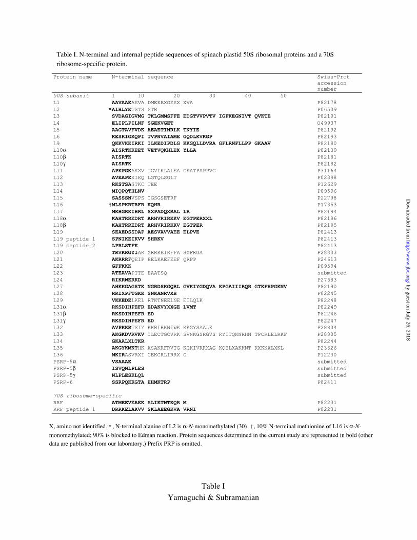
Table I. N-terminal and internal peptide sequences of spinach plastid 50S ribosomal proteins and a 70Sribosome-specific protein.
Protein name N-terminal sequence Swiss-Protaccessionnumber
50S subunit 1 10 20 30 40 50L1 AAVAAEAEVA DMEEEXGESX XVA P82178L2 *AIHLYKTSTS STR P06509L3 SVDAGIGVMG TKLGMMSFFE EDGTVVPVTV IGFKEGNIVT QVKTE P82191L4 ELIPLPILNF SGEKVGET O49937L5 AAGTAVFVDK AEAETINRLK TNYIE P82192L6 KESRIGKQPI TVPNVAIAME GQDLKVKGP P82193L9 QKKVKKIRKI ILKEDIPDLG KKGQLLDVRA GFLRNFLLPP GKAAV P82180L10α AISRTKKEET VETVQKHLEX YLLA P82139L10β AISRTK P82181L10γ AISRTK P82182L11 APKPGKAKXV IGVIKLALEA GKATPAPPVG P31164L12 AVEAPEKIEQ LGTQLSGLT P02398L13 RKSTSASTKC TEE P12629L14 MIQPQTHLNV P09596L15 SASSSNVSPS IGSGSETRF P22798L16 †MLSPKRTRFR KQHR P17353L17 MKHGRKIHRL SXPADQXRAL LR P82194L18α KAHTRREDRT ARHVRIRKKV EGTPERXXL P82196L18β KAHTRREDRT ARHVRIRKKV EGTPER P82195L19 SEAEDSSDAP AESVAVVAEE ELPVE P82413L19 peptide 1 SPNIKEIKVV SHRKV P82413L19 peptide 2 LPRLSTFK P82413L20 TRVKRGYIAR XRRKEIRFFA SXFRGA P28803L21 AKRRRFQEIP EELKAEFEEF QRPP P24613L22 GFFKKK P09594L23 ATEAVAPTTE EAATSQ submittedL24 RIKRWERKD P27683L27 AHKKGAGSTK NGRDSKGQRL GVKIYGDQVA KPGAIIIRQR GTKFHPGKNV P82190L28 RRIXPFTGKK SNKANRVXH P82245L29 VKKEDELKEL RTKTNEELNE EILQLK P82248L31α RKSDIHPEFR EDAKVYXXGE LVMT P82249L31β RKSDIHPEFR ED P82246L31γ RKSDIHPEFR ED P82247L32 AVPKKRTSIY KKRIRKNIWK KKGYSAALK P28804L33 AKGKDVRVKV ILECTGCVRK SVNKGSRGVS RYITQKNRHN TPCRLELRKF P28805L34 GKAALXLTKR P82244L35 AKGYKMKTHK ASAKRFRVTG KGKIVRRXAG KQHLXAKKNT KXKNXLXKL P23326L36 MKIRASVRXI CEKCRLIRRX G P12230PSRP-5α VSAAAE submittedPSRP-5β ISVQNLPLES submittedPSRP-5γ NLPLESKLQL submittedPSRP-6 SSRPQKKGTA HHMKTRP P82411
70S ribosome-specificRRF ATMEEVEAEK SLIETNTKQR M P82231RRF peptide 1 DRRKELAKVV SKLAEEGKVA VRNI P82231
X, amino not identified. *, N-terminal alanine of L2 is α-N-monomethylated (30). †, 10% N-terminal methionine of L16 is α-N-
monomethylated; 90% is blocked to Edman reaction. Protein sequences determined in the current study are represented in bold (other
data are published from our laboratory.) Prefix PRP is omitted.
Table I
Yamaguchi & Subramanian
by guest on July 26, 2018http://w
ww
.jbc.org/D
ownloaded from

Table II. Mass spectrometry of spinach chloroplast 50S ribosomal proteins
LC/MS of TP50 ESI MS of HPLC pool
PRP Sequence Elution Observed Mass Observed Mass SDS-PAGEf
name massa timeb massc differenced masse differenced
(Da) (min) (Da) (Da) (Da) (Da) (Da)
L1 30443.87 33.5 30442.7g -1.2 30447.0 3.1 31.5L2 29661.04g ND ND 30.5L3 28.0 24071.4g 24129.0 25.3L4 27163.50 38.0 27221.8g 58.3 27235.0 71.5 31.0L5 24209.96 37.5 24207.8g -2.2 ND 24.5L6 29.0 20224.9g 20244.0 21.5L9 31.5 17665.8g 17674.8 16.0L10α 35.5 20305.8g ND 21.5L10β ND ND 16.5
L10γ ND ND 16.5L11 16667.62 34.5 16749.1g 81.5 ND 16.5L12 13814.54 41.5 13811.0g -3.5 13813.0 -1.5 17.5L13 23361.83 31.5 23360.2g -1.6 23365.0 3.2 24.5L14 13464.58 37.0 13463.6g -1.0 ND 13.5L15 26.0 22327.9g ND 22.0L16 15327.85 15402.0g 74.1 15467.0 139.2 15.0L17 25.5 12971.5g 13022.4 16.5L18α ND ND 11.5g
L18β ND ND 11.5L19 17566.17 26.5 17564.6g -1.6 17566.0 -0.2 20.0L20 15557.18 ND ND 16.5L21 22762.86 ND 22763.0g 0.1 30.0L22 23114.09g ND 23174.0 59.9 24.5L23 13553.66 ND 13553.5g -0.2 16.0L24 16513.14 26.0 16406.5g -106.6 16425.0 -88.1 16.5L27 23.5 13646.5g 13650.2 16.0L28 25.0 8991.8g 9071.5 13.0L29 27.0 13433.6g 13502.1 15.0L31α ND ND 11.0g
L31β ND ND NAL31γ ND ND NAL32 6503.67 24.5 6503.0g -0.7 6505.0 1.3 11.5L33 7516.81 19.5 7512.0g -4.8 7515.0 -1.8 11.0L34 6767.95 18.0 6767.1g -0.8 6939.3 171.4 NAL35 8433.02 21.0 8432.7g -0.3 8449.9 16.9 12.5L36 4401.35 19.0 4399.0g -2.4 4492.6 91.3 NAPSRP-5 α 9254.97 23.0 9296.0g 41.0 ND NAPSRP-5 β 7065.79 23.0 7065.0 -0.8 ND NAPSRP-5 γ 6638.29 22.5 6636.5 -1.8 ND NAPSRP-6 7387.18g ND ND 12.0a Calculated from translated DNA sequence using ‘peptidesort’ program (31). b Elution time from Fig. 5A. cMassobtained using deconvolution program. d Calculated as observed mass minus sequence mass. e Mass obtained by ESI
MS of HPLC pool containing identified PRP(s) in Fig. 2. f Taken from ref. 2. g Mass values used in Fig. 9. NA, notavailable. ND, not determined.
Table IIYamaguchi & Subramanian
by guest on July 26, 2018http://w
ww
.jbc.org/D
ownloaded from

Table III. Post-translational processing/modification of spinach 50S PRP's and 70S ribosome-specific P-RRF
Plastid encoded (8 proteins)PRP N-terminal sequence Post-translational processing/modification
name1 2 3 4 5 6 7
L2 Met NMA-Ile-His-Leu-Tyr-Lys- 1) fMet removed; 2) α-N-Ala monomethylated
L14 Met-Ile-Gln-Pro-Gln-Thr-His- Formyl group removed
L16 NMM-Leu-Ser-Pro-Lys-Arg-Thr- 1) Formyl group removed; 2) 10% α-N-Met monomethylated; 3) unknown
internal modification
L20 Met Thr-Arg-Val-Lys-Arg-Gly- fMet removed
L22 Met Gly-Phe-Phe-Lys-Lys-Lys- fMet removed
L32 Met Ala-Val-Pro-Lys-Lys-Arg- fMet removed
L33 Met Ala-Lys-Gly-Lys-Asp-Val- fMet removed
L36 Met-Lys-Ile-Arg-Ala-Ser-Val- Formyl group removed
Nuclear encoded (25 50S proteins + P-RRF)Transit peptide cleavage sitePRP
name C-terminal of N-terminal of
Additional post-translational processing/modification during/after import into
chloroplast
transit peptide mature protein -10 -1 1 10
L1 ---SPKSVSFRVL ⇓ AAVAAEAEVA--- No modificationL3 ---XXXXXXXXXX ⇓ SVDAGIGVMG---
L4 ---QTSLSLSIKS ⇓ ELIPLPILNF---
L5 ---XXXXXXXXXX ⇓ AAGTAVFVDK--- No modification
L6 ---XXXXXXXXXX ⇓ KESRIGKQPI---
L9 ---XXXXXXXXXX ⇓ QKKVKKIRKI---
L10 ---XXXXXXXXXX ⇓ AISRTKKEET--- Processed to three forms differing in C-terminal or internal length/chargeL11 ---SHRRLSIVAM ⇓ APKPGKAKKV--- Epsilon-trimethylation of Lys9 and Lys45 (+81.5 Da)
L12 ---TRTTRLRPIA ⇓ AVEAPEKIEQ---
L13 ---TVAFAVAVSA ⇓ RKSTSASTKC--- No modification
L15 ---XXXXXXXXXX ⇓ SASSSNVSPS---
L17 ---XXXXXXXXXX ⇓ MKHGRKIHRL---
L18 ---XXXXXXXXXX ⇓ KAHTRREDRT--- Processed to two forms differing in C-terminal or internal length
L19 ---FPARNSFVVR ⇓ SEAEDSSDAP--- No modification
L21 ---LSRLSLLPVA ⇓ AKRRRFQEIP--- No modification
L23 ---SFGRDLMVAQ ⇓ ATEAVAPTTE--- No modification
L24 ---EGHSPCLIVM ⇓ RIKRWERKDC---
L27 ---XXXXXXXXXX ⇓ AHKKGAGSTK---
L28 ---XXXXXXXXXX ⇓ RPIXPFTGKK---
L29 ---XXXXXXXXXX ⇓ VKKEDELKEL---
L31 ---XXXXXXXXXX ⇓ RKSDIHPEFR--- Processed to three forms differing charge
L34 ---DRCRRFVVRA ⇓ GKAALCLTKR--- No modification
L35 ---TSSPSFTVFA ⇓ AKGYKMKTHK--- No modificationPSRP-5 ---AQKRGTVVAM ⇓ VSAAAEETAG--- Processed to three forms differing in N-terminal length; possible α-N-acetylation
only for alpha form (+41.0 Da)
PSRP-6 ---CGGGVITIEC ⇓ SSRPQKKGTA---
P-RRF ---QNRAGTFRCA ⇓ ATMEEVEAEK---
XX, data not available (cDNA/genomic DNA not sequenced).
Table IIIYamaguchi & Subramanian
by guest on July 26, 2018http://w
ww
.jbc.org/D
ownloaded from

TP50
A
L5e
fg
d
b
a
c
h
i
L2
L9
L3L4
L6
L1
L10γL10β
L10α
L15
L14
L13
L12
L11
L19
L18α
L20
L17L16
L27
L24
L23
L22L21
L31α
L29
L28 L35L32
L33
L34
L18βPSRP-6
PSRP-5α
L31γL31βB
Figure 1Yamaguchi & Subramanian
by guest on July 26, 2018http://w
ww
.jbc.org/D
ownloaded from

L15 L3L17 L12
0 40 80 120 160 200 240
Elution time (min)
A22
0 (
)
0.5
1.0
0
PSRP-5α-γ
L33
L36L34
L27L28L29
L27
L32L35
L24
L3
L15L17
L31α-γ
L21L6
L9
L13
L2L20L18
L22L16
L4
L1L11
L10α-γL23PSRP-6
L5L14
L12
L19
Figure 2Yamaguchi & Subramanian
by guest on July 26, 2018http://w
ww
.jbc.org/D
ownloaded from
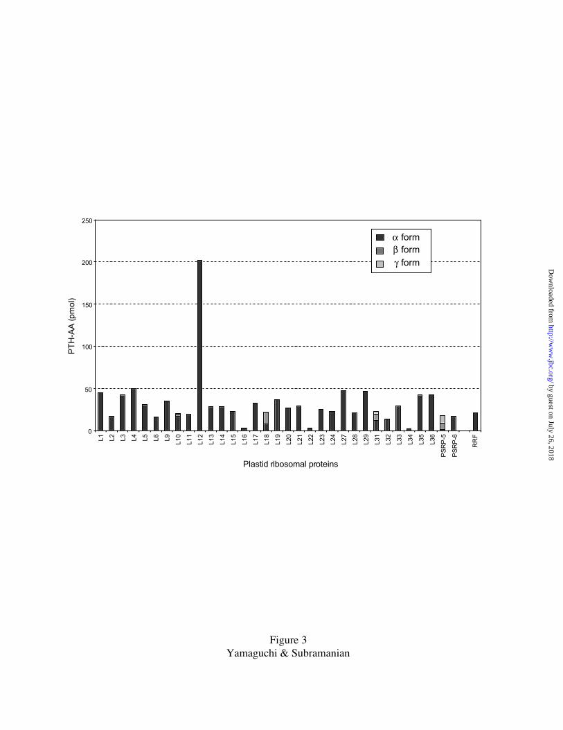
0
50
100
150
200
250
Plastid ribosomal proteins
β form
γ form
α form
PT
H-A
A (p
mol
)
RR
F
PS
RP
-6
PS
RP
-5L1 L2 L3 L4 L5 L6 L9 L10
L11
L12
L13
L14
L15
L16
L17
L18
L19
L20
L21
L22
L23
L24
L27
L28
L29
L31
L32
L33
L34
L35
L36
Figure 3Yamaguchi & Subramanian
by guest on July 26, 2018http://w
ww
.jbc.org/D
ownloaded from

L5Sol ~~~~~~~~~~ ~~~~~~~~~~ ~~~~~~~~~~ ~~~~~~~~~~ ~AAGTAVFVD KAEAETINRL KTNYIEKMVP LLKEEFSYSN ILEVPKVVKI VVNCGIGDASAth MASPSLLQSS ASSFHGRFSP LAAPSSARML SPPLRNVVKV SASGT.VLVE KSEAEKTQRL KTAYLERIIP ALKEEFKYVN IHQVPKVQKI VVNCGIGDAAPpu ~~~~~~~~~~ ~~~~~~~~~~ ~~~~~~~~~~ ~~~~~~~~~~ ~~~~~~~~~~ ~~~~~MVIGL KEKYKTTVTQ ALKEEFQYEN IHEVPRFTKI TINRGLGEASSyn ~~~~~~~~~~ ~~~~~~~~~~ ~~~~~~~~~~ ~~~~~~~~~~ ~~~~~~~~~~ ~~~~~MTQRL KTLYQETILP KLQEEFGYKN IHQVPKLTKV TVNRGLGEASEco ~~~~~~~~~~ ~~~~~~~~~~ ~~~~~~~~~~ ~~~~~~~~~~ ~~~~~~~~~~ ~~~~~~MAKL HDYYKDEVVK KLMTEFNYNS VMQVPRVEKI TLNMGVGEAI
Sol QNAKGLDAAI NELALITGQR PVKTKAKTSI AGFKVREGMT LGIAVTLRGN LMYSFLDRLI NLALPRTRDF QGVNPNSFDG HGNYSVGFRE QSVFPEIKPEAth QNDKGLEAAM KDIALITGQK PIKTRARASI ATFKIREDQP LGIAVTLRGD VMYSFLDRLI NLALPRTRDF QGVSPSSFDG NGNYSIGVKD QGVFPEIRFDPpu QNAKALESSI QELTLITGQK PIVTRAKKSI AGFKIREEVP IGIVVHLRKD KMYSFLEKLI NLTLPRIRDF RGISPKSFDG KGNYNLGLRE QLIFPEIDYDSyn QNAKALESSL TELATITGQK PVVTRARKAI AGFKIREGMP VGVMVTLRSE RMYAFLDRLI NLALPRIRDF RGISPNSFDG RGNYSLGIRE QLIFPEIDYDEco ADKKLLDNAA ADLAAISGQK PLITKARKSV AGFKIRQGYP IGCKVTLRGE RMWEFFERLI TIAVPRIRDF RGLSAKSFDG RGNYSMGVRE QIIFPEIDYD
Sol IVGKARGMDV CITTTAKTDK EAYKLLSLMG MPFREGSGPS T..LVRKKKL KSHHFDAK.K GRRY 220Ath AVGKTRGMDV CISTTAKSDQ EGQKLLALMG MPFREGGGGS TGAIVRKKKL KSHHFDAKGK GKR~ (262) S:81.7% I:72.5%Ppu NIDQIRGLDI SIVTTAKTDQ EGLALLKKLG MPFRES~~~~ ~~~~~~~~~~ ~~~~~~~~~~ ~~~~ (181) S:67.4% I:56.4%Syn TIDQIRGMDV SIITSAQTDE EGRALLKALG MPFRS~~~~~ ~~~~~~~~~~ ~~~~~~~~~~ ~~~~ (180) S:68.9% I:60.0%Eco KVDRVRGLDI TITTTAKSDE EGRALLAAFD FPFRK~~~~~ ~~~~~~~~~~ ~~~~~~~~~~ ~~~~ 178 (179) S:61.5% I:48.6%
L19Sol MASKVLPQAL LVIPSNHSLQ CPPLKKQLGF PIDSNRRFSL SSNCRSNLMV SRASSNLFSS NFSSIFSFPA RNSFVVRSEA EDSSDAPAES VAVVAEEELPAth1 ~~~~~~MQLI RGSTRLLQRH RFSISNTLPF T.SSSCGSWS ANESRKLGFD SFPSTRAATY LISSLKNAGF SSLAVVPKQN F..SSRCFST TGD.SVQGSPAth2 ~~~~~~MQSI RGSTRLLQRH RFSISNTLRF TSSSSCGSWS ENESRKLGFD SCPSSRAATY LISSLKNAGF SSSA.IPKQN FTYASRCFST TGDSSVQGSP
Sol VESEAEAEER PPRQQRVKLG DIMGILNKKA VHAAEELRPV PGIRTGDIVQ IRLEVPE.NK RRLSVYKGIV ISR.QNAGIH TTIRIRRIIA GVGVEIVFPLAth1 ESDCVVPQRI KFKRLDKTAK HIMQIVDKEA VEEVRTLREI PEIKPGYIVQ LKVEVPE.NK RRVSIVKGVV IAR.RNAGLN STFRIRRLVA GVGVESMFPLAth2 ESDCVVPQRI KFKRLDKTAK HIMQIVDKEA VEEVRSLREI PEIKPGYIVQ LKVEVPE.NK RRVSIVKGVV IAR.RNAGLN STFRIRRLVA GVGVESMFPLPpu ~~~~~~~~~~ ~~~~~~MKDN NIKLSLLMKS VEQPLMKTEL PEIKVGDTIR LGLLVKEGSK TREQLCEGVV LSRKKRKSLN TSLTLRCSFQ GVGVERVFFLSyn ~~~~~~~~~~ ~~~~~~~~~M TMNAQAIINS IEAEFLKEDL PTIHVGDTIK VGVKIVEGGK ERIQPYEGTV IA.KRNGGIS ETITVRKIFQ GVGVERVFLLEco ~~~~~~~~~~ ~~~~~~~~~~ ~~~MSNIIKQ LEQEQMKQDV PSFRPGDTVE VKVWVVEGSK KRLQAFEGVV IA.IRNRGLH SAFTVRKISN GEGVERVFQT
Sol YSPNIKEIKV VSHRKVRKAR LYYLRDKLPR LSTFK~~~~~ ~~ 156 (233)Ath1 YSPNLRVIKV VDKKKVRRAK LYYLREKVNA LKKH~~~~~~ ~~ (222) S:62.9% I:50.4%Ath2 YSPNLREIKV VDKKKVRRAK LYYLRDKVNA LKKH~~~~~~ ~~ (225) S:59.4% I:47.7%Ppu NSPRVTFVKV IRRAKVRRAK LYYLRDLLGK ASRLKQIFN~ ~~ (123) S:50.4% I:38.5%Syn HSPRVASIKV LRRGKVRRAK LYYLRDRVGK ATRIKQRFDR AL (122) S:57.3% I:42.7%Eco HSPVVDSISV KRRGAVRKAK LYYLRERTGK AARIKERLN~ ~~ 114 (115) S:52.7% I:37.3%
L34Sol MATLSLLSTG VGAAITNRTP SASLTFITGS RTTNKRVSFN GGSARSGSLH CSFLAPSSSL SSNFSGLSLG LDLTSNTGVS TDRCRRFVVR AGKAALCLTKPpu ~~~~~~~~~~ ~~~~~~~~~~ ~~~~~~~~~~ ~~~~~~~~~~ ~~~~~~~~~~ ~~~~~~~~~~ ~~~~~~~~~~ ~~~~~~~~~~ ~~~~~~~~~~ ~~~~~~~MTKSyn ~~~~~~~~~~ ~~~~~~~~~~ ~~~~~~~~~~ ~~~~~~~~~~ ~~~~~~~~~~ ~~~~~~~~~~ ~~~~~~~~~~ ~~~~~~~~~~ ~~~~~~~~~~ ~~~~~~~MTQEco ~~~~~~~~~~ ~~~~~~~~~~ ~~~~~~~~~~ ~~~~~~~~~~ ~~~~~~~~~~ ~~~~~~~~~~ ~~~~~~~~~~ ~~~~~~~~~~ ~~~~~~~~~~ ~~~~~~~~MK
Sol RS..RSRKSL ARTHGFRLRM STTSGRALLK RRRAKGRKIL CTKTNPSSGK RASP 61 (152)Ppu GTLQGSKRKK IRISGFRARM KTPSGRSILN ERRRKGRKKI MAS~~~~~~~ ~~~~ (46) S:56.8% I:45.5%Syn RTLGGTNRKQ KRTSGFRARM RTHNGRKVIQ ARRSKGRHRL AV~~~~~~~~ ~~~~ (45) S:48.8% I:41.9%Eco RTFQPSVLKR NRSHGFRARM ATKNGRQVLA RRRAKGRARL TVSK~~~~~~ ~~~~ 46 (46) S:50.0% I:50.0%
PSRP-5Sol1 MALLSPLLSL SSVPPITSIA VSSSSFPIKL QN.VS.VAL. LPS...FGQR LVAHGPVIAQ KRGTVVAMVS AAAEETA... .GEDGDQSKV EEANISVQNLSol2 MALLSPLLSL SSVPPITSIA VSSSSFPIKL QN.VS.VAL. LPS...FGQR LVAHGPVIAQ KRGTVVAMVS AAAEETA... .GEDGDQSKV EEANISVQNLPsa MALL..CFNS FTTTPVTS.. .SSSLFPHPT ANPISRVRIG LPTNCLKGFR ILT..PIVQK PRKNSIFIAS AAAGADSNVA DGVEESESKK ESDVVSVDKL
Sol1 PLESKLQLKL EQKIKMKMAK KIRLRRNRLM RKRKLRKRGA WPPSKMKKLK NV α:80 β:58 γ:54 (142)Sol2 PLESKLQLKL EQKIKMKMAK KIRLRRTGLC AKESLRKRGA WPPSKMKKLK NV 80 (142) S:92.5% I:92.5%Psa PLESKLKERE ERMLKMKLAK KIRLKRKRLV QKRRLRKKGN WPPSKMKKLE GV (145) S:65.0% I:53.8%
PSRP-6Sol M.SVSAIFGA RVVTIPSVLR TSSVDGRTVK LQPSTGGSCG GGVITIECSS RPQKKGTAHH MKTRPKKTAR WDIKRGPAVY PPLPPLPAEW TIVSSA.VDEPsa MASVSSIFGC GVSMAPN... .SSLRNKAIR TERRS..ACG G..LLIECSS RPQKKSTAHH MKTRPRKSRL SDRNRKPTVY APLPPLPPDF TIVIPADAST
Sol ADSSSSTTSS SAEIAQSA 69 (116)Psa VDFTPPPPTP SD~~~~~~ (104) S:57.1% I:54.0%
α β γ
Figure 4Yamaguchi & Subramanian
by guest on July 26, 2018http://w
ww
.jbc.org/D
ownloaded from

+17813.4
+15921.7
+131063.4
+111256.6
+91535.6
+101382.0
+81727.3
13811.0
13850.2
A
B
C
Figure 5Yamaguchi & Subramanian
by guest on July 26, 2018http://w
ww
.jbc.org/D
ownloaded from

Cyc. 1
Cyc. 1
SSU 2D/blot
L16 2D/blot
SSU HPLC
Cyc. 1
Ala(L27)
a+NMM deriv.
a+NMM deriv.
NMM
a
b
b
b
Cyc. 2
L16 2D/blot
b
His(L27)
Leu
Cyc. 2
SSU 2D/blot
aNMM deriv.
b Lys
SSU HPLC
Cyc. 2
a
b Lys
aNMM deriv.
Met
Met
Met
5 10 15 20 25 30Elution time (min)
Figure 6Yamaguchi & Subramanian
by guest on July 26, 2018http://w
ww
.jbc.org/D
ownloaded from

S1 S1
L1 L1
RRF
A (30S) B (50S) C (70S)
Figure 7Yamaguchi & Subramanian
by guest on July 26, 2018http://w
ww
.jbc.org/D
ownloaded from

TP70
S3
S2L21
L12
L10α
L10β
L5
L23
L9 PSRP-5
L14
L13
L6S8S9
S13
S18βS15
S12
L11
S21
L16L27L17
L31β
L18αL29
L24
L34
S1α
S1β
PSRP-1
PSRP-2
S6
PSRP-3
S10α/β
S5L1
L4
RRF
L10γ
L19
L3
PSRP-6
L22L15
L2
S7
S17
S16
S20
S19α
S18αS14α S14β
PSRP-4S19β
L20
L28 L32 L35
L33L31αL31γ
L18β
S4
S11
A
B
Figure 8Yamaguchi & Subramanian
by guest on July 26, 2018http://w
ww
.jbc.org/D
ownloaded from

0
5000
10000
15000
20000
25000
30000
35000
L1
L2
L3
L4
L5
L6
L9
L1
0
L1
1
L1
2
L1
3
L1
4
L1
5
L1
6
L1
7
L1
8
L1
9
L2
0
L2
1
L2
2
L2
3
L2
4
L2
5
L2
7
L2
8
L2
9
L3
0
L3
1
L3
2
L3
3
L3
4
L3
5
L3
6
PS
RP
-5
PS
RP
-6
50S ribosomal protein
Ma
ss (
Da
)
Chloroplast
E. coli
Figure 9Yamaguchi & Subramanian
by guest on July 26, 2018http://w
ww
.jbc.org/D
ownloaded from

View from L7/L12 stalk sideView from solvent side
Domain I Domain II
Domain III Domain VI
E. coli Chloroplast
89
10
E. coli Chloroplast
38.2
98
100
101.1
3’3’ 5’4.5S 23S
E. coli Chloroplast
E. coli Chloroplast
59
58.158.358.2
E. coli 23S + 5S rRNA 3-D arrangement
De9
Ad58.2
Ad100
De98
Ad38.2 Ad38.2
Ad100
De98
ExitExit
Cp
St
38.1
38.10
Fig. 10Yamaguchi & Subramanian
90o by guest on July 26, 2018http://w
ww
.jbc.org/D
ownloaded from

Kenichi Yamaguchi and Alap R SubramanianSubunit of an Organelle Ribosome (Chloroplast)
The Plastid Ribosomal Proteins (2): Identification of all the Proteins in the 50S
published online June 28, 2000J. Biol. Chem.
10.1074/jbc.M005012200Access the most updated version of this article at doi:
Alerts:
When a correction for this article is posted•
When this article is cited•
to choose from all of JBC's e-mail alertsClick here
by guest on July 26, 2018http://w
ww
.jbc.org/D
ownloaded from


![SMALL ORGAN4 Is a Ribosome Biogenesis Factor · SMALL ORGAN4 Is a Ribosome Biogenesis Factor Involved in 5.8S Ribosomal RNA Maturation1[OPEN] Rosa Micol-Ponce,a,2,3 Raquel Sarmiento-Mañús,a,2](https://img.pdfslide.us/doc/110x75/60b23bf6ec73ad33ea5d9718/small-organ4-is-a-ribosome-biogenesis-small-organ4-is-a-ribosome-biogenesis-factor.jpg)