Embed Size (px)
Citation preview
The Plasminogen System and Transforming Growth Factor-�in Subjects With Obstructive Sleep Apnea Syndrome:
Effects of CPAP Treatment
Alessia Steffanina MD, Lidia Proietti MD, Caterina Antonaglia MD, Paolo Palange MD,Elena Angelici MD, and Rita Canipari PhD
BACKGROUND: Obstructive sleep apnea syndrome (OSAS) has emerged as a risk factor forcardiovascular disease. A prothrombotic state may affect coagulation and participate in the ath-erosclerotic process in subjects with OSAS. These alterations in coagulation seem to involve theplasminogen activation system. We evaluated the imbalances of the plasminogen activation systemrelated to OSAS, and we assessed the effects of CPAP on the plasminogen activation system.METHODS: Thirty-nine subjects were submitted to a home-based cardiorespiratory sleep study,and 14 healthy subjects (apnea-hypopnea index < 5) were used as controls. Serum levels of uroki-nase-type plasminogen activator (uPA), tissue-type plasminogen activator (tPA), plasminogen ac-tivator inhibitor-1 (PAI-1), and active transforming growth factor-� (TGF-�) were measured.These molecules were reassessed in only 17 of the subjects after 1 month of CPAP. RESULTS:PAI-1 and tPA were significantly higher in the subjects with OSAS compared with the controls,whereas TGF-� and uPA levels were lower. PAI-1 showed a significant positive correlation with theapnea-hypopnea index, percentage of time spent at O2 saturation < 90%, and oxygen desaturationindex, whereas TGF-� was inversely related to all 3 of these parameters. After the CPAP therapy,PAI-1 significantly decreased, whereas TGF-� showed a significant increase, although the valuesdid not reach those of the controls. uPA and tPA did not show significant differences after thetreatment. CONCLUSIONS: Our results suggest an imbalance of fibrinolysis related to OSAS andan improvement of the prothrombotic state after the CPAP treatment. Key words: sleep apnea;CPAP; PAI-1; TGF-�; uPA; tPA. [Respir Care 0;0(0):1–•. © 0 Daedalus Enterprises]
Introduction
Obstructive sleep apnea syndrome (OSAS) is the mostcommon form of sleep-disordered breathing and affectsthousands of people each year. Its frequency in the gen-
eral population is approximately 3–7% of middle-agemen and 2–5% of middle-age women.1 OSAS is char-acterized by the repetitive partial or complete closure ofthe upper airway during sleep that is caused by a col-
Drs Steffanina, Proietti, Palange, and Angelici are affiliated with theLaboratory of Respiratory Pathophysiology, Department of Public Healthand Infectious Diseases, Rome, Italy. Dr Canipari is affiliated with theSection of Histology, Department of Anatomy, Histology, Forensic Med-icine, and Orthopedics, Sapienza University of Rome, Via A Scarpa 16,Rome, Italy. Dr Antonaglia is affiliated with the Department of Pneu-mology and Respiratory Intensive Care Unit, University Hospital of Cat-tinara, Trieste, Italy. At the time of this study, Dr Antonaglia was affil-iated with the Laboratory of Respiratory Pathophysiology, Department ofPublic Health and Infectious Diseases, Rome, Italy. A Steffanina pre-sented a version of this report at the European Respiratory DiseasesAnnual Congress held September 4, 2012, in Vienna, Austria.
This study was supported by La Sapienza University of Rome, AteneoFederato 2009–2010 (to R. C.). The authors have disclosed no conflictsof interest.
The first 2 authors contributed equally to this work.
Correspondence: Rita Canipari PhD, Section of Histology, Department ofAnatomy, Histology, Forensic Medicine, and Orthopedics, Sapienza Uni-versity of Rome, Via A Scarpa 16, 00161 Rome, Italy. E-mail:[email protected].
DOI: 10.4187/respcare.03571
RESPIRATORY CARE • ● ● VOL ● NO ● 1
RESPIRATORY CARE Paper in Press. Published on August 18, 2015 as DOI: 10.4187/respcare.03571
Copyright (C) 2015 Daedalus Enterprises ePub ahead of print papers have been peer-reviewed, accepted for publication, copy edited and proofread. However, this version may differ from the final published version in the online and print editions of RESPIRATORY CARE
lapse of the pharyngeal airway. It is associated withexcessive daytime sleepiness, snoring, recurrent awak-enings and gasping episodes. Obstructive sleep apneamay induce severe intermittent hypoxemia and CO2 re-tention during sleep. Hypoxemia and sleep deprivationappear to be important mechanisms that trigger sys-temic inflammation. Recurrent hypoxemic stress inducesthe release of vasoactive and trophic substances andincreased levels of plasma cytokines, adhesion mole-cules, and C-reactive protein.2,3
Individuals with OSAS have an increased risk for car-diovascular disease and stroke, and OSAS is an indepen-dent risk factor for hypertension.4 Emphasis has been placedon recognizing patients with cardiovascular disease whohave coexisting sleep apnea and understanding the mech-anisms by which sleep apnea may contribute to the pro-gression of their cardiovascular conditions. Moreover, theprevalence of metabolic syndrome in patients with OSASreaches 30–77%.5
OSAS patients have potential markers of thromboticrisk, such as higher plasma levels of fibrinogen6,7 andincreased platelet activation, that decrease after CPAP treat-ment.8 The hypercoagulability resulting from increased co-agulation or impaired fibrinolysis is associated with anincreased risk for atherothrombosis and cardiovascular dis-ease in OSAS.9,10
In recent years, coagulation in OSAS has receivedsome attention, and different studies have focused onthe fibrinolytic activity and the role of the plasminogensystem in subjects with sleep apnea.11-15 There are 2forms of plasminogen activators: tissue-type (tPA) andurokinase-type (uPA). Both are serine proteases thatconvert plasminogen to the active enzyme plasmin. tPAis mostly involved in the dissolution of fibrin in thecirculation, whereas uPA is mainly associated with thepericellular proteolysis during tissue remodeling or tu-mor invasion.16 tPA is currently used in clinical practicefor thrombolysis. tPA and uPA activities are regulatedby specific inhibitors. Plasminogen activator inhibitor-1(PAI-1) is one of the primary regulators of this system,and its principal function in blood is the inhibition offibrinolysis and the stabilization of a thrombus. PAI-1 issynthesized by adipocytes as well as by several othercell types. It has been clearly demonstrated that plasmaPAI-1 levels are increased in obesity and are reduced byweight loss,17,18 but the exact role of PAI-1 in adiposetissue is still controversial. The first connection betweendecreased fibrinolytic capacity, increased plasma PAI-1levels, and cardiovascular disease was reported in youngsurvivors of myocardial infarction.19
Transforming growth factor-� (TGF-�) is secreted bydifferent cell types as an inactive complex and then isactivated by plasmin.20 TGF-� has been shown to be in-volved in cardiovascular disease.21 In fact, it has been
observed that subjects with coronary artery disease presentwith low levels of TGF-� in their serum.22 Among the 3different isoforms (TGF-�1, TGF-�2, and TGF-�3),TGF-�1 is the most prominent in the cardiovascular sys-tem.23 However, its role in OSAS remains largely unex-plored.
There is a link between PAI-1 and TGF-�. In fact, TGF-�induces PAI-1 production, but the increase in PAI-1 mayinhibit TGF-� activation by inhibiting plasminogen acti-vators and, in turn, the activation of plasminogen to plas-min.24
There is limited information about the effect of CPAPtreatment on the plasminogen activation system. Some au-thors have reported higher levels of PAI-1 in subjects withmild to severe OSAS than in controls. However, contrast-ing results can be found in the literature concerningthe relationship between PAI-1 and the severity ofOSAS.11,13,14,25
Our study was designed to assess the presence of ahypercoagulability state in OSAS subjects and in controlsubjects by comparing the levels of uPA, PAI-1, and TGF-�.Additionally, we evaluated the effect of 1 month of CPAPtreatment on plasminogen system activity.
QUICK LOOK
Current knowledge
Obstructive sleep apnea syndrome (OSAS) is the mostcommon form of sleep-disordered breathing. OSAS ischaracterized by the repetitive partial or complete clo-sure of the upper airway during sleep caused by a col-lapse of the pharyngeal airway. Subjects with OSAShave an increased risk for cardiovascular disease andstroke. Subjects with OSAS have markers of throm-botic risk, such as higher plasma fibrinogen and in-creased platelet activation. The hypercoagulability isassociated with an increased risk for atherothrombosisand cardiovascular disease in OSAS.
What this paper contributes to our knowledge
There was a significant decrease of plasminogen acti-vator inhibitor-1 (PAI-1) and an increase of active trans-forming growth factor-� (TGF-�) after CPAP treat-ment, suggesting a decrease in the thrombotic state.CPAP treatment appeared to have beneficial effects oncardiovascular disease in subjects with OSAS. How-ever, the TGF-� values did not reach the values ob-served in the control group, whereas PAI-1 values did,suggesting that longer CPAP treatment may be neededto achieve the desired effect.
EFFECTS OF CPAP TREATMENT IN SUBJECTS WITH OSAS
2 RESPIRATORY CARE • ● ● VOL ● NO ●
RESPIRATORY CARE Paper in Press. Published on August 18, 2015 as DOI: 10.4187/respcare.03571
Copyright (C) 2015 Daedalus Enterprises ePub ahead of print papers have been peer-reviewed, accepted for publication, copy edited and proofread. However, this version may differ from the final published version in the online and print editions of RESPIRATORY CARE
Methods
Subjects
This study included 39 subjects who were referred toour Sleep Disorders Unit between September 2009 andMarch 2010 and between September 2011 and April 2012and 14 healthy subjects from the general population. Ofthe 39 subjects, 17 underwent CPAP therapy, whereas theremaining 22 subjects withdrew from CPAP therapy eitherbecause they did not tolerate the CPAP mask or the devicewas not available. We considered adherence as adequate ifthe mean CPAP use was at least 4 h/night. Table 1 showsthe anthropometric and clinical characteristics, sleep pa-rameters, and smoking habits of controls, subjects withOSAS, and those subjects with OSAS who underwentCPAP therapy, and Table 2 shows the statistical analysisof values presented in Table 1. The experimental protocolwas approved by the Department of Public Health, Sapi-enza University of Rome, according to the Declaration ofHelsinki. Each subject gave informed consent.
Study Protocol
Each subject received a complete general examination,including body mass index (BMI) measurement, biochem-istry, and lipid profile, and was submitted to a cardiore-spiratory sleep study. The exclusion criteria were the di-agnosis of heart failure, diabetes, COPD exacerbation,current infectious and neoplastic diseases, or mental dis-eases.
We performed a full overnight home-based cardiorespi-ratory sleep study using a portable sleep apnea monitoringdevice, SOMNOscreen (SOMNOmedics, Randersacker,Germany). The parameters evaluated were the thoracicand abdominal respiratory movements, respiratory air flowby nasal cannula, body position, snoring, and oxygen sat-uration. Daytime sleepiness was assessed using the Ep-worth Sleepiness Scale. The cardiorespiratory register wasanalyzed according to the American Academy of SleepMedicine 2007 guidelines. In our study, apnea was definedas the cessation of air flow for at least 10 s, and hypopneawas defined as a reduction of at least 30% of the air flowand � 4% of oxygen desaturation or � 50% air flow re-duction with � 3% oxygen desaturation for 10 s or more.From each subject, we manually assessed the number ofapneas and hypopneas per hour of polygraphic recording(apnea-hypopnea index [AHI]), the percentage of the totalpolygraphic recording time passed with an oxygen satura-tion � 90% (time � 90%), and the number of oxygendesaturations of at least 4%/h (oxygen desaturation index[ODI]) as the severity parameters. In the treatment arm,the inclusion criteria were an AHI � 5 in symptomaticsubjects and the consent to CPAP therapy.
The optimal CPAP level for each subject was deter-mined by a titration with the auto-CPAP device iSleep 20(MedicAir, Pogliano Milanese, Milan, Italy). The positiveairway pressure was automatically adjusted, throughout a4-night period, to determine the optimal pressure for main-taining upper airway patency.
A fasting venous blood sample was obtained from eachsubject between 8 and 10 AM after having performed thesleep study. PAI-1, uPA, tPA, and TGF-� were measured.
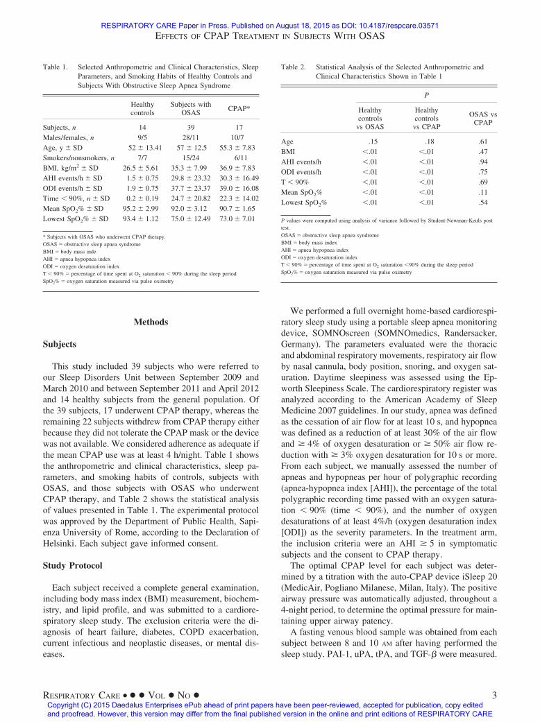
Table 1. Selected Anthropometric and Clinical Characteristics, SleepParameters, and Smoking Habits of Healthy Controls andSubjects With Obstructive Sleep Apnea Syndrome
Healthycontrols
Subjects withOSAS
CPAP*
Subjects, n 14 39 17Males/females, n 9/5 28/11 10/7Age, y � SD 52 � 13.41 57 � 12.5 55.3 � 7.83Smokers/nonsmokers, n 7/7 15/24 6/11BMI, kg/m2 � SD 26.5 � 5.61 35.3 � 7.99 36.9 � 7.83AHI events/h � SD 1.5 � 0.75 29.8 � 23.32 30.3 � 16.49ODI events/h � SD 1.9 � 0.75 37.7 � 23.37 39.0 � 16.08Time � 90%, n � SD 0.2 � 0.19 24.7 � 20.82 22.3 � 14.02Mean SpO2% � SD 95.2 � 2.99 92.0 � 3.12 90.7 � 1.65Lowest SpO2% � SD 93.4 � 1.12 75.0 � 12.49 73.0 � 7.01
* Subjects with OSAS who underwent CPAP therapy.OSAS � obstructive sleep apnea syndromeBMI � body mass indeAHI � apnea hypopnea indexODI � oxygen desaturation indexT � 90% � percentage of time spent at O2 saturation � 90% during the sleep periodSpO2% � oxygen saturation measured via pulse oximetry
Table 2. Statistical Analysis of the Selected Anthropometric andClinical Characteristics Shown in Table 1
P
Healthycontrols
vs OSAS
Healthycontrolsvs CPAP
OSAS vsCPAP
Age .15 .18 .61BMI �.01 �.01 .47AHI events/h �.01 �.01 .94ODI events/h �.01 �.01 .75T � 90% �.01 �.01 .69Mean SpO2% �.01 �.01 .11Lowest SpO2% �.01 �.01 .54
P values were computed using analysis of variance followed by Student-Newman-Keuls posttest.OSAS � obstructive sleep apnea syndromeBMI � body mass indexAHI � apnea hypopnea indexODI � oxygen desaturation indexT � 90% � percentage of time spent at O2 saturation �90% during the sleep periodSpO2% � oxygen saturation measured via pulse oximetry
EFFECTS OF CPAP TREATMENT IN SUBJECTS WITH OSAS
RESPIRATORY CARE • ● ● VOL ● NO ● 3
RESPIRATORY CARE Paper in Press. Published on August 18, 2015 as DOI: 10.4187/respcare.03571
Copyright (C) 2015 Daedalus Enterprises ePub ahead of print papers have been peer-reviewed, accepted for publication, copy edited and proofread. However, this version may differ from the final published version in the online and print editions of RESPIRATORY CARE
The levels of the same markers were measured after 1 monthof CPAP therapy in the subjects who were diagnosed withOSAS.
Laboratory Analysis
To measure uPA, tPA, and PAI-1 antigen levels, venousblood was collected into plastic tubes containing 3.8%sodium citrate (ratio 9:1). The samples were centrifuged at1,500 g for 15 min at �4 °C, and the supernatant wasstored at �80 °C until it was examined. Aliquots of theappropriately diluted samples were assayed by enzyme-linked immunosorbent assay following the instructions ofthe manufacturer (PAI-1/tPA and uPA IMUBIND ELISAkits, American Diagnostica, Pfungstadt, Germany).
Active TGF-� was assayed by adding 100 �l of serumfor 16 h to subconfluent mink lung epithelial cells thatwere stably transfected with a PAI-1 promoter luciferaseconstruct.26 At the end of culture, the cells were washedtwice with phosphate-buffered saline and lysed in 60 �l oflysis buffer (Promega, Madison, Wisconsin). The cell ly-sates were assayed for luciferase activity with an assay kitfrom Promega using a Berthold luminometer. Parallel minklung epithelial cells were incubated with known increasingconcentrations of TGF-� to obtain a standard curve.
Statistics
Statistical analyses were performed using analysis ofvariance followed by the Tukey–Kramer test for com-parisons of multiple groups or paired Student t test forthe comparison of data derived from the 2 groups. Forthe assessment of the correlations, Spearman’s rank-order correlation coefficients (r) and their probability(P) levels were calculated. Values of P � .05 wereconsidered statistically significant. All of the analyseswere performed using SPSS 18.0 for windows (SPSS,Chicago, Illinois).
Results
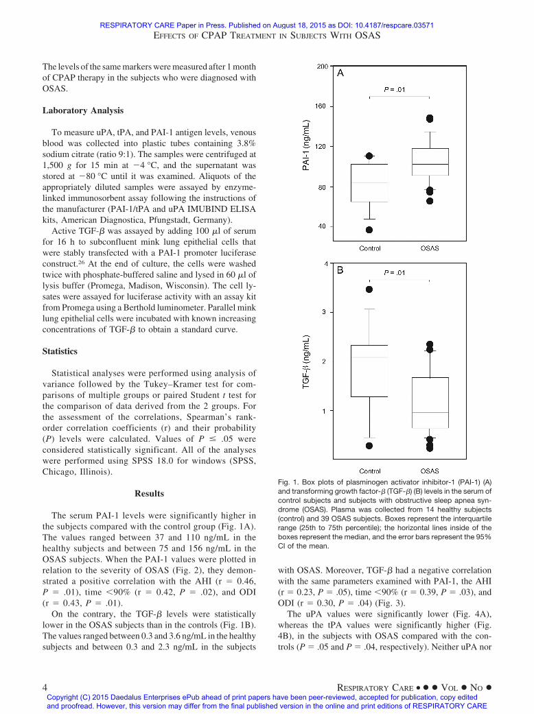
The serum PAI-1 levels were significantly higher inthe subjects compared with the control group (Fig. 1A).The values ranged between 37 and 110 ng/mL in thehealthy subjects and between 75 and 156 ng/mL in theOSAS subjects. When the PAI-1 values were plotted inrelation to the severity of OSAS (Fig. 2), they demon-strated a positive correlation with the AHI (r � 0.46,P � .01), time �90% (r � 0.42, P � .02), and ODI(r � 0.43, P � .01).
On the contrary, the TGF-� levels were statisticallylower in the OSAS subjects than in the controls (Fig. 1B).The values ranged between 0.3 and 3.6 ng/mL in the healthysubjects and between 0.3 and 2.3 ng/mL in the subjects
with OSAS. Moreover, TGF-� had a negative correlationwith the same parameters examined with PAI-1, the AHI(r � 0.23, P � .05), time �90% (r � 0.39, P � .03), andODI (r � 0.30, P � .04) (Fig. 3).
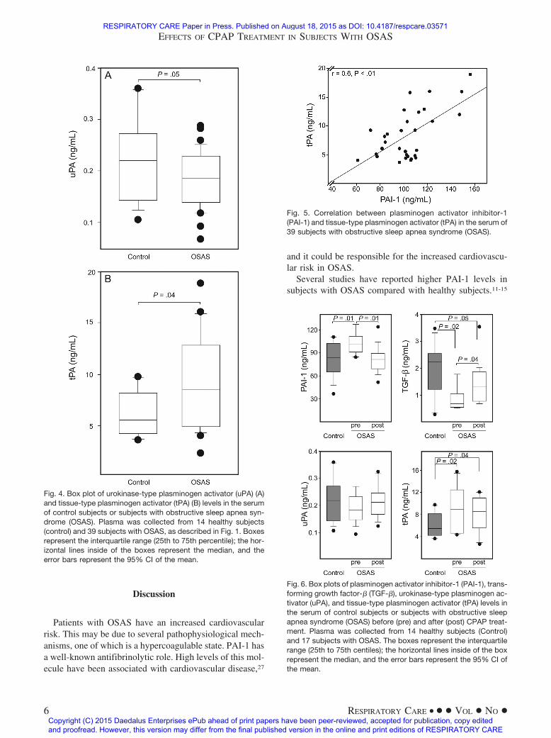
The uPA values were significantly lower (Fig. 4A),whereas the tPA values were significantly higher (Fig.4B), in the subjects with OSAS compared with the con-trols (P � .05 and P � .04, respectively). Neither uPA nor
Fig. 1. Box plots of plasminogen activator inhibitor-1 (PAI-1) (A)and transforming growth factor-� (TGF-�) (B) levels in the serum ofcontrol subjects and subjects with obstructive sleep apnea syn-drome (OSAS). Plasma was collected from 14 healthy subjects(control) and 39 OSAS subjects. Boxes represent the interquartilerange (25th to 75th percentile); the horizontal lines inside of theboxes represent the median, and the error bars represent the 95%CI of the mean.
EFFECTS OF CPAP TREATMENT IN SUBJECTS WITH OSAS
4 RESPIRATORY CARE • ● ● VOL ● NO ●
RESPIRATORY CARE Paper in Press. Published on August 18, 2015 as DOI: 10.4187/respcare.03571
Copyright (C) 2015 Daedalus Enterprises ePub ahead of print papers have been peer-reviewed, accepted for publication, copy edited and proofread. However, this version may differ from the final published version in the online and print editions of RESPIRATORY CARE

tPA showed a correlation with OSAS severity. However,there was a positive correlation between the tPA and PAI-1values (Fig. 5) (r � 0.62, P � .01).
Only 17 of 39 subjects underwent CPAP therapy. How-ever, as shown in Tables 1 and 2, all of the characteristicsof these subjects were not statistically different from thoseof the whole cohort of subjects. One month of CPAPtreatment was effective and led to a significant decrease inthe AHI (2.8 � 2.4, P � .001), time �90% (1.5 � 0.8,P � .001), and ODI (3.6 � 0.6, P � .001); these valueswere not significantly different from the healthy controls(P � .63 and P � .15 versus control, for AHI and time
�90%, respectively). Although the ODI values were sig-nificantly different from the pretreatment values, they didnot reach the values of the healthy subjects (P � .02). Thesubjects’ weight/BMI did not change during the treatmentperiod. After CPAP therapy, we observed a significantdecrease in the PAI-1 levels (�21%, P � .01) and anincrease in active TGF-� (�49%, P � .04) (Fig. 6, A andB). The uPA and tPA levels moderately changed after thetreatment (�7% and �7%, respectively) but did not reachstatistical significance (Fig. 6, C and D). Among the dif-ferent molecules, only PAI-1 levels were not significantlydifferent from the levels of the control subjects after CPAP.
Fig. 2. Correlations between plasminogen activator inhibitor-1(PAI-1) and apnea-hypopnea index (AHI) (A), percentage of timespent at O2 saturation � 90% during the sleep period (Time � 90%)(B), and oxygen desaturation index (ODI) (C) in the serum of sub-jects with obstructive sleep apnea syndrome (OSAS). Bars repre-sent mean � SD of the values obtained in 14 healthy subjects.
Fig. 3. Correlations between transforming growth factor-� (TGF-�)and apnea-hypopnea index (AHI) (A), time � 90% (B), and oxygendesaturation index (C) in the serum of subjects with obstructivesleep apnea syndrome (OSAS). The bar represents the mean � SDof the values obtained in 14 healthy subjects.
EFFECTS OF CPAP TREATMENT IN SUBJECTS WITH OSAS
RESPIRATORY CARE • ● ● VOL ● NO ● 5
RESPIRATORY CARE Paper in Press. Published on August 18, 2015 as DOI: 10.4187/respcare.03571
Copyright (C) 2015 Daedalus Enterprises ePub ahead of print papers have been peer-reviewed, accepted for publication, copy edited and proofread. However, this version may differ from the final published version in the online and print editions of RESPIRATORY CARE
Discussion
Patients with OSAS have an increased cardiovascularrisk. This may be due to several pathophysiological mech-anisms, one of which is a hypercoagulable state. PAI-1 hasa well-known antifibrinolytic role. High levels of this mol-ecule have been associated with cardiovascular disease,27
and it could be responsible for the increased cardiovascu-lar risk in OSAS.
Several studies have reported higher PAI-1 levels insubjects with OSAS compared with healthy subjects.11-15
Fig. 6. Box plots of plasminogen activator inhibitor-1 (PAI-1), trans-forming growth factor-� (TGF-�), urokinase-type plasminogen ac-tivator (uPA), and tissue-type plasminogen activator (tPA) levels inthe serum of control subjects or subjects with obstructive sleepapnea syndrome (OSAS) before (pre) and after (post) CPAP treat-ment. Plasma was collected from 14 healthy subjects (Control)and 17 subjects with OSAS. The boxes represent the interquartilerange (25th to 75th centiles); the horizontal lines inside of the boxrepresent the median, and the error bars represent the 95% CI ofthe mean.
Fig. 4. Box plot of urokinase-type plasminogen activator (uPA) (A)and tissue-type plasminogen activator (tPA) (B) levels in the serumof control subjects or subjects with obstructive sleep apnea syn-drome (OSAS). Plasma was collected from 14 healthy subjects(control) and 39 subjects with OSAS, as described in Fig. 1. Boxesrepresent the interquartile range (25th to 75th percentile); the hor-izontal lines inside of the boxes represent the median, and theerror bars represent the 95% CI of the mean.
Fig. 5. Correlation between plasminogen activator inhibitor-1(PAI-1) and tissue-type plasminogen activator (tPA) in the serum of39 subjects with obstructive sleep apnea syndrome (OSAS).
EFFECTS OF CPAP TREATMENT IN SUBJECTS WITH OSAS
6 RESPIRATORY CARE • ● ● VOL ● NO ●
RESPIRATORY CARE Paper in Press. Published on August 18, 2015 as DOI: 10.4187/respcare.03571
Copyright (C) 2015 Daedalus Enterprises ePub ahead of print papers have been peer-reviewed, accepted for publication, copy edited and proofread. However, this version may differ from the final published version in the online and print editions of RESPIRATORY CARE
However, in the literature, contrasting results can be foundon the relationship between levels of PAI-1 and AHI, theindex of the severity of OSAS. von Kanel et al13 showeda positive correlation, whereas Zamarron et al25 found aninverse correlation.
In our subjects with OSAS, we observed higher PAI-1levels than in the control subjects. Moreover, we haveshown a significant positive correlation between PAI-1and the severity of OSAS, as measured by AHI, time�90%, and ODI.
In the subjects with OSAS, endothelial cells are exposedto intermittent hypoxia/reoxygenation, which leads to theproduction of many vasoactive substances that intervenein the fibrinolysis and coagulation processes in response todifferent hypoxic stimuli. It has been demonstrated that themolecular response to intermittent hypoxia/reoxygenationresults in the activation of nuclear factor kappa B with theconsequent production of inflammatory mediators, such astumor necrosis factor alpha.2 Therefore, the PAI-1 increasecould be due to the activation of inflammatory pathways.In fact, PAI-1 is induced by hypoxia via hypoxia-induciblefactor-1 or -2 in sustained hypoxia as well as via the ac-tivation of nuclear factor kappa B or TGF-� during inter-mittent hypoxia.2
The total proteolytic activity is controlled by modulat-ing the levels of protease inhibitors and/or the local con-centrations of plasminogen activators. Therefore, we alsoinvestigated the presence of the 2 plasminogen activatorstPA and uPA. In our subjects with OSAS, the tPA antigenwas significantly higher than that observed in the controlsubjects, whereas the uPA levels were slightly lower.
It has been shown in epidemiological studies that de-spite being cardioprotective, high plasma tPA concentra-tions positively predict future coronary events. The in-crease of tPA in our study might be a physiological responseto endothelial damage. In fact, it has been demonstratedthat hypoxia can induce the release of tPA from injuredendothelial cells as well as from other cells, such as cere-bral cortical neurons. This mechanism suggests that theelevation of tPA antigen is a result rather than a cause ofatherosclerotic coronary disease. However, the increase ofa factor able to activate the fibrinolytic system, such astPA, being associated with conditions with increased car-diovascular risk, such as OSAS, may appear paradoxical.28
We can hypothesize that higher levels of tPA may repre-sent an attempt to compensate for the hypercoagulablestate in subjects with OSAS. However, the concomitantelevation of PAI-1 and the interaction between plasmino-gen activators and PAI-1 leads to the formation of anenzyme-inhibitor complex, which inhibits plasminogen ac-tivation activity.11 Many authors suggest a protective roleof TGF-� on the cardiovascular system because low levelsof this molecule are related to coronary artery disease witha poor prognosis.22,29,30
TGF-� has been shown to induce PAI-1 synthesis inseveral cell systems. Therefore, an increase of PAI-1 shouldbe correlated with a concomitant increase in TGF-�. In-stead, we found lower levels of active TGF-� and higherlevels of PAI-1 in the subjects with OSAS. This apparentdiscrepancy can be explained by the fact that we measuredactive TGF-�. Although an increase in active TGF-� trig-gers PAI-1 synthesis, the increase in PAI-1 and the con-sequent inhibition of plasminogen activation activity leadto a decrease in plasmin formation and, as a consequence,an impaired TGF-� activation.24,31 In fact, it has beenshown that TGF-� is secreted in a latent form and can beactivated by the uPA/plasmin system.20 In our subjects,TGF-� levels were inversely correlated with AHI, time�90%, and ODI, supporting the hypothesis that the de-crease in TGF-� activity could be involved in the increasedcardiovascular risk related to OSAS.
CPAP is the first-line treatment for OSAS. Evidence inthe literature shows that CPAP therapy reduces cardiovas-cular events,32 most likely through a variety of mecha-nisms. For example, it may reduce inflammatory cytokinelevels, platelet activity, sympathetic nervous system hy-peractivity, and blood pressure.33,34 In agreement with pre-vious studies,15,35 after CPAP treatment, we observed asignificant decrease of PAI-1 levels that was associatedwith a significant increase in active TGF-� levels and amoderate, but not significant, increase in uPA. We areaware of the possible limitations of our results due to therelatively small number of subjects and that the controlsubjects had lower BMI values compared with those of thesubjects with OSAS. However, the fact that after 1 monthof CPAP therapy, we observed a decrease in the levels ofPAI-1 but not relevant changes in BMI suggests that thedecrease of PAI-1 should not be related to BMI.
Another confounding variable in our study is the co-morbidity of OSAS with metabolic syndrome, a conditionoften associated with OSAS.5 In subjects with metabolicsyndrome, an increase in plasma PAI-1 can be observed,and weight loss is associated with a decrease of PAI-1.However, in our subjects, all of the parameters and mol-ecules examined were not significantly different betweenthe 2 groups, with or without metabolic syndrome (datanot shown), probably due to the low number of subjectswith OSAS and metabolic syndrome (13 of 39). Therefore,these results need to be evaluated in a larger number ofsubjects.
The significant decrease of PAI-1 and the increase ofTGF-� that we observed after the CPAP treatment suggesta decrease in the thrombotic state after treatment, whichindicates that CPAP treatment has beneficial effects oncardiovascular disease in subjects with OSAS. However,because in our subjects with OSAS, the TGF-� values didnot reach the values observed in the control group and the
EFFECTS OF CPAP TREATMENT IN SUBJECTS WITH OSAS
RESPIRATORY CARE • ● ● VOL ● NO ● 7
RESPIRATORY CARE Paper in Press. Published on August 18, 2015 as DOI: 10.4187/respcare.03571
Copyright (C) 2015 Daedalus Enterprises ePub ahead of print papers have been peer-reviewed, accepted for publication, copy edited and proofread. However, this version may differ from the final published version in the online and print editions of RESPIRATORY CARE

PAI-1 values did, we can speculate that longer CPAP treat-ment may be needed.
Conclusions
Our study, although performed on a limited number ofsubjects, highlights the importance of focusing on thesemolecules, especially the important link between PAI-1and TGF-�, to better clarify the pathophysiological mech-anisms underlying the hypercoagulability state in patientswith OSAS.
ACKNOWLEDGMENTS
Mink lung epithelial cells were kindly provided by Dr DB Rifkin (NewYork University School of Medicine).
REFERENCES
1. Young T, Palta M, Dempsey J, Skatrud J, Weber S, Badr S. Theoccurrence of sleep-disordered breathing among middle-aged adults.N Engl J Med 1993;328(17):1230-1235.
2. Ryan S, Taylor CT, McNicholas WT. Selective activation of inflam-matory pathways by intermittent hypoxia in obstructive sleep apneasyndrome. Circulation 2005;112(17):2660-2667.
3. Shamsuzzaman AS, Winnicki M, Lanfranchi P, Wolk R, Kara T,Accurso V, Somers VK. Elevated C-reactive protein in patients withobstructive sleep apnea. Circulation 2002;105(21):2462-2464.
4. Lattimore JD, Celermajer DS, Wilcox I. Obstructive sleep apnea andcardiovascular disease. J Am Coll Cardiol 2003;41(9):1429-1437.
5. Lam JC, Lam B, Lam CL, Fong D, Wang JK, Tse HF, et al. Ob-structive sleep apnea and the metabolic syndrome in community-based Chinese adults in Hong Kong. Respir Med 2006;100(6):980-987.
6. Mehra R, Xu F, Babineau DC, Tracy RP, Jenny NS, Patel SR,Redline S. Sleep-disordered breathing and prothrombotic biomark-ers: cross-sectional results of the Cleveland Family Study. Am JRespir Crit Care Med 2010;182(6):826-833.
7. Shamsuzzaman A, Amin RS, Calvin AD, Davison D, Somers VK.Severity of obstructive sleep apnea is associated with elevated plasmafibrinogen in otherwise healthy patients. Sleep Breath. 2014;18(4):761-766.
8. Hui DS, Ko FW, Fok JP, Chan MC, Li TS, Tomlinson B, Cheng G.The effects of nasal continuous positive airway pressure on plateletactivation in obstructive sleep apnea syndrome. Chest 2004;125(5):1768-1775.
9. Drager LF, Polotsky VY, Lorenzi-Filho G. Obstructive sleep apnea:an emerging risk factor for atherosclerosis. Chest 2011;140(2):534-542.
10. von Kanel R, Dimsdale JE. Hemostatic alterations in patients withobstructive sleep apnea and the implications for cardiovascular dis-ease. Chest 2003;124(5):1956-1967.
11. Bagai K, Muldowney JA, 3rd, Song Y, Wang L, Bagai J, Artibee KJ,et al. Circadian variability of fibrinolytic markers and endothelialfunction in patients with obstructive sleep apnea. Sleep 2014;37(2):359-367.
12. Nizankowska-Jedrzejczyk A, Almeida FR, Lowe AA, Kania A,Nastałek P, Mejza F, et al. Modulation of inflammatory and hemo-static markers in obstructive sleep apnea patients treated with man-dibular advancement splints: a parallel, controlled trial. J Clin SleepMed 2014;10(3):255-262.
13. von Kanel R, Loredo JS, Ancoli-Israel S, Mills PJ, Natarajan L,Dimsdale JE. Association between polysomnographic measures ofdisrupted sleep and prothrombotic factors. Chest 2007;131(3):733-739.
14. von Kanel R, Loredo JS, Ancoli-Israel S, Mills PJ, Dimsdale JE.Elevated plasminogen activator inhibitor 1 in sleep apnea and itsrelation to the metabolic syndrome: an investigation in 2 differentstudy samples. Metabolism 2007;56(7):969-976.
15. Zamarron C, Riveiro A, Gude F. Circulating levels of vascular en-dothelial markers in obstructive sleep apnoea syndrome. Effects ofnasal continuous positive airway pressure. Arch Med Sci 2011;7(6):1023-1028.
16. Rijken DC, Lijnen HR. New insights into the molecular mechanismsof the fibrinolytic system. J Thromb Haemost 2009;7(1):4-13.
17. Cesari M, Pahor M, Incalzi RA. Plasminogen activator inhibitor-1(PAI-1): a key factor linking fibrinolysis and age-related subclinicaland clinical conditions. Cardiovasc Ther 2010;28(5):e72-e91.
18. Alessi MC, Juhan-Vague I. PAI-1 and the metabolic syndrome: links,causes, and consequences. Arterioscler Thromb Vasc Biol 2006;26(10):2200-2207.
19. Hamsten A, Wiman B, de Faire U, Blomback M. Increased plasmalevels of a rapid inhibitor of tissue plasminogen activator in youngsurvivors of myocardial infarction. N Engl J Med 1985;313(25):1557-1563.
20. Nunes I, Shapiro RL, Rifkin DB. Characterization of latent TGF-�activation by murine peritoneal macrophages. J Immunol 1995;155(3):1450-1459.
21. Leask A. Potential therapeutic targets for cardiac fibrosis: TGF�,angiotensin, endothelin, CCN2, and PDGF, partners in fibroblastactivation. Circ Res 2010;106(11):1675-1680.
22. Tashiro H, Shimokawa H, Sadamatu K, Yamamoto K. Prognosticsignificance of plasma concentrations of transforming growth fac-tor-� in patients with coronary artery disease. Coron Artery Dis2002;13(3):139-143.
23. Memon AA, Sundquist K, Wang X, Svensson PJ, Sundquist J, ZollerB. Transforming growth factor (TGF)-� levels and unprovoked re-current venous thromboembolism. J Thromb Thrombolysis 2014;38(3):348-354.
24. Odekon LE, Blasi F, Rifkin DB. Requirement for receptor-boundurokinase in plasmin-dependent cellular conversion of latent TGF-�to TGF-�. J Cell Physiol 1994;158(3):398-407.
25. Zamarron C, Ricoy J, Riveiro A, Gude F. Plasminogen activatorinhibitor-1 in obstructive sleep apnea patients with and without hy-pertension. Lung 2008;186(3):151-156.
26. Abe M, Harpel JG, Metz CN, Nunes I, Loskutoff DJ, Rifkin DB. Anassay for transforming growth factor-� using cells transfected with aplasminogen activator inhibitor-1 promoter-luciferase construct. AnalBiochem 1994;216(2):276-284.
27. Nieuwdorp M, Stroes ES, Meijers JC, Buller H. Hypercoagulabilityin the metabolic syndrome. Curr Opin Pharmacol 2005;5(2):155-159.
28. Jansson JH, Olofsson BO, Nilsson TK. Predictive value of tissueplasminogen activator mass concentration on long-term mortality inpatients with coronary artery disease: a 7-year follow-up. Circulation1993;88(5):2030-2034.
29. Grainger DJ. Transforming growth factor � and atherosclerosis: sofar, so good for the protective cytokine hypothesis. ArteriosclerThromb Vasc Biol 2004;24(3):399-404.
30. Cipollone F, Fazia M, Mincione G, Iezzi A, Pini B, Cuccurullo C, etal. Increased expression of transforming growth factor-�1 as a sta-bilizing factor in human atherosclerotic plaques. Stroke 2004;35(10):2253-2257.
31. Sato Y, Tsuboi R, Lyons R, Moses H, Rifkin DB. Characterizationof the activation of latent TGF-� by co-cultures of endothelial cells
EFFECTS OF CPAP TREATMENT IN SUBJECTS WITH OSAS
8 RESPIRATORY CARE • ● ● VOL ● NO ●
RESPIRATORY CARE Paper in Press. Published on August 18, 2015 as DOI: 10.4187/respcare.03571
Copyright (C) 2015 Daedalus Enterprises ePub ahead of print papers have been peer-reviewed, accepted for publication, copy edited and proofread. However, this version may differ from the final published version in the online and print editions of RESPIRATORY CARE

and pericytes or smooth muscle cells: a self-regulating system. J CellBiol 1990;111(2):757-763.
32. Marin JM, Carrizo SJ, Vicente E, Agusti AG. Long-term cardio-vascular outcomes in men with obstructive sleep apnoea-hypop-noea with or without treatment with continuous positive airwaypressure: an observational study. Lancet 2005;365(9464):1046-1053.
33. Martinez-Garcia MA, Capote F, Campos-Rodriguez F, Lloberes P,Díaz de Atauri MJ, Somoza M, et al. Effect of CPAP on bloodpressure in patients with obstructive sleep apnea and resistant hy-
pertension: the HIPARCO randomized clinical trial. JAMA 2013;310(22):2407-2415.
34. Shimizu M, Kamio K, Haida M, Ono Y, Miyachi H, Yamamoto M,et al. Platelet activation in patients with obstructive sleep apneasyndrome and effects of nasal-continuous positive airway pressure.Tokai J Exp Clin Med 2002;27(4):107-112.
35. von Kanel R, Loredo JS, Ancoli-Israel S, Dimsdale JE. Associationbetween sleep apnea severity and blood coagulability: Treatmenteffects of nasal continuous positive airway pressure. Sleep Breath2006;10(3):139-146.
EFFECTS OF CPAP TREATMENT IN SUBJECTS WITH OSAS
RESPIRATORY CARE • ● ● VOL ● NO ● 9
RESPIRATORY CARE Paper in Press. Published on August 18, 2015 as DOI: 10.4187/respcare.03571
Copyright (C) 2015 Daedalus Enterprises ePub ahead of print papers have been peer-reviewed, accepted for publication, copy edited and proofread. However, this version may differ from the final published version in the online and print editions of RESPIRATORY CARE