Embed Size (px)
Citation preview
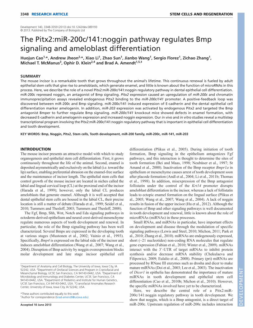
RESEARCH ARTICLE STEM CELLS AND REGENERATION3348
Development 140, 3348-3359 (2013) doi:10.1242/dev.089193© 2013. Published by The Company of Biologists Ltd
INTRODUCTIONThe mouse incisor presents an attractive model with which to studyorganogenesis and epithelial stem cell differentiation. First, it growscontinuously throughout the life of the animal. Second, enamel isdeposited asymmetrically and exclusively on the labial (i.e. toward thelip) surface, enabling preferential abrasion on the enamel-free surfaceand the maintenance of incisor length. The epithelial stem cells thatcontrol growth of the mouse incisor are located in niches called thelabial and lingual cervical loop (CL) at the proximal end of the incisor(Harada et al., 1999); however, only the labial CL producesameloblasts that generate enamel. Although it is well accepted thatdental epithelial stem cells are housed in the labial CL, their preciselocation is still a matter of debate (Harada et al., 1999; Seidel et al.,2010; Tummers and Thesleff, 2003; Tummers and Thesleff, 2008).
The Fgf, Bmp, Shh, Wnt, Notch and Eda signaling pathways inectoderm-derived epithelium and neural crest-derived mesenchymeregulate numerous aspects of tooth development and renewal. Inparticular, the role of the Bmp signaling pathway has been wellcharacterized. Several Bmps are expressed in the developing toothat various stages (Mustonen et al., 2002; Vainio et al., 1993).Specifically, Bmp4 is expressed on the labial side of the incisor andinduces ameloblast differentiation (Wang et al., 2007; Wang et al.,2004). Disruption of Bmp activity by noggin overexpression blocksmolar development and late stage incisor epithelial cell
differentiation (Plikus et al., 2005). During initiation of toothformation, Bmp signaling in the epithelium antagonizes Fgfpathways, and this interaction is thought to determine the sites oftooth formation (Bei and Maas, 1998; Neubüser et al., 1997; StAmand et al., 2000). Inactivation of the Bmp receptor Bmpr1a inepithelium or mesenchyme causes arrest of tooth development soonafter placode formation (Andl et al., 2004; Li et al., 2011b; Thomaset al., 1998). In addition, misexpression of the Bmp antagonistfollistatin under the control of the Krt14 promoter disruptsameloblast differentiation in the incisor, whereas a lack of follistatinleads to ectopic enamel formation on the lingual surface (Plikus etal., 2005; Wang et al., 2007; Wang et al., 2004). A lack of nogginresults in fusion of the upper incisor (Hu et al., 2012). Although thefunction of Bmp and other signaling pathways is well documentedin tooth development and renewal, little is known about the role ofmicroRNAs (miRNAs) in these processes.
Small RNAs, and miRNAs in particular, have important effectson development and disease through the modulation of specificsignaling pathways (Lewis and Steel, 2010; Michon, 2011; Park etal., 2010; Zhang et al., 2010). miRNAs are endogenously expressed,short (~21 nucleotides) non-coding RNA molecules that regulategene expression (Fabian et al., 2010; Winter et al., 2009). miRNAsinteract with the 3�-UTR of target mRNAs to inhibit proteinsynthesis and/or decrease mRNA stability (Chekulaeva andFilipowicz, 2009; Eulalio et al., 2008). Primary (pri) miRNAs areprocessed by RNase III enzymes such as drosha and dicer to makemature miRNAs (Doi et al., 2003; Lee et al., 2003). The inactivationof Dicer1 in epithelia has demonstrated the importance of maturemiRNAs in tooth development and epithelial stem celldifferentiation (Cao et al., 2010b; Michon et al., 2010). However,the specific miRNAs involved have yet to be characterized.
Here, we describe the central role of a Pitx2:miR-200c/141:noggin regulatory pathway in tooth development. Weshow that noggin, which is a Bmp antagonist, is a direct target ofmiR-200c. Upstream regulation of miR-200c includes interaction
1Department of Anatomy and Cell Biology, The University of Iowa, Iowa City, IA52242, USA. 2Department of Orofacial Sciences and Program in Craniofacial andMesenchymal Biology, UCSF, San Francisco, CA 94143-0442, USA. 3Department ofMicrobiology and Immunology and Diabetes Center, UCSF, San Francisco, CA94143-0442, USA. 4Department of Pediatrics and Institute for Human Genetics,UCSF, San Francisco, CA 94143-0442, USA. 5Craniofacial Anomalies ResearchCenter, University of Iowa, Iowa City, IA 52242, USA.
*These authors contributed equally to this work‡Author for correspondence ([email protected])
Accepted 10 June 2013
SUMMARYThe mouse incisor is a remarkable tooth that grows throughout the animal’s lifetime. This continuous renewal is fueled by adultepithelial stem cells that give rise to ameloblasts, which generate enamel, and little is known about the function of microRNAs in thisprocess. Here, we describe the role of a novel Pitx2:miR-200c/141:noggin regulatory pathway in dental epithelial cell differentiation.miR-200c repressed noggin, an antagonist of Bmp signaling. Pitx2 expression caused an upregulation of miR-200c and chromatinimmunoprecipitation assays revealed endogenous Pitx2 binding to the miR-200c/141 promoter. A positive-feedback loop wasdiscovered between miR-200c and Bmp signaling. miR-200c/141 induced expression of E-cadherin and the dental epithelial celldifferentiation marker amelogenin. In addition, miR-203 expression was activated by endogenous Pitx2 and targeted the Bmpantagonist Bmper to further regulate Bmp signaling. miR-200c/141 knockout mice showed defects in enamel formation, withdecreased E-cadherin and amelogenin expression and increased noggin expression. Our in vivo and in vitro studies reveal a multisteptranscriptional program involving the Pitx2:miR-200c/141:noggin regulatory pathway that is important in epithelial cell differentiationand tooth development.
KEY WORDS: Bmp, Noggin, Pitx2, Stem cells, Tooth development, miR-200 family, miR-200c, miR-141, miR-203
The Pitx2:miR-200c/141:noggin pathway regulates Bmpsignaling and ameloblast differentiationHuojun Cao1,*, Andrew Jheon2,*, Xiao Li1, Zhao Sun1, Jianbo Wang1, Sergio Florez1, Zichao Zhang1, Michael T. McManus3, Ophir D. Klein2,4 and Brad A. Amendt1,5,‡
DEVELO
PMENT

3349RESEARCH ARTICLEmiR-200c regulates noggin
of Pitx2 with the shared promoter of miR-200c and miR-141(Mir200c and Mir141 – Mouse Genome Informatics; collectivelymiR-200c/141), and subsequent activation of miR-200c. A secondupstream regulator of miR-200c was identified to be Bmp signaling,thereby indicating a positive-feedback loop between miR-200c andBmp signaling. Furthermore, miR-203 targets the Bmp antagonistBmper and is activated by endogenous Pitx2. Bmper acts similarlyto noggin through the endocytosis of Bmp4 and inhibition of Bmp4signaling (Kelley et al., 2009). We further report that Bmper isexpressed in the dental epithelium during tooth development, addingto the tissue-specific activity of Bmper. Finally, deletion of miR-200c/141 in mice resulted in enamel defects due to downregulationof the cell adhesion protein E-cadherin (E-cad; Cadh1 – MouseGenome Informatics) and of amelogenin (Amel; Amelx – MouseGenome Informatics), an essential protein in enamel formation,confirming our in vitro results in dental epithelial cells.
Sox2 and Tbx1 are dental stem cell markers as they arepredominantly expressed in the CLs of the mouse incisors and/oraffect progenitor cell proliferation (Cao et al., 2010a; Catón et al.,2009; Juuri et al., 2012; Mitsiadis et al., 2008). Other genes also markthe dental stem cells in the labial CL, such as Lgr5, Abcg2, Bmi1,Oct3/4 (Pou5f1 – Mouse Genome Informatics) and Yap1; however,their involvement in stem cell maintenance and proliferation isunknown (Li et al., 2011a; Suomalainen and Thesleff, 2010).Recently, E-cad was shown to regulate dental epithelial stem cellproliferation and migration in the mouse incisor CL (Li et al., 2012).Our in vivo and in vitro studies demonstrate a central role for miR-200c/141 in the regulation of stem cells and ameloblasts during dentalepithelial cell differentiation and tooth development.
MATERIALS AND METHODSAnimalsAnimals were housed in the Program of Animal Resources of the Institute ofBiosciences and Technology at the Texas A&M Health Science Center or inthe animal facilities at UCSF. Experimental procedures were approved by theInstitutional Animal Care and Use Committee (IACUC) and animals werehandled in accordance with the principles and procedure of the Guide for theCare and Use of Laboratory Animals. Procedures for the generation of miR-200c/141 knockout mice were described previously (Park et al., 2012).Briefly, the miR-200c/141 knockout construct was generated by a previouslydescribed ‘knockout-first’ approach (Testa et al., 2004). Two homology armsat the 5� and 3� ends of the targeting vector mediated gene-specific targetingby homologous recombination. Targeting led to insertions of a promoterlesslacZ reporter with an IRES and a poly(A) signal, β-actin-driven neomycinselection marker with a poly(A) signal, and the miR-200c/141 stem-loopflanked by loxP sites into the miR-200c/141 locus. Germline-transmitted mice(lacZ-neo-flox) were crossed with germline deleter Cre (under the control ofthe beta-actin promoter) mice to produce mice with a reporter-tagged nullallele [lacZ knockout allele (lacZ-KO)]. The Pitx2Cre;Dicer1loxP/loxP
(Pitx2Cre/Dicer1) and Krt14-PITX2C transgenic mice have been describedpreviously (Cao et al., 2010b; Venugopalan et al., 2008). We analyzed 12 miR-200c knockout mice and they all showed enamel defects by microcomputedtomography (μCT) or histology. The defect in tooth eruption (third molars)was observed by μCT in all three of the specimens that we scanned.
miRNA microarrays and qPCRmRNA microarray analysis comparing gene expression in the incisor ofpostnatal day (P) 0 control and Pitx2Cre/Dicer1 has been described (Cao etal., 2010b). For miRNA comparison between the labial CL cells and pre-ameloblast and ameloblast cells, these two regions from the P0 mouse lowerincisors were manually dissected under a dissection microscope. Total RNAincluding miRNA from these tissues was prepared using the miRNeasy MiniKit (Qiagen). Lower incisors from P0 control and Krt14-PITX2C mouseincisors were also dissected and total RNA was prepared using themiRNeasy Mini Kit. miRNA microarray analysis was performed by LC
Sciences using µParaflo Microfluidic Biochip version 14, which detectsmiRNA transcripts listed in Sanger miRBase release 14.0. Quantitative real-time PCR (qPCR) analysis of miRNAs was performed using TaqManmicroRNA assay probes (Applied Biosystems), and U6B probe (AppliedBiosystems) was used as a reference for normalization. Microarray data areavailable at GEO under accession number GSE48583.
mRNA microarray and qPCR gene expression analysesCodeLink Mouse Whole Genome chips (Applied Microarrays) were usedfor DNA microarray analyses. Total RNAs were reverse transcribed usingoligo(dT) primers according to the manufacturer’s instructions (iScriptSelect cDNA Synthesis Kit, Bio-Rad). cDNA levels were normalized byPCR amplification with primers to beta-actin (5�-GCCTTCCTTC -TTGGGTATG-3� and 5�-ACCACCAGACAGCACTGTG-3�). Primersequences are listed in supplementary material Table S1.
HistologyEmbryos were fixed with 4% paraformaldehyde in PBS for 4 hours orovernight. Following fixation, samples were dehydrated through a gradedethanol series, embedded in paraffin wax and sectioned (7 μm). StandardHematoxylin and Eosin (H&E) staining was used to examine tissuemorphology. μCT was performed on a MicroXCT-200 (Xradia) through theMicro-CT Imaging Facility at UCSF.
Immunohistochemistry and immunofluorescenceCraniofacial tissue sections (7 μm) were used for immunohistochemistryand immunofluorescence. Antigen retrieval was performed by autoclavingin 0.1 M Tris-HCl buffer (pH 9.0) for 5 minutes. Primary antibodies againstnoggin (Abcam ab16054, 1:300), E-cad (BD Biosciences 70177, 1:500),PITX2 (Capra Sciences PA-1023, 1:500) and Amel (Santa Cruz L0506,1:200) were diluted in TBS (0.5% Tween in phosphate-buffered saline)containing 0.1% Triton X-100, 5% goat serum and 1% BSA, incubatedovernight at 4°C and detected with a biotinylated goat anti-rabbit IgGconjugate (Vector Labs, 1:200) using the avidin-biotin complex (VectorLabs) and AEC Staining Kit (Sigma) or fluorescent secondary antibody(Invitrogen). The cell immunofluorescence assays used cells fixed withparaformaldehyde. Cells were blocked with 10% goat serum and incubatedwith E-cad antibody (BD Biosciences, 1:500) overnight at 4°C. FITC-conjugated secondary antibody was used for detection. DAPI was used forcounterstaining.
DNA cloning, cell culture, transient transfection, luciferase assayand western blottingA 288 bp genomic DNA fragment upstream of miR-200c was cloned intothe pSilencer4.1 vector (Ambion) using primers 5�-AAGAAGGG -GCTTCCAGGTTA-3� and 5�-GGAAGTGTCCCAAATGACG-3�. TheNog 3�-UTR was cloned downstream of the luciferase gene in the pGL3vector (Promega) using primers 5�- GCCCAGACACTTGATGGAT-3� and5�-TCCTGTTCTGCACTTCTTCCT-3�. The PCR-driven overlap extensionmethod was used to mutate the binding site of miR-200c in the 3�-UTR ofNog. A 2 kb DNA fragment including the Pitx2 binding site upstream ofmiR-200c was cloned into the pTK-Luc vector using primers 5�-TCAGTGGATCCTCTTGCTATGCACGTTTTCG-3� and 5�-TGACTG -GATCCGCTTGCTTGCACGATAATCA-3�; this vector construct uses theminimal TK promoter (Amendt et al., 1999). The QuikChange Site-DirectedMutagenesis Kit (Agilent Technologies) was used to mutate the Pitx2binding site from TAATCC to TAATTA 4029 bp upstream of pre-miR-200c.A 1 kb DNA fragment that includes the phospho (p)-Smad1 binding siteupstream of miR-200c was cloned into pTK-Luc using primers 5�-TCATAGGATCCCCTATGGCAGGAGGACACAC-3� and 5�-ATACTG -GATCCAGACAGACCACCGAATGGAC-3�.
LS-8 oral epithelial-like cells, which are derived from neonatal mousemolar tissue (Chen et al., 1992), were cultured and transfected byelectroporation as described (Amen et al., 2007). Cultures were fed for 24hours prior to transfection, resuspended in PBS and mixed with 2 μg miRNA(p-Silencer), PITX2 and Bmpr1a (pcDNA3.1) expression plasmids, 0.1 μgreporter plasmid (pGL3) and 0.5 μg β-galactosidase plasmid. Transfected cellswere incubated for 48 hours in 60 mm culture dishes, lysed and assayed forreporter activities and protein content by the Bradford assay (Bio-Rad). D
EVELO
PMENT
3350
Luciferase was measured using reagents from Promega. β-galactosidase wasmeasured using Galacto-Light Plus reagents (Tropix). All luciferase activitieswere normalized to β-galactosidase activity and are shown as mean-folddifferences relative to empty luciferase plasmids. DNAs were double CsClbanded for purity and cells were transfected by electroporation. To measureendogenous protein, cell lysates (10 μg) were separated on a 12% SDS-polyacrylamide gel. Following SDS-PAGE, the proteins were transferred toPVDF filters (Millipore), immunoblotted using primary antibody againstnoggin (Abcam ab16054, 1:500), PITX2 (Capra Sciences PA-1023, 1:500),Bmper (R&D Systems MAB2299, 1:500), p-Smad1/5/8 (Cell Signaling9511L, 1:500), Smad1 (Cell Signaling D59D7, 1:500) and beta-tubulin (SantaCruz L0811, 1:2000). ECL reagents (Amersham) were used for detection.
Chromatin immunoprecipitation (ChIP) assayChIP assays were performed as previously described using the ChIP AssayKit (Upstate Biotechnology) with the following modifications (Amen et al.,2007; Diamond et al., 2006). Unstimulated LS-8 cells were fed for 24 hours,harvested and plated in 60 mm dishes. Cells were cross-linked with 1%formaldehyde for 10 minutes at 37°C. All PCR reactions were performed atan annealing temperature of 60°C. The following primers were used toamplify the miR-200c promoter region containing the Pitx2 binding site(sense, 5�-AAATGATGGCTGTCCTCGTC-3�; antisense, 5�-AGTGGG -AGAAGCCCAGGTAT-3�) or the p-Smad1 binding site (sense, 5�-CTGGTTTTGGCCTCAGTGAT-3�; antisense, 5�-TACCCAACCAGT-CCACCTTC-3�). All ChIP PCR products were confirmed by size andsequencing. As controls, the primers were used in PCR experiments withoutchromatin; normal rabbit IgG replaced the PITX2 antibody to revealnonspecific immunoprecipitation of chromatin. Three parallel real-timePCRs were also performed in triplicate using these primers to quantify theenrichment of DNA pulled down by the PITX2 antibody as compared with
RESEARCH ARTICLE Development 140 (16)
the DNA pulled down by the IgG control. Primers located 6.7 kb upstreamof pre-miR-200c (sense, 5�-ATGTCTGCTGCTTCCACACA-3�; antisense,5�-CTGCTGTGTCATGCTCCCTA-3�) were used as controls because thisfragment does not contain a Pitx2 binding site or p-Smad1 binding site.
Lentiviral expression constructs and infectionThe pLL3.7 vector (Addgene, 11795) was used to make lentiviral miRNAexpression constructs. miR-200c/141 was amplified using primers 5�-TATACTGTACAAGTAGCTCAGTGATGGGCAAGTCAG-3� and 5�-TTCTGGAATTCTCCCCTTTGCCAAGTGATAC-3�. The insert wasligated into pLL3.7 using BsrGI and EcoRI. Second generation lentivirusplasmids (psPAX2 and pMD2.G, Addgene, 12260 and 12259) were used forlentivirus production. Briefly, 293FT cells were transfected with lentivirusplasmids using Fugene HD (Promega), medium collected after 28 hours,centrifuged and filtered to obtain virus. For lentivirus infection, LS-8 cellswere subcultured and transferred to 6 cm dishes at 20% confluence. Viruswas added immediately after plating and cultured for 2 weeks with mediumchange every 2-3 days.
Statistical analysisTwo-tailed unpaired Student’s t-test was used to determine the differencebetween two sets of values. Error bars indicate mean ± s.e. All experimentswere repeated at least three times.
RESULTSmiR-200c represses noggin in dental epithelial-likecellsInactivation of Dicer1 using Pitx2Cre, Wnt1Cre and Krt14Cre hasdemonstrated that mature miRNAs play central roles in tooth
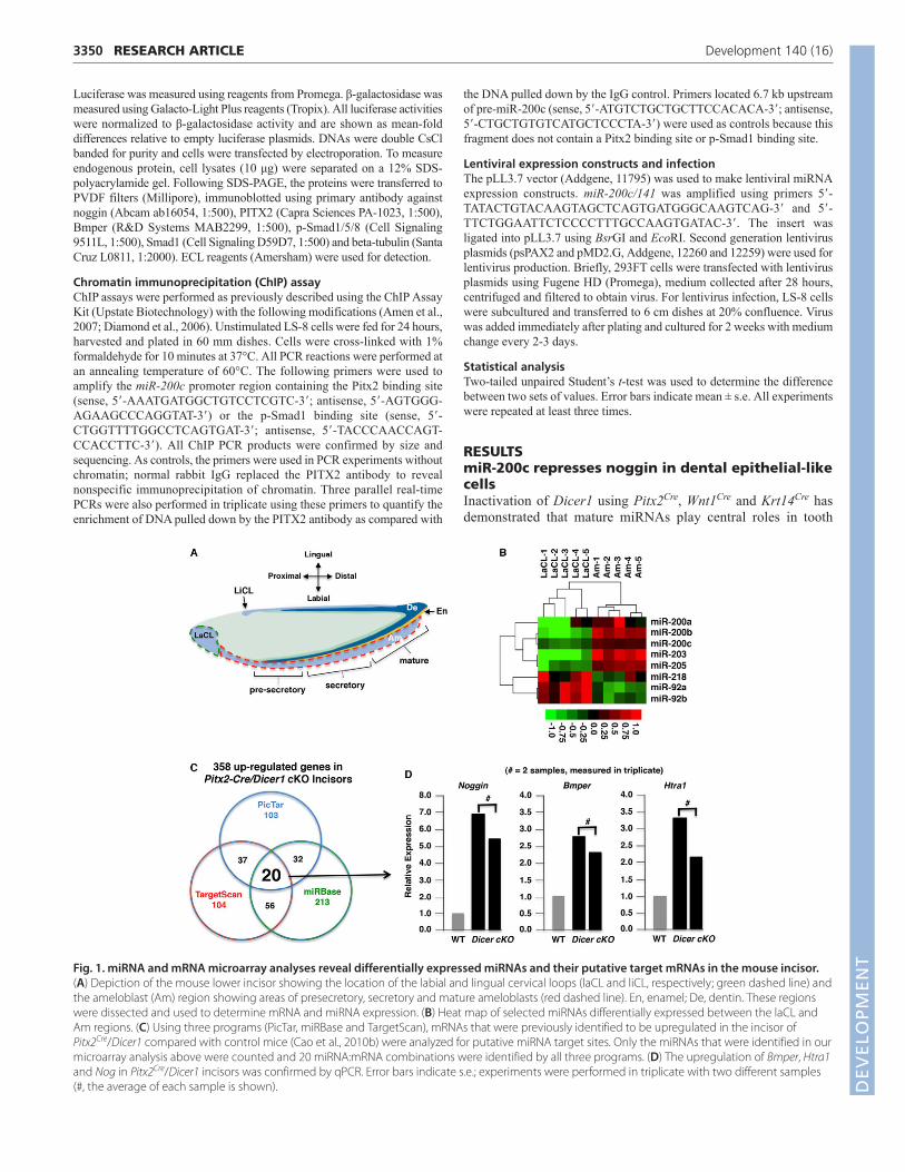
Fig. 1. miRNA and mRNA microarray analyses reveal differentially expressed miRNAs and their putative target mRNAs in the mouse incisor.(A) Depiction of the mouse lower incisor showing the location of the labial and lingual cervical loops (laCL and liCL, respectively; green dashed line) andthe ameloblast (Am) region showing areas of presecretory, secretory and mature ameloblasts (red dashed line). En, enamel; De, dentin. These regionswere dissected and used to determine mRNA and miRNA expression. (B) Heat map of selected miRNAs differentially expressed between the laCL andAm regions. (C) Using three programs (PicTar, miRBase and TargetScan), mRNAs that were previously identified to be upregulated in the incisor ofPitx2Cre/Dicer1 compared with control mice (Cao et al., 2010b) were analyzed for putative miRNA target sites. Only the miRNAs that were identified in ourmicroarray analysis above were counted and 20 miRNA:mRNA combinations were identified by all three programs. (D) The upregulation of Bmper, Htra1and Nog in Pitx2Cre/Dicer1 incisors was confirmed by qPCR. Error bars indicate s.e.; experiments were performed in triplicate with two different samples(#, the average of each sample is shown). D
EVELO
PMENT

development and that these defects are associated with miRNAcontrol of epithelial stem cell proliferation and differentiation (Caoet al., 2010b; Michon et al., 2010). We set out to discover whichspecific miRNAs are important in these processes by comparing themiRNA expression profiles of the labial CL (laCL) and ameloblast(Am) regions (Fig. 1A) in wild-type mouse incisors usingmicroarray analysis. Numerous miRNAs were identified asdifferentially expressed between the labial CL and ameloblastregions. Subsets of these were identified as potential miRNAstargeting Bmp signaling antagonists, as discussed below (Fig. 1B).To identify miRNA target genes important in tooth development,we utilized data generated from a comparison of mRNA expressionin the incisors of control and Pitx2Cre/Dicer1 conditional knockout(cKO) mice (Cao et al., 2010b). These analyses revealed 358transcripts that were upregulated greater than 2-fold inPitx2Cre/Dicer1 cKO incisors. Three separate programs (PicTar,TargetScan, miRBase) were used to predict the presence of miRNAbinding elements in these genes, and 20 miRNA:mRNA interactionswere predicted to be important in the dental epithelium (Fig. 1C).Three of these upregulated genes encoded Bmp signalingantagonists, namely noggin (Nog), Bmper and Htra1, and qPCRconfirmed the upregulation (Fig. 1D). We focused on the miR-200c:Nog interaction for several reasons: Nog expression wasincreased over 6-fold in Pitx2Cre/Dicer1 cKO mice; theoverexpression of Nog has previously been shown to inhibit
3351RESEARCH ARTICLEmiR-200c regulates noggin
ameloblast differentiation (Hu et al., 2012; Plikus et al., 2005); andthe 3�-UTR of Nog was predicted to contain a conserved miR-200cbinding site. Because miR-200c was one of the miRNAs shown tobe differentially expressed in ameloblasts relative to the labial CL(Fig. 1B), we initiated studies of the miR-200c:noggin pathway.
Noggin was upregulated in P2 Pitx2Cre/Dicer1 cKO incisorscompared with those of control mice (Fig. 2A). The wild-typeincisors show low levels of noggin expression. To test whether thepredicted miR-200c element in the 3�-UTR of Nog was functional(Fig. 2B), we ligated this sequence downstream of the luciferasegene in the pGL3 plasmid and co-transfected this construct with amiR-200c expression plasmid into LS-8 dental epithelial-like cells(Fig. 2C). Luciferase activity was approximately halved with co-transfection of miR-200c compared with empty vector, and,importantly, mutation of the predicted miR-200c binding site in Nogabolished this repression (Fig. 2C). Furthermore, transfection ofmiR-200c decreased endogenous Nog expression in LS-8 cells(Fig. 2D).
We also characterized the miR-203:Bmper interaction(supplementary material Fig. S1). Similar to miR-200c, miR-203was differentially expressed in ameloblasts relative to the labial CL(Fig. 1B) and was responsive to PITX2 (supplementary materialFig. S1A). miR-203 targeted Bmper through a conserved sequencein its 3�-UTR, which when mutated resulted in the loss of inhibitionby miR-203 (supplementary material Fig. S1B). The inhibition of
Fig. 2. Characterization of the miR-200c:noggin regulatory pathway. (A) Noggin (green) was present at higher levels in the labial CL (outlined) ofPitx2Cre/Dicer1 (bottom) and miR-200c/141 null (not shown) incisors than in controls (top) at P2. (B) The target sequence of miR-200c in the 3�-UTR ofNog is highly conserved in vertebrates, with 100% conservation of the seed sequence (underlined). (C) Normalized luciferase activity of the 3�-UTR Nog-luciferase reporter (WT Nog 3�UTR) with empty plasmid (Vector) or CMV-miR-200c (miR-200c) shows loss of luciferase activity with expression of miR-200c. There is no loss of luciferase activity when the miR-200c seed sequence is mutated (Mut Nog 3�UTR). Error bars indicate s.e.; five independentexperiments (n=5). (D) Western blot analysis shows a decrease in noggin levels when miR-200c is overexpressed in LS-8 oral epithelial-like cells. Beta-tubulin provided a loading control. D
EVELO
PMENT

3352
Bmper by miR-203 was further substantiated by western blotanalysis of LS-8 cells transfected with a miR-203 expressionplasmid (supplementary material Fig. S1C).
Endogenous Pitx2 binds to the miR-200c 5�flanking chromatin and activates miR-200cexpressionTo understand the transcriptional regulation of miR-200c, wescreened for potential conserved transcription factor binding sites upstream of pre-miR-200c using ECR Browser(http://ecrbrowser.dcode.org). Notably, miR-200c and miR-141 areadjacent to each other and share the same promoter region onchromosome 6. Although miR-141 was not differentially regulatedin this miRNA screen, miR-141 was previously reported to beupregulated in ameloblasts compared with the labial CL, similarlyto miR-200c (Jheon et al., 2011). However, miR-141 was expressedat significantly lower levels than miR-200c when tested by qPCR(data not shown). To determine whether PITX2 activates miR-200ctranscription in vivo, we compared miRNA expression profilesbetween control and Krt14-PITX2 overexpression mouse incisors.This analysis revealed the upregulation of miR-200c in Krt14-
RESEARCH ARTICLE Development 140 (16)
PITX2 incisors (Fig. 3A). A conserved Pitx2 binding site wasidentified ~4 kb upstream of pri-miR-200c (Fig. 3B).
The importance of Pitx2 during craniofacial and tooth developmenthas been well documented (Lu et al., 1999; Venugopalan et al., 2011),and Pitx2 is localized to the dental epithelium (Hjalt et al., 2000). Totest the interaction of Pitx2 with this putative binding site, weperformed chromatin immunoprecipitation (ChIP) assays in LS-8cells, which express Pitx2 (Green et al., 2001). Endogenous Pitx2bound to the miR-200c/141 promoter, whereas anti-IgG antisera didnot immunoprecipitate this chromatin region (Fig. 3C). As a furthercontrol, sequence upstream of the Pitx2 binding site, using either anti-PITX2 or IgG antisera in the ChIP assay, was not amplified (Fig. 3D).ChIP using anti-PITX2 antibody showed an ~8-fold enrichment ofchromatin containing the putative Pitx2 binding site (Fig. 3E).
To confirm that Pitx2 regulates miR-200c expression through thisPitx2 binding site, we ligated a 2 kb gene fragment spanning theidentified Pitx2 binding element into the pTK-Luc vector upstreamof the minimal TK promoter (TK-Luc). Co-transfection of thereporter and PITX2A expression plasmids showed a ~6-fold increasein luciferase activity compared with co-transfection with emptyvector (Fig. 3F). The activation was impaired when the Pitx2
Fig. 3. Pitx2 directly binds to and activates expression of the miR-200c/141 cluster. (A) Heat map showing an increase in miR-200c expression froman miRNA microarray experiment comparing wild-type (control) and Krt14-PITX2 overexpression mouse incisors. (B) A Pitx2 binding site (TAATCC) wasidentified at position −4029 bp of the miR-200c/141 promoter (+1 bp was assigned at the start of pre-miR-200c). Primers (arrows) to amplify the putativePitx2 binding site region (−4193 to –3949), as well as a control region (−6897 to –6743) for ChIP experiments, are indicated. (C) ChIP analysis in LS-8 cellsdemonstrated the interaction of endogenous Pitx2 with the putative Pitx2 binding site in the promoter region of miR-200c/141. (D) Control ChIPexperiments showed no amplification or enrichment of the 154 bp DNA fragment with anti-PITX2 antibody. IP, immunoprecipitate. (E) A 244 bp DNAfragment was amplified and enriched ~8-fold following ChIP using anti-PITX2 antibody but not IgG. (F) Luciferase assays demonstrate activation of miR-200c/141 by PITX2. Luciferase plasmids (Luc) containing the wild-type miR-200c promoter region (miR-200c-Luc) or a mutated Pitx2 binding site (Mut-Luc) were co-transfected with empty vector (Vector) or CMV-PITX2 (PITX2A). Four independent experiments (n=4). (G) Levels of endogenous miR-200cwere measured by TaqMan qPCR in LS-8 cells transfected with CMV-PITX2. Three independent experiments (n=3); **P<0.01. All error bars indicate s.e. D
EVELO
PMENT

binding site was mutated from TAATCC to TAATTA (Mut TK-Luc)(Fig. 3F). Furthermore, transfection of PITX2A increasedendogenous miR-200c expression in LS-8 cells ~6-fold (Fig. 3G).
miR-200c and Bmp signaling form a regulatorypositive-feedback loopTo investigate whether miR-200c regulates Bmp signaling, weconstructed a Bmp reporter plasmid containing two Bmp-responsiveelements (2×BRE) upstream of the minimal TK promoter andluciferase gene. Co-transfection of miR-200c and the BRE-containing plasmid into LS-8 cells resulted in a ~1.6-fold increasein reporter activity compared with empty vector (supplementarymaterial Fig. S2A). Western blot analysis of miR-200c-transfectedcell lysates revealed increased levels of p-Smad1/5/8 protein, amarker of Bmp signaling (supplementary material Fig. S2B).However, pan-Smad1 protein was not affected (supplementarymaterial Fig. S2B). Notably, LS-8 cells express endogenous miR-200c and Pitx2 at low levels (data not shown). These cells arederived from neonatal mouse oral epithelial tissues and expressdental epithelial differentiation factors (Chen et al., 1992).
Because Bmp signaling plays a role in dental epithelial celldifferentiation, we asked whether Bmp signaling regulates miR-200c expression. Analysis of the 5� flanking region of miR-200c/141 identified several potential SMAD binding sites (Fig. 4A).ChIP assays using an anti-p-Smad1/5/8 antibody revealedendogenous p-Smad1/5/8 binding to the most proximal SMAD sitein the miR-200c promoter (Fig. 4B). As an additional control, wecould not immunoprecipitate chromatin upstream of the identifiedSMAD binding sites in the miR-200c promoter using anti-p-Smad1/5/8 or IgG antisera (Fig. 4C). Endogenous p-Smad1/5/8binding to this region was enriched ~30-fold, as assessed by qPCR(Fig. 4D). Furthermore, co-transfection of the constitutively
3353RESEARCH ARTICLEmiR-200c regulates noggin
activated Bmpr1a expression plasmid and miR-200c TK-Lucplasmids in CHO cells resulted in a ~3.5-fold increase in luciferaseactivity compared with empty vector (Fig. 4E). Bmpr1a encodes atype I Bmp receptor that is expressed in the dental and palateepithelium and mesenchyme at early stages of development andmediates phosphorylation of Smad1 (Bonilla-Claudio et al., 2012;He et al., 2010). Importantly, in LS-8 cells transfected with miR-200c, qPCR analysis indicated increased Amel expression, a geneknown to be responsive to Bmp in the dental epithelium and amarker for differentiated dental epithelial cells (Fig. 4F) (Arakaki etal., 2012; Gluhak-Heinrich et al., 2010).
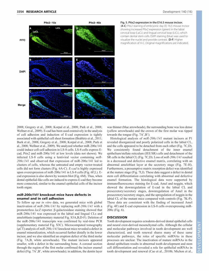
Pitx2 is highly expressed in the incisor cervicalloop stem cell nichePitx2 is highly expressed in the CLs, with less expression inameloblasts (Fig. 5A-F), and miR-200c expression was initiated inthe labial CL and highly expressed in pre-ameloblasts (Fig. 1B).Pitx2 is expressed in differentiated ameloblast cells at low levels(Hjalt et al., 2000; Mucchielli et al., 1997). These data suggest that,as the transient progenitor cells in the stellate reticulum of the CLintercalate into the enamel epithelium, they express miR-200c,which turns on the differentiation program of E-cad expression, celladhesion and ameloblast differentiation. Thus, as the progenitorcells migrate to the distal tip of the growing incisor they begin theirdifferentiation and Pitx2 expression decreases, while Bmp signalingcoordinates the continued expression of miR-200c.
miR-200c expression increases cell adhesion in LS-8 dental epithelial-like cellsPrevious reports on miR-200 family members, and specificallymiR-200c/141, describe their role in the regulation of cell-celladhesion in various cell types (Brabletz et al., 2011; Burk et al.,
Fig. 4. Endogenous p-Smad1/5/8 binds tothe miR-200c promoter and Bmp signalingactivates the miR-200c promoter. (A) Severalputative SMAD binding sites were identified inthe miR-200c/141 promoter region at positions−523, −338 and −130 bp (relative to thetranscription start site of pre-miR-200c/141).Primers (arrows) to amplify the most proximalSMAD binding site region (−8 to –183), as wellas a control region (−6897 to –6743) for ChIPexperiments, are indicated. (B) ChIP analysis inLS-8 cells demonstrated the interaction ofendogenous Smad1/5/8 with the SMADbinding site in the promoter region of miR-200c/141. (C) Control ChIP experimentsshowed no amplification or enrichment of the154 bp DNA fragment with anti-Smad1/5/8antibody. (D) A 175 bp DNA fragment wasamplified and enriched ~30-fold followingChIP using anti-Smad1/5/8 antibody but notIgG. (E) Luciferase reporter assays intransfected CHO cells. The miR-200c/141promoter is activated when co-transfectedwith constitutively active Bmpr1a. (F) qPCRanalysis of amelogenin (Amel) in LS-8 cellstransfected with miR-200c. All error barsindicate s.e.
DEVELO
PMENT
3354
2008; Gregory et al., 2008; Korpal et al., 2008; Park et al., 2008;Wellner et al., 2009). E-cad has been used extensively in the analysisof cell adhesion and induction of E-cad expression is tightlyassociated with epithelial cell sheet formation (Brabletz et al., 2011;Burk et al., 2008; Gregory et al., 2008; Korpal et al., 2008; Park etal., 2008; Wellner et al., 2009). We analyzed whether miR-200c/141could induce cell-cell adhesion in LS-8 cells. LS-8 cells express E-cad, Pitx2 and miR-200c/141 at low levels (data not shown). Weinfected LS-8 cells using a lentiviral vector containing miR-200c/141 and observed that expression of miR-200c/141 led toclusters of cells, whereas the untreated and empty vector-treatedcells did not form clusters (Fig. 6A-C). E-cad is highly expressedupon overexpression of miR-200c/141 in LS-8 cells (Fig. 6F,L). E-cad expression is also shown by western blot (Fig. 6M). Thus, whendental epithelial-like cells are induced to express E-cad they becomemore connected, similar to the enamel epithelial cells of the incisortooth organ.
miR-200c/141 knockout mice have defects inenamel and in cell adhesionTo follow up our in vitro data, we generated mice with globalinactivation of miR-200c/141 by replacing miR-200c/141 with apromoterless lacZ reporter. β-galactosidase staining showed thatmiR-200c/141 was expressed in the labial and lingual CLs andameloblasts (supplementary material Fig. S3A,B,D-F). Deletion ofthe miR-200c/141 transcripts was confirmed by real-time PCR(supplementary material Fig. S3C). Microcomputed tomography(μCT) analysis of miR-200c/141 knockout mice revealed a defect inenamel mineralization, which occurred further distally in the lowerincisor, and we also identified an eruption defect of the third molar(Fig. 7A,B, white arrowheads). The mutant incisors appearedsmaller, with a defect in the surrounding bone. A coronal sectionthrough the region of the first molar confirmed the incisor enameldefect (Fig. 7A�,B�, white arrowheads); in addition, the dentin layer
RESEARCH ARTICLE Development 140 (16)
was thinner (blue arrowheads), the surrounding bone was less dense(yellow arrowheads) and the crown of the first molar was tippedtowards the tongue (Fig. 7A�,B�).
Histological analysis of miR-200c/141 mutant incisors at P1revealed disorganized and poorly polarized cells in the labial CL,and the cells appeared to be detached from each other (Fig. 7C,D).We consistently found detachment of the inner enamelepithelium/stellate reticulum (IEE/SR) cells and detachment of theSR cells in the labial CL (Fig. 7C,D). Loss of miR-200c/141 resultedin a decreased and defective enamel matrix, correlating with anabnormal ameloblast layer at the secretory stage (Fig. 7E-H).Furthermore, a presumptive matrix resorption defect was identifiedat the mature stage (Fig. 7I,J). These data suggest a defect in dentalstem cell differentiation correlating with abnormal and defectiveenamel formation. The histological data were supported byimmunofluorescence staining for E-cad, Amel and noggin, whichshowed the downregulation of E-cad in the labial CL andpresecretory/secretory stages, downregulation of Amel in thepresecretory/secretory stages, and the upregulation of noggin in thelabial CL of the mutant mice compared with controls (Fig. 7K-P).These data are consistent with the finding of increased Amel(Fig. 4F) and E-cad expression in LS-8 cells overexpressing miR-200c (Fig. 6).
DISCUSSIONTooth development requires ectoderm-derived dental epithelial cellsand neural crest-derived mesenchymal cells. Although the cellularand molecular pathways involved in tooth development are wellcharacterized, and tooth renewal shares many of these samemolecular pathways, the roles of specific miRNAs in theseprocesses are unclear. The inactivation of mature miRNAs in thedental epithelium results in abnormal tooth development and stemcell differentiation and revealed a role for epithelial miRNAs intooth development and renewal (Cao et al., 2010b; Michon et al.,
Fig. 5. Pitx2 expression in the E16.5 mouse incisor. (A-C) Pitx2 staining of embryonic day (E) 16.5 mouse incisorshowing increased Pitx2 expression (green) in the labialcervical loop (LaCL) and lingual cervical loop (LiCL), whichcontain dental stem cells. DAPI staining (blue) was used tovisualize the nuclei and provide contrast. (D-F) Highermagnification of A-C. Original magnifications are indicated.
DEVELO
PMENT

2010). However, it is as yet unclear which specific miRNAs andtarget genes are important for these processes. Our in vivo and invitro studies demonstrate that a Pitx2:miR-200c/141:nogginregulatory pathway is essential for tooth development and renewal.
Pitx2, which is the earliest transcriptional marker in toothdevelopment, is highly expressed in the labial CL, with decreasedexpression in ameloblasts, and induces a transcriptional programthat involves miR-200c. Pitx2 activates miR-200c through the Pitx2binding site in the promoter region of miR-200c/141. miR-200crepresses translation of Nog through a conserved sequence in its 3�-UTR, thereby increasing Bmp signaling. Bmp signaling is alsoincreased with repression of Bmper by miR-203. Furthermore, miR-200c overexpression upregulated the expression of E-cad and Amel.Bmps induce high levels of expression of the enamel matrix proteinameloblastin in the dental epithelium (Arakaki et al., 2012; Gluhak-Heinrich et al., 2010); noggin inhibits Bmp signaling and stronglydownregulates endogenous ameloblastin expression (Wang et al.,2004); and Krt14-Nog transgenic mice show repression ofameloblast differentiation and increased epithelial proliferation inboth the labial and lingual CL (Wang et al., 2007). Noggin levelswere increased in Pitx2Cre/Dicer1 cKO mouse incisors, confirmingthe role of miRNAs, and specifically miR-200c, in its regulation.In addition to regulation by Pitx2, miR-200c is also regulated by
3355RESEARCH ARTICLEmiR-200c regulates noggin
Bmp signaling, indicating a positive-feedback loop between miR-200c and Bmp signaling.
Interestingly, Bmp signaling promotes the early stage of somaticcell reprogramming by inducing a mesenchyme-to-epithelialtransition (MET) (Samavarchi-Tehrani et al., 2010). Bmp signalinginduces a program of miRNA expression, which includes the miR-200 family and miR-205, that is associated with MET (Samavarchi-Tehrani et al., 2010). The increase in miR-200 family expression isalso associated with embryonic stem cell (ESC) differentiation and,conversely, Zeb1 is expressed in normal undifferentiated ESCs andcancer cells (Bar et al., 2008; Ben-Porath et al., 2008; Wellner etal., 2009). Increased miR-200c/141 expression results in therepression of noggin, which increases Bmp signaling, and wesuggest that this contributes to the transition of dental stem cells todifferentiated epithelial cells.
Comparison of Pitx2Cre/Dicer1 cKO mouse incisors with those ofthe miR-200c/141 knockout mouse reveals similar defects in dentalepithelial cell differentiation (Cao et al., 2010b). In both mousemodels the labial CL has expanded, presumably owing to increasedprogenitor cell proliferation, with a concomitant decrease inepithelial cell differentiation as demonstrated by the reduction indental epithelial differentiation markers. Interestingly, the incisorphenotype observed in the global knockout of all mature miRNAs
Fig. 6. miR-200c/141 overexpression in LS-8 cells inducesE-cad expression and cell-cell adhesion. (A-C) Morphologyof LS-8 cells that were left untreated (A), transduced withempty vector (B), or transduced with lentiviral miR-200c/141(C). (D-F) Immunofluorescence staining shows increased levelsof E-cad and cell-cell adherence with miR-200c/141 expression(F). (G-I) DAPI staining of LS-8 nuclei. (J-L) Overlay of E-cad andDAPI stains. (M) Western blot of E-cad expression in LS-8 cellstransduced with empty vector or lentiviral miR-200c/141,shown at two concentrations of cell lysate. Gapdh provided aloading control.
DEVELO
PMENT

3356
in the Pitx2Cre/Dicer1 cKO is similar to that of the miR-200c/141knockout, suggesting that the miR-200 family plays a major role inincisor development and renewal. A major difference between thetwo mice is the expansion of the stem cells and production ofmultiple CL regions giving rise to multiple and branched incisors inthe Pitx2Cre/Dicer1 cKO (Cao et al., 2010b).
Previous reports on miR-200 family members, and specificallymiR-200c/141, describe their role in the regulation of cell-celladhesion in various cell types (Brabletz et al., 2011; Burk et al.,2008; Gregory et al., 2008; Korpal et al., 2008; Park et al., 2008;Wellner et al., 2009). Zeb1 and Zeb2 are transcriptional repressorsof E-cad and targeting of these genes by the miR-200 familyincreases E-cad expression in epithelial cells. Interestingly, LS-8dental epithelial cells formed adherent colonies uponoverexpression of miR-200c/141, which correlated with an increasein E-cad levels. The regulation of E-cad by miR-200c is likely to berequired for the adhesion of ameloblasts to the stratum intermedium(SI), which is an integral interaction for tooth development (Jheon
RESEARCH ARTICLE Development 140 (16)
et al., 2011). This is supported by studies in mice harboring aconditional inactivation of E-cad, which showed a compromise inthe ameloblast-SI interface (Li et al., 2012). However, it is alsolikely that miR-200c plays a role in stem cell adhesion in the labialCL because conditional inactivation of E-cad also led to defects inattachment of the SR cells to the outer enamel epithelium (OEE),along with migration defects (Li et al., 2012). The labial CL consistsof the IEE and OEE, as well as SR cells, which although epithelial-derived exhibit a mesenchyme-like morphology and are housedbetween the IEE and OEE (Fig. 8A) (Harada et al., 1999; Tummersand Thesleff, 2003; Tummers and Thesleff, 2008). Notably, LS-8cells were isolated from the oral/enamel epithelium of embryonicmouse teeth (Chen et al., 1992; Xu et al., 2006), and their lack of adental epithelial phenotype in culture, along with the low expressionof Amel and the absence of expression of many ameloblast-specificgenes such as ameloblastin, enamelin and tuftelin (data not shown),suggests a predominantly SR-like cell population. These cells mightnot be fully differentiated as they lack a true dental epithelial
Fig. 7. miR-200c/141 knockout mice exhibit tooth and bone defects. (A,B) μCT analysis of the hemi-mandible from control (wild-type) and miR-200c/141 knockout (KO) mice show that enamel mineralization occurs further distally (white arrowheads) and that eruption of the third molar iscompromised (red arrowhead) in the KO (n=3). (A�,B�) Coronal sections at the region of the first molar (red dotted lines in A,B) show an enamel defect(white arrowheads), a dentin defect (blue arrowheads), a decrease in alveolar bone (yellow arrowheads) and that the angle of the molar is shifted (greenline) with the crown tipped towards the tongue in KO mice. (C-J) H&E staining at various stages of amelogenesis in the lower incisor. Cells in the labialCL (LaCL) appear detached, in part, in the KO mice (D) relative to controls (C); the yellow arrowhead indicates an IEE/SR detachment and the redarrowhead indicates SR detachment. At the secretory stage, a thinner layer of enamel matrix is secreted in KO mice (F) compared with controls (E) andthe single row of ameloblasts is disrupted (H, double asterisks) compared with controls (G). The enamel matrix has detached from the ameloblasts dueto processing (G, asterisk). At the mature stage, there is some retention of enamel matrix (J, arrowhead) in KO mice compared with controls (I). n=7. (K-P) Immunofluorescence staining for E-cad (K-L�), Amel (M,N) and noggin (O,P) in the laCL and presecretory/secretory stages (white dashed lines).Lower levels of E-cad and Amel (white arrowheads) and higher levels of noggin are detected in KO mice compared with controls. Am, ameloblasts; En,enamel; SI, stratum intermedium; SR, stellate reticulum. Scale bars: 1 mm in A-B�; 50 μm in C-J; 20 μm in K-P.
DEVELO
PMENT

signature (low Amel, Pitx2, Tbx1 and E-cad and miR-200 familyexpression). These cells are converted to epithelial sheets byoverexpressing miR-200c, which is highly expressed in epithelialcells such as MDCK cells. Thus, they might behave similarly to theSR cells in transitioning to polarized epithelial cells through theexpression of miR-200c. We are currently working to establish amiRNA-regulated program to generate dental epithelial cells fromother types of cells. Although it is clear that enamel epithelial stemcells are housed in the labial CL, it is as yet unknown whether thesecells reside in the SR, in the OEE, or in both regions (Harada et al.,1999; Ohshima et al., 2005; Parsa et al., 2010; Seidel et al., 2010;Tummers and Thesleff, 2003; Tummers and Thesleff, 2008). Onehypothesis is that miR-200c in the labial CL would increase Bmpand E-cad expression in the putative stem cells of the SR, inducecell adhesion and invasion into the OEE, which in turn differentiatesto the IEE and ultimately ameloblasts (Fig. 8A) (Harada et al., 1999;Tummers and Thesleff, 2003). In miR-200c/141 knockout mice, theexpression of E-cad is reduced, which would lead to delayedmigration of the IEE and ultimately to a delay in enamel formation.However, the function of miR-200c might not be limited to thelabial CL. Although the effects observed at various ameloblaststages with the lack of miR-200c might be an indirect consequenceof initial labial CL defects, it is clear that miR-200c directly affectsdental epithelial cell differentiation. Further experiments will berequired to better understand the functions of miR-200c in the labialCL and ameloblasts.
3357RESEARCH ARTICLEmiR-200c regulates noggin
We have identified a Pitx2:miR-200c:noggin pathway in theregulation of dental stem cell differentiation. Pitx2 induces atranscriptional program involving miR-200c, which directly targetsand inhibits noggin expression (Fig. 8B). Decreased noggin leads toincreased Bmp signaling activity and epithelial cell differentiation.Notably, the increased Bmp activity feeds back to activate miR-200c/141 to maintain the differentiation pathway. The Pitx2:miR-200c:noggin regulatory pathway activates E-cad expression andpromotes adhesion of the SR cells (Fig. 8B).
AcknowledgementsWe thank Drs Elena Semina and Rena D’Souza for reagents and discussions;and Angela Ho, Sabra Djomehri, Diana Gutierrez, Shan Gao and AdriannaCavender for technical assistance. Micro XCT imaging work was performed atthe Division of Biomaterials and Bioengineering Micro-CT Imaging Facility,UCSF.
FundingThe micro-CT imaging facility at UCSF is generously supported by theDepartment of Health and Human Services/NIH S10 Shared InstrumentationGrant S10RR026645; and the Departments of Preventive and RestorativeDental Sciences and Orofacial Sciences, School Of Dentistry, UCSF. The CarverCollege of Medicine and College of Dentistry at the University of Iowaprovided support for this work. This work was supported by the NationalInstitutes of Health [K99-DE022059 to A.H.J., DE021420 to O.D.K., andDE13941 to B.A.A.]. Deposited in PMC for release after 12 months.
Competing interests statementThe authors declare no competing financial interests.
Author contributionsH.C., A.J., X.l., Z.S., J.W., S.F. and Z.Z. performed experiments, analyzed dataand made figures; H.C., A.J., O.D.K. and B.A.A. designed experiments andwrote the manuscript; M.T.M., O.D.K. and B.A.A. provided reagents, mice andtechnical support; H.C., A.J., O.D.K. and B.A.A. designed protocols andanalyzed data.
Supplementary materialSupplementary material available online athttp://dev.biologists.org/lookup/suppl/doi:10.1242/dev.089193/-/DC1
ReferencesAmen, M., Liu, X., Vadlamudi, U., Elizondo, G., Diamond, E., Engelhardt, J. F.
and Amendt, B. A. (2007). PITX2 and β-catenin interactions regulate Lef-1isoform expression. Mol. Cell. Biol. 27, 7560-7573.
Amendt, B. A., Sutherland, L. B. and Russo, A. F. (1999). Multifunctional role ofthe Pitx2 homeodomain protein C-terminal tail. Mol. Cell. Biol. 19, 7001-7010.
Andl, T., Ahn, K., Kairo, A., Chu, E. Y., Wine-Lee, L., Reddy, S. T., Croft, N. J.,Cebra-Thomas, J. A., Metzger, D., Chambon, P. et al. (2004). EpithelialBmpr1a regulates differentiation and proliferation in postnatal hair follicles andis essential for tooth development. Development 131, 2257-2268.
Arakaki, M., Ishikawa, M., Nakamura, T., Iwamoto, T., Yamada, A.,Fukumoto, E., Saito, M., Otsu, K., Harada, H., Yamada, Y. et al. (2012). Roleof epithelial-stem cell interactions during dental cell differentiation. J. Biol.Chem. 287, 10590-10601.
Bar, M., Wyman, S. K., Fritz, B. R., Qi, J., Garg, K. S., Parkin, R. K., Kroh, E. M.,Bendoraite, A., Mitchell, P. S., Nelson, A. M. et al. (2008). MicroRNAdiscovery and profiling in human embryonic stem cells by deep sequencingof small RNA libraries. Stem Cells 26, 2496-2505.
Bei, M. and Maas, R. (1998). FGFs and BMP4 induce both Msx1-independentand Msx1-dependent signaling pathways in early tooth development.Development 125, 4325-4333.
Ben-Porath, I., Thomson, M. W., Carey, V. J., Ge, R., Bell, G. W., Regev, A. andWeinberg, R. A. (2008). An embryonic stem cell-like gene expressionsignature in poorly differentiated aggressive human tumors. Nat. Genet. 40,499-507.
Bonilla-Claudio, M., Wang, J., Bai, Y., Klysik, E., Selever, J. and Martin, J. F.(2012). Bmp signaling regulates a dose-dependent transcriptional program tocontrol facial skeletal development. Development 139, 709-719.
Brabletz, S., Bajdak, K., Meidhof, S., Burk, U., Niedermann, G., Firat, E.,Wellner, U., Dimmler, A., Faller, G., Schubert, J. et al. (2011). The ZEB1/miR-200 feedback loop controls Notch signalling in cancer cells. EMBO J. 30, 770-782.
Burk, U., Schubert, J., Wellner, U., Schmalhofer, O., Vincan, E., Spaderna, S.and Brabletz, T. (2008). A reciprocal repression between ZEB1 and members
Fig. 8. A model for the role of miR-200c/141 in the adhesion of stemcells in the stellate reticulum to the outer enamel epithelium. (A) Depiction of the incisor labial cervical loop and associated cells.Putative epithelial dental stem cells are located in the stellate reticulumand the outer enamel epithelium (indicated in green). (B) Model of thePitx2:miR-200c/141:noggin regulatory pathway in the regulation of toothdevelopment and renewal. Pitx2 directly activates miR-200c/141 and miR-203, which directly inhibit noggin and Bmper, which inhibit Bmpsignaling. Bmp feeds back to activate miR-200c/141 and miR-203expression. miR-200c/141 and miR-203 indirectly regulate amelogenin(Amel) and E-cadherin (E-cad) expression. These factors regulate toothdevelopment and renewal.
DEVELO
PMENT

3358 RESEARCH ARTICLE Development 140 (16)
of the miR-200 family promotes EMT and invasion in cancer cells. EMBO Rep. 9,582-589.
Cao, H., Florez, S., Amen, M., Huynh, T., Skobe, Z., Baldini, A. and Amendt,B. A. (2010a). Tbx1 regulates progenitor cell proliferation in the dentalepithelium by modulating Pitx2 activation of p21. Dev. Biol. 347, 289-300.
Cao, H., Wang, J., Li, X., Florez, S., Huang, Z., Venugopalan, S. R., Elangovan,S., Skobe, Z., Margolis, H. C., Martin, J. F. et al. (2010b). MicroRNAs play acritical role in tooth development. J. Dent. Res. 89, 779-784.
Catón, J., Luder, H.-U., Zoupa, M., Bradman, M., Bluteau, G., Tucker, A. S.,Klein, O. and Mitsiadis, T. A. (2009). Enamel-free teeth: Tbx1 deletion affectsamelogenesis in rodent incisors. Dev. Biol. 328, 493-505.
Chekulaeva, M. and Filipowicz, W. (2009). Mechanisms of miRNA-mediatedpost-transcriptional regulation in animal cells. Curr. Opin. Cell Biol. 21, 452-460.
Chen, L. S., Couwenhoven, R. I., Hsu, D., Luo, W. and Snead, M. L. (1992).Maintenance of amelogenin gene expression by transformed epithelial cells ofmouse enamel organ. Arch. Oral Biol. 37, 771-778.
Diamond, E., Amen, M., Hu, Q., Espinoza, H. M. and Amendt, B. A. (2006).Functional interactions between Dlx2 and lymphoid enhancer factor regulateMsx2. Nucleic Acids Res. 34, 5951-5965.
Doi, N., Zenno, S., Ueda, R., Ohki-Hamazaki, H., Ui-Tei, K. and Saigo, K.(2003). Short-interfering-RNA-mediated gene silencing in mammalian cellsrequires Dicer and eIF2C translation initiation factors. Curr. Biol. 13, 41-46.
Eulalio, A., Huntzinger, E. and Izaurralde, E. (2008). Getting to the root ofmiRNA-mediated gene silencing. Cell 132, 9-14.
Fabian, M. R., Sonenberg, N. and Filipowicz, W. (2010). Regulation of mRNAtranslation and stability by microRNAs. Annu. Rev. Biochem. 79, 351-379.
Gluhak-Heinrich, J., Guo, D., Yang, W., Harris, M. A., Lichtler, A., Kream, B.,Zhang, J., Feng, J. Q., Smith, L. C., Dechow, P. et al. (2010). New roles andmechanism of action of BMP4 in postnatal tooth cytodifferentiation. Bone 46,1533-1545.
Green, P. D., Hjalt, T. A., Kirk, D. E., Sutherland, L. B., Thomas, B. L., Sharpe, P.T., Snead, M. L., Murray, J. C., Russo, A. F. and Amendt, B. A. (2001).Antagonistic regulation of Dlx2 expression by PITX2 and Msx2: implications fortooth development. Gene Expr. 9, 265-281.
Gregory, P. A., Bert, A. G., Paterson, E. L., Barry, S. C., Tsykin, A., Farshid, G.,Vadas, M. A., Khew-Goodall, Y. and Goodall, G. J. (2008). The miR-200family and miR-205 regulate epithelial to mesenchymal transition by targetingZEB1 and SIP1. Nat. Cell Biol. 10, 593-601.
Harada, H., Kettunen, P., Jung, H.-S., Mustonen, T., Wang, Y. A. and Thesleff,I. (1999). Localization of putative stem cells in dental epithelium and theirassociation with Notch and FGF signaling. J. Cell Biol. 147, 105-120.
He, F., Xiong, W., Wang, Y., Matsui, M., Yu, X., Chai, Y., Klingensmith, J. andChen, Y. (2010). Modulation of BMP signaling by Noggin is required for themaintenance of palatal epithelial integrity during palatogenesis. Dev. Biol. 347,109-121.
Hjalt, T. A., Semina, E. V., Amendt, B. A. and Murray, J. C. (2000). The Pitx2protein in mouse development. Dev. Dyn. 218, 195-200.
Hu, X., Wang, Y., He, F., Li, L., Zheng, Y., Zhang, Y. and Chen, Y. P. (2012).Noggin is required for early development of murine upper incisors. J. Dent. Res.91, 394-400.
Jheon, A. H., Li, C.-Y., Wen, T., Michon, F. and Klein, O. D. (2011). Expression ofmicroRNAs in the stem cell niche of the adult mouse incisor. PLoS ONE 6,e24536.
Juuri, E., Saito, K., Ahtiainen, L., Seidel, K., Tummers, M., Hochedlinger, K.,Klein, O. D., Thesleff, I. and Michon, F. (2012). Sox2+ stem cells contribute toall epithelial lineages of the tooth via Sfrp5+ progenitors. Dev. Cell 23, 317-328.
Kelley, R., Ren, R., Pi, X., Wu, Y., Moreno, I., Willis, M., Moser, M., Ross, M.,Podkowa, M., Attisano, L. et al. (2009). A concentration-dependentendocytic trap and sink mechanism converts Bmper from an activator to aninhibitor of Bmp signaling. J. Cell Biol. 184, 597-609.
Korpal, M., Lee, E. S., Hu, G. and Kang, Y. (2008). The miR-200 family inhibitsepithelial-mesenchymal transition and cancer cell migration by directtargeting of E-cadherin transcriptional repressors ZEB1 and ZEB2. J. Biol. Chem.283, 14910-14914.
Lee, Y., Ahn, C., Han, J., Choi, H., Kim, J., Yim, J., Lee, J., Provost, P., Rådmark,O., Kim, S. et al. (2003). The nuclear RNase III Drosha initiates microRNAprocessing. Nature 425, 415-419.
Lewis, M. A. and Steel, K. P. (2010). MicroRNAs in mouse development anddisease. Semin. Cell Dev. Biol. 21, 774-780.
Li, L., Kwon, H. J., Harada, H., Ohshima, H., Cho, S.-W. and Jung, H.-S.(2011a). Expression patterns of ABCG2, Bmi-1, Oct-3/4, and Yap in thedeveloping mouse incisor. Gene Expr. Patterns 11, 163-170.
Li, L., Lin, M., Wang, Y., Cserjesi, P., Chen, Z. and Chen, Y. (2011b). BmprIa isrequired in mesenchymal tissue and has limited redundant function withBmprIb in tooth and palate development. Dev. Biol. 349, 451-461.
Li, C. Y., Cha, W. H., Luder, H. U., Charles, R. P., McMahon, M., Mitsiadis, T. A.and Klein, O. D. (2012). E-cadherin regulates the behavior and fate ofepithelial stem cells and their progeny in the mouse incisor. Dev. Biol. 366, 357-366.
Lu, M. F., Pressman, C., Dyer, R., Johnson, R. L. and Martin, J. F. (1999).Function of Rieger syndrome gene in left-right asymmetry and craniofacialdevelopment. Nature 401, 276-278.
Michon, F. (2011). Tooth evolution and dental defects: from genetic regulationnetwork to micro-RNA fine-tuning. Birth Defects Res. A Clin. Mol. Teratol. 91, 763-769.
Michon, F., Tummers, M., Kyyrönen, M., Frilander, M. J. and Thesleff, I.(2010). Tooth morphogenesis and ameloblast differentiation are regulated bymicro-RNAs. Dev. Biol. 340, 355-368.
Mitsiadis, T. A., Tucker, A. S., De Bari, C., Cobourne, M. T. and Rice, D. P. C.(2008). A regulatory relationship between Tbx1 and FGF signaling duringtooth morphogenesis and ameloblast lineage determination. Dev. Biol. 320,39-48.
Mucchielli, M.-L., Mitsiadis, T. A., Raffo, S., Brunet, J.-F., Proust, J.-P. andGoridis, C. (1997). Mouse Otlx2/RIEG expression in the odontogenicepithelium precedes tooth initiation and requires mesenchyme-derivedsignals for its maintenance. Dev. Biol. 189, 275-284.
Mustonen, T., Tümmers, M., Mikami, T., Itoh, N., Zhang, N., Gridley, T. andThesleff, I. (2002). Lunatic fringe, FGF, and BMP regulate the Notch pathwayduring epithelial morphogenesis of teeth. Dev. Biol. 248, 281-293.
Neubüser, A., Peters, H., Balling, R. and Martin, G. R. (1997). Antagonisticinteractions between FGF and BMP signaling pathways: a mechanism forpositioning the sites of tooth formation. Cell 90, 247-255.
Ohshima, H., Nakasone, N., Hashimoto, E., Sakai, H., Nakakura-Ohshima, K.and Harada, H. (2005). The eternal tooth germ is formed at the apical end ofcontinuously growing teeth. Arch. Oral Biol. 50, 153-157.
Park, S.-M., Gaur, A. B., Lengyel, E. and Peter, M. E. (2008). The miR-200 familydetermines the epithelial phenotype of cancer cells by targeting the E-cadherin repressors ZEB1 and ZEB2. Genes Dev. 22, 894-907.
Park, C. Y., Choi, Y. S. and McManus, M. T. (2010). Analysis of microRNAknockouts in mice. Hum. Mol. Genet. 19 R2, R169-R175.
Park, C. Y., Jeker, L. T., Carver-Moore, K., Oh, A., Liu, H. J., Cameron, R.,Richards, H., Li, Z., Adler, D., Yoshinaga, Y. et al. (2012). A resource for theconditional ablation of microRNAs in the mouse. Cell Reports 1, 385-391.
Parsa, S., Kuremoto, K., Seidel, K., Tabatabai, R., Mackenzie, B., Yamaza, T.,Akiyama, K., Branch, J., Koh, C. J., Al Alam, D. et al. (2010). Signaling byFGFR2b controls the regenerative capacity of adult mouse incisors.Development 137, 3743-3752.
Plikus, M. V., Zeichner-David, M., Mayer, J.-A., Reyna, J., Bringas, P.,Thewissen, J. G. M., Snead, M. L., Chai, Y. and Chuong, C.-M. (2005).Morphoregulation of teeth: modulating the number, size, shape anddifferentiation by tuning Bmp activity. Evol. Dev. 7, 440-457.
Samavarchi-Tehrani, P., Golipour, A., David, L., Sung, H.-K., Beyer, T. A.,Datti, A., Woltjen, K., Nagy, A. and Wrana, J. L. (2010). Functional genomicsreveals a BMP-driven mesenchymal-to-epithelial transition in the initiation ofsomatic cell reprogramming. Cell Stem Cell 7, 64-77.
Seidel, K., Ahn, C. P., Lyons, D., Nee, A., Ting, K., Brownell, I., Cao, T., Carano,R. A., Curran, T., Schober, M. et al. (2010). Hedgehog signaling regulates thegeneration of ameloblast progenitors in the continuously growing mouseincisor. Development 137, 3753-3761.
St Amand, T. R., Zhang, Y., Semina, E. V., Zhao, X., Hu, Y., Nguyen, L., Murray,J. C. and Chen, Y. (2000). Antagonistic signals between BMP4 and FGF8define the expression of Pitx1 and Pitx2 in mouse tooth-forming anlage. Dev.Biol. 217, 323-332.
Suomalainen, M. and Thesleff, I. (2010). Patterns of Wnt pathway activity in themouse incisor indicate absence of Wnt/β-catenin signaling in the epithelialstem cells. Dev. Dyn. 239, 364-372.
Testa, G., Schaft, J., van der Hoeven, F., Glaser, S., Anastassiadis, K., Zhang,Y., Hermann, T., Stremmel, W. and Stewart, A. F. (2004). A reliable lacZexpression reporter cassette for multipurpose, knockout-first alleles. Genesis38, 151-158.
Thomas, B. L., Tucker, A. S., Ferguson, C., Qiu, M., Rubenstein, J. L. R. andSharpe, P. T. (1998). Molecular control of odontogenic patterning: positionaldependent initiation and morphogenesis. Eur. J. Oral Sci. 106 Suppl. 1, 44-47.
Tummers, M. and Thesleff, I. (2003). Root or crown: a developmental choiceorchestrated by the differential regulation of the epithelial stem cell niche inthe tooth of two rodent species. Development 130, 1049-1057.
Tummers, M. and Thesleff, I. (2008). Observations on continuously growingroots of the sloth and the K14-Eda transgenic mice indicate that epithelialstem cells can give rise to both the ameloblast and root epithelium celllineage creating distinct tooth patterns. Evol. Dev. 10, 187-195.
Vainio, S., Karavanova, I., Jowett, A. and Thesleff, I. (1993). Identification ofBMP-4 as a signal mediating secondary induction between epithelial andmesenchymal tissues during early tooth development. Cell 75, 45-58.
Venugopalan, S. R., Amen, M. A., Wang, J., Wong, L., Cavender, A. C.,D’Souza, R. N., Akerlund, M., Brody, S. L., Hjalt, T. A. and Amendt, B. A.(2008). Novel expression and transcriptional regulation of FoxJ1 during oro-facial morphogenesis. Hum. Mol. Genet. 17, 3643-3654. D
EVELO
PMENT

3359RESEARCH ARTICLEmiR-200c regulates noggin
Venugopalan, S. R., Li, X., Amen, M. A., Florez, S., Gutierrez, D., Cao, H.,Wang, J. and Amendt, B. A. (2011). Hierarchical interactions ofhomeodomain and forkhead transcription factors in regulating odontogenicgene expression. J. Biol. Chem. 286, 21372-21383.
Wang, X.-P., Suomalainen, M., Jorgez, C. J., Matzuk, M. M., Werner, S. andThesleff, I. (2004). Follistatin regulates enamel patterning in mouse incisors byasymmetrically inhibiting BMP signaling and ameloblast differentiation. Dev.Cell 7, 719-730.
Wang, X.-P., Suomalainen, M., Felszeghy, S., Zelarayan, L. C., Alonso, M. T.,Plikus, M. V., Maas, R. L., Chuong, C.-M., Schimmang, T. and Thesleff, I.(2007). An integrated gene regulatory network controls stem cell proliferationin teeth. PLoS Biol. 5, e159.
Wellner, U., Schubert, J., Burk, U. C., Schmalhofer, O., Zhu, F., Sonntag, A.,Waldvogel, B., Vannier, C., Darling, D., zur Hausen, A. et al. (2009). The
EMT-activator ZEB1 promotes tumorigenicity by repressing stemness-inhibiting microRNAs. Nat. Cell Biol. 11, 1487-1495.
Winter, J., Jung, S., Keller, S., Gregory, R. I. and Diederichs, S. (2009). Manyroads to maturity: microRNA biogenesis pathways and their regulation. Nat.Cell Biol. 11, 228-234.
Xu, Y., Zhou, Y. L., Luo, W., Zhu, Q. S., Levy, D., MacDougald, O. A. andSnead, M. L. (2006). NF-Y and CCAAT/enhancer-binding protein alphasynergistically activate the mouse amelogenin gene. J. Biol. Chem. 281, 16090-16098.
Zhang, Z., Florez, S., Gutierrez-Hartmann, A., Martin, J. F. and Amendt, B. A.(2010). MicroRNAs regulate pituitary development, and microRNA 26bspecifically targets lymphoid enhancer factor 1 (Lef-1), which modulatespituitary transcription factor 1 (Pit-1) expression. J. Biol. Chem. 285, 34718-34728.
DEVELO
PMENT