Embed Size (px)
Citation preview

The pig as an animal model for Cystic Fibrosis –
approaches to overcome the lethal intestinal phenotype
von Michaela Désirée Dmochewitz

Inaugural-Dissertation zur Erlangung der Doktorwürde
der Tierärztlichen Fakultät der Ludwig-Maximilians-Universität München
The pig as an animal model for Cystic Fibrosis –
approaches to overcome the lethal intestinal phenotype
von Michaela Désirée Dmochewitz
aus Ulm
München 2016

Aus dem Veterinärwissenschaftlichen Department der Tierärztlichen Fakultät
der Ludwig-Maximilians-Universität München
Lehrstuhl für Molekulare Tierzucht und Biotechnologie
Arbeit angefertigt unter der Leitung von Univ.-Prof. Dr. Eckhard Wolf
Mitbetreuung durch Dr. Nikolai Klymiuk und Dr. Andrea Bähr
Gedruckt mit Genehmigung der Tierärztlichen Fakultät
der Ludwig-Maximilians-Universität München
Dekan: Univ.-Prof. Dr. Joachim Braun
Berichterstatter: Univ.-Prof. Dr. Eckhard Wolf
Korreferenten: Univ.-Prof. Dr. Mathias Ritzmann
Univ.-Prof. Dr. Katrin Hartmann
Priv.-Doz. Dr. Birgit Viertlböck
Univ.-Prof. Dr. Rüdiger Wanke
Tag der Promotion: 16.07.2016

Für Miky
Just because something doesn’t do what you planned it to do
doesn’t mean it’s useless.
Thomas A. Edison

During the preparation of this thesis, the following review was published:
Dmochewitz, M. and E. Wolf (2015). "Genetic engineering of pigs for the creation of
translational models of human pathologies." Animal Frontiers 5(1).
Parts of this study were presented at the following conference:
Opening Conference COST Action BM1308 “Sharing Advances on Large Animal
Models (SALAAM)”, Gene Center, LMU Munich, December 15-16, 2014.

Table of contents I
TABLE OF CONTENTS
I. INTRODUCTION ...................................................................................... 1
II. REVIEW OF THE LITERATURE .......................................................... 3
1. Cystic Fibrosis ............................................................................................3
1.1. Genetics of Cystic Fibrosis ..........................................................................4
1.2. Pathophysiology of Cystic Fibrosis .............................................................5
1.3. Phenotypical abnormalities in Cystic Fibrosis .............................................6
1.4. Diagnosis of Cystic Fibrosis ........................................................................7
1.5. Treatment strategies for Cystic Fibrosis.......................................................8
2. Animal models of Cystic Fibrosis ...........................................................10
2.1. Cystic Fibrosis mice models ......................................................................10
2.2. Cystic Fibrosis rat model............................................................................11
2.3. Cystic Fibrosis ferret model .......................................................................12
2.4. Cystic Fibrosis zebrafish model .................................................................13
2.5. Cystic fibrosis pig models ..........................................................................13
2.5.1. The value and uniqueness of the porcine CF model regarding the
respiratory phenotype .................................................................................15
2.5.2. The lethal intestinal phenotype impairs the utility of the CF pig model –
approaches to overcome this limitation ......................................................17
III. ANIMALS, MATERIALS AND METHODS ........................................ 22
1. Animals ......................................................................................................22
2. Materials ...................................................................................................22
2.1. Chemicals ...................................................................................................22
2.2. Consumables ..............................................................................................24

Table of contents II
2.3. Devices .......................................................................................................25
2.4. Antibodies, drugs, enzymes and oligonucleotides .....................................27
2.5. Buffers and solutions ..................................................................................30
2.6. Kits .............................................................................................................36
2.7. Other reagents ............................................................................................36
2.8. Software .....................................................................................................37
3. Methods .....................................................................................................37
3.1. Production of CFTR-/-, CFTR+/- and WT piglets by breeding ....................37
3.2. Generation of gut-modified CFTR-/- founder animals ................................38
3.3. Management of neonatal gut-modified CFTR-/- piglets .............................39
3.4. Necropsy of CF piglets ...............................................................................40
3.5. Preparation of the airways explanted from WT and CFTR-/- piglets for
experiments on mucociliary transport ........................................................42
3.6. Analysis at the genomic level ....................................................................44
3.6.1. Genotyping of piglets .................................................................................44
3.6.2. Sequencing of PCR products......................................................................47
3.7. Expression analysis at the RNA level ........................................................49
3.7.1. RNA isolation .............................................................................................49
3.7.2. cDNA synthesis ..........................................................................................51
3.7.3. qPCR ..........................................................................................................52
3.8. Expression analysis at the protein level .....................................................54
3.8.1. Immunohistochemical and immunofluorescent detection of CFTR ..........54
3.8.2. Detection of CFTR by Western Blot ..........................................................58
Table of contents III
IV. RESULTS .................................................................................................. 62
1. Transgenic expression of CFTR in the intestine modifies the severity of
meconium ileus in gut-modified CFTR-/- piglets ....................................62
1.1. Generation of gut-modified CFTR-/- piglets ...............................................62
1.2. Analysis of the intestinal phenotype in gut-modified CFTR-/- piglets .......63
1.3. Selection of founder animals for recloning ................................................68
1.4. Analysis of gut-modified CFTR-/- piglets at the genomic level .................71
1.5. Expression analysis of CFTR-/- and WT piglets at the protein level ..........77
1.6. Expression analysis of gut-modified CFTR-/- piglets at the RNA level .....89
2. Modifier genes contribute to the severity of meconium ileus in CFTR-/-
piglets .......................................................................................................100
2.1. Establishment of a breeding herd for the production of WT, CFTR+/- and
CFTR-/- piglets ..........................................................................................100
2.2. Finding CFTR-/- piglets with an improved intestinal phenotype ..............103
V. DISCUSSION ......................................................................................... 111
VI. SUMMARY............................................................................................. 121
VII. ZUSAMMENFASSUNG ....................................................................... 123
VIII. INDEX OF FIGURES ............................................................................ 126
IX. INDEX OF TABLES .............................................................................. 129
X. REFERENCE LIST ............................................................................... 131
XI. ACKNOWLEDGEMENT ..................................................................... 155
Index of abbreviations IV
INDEX OF ABBREVIATIONS
aq. bidest. Bidistilled water
aq. dest. Distilled water
AAV Adeno-associated virus
APS Ammonium persulfate
ASL Airway surface liquid
ATP Adenosine triphosphate
BAC Bacterial artificial chromosome
BCA Bicinchoninic acid
BMI Body mass index
bp Base pair
BSA Bovine serum albumin
BW Body weight
cAMP Cyclic adenosine monophosphate
CBAVD Congenital bilateral absence of the vas deferens
cDNA Complementary DNA
CF Cystic Fibrosis
CFRD Cystic Fibrosis-related diabetes
CFTR Cystic Fibrosis Transmembrane Conductance Regulator
CT Computed tomography
DAB 3.3’ Diaminobenzidine tetrahydrochloride
dATP Deoxyadenosine triphosphate
dCTP Deoxycytidine triphosphate
dGTP Deoxyguanosine triphosphate
DIOS Distal intestinal obstruction syndrome
DNA Deoxyribonucleic acid
DNase Deoxyribonuclease
dNTP Deoxynucleoside triphosphate
DTT Dithiothreitol
dTTP Deoxythymidine triphosphate
EDTA Ethylenediaminetetraacetic acid
E.g. For example

Index of abbreviations V
ENaC Epithelial sodium channel
EPI Exocrine pancreatic insufficiency
ET Embryo transfer
EtOH Ethanol
FBC Focal biliary cirrhosis
FDA US Food and Drug Administration
gDNA Genomic DNA
h Hour
hCG Human chorionic gonadotropin
H.E. Hematoxylin and eosin
HCl Hydrochloric acid
HIER Heat-induced epitope retrieval
HR Homologous recombination
i.c. Intracardial
i.e. That is
i.m. Intramuscular
IF Immunofluorescence
Ig Immunoglobulin
IHC Immunohistochemistry
IRT Immunoreactive trypsinogen
IU International unit
kb Kilobase
KO Knockout
L Liter
LMU Ludwig-Maximilians-University of Munich
M Mole
MCT Mucociliary transport
mg Milligramm
MI Meconium ileus
min Minute
mL Milliliter
mM Millimolar
mmol Millimole
MOPS 3-(N-morpholino) propanesulfonic acid

Index of abbreviations VI
mRNA Messenger RNA
MVG Moorversuchsgut
NaCl Sodium chloride
ng Nanogram
NT Nuclear transfer
NTC Non-template control
OCT Optimal cutting temperature compound
ORCC Outward rectifying chloride channel
PCR Polymerase chain reaction
PFA Paraformaldehyde
pH Negative log of hydrogen ion concentration in a water-based solution
PIER Proteinase-induced epitope retrieval
PMSG Pregnant mare’s serum gonadotropin
qPCR Quantitative PCR
rAAV Recombinant Adeno-associated virus
rcf Relative centrifugal force
RNA Ribonucleic acid
RNase Ribonuclease
rpm Revolutions per minute
RT Room temperature
s Second
SCNT Somatic cell nuclear transfer
SDS Sodium dodecyl sulfate
SDS PAGE SDS polyacrylamide gel electrophoresis
SNP Single nucleotide polymorphism
TAE Tris acetate buffer
TALEN Transcription activator-like effector nuclease
TBS Tris buffered saline
TEMED Tetraethylethylenediamine
TRIS Tris (hydroxymethyl)-aminomethane
U Unit
UV Ultraviolet
V. Vene
WB Western Blot

Index of abbreviations VII
WT Wild type
w/v Weight per volume solution
μg Microgram
µL Microliter

I. Introduction 1
I. INTRODUCTION
Cystic Fibrosis (CF) is one of the most common life-threatening hereditary
diseases in people of European descent. Mutations in the gene coding for the
Cystic Fibrosis Transmembrane Conductance Regulator (CFTR) lead to defective
anion transport on epithelial surfaces and thereby cause a multisystemic disease
mainly affecting the airways and the pancreas (Davies, Alton et al. 2007). From
childhood on, patients have to undergo a multitude of symptomatic treatments for
the management of pulmonary and gastrointestinal disease (Cystic Fibrosis
Foundation 2016). Efforts in intensive symptomatic therapy have revolutionized
the treatment of CF patients, leading to an increase of the median survival age
from early childhood for a child diagnosed with CF in the 1950s to about 37 years
for a CF patient today (Dodge, Lewis et al. 2007, MacKenzie, Gifford et al.
2014). However, treatment options are still mainly limited to symptomatic
therapy.
For a better understanding of pathophysiological mechanisms involved in the
onset of the disease and evaluation of novel therapeutic approaches aiming at the
underlying cause of CF, animal models are an essential tool. Efforts in genome
manipulation during the past decade have facilitated the establishment of different
animal models for human genetic disorders. From the five CF animal models
developed so far (mouse, rat, ferret, pig and zebrafish), the porcine CF model
proves to be the model showing the closest resemblance to the typical CF
phenotype in humans (Cutting 2015). Until now, a range of important insights,
mainly regarding the inflammatory and infectious processes involved in the
development of pulmonary disease, have been gained by studies on porcine CF
tissue. However, the utility of the porcine CF model is still hampered, as CF
piglets cannot reach adulthood due to a severe meconium ileus that occurs in
100 % of all cases. This neonatal intestinal obstruction proves to be fatal if not
surgically corrected (Rogers, Stoltz et al. 2008, Klymiuk, Mundhenk et al. 2012).

I. Introduction 2
The aim of this thesis was to evaluate two different approaches for the rescue of
the lethal intestinal phenotype in CF piglets. The transgenic approach was driven
by the idea that selective expression of CFTR in the intestine of a CF pig should
ameliorate meconium ileus without interfering with the typical phenotype seen in
other organ systems. The second approach focused on the finding that modifier
genes are known to strongly influence the severity of meconium ileus in human
CF patients. Therefore, it was evaluated whether modifier genes might also play a
contributive role in the susceptibility for meconium ileus in CF piglets.

II. Review of the literature 3
II. REVIEW OF THE LITERATURE
1. Cystic Fibrosis
Cystic Fibrosis (CF) is the most common lethal autosomal recessive monogenic
disease in people from Caucasian origin with a prevalence of about 70,000
affected individuals worldwide (Cutting 2015). Although there was no specific
characterization of the disease until the early 20th century, historic texts give a hint
that characteristic symptoms of the disease, like the association between elevated
sweat levels and early death, were identified much earlier:
Das Kind stirbt bald wieder, dessen Stirne beim Küssen salzig schmeckt.
“The child whose brow tastes salty when kissed will soon die.”
Almanac of Children’s Songs and Games from Switzerland (Rochholz 1857).
In 1938, Dorothy Anderson was the first to describe in detail the clinic and
pathology of infants suffering from neonatal intestinal obstruction and intestinal
and respiratory complications. Due to the characteristic histology of the pancreas
with cyst-like dilated and obstructed acini and ducts surrounded by fibrotic tissue
she named this disease Cystic Fibrosis (Anderson 1938). 13 years later, Kessler
(1951) hypothesized, observing that CF patients were more susceptible to heat
prostration and sweat depletion, that exocrine glands in CF patients were
inadequate in function. This lead to the insight that the electrolyte transport is
disturbed and that CF patients show elevated sodium and chloride levels in sweat
(di Sant'Agnese, Darling et al. 1953), resulting in the development of the “sweat
test” as a valuable diagnostic tool for CF, which is still in common use today. In
doing so, sweating is stimulated by pilocarpine iontophoresis and sodium and
chloride concentrations can be measured in the collected sweat (Gibson and
Cooke 1959). This major diagnostic breakthrough did not result in a better
understanding and therapy of CF until 1989, when Riordan, Rommens et al.
(1989) identified the underlying genetic cause of CF, i.e. mutations in the gene
encoding the anion channel Cystic Fibrosis Transmembrane Conductance
Regulator (CFTR).

II. Review of the literature 4
1.1. Genetics of Cystic Fibrosis
The CFTR gene is located on chromosome 7, consists of 27 exons spanning about
250 kb and encodes the CFTR anion channel. CFTR is a unique member of the
ATP-binding cassette (ABC) family of membrane transport proteins (Riordan
2005), playing an important role in the chloride and bicarbonate transport across
epithelia. The CFTR protein comprises 1,480 amino acids, is predominantly
expressed at the apical membrane of epithelial cells (Marino, Matovcik et al.
1991, Denning, Ostedgaard et al. 1992) and occupies five functional domains –
two membrane-spanning domains (MSD1 and MSD2) forming the actual channel
through the membrane, two asymmetric nucleotide-binding domains (NBD1 and
NBD2) regulating the gating of the channel by binding and hydrolysis of ATP,
and the R-domain that mediates channel activity by cAMP-dependent
phosphorylation (Sheppard and Welsh 1999). Up to 2,000 mutations in the CFTR
gene have been reported to the CFTR mutation database. The most common
mutation F508del, characterized by a deletion of phenylalanine at position 508, is
found on at least one allele of 85 % of CF patients (Bell, De Boeck et al. 2015,
Cystic Fibrosis Mutation Database 2015). Based on the functional effect on the
CFTR protein, mutations are currently classified into six distinct groups (Welsh
and Smith 1993). Mutations leading to deficient protein synthesis, like premature
stop codons, frameshifts or nonsense mutations, are summarized in class I,
whereas mutations in class II, amongst them the F508del mutation, lead to a
failure in protein processing and subsequent degradation of the protein by cell
quality control mechanisms in the endoplasmatic reticulum. In class III, mutations
leading to alterations in the regulation of gating by failure to respond to cAMP
stimulation are combined. Mutations of these first three classes produce the
classic CF phenotype. In contrast, mutations of classes IV to VI often cause
milder disease patterns due to defective chloride conductance (class IV), altered
stability of mRNA (class V) or decreased stability of the mature protein (class VI)
(Proesmans, Vermeulen et al. 2008, Amaral and Farinha 2013).
II. Review of the literature 5
1.2. Pathophysiology of Cystic Fibrosis
CFTR does not only play an important role in the chloride and bicarbonate
transport across epithelia, but also regulates the electrolyte transport of other
channels via osmolar interaction. E.g., sodium absorption via the epithelial
sodium channel ENaC and chloride efflux via the outwardly rectifying chloride
channel ORCC are inhibited as a consequence (Reddy, Light et al. 1999, Reddy
and Quinton 2001). The exact mechanism of how the dysfunction of CFTR leads
to a disease that affects a range of different organs still remains unclear. However,
the major outcome, the secretion by exocrine glands in the airways, intestine and
pancreas with increased viscosity, decreased volume and lowered pH, is
indisputable. Two main prevalent theories, the “high salt” and the “low volume”
hypothesis try to answer the question of how the pathology of respiratory disease
is exactly evoked. The “high salt” hypothesis focuses on the idea that reduced
chloride conductance leads to an increase of NaCl in the airway surface liquid
(ASL) that inhibits a naturally secreted antimicrobial protein and therefore
predisposes for the development of infections (Goldman, Anderson et al. 1997).
In contrast, the “low volume” theory claims that defective chloride secretion leads
to less water efflux due to an osmotic gradient that is further intensified by the
above-mentioned influences on other ion channels. The consequence is a liquid
depletion on the epithelial surface causing an increase in viscosity of epithelial
secretions that interferes with the function of different organs by damaging tissue
and eventually destroying the organ (Boucher 2007, Ratjen 2009). In support of
the “low volume” theory, studies have shown that ASL of healthy and CF
individuals do not differ in the concentration of sodium and chloride (Knowles,
Robinson et al. 1997) and that ASL volume is indeed reduced in CF airway
epithelia (Matsui, Grubb et al. 1998).

II. Review of the literature 6
1.3. Phenotypical abnormalities in Cystic Fibrosis
Cystic Fibrosis is a multisystemic disease affecting the airways, the
gastrointestinal tract including the hepatobiliary system and pancreas, the
reproductive tract and the sweat glands. The majority of CF patients die of
pulmonary insufficiency associated with bronchial infections (Bell, De Boeck et
al. 2015). The depletion of ASL causes disturbances in the mucociliary clearance
leading to an increased deposit of infectious agents in the lungs. The consequence
is a vicious circle of chronic inflammatory and obstructive processes like
bronchiectasis and chronic infections with a predominance of Staphylococcus (S.)
aureus and Pseudomonas (P.) aeruginosa in the lungs and sinuses (Proesmans,
Vermeulen et al. 2008). The genesis of the inflammation in the airways still
remains unclear. It is controversially debated whether inflammation is a
consequence of infection or occurs primarily in the absence of infectious agents
(Heijerman 2005). Regardless of the precise mechanism of pathogenesis, the end
stages of airway disease are characterized by a series of pulmonary exacerbations
leading to a decline in lung function and an increase in respiratory and systemic
symptoms, resulting in fatal respiratory failure (Rosenfeld, Emerson et al. 2001).
About 10-20 % of all CF newborns suffer from meconium ileus (MI), a life-
threatening obstruction of the intestine by fetal faeces, which is treated either
conservatively by enemas or by surgical intervention. MI can be classified into
simple and complex forms, the latter being complicated by additional intestinal
abnormalities such as atresia or microcolon. Depending on the severity of MI,
surgical methods range from simple enterostomy to resection of intestinal
segments. Interestingly, although undergoing complicated surgical interventions,
prolonged hospitalization and being infected with P. aeruginosa earlier, the long-
term prognosis of CF patients presenting with MI is not affected negatively
(Kappler, Feilcke et al. 2009). The prevalence of MI at birth shows a high
correlation with more severe mutations like F508del and predisposes patients to
develop the distal intestinal obstruction syndrome (DIOS) in later life (Kelly and
Buxbaum 2015).

II. Review of the literature 7
Another feature strongly associated with more severe mutations is pancreas
insufficiency that occurs in 85 % of CF patients. Exocrine pancreas dysfunction
(EPI) caused by extensive structural damage of the ducts and acini leads to
general malnutrition, growth retardation and fat-soluble vitamin deficiency. The
treatment by substitution of vitamins and pancreatic enzymes is well established.
CF patients are also predisposed to develop pancreatitis and CF-related diabetes
(CFRD), which is known to contribute to the progression of respiratory disease
(Katkin and Schultz 2010, Brennan and Schrijver 2016). Early insulin therapy
improves the lung function and is also associated with better weight gain
(Hameed, Jaffé et al. 2015).
With improved life expectancy of CF patients, liver disease characterized by focal
biliary cirrhosis (FBC), cholangiectasis, cholelithiasis and fatty liver has become
the second most life-threatening feature after respiratory failure (Efrati, Barak et
al. 2003). FBC is commonly treated by oral supplementation of the bile salt
ursodeoxycholic acid that normalizes liver function. Only if hepatobiliary disease
is complicated by portal hypertension, a liver transplantation is considered (Kelly
and Buxbaum 2015).
All males with classic CF suffer from infertility due to the congenital bilateral
absence of the vas deferens (CBAVD) and obstructive azoospermia. In contrast,
only a small percentage of women affected exhibit reduced fertility, however,
without any signs of structural abnormalities (O'Sullivan and Freedman 2009). It
is of note that even men carrying only one mutation or being diagnosed with
atypical CF without any other characteristic symptoms, are at high risk to suffer
from CBAVD or other fertility problems (Mak, Zielenski et al. 1999).
1.4. Diagnosis of Cystic Fibrosis
As mentioned above, the gold standard for the diagnosis of CF in patients
exhibiting clinical signs or symptoms is still the sweat test developed by Gibson
and Cooke (1959). If chloride concentrations are greater than 60 mmol/L, a
definitive diagnosis of CF can be reached, whereas intermediate concentrations
between 30-59 mmol/L are only indicative of CF. In such cases, CF can only be
diagnosed by identifying two CF-causing mutations on both alleles. Even for

II. Review of the literature 8
patients with diagnostic chloride concentrations, a screening for mutations by
molecular testing is commonly conducted. In order to detect CF patients before
showing any clinical signs, a newborn screening for CF was established in over 40
states worldwide. Immunoreactive trypsinogen (IRT), an enzyme elevated by
pancreatic injury and therefore indicative of CF, is routinely measured in the
blood of newborns. If abnormal levels are detected, further tests such as mutation
screening or sweat tests are performed to confirm the diagnosis of CF (Farrell,
Rosenstein et al. 2008, O'Sullivan and Freedman 2009). In days of powerful
molecular-genetic tools, screening for mutations in the CFTR gene can be
conducted easily. Generally, a panel of 23 mutations known to cause classic CF
and occurring with a frequency of more than 0.1 % in CF patients’ chromosomes,
is screened for by commercially available analyte specific reagents (Strom,
Janeszco et al. 2006). In the event that mutations cannot be identified by this
method, an additional assay is conducted. By sequencing all exons, splice
junctional sequences, the promoter and parts of intron 11 and 19, 98.7 % of all
reported mutations are detected (Strom, Huang et al. 2003).
1.5. Treatment strategies for Cystic Fibrosis
Although the causative agent of CF has been discovered more than 25 years ago,
treatment is still focused on symptomatic therapy. For an effective treatment,
close monitoring and early and aggressive intervention is recommended to fulfil
the primary goals – maintaining good lung function as long as possible, assuring
adequate growth and managing CF-related complications. As good nutritional
status is strongly correlated with improved pulmonary function (Konstan, Butler
et al. 2003), it is essential for CF patients with pancreatic insufficiency to keep a
balanced, high-caloric diet with supplementation of fat-soluble vitamins and
pancreatic enzymes (Borowitz, Baker et al. 2002). Management of respiratory
problems depends on the different stages of CF lung disease – from airway
clearing techniques and mucolytic inhalation in the pre-infectious status to
aggressive intravenous and aerosol antibiotic treatment and eventual lung
transplantation in the end stage (Davies, Alton et al. 2007). In the case of CFRD

II. Review of the literature 9
and hepatobiliary disease, additional medication with insulin and ursodeoxycholic
acid, respectively, is recommended (Kelly and Buxbaum 2015).
Over the last decades, efforts in the multidisciplinary symptomatic treatment have
improved the survival age from early childhood in the 1950s to a median survival
age of 37 years today (Cutting 2015). Nevertheless, novel therapeutic approaches
with the aim of curing the disease by restoring the CFTR function have only
evolved recently.
Gene therapy approaches for CF are based upon the concept of gene
complementation, meaning that an additional WT CFTR cDNA copy is delivered
to CF-affected cells, an approach feasible for CF patients with any genetic
mutation (Gill and Hyde 2014). First results from clinical phase IIB trials with the
non-viral, liposome-based vector GL67A indicate that gene therapy can lead to an
improvement in lung function (Alton, Armstrong et al. 2015).
Another therapeutic approach is the development of novel drugs, the so-called
CFTR modulators, which target specific mutation classes directly (Amaral and
Kunzelmann 2007). “Suppressors” are suitable for mutations of class I by
promoting the read-through of premature stop codons and thereby permitting the
translation of the full-length protein. Ataluren has been successfully used in
clinical phase III trials, showing a beneficial effect on lung function, but can only
be used for the 5 % of CF patients carrying class I mutations (Wilschanski, Miller
et al. 2011). “Correctors” act as chaperones by allowing the mutant protein to
escape degradation and reach the cell membrane and are therefore suitable for
class II mutations. Treatment of CF patients carrying the most common F508del
mutation with the corrector Lumacaftor alone is insufficient, whereas in
combination with the potentiator Ivacaftor it is now a commercially available drug
approved by the US Food and Drug Administration (FDA) (Cystic Fibrosis
Foundation 2016). “Potentiators” increase channel gating activity and are used for
the treatment of class III mutations (Clancy and Jain 2012, Bell, De Boeck et al.
2015, Brennan and Schrijver 2016). The potentiator Ivacaftor, the first FDA-
approved molecular therapeutic for CF, can be successfully used in the treatment
of about 3 % of CF patients that carry the G551D mutation by increasing chloride
transport through improved channel activity (Van Goor, Hadida et al. 2009).

II. Review of the literature 10
2. Animal models of Cystic Fibrosis
Animal models are a unique tool for a better understanding of the
pathophysiological mechanisms in Cystic Fibrosis and the development or testing
of new therapeutic approaches. Among other things, they allow for the study of
early pathogenesis prior to clinical manifestations and the controlled evaluation of
therapeutic effects. The relevance of a disease model increases if a systematic
comparison of different model species is possible (Olivier, Gibson-Corley et al.
2015). The rapid technological progress in the genetic manipulation of animals in
the last decades (Whitelaw, Sheets et al. 2016) has facilitated the efficient
development of animal models for Cystic Fibrosis in five different species, a
unique feature among human genetic disorders (Cutting 2015). Each of these
model organisms has its specific advantages, but also major limitations.
2.1. Cystic Fibrosis mice models
Historically, the first animal model for CF was the CF mouse model. Mice models
bear several advantages – methods for genomic manipulation are well established,
the reproduction rate is high and the costs of maintaining and housing are low.
Since the first mouse strain carrying a mutation in the Cftr gene was generated in
1992 (Snouwaert, Brigman et al. 1992), at least 14 different CF mouse models,
carrying null and clinically relevant mutant Cftr forms like F508del, were
developed (Scholte, Davidson et al. 2004, Fisher, Zhang et al. 2011). Gene
targeting approaches as well as environmental influences cause slight variations in
the severity of the phenotype, but there is consensus in the important phenotypical
abnormalities (Davidson and Rolfe 2001, Wilke, Buijs-Offerman et al. 2011). The
most prominent hallmark feature in CF mice is the severe intestinal phenotype
characterized by electrophysiological abnormalities, mucus accumulation in the
dilated crypts, goblet cell hyperplasia, a general failure to thrive and death around
weaning due to intestinal obstruction. The physiological processes involved in the
intestinal blockage are reminiscent of the MI complications in human CF infants,
but advance differently from the clinical point of view (Guilbault, Saeed et al.
2007). In general, pancreatic insufficiency is clearly less pronounced than in

II. Review of the literature 11
human CF patients, probably because of a relatively low expression of CFTR in
the murine pancreatic ducts and the existence of an alternative calcium-activated
fluid secretory pathway (Gray, Winpenny et al. 1995). Another feature absent in
CF mice is obvious liver pathology. However, the murine CF gallbladder shows
abnormalities like distension, inflammatory processes or an obstruction with
vicious bile leading to emptying defects (Debray, Rainteau et al. 2012). An
interesting feature in CF mice is that a lack of CFTR leads to abnormalities in
enamel development in the incisors (Wright, Kiefer et al. 1996). Although most of
the male CF mice exhibit reduced fertility, the complete bilateral absence of the
vas deferens typical for humans is missing probably due to the presence of
additional electrolyte transport mechanisms in the reproductive tract (Leung,
Wong et al. 1996) . The main problem limiting the usage of the mouse as an
animal model for CF is the absence of the respiratory disease typical for human
CF. Although CF mice show some distinct pathological abnormalities like defects
in mucociliary clearance or pulmonary parenchymal thickening, they neither
develop spontaneous chronic infection nor inflammation of the lung. The reasons
for this discrepancy still remain unclear, but potential explanations could be
differences in the distribution of secretory cells and submucosal glands or the
existence of alternative chloride channels (Grubb, Paradiso et al. 1994, Grubb and
Boucher 1999).
In conclusion, CF mouse models have given important insights into disease
mechanisms, like the role of alternative secretory pathways or potential modifier
genes and the pathology of intestinal disease. They allow for the evaluation of
novel therapeutic approaches, but due to their deficiency of recapitulating human
airway disease, other model species are essential for CF research.
2.2. Cystic Fibrosis rat model
In 2014, the first Cftr-/- rat was generated by genome editing with Zinc finger
nucleases. Advantages of the rat as a model species for CF are the fast
reproduction rate, and, unlike mice, a distribution pattern of submucosal glands in
the airways comparable to humans and a size allowing for surgical interventions
and easier tissue collection. Similar to CF mice, Cftr-/- rats show a failure to thrive

II. Review of the literature 12
with decreased survival at weaning because of intestinal obstruction and abnormal
enamel deposition in teeth, but no obvious signs of hepatobiliary or pancreatic
changes. Histological changes in the intestine include cell sloughing, mucus-
dilated crypts and bacterial overgrowth. Short-circuit measurements by Ussing-
chamber confirm the absence of CFTR in the intestine. Similar to human CF,
male CF KO rats exhibit bilateral absence of the vas deferens and abnormalities in
the airways like reduced tracheal circumference and reduced submucosal gland
area. Unfortunately, the lungs appear normal without any signs of pathological or
inflammatory processes (Tuggle, Birket et al. 2014).
2.3. Cystic Fibrosis ferret model
Since the structure of the airways resembles humans and the ferret lung is a
known disease model for lung infections, the ferret is considered to be a
promising model species for CF. Ferrets that are heterozygous for a disruption of
the CFTR gene were generated by recombinant Adeno-associated virus -mediated
(rAAV) gene targeting (Sun, Yan et al. 2008) and selectively bred for the
production of litters containing WT, CFTR+/- and CFTR-/- kits (Sun, Sui et al.
2010). CF KO kits suffer from lethal MI with a penetrance of 75 %, but even
ferrets that are able to remove MI on their own fail to thrive and die within a few
days if not reared with a mixture of oral laxatives, proton pump inhibitors,
antibiotics and pancreatic enzyme supplementation. Surviving CF kits exhibit
crypts dilated by vicious mucus, villous atrophy, lymphoplasmacytic
inflammation and bacterial overgrowth of the intestine. Although the
hepatobiliary system appears normal at birth, gallbladder disease with mucous
changes and mucosal proliferation occurs in CF KO ferrets older than one month.
Additionally, liver enzymes are elevated, indicating early functional liver disease
(Sun, Olivier et al. 2014). CF KO kits are born with only mild signs of exocrine
pancreatic disease and slight abnormalities in endocrine function. However,
during the first months a progressive destruction and remodeling of the exocrine
and endocrine tissue occurs and ferrets develop exocrine pancreatic insufficiency
and characteristics of CF-related diabetes (Olivier, Yi et al. 2012). In all male CF
KO ferrets, the vas deferens is either completely absent or degenerated (Sun, Sui

II. Review of the literature 13
et al. 2010). Most importantly, ferrets develop lung disease very similar to
humans with a high susceptibility to bacterial infections, impaired mucociliary
clearance and characteristic histological changes like mucus-obstruction of the
airways and submucosal glands, atelectasis and inflammation (Sun, Olivier et al.
2014).
2.4. Cystic Fibrosis zebrafish model
A model organism principally used for the examination of early developmental
effects of Cftr is the zebrafish. Studies on cftr knockdown zebrafish embryos
show that bacterial clearance after P. aeruginosa infection is clearly reduced in
comparison to other pathogens commonly isolated in CF patients, indicating a
Cftr-mediated specific resistance to P. aeruginosa (Phennicie, Sullivan et al.
2010). Other important insights gained from zebrafish cftr mutants generated by
TALENs are the important role of Cftr in the regulation of fluid secretion into the
Kupffer’s vesicles - thereby affecting left-right asymmetry (Navis, Marjoram et
al. 2013) - and the fact that pancreatic destruction occurs without major defects in
the early organ development (Navis and Bagnat 2015).
2.5. Cystic fibrosis pig models
Genetically-engineered pigs harbor great potential for use in biomedical and
translational research as they share many important features with humans. The
similarity in terms of anatomy, physiology, histology, immunology, metabolism
and also genomics is high, the short generation time and large litter size facilitate
rapid production of enough animals and environmental influences can be
standardized. Additionally, the porcine lifespan allows for long-term studies
regarding effects of therapeutic treatments and pathogenesis, and the body size
allows for testing of human devices and surgical interventions as well as efficient
and easy tissue collection (Rogers, Abraham et al. 2008, Aigner, Renner et al.
2010).
II. Review of the literature 14
To date, three different porcine CF models either carrying a completely disrupted
CFTR gene (Rogers, Stoltz et al. 2008, Klymiuk, Mundhenk et al. 2012) or the
most common human mutation F508del (Rogers, Hao et al. 2008) have been
generated by different genetic approaches. Rogers and coworkers targeted the
CFTR locus in fetal fibroblasts by using rAAV vectors, followed by somatic cell
nuclear transfer (SCNT) and embryo transfer, resulting in male heterozygous
CFTR+/- offspring that was bred selectively over two generations to generate
CFTR-/- piglets. The first European CF pig model was established by Klymiuk,
Mundhenk et al. (2012) via sequential targeting of the CFTR locus in primary
kidney cells using large vectors based on modified bacterial artificial
chromosomes (BACs).
CF pigs show many of the characteristic phenotypic abnormalities found in
human CF patients. Exocrine pancreas destruction in newborn piglets is
histopathologically characterized by small, degenerated lobes with a decreased
number of acinar cells, dilated ducts filled with eosinophilic material and signs of
mild inflammation, surrounded by undifferentiated connective tissue (Meyerholz,
Stoltz et al. 2010, Klymiuk, Mundhenk et al. 2012). Although the endocrine
pancreatic tissue seems morphologically normal at birth, neonatal CF pigs exhibit
functional abnormalities in glycaemic regulation and insulin secretion and,
according to human CFRD, they tend to spontaneously develop hyperglycemia.
This observation refuted the long-standing hypothesis that endocrine changes are
mainly caused by the destruction of the exocrine pancreas (Uc, Olivier et al.
2015). Human focal biliary cirrhosis is mirrored in the CF piglet in a milder form
with hyperplastic bile ducts surrounded by fibrotic tissue and chronic cellular
inflammation. Gallbladder disease is reported in 15-30 % of human CF patients,
but is a consistent finding in CF piglets, which have a micro-gallbladder
obstructed by viscous bile and mucus and signs of epithelial proliferation (Welsh,
Rogers et al. 2009). Pancreatic and biliary secretions are altered in terms of
volume, pH and content of enzymes, suggesting an involvement in disease
pathogenesis (Uc, Giriyappa et al. 2012).

II. Review of the literature 15
CF piglets also exhibit another feature commonly found in human male CF
patients, i.e. a degenerated or absent vas deferens sometimes accompanied by
epididymis atresia without tubular structures (Pierucci-Alves, Akoyev et al.
2011). Similar to CF ferrets, CF piglets show a defect in the mineralization of
enamel, suggesting an important role of CFTR in the maturation and bioformation
of enamel (Chang, Lacruz et al. 2011). A very interesting finding is that the
absence of CFTR leads directly to defects in the peripherous nervous system, a
feature that is known from human CF patients but was thought to be caused by the
secondary manifestations of the disease (Reznikov, Dong et al. 2013).
2.5.1. The value and uniqueness of the porcine CF model regarding the
respiratory phenotype
Concerning pulmonary disease, the pig bears several advantages over other model
species. The porcine lung has been an established model for both the healthy and
sick human lung and therefore has been commonly used for the evaluation of
pulmonary therapeutics long before CF pig models have been established (Rogers,
Hao et al. 2008). The anatomy, histology and morphology of the human and
porcine airways show great levels of similarity (Judge, Hughes et al. 2014). Lung
structure, tissue modeling, volume and vascular circulation are comparable, and
the distribution pattern of submucosal glands throughout the cartilaginous airways
and the secretory functions that are known to affect the pulmonary phenotype in
CF (Widdicombe and Wine 2015) resemble those in humans. Physiological and
biochemical processes important for CF pathogenesis like transepithelial
electrolyte transport or mucociliary clearance are reported to lay in the same range
in humans and pigs, and in general, the porcine lung is susceptible to infection by
CF-relevant human pathogens like P. aeruginosa or S. aureus (Rogers, Abraham
et al. 2008). Another important factor for the study of infection and inflammation
in CF patients is the role of the porcine innate immune system in inflammatory
processes that shows great parallels to humans (Bréa, Meurens et al. 2012). The
characterization of cellular and tissue expression patterns of porcine CFTR both
on mRNA and protein level revealed that the distribution is similar to humans in

II. Review of the literature 16
all major CF-relevant organ systems, supporting the suitability of the pig for CF
research (Plog, Mundhenk et al. 2010).
Taken together, it does not surprise that, in contrast to most other animal models,
CF piglets exhibit several phenotypic abnormalities in the airways. At birth, the
lungs and submucosal glands appear morphologically normal and show no signs
of infection or inflammation (Rogers, Stoltz et al. 2008), whereas the tracheas are
misshapen with thickened cartilage and enlarged smooth muscle bundles
(Klymiuk, Mundhenk et al. 2012). Similar to human CF, neonatal piglets have
hypoplastic sinuses and spontaneously develop chronic infection (Chang, Pezzulo
et al. 2012). Like human CF patients, CF pigs suffer from lung disease
characterized by inflammation, remodeling of tissue and accumulation of mucus
accompanied by defects in the eradication of bacteria (Stoltz, Meyerholz et al.
2010).
A range of important insights into the molecular mechanisms involved in CF
pathogenesis have been gained on CF pigs or porcine CF tissue. Experiments on
early pathogenesis of lung disease show that anatomical defects in the airways
caused by a lack of CFTR might contribute to the onset of respiratory disease. The
detection of sinus hypoplasia in newborn CF piglets prior to CF-related sinusitis
could clear up the question whether sinus hypoplasia in human CF patients results
out of chronical infectious processes or is a developmental consequence of lack of
CFTR (Chang, Pezzulo et al. 2012). Inspired by the finding that CF piglets show
structural abnormalities of the trachea, it was demonstrated by CT scans in CF
infants and by sonography in adult CF patients that similar features are present in
humans (Meyerholz, Stoltz et al. 2010, Diwakar, Adam et al. 2015). The
hypothesis that air trapping and airway obstruction in human CF patients are
mainly secondary consequences of infection and inflammation was refuted due to
the fact that newborn piglets develop these features spontaneously in the absence
of inflammatory processes. A potential explanation for these findings is the
reduction in size of the proximal airways (Adam, Michalski et al. 2013). Another
indication for the contribution of structural airway defects to early disease
pathogenesis was given by the fact that structural abnormalities lead to irregular
particle distribution and increased particle deposition in the porcine lung
(Awadalla, Miyawaki et al. 2014). In addition, the porcine CF model has enabled
some breakthrough studies on the genesis of inflammation and infection in CF.
II. Review of the literature 17
CF piglets show no evidence of inflammatory processes in the lung until they
become infected as a result of impaired eradication of bacteria, indicating that
infection is the causative agent that triggers inflammation and not vice versa
(Stoltz, Meyerholz et al. 2010). A possible explanation for defective eradication
of bacteria in the porcine CF lung is given by Pezzulo, Tang et al. (2012). They
postulate that decreased pH of the airway surface liquid, caused by defective
HCO3- secretion, leads to impairment in the killing of pathogens. Hoegger,
Fischer et al. (2014) demonstrated that besides defects in the secreted
antimicrobial activity, a second important lung defence mechanism, the
mucociliary transport (MCT), is disturbed. Primary MCT defects occur
independently from depleted airway surface liquid due to impaired detachment of
mucus strands from submucosal glands, a feature that might be mediated by the
absence of bicarbonate transport (Birket, Chu et al. 2014). Studies on explanted
tracheas from CF and WT piglets demonstrate that incubation with CF-relevant
pathogens triggers CFTR-dependent ASL-secretion, a feature that would be
lacking in CF patients, thereby facilitating infectious processes (Luan,
Campanucci Vó et al. 2014). Most importantly, studies on electrolyte transport in
porcine CF airway tissue have questioned the long-standing hypothesis that
sodium absorption is enlarged and periciliary liquid depth is reduced in CF
epithelia. Results indicate that HCO3- and Cl- transport are the main pathways
disturbed by the loss of CFTR function without any alteration in sodium
absorption (Chen, Stoltz et al. 2010), a concept that could be confirmed by
experiments on human airway epithelia (Itani, Chen et al. 2011). First trials
supporting the role of the CF pig for in vivo testing of new therapeutic approaches
have been performed using adenovirus-mediated gene transfer to correct ion
transport in porcine CF sinus epithelia (Potash, Wallen et al. 2013).
2.5.2. The lethal intestinal phenotype impairs the utility of the CF pig
model – approaches to overcome this limitation
The most prominent feature in CF piglets is intestinal disease with fatal meconium
ileus. In contrast to CF infants who suffer from MI in about 15 % of all cases, MI
in CF piglets occurs with a prevalence of 100 %. The site of obstruction with fetal

II. Review of the literature 18
faeces and mucus ranges from the distal part of the jejunum to the beginning of
the spiral colon. An atretic and stenotic microcolon with reduced diameter and
pliability is found distal to the site of obstruction. MI proves to be fatal within 24-
36 h after birth if it is not corrected surgically by ileostomy or cecostomy, as
piglets develop severe enteritis and peritonitis or even perforation of the intestine
(Stoltz, Meyerholz et al. 2010). Rather than influences of the abnormal pancreas,
the primary cause for this intestinal blockage is the primary lack of CFTR in the
intestine (Stoltz, Rokhlina et al. 2013). Another intestinal lesion found in CF pigs
is diverticulosis at the mesenterial side of the jejunum distended with meconium,
caused by herniation of the tuna mucosa and submucosa through the hypertrophic
tuna muscularis, predisposing the intestine to rupture (Meyerholz, Stoltz et al.
2010, Klymiuk, Mundhenk et al. 2012). Both of these pathological findings are
also reported in human CF patients, however, with a lower incidence rate and less
severity. Histopathological findings include the obstruction of duodenal glands
with mucus, atrophy of the epithelium and mucinous hyperplasia that is more
pronounced in the intestine distal to the site of obstruction, whereas no obvious
signs of primary inflammation are found (Meyerholz, Stoltz et al. 2010).
In conclusion, pathological findings in all porcine CF models strongly resemble
the pathology found in human CF patients, thereby making the porcine CF pig an
ideal model organism for CF research. However, the CF pig is hampered by the
fatal intestinal obstruction with meconium. Although many studies can be
performed on tissue or cell cultures derived from neonatal CF piglets, an essential
aspect of an ideal model organism is that it can reach adulthood and thereby
allows for long-term studies regarding pathogenesis and evaluation of potential
therapies. To overcome the lethal intestinal obstruction in CF piglets, several
approaches from different kind of scientific views are imaginable or have already
been tried.

II. Review of the literature 19
Surgical approach
As mentioned above, MI is also a feature that occurs in about 15 % of human CF
newborns and has to be treated vigorously to assure survival (van der Doef,
Kokke et al. 2011). Depending on the severity of MI, therapeutic options range
from initial treatments with enemas using gastrografin or N-acetyl-cystein to
complex abdominal surgeries like anastomosis or enterostomy (Karimi, Gorter et
al. 2011). In CF piglets, non-surgical treatment with laxatives alone is not enough
to rescue the severe intestinal phenotype. To ensure their survival, they have to
undergo ileostomy or cecostomy to bypass the obstruction. Perioperatively,
piglets have to be treated supportively with intravenous fluids, analgesics and
prophylactic antibiotics (Stoltz, Meyerholz et al. 2010). However, surgical
correction of the MI is associated with a high rate of postoperative mortality in
piglets; one of the reasons could be the long time between birth and surgery that is
needed to obtain genotyping results. In order to shorten this interval, Guillon,
Chevaleyre et al. (2015) have developed a new rapid method for the identification
of CF piglets via CT scan. It has to be said, however, that a surgical correction is
sometimes not feasible due to atretic parts of the large intestine and that very high
surgery- and intensive care-related costs hinder the production of living CF piglets
on a routine basis via this approach.
Transgenic approach
The idea to rescue the severe intestinal phenotype in CF animals by transgenic
expression of CFTR under the control of a gut-specific promoter is not new. The
transgenic CFTR protein is selectively expressed in the intestine, thereby
correcting the obstructive phenotype that is caused by the lack of CFTR, and in no
other organ systems. Therefore, transgenic CF animals should exhibit all the
characteristic features of CF apart from intestinal disease. Already in 1994, Zhou,
Dey et al. demonstrated that expression of human CFTR, driven by a rat fatty-acid
binding protein promoter, corrected the lethal obstructive phenotype in CF mice
and restored chloride secretion (Zhou, Dey et al. 1994). This concept was
confirmed by Ostedgaard, Meyerholz et al. (2011) for a truncated CFTR protein,
which might be applied by viral gene therapy. A transgenic approach also works
for the porcine CF model, as piglets with selective expression of CFTR under the

II. Review of the literature 20
control of the rat fatty-acid binding protein promoter are able to survive
meconium ileus and develop lung disease and other typical organ manifestations
over time. An expression rate of about 20 % of WT CFTR seems to be sufficient
to rescue the intestinal phenotype; however, piglets still have to be treated
supportively with gastrografin enemas to ensure survival (Stoltz, Rokhlina et al.
2013).
Contribution of genetic modifiers to disease severity
Studies on monozygotic and dizygotic human CF twin pairs have demonstrated
that disease severity shows higher concordance in homozygous twins. This
implies that the phenotypic manifestation of the basic mutation is influenced by
additional genetic factors, so-called modifier genes (Bronsveld, Mekus et al.
2001). It is now known that modifier genes contribute to the variability of several
features in CF – severity of obstructive lung disease, establishment of chronic
infection with P. aeruginosa, risk for development of CFRD, low BMI and also
neonatal MI (Cutting 2015). The heritability both for developing MI and the
absence of MI is estimated to be 1.0. This finding indicates the high contribution
of modifier genes responsible for increased susceptibility to MI as well as
protection against MI (Blackman, Deering-Brose et al. 2006). Genome-wide
analysis has revealed several potential candidate regions, including
polymorphisms in the genes coding for several members of the solute transport
family of proteins, SLC4A4, SLC6A14 and SLC26A9 (Dorfman, Li et al. 2009,
Sun, Rommens et al. 2012). SNPs in the MSRA gene coding for the methionine
sulfoxide reductase have a protective effect against MI, a hypothesis supported by
the fact that mice lacking MSRA expression do not show the typical intestinal
obstruction around weaning (Henderson, Doshi et al. 2012).
The high contribution of modifier genes to intestinal obstruction in humans and
murine CF models permits the assumption that intestinal disease severity in CF
pigs might also be modified. It has to be mentioned that there is no knowledge
about the existence of modifier genes in the porcine genome that might modulate
the CF phenotype. In addition, insights about potential candidate genes occurring
in mice and humans cannot be directly adapted to the pig.
One of the possible candidate genes in the porcine genome is the CLCA4b gene
encoding a member of the calcium-activated chloride channel regulator protein

II. Review of the literature 21
family. In CF humans, CLCA4 is known to modify the intestinal phenotype by
modulation of residual chloride conductance (Kolbe, Tamm et al. 2013). A
species-specific duplication of the corresponding pCLCA4 gene occurs in the pig.
In contrast to CLCA4a, CLCA4b is selectively expressed in small and large
intestine epithelial crypts that are the only intestinal cell type to express CFTR,
suggesting a potential involvement in the modulation of MI. In addition, a
deleterious mutation in the CLCA4b gene occurs naturally with a high prevalence
in the pig population without obviously affecting the phenotype in WT animals.
This might be caused by the fact that carriers of this deletion bear a selective
advantage over other animals (Plog, Klymiuk et al. 2015).
The aim of my thesis was to find a way to increase the life expectancy of the
porcine CF model by evaluation of two different approaches to overcome the
lethal intestinal phenotype in CFTR-/- piglets. In the first part of this study the
impact of transgenic expression of CFTR in the intestine on the severity of
meconium ileus in CFTR-/- piglets is examined with a special focus on expression
analysis of CFTR and the gut-specific promoter FABP2 at mRNA level. In
addition, the porcine CF model is further characterized both at genomic and
protein level. The second part of my thesis focuses on the role of modifier genes
in the development of meconium ileus in CFTR-/- piglets. The influence of the
candidate gene CLCA4b on meconium ileus is investigated and other candidate
loci with a potential impact on the intestinal phenotype are screened for by a
genome-wide analysis. In addition, mucociliary transport mechanisms in CF KO
and WT piglets are examined.
III. Animals, materials and methods 22
III. ANIMALS, MATERIALS AND METHODS
1. Animals
The animals included in this work were wild type (WT), heterozygous CFTR+/- or
homozygous CFTR-/- piglets generated by breeding of heterozygous female and
male CFTR+/- pigs. In addition, CFTR-/- piglets carrying an additional transgene
for the expression of CFTR under the control of the FABP2 promoter were
produced by somatic cell nuclear transfer (SCNT) followed by embryo transfer
(ET). These piglets are designated as gut-modified CFTR-/- piglets in the
following. For the generation of heterozygous CFTR+/- piglets, the CFTR locus on
one allele in primary kidney cells of a 3-months-old male WT boar was targeted
with a bacterial artificial chromosome (BAC) vector containing large regions of
homology. This lead to the insertion of a STOP box and neo®/kan® resistance
cassette adjacent to the start codon in exon 1. Correctly modified cell clones were
used for SCNT and embryos transferred laparoscopically to estrus-synchronized
gilts (Klymiuk, Mundhenk et al. 2012). All animal experiments were approved by
the responsible animal welfare authority (Regierung von Oberbayern, AZ 55.2-1-
54-2531-78-07, 55.2-1-54-2532-70-12 and 55.2-1-54-2532-163-2014).
2. Materials
2.1. Chemicals
2-Mercaptoethanol Roth, Karlsruhe
2-Propanol Roth, Karlsruhe
Acetic Acid (glacial) Roth, Karlsruhe
Acrylamide (30 %) BioRad, Munich
Agarose UltraPureTM Invitrogen, Karlsruhe
Albumin fraction V Roth, Karlsruhe
AmershamTM ECLTM western blotting GE Healthcare, Munich
detection reagent
Ammonium persulfate (APS) BioRad, Munich

III. Animals, materials and methods 23
Bicinchoninic acid Sigma, Steinheim
Bromophenolblue Roth, Karlsruhe and Merck,
Darmstadt
Chloroform (Trichloromethane) Roth, Karlsruhe
Complete proteinase inhibitor cocktail tablets Roche Diagnostics, Mannheim
Coomassie® Brilliant Blue G250 Merck, Darmstadt
CuSO4 (cupric sulfate) Sigma, Steinheim
DAB (3.3’ Diaminobenzidine tetrahydrochloride) KemEnTec, Denmark
EDTA (Ethylenediaminetetraacetic acid) Roth, Karlsruhe and VWR,
Darmstadt
Ethanol Roth, Karlsruhe
Ethidiumbromide (1 mg/mL) Merck, Darmstadt
Formaldehyde solution, 37 % Sigma, Steinheim
Formamide Roth, Karlsruhe
Glycerin (Glycerol) Roth, Karlsruhe and Sigma,
Steinheim
Glycine Roth, Karlsruhe
H2O2 (Hydrogen peroxide 37 %) Roth, Karlsruhe
HCl (Hydrochloric acid 25 %) VWR, Darmstadt
HCl (Hydrochloric acid, 37 %) Roth, Karlsruhe
Histokitt Glaswarenfabrik Karl Hecht,
Sondheim/Rhön
KCl (Potassium chloride) Merck, Darmstadt
KH2PO4 (Potassium dihydrogen phosphate) Merck, Darmstadt
Mayer‘s Hemalum solution Merck, Darmstadt
Methanol Roth, Karlsruhe and Sigma,
Steinheim
MgCl2 (Magnesium chloride) Fluka Chemie, Schweiz
MOPS (3-(N-morpholino) propanesulfonic acid) Roth, Karlsruhe
Na2HPO4 Merck, Darmstadt
(Di-sodiumhydrogenphosphate-2-hydrate)
NaCl (Sodium chloride) Merck, Darmstadt
NaOH (Sodium hydroxide 2N) Roth, Karlsruhe and VWR,
Darmstadt
III. Animals, materials and methods 24
Paraformaldehyde Sigma, Steinheim
Perfadex® XVIVO Perfusion, Sweden
Ponceau S Sigma, Steinheim
Roti-Histofix 4 % Roth, Karlsruhe
SDS (Sodium dodecyl sulfate) Serva Electrophoresis,
Heidelberg
Sucrose Sigma, Steinheim
SuperBlock (PBS) blocking buffer Thermo Scientific, Schwerte
Target Retrieval Solution Dako, Hamburg
Temed (Tetraethylethylenediamine) Roth, Karlsruhe
Tissue-Tek® O.C.T Compound Sakura, Staufen
Tris (Tris-(hydroxymethyl)-aminomethane) Roth, Karlsruhe
Tri-sodium citrate dihydrate Roth, Karlsruhe
Triton X-100 Roth, Karlsruhe
TRIzol® Reagent Thermo Scientific, Schwerte
Tween®20 Sigma, Steinheim
Universal Agarose Bio&SELL, Nürnberg
Vectashield Antifade Mounting Medium with Vector Laboratories, USA
DAPI
w/v nonfat dry milk (blotting grade) Roth, Karlsruhe
Xylene VWR, Darmstadt
Xylene cyanole Roth, Karlsruhe
2.2. Consumables
AmershamTM HyperfilmTM ECL GE Healthcare, Munich
high performance chemiluminescense film
Centrifuge tubes (15 mL, 50 mL) Falcon, Becton Dickinson,
Heidelberg
Cover slips for histology Roth, Karlsruhe
Disposable plastic pipettes Falcon, Becton Dickinson,
Heidelberg
Disposal polypropylene bags Sarstedt AG+Co, Nümbrecht
EDTA Monovette® blood collection system Sarstedt, Nümbrecht

III. Animals, materials and methods 25
Gel blotting paper (Whatman paper) BioRad, Munich
Glass Pasteur pipettes Brand, Wertheim
Immobilon®-P nitrocellulose transfer Millipore, Billerica, USA
membranes
LightCycler® 480 Multiwell Plates 96 Roche Diagnostics, Mannheim
LightCycler® 480 Sealing Foil Roche Diagnostics, Mannheim
Microscope slides Star Frost® Engelbrecht, Edermünde
OP-Cover (60 × 90 cm) A. Albrecht, Aulendorf
Parafilm® M American Can Company, USA
PCR reaction tubes (0.2 mL) Brand, Wertheim
Pipet tips Eppendorf, Hamburg
Pipet tips with filter Thermo Scientific, Schwerte
Prot, ElecTM tips BioRad, Munich
SafeGrip® latex gloves SLG, Munich
Safe-Lock reaction tubes (1.5 mL, 2 mL) Eppendorf, Hamburg
Saran Barrier food wrap Dow, USA
Sempercare® nitrile gloves Satra Technology Center, USA
Tissue culture dishes (60 mm × 15 mm) Sarstedt, Nümbrecht
Tissue culture plate, 96 well Falcon, Becton Dickinson,
Heidelberg
Uni-Link embedding cassettes Engelbrecht, Edermünde
2.3. Devices
Accu-jet® pro pipette controller Brand, Wertheim
Agarose gel electrophoresis chamber WG - Biotech, Ebersberg and
OWL Inc., USA
Autotechnikon Histomaster 2050/Dl Bavimed, Birkenau
Axiovert 200 M Zeiss, Oberkochen
Benchtop 96 tube working rack Stratagene, USA
Chyo Petit Balance MK-2000B YMC Co, Japan
CryoStar NX70 Thermo Scientific, Schwerte
Eppendorf Centrifuge 5415 D Eppendorf, Hamburg
Eppendorf Centrifuge 5417 R Eppendorf, Hamburg
III. Animals, materials and methods 26
Eppendorf Centrifuge 5810 R Eppendorf, Hamburg
Gel documentation system BioRad, Munich
GeneQuant Pro spectrophotometer Thermo Scientific, Schwerte
HM 315 microtome Microm, Walldorf
Homogenizer ART-Micra D-8 ART, Müllheim
Hybridization oven Bachofer, Reutlingen
Incubator Memmert, Schwabach
inoLab® pH meter 7110 WTW, Weilheim
Labcycler SensoQuest, Göttingen
LightCycler® 96 Roche Diagnostics, Mannheim
Mastercycler® gradient Eppendorf, Hamburg
Mettler PM 6000 Mettler - Toledo, Gießen
Microwave DAEWOO, Korea
Mini-Protean Tetra Cell BioRad, Munich
Optimax X-ray Film processor 1170-1-0000 PROTEC, Oberstenfeld
Pipettes (1000 µL, 200 µL, 20 µL, 10 µL, 2 µL) Gilson Inc, USA
Polytron homogenizer PT 2500E Kinematica, Switzerland
Power Pac 300 gel electrophoresis unit BioRad, Munich
Power Station 300 gel electrophoresis unit Labnet International, USA
RH Basic heating plate with magnetic stirrer IKA, Staufen
Select vortexer Select BioProducts, USA
SunriseTM microplate reader for ELISA Tecan, Austria
TBS 88 paraffin embedding system Medite, Burgdorf
Thermomixer 5436 Eppendorf, Hamburg
Trans Blot® SD Semi-Dry transfer cell BioRad, Munich
Varioklav 400 autoclave H+P Labortechnik,
Oberschleißheim
WB 6 water bath Preiss-Daimler Group,
Puschwitz
X-ray cassette Rego, Augsburg

III. Animals, materials and methods 27
2.4. Antibodies, drugs, enzymes and oligonucleotides
Primary antibodies
Monoclonal mouse anti-CFTR, clone CF3 abcam, UK
Monoclonal mouse anti-CFTR; Antibody 528 CF Foundation, USA
Monoclonal mouse anti-CFTR; Antibody 570 CF Foundation, USA
Monoclonal mouse anti-CFTR; Antibody 596 CF Foundation, USA
Monoclonal mouse anti-CFTR; Antibody 660 CF Foundation, USA
Monoclonal mouse anti-CFTR; Antibody 769 CF Foundation, USA
Monoclonal mouse anti-human CFTR; R&D Systems, Wiesbaden
clone #24-1
Polyclonal goat anti-CFTR; N-20 Santa Cruz, Heidelberg
Polyclonal rabbit anti-CFTR; #2269 Cell Signaling, Frankfurt am
Main
Polyclonal rabbit anti-CFTR; H-182 Santa Cruz, Heidelberg
Secondary antibodies
Biotinylated goat anti-mouse IgM; BA-2020 Vector Laboratories, USA
Biotinylated goat anti-rabbit IgG; BA-1000 Vector Laboratories, USA
Biotinylated polyclonal AffiniPure Jackson ImmunoResearch, USA
goat-anti-mouse IgG
Biotinylated polyclonal goat anti-mouse IgG, Dako, Hamburg
#E0433
Biotinylated rabbit anti-goat IgG, BA-5000 Vector Laboratories, USA
HRP-conjugated goat anti-rabbit IgG Cell Signaling, Frankfurt am
Main
HRP-conjugated polyclonal AffiniPure Jackson ImmunoResearch, USA
mouse anti-goat IgG
HRP-conjugated polyclonal goat anti-mouse IgG Dianova, Hamburg
Alexa Fluor 488-conjugated AffiniPure Jackson ImmunoResearch, USA
donkey anti-mouse IgG

III. Animals, materials and methods 28
Drugs
Altrenogest (Regumate®) Serumwerke Bernburg,
Bernburg
Azaperon (Stresnil®) Janssen Pharmaceutica,
Belgium
Choriongonadotropine (hCG) (Ovogest®) Intervet, Unterschleißheim
Cloprostenol (Estrumate®) Intervet, Unterschleißheim
Diatrizoate meglumine and diatrizoate sodium Bayer Health Care AG,
solution (Gastrografin®) Leverkusen
Embutramid, Mebezonium, Tetracain (T61®) Intervet, Unterschleißheim
Glucose solution 50 % B. Braun, Melsungen
Ketamine hydrochloride (Ursotamin®) Serumwerke Bernburg,
Bernburg
Omeprazol (OMEP®) Hexal AG, Holzkirchen
Pancreatin (Eurobiol®) Laboratoires Mayoli Spindler,
France
PMSG (Intergonan®) Intervet, Unterschleißheim
Potassium chloride solution 7.5 % B. Braun, Melsungen
Sodium chloride solution 0.9 % ad us. vet. B. Braun, Melsungen
Enzymes
BigDye® Terminator v3.1 Applied Biosystems,
Weiterstadt
DNase I, RNase-free (1 U/µL) Thermo Scientific, Schwerte
FastStart Essential DNA Green Master Roche Diagnostics, Mannheim
Proteinase K, ready to use Dako, Hamburg
RevertAid H Minus Reverse Transcriptase Thermo Scientific, Schwerte
(200 U/µL)
SuperScript® III Reverse Transcriptase Thermo Scientific, Schwerte
(200 U/µL)
Taq DNA Polymerase (5 U/mL) Agrobiogen, Hilgertshausen
Uracil N-Glycosylase (UNG) (1 U/µL) Thermo Scientific, Schwerte

III. Animals, materials and methods 29
Oligonucleotides
All oligonucleotides were designed with the PrimerQuest Tool by IDT and were
purchased from Thermo Scientific, Schwerte.
CFTR3f 5-GAC AGT ACT GCT TAG TGG TCA G-3
CFTR3r 5-CAG ATC TAG AAT TCT GGT ATG-3
Neo3f 5-GAG AAC CTG CGT GCA ATC CAT C-3
Neo2f 5-GAG ATG AGG AAG AGG AGA ACA G-3
CFTR2f 5-GAT CTG CTA CTT AGT AGT TAC TG-3
CFTR2r 5-CAA TTC CTC AAG TAC AGA TC-3
Cg1f 5-AAT GAC ATC ACE GCA GGT CAG A-3
Cg2f 5-AGA AGA GTA GGG CCT TTG GCA T-3
Cg1r 5-TGG CTG AAC TGA GCG AAC AAG T-3
Cg2r 5-ATT CCA CCA CTG CTC CCA TTC A -3
Cg3r 5-TGG CTC ATC ACA TGC AAA GCA G-3
Cg4r 5-CCT CCA GTA TGT GTG CCA AGA A-3
Cg5r 5-AGC ACA TGT GGG TCT TAG AGT ACG-3
STOPr 5-CCA ATT ATG TCA CAC CAC AG-3
CFTR7118rv 5-GAG AAG ATG CTG GCC TTT TCC-3
CFTR5armf 5-TGA GCA GTT TGT CTG GAG CAT-3
CFTR7160rv 5-TCC AAA GCT CAG CTA GAC ACC-3
pACT954f 5-CGC TCG TGG TCG ACA ACG-3
pACT1919r 5-CTG GAT GGC CAC GTA CAT G-3
FEqf1 5-TGG CAT TTG ATG GTG CCT GGA A-3
FEqf2 5-TGC CTG GAA GAT AGA CCG CAA T-3
FEqf3 5-ACG GAA CTG AAC TCA CTG GGA A-3
FEqr1 5-GCT GCA AGT TTC CTC TTT ACC ACG-3
FEqr2 5-GTC ATG AGC TGC AAG TTT CCT C-3
FEqr3 5-TTC CCA GTG AGT TCA GTT CCG T-3
FEqr4 5-TGG GCT GTG CTC CAA GAA CAA T-3
CEqf1 5-AGA GTC ACA AGA GAG CCC ATC A-3
CEqf2 5-ACA TCC CTT CTC TGG GAA ATG GGT-3
CEqf3 5-AGT GAA CGT GTC TGC TGC CAG TAA-3
CEqr1 5-ACT TAC TGG CAG CAG ACA CGT T-3
III. Animals, materials and methods 30
CEqr2 5-TGC CTG GAG CTT CTG TGG TAA GAA-3
CEqr3 5-ACA TCT ACC TGT AGT GCC CAG A-3
Lars1f 5-CCT GGC AAG CTT GAT TTT G-3
Lars1ra 5-CTA AAG TCT TGT TTC TTG C-3
Lars2f 5-AAC CTG AAC AAG TTT GAT GAA G-3
Lars2r 5-AAG GCA AGT CCA CAG AAG GC-3
CFgutf1 5-CCG TGA ATG TGG ACG AAGT-3
CFgutr1 5-TCG AGC TCC TAA TGC CAA AG-3
CFgutf2 5-GAA GAA GAG GTG CAA GAA ACA AG-3
CFgutr2 5-CTG GCA ACT AGA AGG CTC AG-3
CFgutf3 5-TGA AGG AGG AAA CAG AAG AAG AG-3
CFgutr3 5-CAG ATG GCT GGC AAC TAG AA-3
CFgutf4 5-TTC TAG TTG CCA GCC ATC TG-3
CFgutr4 5-TAG GAA AGG ACA GTG GGA GT-3
CLCA4b1f 5-GAG AGG TAT TCA AGG CCA CTT AG-3
CLCA4b2f 5-TTC CAC ATG CTA CAG GAA GAA A-3
CLCA4b3f 5- CAG CTT GGA TCT GGC ATT ACT-3
CLCA4b1r 5- CTC CTC TGT ACG TCT CCT GTT A-3
CLCA4b2r 5- GAG GGC CTT AGG AAG AGA GA-3
CLCA4b3r 5-CAC AGC ACC TTC AGA GTT GA-3
2.5. Buffers and solutions
Water deionized in a Millipore device (EASYpure® II, pure aqua, Schnaitsee) and
termed as aq. bidest. was used as solvent. All buffers and other solutions were
stored at room temperature if not indicated otherwise.
Buffers and solutions for PCR and agarose gels
DNA loading buffer (10 ×)
10 % glycerol in aq. bidest.
1 spatula tip of Bromophenol Blue
Add 0.5 M NaOH until color turns blue.
Stored aliquoted at 4 °C.

III. Animals, materials and methods 31
dNTP-mix
2 mM dATP, dCTP, dGTP, dTTP
Mixed in aq. bidest.
Stored aliquoted at -20 °C.
TAE buffer (50 ×)
242 g 2 M Tris
100 mL 0.5 M EDTA (pH 8.0)
57 mL glacial acetic acid
Ad 1000 mL aq. bidest.
Buffer solution was filtrated and autoclaved for storage.
Before usage, the buffer solution was diluted to single concentration.
Gene RulerTM 1 kb DNA molecular weight standard
100 µL Gene RulerTM 1 kb DNA molecular weight standard
100 µL 6 × loading dye
400 µL aq. bidest.
Stored aliquoted at -20 °C.
Sequencing buffer (5 ×)
17.5 mL 1 M Tris/HCl (pH 9.0)
125 µL 1 M MgCl2
Ad 50 mL aq. bidest.
Stored aliquoted at -20 °C.
Buffers and solutions for RNA isolation and cDNA synthesis
10 mM Tris/HCl pH 8.0
10 mM Tris
Adjust pH to 8.0 with HCl.
10 mM dNTPs
10 mM dATP, dCTP, dGTP, dTTP
Mixed in aq. bidest.
Stored aliquoted at -20 °C.

III. Animals, materials and methods 32
25 × MOPS
2 M MOPS
500 mM NaOAc
10 mM EDTA
Mixed in aq. bidest.
Stored in the dark at 4 °C.
RNA denaturating buffer
750 µL formamide
250 µL formaldehyde
150 µL 25 × MOPS
116 µL glycerol 86 %
122 µL bromophenol blue 0.25 %
122 µL xylene cyanole 0.25 %
6 µL EtBr
Stored aliquoted at -80 °C.
Buffers and solutions for fixation and immunological detection
Methacarn
60 % methanol
30 % chloroform
10 % acetic acid (glacial)
Prepared freshly, stored in the dark.
4 % PFA solution
4 % PFA
1 × PBS buffer
Solution was heated to 50 °C in a water bath and was vigorously shaken
several times. For complete dissolution of PFA, 200 µL of 5 M NaOH
were added to raise the pH. After cooling down to room temperature, the
solution was adjusted to pH 7.4 with HCl, filtered and stored at 4 °C.

III. Animals, materials and methods 33
2 % PFA solution
2 % PFA
1 × PBS buffer
Solution was heated to 50 °C on a warming plate and mixed with a
magnetic stirrer. For complete dissolution of PFA, 200 µL of 5 M NaOH
were added to raise the pH. After cooling down to room temperature, the
solution was adjusted to pH 7.4 with HCl, filtered and stored at 4 °C.
30 % sucrose
300 g sucrose
Ad 1000 mL aq. bidest.
After filtration the solution was stored at 4 °C.
Citrate buffer
10 mM sodium citrate pH 6
0.05 % Tween 20
Sodium citrate and aq. bidest. were mixed and pH was adjusted to 6 with
HCl. Tween 20 was added and the solution was mixed well.
10 × TBS buffer for IHC
90 g NaCl
60.5 g Tris
Ad 1000 mL aq. bidest.
Adjusted to pH 7.6.
Concentrated stock solution of buffer was autoclaved and diluted to single
concentration with aq. bidest. before use.
DAB solution
One tablet DAB was dissolved in 10 mL aq. bidest. in the dark and was
shaken several times.
Aliquots were stored at -20 °C.

III. Animals, materials and methods 34
Buffers and solutions for Western Blots
Laemmli buffer (5 ×)
65.5 mL 1 M Tris pH 6.8
100 mL Glycerol (100 %)
2 mL 0.5 M EDTA pH 8
20 g SDS
0.01 % bromophenol blue
Ad 200 mL aq. bidest.
PBS buffer (10 ×)
136 mM NaCl
8.1 mM Na2HPO4
2.7 mM KCl
1.5 mM KH2PO4
Adjusted to pH 7.4.
After filtration, buffer solution was autoclaved and diluted to single
concentration with aq. bidest. before use.
Complete Proteinase inhibitor solution (25 ×)
One tablet was dissolved in 2 mL aq. bidest. to obtain a 25 × stock
solution.
Stored at 4 °C and used within 12 weeks.
Protein extraction buffer (1 × Laemmli)
2 mL 1 M Tris (pH7.5)
2 mL Triton X-100
20 mL 5 × Laemmli buffer
4 mL 25 × Complete proteinase inhibitor solution
76 mL aq. bidest.

III. Animals, materials and methods 35
Ponceau S solution
0.2 g Ponceau
3 mL glacial acetic acid
Ad 100 mL aq. bidest.
SDS running buffer (10 ×)
30.3 g Tris
144 g glycine
10 g SDS
Ad 1000 mL aq. bidest.
Stored at 4 °C.
Buffer was diluted to single concentration with aq. bidest. before use.
Transfer buffer (10 ×)
58 g Tris
29 g glycine
3.7 g SDS
Ad 1000 mL aq. bidest.
Stored at 4 °C.
Transfer buffer (1 ×)
20 mL 10x Transfer buffer
140 mL aq. bidest.
40 mL methanol
Prepared freshly.
TBS buffer for WB (10 ×)
80 g NaCl
30 g Tris
Ad 1000 mL aq. bidest.
Adjusted to pH 7.4.
Concentrated stock buffer was autoclaved.
III. Animals, materials and methods 36
TBS-T (0.1 %) (1 ×)
100 mL 10x TBS
1 mL Tween-20
Ad 1000 mL aq. bidest.
TBS-T-BSA
0.1 % TBS-T
5 % BSA
TBS-T-w/v nonfat dry milk
0.1 % TBS-T
5 % w/v nonfat dry milk
Elution buffer
62.5 mM Tris/HCl pH 6.7
2 % SDS
100 mM 2-mercaptoethanol
2.6. Kits
Direct-zolTM RNA MiniPrep Kit Zymo Research, Freiburg
Double Pure Kombi Kit Bio&SELL, Nürnberg
Easy-DNATM Kit Invitrogen, Karlsruhe
Isohelix DDK DNA Isolation Kit Cell Projects Limited, UK
NexttecTM Genomic DNA Isolation Kit Nexttec GmbH, Leverkusen
Vectastain ABC Komplex Vector Laboratories, UK
2.7. Other reagents
0.1 M DTT for cDNA synthesis Thermo Scientific, Schwerte
10 × PCR buffer Agrobiogen, Hilgertshausen
10 × DNase reaction buffer with MgCl2 Thermo Scientific, Schwerte
5 × First-Strand buffer for cDNA synthesis Thermo Scientific, Schwerte
5 × RevertAid reaction buffer Thermo Scientific, Schwerte

III. Animals, materials and methods 37
6 × DNA loading dye Thermo Scientific, Schwerte
Bovine serum albumin (BSA) fraction V Roth, Karlsruhe
dNTPs (dATP, dCTP, dGTP, dTTP) Thermo Scientific, Schwerte
Gelatin from cold water fish skin Sigma, Steinheim
Gene RulerTM (1 kb DNA ladder) Thermo Scientific, Schwerte
DNA molecular weight standard
Oligo (dT) primer Thermo Scientific, Schwerte
PageRulerTM prestained protein ladder Thermo Scientific, Schwerte
Random primer hexamers (600 μg/µL) Thermo Scientific, Schwerte
RiboLock RNase inhibitor (40 U/µL) Thermo Scientific, Schwerte
Serum (goat, rabbit, pig) MP Biomedicals, France
Unstained protein molecular weight marker Thermo Scientific, Schwerte
2.8. Software
BioEdit Sequence Alignment Editor
FinchTV Version 1.3.1, Geospiza Inc.
GraphPad PRISM version 5.02
LightCycler® 96 Application Software 1.1.0.1320
ImageJ 1.49m, National Institutes of Health, USA
AxioVision 4.0, Carl Zeiss Microscopy
3. Methods
3.1. Production of CFTR-/-, CFTR+/- and WT piglets by breeding
Male CFTR+/- piglets generated by SCNT were grown to maturity and mated to
female WT sows. Female and male CFTR+/- offspring were kept as a breeding
herd and selectively mated to each other to produce litters with WT, CFTR+/- and
CFTR-/- piglets. Female CFTR+/- gilts were either estrus-synchronized by oral
administration of altrenogest (5 mL/animal) over a period of at least 15 days or
mated when estrus appeared naturally. Pregnancy was controlled in regular
intervals of about 3 weeks via medical ultrasonic and birth routinely induced by
i.m. injection of 2 mL of cloprostenol on day 113 of the pregnancy.

III. Animals, materials and methods 38
Sows farrowed usually between 24 and 36 hours after the cloprostenol injection
and were surveyed during the birth process.
3.2. Generation of gut-modified CFTR-/- founder animals
Gut-modified CFTR-/- piglets were generated by additive gene transfer followed
by SCNT and ET. For this purpose, an expression vector constructed by N.
Klymiuk, Chair for Molecular Animal Breeding and Biotechnology, LMU
Munich was transfected into the primary kidney cells of CFTR-/- piglets. The
DNA construct consisted of a 1.2 kb fragment of the porcine Fatty-acid Binding
protein 2 (FABP2) promoter, a 215 bp long part of the porcine β-hemoglobin
(HBB) gene, the 4.8 kb coding region of the porcine CFTR followed by the 277 bp
long polyadenylation signal of the bovine Growth Hormone (bGH) gene and a
puromycin® resistance cassette.
Stable transfected pooled cell clones were recognized by positive antibiotic
selection, taken as donors for SCNT using in vitro matured oocytes and cloned
embryos laparoscopically transferred to gilts (Kurome, Kessler et al. 2015). An
overview of the routine workflow is given in Figure 1. Gilts were pre-
synchronized by oral administration of altrenogest for at least 15 days, i.m.
injection of 750 IU of PMSG 36 hours after the last altrenogest administration and
i.m. injection of 750 IU of hCG 3 days after PMSG injection. ET was performed
about 36 hours after hCG injection. Pregnancy was confirmed by ultrasonic
examinations in regular intervals and birth routinely induced by 2 mL i.m.
injection of cloprostenol on day 114 of the pregnancy. Piglets with a promising
phenotype could be recloned by explantation of the euthanized animal’s kidney
and establishment of cell cultures of cells from this kidney that could be used for
NT and SCNT.
Cell culture experiments were executed by A. Wünsch, Chair for Molecular
Animal Breeding and Biotechnology, LMU Munich; SCNT and ET were
performed by M. Kurome, B. Kessler, V. Zakhartchenko and T. Güngör, Chair for
Molecular Animal Breeding and Biotechnology, LMU Munich.

III. Animals, materials and methods 39
Figure 1: Generation of gut-modified CFTR-/- piglets. CFTR-/- kidney cells
transfected with the transgenic DNA construct are used for somatic cell nuclear
transfer and embryo transfer.
3.3. Management of neonatal gut-modified CFTR-/- piglets
After delivery, gut-modified CFTR-/- piglets were kept under 24 h surveillance and
supportive treatments were undertaken to increase the probability of removing
meconium ileus. The piglets were weighed immediately after birth, received ear
marks for identification and their tails were docked to serve as material for
genotyping. All piglets were treated with repeated gastrografin enemas every 8 h
to facilitate deposit of meconium, received omeprazol as gastric protection every
12 h and pancreatic enzymes were substituted every 24 h using pancreatin. For
circulation support, prewarmed 0.9 % NaCl / 5 % glucose / 0.2 % KCl was given
every 4 h via stomach tube. If the piglets were not able to drink themselves, the
sow was milked and the animals were fed at least 20 mL of milk via stomach tube
every two hours to ensure colostrum intake. Every 2 h, the general condition of
the piglets was checked with special attention to motility, defecation and
abdominal pressure.

III. Animals, materials and methods 40
Piglets were treated according to this protocol:
Table 1: Treatment protocol for gut-modified CFTR-/- piglets.
Birth Earmarks and tail docking
Weighing
Ensuring colostrum intake
1 mL gastrografin enema
Birth + 4 h 10 crushed grains of Eurobiol® and half the content of one
capsule of OMEP® are mixed with 20 mL 0.9 % NaCl /
5 % glucose / 0.2 % KCl and fed via stomach tube
Birth + 8 h 20 mL 0.9 % NaCl / 5 % glucose / 0.2 % KCl via stomach
tube
1 mL gastrografin enema
Birth + 12 h 20 mL 0.9 % NaCl / 5 % glucose / 0.2 % KCl via stomach
tube
Birth + 16 h 10 crushed grains of Eurobiol® in 20 mL 0.9 % NaC l/ 5 %
glucose / 0.2 % KCl via stomach tube
1 mL gastrografin enema
Birth + 20 h 20 mL 0.9 % NaCl / 5 % glucose / 0.2 % KCl via stomach
tube
Birth + 24 h 20 mL 0.9 % NaCl / 5 % glucose / 0.2 % KCl via stomach
tube
1 mL gastrografin enema
Weight control
If the general condition of a piglet started to deteriorate considerably without any
signs of meconium removal and the piglet showed a bloated, pear-shaped, hard
abdomen, the piglet was anaesthetized i.m. with ketamine hydrochloride
(2 mL/10 kg BW) and azaperone (0.5 mL/kg BW) followed by euthanization via
i.c. injection of Embutramid, Mebezonium, Tetracain (1 mL/10 kg BW).
3.4. Necropsy of CF piglets
In order to compare intestinal phenotypes and expression patterns, samples were
taken from gut-modified CFTR-/-, WT, CFTR+/- and CFTR-/- piglets. After
euthanization, a series of pictures was taken to document the severity of the
intestinal phenotype, including pictures of the opened abdomen, the intestinal
convolute in whole and the intestine laid out in full length. A range of organ
samples were taken for expression analysis at protein and RNA level. Tissue

III. Animals, materials and methods 41
samples were collected from the lung, trachea, heart, liver, kidney, pancreas and
from different parts of the intestine (duodenum, proximal and distal jejunum,
ileum and colon).
Figure 2: Sampling of the intestine. The different parts of the intestine used for
sample collection are defined as follows: The duodenum reaches from pylorus to
the plica duodenocolica; samples from the proximal and distal jejunum were
collected within the first and the last third, respectively; the ileum is defined by
the length of the plica ileocaecalis and colon samples were taken from the gyri
centripetales of the colon ascendens.
For RNA and protein isolation purposes, organ samples were cut into small pieces
and quick-frozen on dry ice as fast as possible to prevent degradation. Samples
were wrapped in tin foil and stored at -80 °C until further examination. After the
sampling of each organ, the surgical instruments were disinfected with 70 %
ethanol to prevent contamination.
For immunological detection of CFTR on cryo-sections, tissue was frozen in two
different ways. In the first freezing method, organs were cut into thin slices with a
sharp scalpel knife, embedded in cryo-molds containing optimal cutting
temperature (OCT) compound and frozen quickly on dry ice. The second freezing
method consisted of a fixation step of the tissue block in 2 % PFA at RT for 1 h,
followed by incubation in ice-cold 30 % sucrose overnight. The next day, tissue
slides were embedded in OCT in cryo-molds and quick-frozen in a

III. Animals, materials and methods 42
70 % ethanol/dry ice bath. Until further processing, cryo-molds were stored at -
80 °C.
For immunological detection of CFTR in paraffin sections, 1 cm × 1 cm × 1 cm
blocks were cut out of organs with a sharp scalpel knife and put in falcons
containing either methacarn solution or Roti-Histofix 4 % for fixation. Falcons
containing methacarn solution had to be stored on ice protected from light. About
6 h after collecting the samples, the tissue blocks were cut into thin slices with a
sharp microtome knife and the slices were put into embedding cassettes for further
fixation in the respective fixation solution. After about 24 h, cassettes in
methacarn solution were transferred directly to 70 % ethanol while cassettes in
4 % PFA were first watered with tap water for 30 min, then watered with aq. dest.
for 30 min and finally transferred to 70 % ethanol. Cassettes were stored in 70 %
ethanol until embedding in paraffin.
3.5. Preparation of the airways explanted from WT and CFTR-/- piglets
for experiments on mucociliary transport
Before starting, the transport solution Perfadex® was prepared by adjusting the
pH to 7.4 with 1 M Tris solution. Piglets were anaesthetized i.m. with ketamine
hydrochloride (2 mL/10 kg BW) and azaperone (0.5 mL/kg BW), followed by
euthanization via i.c. injection of Embutramid, Mebezonium, Tetracain
(1 mL/10 kg BW) (Figure 3 (1)). The thorax was opened (Figure 3 (2)) and,
starting from the larynx, the trachea, the lungs and the heart were explanted
(Figure 3 (3)). The heart and the mediastinum containing the esophagus and big
blood vessels were removed, while repeatedly bathing the airways in Perfadex®
solution (Figure 3 (4)). Pulmonary tissue was carefully pulled away with forceps
in a petri dish filled with Perfadex® (Figure 3 (5)) until only the bronchial system
was left (Figure 3 (6 and 7)). Airways were sent in 50 mL falcons containing
Perfadex® solution at 4 °C to the Mucin Biology Group, Gothenburg for further
analysis (Figure 3 (8)).

III. Animals, materials and methods 43
Figure 3: Preparation of the airways.
All experiments on WT and CF KO tracheas were performed by Anna Ermund
from the Mucin Biology Group at the University of Gothenburg.
For video microscopy imaging of living tissue, tracheas were mounted in a petri
dish containing Krebs buffer aerated with 95 % O2 /5 % CO2 (10 mM glucose,
5.1 mM Na-glutamate, 5.7 mM Na-pyruvate, 116 mM NaCl, 1.3 mM CaCl2,
3.6 mM KCl, 1.4 mM KH2PO4, 23 mM NaHCO3, and 1.2 mM MgSO4, pH 7.4).
Mucus was stained with Alcian blue 8GX. Time lapses were acquired on a Nikon
stereo microscope and a Nikon color camera and strand velocity was calculated
using the program NIS elements.
For Muc5b and Muc5ac antibody staining, WT and CF KO airways were fixed in
4 % PFA, paraffin embedded, sectioned and then used for immunofluorescence
microscopy. The rabbit anti-Muc5b antibody was a kind gift from Dr. Mehmet
Kesimer, UNC Chapel Hill and the Muc5ac antibody was the mouse monoclonal
antibody clone 45M1 purchased from Sigma Aldrich. Secondary antibodies were
donkey anti-rabbit Alexa Fluor 488 and donkey anti-mouse Alexa Fluor 555
(Thermo Scientific, Schwerte). Images were acquired with an upright LSM 700
Axio Examiner 2.1 confocal imaging system.
III. Animals, materials and methods 44
For scanning electron microscopy (SEM), WT and CF KO airways were fixed in
Karnovsky fixative (2 % paraformaldehyde and 2.5 % glutaraldehyde in 0.05 M
sodium cacodylate buffer, pH 7.2) for 24 h. Specimens for SEM were dehydrated
in ethanol followed by immersion in hexamethyldisilazane and evaporation in a
fume hood. The dried specimens were mounted on stubs and sputter-coated with
palladium before examination in a Zeiss 982 Gemini field emission scanning
electron microscope.
3.6. Analysis at the genomic level
As the state of health of CFTR-/- piglets started to deteriorate after about 12 h post-
partum because of failure to remove meconium, a very fast genotyping protocol
had to be established to differentiate between genotypes, including rapid DNA
isolation and a time-saving genotyping Polymerase Chain Reaction (PCR).
3.6.1. Genotyping of piglets
Three DNA isolation procedures using different starting materials were compared
in order to establish the fastest and most reliable protocol.
Isolation of genomic DNA from tails
Genomic DNA from freshly docked tails of newborn piglets was isolated using
the nexttecTM Genomic DNA Isolation Kit from Tissue and Cells (nexttec GmbH,
Leverkusen) according to the manufacturer’s instructions. A very small amount of
tissue (about 3 small pieces with a diameter < 1 mm) was incubated in lysis buffer
in a 2 mL Eppendorf tube using a thermomixer with 1,200 rpm at 60 °C for
30 min. 3 µL DTT were added to each sample to ensure proper lysis and to
increase the DNA yield. The sample was vortexed vigorously several times to
accelerate the lysis of tissue pieces. After 30 min, the lysate was purified on an
equilibrated cleaning column and the eluate containing the purified DNA was
immediately used for PCR.
III. Animals, materials and methods 45
Isolation of genomic DNA from buccal swabs
The Isohelix DDK DNA Isolation Kit (Cell Projects Limited, UK) was employed
for the isolation of genomic DNA from buccal swabs. For this purpose, buccal
cells were collected by scratching the oral mucosa of newborn piglets with
Isohelix buccal swabs. Afterwards, the DNA was isolated according to the
manufacturer’s instructions taking all optional steps to increase DNA yield and
improve purity. The volume for resuspension of the DNA pellet was decreased to
an amount of 55 µL TE solution to achieve a higher DNA concentration. The
DNA concentration was measured at 260 nm with the GeneQuant Pro photometer
and the purity was checked by measuring 260 nm/ 280 nm and 260 nm/ 230 nm
ratios. The DNA sample was then immediately used as a template for PCR.
Isolation of genomic DNA from blood samples
For the isolation of genomic DNA from blood samples, the Easy-DNATM Kit,
protocol #2 – 30 min DNA extraction from blood samples (InvitrogenTM,
Karlsruhe) was used. 1 mL of venous blood was taken from the V. cava with
EDTA monovettes and the DNA was isolated according to the manufacturer’s
instructions. The DNA pellet was resuspended in 105 µL T-buffer to increase
DNA yield and to stabilize the DNA. After isolating the DNA, the concentration
and purity were measured using the GeneQuant Pro spectrophotometer, the DNA
was diluted to a concentration of 100 ng/µL and then used as a template for PCR.
PCR
Two different sets of primers detecting the WT and the transgenic sequence of the
CFTR locus were tested for genotyping of CF piglets. To identify gut-modified
CFTR-/- piglets, an additional PCR detecting specific parts of the expression
vector was established. To identify optimal conditions, different annealing
temperatures were tested via temperature gradient PCR and different primer
concentrations were checked. The PCR components were mixed on ice to a final
volume of 25 µL in 0.2 mL reaction tubes. Previously isolated genomic DNA
from WT, CFTR+/- and CFTR-/- piglets served as control and aq. dest. was used as
a non-template control. Details for master mix ingredients and PCR conditions are
listed in Table 2 and Table 3 below.

III. Animals, materials and methods 46
Table 2: Master mix composition for genotyping PCRs.
10 × PCR buffer 2.5 µL
MgCl2 (15 mM) 2.5 µL
dNTPs (2 mM) 2.5 µL
Primer f (10 µM) Xx µL
Primer r (10 µM) Xx µL
Taq Polymerase (5 U/µL) 0.2 µL
aq. dest. Ad 24 µL
DNA template 1 µL
Table 3: Cycler protocol for genotyping PCRs.
Denaturation 95 °C 5 min
Denaturation 95 °C 30 s
35 × Annealing Xx °C 30 s
Elongation 72 °C 1 min
Final elongation 72 °C 10 min
Termination 4 °C 15 min
Agarose gel electrophoresis
To prove the presence of amplified DNA, agarose gel electrophoresis was
employed. For this purpose, a 1 % agarose gel was prepared by heating 1 × TAE
buffer with 1 g/100 mL Universal Agarose in the microwave until the agarose had
completely dissolved. After cooling down to about 55 °C, ethidium bromide was
added to the mixture in a concentration of 0.5 µg/mL and the gel was poured into
an electrophoresis chamber (OWL Inc., USA) for polymerization. After
termination of the PCR, 2.5 µL 10 × DNA loading buffer were added to each PCR
sample and the chamber was filled with 1 × TAE buffer as running buffer. 6 µL of
Gene RulerTM 1 kb DNA molecular weight standard for the determination of the
fragment size as well as the samples were pipetted into individual gel slots. By
application of an electric field with a voltage of about 130 V, the DNA samples
were separated according to their size and were visualized under UV light
afterwards. The size of the bands could be estimated on the basis of the DNA
marker.

III. Animals, materials and methods 47
3.6.2. Sequencing of PCR products
For verification of the correct amplification of the DNA, bands of the correct size
were cut out of the gel, the DNA was eluted and then sequenced.
DNA elution
For sequencing purposes, amplified DNA as well as a Gene RulerTM 1 kb DNA
molecular weight standard were loaded onto a 1 % UltraPureTM Agarose/ 1 × TAE
buffer gel. Under UV light, bands of the correct size were cut out of the gel. DNA
was eluted from the gel according to “protocol 1: isolation of DNA from agarose
gels” from the Bio&Sell Double Pure Kombi Kit. All steps were performed
according to the manufacturer’s instructions. For final DNA elution (step 8),
30 µL of elution buffer, preheated to 50 °C, were used to increase the DNA yield.
To confirm the successful elution,
2 µL DNA elute
2 µL 10 × DNA loading buffer
15 µL aq. bidest.
were mixed together and loaded on a 1 % agarose gel. A Gene RulerTM 1 kb DNA
molecular weight standard was used as marker.
Sequencing PCR
The DNA base sequences of the amplified DNA were determined via the Sanger-
DNA sequencing approach. For this purpose, PCR components were mixed on ice
to a final volume of 10 µL in 0.2 mL reaction tubes (see Table 4). Each sample
was sequenced twice, once with a forward, and once with a reverse primer. The
same primers used for genotyping were employed for the sequencing reaction.

III. Animals, materials and methods 48
Table 4: Master mix composition for sequencing PCRs.
5 × Sequencing buffer 4 µL
BigDye 1 µL
Primer (10 µM) 1 µL
aq. dest. 2 µL
DNA template 2 µL
Table 5: Cycler protocol for sequencing PCRs.
Denaturation 95 °C 1 min
Denaturation 95 °C 5 s
40 × Annealing 50 °C 10 s
Elongation 60 °C 4 min
Termination 4 °C 15 min
Ethanol Precipitation
After terminating the PCR run, an ethanol precipitation was performed for
purification of the cycle-sequenced products. 2.5 µL of 125 mM EDTA and 30 µL
of 100 % EtOH, precooled to -20 °C, were added to the amplified PCR mix and
the mixture was transferred to a fresh 1.5 mL tube. Subsequently, the samples
were incubated on ice for 15 min. After centrifugation at 13,000 rpm for 30 min at
4 °C, the supernatant was removed with a pipette and the pellet was washed in
50 µL of 70 % EtOH. Afterwards the samples were centrifuged at 13,000 rpm for
2.5 min, the liquid supernatant was removed with a pipette and the pellet was air-
dried for 6 min. The pellet was resuspended in 30 µL aq. dest. and transferred to a
sequencing plate. The analysis of the fluorescently labeled DNA fragments was
carried out at the Genome Analysis Center, Helmholtzzentrum Munich, with the
automated capillary electrophoresis system ABI 3730 48-capillary DNA
Analyzer.
Nucleotide sequences were evaluated bioinformatically with FinchTV Version
1.3.1, Geospiza Inc. and the BioEdit Sequence Alignment Editor.

III. Animals, materials and methods 49
3.7. Expression analysis at the RNA level
For expression analysis at the RNA level, RNA had to be isolated from tissue
samples and reverse transcribed into cDNA. An absolute quantification of CFTR
and gut-modified CFTR expression was performed via quantitative PCR (qPCR)
in comparison to the ubiquitously expressed housekeeping gene TBP2. An
additional qPCR experiment was established for FABP2, as the FABP2-promoter
served as intestine-specific promoter for the gut-modified CFTR construct.
3.7.1. RNA isolation
Two different methods for the isolation of tRNA from tissue material were
compared – the time-consuming acid guanidinium thiocyanate-phenol-chloroform
extraction with TRIzol® and the RNA isolation via Direct-zol™ RNA MiniPrep
Kit.
RNA isolation with TRIzol®
Tissue samples stored at -80 °C were put into liquid nitrogen and ground
thoroughly using a pestle and mortar standing in liquid nitrogen. All working
steps were performed under a hood in a seperate room where no DNA isolation
was performed to avoid possible DNA contamination. A maximal amount of
100 mg of tissue powder was transferred with a microspoon to a 2 mL tube filled
with 1 mL TRIzol® and homogenized immediately for 10 s at 30,000 rpm with
the Polytron PT 2500 E rotor stator. Between different samples, the rotor was
cleaned with aq. dest., 0.2 M NaOH and aq. dest. again to avoid carry-over. After
an incubation step of 10 min at RT, the samples were centrifuged at 12,000 rcf for
10 min at 4 °C and the supernatant was transferred to new 1.5 mL tubes. 200 µL
of chloroform were added to each sample, which were shaken vigorously for
about 15 s and incubated at RT for 3 min. Following centrifugation at 12,000 rcf
for 15 min at 4 °C, the mixture separated into the lower phenol/chloroform phase,
an interphase and the upper aqueous phase containing the RNA. The upper phase
was transferred carefully with a pipette to a new 1.5 mL tube without disturbing
the interphase. For RNA precipitation, the aqueous phase was mixed with 500 µL
of isopropyl alcohol and the samples were incubated for 10 min at RT. After

III. Animals, materials and methods 50
centrifugation at 12,000 rcf for 10 min at 4 °C, the precipitated RNA formed a
pellet at the bottom of the tube and the supernatant could be removed. The pellet
was washed with 1 mL of 75 % ethanol, vortexed vigorously and subsequently
centrifuged at 7,500 rcf for 5 min at 4 °C. The supernatant was removed
completely and the pellet was left to air-dry at RT for 6 min. The RNA pellet was
resuspended in 10 mM Tris, pH 8; the volume was chosen according to the size of
the pellet, and incubated for 10 min at 60 °C.
The RNA concentration was determined by measuring the absorbance at a
wavelength of 260 nm and 280 nm with a spectrophotometer. The 260 nm/
280 nm and 260 nm/ 230 nm ratios were used to assess the purity of the RNA. If
the ratio was lower than 1.9 and 2.0, respectively, this could be due to a
contamination of the RNA with chemicals with stronger absorbance, like TRIzol®
or protein.
If required, the overall quality of the isolated RNA was assessed by gel
electrophoresis on a denaturating 1 % agarose gel. The gel was prepared by
heating 1.5 g Universal agarose with 135 mL aq. dest. in the microwave until the
agarose was completely dissolved. Before the gel was poured into a gel
electrophoresis chamber in the hood, 6 mL 25 × MOPS and 7.5 mL formaldehyde
were added to the mixture. 1 × MOPS buffer was used as running buffer. The
RNA samples were prepared as follows:
10 µL aq. dest.
5 µL Denaturating Buffer
2 µL RNA (500 ng/µL)
Accordingly, a Gene RulerTM 1 kb DNA molecular weight standard was prepared:
4 µL aq. dest.
5 µL Denaturating Buffer
6 µL Gene RulerTM 1 kb DNA molecular weight standard
The samples were then incubated at 55 °C in a thermomixer for 15 min to
denature the secondary structures and finally cooled on ice for 2 min. Samples
were loaded on the gel and the integrity of the RNA was determined by observing
the visibility of intensive 18S and 28S bands.

III. Animals, materials and methods 51
RNA isolation with the Direct-zol™ RNA MiniPrep Kit
Tissue samples were prepared as mentioned above, the tissue was homogenized in
TRIzol® and the supernatant was transferred to a new tube after centrifugation to
remove any particulate debris. Afterwards, all steps of part II (RNA purification)
were executed according to the manufacturer’s instructions. RNA concentration
and integrity were determined as mentioned above.
DNase digestion
To remove a possible contamination with genomic DNA, RNA samples were
treated with DNase I, RNase-free, an endonuclease that digests single- and
doublestranded DNA. For this purpose,
2 µL 10 × reaction buffer with MgCl2
2.5 µL DNase I, RNase-free
10.5 µL aq. dest.
5 µL RNA (500 ng/µL)
were mixed together and the samples were incubated at 37 °C. DNase was
inactivated by heating the mixture to 65 °C for 10 min after addition of 2 µL
50 mM EDTA.
3.7.2. cDNA synthesis
8 µL of the DNase digest, equivalent to 1000 ng RNA, were applied for cDNA
synthesis. Two different reverse transcriptase enzymes were tested in order to
obtain the optimal results. The integrity of the obtained cDNA was confirmed by
amplification of the ubiquitously expressed housekeeping gene ACTB. To check
for a possible contamination with gDNA, the DNase digest was loaded on the gel
in addition to the cDNA samples.
cDNA synthesis with RevertAid H Minus Reverse Transcriptase
All steps including all optional procedures were executed according to the
manufacturer’s instructions. To ensure an as complete as possible cDNA
synthesis, 1 µL of Oligo (dT) and 1 µL of Random Primer Hexamers were used as
primers. The obtained cDNA was stored at -20 °C until further processing.
III. Animals, materials and methods 52
cDNA synthesis with SuperScriptTM III Reverse Transcriptase
First strand cDNA synthesis with SuperScriptTM III Reverse Transcriptase was
executed according to the manufacturer’s instructions. Oligo (dT) was used as
primer. The obtained cDNA was stored at -20 °C until further processing.
3.7.3. qPCR
The aim of qPCR is to determine the amount of an expressed gene in a tissue
sample by measuring the number of copies of an mRNA transcript in relation to
the inserted amount of RNA. The amplification of the selected reverse-transcribed
cDNA can be monitored in real-time by application of a fluorescent dye like
SYBR Green that intercalates into double-stranded DNA and thereby causes the
fluorescence to rise with increased concentration of the amplified product.
The amount of expression of four different genes was determined in the collected
tissue samples of WT, CFTR-/- and gut-modified CFTR-/- piglets. The expression
of gut-modified CFTR was measured by amplification of a specific sequence of
the expression vector spanning the different domains of the vector. The expression
level of CFTR could be determined by amplification of a region lying within the
coding sequence of CFTR, thereby detecting both endogenous and transgenic
CFTR. As the promoter of FABP2 served as intestine-specific promoter for the
gut-modified expression vector, another qPCR was set up for the determination of
FABP2. To enable the comparison between different animals, a part of TBP2, a
housekeeping gene coding for the transcription factor TATA-box binding
protein 2, was amplified.
Primers for the different sets of qPCR were tested first in an endpoint PCR run.
cDNA from intestinal WT and gut-modified CFTR-/- piglets tissue, respectively,
served as a template. Promising primer pairs were then tested for their suitability
in qPCR. For this purpose, the following four serial 1:4 dilutions were prepared
from cDNA and served as template in duplicates:
1:4 10 µL cDNA + 30 µL aq. bidest.
1:16 10 µL 1:4 dilution + 30 µL aq. bidest.
1:64 10 µL 1:16 dilution + 30 µL aq. bidest.
1:256 10 µL 1:64 dilution + 30 µL aq. bidest.

III. Animals, materials and methods 53
Different primer concentrations and annealing temperatures were tested to achieve
optimal results. PCR components were mixed on ice to a final volume of 10 µL in
a LightCycler® 480 Multiwell Plate 96. To eliminate a carry-over DNA
contamination from previous runs, Uracil N-Glycosylase (UNG) was included in
the master mix. The PCR reaction batch and conditions are described in Table 6
and Table 7. A melting curve analysis was performed following the two-step
amplification process to assess that one single, specific product was amplified.
Table 6: Master mix composition for qPCRs.
FastStart Essential DNA Green Master 6.25 µL
UNG 0.075 µL
Primer f (10 µM) Xx µL
Primer r (10 µM) Xx µL
aq. bidest. Ad 10 µL
Template (cDNA) 2.5 µL
Table 7: Cycler protocol for qPCRs.
UNG activation 50 °C 2 min
Denaturation 95 °C 10 min
Denaturation 95 °C 10 s 45 ×
Annealing Xx °C Xx s
Melting 95 °C 10 s
Cooling 37 °C 30 s
To enable a comparison between the different runs and thus establish the optimal
conditions, standard curves were constructed and analyzed according to Bustin
(2004). If the linear regression analysis showed a high coefficient of
determination (R2 > 0.98) and the slope of the standard curve was approximately
3.32 (equivalent to an amplification efficiency of 100 %), these conditions were
used for the detection of the corresponding genes.
Four serial four-fold dilutions of the cDNA of all organ samples of an individual
served as template performed in duplicates. A non-template control (NTC) served
as a negative control. Standard curves were established for each tissue and the

III. Animals, materials and methods 54
expression of the selected genes was quantified based on the amplification
efficiency and Ct values.
To verify the correct amplification, qPCR products were sequenced via the Sanger
approach following the same protocol as mentioned under 3.6.2 “Sequencing of
PCR products” above.
3.8. Expression analysis at the protein level
The presence of the CFTR protein should be demonstrated in a set of selected
tissue samples (see 3.4). Immunohistochemical (IHC) and immunofluorescent (IF)
staining was employed both for the identification and localization of CFTR in
tissue sections, whereas western blot (WB) analysis was used for the detection of
CFTR in protein lysates extracted from snap-frozen tissue. Tissue from WT and
CFTR-/- piglets, respectively, served as positive and negative controls.
3.8.1. Immunohistochemical and immunofluorescent detection of CFTR
The aim of this study was to establish an immunological assay for the reliable and
specific detection of CFTR in porcine tissue sections. A range of antibodies for
the detection of porcine CFTR in paraffin-embedded or cryo-sections are
commercially available. However, evidence for the specificity was limited by the
unavailability of a real negative control. Therefore, the usage of these antibodies
for specific detection of porcine CFTR was evaluated with WT lung and intestinal
tissue as a positive control and tissue of the well-characterized CFTR-/- piglets,
that are known not to express any CFTR, as a real negative control.
Processing of fixated tissue material
Formalin- and methacarn-fixated tissue was routinely processed in a carousel
tissue processor (Autotechnikon Histomaster 2050/Dl), as described in Table 8.
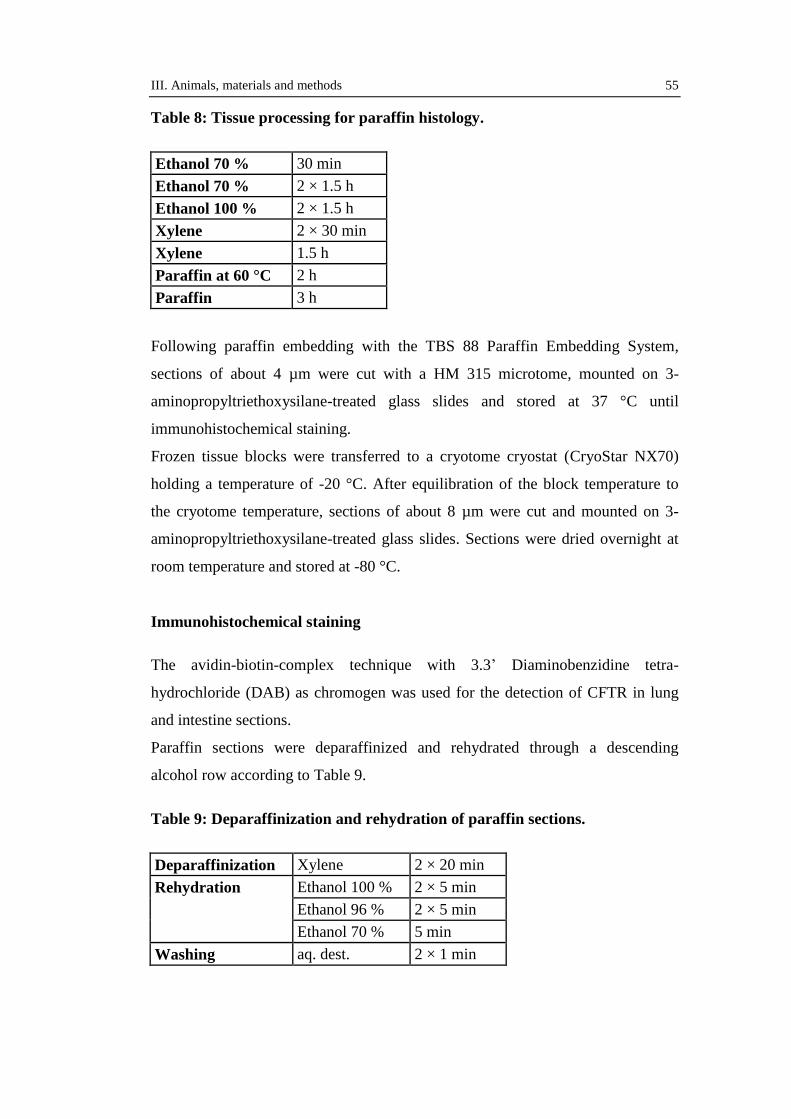
III. Animals, materials and methods 55
Table 8: Tissue processing for paraffin histology.
Ethanol 70 % 30 min
Ethanol 70 % 2 × 1.5 h
Ethanol 100 % 2 × 1.5 h
Xylene 2 × 30 min
Xylene 1.5 h
Paraffin at 60 °C 2 h
Paraffin 3 h
Following paraffin embedding with the TBS 88 Paraffin Embedding System,
sections of about 4 µm were cut with a HM 315 microtome, mounted on 3-
aminopropyltriethoxysilane-treated glass slides and stored at 37 °C until
immunohistochemical staining.
Frozen tissue blocks were transferred to a cryotome cryostat (CryoStar NX70)
holding a temperature of -20 °C. After equilibration of the block temperature to
the cryotome temperature, sections of about 8 µm were cut and mounted on 3-
aminopropyltriethoxysilane-treated glass slides. Sections were dried overnight at
room temperature and stored at -80 °C.
Immunohistochemical staining
The avidin-biotin-complex technique with 3.3’ Diaminobenzidine tetra-
hydrochloride (DAB) as chromogen was used for the detection of CFTR in lung
and intestine sections.
Paraffin sections were deparaffinized and rehydrated through a descending
alcohol row according to Table 9.
Table 9: Deparaffinization and rehydration of paraffin sections.
Deparaffinization Xylene 2 × 20 min
Rehydration Ethanol 100 % 2 × 5 min
Ethanol 96 % 2 × 5 min
Ethanol 70 % 5 min
Washing aq. dest. 2 × 1 min

III. Animals, materials and methods 56
Sections from formalin-fixated tissue had to be pre-treated with antigen retrieval
agents to break the protein cross-links formed by formalin fixation and thereby
restoring hidden antigenic sites. In general, at least one heat-induced epitope
retrieval (HIER) technique, in this case heating by microwave, and the proteinase-
induced epitope retrieval (PIER) with proteinase K were evaluated. For selected
antibodies, three different HIER methods were tested. Dilutions of H2O2, serum
and antibodies were prepared with TBS buffer. All steps were performed at RT if
not indicated otherwise. An overview of the workflow of the standard IHC
protocol with HIER is given in Table 10.
Table 10: Standard IHC protocol with HIER.
Procedure Agent Conditions
STEP 1 HIER 10 mM Citrate
buffer pH 6 +
0.05 % Tween
800 W until boiling, 15
min 320 W in microwave
Cooling 30 min
10 mM Citrate
buffer pH 6 +
0.05 % Tween
45 min at 800 W in
pressure cooker in
microwave
Cooling 20 min
DAKO target
retrieval solution
20 min at 97 °C in water
bath
Cooling 20 min
STEP 2 Washing TBS buffer 10 min
STEP 3 Blocking of
endogenous
peroxidase activity
1 % H2O2 15 min
STEP 4 Washing TBS buffer 10 min
STEP 5 Serum blocking 5 % serum 1 h at RT
STEP 6 Primary antibody 4 °C overnight
STEP 7 Washing TBS buffer 10 min
STEP 8 Secondary
antibody
1 h at RT
STEP 9 Washing TBS buffer 10 min
STEP 10 Detection ABC complex 30 min
STEP 11 Washing TBS buffer 10 min
STEP 12 Substrate DAB
STEP 13 Rinsing Floating tap water 5 min
STEP 14 Counterstaining Mayer’s Hemalum 30 s
STEP 15 Rinsing Floating tap water 5 min

III. Animals, materials and methods 57
In paraffin sections that were treated with proteinase K, endogenous peroxidase
activity was first blocked with 1 % H2O2 for 15 min, followed by a 10 min
washing step with TBS buffer. Next, sections were incubated with proteinase K as
antigen retrieval and subsequently all steps from the standard protocol, beginning
with STEP 4, were executed.
For sections from methacarn-fixated tissue that did not need any antigen retrieval,
the standard protocol was used from STEP 2 onwards.
Cryo-sections were treated in two different ways. In the first protocol (cryo
method 1), tissue frozen in OCT compound on dry ice was used, while in the
second protocol (cryo method 2), tissue incubated in 2 % PFA for 1 h at RT,
subsequently incubated in ice-cold 30 % sucrose overnight and frozen in OCT
compound in an ethanol/dry ice bath was used. For cryo method 1, cryo-sections
were air-thawed and fixated in 4 % PFA for 5 min first. After two washing steps
with TBS for 2 × 5 min, sections were permeabilized by incubation in 100 %
EtOH at -20 °C for 2 min followed by another washing step with TBS for 5 min.
Afterwards, all steps from the standard protocol, beginning with STEP 3, were
performed. In the second protocol (cryo method 2), cryo-sections were air-thawed,
rehydrated in PBS for 2 × 5 min and then permeabilized by incubation in 0.2 %
Triton X-100 for 5 min. After a washing step in TBS buffer for 5 min, all steps
from the standard protocol, beginning with STEP 3, were executed. Deviating
from the standard protocol, serum was diluted in Super-Block and the sections
were only incubated for 45 min in STEP 5 and the primary antibody was
incubated for 2 h at 37 °C in STEP 6. For details on primary and secondary
antibodies, dilutions and serum, see Table 17.
Immunofluorescent detection of CFTR
For immunofluorescent staining of CFTR, only cryo-sections from tissue frozen
with the second freezing method (1 h 2 % PFA RT, 30 % ice-cold sucrose
overnight and freezing in OCT compound in ethanol/dry ice bath) were used.
Frozen tissue blocks were processed as described above. Cryo-sections were air-
thawed, rehydrated in PBS for 2 × 5 min and permeabilized in 0.2 % Triton X-100
for 5 min. After two washing steps with PBS for 5 min, sections were incubated
with 5 % donkey serum in Super-block for 45 min to reduce non-specific binding.

III. Animals, materials and methods 58
Subsequently, sections were incubated with the primary antibody Ab 596 from the
CF Foundation, USA (1:50 in Super-Block) for 2 h at 37 °C and washed for 10
min in PBS. Sections were then incubated with the alexa fluor 488-conjugated
secondary antibody donkey anti-mouse diluted 1:250 in 2 % pig serum for 1 h.
From incubation with the secondary antibody onwards, all steps were performed
in the dark to avoid bleaching of the fluorescence. After a last washing step with
PBS for 10 min, slides were coverslipped with Vectashield antifade solution
containing 4',6-diamidino-2-phenylindole (DAPI) for the staining of cell nuclei
and afterwards stored at 4 °C in the dark. Images were acquired with an inverted
epifluorescence microscope (Axiovert 200 M; Zeiss, Oberkochen).
3.8.2. Detection of CFTR by Western Blot
The applicability of commercially available antibodies for the detection of CFTR
in protein lysates via Western blot (WB) was evaluated with tissue of a WT piglet,
human liver tissue and human T84 cells as a positive control and tissue of a
CFTR-/- piglet as a real negative control.
Protein isolation from tissue
40 mg of snap frozen tissue material were transferred to a 12 mL tube containing
1 mL of protein extraction buffer and was homogenized for 2 min on ice with the
homogenizer ART-Micra D-8 at 23,500 rpm. To avoid contamination, the rotor
was cleaned three times with aq. bidest. and once with PBS between different
samples. Protein extracts were left on ice for 30 min, transferred to 1.5 mL
Eppendorf tubes and incubated for 15 min at 60 °C. After cooling on ice for
3 min, the samples were centrifuged at 15,000 rcf for 5 min at 4 °C and the
supernatant was transferred to a new 1.5 mL Eppendorf tube. Protein lysates were
stored at -20 °C until further processing.
Measurement of protein concentration by BCA assay
To quantify the total amount of protein in the extracted tissue lysates, the
Bicinchoninic acid (BCA) assay was used. The principle of this method is that the
protein concentration is indicated by a change of color in the sample solution from
green to purple in proportion to the protein concentration, which can then be

III. Animals, materials and methods 59
measured photometrically by establishing a calibration curve using protein
standard solutions of known concentration.
For this purpose, protein standard solutions of known concentration were prepared
by a six-fold dilution row (50 µL each) of a BSA stock solution (4 mg/mL) in
PBS/protein extraction buffer (4:1 v:v). 10 µL of each protein lysate were diluted
with 40 µL PBS. Bicinchoninic acid was mixed with 4 % CuSO4-solution in a
ratio of 50:1 and 200 µL of this mixture were pipetted to each standard and
unknown sample. 100 µL of each reaction mixture were pipetted in duplicates to a
96 well plate and the plate was incubated at 37 °C for 30 min. Extinction values
were measured at 562 nm with the SunriseTM microplate reader for ELISA and
protein concentrations were determined via the standard curve.
SDS PAGE
Proteins were separated according to their size by gel electrophoresis. The SDS
PAGE is based on the principle that sodium dodecyle sulfate (SDS) denatures
proteins and covers them with a negative charge so that they can be separated
according to their molecular weight in a polymerized gel matrix when an
electrical field is applied.
An 8 % SDS polyacrylamide separating gel was prepared according to Table 11,
poured into the Mini Protean® Tetra system and covered with a layer of water for
polymerization.
Table 11: Composition of 8 % SDS polyacrylamide separating gel.
aq. dest. 4.63 mL
1.5 M Tris (pH 8.8) 2.5 mL
30 % Acrylamid/Bis Solution 2.67 mL
10 % SDS 100 µL
10 % APS 100 µL
Temed 5 µL
After 45 min, the water was removed carefully, the separating gel was overlaid
with a 5 % stacking SDS polyacrylamide gel (see Table 12) and a 15-well comb
inserted at the top.
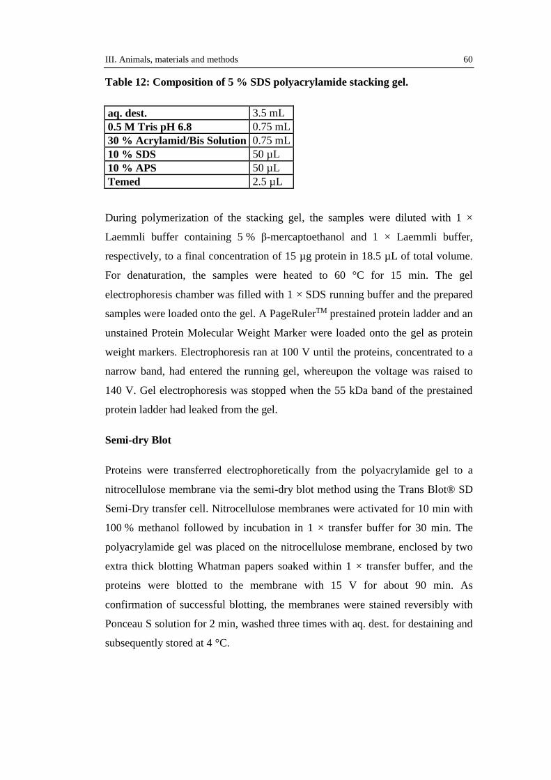
III. Animals, materials and methods 60
Table 12: Composition of 5 % SDS polyacrylamide stacking gel.
aq. dest. 3.5 mL
0.5 M Tris pH 6.8 0.75 mL
30 % Acrylamid/Bis Solution 0.75 mL
10 % SDS 50 µL
10 % APS 50 µL
Temed 2.5 µL
During polymerization of the stacking gel, the samples were diluted with 1 ×
Laemmli buffer containing 5 % β-mercaptoethanol and 1 × Laemmli buffer,
respectively, to a final concentration of 15 µg protein in 18.5 µL of total volume.
For denaturation, the samples were heated to 60 °C for 15 min. The gel
electrophoresis chamber was filled with 1 × SDS running buffer and the prepared
samples were loaded onto the gel. A PageRulerTM prestained protein ladder and an
unstained Protein Molecular Weight Marker were loaded onto the gel as protein
weight markers. Electrophoresis ran at 100 V until the proteins, concentrated to a
narrow band, had entered the running gel, whereupon the voltage was raised to
140 V. Gel electrophoresis was stopped when the 55 kDa band of the prestained
protein ladder had leaked from the gel.
Semi-dry Blot
Proteins were transferred electrophoretically from the polyacrylamide gel to a
nitrocellulose membrane via the semi-dry blot method using the Trans Blot® SD
Semi-Dry transfer cell. Nitrocellulose membranes were activated for 10 min with
100 % methanol followed by incubation in 1 × transfer buffer for 30 min. The
polyacrylamide gel was placed on the nitrocellulose membrane, enclosed by two
extra thick blotting Whatman papers soaked within 1 × transfer buffer, and the
proteins were blotted to the membrane with 15 V for about 90 min. As
confirmation of successful blotting, the membranes were stained reversibly with
Ponceau S solution for 2 min, washed three times with aq. dest. for destaining and
subsequently stored at 4 °C.

III. Animals, materials and methods 61
Western Immunoblot
CFTR was detected via antigen-antibody coupling in an immunoblot. All steps
were conducted in 50 mL falcons in a hybridization oven at room temperature if
not indicated otherwise. Primary antibodies were diluted in TBS-T-BSA, while
secondary antibodies were diluted in TBS-T-w/v nonfat dry milk. An overview of
the standard protocol is given in Table 13.
Table 13: Standard western blot protocol.
Procedure Agent Conditions
STEP 1 Activation 100 % methanol 1 min
STEP 2 Washing TBS-T 3 × 5 min
STEP 3 Blocking TBS-T-w/v nonfat
dry milk
1 h
STEP 4 Washing TBS-T 1 × 15 min, 2 × 5 min
STEP 5 Primary antibody 4 °C overnight
STEP 6 Washing TBS-T 1 × 15 min, 2 × 5 min
STEP 7 Secondary antibody 1 h
STEP 8 Washing TBS-T 1 × 15 min, 2 × 5 min
STEP 9 Detection ECLTM detection
reagent
Signals were detected with the ECL detection reagent on HyperfilmTM ECL
chemiluminescense films in a darkroom. After suitable exposition time, films
were developed in the Optimax X-ray Film processor 1170-1-0000.
An overview of the tested antibodies and dilutions is given in Table 18.
Stripping of the membranes
If membranes were to be used in further experiments, bound antibodies had to be
removed by stripping of the membranes. For this purpose, the membranes were
incubated in elution buffer for 30 min at 70 °C, followed by two washing steps in
aq. bidest. for 10 min and one washing step in TBS-T in a new falcon for 10 min.
Afterwards, the membranes could be used for another antibody incubation,
starting with STEP 3 of the standard protocol.

IV. Results 62
IV. RESULTS
1. Transgenic expression of CFTR in the intestine modifies
the severity of meconium ileus in gut-modified CFTR-/-
piglets
Since a surgical correction of the fatal intestinal obstruction in newborn CF piglets
is not feasible to produce large numbers of living animals, we decided to take
another approach. By transgenic expression of porcine CFTR under the control of
a promoter selectively expressed in the intestine, the severity of MI should be
ameliorated and thereby ensure the survival of the CF piglets without affecting the
other typical organ manifestations. As those pigs should have the potential to
avoid MI, they are designated as potentially “gut-modified” in the following.
1.1. Generation of gut-modified CFTR-/- piglets
Gut-modified CFTR-/- piglets were generated by additive gene transfer of the
expression vector (see Figure 4) in porcine CFTR-/- kidney cells, followed by
SCNT and ET. This work was performed by N. Klymiuk, A. Wünsch,
M. Kurome, V. Zakhartchenko, B. Kessler and T. Güngör, Chair for Molecular
Animal Breeding and Biotechnology, LMU Munich.
Figure 4: DNA construct for generation of gut-modified CFTR-/- piglets. The
expression vector consists of different functional parts: a 1.2 kb fragment of the
porcine fatty-acid binding protein 2 promoter (FABP2) for the selective
expression in the intestine, a 215 bp long part of the porcine β-hemoglobin (HBB)
gene to increase the expression capacity by introducing a synthetic intron, the
4.8 kb coding region of the porcine CFTR followed by the 277 bp long

IV. Results 63
polyadenylation signal of the bovine Growth Hormone (bGH) gene as termination
signal and a puromycin® (puro®) resistance cassette for positive selection.
A total of 1401 gut-modified CFTR-/- embryos were transferred to 12 pre-
synchronized sows, resulting in four pregnancies. Three pregnancies went to full
term with the birth of 13 living and 5 stillborn piglets. The efficiency of NT
experiments, as defined by the number of living piglets per number of transferred
embryos and per number of successful embryos, respectively, was about 0.9 %
and 3.7 %. 25 % of all performed ETs resulted in the birth of living offspring. To
exclude a potential influence on the outcome of pregnancy rates and the
phenotype, CFTR-/- kidney cells from different genetic backgrounds were
employed for additive gene transfer. In the first 11 experiments, cells from a
cloned CFTR-/- piglet (#1267) and pooled cells from several cloned CFTR-/-
piglets (#1268, 1270, 1274) were used. In the last NT experiment, pooled cells
from CFTR-/- piglets (#2353, 2358) produced by normal breeding were employed
to avoid the negative effects of repeated reproduction by cloning (for details see
Table 14).
1.2. Analysis of the intestinal phenotype in gut-modified CFTR-/- piglets
Immediately after birth, the gut-modified CF piglets were repeatedly treated with
enemas. Medication for gastric protection and circulation support was applied and
pancreatic enzymes were supplemented to increase the likelihood of MI removal
(for details see treatment protocol in Table 1). The gut-modified CF piglets from
the litter of sow 1 and sow 12 exhibited a clearly impaired viability since birth, as
they were unable to suckle on their own and had to be fed artificially to ensure
colostrum and energy intake. The piglets were kept under 24 h surveillance and as
soon as a piglet started to show a considerably deteriorated behavior,
characterized by reduced motility, inability to suckle milk and a pear-looking,
bloated abdomen with enhanced abdominal pressure in the absence of any signs of
meconium removal, it was euthanized for controlled phenotypical analysis and
sample collection.

IV. Results 64
All 13 living piglets had to be euthanized within the first 24 h after birth as, in
spite of all the supportive measures taken, they were not able to remove
meconium and therefore developed the severe characteristic symptoms mentioned
above. For a detailed evaluation of the severity, localization and extension of MI
in comparison to “normal” CF piglets and WT piglets of the same age, see Figure
5 and
Figure 6. Summarized, the severity of the intestinal phenotype was improved in
comparison to normal CF KO piglets in all examined gut-modified CFTR-/-
piglets. The localization of stuck meconium was consistently found in more distal
parts of the intestine and in a large percentage of gut-modified CF piglets,
diverticulosis was less pronounced.

IV. Results 65
Table 14: Results of SCNT experiments using gut-modified CFTR-/- embryos.
Recipients Genetic background No. of transferred
embryos
No. of successful
embryos
Pregnancy Result
Living piglets Stillborn piglets
Sow 1 #1267 121 121 + 3 (#2128-2130) 3 (#2131-2133)
Sow 2 #1267 120 - - - -
Sow 3 #1267 89 - + Preterm abortion
Sow 4 #1267 99 - - - -
Sow 5 #1267 111 - - - -
Sow 6 #1267 136 136 + 6 (#2477-2482) 2 (#2494, 2507)
Sow 7 #1267 119 - - - -
Sow 8 #1267 118 - - - -
Sow 9 #1267 95 - - - -
Sow 10 #1267 139 - - - -
Sow 11 #1268, 1270, 1274 159 - - - -
Sow 12 #2353, 2358 95 95 + 4 (#2736-2739) -
Total 1401 352 3 13 5
Efficiency ~0.9 % ~3.7 % 25 % ~72.2 % ~27.8 %
Efficiency of NT experiments is calculated as number of living piglets per number of transferred or successful embryos. Pregnancy rate is calculated
as number of pregnancies resulting in the birth of living piglets per ETs in total. Percentage of living and stillborn piglets, respectively, is calculated
by correspondent number of piglets per total number of born piglets.

IV. Results 66
Figure 5: Intestinal phenotype of WT, gut-modified CFTR-/- and CFTR-/-
piglets. (A) Detoriation of the general condition in a gut-modified CF piglet is
illustrated by the decreased ability to move on its own and a severely bloated
abdomen with prominent blood vessels (black arrow). (B) View of the intestinal
convolute after opening the abdominal cavity and after complete resection. White
asterisks mark the presence of meconium in dilated intestinal loops. In WT
piglets, meconium has been completely deposited after about 24 h and the
intestine shows a healthy brownish color and intestinal loops of equal diameter.
Both in normal and gut-modified CF piglets, intestinal loops are blocked by black
meconium. Compared to normal CF piglets, the blockage by meconium appears in
the more distal parts of the intestine in gut-modified CF piglets, extending until
the beginning of the colon. Discontinuous black arrows point to the cecum, which
is severely enlarged by stuck meconium in gut-modified CF piglets. The colon is

IV. Results 67
marked by black arrows. Gut-modified CF piglets exhibit the microcolon
characteristic for normal CF piglets, although a small portion of the meconium
has entered the proximal parts of the colon ascendens.
Figure 6: Range of MI in WT, gut-modified CFTR-/- (gut CF) and CFTR-/-
piglets. As a reference point, the cecum is marked by discontinuous black arrows.
The range of MI (marked by white asterisks) differs in the location between gut-
modified and normal CF piglets. In all examined normal CF piglets, stuck
meconium is mainly located in the middle and distal parts of the jejunum, whereas
gut-modified CF piglets exhibit a slightly improved phenotype with meconium
consistently localized in the distal jejunum, cecum and proximal colon. The
prominent feature of diverticulosis (black arrow), that is consistently found in all
normal CF piglets and illustrates the severity of the intestinal phenotype, is
considerably less pronounced or even missing in gut-modified CF piglets.
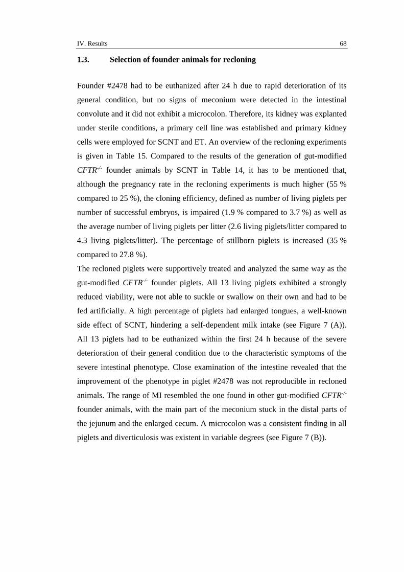
IV. Results 68
1.3. Selection of founder animals for recloning
Founder #2478 had to be euthanized after 24 h due to rapid deterioration of its
general condition, but no signs of meconium were detected in the intestinal
convolute and it did not exhibit a microcolon. Therefore, its kidney was explanted
under sterile conditions, a primary cell line was established and primary kidney
cells were employed for SCNT and ET. An overview of the recloning experiments
is given in Table 15. Compared to the results of the generation of gut-modified
CFTR-/- founder animals by SCNT in Table 14, it has to be mentioned that,
although the pregnancy rate in the recloning experiments is much higher (55 %
compared to 25 %), the cloning efficiency, defined as number of living piglets per
number of successful embryos, is impaired (1.9 % compared to 3.7 %) as well as
the average number of living piglets per litter (2.6 living piglets/litter compared to
4.3 living piglets/litter). The percentage of stillborn piglets is increased (35 %
compared to 27.8 %).
The recloned piglets were supportively treated and analyzed the same way as the
gut-modified CFTR-/- founder piglets. All 13 living piglets exhibited a strongly
reduced viability, were not able to suckle or swallow on their own and had to be
fed artificially. A high percentage of piglets had enlarged tongues, a well-known
side effect of SCNT, hindering a self-dependent milk intake (see Figure 7 (A)).
All 13 piglets had to be euthanized within the first 24 h because of the severe
deterioration of their general condition due to the characteristic symptoms of the
severe intestinal phenotype. Close examination of the intestine revealed that the
improvement of the phenotype in piglet #2478 was not reproducible in recloned
animals. The range of MI resembled the one found in other gut-modified CFTR-/-
founder animals, with the main part of the meconium stuck in the distal parts of
the jejunum and the enlarged cecum. A microcolon was a consistent finding in all
piglets and diverticulosis was existent in variable degrees (see Figure 7 (B)).

IV. Results 69
Table 15: Recloning experiments of founder #2478.
Recipients Recloning of
founder
No. of
transferred
embryos
No. of successful
embryos
Pregnancy Result
Living piglets Stillborn piglets
Sow 1 #2478 194 194 + 1 (#2870) 2 (#2869, 2871)
Sow 2 #2478 118 118 + 1 (#2906) 1 (#2906)
Sow 3 #2478 119 - - - -
Sow 4 #2478 152 - - - -
Sow 5 #2478 187 - - - -
Sow 6 #2478 135 - - - -
Sow 7 #2478 122 122 + 2 (#4008, 4009) -
Sow 8 #2478 127 127 + 4 (#4051-4054) -
Sow 9 #2478 134 134 + 5 (#4280, 4283-4286) 4 (#4279, 4281, 4282, 4287)
Total 1288 695 5 13 7
Efficiency ~1.0 % ~1.9 % ~55 % 65 % 35 %
Efficiency of NT experiments is calculated as number of living piglets per number of transferred or successful embryos. Pregnancy rate is calculated
as number of pregnancies resulting in the birth of living piglets per ETs in total. Percentage of living and stillborn piglets, respectively, is calculated
by correspondent number of piglets per total number of born piglets.

IV. Results 70
Figure 7: Phenotype of recloned founder piglets. (A) Abnormal findings in
recloned piglets. All recloned piglets show reduced viability, a high percentage of
them has an enlarged tongue and the number of stillborn piglets or mummies is
increased. (B) Intestinal phenotype of two exemplary piglets. The cecum is
marked with a discontinuous black arrow, the continuous black arrow points to
the microcolon. The range of MI is marked with white asterisks. Meconium is
stuck in the distal parts of the jejunum, cecum and the beginning of the colon
ascendens. All piglets exhibit a microcolon with reduced diameter. Thus, the
range of MI resembles other founder animals with a slight improvement compared
to normal CF KO piglets, but not the clearly improved phenotype of #2478. The
range of diverticulosis differs strongly between all examined recloned piglets.

IV. Results 71
1.4. Analysis of gut-modified CFTR-/- piglets at the genomic level
Considering the fact that the health of normal CFTR-/- piglets started to deteriorate
within 24 h after birth because of the severe intestinal phenotype, it was essential
to establish a rapid method for the genotyping of gut-modified CF piglets,
including a time-sparing DNA isolation method and an extremely shortened PCR
protocol.
DNA isolation
Three different fast DNA isolation kits were compared in terms of suitability, time
duration and reliability of PCR results – the nexttecTM Genomic DNA Isolation
Kit from Tissue and Cells, the Easy-DNATM Kit, protocol #2 – 30 min DNA
extraction from blood samples and the Isohelix DDK DNA Isolation Kit.
Table 16: Comparison of DNA isolation methods.
Source Suitability Time
duration
Reliability of
PCR results
Nexttec kit Docked
tail
Tail docking is routinely
performed in all litters to
prevent tail biting
60 min high
Easy-DNA
kit
Blood Blood collection is
critical in neonatals
30 min low
Isohelix kit Buccal
swabs
Easy to handle 90 min medium-high
After comparing all critical aspects, the DNA isolation from docked tails via the
nexttec kit proved to be the safest, fastest and most reliable method for the
isolation of genomic DNA in newborn piglets.
Establishment of genotyping PCRs
To confirm the CFTR-/- genotype, two sets of regular PCR amplifying the WT and
the mutated CFTR sequence were established. The integration of the gut-modified
CFTR expression vector in the CFTR-/- genome was confirmed by amplifying a
specific part of the expression vector. As the nexttec DNA isolation protocol is
very time-saving but produces DNA of inferior quality, a range of primer pairs for
amplification of the WT (see Figure 8) and mutant CFTR (see Figure 9) sequence

IV. Results 72
had to be tested and best pairs were further optimized. To determine whether the
gut-modified CFTR expression vector had been integrated in the CFTR-/- genome,
primer pairs amplifying specific parts of the expression vector in the 3’- and 5’-
UTR-regions flanking the porcine CFTR sequence were tested and the conditions
were further optimized (see Figure 10).
Figure 8: Testing of primer pairs for the detection of WT CFTR. Primer pairs
were first tested with an annealing temperature of 58 °C and a starting volume of
0.5 µL each. DNA isolated with the nexttec 1 h-protocol from WT, CFTR+/- (het)
and CFTR-/- (hom) piglets served as template. Conditions of promising pairs
(1 and 12) were optimized by temperature gradient PCR and comparison of
different primer concentrations.
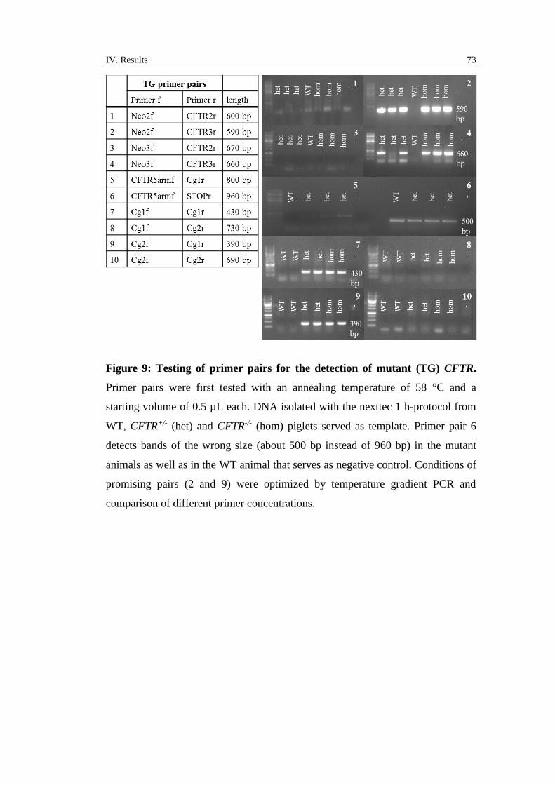
IV. Results 73
Figure 9: Testing of primer pairs for the detection of mutant (TG) CFTR.
Primer pairs were first tested with an annealing temperature of 58 °C and a
starting volume of 0.5 µL each. DNA isolated with the nexttec 1 h-protocol from
WT, CFTR+/- (het) and CFTR-/- (hom) piglets served as template. Primer pair 6
detects bands of the wrong size (about 500 bp instead of 960 bp) in the mutant
animals as well as in the WT animal that serves as negative control. Conditions of
promising pairs (2 and 9) were optimized by temperature gradient PCR and
comparison of different primer concentrations.

IV. Results 74
Figure 10: Testing of primer pairs for the detection of the gut-modified
CFTR expression vector. Primer pair 1 is located in the 5’-UTR region; the
other primer pairs are located in the 3’-UTR region of the expression vector.
Primer pairs were first tested with an annealing temperature of 58 °C and a
starting volume of 0.5 µL each. DNA from gut-modified CFTR-/- (gutCF) and WT
piglets isolated with the nexttec 1 h-protocol served as template. Primer pair 3
also detects a signal in the WT piglets that serve as negative controls. Conditions
for the promising primer pair 2 were optimized by temperature gradient PCR and
comparison of different primer concentrations.
In conclusion, the time necessary for the genotyping of gut-modified CF piglets
could be minimized to less than three hours between birth and genotyping results,
including one hour for DNA isolation and one hour for the final genotyping PCRs
(see Figure 11). DNA isolated with the nexttec 1 h-protocol was of sufficient
quality for regular genotyping PCR.

IV. Results 75
Figure 11: Example of genotyping gut-modified CFTR-/- (gutCF), WT,
CFTR+/- (het) and CFTR-/- (hom) piglets. All three genotyping PCRs detecting
the WT, the mutant (TG) and the gut-modified (gutCF) CFTR sequence are run
under the same conditions in one machine. The time duration of all PCR steps was
minimized as far as possible, until the whole run took about one hour.
Verification of the correct amplification of the PCR products
The detection of bands of the correct bp size using the agarose gel electrophoresis
has to be approved by a further method. To confirm that primers annealed to the
correct DNA sequences and that the requested base sequences were amplified,
DNA was eluted from bands of the correct bp size and sequenced via the Sanger
sequencing approach. The detailed comparison of the base sequences derived
from sequencing chromatograms to the target sequences revealed that all primers
annealed correctly to the sequences and that target and amplified sequences were
identical (see Figure 12).

IV. Results 76
Figure 12: Verification of genotyping PCR products. (A) Electropherograms of
PCR products amplified with primers for WT, mutant (TG) and gut-modified
CFTR. In the mutant and WT CFTR chromatograms, the 5’-parts are identical as
the forward primer Cg2f, which is used for both PCRs, anneals to a part of the
CFTR sequence present in both sequences. The sequences specific for WT and
mutant CFTR further downstream are highlighted in red (WT) and yellow (TG).
(B) Comparison of amplified sequences to target sequences.

IV. Results 77
1.5. Expression analysis of CFTR-/- and WT piglets at the protein level
For a functional evaluation of the porcine CF model, the absence of CFTR
expression had to be confirmed at the protein level. Therefore, CFTR expression
in WT and CF KO piglets was examined by immunohistochemical or
immunofluorescent approaches and by western blot. A range of commercially
available primary antibodies are described to detect CFTR in tissue derived from
pigs. Exclusively in this study, the specificity of the antibodies was evaluated with
a best-optimal negative control, tissue derived from CF KO animals.
Detection of CFTR by immunohistochemistry and immunofluorescence
Initially, the utility of the well-characterized antibody Ab 596 (CF Foundation,
USA) for the detection of CFTR in cryo-sections from tissue frozen with the
complicated freezing method 2 was evaluated in IF and IHC, according to a
published protocol (Stoltz, Rokhlina et al. 2013). However, the primary aim of the
study was to find an antibody that reliably detected CFTR in paraffin-embedded
tissue, as the morphology of paraffin-embedded tissue is better preserved and the
fixation and sectioning works very well compared to cryo-sections. For a detailed
evaluation of the antibodies, at least one HIER and one PIER method were tested
using formalin-fixated tissue. As some antibody-specific protocols preferred the
usage of cryo-sections for IHC, the specificity of all antibodies was also evaluated
on paraffin slides from methacarn-fixated tissue. Methacarn fixation is reported to
produce tissue slides with a quality similar to cryo-sections (Mitchell, Ibrahim et
al. 1985). The specificity of the antibodies from the CF Folding consortium was
also evaluated using cryo-sections from tissue frozen with freezing method 1with
cryo method 1, an established protocol for the detection of CFTR in human tissue
(Kreda and Gentzsch 2011). Table 17 gives an overview of all antibodies,
fixations and conditions tested in immunohistochemistry.

IV. Results 78
Table 17: Overview of all tested immunohistochemical conditions.
Primary antibody Fixation Antigen retrieval Dilution Secondary antibody Dilution Serum
CF Folding
consortium Ab 528
Formalin Microwave 1:100; 1:300; 1:900 Biotinylated goat anti-
mouse IgG + 5 % serum
pig
1:200 Goat
Proteinase K 1:200; 1:600
Methacarn - 1:100; 1:300; 1:900
Cryo method 1 - 1:250; 1:1,000 Biotinylated AffiniPure
goat anti-mouse IgG
1:250
CF Folding
consortium Ab 570
Formalin Microwave 1:100; 1:300; 1:900 Biotinylated goat anti-
mouse IgG + 5 % serum
pig
1:200 Goat
Proteinase K 1:200; 1:600
Methacarn - 1:100; 1:300; 1:900
Cryo method 1 - 1:250; 1:1,000 Biotinylated AffiniPure
goat anti-mouse IgG
1:250
CF Folding
consortium Ab 596
Formalin Microwave 1:100; 1:300; 1:900 Biotinylated goat anti-
mouse IgG + 5 % serum
pig
1:200 Goat
Water bath 1:300; 1:900; 1:2,700
Pressure cooker 1:300; 1:900; 1:2,700
Proteinase K 1:200; 1:600
Methacarn - 1:100; 1:300; 1:900
Cryo method 1 - 1:250; 1:1,000 Biotinylated AffiniPure
goat anti-mouse IgG
1:250
Cryo method 2 - 1:300; 1:900 Biotinylated AffiniPure
goat anti-mouse IgG + 2 %
serum pig
1:250

IV. Results 79
CF Folding
consortium Ab 660
Formalin Microwave 1:100; 1:300; 1:900 Biotinylated goat anti-
mouse IgG + 5 % serum
pig
1:200 Goat
Proteinase K 1:200; 1:600
Methacarn - 1:100; 1:300; 1:900
Cryo method 1 - 1:250; 1:1,000 Biotinylated AffiniPure
goat anti-mouse IgG
1:250
CF Folding
consortium Ab 769
Formalin Microwave 1:100; 1:300; 1:900 Biotinylated goat anti-
mouse IgG + 5 % serum
pig
1:200 Goat
Proteinase K 1:200; 1:600
Methacarn - 1:100; 1:300; 1:900
Cryo method 1 - 1:250; 1:1,000 Biotinylated AffiniPure
goat anti-mouse IgG
1:250
Abcam CF3 Formalin Microwave 1:100; 1:300; 1:900; 1:1,500;
1:1,800; 1:3,000; 1:6,000;
1:12,000
Biotinylated goat anti-
mouse IgM + 2 % serum
pig
1:100 Goat
Water bath 1:1,500; 1:3,000; 1:6,000;
1:12,000
Proteinase K 1:1,500; 1:3,000; 1:6,000;
1:12,000
Methacarn - 1:100; 1:300; 1:900; 1:1,500;
1:1,800; 1:3,000; 1:6,000;
1:12,000

IV. Results 80
Cell Signaling #2269 Formalin Microwave 1:100; 1:300; 1:900 Biotinylated goat anti-
rabbit IgG + 5 % serum
pig
1:200 Goat
Proteinase K 1:300
Methacarn - 1:100; 1:300; 1:900
R&D 24-1 Formalin Microwave 1:25; 1:75; 1:250 Biotinylated goat anti-
mouse IgG + 5 % serum
pig
1:200 Goat
Water bath 1:150; 1:300; 1:900
Pressure cooker 1:150; 1:300; 1:900
Proteinase K 1:100; 1:350
- 1:100; 1:200; 1:500
Methacarn - 1:100; 1:300; 1:900
Santa Cruz H 182 Formalin Microwave 1:100; 1:300; 1:900 Biotinylated goat anti-
rabbit IgG + 5 % serum
pig
1:200 Goat
Proteinase K 1:50; 1:100; 1:200; 1:300
Methacarn - 1:100; 1:300; 1:900
Santa Cruz N-20 Formalin Microwave 1:100; 1:300; 1:900 Biotinylated rabbit anti-
goat IgG + 5 % serum pig
1:200 Rabbit
Proteinase K 1:50; 1:150; 1:450
Methacarn - 1:100; 1:300; 1:900
The single method specifically detecting CFTR is marked in red.

IV. Results 81
Staining with the primary antibody Ab 596 (CF Foundation, USA) of cryo-
sections derived from tissue frozen with the complicated freezing method 2
worked reliably using cryo method 2 for the specific detection of CFTR in
intestinal tissue both in IF and IHC (see Figure 13 and Figure 14). Staining
patterns in IF and IHC were identical.
In WT animals, CFTR was strongly expressed in the crypts of the intestine and
showed a decreasing gradient along the villi from the crypts to the villous top.
Several single villous cells with normal enterocyte-like morphology, presumably
an equivalent to the CFTR High expresser cells found in human and rat proximal
small intestine (Jakab, Collaco et al. 2013), showed a strong CFTR signal. In the
staining with IF detection, fetal faeces showed a strong autofluorescence, a feature
that was reproduced using IHC, where fetal faeces were stained in brown in the
negative control. In WT lung tissue, a very weak CFTR signal was detected in the
submucosal glands, whereas the respiratory epithelium was not stained. In CF KO
animals, CFTR was neither detected in intestinal nor in lung tissue. Staining with
antibody Ab 596 confirmed thereby the absence of any CFTR protein in the
CFTR-/- piglets.

IV. Results 82
Figure 13: Immunofluorescent detection of CFTR in the intestine of WT and CFTR-/- piglets with Ab 596. Images show staining for cell nuclei
(DAPI, blue) and CFTR (red). Intestinal crypts are marked with a white arrow. In WT piglets, CFTR is mainly localized in the intestinal crypts with a
decreasing gradient along the villous axis and strong staining of single villous cells. In CF KO piglets, CFTR is not detected, faeces show a strong
autofluorescence (marked with white asterisks).

IV. Results 83
Figure 14: Immunohistochemical detection of CFTR in WT and CFTR-/-
intestine and lung with Ab 596. (A) Detection of CFTR in the intestine of WT
and CF KO piglets. Intestinal crypts are marked with black arrows. The
discontinuous arrow points to a single strongly-stained cell. Negative controls

IV. Results 84
(neg) are stained with the secondary antibody only. Faeces are stained in brown
both in the stained slide and the negative control of the CF KO intestine (marked
with black asterisks). (B) Detection of CFTR in the lung from WT and CF KO
piglets. Black arrows point to submucosal glands.
Since the detection of CFTR on cryo-sections with Ab 596 was not sensitive
enough to detect CFTR in lung tissue and the tissue morphology showed freezing
artefacts in spite of the cryo-protective freezing process, several commercially
available antibodies for the detection of CFTR on paraffin-embedded tissue were
tested. As the lung is the major organ of interest in CF, the antibodies were first
evaluated on lung tissue from WT and CFTR-/- piglets (see Figure 15). Two
antibodies showed an obvious difference of staining pattern between WT and CF
KO piglets, all other antibodies detected no specific signal. Ab 596 (1:900, HIER
by water bath) showed strong apical cytoplasmatic staining of the respiratory
epithelium and very weak staining of the submucosal glands in formalin-fixated
WT lung tissue, whereas the CF KO lung tissue was negative (see Figure 15 (B)).
Ab 24-1 (1:900, HIER by pressure cooker) recognized a weak apical
cytoplasmatic signal of the respiratory epithelium and submucosal glands with
very strong staining on the apical surface of the respiratory epithelium in
formalin-fixated WT tissue, while CF KO tissue was negative (see Figure 15 (D)).
However, when these two antibodies were employed under the same conditions
for the detection of CFTR in intestinal tissue, they detected the same staining
pattern both in WT and CF KO tissue (see Figure 16). To exclude a single animal-
specific effect, both antibodies were tested with the same conditions on a larger
number of WT and CF KO animals. As the unspecific staining on intestinal tissue
was reproducible for all tested animals, these antibodies were not suitable for a
specific detection of CFTR. None of the other evaluated antibodies detected a
specific CFTR signal in WT and simultaneously no signal in CF KO piglets in
lung and intestinal paraffin-embedded tissue. Either the signal pattern was
completely unspecific with strong staining of the wrong tissue, e.g. smooth
muscle (see Figure 15 (C)), or the expression pattern seemed correct, however,
with no difference between WT and CF KO animals. Cryo method 1 did not give
a specific signal for CFTR, when porcine tissue was used.

IV. Results 85
Figure 15: Testing of antibodies on paraffin-embedded lung tissue from WT
and CFTR-/- piglets. (A) Ab 528 (CF Foundation, USA), 1:900 on methacarn-
fixated lung tissue. (B) Ab 596 (CF Foundation, USA), 1:900 on formalin-fixated
lung tissue with HIER by water bath. (C) Ab CF3 (abcam, UK), 1:12,000 on

IV. Results 86
formalin-fixated tissue with PIER by proteinase K. (D) Ab 24-1 (R&D systems,
Wiesbaden), 1:900 on formalin-fixated tissue with HIER by pressure cooker.
Figure 16: Unspecific staining of CFTR in the intestine of WT and CFTR-/-
piglets. (A) IHC on formalin-fixated intestinal tissue with HIER by water bath
using Ab 596 in a dilution of 1:900. The staining shows the correct pattern, with
main localization in the intestinal crypts (marked with a black arrow), but WT and
CF KO slides show no difference in staining patterns. (B) IHC on formalin-
fixated intestinal tissue with HIER by pressure cooker using ab 24-1 in a dilution
of 1:900. Staining shows the correct pattern, but no difference between WT and
CF KO tissue.
Detection of CFTR by western blot
The aim of this study was to evaluate whether commercially available antibodies
for the detection of CFTR in WB can specifically recognize CFTR in native tissue
derived from WT and CF KO piglets. For testing the sensitivity of the antibodies,

IV. Results 87
protein lysates from pig organs with high (proximal jejunum) and low (lung and
heart) expression levels of CFTR were employed for analysis. As all antibodies
were designated to detect human CFTR, human liver and T84 cells, a human
colon carcinoma cell line with endogenous CFTR expression levels, were
additionally used as positive controls. Table 18 gives an overview of the tested
primary and secondary antibodies with correspondent dilutions.
None of the tested antibodies specifically recognized the two typical CFTR bands
(B band and C band) at about 150 kDa and 180 kDa in porcine tissue lysates. The
B band indicates the presence of the core-glycosylated form of the protein and the
C band the fully-glycosylated mature form of CFTR (Farinha, Penque et al.
2004). Either no bands of the correct size were detected (see Figure 17 (B)) or
bands of the correct size were also detected in the negative control, tissue derived
from the CF KO piglet (see Figure 17 (A)). Only one antibody, Ab 24-1 (R&D
systems, Wiesbaden) gave a specific signal for the detection of endogenous levels
of human CFTR in human T84 cells (see Figure 17 (A)).
Table 18: Overview of tested western blot conditions.
Primary antibody Dilution Secondary antibody Dilution
CF Folding
consortium Ab 528
1:1,000 HRP goat anti-mouse IgG 1:5,000
CF Folding
consortium Ab 570
1:1,000 HRP goat anti-mouse IgG 1:5,000
CF Folding
consortium Ab 596
1:2,000 HRP goat anti-mouse IgG 1:5,000
CF Folding
consortium Ab 660
1:750 HRP goat anti-mouse IgG 1:5,000
CF Folding
consortium Ab 769
1:1,000 HRP goat anti-mouse IgG 1:5,000
Cell Signaling #2269 1:1,000 HRP goat anti-rabbit IgG 1:2,000
Abcam CF3 1:1,500 HRP goat anti-mouse IgG 1:5,000
R&D 24-1 1:400 HRP goat anti-mouse IgG 1:5,000
Santa Cruz H 182 1:1,200 HRP goat anti-rabbit IgG 1:2,000
Santa Cruz N-20 1:200 HRP mouse anti-goat IgG 1:5,000

IV. Results 88
Figure 17: Detection of CFTR by WB. (A) Detection of CFTR with antibody
R&D 24-1. The sharp B band and C band (red arrows) typical for the core- and
fully glycosylated CFTR protein are detected in protein lysate from human T84
cells. In porcine tissue, bands of the size of the C band are detected both in WT
and CF KO piglets. (B) Detection of CFTR with antibody Ab 528. No bands of
the correct size are detected.

IV. Results 89
1.6. Expression analysis of gut-modified CFTR-/- piglets at the RNA level
The detection of endogenous CFTR protein levels by immunological methods
proved to be difficult, limited by both specificity and sensitivity of the available
antibodies. Thus, the expression levels of CFTR were examined at the more
sensitive mRNA level. To assess whether gut-modified CFTR is transcribed into
mRNA and whether gut-modified CFTR transcripts are restricted to the intestine,
the expression level of gut-modified CFTR in different parts of the intestine, lung,
trachea, heart, liver, kidney and pancreas was measured using qPCR. To
determine the expression levels of CFTR in WT animals, another qPCR was set
up for the detection of endogenous CFTR transcripts. In addition, gut-modified
CFTR expression was compared to the transcript level of FABP2, whose promoter
was used to drive the transgene. To enable a comparison between different
animals and organs, the expression of TBP2, a lowly expressed housekeeping
gene coding for the transcription factor TATA-box binding protein 2, was
determined.
The comparison of two methods for the isolation of RNA from tissue, the RNA
isolation with Trizol® and the Direct-zol™ RNA MiniPrep Kit revealed that the
purity and concentration of the extracted RNA was higher when RNA was
isolated with Trizol®. The quality of RNA and cDNA was controlled at different
levels. RNA concentration and purity were measured with a photometer and only
if the ratios for the wavelengths 260 nm/ 280 nm and 260 nm/ 230 nm were above
1.8 and 1.9, respectively, the RNA was employed for cDNA synthesis. The
integrity of RNA was assessed by electrophoresis on a denaturating RNA gel;
RNA of good overall quality exhibited sharp 28S and 18S ribosomal RNA bands
with an approximate intensity of 2:1 under UV light. Degraded RNA appeared as
a smear and was not employed for any further analysis (see Figure 18 (A)).

IV. Results 90
The integrity of cDNA and the possible contamination with gDNA was assessed
in a PCR amplifying a part of the ubiquitously expressed housekeeping gene beta-
actin. Compared to the RevertAid Reverse Transcriptase, the intensity of bands in
the control actin-PCR on cDNA appeared stronger when the SuperScriptTM III
Reverse Transcriptase was used for cDNA synthesis, implying a more efficient
reverse transcription. Only if sharp bands were detected in the samples containing
cDNA template and the samples containing the DNase digest were clearly
negative, cDNA was employed for further qPCR analysis (see Figure 18 (B)).
Figure 18: Quality assessment of RNA and cDNA. (A) Assessment of RNA
integrity using a denaturating RNA gel. Overall quality of loaded RNA samples
appears to be good, as sharp 28s and 18s bands in an approximate ratio of 2:1 are
visible. (B) beta-actin PCR on cDNA and corresponding DNase digest templates.
cDNA as template gives sharp bands, whereas DNase digest samples are negative,
indicating no contamination with gDNA.

IV. Results 91
Primers for qPCR were first evaluated in endpoint-PCR. Primers for the detection
of gut-modified CFTR were already tested for genotyping (see Figure 10) and the
utility of primers for the amplification of TBP2 was already proven in previous
studies. Primers for the detection of CFTR were designed to anneal to the 3’-UTR
or 5’-UTR regions of CFTR that are distinct between endogenous and transgenic
CFTR. However, all primers tested for the detection of endogenous CFTR also
amplified unspecific products and were therefore not suitable for the
determination of endogenous CFTR (see Figure 19 (A) and (B)). Therefore, the
exon-spanning primers Lars2f and Lars2r, lying within the coding region of
CFTR, were utilized, enabling a direct comparison of CFTR expression between
WT and gut-modified CF animals (see Figure 19 (C)). For the detection of FABP2
expression, exon-spanning primers were evaluated (see Figure 20) and the best
primer pair 6, FEqf2 and FEqr3 was finally utilized for qPCR.
Final conditions such as primer volumes, annealing temperature and duration of
PCR steps were optimized in the qPCR by comparing the slopes of standard
curves that were calculated from serial dilutions of cDNA templates. If the linear
regression analysis showed a high coefficient of determination (R2 > 0.98) and the
slope of the standard curve was approximately -3.32 (equivalent to an
amplification efficiency of 100 %), these conditions were used for the
determination of the correspondent genes. Final optimized conditions are given in
Table 19.

IV. Results 92
Figure 19: Testing of primer pairs for the detection of CFTR in qPCR. Primer
pairs were first tested with an annealing temperature of 58 °C and a starting
volume of 0.5 µL each. (A) Primer pairs for the detection of endogenous CFTR
were tested first with WT gDNA as template. (B) Specificity of promising primer
pairs was evaluated with gDNA from WT and CFTR-/- piglets (hom) as template.
All primer pairs amplify a product both in WT and CFTR-/- piglets. (C) Exon-
spanning primer pairs 9 and 10 are designed to amplify products lying within the
coding region of CFTR. Primer pair 10 detects a specific band in cDNA isolated
from WT intestine (duodenum (Duo), proximal jejunum (prox Jej) and distal
jejunum (dist Jej)), but not in gDNA or in cDNA from the duodenum of CFTR-/-
piglets (hom Duo).

IV. Results 93
Figure 20: Testing of primer pairs for the detection of FABP2 in qPCR.
Primer pairs were first tested with an annealing temperature of 58 °C and a
starting volume of 0.5 µL each. Exon-spanning primers were tested with WT
cDNA from intestinal tissue material (proximal jejunum (prox Jej) and distal
jejunum (dist Jej)).
Table 19: Final conditions for qPCR.
Gene Primer pair Concentration Annealing
temperature
Annealing time
TBP2 tbpf/tbpr 0.7 µL 63 °C 60 s
FABP2 FEqf2/FEqr3 0.3 µL 60 °C 30 s
gutCFTR CFgutf2/CFgutr2 0.6 µL 65 °C 30 s
CFTR Lars2f/Lars2r 0.8 µL 63 °C 60 s
The expression level, measured in copies per ng RNA inserted into cDNA
synthesis, was calculated for each examined gene from Ct -values and the PCR
efficiency derived from the slopes of standard curves. The measurement of the
expression of the housekeeping gene TBP2 served as internal quality control.
Especially the integrity of cDNA from pancreatic tissue was evaluated on the
basis of the TBP2 expression. If no TBP2 expression was detected in a specific
organ sample, the cDNA was not further analyzed. The expression of TBP2 was
approximately within the same range of several hundred copies per ng RNA for
all examined organs and genotypes (see Figure 21).

IV. Results 94
Figure 21: Expression levels of TBP2 in different organs and genotypes.
Expression levels of TBP2 in copies per ng RNA inserted in cDNA synthesis are
calculated from Ct-values and PCR efficiency. The exemplary graphs show that in
cDNA from pancreatic or tracheal tissue of several animals no expression of
TBP2 is detected, meaning that cDNA synthesis unsuccessful.
The FABP2 expression was investigated for the evaluation of the utility as
intestine-specific promoter for the gut-modified construct. The expression pattern
of FABP2 did not differ among different genotypes. As expected, expression
levels lying within a range of several hundred to several thousand copies per ng
inserted RNA could be detected throughout the small intestine. Expression levels
increased from the duodenum to the proximal parts of the jejunum, where the
highest levels of mRNA expression were determined. From the proximal jejunum
to the ileum, a clear decline of the FABP2 expression was observed (see Figure
22). In the other examined organ systems, as well as in the colon, no quantifiable
amount of transcripts was measured. It is of note; that FABP2 expression was

IV. Results 95
consistently higher in the intestine of CF KO or gut-modified CF piglets,
correlating with the suggested suitability of increased FABP2 levels as marker for
epithelial intestinal damage (Schellekens, Grootjans et al. 2014).
Figure 22: Expression levels of FABP2 in different organs and genotypes.
Expression levels of FABP2 in copies per ng RNA inserted in cDNA synthesis are
calculated from Ct -values and PCR efficiency. FABP2 expression shows a
consistent pattern throughout all examined genotypes. Expression levels increase

IV. Results 96
from duodenum to proximal jejunum, where the highest levels are detected, and
decrease up to the ileum. In the colon, no quantifiable expression can be
measured.
Expression levels of the gut-modified CFTR construct were examined in eight
founder animals and two recloned piglets. In WT and CF KO piglets, no
quantifiable amount of expression was detected. In five founder animals (#2128,
2130, 2477, 2480 and 2482) and in the recloned piglets (#2870 and 2906),
expression of gut-modified CFTR was limited to the small intestine, with the
highest expression levels in the proximal parts. This expression level
corresponded to the expression of FABP2. Three founder animals (#2129, 2479
and 2736) showed expression of the gut-modified construct throughout all the
examined organ systems, although the expression levels in the intestine were
higher compared to other organs (see Figure 23).

IV. Results 97
Figure 23: Expression levels of gut-modified CFTR in founder animals and
recloned piglets. Expression levels of gut-modified CFTR in copies per ng RNA
inserted in cDNA synthesis are calculated from Ct -values and PCR efficiency. In
founder #2477 and recloned piglet #2906, the expression of gut-modified CFTR is
limited to the small intestine (duodenum, proximal and distal jejunum and ileum).
Founder #2129 shows expression in all examined organs, with the highest levels
in the proximal jejunum.
For direct comparison of CFTR expression in WT and gut-modified CF animals,
the expression of a part of the coding region found in both genotypes was
analyzed. In CF KO animals, no CFTR expression was measurable. The overall
expression level of CFTR in WT animals was relatively low, lying within a range
of several hundred copies per ng inserted RNA for the proximal parts of the small
intestine, where the highest expression levels were detected. The whole intestine
was the organ with the highest expression levels, showing a strong decline from
the duodenum to the colon (see Figure 24 (A)). In the airways, only very small
amounts of CFTR were expressed. CFTR expression in gut-modified CFTR-/-
piglets corresponded to the expression of gut-modified CFTR in terms of organ
expression patterns. In five founder animals (#2128, 2130, 2477, 2480 and 2482)
and in the recloned piglets (#2870 and 2906), expression of CFTR was limited to
the small intestine. Three founder animals (#2129, 2479 and 2736) showed
expression of CFTR in all examined organs. CFTR expression levels were
negatively correlated with the severity of the intestinal phenotype. Founder #2477
and recloned piglet #2906 showed the strongest CFTR expression levels of all
examined animals. The correlation of CFTR expression pattern and expression
levels of these two animals with their intestinal phenotype revealed that the
severity of MI was less pronounced when the expression levels of CFTR were
higher in the distal parts of the small intestine (see Figure 24). However, even in
these piglets, the expression levels of CFTR were not sufficient to rescue them
from the lethal intestinal obstruction and to ensure their survival.
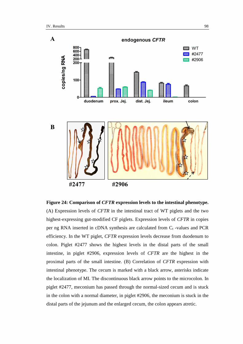
IV. Results 98
Figure 24: Comparison of CFTR expression levels to the intestinal phenotype.
(A) Expression levels of CFTR in the intestinal tract of WT piglets and the two
highest-expressing gut-modified CF piglets. Expression levels of CFTR in copies
per ng RNA inserted in cDNA synthesis are calculated from Ct -values and PCR
efficiency. In the WT piglet, CFTR expression levels decrease from duodenum to
colon. Piglet #2477 shows the highest levels in the distal parts of the small
intestine, in piglet #2906, expression levels of CFTR are the highest in the
proximal parts of the small intestine. (B) Correlation of CFTR expression with
intestinal phenotype. The cecum is marked with a black arrow, asterisks indicate
the localization of MI. The discontinuous black arrow points to the microcolon. In
piglet #2477, meconium has passed through the normal-sized cecum and is stuck
in the colon with a normal diameter, in piglet #2906, the meconium is stuck in the
distal parts of the jejunum and the enlarged cecum, the colon appears atretic.

IV. Results 99
To confirm that primers used for qPCR amplified the correct products, a melt
curve analysis was performed following each qPCR run (data not shown) and the
PCR products were sequenced via the Sanger approach (see Figure 25). The
correct amplification of the primers used for the determination of gut-modified
CFTR had already been proven. All amplified sequences showed 100 % identity
with the target sequences. Sequencing confirmed that very low levels of FABP2
are also expressed in other organs apart from the intestine.
Figure 25: Sequencing of qPCR products. (A) Extract of electropherograms
from the sequenced qPCR products. (B) Comparison of amplified sequences and
target sequences.

IV. Results 100
2. Modifier genes contribute to the severity of meconium
ileus in CFTR-/- piglets
As is known, the severity of the intestinal phenotype in human CF patients is
strongly influenced by modifier genes either increasing or decreasing the
susceptibility for MI (Blackman, Deering-Brose et al. 2006). Therefore, the
second approach for the rescue of the lethal intestinal phenotype in CF KO piglets
was to study if modifier genes might also play a role in the development of MI in
the porcine CF model.
2.1. Establishment of a breeding herd for the production of WT,
CFTR+/- and CFTR-/- piglets
In order to examine whether there was a naturally occurring variability in the
severity of MI in CF KO piglets, the intestinal phenotype of a large number of CF
KO piglets had to be analyzed. By selective mating of female and male CFTR+/-
pigs, litters containing piglets of all genotypes were produced for different
collaboration projects. To minimize inbreeding, mating between littermates was
avoided if possible. In 35 litters with 10.4 animals per litter on average, a total of
364 piglets were born. Approximately following the Mendelian rules, 99 piglets
were WT (27.2 %), 91 piglets were CF KO (25 %) and 174 piglets were CFTR+/-
(47.8 %). It is of note, that in contrast to data reported for male humans carrying
one CFTR mutation (Mak, Zielenski et al. 1999), we could not observe any
problems with reduced fertility or infertility in the CFTR+/- breeding boars.
Experiments on mucus transport in explanted WT and CFTR-/- airways
The main part of CF piglets was produced, genotyped and used for tissue
sampling to study mucociliary clearance in explanted tracheas by Anna Ermund
and Gunnar C. Hansson from the Mucin Biology Group at the University of
Gothenburg. The video microscopy imaging of living tracheal tissue showed that
the airways were cleaned by mucus in strands that move towards the larynx and
not by a continuous mucus layer. In CF KO airways, mucus strands are

IV. Results 101
transported with a much slower velocity compared to WT airways - a feature that
can be imitated in WT airways when bicarbonate is removed from the Krebs
buffer (see Figure 26).
Figure 26: Video microscopy imaging of living WT and CFTR-/- tracheal
tissue. (A) Mucociliary clearance in living tracheal tissue; first and last frame
from video microscopy imaging. Mucus is stained by Alcian blue, arrows of the
same color point to identical strands. Mucus does not form a continuous layer but
forms strands. Within a period of 5 min, mucus strands have moved in the WT
trachea but not in the trachea of CF KO piglets. (B) Calculation of transport

IV. Results 102
velocity of strands. Statistics calculated with Kruskal-Wallis non-parametric test.
Each data point represents the mean of several time lapses of one animal. Data
provided by Anna Ermund, Mucin Biology Group, University of Gothenburg.
Mucus strands are secreted by submucosal glands and should therefore mostly
consist of Mucin 5b (Muc5b), that is expressed by mucous cells in the submucosal
glands and by goblet cells of the surface epithelium (Meyerholz, Lambertz et al.
2015). However, immunofluorescent studies with antibodies for Mucin 5ac
(Muc5ac) and Muc5b and scanning electron microscopy revealed that mucus
strands released by submucosal glands were coated by mucus from goblet cells of
the surface epithelium. As goblet cells mainly express Muc5ac, similar amounts
of Muc5ac and Muc5b are found in the airway mucus. In terms of mucus
secretion and mucin contents, WT and CF KO mucus in the trachea do not differ
(see Figure 27).
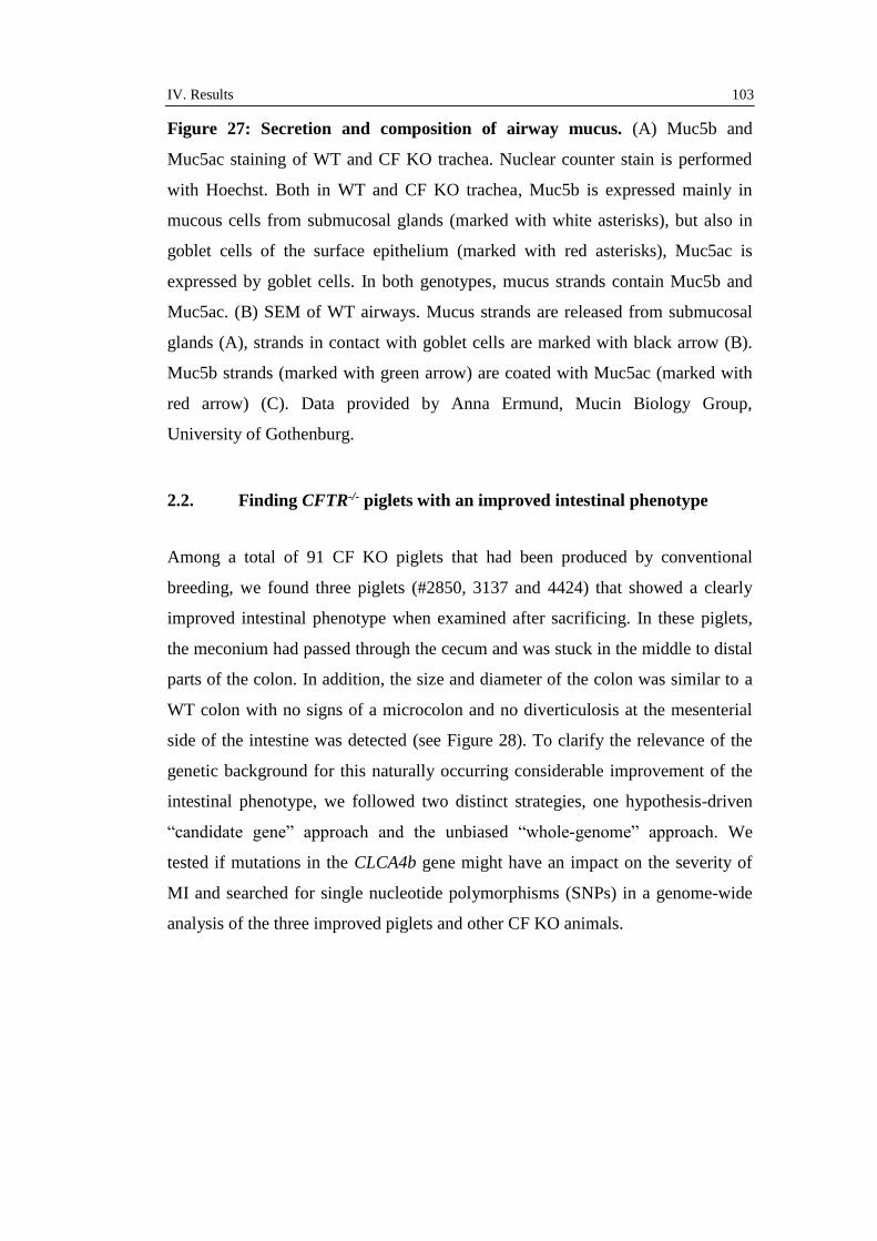
IV. Results 103
Figure 27: Secretion and composition of airway mucus. (A) Muc5b and
Muc5ac staining of WT and CF KO trachea. Nuclear counter stain is performed
with Hoechst. Both in WT and CF KO trachea, Muc5b is expressed mainly in
mucous cells from submucosal glands (marked with white asterisks), but also in
goblet cells of the surface epithelium (marked with red asterisks), Muc5ac is
expressed by goblet cells. In both genotypes, mucus strands contain Muc5b and
Muc5ac. (B) SEM of WT airways. Mucus strands are released from submucosal
glands (A), strands in contact with goblet cells are marked with black arrow (B).
Muc5b strands (marked with green arrow) are coated with Muc5ac (marked with
red arrow) (C). Data provided by Anna Ermund, Mucin Biology Group,
University of Gothenburg.
2.2. Finding CFTR-/- piglets with an improved intestinal phenotype
Among a total of 91 CF KO piglets that had been produced by conventional
breeding, we found three piglets (#2850, 3137 and 4424) that showed a clearly
improved intestinal phenotype when examined after sacrificing. In these piglets,
the meconium had passed through the cecum and was stuck in the middle to distal
parts of the colon. In addition, the size and diameter of the colon was similar to a
WT colon with no signs of a microcolon and no diverticulosis at the mesenterial
side of the intestine was detected (see Figure 28). To clarify the relevance of the
genetic background for this naturally occurring considerable improvement of the
intestinal phenotype, we followed two distinct strategies, one hypothesis-driven
“candidate gene” approach and the unbiased “whole-genome” approach. We
tested if mutations in the CLCA4b gene might have an impact on the severity of
MI and searched for single nucleotide polymorphisms (SNPs) in a genome-wide
analysis of the three improved piglets and other CF KO animals.

IV. Results 104
Figure 28: Improved intestinal phenotype in CFTR-/- piglets. (A) Features of
improvement. Small parts of the meconium are removed naturally (black arrow
points to removed meconium) and no herniation of mucosa is detected at the
mesenterial side of the intestine. After opening the abdomen, it becomes obvious
that the cecum (white arrow) is enlarged but filled with milk, and meconium is
visible in the colon gyri (white asterisks). (B) Intestine laid out in full length.
Meconium passed through the cecum (black arrow) and milk is found in the

IV. Results 105
proximal to middle parts of the colon (black asterisks). The diameter of the colon
is enlarged in comparison to “normal” CF KO piglets.
Influence of CLCA4b integrity on meconium ileus severity in CFTR-/- piglets
CLCA4 is known to modulate residual chloride conductance in the intestine of
human CF patients (Kolbe, Tamm et al. 2013). In the pig genome, a naturally
occurring duplication, CLCA4b, occurs, that is especially abundant in intestinal
cells and co-expressed with CFTR. A large percentage of the pig population has a
deleterious mutation within this gene. The 10 bp deletion leads to a loss of the WT
splice acceptor site in intron 7, resulting in alternative splicing to a sequence
38 bp upstream of exon 8. This causes a frameshift in protein translation and
generates a premature stop codon in exon 8 (see Figure 29 (A)) (Plog, Klymiuk et
al. 2015). We examined if there was correlation between the CLCA4b integrity
and the improved intestinal phenotype in the three CF KO piglets. Three primer
pairs amplifying the region containing the mutation were compared and primer
pair CLCA4b2f/CLCA4b2r was ultimately utilized for PCR (see Figure 29 (B)).
Sequencing the candidate region of CLCA4b in two of the three improved CF KO
piglets (#2850 and #3137) and 15 other CF KO piglets revealed that piglet #2850
was homozygous for the deletion in CLCA4b, whereas piglet #3137 was
heterozygous. Three other examined CF KO piglets without improved phenotype
were homozygous for the deletion, eight heterozygous and in four CF KO piglets
only WT alleles were detected. These results indicate that the deletion in CLCA4b
has no effect on the severity of MI and that CLCA4b is therefore no potential
modifier gene for the intestinal phenotype in the porcine CF model.

IV. Results 106
Figure 29: Sequencing of the candidate region in CLCA4b. (A) Deletion in
CLCA4b causes alternative splicing (WT and cryptic splice acceptor sites are
marked with a black and red arrow, respectively) and the transcription of a protein
with a premature stop codon (marked with asterisk). (B) Testing of primers for the
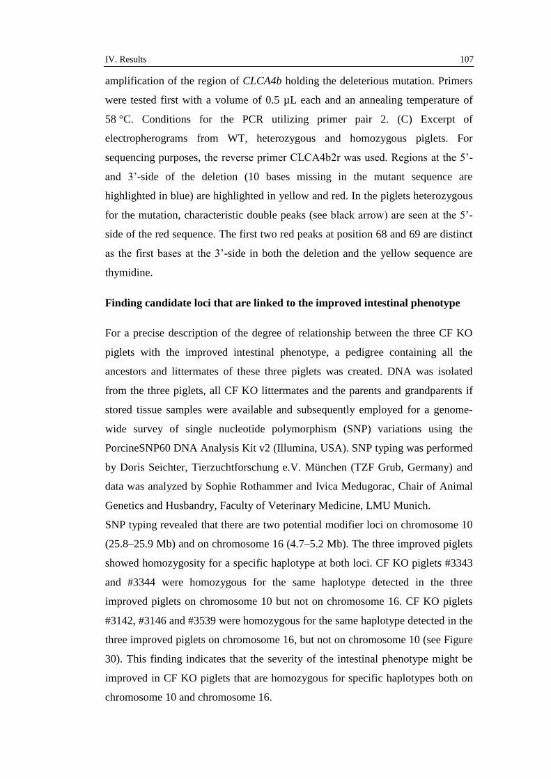
IV. Results 107
amplification of the region of CLCA4b holding the deleterious mutation. Primers
were tested first with a volume of 0.5 µL each and an annealing temperature of
58 °C. Conditions for the PCR utilizing primer pair 2. (C) Excerpt of
electropherograms from WT, heterozygous and homozygous piglets. For
sequencing purposes, the reverse primer CLCA4b2r was used. Regions at the 5’-
and 3’-side of the deletion (10 bases missing in the mutant sequence are
highlighted in blue) are highlighted in yellow and red. In the piglets heterozygous
for the mutation, characteristic double peaks (see black arrow) are seen at the 5’-
side of the red sequence. The first two red peaks at position 68 and 69 are distinct
as the first bases at the 3’-side in both the deletion and the yellow sequence are
thymidine.
Finding candidate loci that are linked to the improved intestinal phenotype
For a precise description of the degree of relationship between the three CF KO
piglets with the improved intestinal phenotype, a pedigree containing all the
ancestors and littermates of these three piglets was created. DNA was isolated
from the three piglets, all CF KO littermates and the parents and grandparents if
stored tissue samples were available and subsequently employed for a genome-
wide survey of single nucleotide polymorphism (SNP) variations using the
PorcineSNP60 DNA Analysis Kit v2 (Illumina, USA). SNP typing was performed
by Doris Seichter, Tierzuchtforschung e.V. München (TZF Grub, Germany) and
data was analyzed by Sophie Rothammer and Ivica Medugorac, Chair of Animal
Genetics and Husbandry, Faculty of Veterinary Medicine, LMU Munich.
SNP typing revealed that there are two potential modifier loci on chromosome 10
(25.8–25.9 Mb) and on chromosome 16 (4.7–5.2 Mb). The three improved piglets
showed homozygosity for a specific haplotype at both loci. CF KO piglets #3343
and #3344 were homozygous for the same haplotype detected in the three
improved piglets on chromosome 10 but not on chromosome 16. CF KO piglets
#3142, #3146 and #3539 were homozygous for the same haplotype detected in the
three improved piglets on chromosome 16, but not on chromosome 10 (see Figure
30). This finding indicates that the severity of the intestinal phenotype might be
improved in CF KO piglets that are homozygous for specific haplotypes both on
chromosome 10 and chromosome 16.

IV. Results 108

IV. Results 109
Figure 30: Pedigree and SNP-typing of three CFTR-/- piglets with improved
intestinal phenotype. The three improved CF KO piglets #2850, #3137 and
#4424 are highlighted in red. Filled and empty boxes indicate the haplotypes of
the two alleles (the potential locus on chromosome 10 is highlighted in red, the
potential locus on chromosome 16 in purple). The three improved piglets are the
only CF KO piglets that are homozygous for a specific haplotype (filled box) for
both events.
As we assumed that two separate autosomal recessive events had to combine in
order to achieve an improvement of the intestinal phenotype in CF KO piglets, the
probability to generate these improved CF KO piglets by mating of CFTR+/- pigs
was calculated according to Mendelian rules. Only 25 % of a litter are CF KO
piglets, therefore, the probability that a piglet is both a CF KO and homozygous
for the SNPs found on chromosome 10 and 16, is very low if the parents are not
selected for the specific haplotypes (see Table 20). For an efficient production of
CF KO piglets that are homozygous for both potential modifiers, it is important
for future breeding attempts that all pigs of the breeding herd will be screened for
the two candidate loci.
Table 20: Influence of the parents’ genotype on the probability to generate
CFTR-/- piglets with an improved intestinal phenotype.
Genotype of
parents
Probability for
piglets homozygous
for both events
(A/A; B/B)
Probability for CF
KO piglets
homozygous for both
events (A/A; B/B)
P1: A/a; B/b
P2: A/a; B/b
6.25 % ~1.56 %
P1: A/A; B/b
P2: A/a; B/b
12.5 % ~3.13 %
P1 A/A; B/b
P2: A/A; B/b
P1: A/A; B/B
P2: A/a; B/b
25 % 6.25 %
P1: A/A; B/b
P2: A/A; B/B
50 % 12.5 %
P1: A/A; B/B
P2: A/A; B/B
100 % 25 %

IV. Results 110
Parents’ (P1 and P2) haplotypes for the two candidate loci A and B are written in
capital letters for the desired haplotype and in lowercase for the WT haplotype.
Probabilities are calculated according to Mendelian rules.

V. Discussion 111
V. DISCUSSION
Although many important insights into pathological mechanisms have been
gained during the last decades and novel forms of therapy have been developed,
Cystic Fibrosis still remains a disease that severely reduces the life expectancy of
affected patients and thus warrants all the scientific efforts concerning this field
(Spielberg and Clancy 2016). Animal models are a unique and essential tool for a
better understanding of CF and the testing of new therapeutic approaches. Out of
all established animal models for CF, the porcine model shows the closest
resemblance to the classic human CF phenotype and thus enables a simplified
translation of findings to humans. Studies on neonatal tracheal tissue from CF KO
and WT piglets generated by breeding have given important new insights into
mucus composition and mucociliary transport in the airways (see Figure 26 and
Figure 27) and many collaboration partners are interested in tissue from newborn
piglets. However, since not all experiments can be performed on neonatal piglets,
the availability of older, living CF pigs is required. For this purpose, the main
limitation of the utility of the porcine CF model has to be overcome - the lethal
neonatal intestinal obstruction by meconium. In this thesis, two different
approaches to rescue the severe intestinal phenotype were evaluated; one
approach was the selective transgenic expression of CFTR in the intestine, while
the second approach investigated the contribution of modifier genes to an
improved intestinal phenotype in CF KO piglets. In addition, I characterized the
porcine CF model with different molecular biological methods.
The detection of CFTR in the intestine is only successful using one particular
antibody under very specific conditions
The investigation of CFTR at protein level proved to be very difficult and it
turned out that the detection was only successful in the intestine using one
particular antibody, Ab 596, under very specific conditions. Ten commercially
available primary antibodies that were described to detect porcine CFTR were

V. Discussion 112
evaluated for their specificity and sensitivity in paraffin-embedded and frozen
sections. In general, the advantages of frozen sections are that the antigenicity is
often better preserved, whereas the tissue and cellular morphology are better and
the handling and storage are easier when paraffin-embedded slides are used
(Fischer, Jacobson et al. 2008, Wild 2013).
In paraffin-embedded tissue, none of the examined antibodies recognized the
correct CFTR protein, even though different antigen retrieval techniques both for
heat-induced and proteolytic-induced epitope retrieval were tested. Although the
antibodies detected the typical expression pattern of CFTR in WT intestine with
the strongest signal intensity in the epithelial crypts and a decrease along the
crypt-villus axis, the main problem was that the staining pattern was identical with
that of CF KO intestine (see Figure 16). Interestingly, this feature could also be
detected in western blots, where several antibodies recognized a band of the
correct size both in WT and CF KO tissue (see Figure 17 (A)). A potential
explanation for this finding could be the fact that in this thesis the specificity of
antibodies was evaluated using porcine KO tissue as a “gold standard” negative
control and not only by staining with the secondary antibody; an experiment that
has not been done like this before, as no “real” negative control had been available
until previously. As CFTR is a member of the ABC transporters that are
characterized by highly-conserved motifs in the nucleotide-binding domains
(Rees, Johnson et al. 2009), antibodies against CFTR might also recognize
epitopes on other membrane transport proteins with a distribution pattern similar
to CFTR. This idea is fortified by a study in which antibodies against different
regions of CFTR recognized not only CFTR, but also immunologically related
proteins with a comparable molecular mass and distribution pattern (Walker,
Watson et al. 1995).
For the detection of human CFTR in frozen sections with the antibodies from the
CF folding consortium, a detailed protocol was available (Kreda and Gentzsch
2011). As the amino acid sequences from all purchased antibodies showed a
100 % identity between human and pig, it was expected that this protocol should
also work for the specific detection of CFTR in porcine tissue. However, neither
in frozen sections nor in methacarn sections with expected antigenicity
comparable to frozen sections (Mitchell, Ibrahim et al. 1985), a specific staining

V. Discussion 113
of CFTR was detected with this protocol. This indicates that antigen-antibody
interactions differ between species in spite of identical epitope sequences.
Only with one very specialized protocol the specific detection of CFTR worked
reliably on cryo-sections from the intestine of WT animals, while CF KO tissue
was negative. In the WT lung, only a very weak staining of the submucosal glands
was detected. This finding can be easily explained by the fact that CFTR
antibodies are often not sensitive enough to detect endogenous levels of the
protein that is expressed only in very low abundance in many tissues (Doucet,
Mendes et al. 2003). The expression analysis of CFTR at mRNA level showed
that CFTR expression in the proximal jejunum is about ten times higher than in
the lung (see Figure 24). There might be two possible issues contributing to the
specific detection of CFTR with this protocol. The freezing method with pre-
incubation in 2 % PFA and 30 % sucrose before snap-freezing embedded in OCT
compound in an ethanol/dry ice bath should only lead to a better conservation of
tissue morphology, but not interfere with the antigenicity of epitopes, which is in
general better preserved in frozen than in paraffin-embedded tissue (Wild 2013,
Hare, George et al. 2014). The second main difference to all other examined
protocols was the treatment of sections with Triton X-100. Triton X-100 is known
to increase the permeability of tissue for antibodies by destroying the membrane
structure (Chen, Cho et al. 2010) and might thereby lead to a better detection of
CFTR with Ab 596 that recognizes a part of the intracytoplasmatic NBD2 that
could otherwise be difficult to access. One problem with studies on the
distribution pattern of CFTR is, that even when only experiments on porcine
tissue are taken into account, the results are inconsistent. Plog, Mundhenk et al.
(2010) describe that porcine CFTR is expressed in ciliated epithelial cells and
goblet cells in the nasal, tracheal, and bronchial mucosa but not in the alveolar or
bronchiolar epithelia or submucosal glands. In contrast, Ballard, Trout et al.
(1999) localized CFTR mainly to the ducts of submucosal glands and in weaker
intensity to cells of the bronchial surface epithelium, an expression pattern
corresponding to the weak staining of submucosal glands we detected in WT lung
tissue (see Figure 14 (B)). Therefore, it is strongly recommended to repeat the
working protocol with other antibodies in order to evaluate if these antibodies can
be utilized for the specific detection of CFTR and if they recognize the same

V. Discussion 114
distribution pattern as Ab 596. Only when a panel of antibodies recognizing
different regions of CFTR detects the identical distribution pattern, a reliable
conclusion on the expression of CFTR in porcine tissue can be reached. As the
primary aim was to find an antibody that specifically detected CFTR in paraffin-
embedded tissue, a panel of monoclonal antibodies recognizing specific peptide
sequences of porcine CFTR were generated by immunization of rats and mice via
the hybridoma technique by the AG Kremmer, Institute of Molecular
Immunology Helmholtz Center Munich (Rottach, Kremmer et al. 2008). These
antibodies are currently evaluated using WT and CF KO tissue and will be
analyzed in more detail both for their utility for western blot and
immunohistochemistry in the future.
Not only the intensity, but also the localization of CFTR expression affects the
severity of meconium ileus in gut-modified CFTR-/- piglets
The correlation of CFTR expression levels with the intestinal phenotype in gut-
modified CFTR-/- piglets revealed that the improvement of the MI was more
pronounced when CFTR expression levels were higher in the distal parts of the
intestine. In contrast, when high CFTR expression levels were restricted to the
proximal parts of the small intestine, the intestinal phenotype did not differ from
normal CF KO piglets. In the ileum of founder #2477 with the clearest
improvement of MI, almost the same levels of CFTR were detected as in the WT
ileum, albeit without an increased survival rate (see Figure 24). This finding
remains contrary to a CF mouse model, in which only 5 % of normal CFTR
expression levels assured the survival from intestinal disease (Dorin, Farley et al.
1996). However, a direct translation of findings in CF mice to the porcine CF
model is difficult due to differences in the pathological mechanisms involved in
the obstructive intestinal disease in these two species. Another study performed by
Stoltz, Rokhlina et al. (2013) in CF KO piglets states that 20 % of WT CFTR
mRNA expression in the ileum of gut-corrected CFTR-/- piglets is sufficient to
rescue the lethal intestinal phenotype. This contradiction might be explained by
the fact that in this model CFTR expression was only measured in one part of the
intestine, the ileum, and not in five different regions of the intestine as in our

V. Discussion 115
study. Therefore, it is unclear, whether higher levels of CFTR were detected in
these piglets in other parts of the intestine that were the underlying reason for the
increased survival and whether the expression levels in the ileum do have a value
in predicting the severity of the intestinal phenotype.
We claim that in our examined gut-modified CFTR-/- piglets the survival was not
rescued, in spite of high CFTR expression levels in proximal parts of the intestine,
due to the lack of CFTR expression in the colon. We hypothesize that the
localization of transgenic CFTR expression in the colon is as important as the
intensity of the expression levels. There are several potential reasons why the
prevalence of MI in CF piglets is so much higher than in other animal models for
CF (ferret 75 %; mice develop DIOS-like obstruction around weaning) or affected
humans (up to 20 %) (Borowitz and Gelfond 2013). Studies performed by Plog,
Mundhenk et al. (2010) justify the 100 % incidence of MI in pigs with the strong
expression of CFTR in the ileum and colon of WT piglets. Apart from the missing
CFTR expression in the ileum and colon, other features are known to affect the
severity and incidence of MI. The existence of alternative chloride currents in the
intestine contributes to a modulation of gastrointestinal disease severity in CF
mice and might be a reason for the less pronounced intestinal phenotype in CF
mice models in comparison to the porcine model (Rozmahe, Wilschanski et al.
1996). CF patients carrying severe mutations in the CFTR gene are at a higher risk
to develop MI (Dupuis, Keenan et al. 2015), so the fact that CF pigs and ferrets
carry a null mutation should explain the high incidence of MI in these models.
Other findings contributing to the high incidence of MI are the specific anatomy
of the porcine large intestine, physiological differences in the secretory
mechanisms in the large intestine in relation to other species or the narrow inbred-
status of the experimental CF herd (Rogers, Stoltz et al. 2008, Sun, Sui et al.
2010).
Another indication that missing CFTR expression in the colon is primarily
responsible for the severe intestinal disease in CF pigs is the atretic microcolon
found in CF KO piglets (Meyerholz, Stoltz et al. 2010, Klymiuk, Mundhenk et al.
2012) that seems to be the main obstacle to the removal of meconium. Whether
the atresia of the colon is really caused primarily by a developmental effect due to
the lack of CFTR or develops subsequently because the functional obstruction

V. Discussion 116
leads to a missing distension by luminal contents (Amodio, Berdon et al. 2012),
should be analyzed in greater detail. However, the fact that from the first trimester
onwards CFTR is expressed throughout the intestine in high levels during fetal
development, gives a strong hint to the important role of CFTR in organ
development (Tizzano, Chitayat et al. 1993). Only recently, Than, Linnekamp et
al. (2016) demonstrated the importance of CFTR in the regulation of large
intestinal stem cells since CFTR acts as a tumor suppressor gene for colorectal
cancer in mice and humans. This implicates that CFTR expression in the distal
parts of the intestine is a major contributor to the correct formation and
development of the intestine.
An important question in this context is why the expression of CFTR is so high in
the colon of pigs. This might be due to an adaptive process to the normal
physiology of digestion in the porcine large intestine. CFTR-expressing goblet
cells in the intestine form the characteristic two-layer mucus that serves as part of
the innate immune system in form of a mucosal barrier (Kreda, Davis et al. 2012).
In the porcine large intestine, dietary fibers are fermented to short chain fatty-
acids (Jha and Berrocoso 2016) that stimulate colonic mucin production (Barcelo,
Claustre et al. 2000) and gut microbiota might even utilize released mucins as
energy source (Stanley, Ram et al. 1986). The magnified stimulation of mucus
production and digestion by gut microbiota might therefore have led to an
adaptive increase of goblet cell numbers in the large intestine.
The expression pattern of porcine FABP2 limits the utility of the promoter to
drive a selective expression of CFTR in the intestine
In order to achieve a selective expression of CFTR in the intestine of gut-modified
CFTR-/- piglets, the promoter of the porcine FABP2 gene was employed. The
FABP2 protein is involved in the uptake and cellular transport of dietary long
fatty-acids and therefore it is expressed predominantly and in high abundance in
the intestine (Jiang and Li 2006). As the mRNA of FABP2 is the most abundant
RNA sequence in the intestine (Bass 1988) and the cytosolic FABPs represent up
to 2 % of cytosolic proteins in the enterocytes (Ockner and Manning 1974), the
expression levels of the transgenic construct were expected to be high. In contrast
V. Discussion 117
to other studies that employed the FABP2 promoter from the rat for the transgenic
expression of CFTR in the mouse (Zhou, Dey et al. 1994) and pig (Stoltz,
Rokhlina et al. 2013), we decided to use a homologous promoter because its
activity should be stronger and the distribution pattern more precise in the same
species (Rathore and Sunilkumar 2005). In 33 % of all examined gut-modified
CFTR-/- founder animals, however, CFTR expression was not limited to the
intestine but could be detected, albeit with lower abundance, in other CF-relevant
organ systems such as the airways or the pancreas (see Figure 23). Therefore, the
utility of these animals would be limited as they would most likely not develop
the characteristic CF phenotype. This finding remains contrary to the US
transgenic porcine CF model that utilized the rat FABP2 promoter for the
selective expression of CFTR in the intestine and did not exhibit any extra-
intestinal expression (Stoltz, Rokhlina et al. 2013). However, consistent with our
results, in a CF mouse model expressing human CFTR under the control of the rat
FABP2 promoter, several lines showed an extra-intestinal expression (Zhou, Dey
et al. 1994), indicating that both rat and porcine FABP2 expression might not be
limited to the intestine. Measuring the expression levels of FABP2 by quantitative
PCR and sequencing of amplification products revealed that very high levels are
consistently found in all parts of the small intestine, with highest levels in the
jejunum, but also that small levels are detected in all other examined organs (see
Figure 22 and Figure 25). Other studies confirm that both rat and porcine FABP2
transcripts are detected in lower levels in organs other than the intestine (Gordon,
Elshourbagy et al. 1985, Jiang and Li 2006). Whether the detected CFTR
expression in extra-intestinal organs in our gut-modified CFTR-/-piglets was
triggered by the low amount of FABP2 expression in other organs or was caused
by the specific integration side of the transgenic vector, could only be clarified by
a whole genome sequencing of these piglets. However, expression patterns and
the corresponding results in the transgenic mouse model (Zhou, Dey et al. 1994)
provide a strong indication that the FABP2 promoter is not suitable to drive an
intestine-selective CFTR expression.
Another aspect concerning the utility of the FABP2 as promoter for the expression
of CFTR is the stage-specific expression during fetal development. As we
conclude that CFTR expression is essential for the development of the intestine

V. Discussion 118
and CFTR expression can be detected as early as in the first trimester of
pregnancy in humans (Tizzano, Chitayat et al. 1993), a similar developmental
expression of FABP2 might be important to prevent the formation of a
microcolon. A closer look at developmental expression patterns of rat and mouse
FABP2 showed that this gene is only expressed during the late trimester of
pregnancy in very low levels with a four-fold increase of expression levels within
the first 24 h after birth by gene induction (Gordon, Elshourbagy et al. 1985,
Green, Cohn et al. 1992). In contrast, studies on fetal human tissue report a
FABP2 expression very early in the gestational period (Levy, Menard et al. 2009).
As no studies on the developmental expression pattern of porcine FABP2 exist,
the suitability of the promoter to drive CFTR expression during early fetal
development remains a feature that should be further investigated.
The last aspect that should be considered in the evaluation of the usefulness of the
FABP2 promoter is that expression analysis of gut-modified CFTR-/- piglets
indicates the necessity of CFTR expression in the large intestine to ensure survival
(see Figure 24). Therefore, an ideal promoter should be characterized by high
expression in the distal parts of the intestine. FABP2 expression analysis at
mRNA level showed that FABP2 is predominantly expressed in the small
intestine and no measurable amounts are detected in the colon (see Figure 22).
Corresponding to this finding, FABP2 protein contents in the human small
intestine are significantly higher than in the colon, with highest levels in the
jejunum (Pelsers, Namiot et al. 2003). The absence of FABP2 expression in the
colon may have been the underlying reason for the severity of MI that could not
be improved to a sufficient degree in our gut-modified CFTR-/- piglets.
The genetic background of CFTR-/- piglets influences the severity of
meconium ileus
The finding with the strongest impact on future research concerning the porcine
CF model was that the severity of meconium ileus shows variability among CF
KO piglets produced by conventional breeding (see Figure 28). Until now all
groups working with CF pigs have reported a 100 % penetrance of MI,
independent from the mutation in the CFTR gene (Rogers, Stoltz et al. 2008,

V. Discussion 119
Ostedgaard, Meyerholz et al. 2011, Klymiuk, Mundhenk et al. 2012). In contrast,
in other species a high variability of the severity of the intestinal phenotype is
reported in CF, which is mainly influenced by the genetic background. MI in
human CF patients is one of several CF manifestations, such as CFRD (Blackman,
Commander et al. 2013) or susceptibility to chronic infections with P. aeruginosa
(Emond, Louie et al. 2012), which show very high heritability values. This
indicates the strong contribution of genetic factors apart from the CFTR mutation
to the phenotypic variety (Brennan and Schrijver 2016). Already in 1996, in CF
mice models the existence of secondary genetic factors influencing the intestinal
phenotype was described (Rozmahe, Wilschanski et al. 1996). Since, a range of
potential candidate genes like members of the solute carrier protein family or
multiple apical plasma membrane constituents have been identified by genome-
wide association studies (Sun, Rommens et al. 2012). However, in this study we
could demonstrate for the first time the presence of porcine modifier genes
contributing to a variable severity of MI in the CF pig model (see Figure 30).
Only in rare cases modifier genes associated with intestinal obstruction were
shown to have the same effect in two different species, like CLCA1 (Van der
Doef, Slieker et al. 2010) or MSRA (Henderson, Doshi et al. 2012) in CF mice
and humans. Nevertheless, it is important to compare the two candidate loci on
chromosome 10 and 16 in the porcine genome to regions in the human genome
syntenic to these loci. Thereby we can examine whether genes reported to modify
the CF phenotype in humans lay within this region and might therefore also be
potential modifier genes for the porcine model. However, first analyses indicate
that no described human modifier gene is expressed in the region of interest. Next
steps will include a more detailed examination of the two porcine candidate loci in
order to determine whether genes with a potential functional correlation with the
intestinal obstruction are expressed in this region. The majority of human modifier
genes for MI encode proteins that might interact directly with the functional
defects leading to the CF phenotype, e.g. by regulation of transepithelial
electrolyte and bicarbonate transport (Dorfman, Li et al. 2009) or modulation of
proteolysis of the abnormal proteinaceous meconium (Henderson, Doshi et al.
2012). In future studies it has to be clarified, whether SNPs lie within the coding
region of a gene, directly interfering with protein function, or are located at
V. Discussion 120
regulatory regions, affecting the binding of transcription factors and thereby
increasing or suppressing gene expression (Bush 2006).
In parallel to a detailed analysis of the two candidate loci it will be essential to
produce new CF piglets with an improved intestinal phenotype in order to
investigate if these piglets can completely remove meconium and survive the first
critical days of life. These piglets can be generated by two different ways – either
by reclonation of one of the improved CF piglets via SCNT and ET or by
conventional breeding after a screening of the breeding herd for the desired
haplotypes. The generation of “gut-improved” CF piglets by SCNT and ET is a
fast and efficient method to produce a small number of animals with the desired
genotype and is therefore suited for the evaluation of the intestinal phenotype as a
kind of “proof of concept” study. However, it has to be taken into account that in
the recloned piglets from gut-modified CF founder #2478, the improved intestinal
phenotype was not reproducible (see Figure 7) and that recloned “gut-improved”
piglets might therefore not exhibit the same level of improvement as the donor
animals. In addition, the production of large numbers of viable animals via this
method is not feasible due to low cloning efficiency (see Table 14 and Table 15)
and a high rate of phenotypical abnormalities (see Figure 7), which might be due
to epigenetic reprogramming or genomic damage of the donor cells (Cho, Kim et
al. 2007). Therefore, all effort will be made to produce these gut-improved CF
piglets by conventional breeding in the future. By screening of the breeding herd
for the two candidate loci we will be able to selectively mate animals with a
specific haplotype to increase the probability of CF KO piglets homozygous for
both loci according to Table 20. If “gut-improved” CF KO piglets can survive the
first critical days, they will make an ideal animal model for Cystic Fibrosis. In
contrast to the porcine CF model with transgenic expression of CFTR in the
intestine, the severity of meconium ileus is rescued in these piglets by naturally
occurring modifier genes, whose existence is also described in human CF patients.
Therefore, a living “gut-improved” CF pig will be an ideal large animal model for
the investigation of unknown pathological mechanisms and testing of novel forms
of therapy. It opens new prospects for CF research and can thereby lead to a better
understanding of Cystic Fibrosis or even the development and evaluation of
potential curative treatments.
VI. Summary 121
VI. SUMMARY
The pig as an animal model for Cystic Fibrosis – approaches to overcome the
lethal intestinal phenotype
Cystic Fibrosis (CF) is one of the most common lethal autosomal recessive
hereditary diseases in people from Caucasian origin. Mutations in the gene
encoding the anion channel Cystic Fibrosis Transmembrane Conductance
Regulator (CFTR) cause defective transepithelial electrolyte transport. The
consequence of this pathophysiological mechanism is a multisystemic disease
mainly affecting the airways, the gastrointestinal tract and the pancreas. Chronic
inflammations and infections of the respiratory tract are the main reasons for the
decreased survival rate of CF patients. Although novel drugs, so-called
“modulators”, targeting specific mutation classes directly at the genetic or protein
level have been developed in recent years, treatment options are still primarily
limited to symptomatic therapy. So far, five different established animal models
are available for CF research. As the porcine CF model bears the strongest
resemblance to the classic human CF phenotype, important insights into the
underlying pathological mechanisms involved in the development of airway
disease have been gained by studying this model organism. The main aspect
limiting the suitability of the porcine CF model is the lethal neonatal meconium
ileus (MI) that occurs with a penetrance of 100 % and proves to be fatal if not
surgically corrected.
In this thesis, I evaluated two different approaches to rescue the lethal intestinal
phenotype of CF piglets. The first approach focused on the idea that the transgenic
expression of CFTR under the control of the intestine-specific fatty-acid binding
protein (FABP2) promoter should ameliorate the intestinal obstruction in CF
piglets without interfering with other typical organ manifestations of CF. Despite
an elaborate supporting treatment, none of the transgenic gut-modified piglets was
able to remove the meconium completely. The examination of the intestinal
phenotype consistently revealed an improvement of MI, indicated by a more distal
localization and the absence of diverticulosis. The establishment of a fast
genotyping protocol in less than three hours and the sequencing of amplified DNA

VI. Summary 122
confirmed the genetic CF background and the integration of the transgenic vector
into the CF genome. By quantitative PCR, I determined the expression level of
CFTR in different parts of the intestine and other CF-relevant organs and could
demonstrate that 33 % of all animals expressed CFTR in all organs. In all other
tested animals, the expression of CFTR was restricted to the small intestine and
correlated positively with the expression of FABP2. A correlation of the intestinal
phenotype with CFTR expression levels revealed that the higher the expression
levels were in the distal parts of the intestine, the more distinct was the
improvement in the phenotype. In addition, I confirmed the absence of CFTR in
the intestine and lungs of CF piglets by immunohistochemistry and
immunofluorescence.
In the second part of the thesis, I investigated whether modifier genes contributed
to the varying severity of meconium ileus that naturally occurred when CF pigs
were produced by conventional breeding. In a hypothesis-driven “candidate gene”
approach, I demonstrated by sequencing that the integrity of CLCA4b does not
affect the intestinal phenotype in CF piglets. The genome-wide search for single
nucleotide polymorphisms (SNPs) in CF piglets with an improved phenotype in
comparison to other CF piglets revealed two potential candidate loci on
chromosome 10 and 16, which have to be combined in an autosomal recessive
way to improve the intestinal phenotype.
In conclusion, I could demonstrate two different ways to improve the lethal
intestinal phenotype in a porcine model for Cystic Fibrosis in this thesis. The
intestinal phenotype of transgenic gut-modified CF piglets was clearly alleviated,
however, not to a sufficient degree to ensure survival. Presumably, this was
caused by the absence of CFTR expression in the colon. In addition, the FABP2
promoter was not suitable to drive an intestine-specific CFTR expression with
high levels in the distal parts. In contrast, the second approach looks very
promising. The discovery of potential modifier genes that contribute to a variable
intestinal phenotype in CF piglets harbors great potential for the development of a
living porcine CF model.

VII. Zusammenfassung 123
VII. ZUSAMMENFASSUNG
Das Schwein als Tiermodell für Mukoviszidose - Ansätze zur Behebung des
tödlichen intestinalen Phänotyps
Mukoviszidose, auch zystische Fibrose (CF) genannt, gehört zu einer der
häufigsten tödlichen autosomal rezessiven Erbkrankheiten bei Menschen mit
kaukasischem Hintergrund. Mutationen des CFTR-Gens, das für den Anionen-
kanal Cystic Fibrosis Transmembrane Conductance Regulator (CFTR) codiert,
verursachen einen fehlerhaften Elektrolyttransport an der Epitheloberfläche. Die
Folge ist eine multisystemische Erkrankung, die vor allem die Luftwege, den
Darmtrakt und die Bauchspeicheldrüse betrifft. Chronische Entzündungen und
Infektionen des Atemtrakts sind die Hauptursachen für die eingeschränkte
Lebenserwartung von CF-Patienten. Obwohl in den letzten Jahren neue
Medikamente, sogenannte Modulatoren, entwickelt wurden, die gezielt auf
Genom- oder Proteinebene spezielle Mutationsklassen angreifen, beschränkt sich
die Behandlung auch heutzutage vorrangig auf eine symptomatische Therapie.
Fünf verschiedene etablierte Tiermodelle stehen bislang der CF-Forschung zur
Verfügung. Da das Schweinemodell dem klassischen Phänotyp beim Menschen
am stärksten ähnelt, konnten anhand dieses Tiermodells wichtige Erkenntnisse
über die pathophysiologischen Prozesse, die der Entwicklung der Erkrankung der
Atemwege zugrunde liegen, gewonnen werden. Der Nutzen des CF-
Schweinemodells ist jedoch enorm eingeschränkt, da es bei neugeborenen CF-
Ferkeln mit einer Inzidenz von 100 % zu einem Mekoniumileus (MI) kommt, der
ohne Operation tödlich verläuft.
In meiner Dissertation entwickelte ich zwei verschiedene Ansätze, um den
tödlichen Phänotyp im Darm von neugeborenen CF-Ferkeln zu beheben. Der erste
Ansatz basierte auf der Hypothese, dass eine transgene Expression von CFTR
unter der Kontrolle des Promoters des nur im Darm exprimierten Gens FABP2
(fatty-acid binding protein) zu einer Verbesserung des MI führen sollte, ohne
dabei andere CF-relevante Organe zu beeinflussen.

VII. Zusammenfassung 124
Trotz einer ausgeprägten unterstützenden Behandlung konnte keines der
transgenen „darmverbesserten“ Ferkel das Mekonium vollständig ausscheiden.
Die visuelle Begutachtung des Darmtrakts zeigte bei allen Tieren eine
Verbesserung des MI mit einer distaleren Lokalisation und einem Fehlen der
Divertikulose. Die Genotypisierung und Sequenzierung der amplifizierten DNA
bestätigte die Genetik der CF-Ferkel und die Integration des transgenen Vektors
in das Genom. Mit quantitativer PCR wurde die Menge an exprimiertem CFTR in
verschiedenen Darmabschnitten und anderen CF-relevanten Organen bestimmt.
Dabei exprimierten 33 % der untersuchten Tiere CFTR in allen Organen. Bei allen
anderen getesteten Tieren war die Expression ausschließlich auf den Dünndarm
beschränkt und korrelierte positiv mit dem Expressionsmuster von FABP2. Die
Korrelation zwischen dem Darmphänotyp und der Menge an exprimiertem CFTR
zeigte, dass die Verbesserung des Darmphänotyps umso ausgeprägter war, je
höher die Expression in distaleren Darmabschnitten war. Zusätzlich konnte ich
mit immunhistochemischen und Immunfluoreszenz-Methoden bestätigen, dass im
Darm und in der Lunge von CF Ferkeln kein CFTR Protein exprimiert wird.
Im zweiten Teil der Dissertation wurde untersucht, ob Modifier-Gene für die
unterschiedliche Ausprägung des Darmphänotyps, die natürlicherweise bei
gezüchteten CF Ferkeln auftrat, verantwortlich sind. In einem hypothesen-
gesteuerten Kandidatengen-Ansatz wurde gezeigt, dass Deletionen im CLCA4b-
Gen in keinem Zusammenhang mit dem verbesserten Darmphänotyp stehen. Die
genomweite Suche nach Einzelnukleotidpolymorphismen, in denen sich CF-
Ferkel mit verbessertem Phänotyp von anderen CF-Ferkeln unterscheiden, ergab
zwei potentielle Kandidatenregionen auf den Chromosomen 10 und 16, die
autosomal rezessiv miteinander kombiniert sein müssen.
Zusammengefasst beschreibt diese Dissertation zwei verschiedene Ansätze, den
tödlichen intestinalen Phänotyp in einem Schweinemodell für Mukoviszidose zu
beheben. Die Ausprägung des Darmphänotyps in dem transgenen CF-
Schweinemodell konnte zwar deutlich verbessert werden, jedoch nicht in einem
solchen Maß, dass das Überleben der Tiere gewährleistet wurde. Die Ursache
dafür liegt vermutlich in der fehlenden Expression von CFTR im Colon. Des
Weiteren ist der FABP2 Promoter ungeeignet, eine darmspezifische CFTR-
Expression mit hohen Spiegeln im Dickdarm zu bewirken.

VII. Zusammenfassung 125
Im Gegensatz dazu ist der zweite Ansatz durchaus vielversprechend. Die
Entdeckung von möglichen Modifier-Genen, die den Darmphänotyp von CF-
Ferkeln beeinflussen, birgt großes Potential für die Entwicklung eines lebenden
CF-Schweinemodells.

VIII. Index of figures 126
VIII. INDEX OF FIGURES
Figure 1: Generation of gut-modified CFTR-/- piglets..................................... 39
Figure 2: Sampling of the intestine. ................................................................ 41
Figure 3: Preparation of the airways. .............................................................. 43
Figure 4: DNA construct for generation of gut-modified CFTR-/- piglets. ..... 62
Figure 5: Intestinal phenotype of WT, gut-modified CFTR-/- and CFTR-/-
piglets ............................................................................................... 66
Figure 6: Range of MI in WT, gut-modified CFTR-/- (gut CF) and CFTR-/-
piglets. .............................................................................................. 67
Figure 7: Phenotype of recloned founder piglets. ........................................... 70
Figure 8: Testing of primer pairs for the detection of WT CFTR. .................. 72
Figure 9: Testing of primer pairs for the detection of mutant (TG) CFTR. .... 73
Figure 10: Testing of primer pairs for the detection of the gut-modified CFTR
expression vector. ............................................................................ 74
Figure 11: Example of genotyping gut-modified CFTR-/- (gutCF), WT, CFTR+/-
(het) and CFTR-/- (hom) piglets. ...................................................... 75
Figure 12: Verification of genotyping PCR products. ...................................... 76

VIII. Index of figures 127
Figure 13: Immunofluorescent detection of CFTR in the intestine of WT and
CFTR-/- piglets with Ab 596. ........................................................... 82
Figure 14: Immunohistochemical detection of CFTR in WT and CFTR-/-
intestine and lung with Ab 596. ....................................................... 83
Figure 15: Testing of antibodies on paraffin-embedded lung tissue from WT
and CFTR-/- piglets. .......................................................................... 85
Figure 16: Unspecific staining of CFTR in the intestine of WT and CFTR-/-
piglets. .............................................................................................. 86
Figure 17: Detection of CFTR by WB. ............................................................. 88
Figure 18: Quality assessment of RNA and cDNA. .......................................... 90
Figure 19: Testing of primer pairs for the detection of CFTR in qPCR. ........... 92
Figure 20: Testing of primer pairs for the detection of FABP2 in qPCR. ......... 93
Figure 21: Expression levels of TBP2 in different organs and genotypes. ....... 94
Figure 22: Expression levels of FABP2 in different organs and genotypes. ..... 95
Figure 23: Expression levels of gut-modified CFTR in founder animals and
recloned piglets. ............................................................................... 97
Figure 24: Comparison of CFTR expression levels to the intestinal phenotype. ..
......................................................................................................... 98
Figure 25: Sequencing of qPCR products. ........................................................ 99
VIII. Index of figures 128
Figure 26: Video microscopy imaging of living WT and CFTR-/- tracheal tissue.
....................................................................................................... 101
Figure 27: Secretion and composition of airway mucus. ................................ 103
Figure 28: Improved intestinal phenotype in CFTR-/- piglets. ........................ 104
Figure 29: Sequencing of the candidate region in CLCA4b. ........................... 106
Figure 30: Pedigree and SNP-typing of three CFTR-/- piglets with improved
intestinal phenotype. ...................................................................... 109

IX. Index of tables 129
IX. INDEX OF TABLES
Table 1: Treatment protocol for gut-modified CFTR-/- piglets. ..................... 40
Table 2: Master mix composition for genotyping PCRs................................ 46
Table 3: Cycler protocol for genotyping PCRs. ............................................. 46
Table 4: Master mix composition for sequencing PCRs. ............................... 48
Table 5: Cycler protocol for sequencing PCRs. ............................................. 48
Table 6: Master mix composition for qPCRs................................................. 53
Table 7: Cycler protocol for qPCRs. .............................................................. 53
Table 8: Tissue processing for paraffin histology. ......................................... 55
Table 9: Deparaffinization and rehydration of paraffin sections. .................. 55
Table 10: Standard IHC protocol with HIER. .................................................. 56
Table 11: Composition of 8 % SDS polyacrylamide separating gel. ............... 59
Table 12: Composition of 5 % SDS polyacrylamide stacking gel. .................. 60
Table 13: Standard western blot protocol. ....................................................... 61

IX. Index of tables 130
Table 14: Results of SCNT experiments using gut-modified CFTR-/- embryos. .
......................................................................................................... 65
Table 15: Recloning experiments of founder #2478. ....................................... 69
Table 16: Comparison of DNA isolation methods. .......................................... 71
Table 17: Overview of all tested immunohistochemical conditions. ............... 78
Table 18: Overview of tested western blot conditions. .................................... 87
Table 19: Final conditions for qPCR. .............................................................. 93
Table 20: Influence of the parents’ genotype on the probability to generate
CFTR-/- piglets with an improved intestinal phenotype. ................ 109

X. Reference list 131
X. REFERENCE LIST
Adam, R. J., A. S. Michalski, C. Bauer, M. H. Abou Alaiwa, T. J. Gross, M. S.
Awadalla, D. C. Bouzek, N. D. Gansemer, P. J. Taft, M. J. Hoegger, A. Diwakar,
M. Ochs, J. M. Reinhardt, E. A. Hoffman, R. R. Beichel, D. K. Meyerholz and D.
A. Stoltz (2013). "Air trapping and airflow obstruction in newborn cystic fibrosis
piglets." Am J Respir Crit Care Med 188(12): 1434-1441.
Aigner, B., S. Renner, B. Kessler, N. Klymiuk, M. Kurome, A. Wunsch and E.
Wolf (2010). "Transgenic pigs as models for translational biomedical research." J
Mol Med (Berl) 88(7): 653-664.
Alton, E. W., D. K. Armstrong, D. Ashby, K. J. Bayfield, D. Bilton, E. V.
Bloomfield, A. C. Boyd, J. Brand, R. Buchan and R. Calcedo (2015). "Repeated
nebulisation of non-viral CFTR gene therapy in patients with cystic fibrosis: a
randomised, double-blind, placebo-controlled, phase 2b trial." The Lancet
Respiratory Medicine 3(9): 684-691.
Amaral, M. and C. Farinha (2013). "Rescuing mutant CFTR: a multi-task
approach to a better outcome in treating cystic fibrosis." Current pharmaceutical
design 19(19): 3497-3508.
Amaral, M. D. and K. Kunzelmann (2007). "Molecular targeting of CFTR as a
therapeutic approach to cystic fibrosis." Trends in pharmacological sciences
28(7): 334-341.
Amodio, J., W. Berdon, S. Abramson and C. Stolar (2012). "Microcolon of
prematurity: a form of functional obstruction." American Journal of
Roentgenology 146(2): 239-244.

X. Reference list 132
Anderson, D. H. (1938). "Cystic Fibrosis of the pancreas and its relation to celiac
disease: a clinical and pathological study." Am J Dis Child 56: 344-399.
Awadalla, M., S. Miyawaki, M. H. Abou Alaiwa, R. J. Adam, D. C. Bouzek, A. S.
Michalski, M. K. Fuld, K. J. Reynolds, E. A. Hoffman, C. L. Lin and D. A. Stoltz
(2014). "Early airway structural changes in cystic fibrosis pigs as a determinant of
particle distribution and deposition." Ann Biomed Eng 42(4): 915-927.
Ballard, S. T., L. Trout, Z. Bebök, E. J. Sorscher and A. Crews (1999). "CFTR
involvement in chloride, bicarbonate, and liquid secretion by airway submucosal
glands." American Journal of Physiology - Lung Cellular and Molecular
Physiology 277(4): L694-L699.
Barcelo, A., J. Claustre, F. Moro, J. Chayvialle, J. Cuber and P. Plaisancie (2000).
"Mucin secretion is modulated by luminal factors in the isolated vascularly
perfused rat colon." Gut 46(2): 218-224.
Bass, N. M. (1988). "The cellular fatty-acid binding proteins: aspects of structure,
regulation, and function." Int Rev Cytol 111: 143-184.
Bell, S. C., K. De Boeck and M. D. Amaral (2015). "New pharmacological
approaches for cystic fibrosis: Promises, progress, pitfalls." Pharmacology &
Therapeutics 145: 19-34.
Birket, S. E., K. K. Chu, L. Liu, G. H. Houser, B. J. Diephuis, E. J. Wilsterman,
G. Dierksen, M. Mazur, S. Shastry and Y. Li (2014). "A functional anatomic
defect of the cystic fibrosis airway." American journal of respiratory and critical
care medicine 190(4): 421-432.
X. Reference list 133
Blackman, S. M., C. W. Commander, C. Watson, K. M. Arcara, L. J. Strug, J. R.
Stonebraker, F. A. Wright, J. M. Rommens, L. Sun and R. G. Pace (2013).
"Genetic modifiers of cystic fibrosis-related diabetes." Diabetes: DB_130510.
Blackman, S. M., R. Deering-Brose, R. McWilliams, K. Naughton, B. Coleman,
T. Lai, M. Algire, S. Beck, J. Hoover-Fong, A. Hamosh, M. D. Fallin, K. West, D.
E. Arking, A. Chakravarti, D. J. Cutler and G. R. Cutting (2006). "Relative
Contribution of Genetic and Non-genetic Modifiers to Intestinal Obstruction in
Cystic Fibrosis." Gastroenterology 131(4): 1030-1039.
Borowitz, D., R. D. Baker and V. Stallings (2002). "Consensus report on nutrition
for pediatric patients with cystic fibrosis." Journal of pediatric gastroenterology
and nutrition 35(3): 246-259.
Borowitz, D. and D. Gelfond (2013). "Intestinal complications of cystic fibrosis."
Current opinion in pulmonary medicine 19(6): 676-680.
Boucher, R. (2007). "Evidence for airway surface dehydration as the initiating
event in CF airway disease." Journal of internal medicine 261(1): 5-16.
Bréa, D., F. Meurens, V. Dubois A , J. Gaillard, C. Chevaleyre, M. L. Jourdan, N.
Winter, B. Arbeille, M. Si-Tahar, F. Gauthier and S. Attucci (2012). "The pig as a
model for investigating the role of neutrophil serine proteases in human
inflammatory lung diseases." Biochem J 447(Pt 3): 363-370.
Brennan, M.-L. and I. Schrijver (2016). "Cystic Fibrosis: A Review of Associated
Phenotypes, Use of Molecular Diagnostic Approaches, Genetic Characteristics,
Progress, and Dilemmas." The Journal of Molecular Diagnostics 18(1): 3-14.

X. Reference list 134
Bronsveld, I., F. Mekus, J. Bijman, M. Ballmann, H. R. de Jonge, U. Laabs, D. J.
Halley, H. Ellemunter, G. Mastella, S. Thomas, H. J. Veeze and B. Tümmler
(2001). "Chloride conductance and genetic background modulate the cystic
fibrosis phenotype of ΔF508 homozygous twins and siblings." J Clin Invest
108(11): 1705-1715.
Bush, A. (2006). Cystic fibrosis in the 21st century, Karger Medical and Scientific
Publishers.
Bustin, S. A. (2004). AZ of quantitative PCR, International University Line San
Diego, CA.
Chang, E. H., R. S. Lacruz, T. G. Bromage, P. Bringas, M. J. Welsh, J. Zabner
and M. L. Paine (2011). "Enamel Pathology Resulting from Loss of Function in
the Cystic Fibrosis Transmembrane Conductance Regulator in a Porcine Animal
Model." Cells Tissues Organs 194(2-4): 249-254.
Chang, E. H., A. A. Pezzulo, D. K. Meyerholz, A. E. Potash, T. J. Wallen, L. R.
Reznikov, J. C. Sieren, P. H. Karp, S. Ernst, T. O. Moninger, N. D. Gansemer, P.
B. McCray, Jr., D. A. Stoltz, M. J. Welsh and J. Zabner (2012). "Sinus hypoplasia
precedes sinus infection in a porcine model of cystic fibrosis." Laryngoscope
122(9): 1898-1905.
Chen, J. H., D. A. Stoltz, P. H. Karp, S. E. Ernst, A. A. Pezzulo, T. O. Moninger,
M. V. Rector, L. R. Reznikov, J. L. Launspach, K. Chaloner, J. Zabner and M. J.
Welsh (2010). "Loss of anion transport without increased sodium absorption
characterizes newborn porcine cystic fibrosis airway epithelia." Cell 143(6): 911-
923.
Chen, X., D.-B. Cho and P.-C. Yang (2010). "Double staining
immunohistochemistry." North American Journal of Medical Sciences 2(5): 241-
245.

X. Reference list 135
Cho, S.-K., J.-H. Kim, J.-Y. Park, Y.-J. Choi, J.-I. Bang, K.-C. Hwang, E.-J. Cho,
S.-H. Sohn, S. J. Uhm, D.-B. Koo, K.-K. Lee, T. Kim and J.-H. Kim (2007).
"Serial cloning of pigs by somatic cell nuclear transfer: Restoration of phenotypic
normality during serial cloning." Developmental Dynamics 236(12): 3369-3382.
Clancy, J. P. and M. Jain (2012). "Personalized Medicine in Cystic Fibrosis."
American Journal of Respiratory and Critical Care Medicine 186(7): 593-597.
Cutting, G. R. (2015). "Cystic fibrosis genetics: from molecular understanding to
clinical application." Nat Rev Genet 16(1): 45-56.
Cystic Fibrosis Foundation. (2016). "CF Clinical Care Guidelines." 2016, from
https://www.cff.org/For-Caregivers/CF-Clinical-Care-Guidelines/.
Cystic Fibrosis Foundation. (2016). "Drug development pipeline." Retrieved
05.01.2016, 2016, from https://tools.cff.org/research/drugdevelopmentpipeline/.
Cystic Fibrosis Mutation Database. (2015). "CFMDB Statistics." Retrieved 17
Dec, 2015, from http://www.genet.sickkids.on.ca/StatisticsPage.html.
Davidson, D. J. and M. Rolfe (2001). "Mouse models of cystic fibrosis." Trends in
Genetics 17(10): S29-S37.
Davies, J. C., E. W. F. W. Alton and A. Bush (2007). "Cystic fibrosis." BMJ
335(7632): 1255-1259.
Debray, D., D. Rainteau, V. Barbu, M. Rouahi, H. E. Mourabit, S. Lerondel, C.
Rey, L. Humbert, D. Wendum, C. H. Cottart, P. Dawson, N. Chignard and C.
Housset (2012). "Defects in Gallbladder Emptying and Bile Acid Homeostasis in
Mice With Cystic Fibrosis Transmembrane Conductance Regulator Deficiencies."
Gastroenterology 142(7): 1581-1591.e1586.

X. Reference list 136
Denning, G. M., L. S. Ostedgaard, S. H. Cheng, A. E. Smith and M. J. Welsh
(1992). "Localization of cystic fibrosis transmembrane conductance regulator in
chloride secretory epithelia." J Clin Invest 89(1): 339-349.
di Sant'Agnese, P. A., R. C. Darling, G. A. Perera and E. Shea (1953). "Abnormal
electrolyte composition of sweat in cystic fibrosis of the pancreas. Clinical
Significance and Relationship to the Disease." Pediatrics 12(5): 549-563.
Diwakar, A., R. J. Adam, A. S. Michalski, M. M. Tamegnon, A. J. Fischer, J. L.
Launspach, R. A. Horan, S. C. Kao, K. Chaloner, D. K. Meyerholz and D. A.
Stoltz (2015). "Sonographic evidence of abnormal tracheal cartilage ring structure
in cystic fibrosis." The Laryngoscope 125(10): 2398-2404.
Dodge, J. A., P. A. Lewis, M. Stanton and J. Wilsher (2007). "Cystic fibrosis
mortality and survival in the UK: 1947–2003." European Respiratory Journal
29(3): 522-526.
Dorfman, R., W. Li, L. Sun, F. Lin, Y. Wang, A. Sandford, P. D. Paré, K. McKay,
H. Kayserova, T. Piskackova, M. Macek, K. Czerska, D. Sands, H. Tiddens, S.
Margarit, G. Repetto, M. K. Sontag, F. J. Accurso, S. Blackman, G. R. Cutting, L.
C. Tsui, M. Corey, P. Durie, J. Zielenski and L. J. Strug (2009). "Modifier gene
study of meconium ileus in cystic fibrosis: statistical considerations and gene
mapping results." Hum Genet 126(6): 763-778.
Dorin, J., R. Farley, S. Webb, S. Smith, E. Farini, S. Delaney, B. Wainwright, E.
Alton and D. Porteous (1996). "A demonstration using mouse models that
successful gene therapy for cystic fibrosis requires only partial gene correction."
Gene therapy 3(9): 797-801.

X. Reference list 137
Doucet, L., F. Mendes, T. Montier, P. Delépine, D. Penque, C. Férec and M. D.
Amaral (2003). "Applicability of Different Antibodies for the
Immunohistochemical Localization of CFTR In Respiratory and Intestinal Tissues
of Human and Murine Origin." Journal of Histochemistry & Cytochemistry 51(9):
1191-1199.
Dupuis, A., K. Keenan, C. Y. Ooi, R. Dorfman, M. K. Sontag, L. Naehrlich, C.
Castellani, L. J. Strug, J. M. Rommens and T. Gonska (2015). "Prevalence of
meconium ileus marks the severity of mutations of the Cystic Fibrosis
Transmembrane Conductance Regulator (CFTR) gene." Genet Med.
Efrati, O., A. Barak, D. Modan-Moses, A. Augarten, D. Vilozni, D. Katznelson,
A. Szeinberg, J. Yahav and Y. Bujanover (2003). "Liver cirrhosis and portal
hypertension in cystic fibrosis." European journal of gastroenterology &
hepatology 15(10): 1073-1078.
Emond, M. J., T. Louie, J. Emerson, W. Zhao, R. A. Mathias, M. R. Knowles, F.
A. Wright, M. J. Rieder, H. K. Tabor and D. A. Nickerson (2012). "Exome
sequencing of extreme phenotypes identifies DCTN4 as a modifier of chronic
Pseudomonas aeruginosa infection in cystic fibrosis." Nature genetics 44(8): 886-
889.
Farinha, C. M., D. Penque, M. Roxo-Rosa, G. Lukacs, R. Dormer, M. McPherson,
M. Pereira, A. G. Bot, H. Jorna and R. Willemsen (2004). "Biochemical methods
to assess CFTR expression and membrane localization." Journal of Cystic Fibrosis
3: 73-77.
Farrell, P. M., B. J. Rosenstein, T. B. White, F. J. Accurso, C. Castellani, G. R.
Cutting, P. R. Durie, V. A. LeGrys, J. Massie and R. B. Parad (2008). "Guidelines
for diagnosis of cystic fibrosis in newborns through older adults: Cystic Fibrosis
Foundation consensus report." The Journal of pediatrics 153(2): S4-S14.

X. Reference list 138
Fischer, A. H., K. A. Jacobson, J. Rose and R. Zeller (2008). "Cryosectioning
Tissues." Cold Spring Harbor Protocols 2008(8): 4991.
Fisher, J. T., Y. Zhang and J. F. Engelhardt (2011). "Comparative Biology of
Cystic Fibrosis Animal Models." Methods Mol Biol 742: 311-334.
Gibson, L. E. and R. E. Cooke (1959). "A test for concentration of electrolytes in
sweat in cystic fibrosis of the pancreas utilizing pilocarpine by iontophoresis."
Pediatrics 23(3): 545-549.
Gill, D. R. and S. C. Hyde (2014). "Delivery of genes into the CF airway." Thorax
69(10): 962-964.
Goldman, M. J., G. M. Anderson, E. D. Stolzenberg, U. P. Kari, M. Zasloff and J.
M. Wilson (1997). "Human β-Defensin-1 Is a Salt-Sensitive Antibiotic in Lung
That Is Inactivated in Cystic Fibrosis." Cell 88(4): 553-560.
Gordon, J., N. Elshourbagy, J. Lowe, W. Liao, D. Alpers and J. Taylor (1985).
"Tissue specific expression and developmental regulation of two genes coding for
rat fatty-acid binding proteins." Journal of Biological Chemistry 260(4): 1995-
1998.
Gray, M. A., J. P. Winpenny, B. Verdon, H. McAlroy and B. E. Argent (1995).
"Chloride channels and cystic fibrosis of the pancreas." Bioscience Reports 15(6):
531-541.
Green, R. p., S. m. Cohn, J. c. Sacchettini, K. e. Jackson and J. i. Gordon (1992).
"The mouse intestinal fatty-acid binding protein gene: nucleotide sequence,
pattern of developmental and regional expression, and proposed structure of its
protein product." DNA and cell biology 11(1): 31-41.

X. Reference list 139
Grubb, B. R. and R. C. Boucher (1999). "Pathophysiology of Gene-Targeted
Mouse Models for Cystic Fibrosis." Physiological Reviews 79(1): S193-S214.
Grubb, B. R., A. M. Paradiso and R. C. Boucher (1994). "Anomalies in ion
transport in CF mouse tracheal epithelium." American Journal of Physiology-Cell
Physiology 267(1): C293-C300.
Guilbault, C., Z. Saeed, G. P. Downey and D. Radzioch (2007). "Cystic Fibrosis
Mouse Models." American Journal of Respiratory Cell and Molecular Biology
36(1): 1-7.
Guillon, A., C. Chevaleyre, C. Barc, M. Berri, H. Adriaensen, F. Lecompte, T.
Villemagne, J. Pezant, R. Delaunay, J. Moënne-Loccoz, P. Berthon, A. Bähr, E.
Wolf, N. Klymiuk, S. Attucci, R. Ramphal, P. Sarradin, D. Buzoni-Gatel, M. Si-
Tahar and I. Caballero (2015). "Computed Tomography (CT) Scanning Facilitates
Early Identification of Neonatal Cystic Fibrosis Piglets." PLoS One 10(11).
Hameed, S., A. Jaffé and C. F. Verge (2015). "Advances in the detection and
management of cystic fibrosis related diabetes." Current opinion in pediatrics
26(4): 525-533.
Hare, D. J., J. L. George, L. Bray, I. Volitakis, A. Vais, T. M. Ryan, R. A. Cherny,
A. I. Bush, C. L. Masters, P. A. Adlard, P. A. Doble and D. I. Finkelstein (2014).
"The effect of paraformaldehyde fixation and sucrose cryoprotection on metal
concentration in murine neurological tissue." Journal of Analytical Atomic
Spectrometry 29(3): 565-570.
Heijerman, H. (2005). "Infection and inflammation in cystic fibrosis: a short
review." Journal of Cystic Fibrosis 4: 3-5.

X. Reference list 140
Henderson, L. B., V. K. Doshi, S. M. Blackman, K. M. Naughton, R. G. Pace, J.
Moskovitz, M. R. Knowles, P. R. Durie, M. L. Drumm and G. R. Cutting (2012).
"Variation in MSRA modifies risk of neonatal intestinal obstruction in cystic
fibrosis." PLoS Genet 8(3): e1002580.
Hoegger, M. J., A. J. Fischer, J. D. McMenimen, L. S. Ostedgaard, A. J. Tucker,
M. A. Awadalla, T. O. Moninger, A. S. Michalski, E. A. Hoffman, J. Zabner, D.
A. Stoltz and M. J. Welsh (2014). "Impaired Mucus Detachment Disrupts
Mucociliary Transport in a Piglet Model of Cystic Fibrosis." Science 345(6198):
818-822.
Itani, O. A., J.-H. Chen, P. H. Karp, S. Ernst, S. Keshavjee, K. Parekh, J. Klesney-
Tait, J. Zabner and M. J. Welsh (2011). "Human cystic fibrosis airway epithelia
have reduced Cl− conductance but not increased Na+ conductance." Proceedings
of the National Academy of Sciences 108(25): 10260-10265.
Jakab, R. L., A. M. Collaco and N. A. Ameen (2013). "Characterization of CFTR
High Expresser cells in the intestine." Am J Physiol Gastrointest Liver Physiol
305(6): G453-465.
Jha, R. and J. F. D. Berrocoso (2016). "Dietary fiber and protein fermentation in
the intestine of swine and their interactive effects on gut health and on the
environment: A review." Animal Feed Science and Technology 212: 18-26.
Jiang, Y.-Z. and X.-W. Li (2006). "Molecular Cloning and Tissue-specific
Expression of Intestinal-type Fatty-acid Binding Protein in Porcine." Acta
Genetica Sinica 33(2): 125-132.
Judge, E. P., J. M. L. Hughes, J. J. Egan, M. Maguire, E. L. Molloy and S. O’Dea
(2014). "Anatomy and Bronchoscopy of the Porcine Lung. A Model for
Translational Respiratory Medicine." American Journal of Respiratory Cell and
Molecular Biology 51(3): 334-343.

X. Reference list 141
Kappler, M., M. Feilcke, C. Schröter, A. Kraxner and M. Griese (2009). "Long-
term pulmonary outcome after meconium ileus in cystic fibrosis." Pediatric
Pulmonology 44(12): 1201-1206.
Karimi, A., R. R. Gorter, C. Sleeboom, C. M. F. Kneepkens and H. A. Heij
(2011). "Issues in the management of simple and complex meconium ileus."
Pediatr Surg Int 27(9): 963-968.
Katkin, J. and K. Schultz (2010). "Cystic fibrosis: overview of gastrointestinal
disease." UpToDate. Waltham, MA: UpToDate.
Kelly, T. and J. Buxbaum (2015). "Gastrointestinal Manifestations of Cystic
Fibrosis." Digestive Diseases and Sciences 60(7): 1903-1913.
Kessler, W. H. a. A. D. H. (1951). "Heat prostration in fibrocystic disease of the
pancreas and other conditions." Pediatrics 8(5): 648-656.
Klymiuk, N., L. Mundhenk, K. Kraehe, A. Wuensch, S. Plog, D. Emrich, M. C.
Langenmayer, M. Stehr, A. Holzinger, C. Kroner, A. Richter, B. Kessler, M.
Kurome, M. Eddicks, H. Nagashima, K. Heinritzi, A. D. Gruber and E. Wolf
(2012). "Sequential targeting of CFTR by BAC vectors generates a novel pig
model of cystic fibrosis." J Mol Med (Berl) 90(5): 597-608.
Knowles, M. R., J. M. Robinson, R. E. Wood, C. A. Pue, W. M. Mentz, G. C.
Wager, J. T. Gatzy and R. C. Boucher (1997). "Ion composition of airway surface
liquid of patients with cystic fibrosis as compared with normal and disease-control
subjects." J Clin Invest 100(10): 2588-2595.
Kolbe, E. W., S. Tamm, S. Hedtfeld, T. Becker, B. Tummler and F. Stanke
(2013). "CLCA4 variants determine the manifestation of the cystic fibrosis basic
defect in the intestine." Eur J Hum Genet 21(6): 691-694.

X. Reference list 142
Konstan, M. W., S. M. Butler, M. E. B. Wohl, M. Stoddard, R. Matousek, J. S.
Wagener, C. A. Johnson and W. J. Morgan (2003). "Growth and nutritional
indexes in early life predict pulmonary function in cystic fibrosis." The Journal of
Pediatrics 142(6): 624-630.
Kreda, S. M., C. W. Davis and M. C. Rose (2012). "CFTR, Mucins, and Mucus
Obstruction in Cystic Fibrosis." Cold Spring Harbor Perspectives in Medicine
2(9): a009589.
Kreda, S. M. and M. Gentzsch (2011). "Imaging CFTR protein localization in
cultured cells and tissues." Cystic Fibrosis: Diagnosis and Protocols, Volume II:
Methods and Resources to Understand Cystic Fibrosis: 15-33.
Kurome, M., B. Kessler, A. Wuensch, H. Nagashima and E. Wolf (2015). Nuclear
Transfer and Transgenesis in the Pig. Nuclear Reprogramming. N. Beaujean, H.
Jammes and A. Jouneau, Springer New York. 1222: 37-59.
Leung, A., P. Wong, J. Yankaskas and R. Boucher (1996). "cAMP-but not Ca
(2+)-regulated Cl-conductance is lacking in cystic fibrosis mice epididymides and
seminal vesicles." American Journal of Physiology-Cell Physiology 271(1):
C188-C193.
Levy, E., D. Menard, E. Delvin, A. Montoudis, J. F. Beaulieu, G. Mailhot, N.
Dube, D. Sinnett, E. Seidman and M. Bendayan (2009). "Localization, function
and regulation of the two intestinal fatty-acid-binding protein types." Histochem
Cell Biol 132(3): 351-367.
Luan, X., A. Campanucci Vó, M. Nair, O. Yilmaz, G. Belev, T. E. Machen, D.
Chapman and J. P. Ianowski (2014). "Pseudomonas aeruginosa triggers CFTR-
mediated airway surface liquid secretion in swine trachea." Proc Natl Acad Sci U
S A 111(35): 12930-12935.

X. Reference list 143
MacKenzie, T., A. H. Gifford, K. A. Sabadosa, H. B. Quinton, E. A. Knapp, C. H.
Goss and B. C. Marshall (2014). "Longevity of Patients With Cystic Fibrosis in
2000 to 2010 and Beyond: Survival Analysis of the Cystic Fibrosis Foundation
Patient RegistryLifetime of Patients With Cystic Fibrosis in 2000 to 2010 and
Beyond." Annals of Internal Medicine 161(4): 233-241.
Mak, V., J. Zielenski, L.-C. Tsui, P. Durie, A. Zini, S. Martin, T. B. Longley and
K. A. Jarvi (1999). "Proportion of cystic fibrosis gene mutations not detected by
routine testing in men with obstructive azoospermia." Jama 281(23): 2217-2224.
Marino, C. R., L. M. Matovcik, F. S. Gorelick and J. A. Cohn (1991).
"Localization of the cystic fibrosis transmembrane conductance regulator in
pancreas." Journal of Clinical Investigation 88(2): 712.
Matsui, H., B. R. Grubb, R. Tarran, S. H. Randell, J. T. Gatzy, C. W. Davis and R.
C. Boucher (1998). "Evidence for Periciliary Liquid Layer Depletion, Not
Abnormal Ion Composition, in the Pathogenesis of Cystic Fibrosis Airways
Disease." Cell 95(7): 1005-1015.
Meyerholz, D. K., A. M. Lambertz, L. R. Reznikov, G. K. Ofori-Amanfo, P. H.
Karp, P. B. McCray, M. J. Welsh and D. A. Stoltz (2015). "Immunohistochemical
Detection of Markers for Translational Studies of Lung Disease in Pigs and
Humans." Toxicologic Pathology.
Meyerholz, D. K., D. A. Stoltz, E. Namati, S. Ramachandran, A. A. Pezzulo, A.
R. Smith, M. V. Rector, M. J. Suter, S. Kao, G. McLennan, G. J. Tearney, J.
Zabner, P. B. McCray, Jr. and M. J. Welsh (2010). "Loss of cystic fibrosis
transmembrane conductance regulator function produces abnormalities in tracheal
development in neonatal pigs and young children." Am J Respir Crit Care Med
182(10): 1251-1261.

X. Reference list 144
Meyerholz, D. K., D. A. Stoltz, A. A. Pezzulo and M. J. Welsh (2010). "Pathology
of gastrointestinal organs in a porcine model of cystic fibrosis." Am J Pathol
176(3): 1377-1389.
Mitchell, D., S. Ibrahim and B. A. Gusterson (1985). "Improved
immunohistochemical localization of tissue antigens using modified methacarn
fixation." Journal of Histochemistry & Cytochemistry 33(5): 491-495.
Navis, A. and M. Bagnat (2015). "Loss of cftr function leads to pancreatic
destruction in larval zebrafish." Developmental Biology 399(2): 237-248.
Navis, A., L. Marjoram and M. Bagnat (2013). "Cftr controls lumen expansion
and function of Kupffer’s vesicle in zebrafish." Development 140(8): 1703-1712.
O'Sullivan, B. P. and S. D. Freedman (2009). "Cystic fibrosis." The Lancet
373(9678): 1891-1904.
Ockner, R. K. and J. A. Manning (1974). "Fatty-acid-binding protein in small
intestine identification, isolation, and evidence for its role in cellular fatty-acid
transport." Journal of Clinical Investigation 54(2): 326.
Olivier, A. K., K. N. Gibson-Corley and D. K. Meyerholz (2015). "Animal
models of gastrointestinal and liver diseases. Animal models of cystic fibrosis:
gastrointestinal, pancreatic, and hepatobiliary disease and pathophysiology."
American Journal of Physiology - Gastrointestinal and Liver Physiology 308(6):
G459-G471.
Olivier, A. K., Y. Yi, X. Sun, H. Sui, B. Liang, S. Hu, W. Xie, J. T. Fisher, N. W.
Keiser and D. Lei (2012). "Abnormal endocrine pancreas function at birth in
cystic fibrosis ferrets." The Journal of clinical investigation 122(122 (10)): 3755-
3768.

X. Reference list 145
Ostedgaard, L. S., D. K. Meyerholz, J. H. Chen, A. A. Pezzulo, P. H. Karp, T.
Rokhlina, S. E. Ernst, R. A. Hanfland, L. R. Reznikov, P. S. Ludwig, M. P.
Rogan, G. J. Davis, C. L. Dohrn, C. Wohlford-Lenane, P. J. Taft, M. V. Rector, E.
Hornick, B. S. Nassar, M. Samuel, Y. Zhang, S. S. Richter, A. Uc, J. Shilyansky,
R. S. Prather, P. B. McCray, Jr., J. Zabner, M. J. Welsh and D. A. Stoltz (2011).
"The DeltaF508 mutation causes CFTR misprocessing and cystic fibrosis-like
disease in pigs." Sci Transl Med 3(74): 74ra24.
Ostedgaard, L. S., D. K. Meyerholz, D. W. Vermeer, P. H. Karp, L. Schneider, C.
D. Sigmund and M. J. Welsh (2011). "Cystic fibrosis transmembrane conductance
regulator with a shortened R domain rescues the intestinal phenotype of CFTR-/-
mice." Proc Natl Acad Sci U S A 108(7): 2921-2926.
Pelsers, M. M. A. L., Z. Namiot, W. Kisielewski, A. Namiot, M. Januszkiewicz,
W. T. Hermens and J. F. C. Glatz (2003). "Intestinal-type and liver-type fatty-
acid-binding protein in the intestine. Tissue distribution and clinical utility."
Clinical Biochemistry 36(7): 529-535.
Pezzulo, A. A., X. X. Tang, M. J. Hoegger, M. H. Alaiwa, S. Ramachandran, T.
O. Moninger, P. H. Karp, C. L. Wohlford-Lenane, H. P. Haagsman, M. van Eijk,
B. Banfi, A. R. Horswill, D. A. Stoltz, P. B. McCray, Jr., M. J. Welsh and J.
Zabner (2012). "Reduced airway surface pH impairs bacterial killing in the
porcine cystic fibrosis lung." Nature 487(7405): 109-113.
Phennicie, R. T., M. J. Sullivan, J. T. Singer, J. A. Yoder and C. H. Kim (2010).
"Specific Resistance to Pseudomonas aeruginosa Infection in Zebrafish Is
Mediated by the Cystic Fibrosis Transmembrane Conductance Regulator." Infect
Immun 78(11): 4542-4550.
Pierucci-Alves, F., V. Akoyev, J. C. Stewart, L. H. Wang, K. S. Janardhan and B.
D. Schultz (2011). "Swine Models of Cystic Fibrosis Reveal Male Reproductive
Tract Phenotype at Birth." Biol Reprod 85(3): 442-451.

X. Reference list 146
Plog, S., N. Klymiuk, S. Binder, M. J. Van Hook, W. B. Thoreson, A. D. Gruber
and L. Mundhenk (2015). "Naturally Occurring Deletion Mutants of the Pig-
Specific, Intestinal Crypt Epithelial Cell Protein CLCA4b without Apparent
Phenotype." PLoS ONE 10(10): e0140050.
Plog, S., L. Mundhenk, M. K. Bothe, N. Klymiuk and A. D. Gruber (2010).
"Tissue and cellular expression patterns of porcine CFTR: similarities to and
differences from human CFTR." J Histochem Cytochem 58(9): 785-797.
Potash, A. E., T. J. Wallen, P. H. Karp, S. Ernst, T. O. Moninger, N. D.
Gansemer, D. A. Stoltz, J. Zabner and E. H. Chang (2013). "Adenoviral gene
transfer corrects the ion transport defect in the sinus epithelia of a porcine CF
model." Mol Ther 21(5): 947-953.
Proesmans, M., F. Vermeulen and K. De Boeck (2008). "What's new in cystic
fibrosis? From treating symptoms to correction of the basic defect." Eur J Pediatr
167(8): 839-849.
Rathore, K. S. and G. Sunilkumar (2005). "Homologous promoter use in genetic
modification." ISB News Report, September: 6-7.
Ratjen, F. A. (2009). "Cystic fibrosis: pathogenesis and future treatment
strategies." Respiratory care 54(5): 595-605.
Reddy, M. M., M. J. Light and P. M. Quinton (1999). "Activation of the epithelial
Na+ channel (ENaC) requires CFTR Cl- channel function." Nature 402(6759):
301-304.
Reddy, M. M. and P. M. Quinton (2001). "Selective activation of cystic fibrosis
transmembrane conductance regulator Cl- and HCO3- conductances." JOP 2(4
Suppl): 212-218.

X. Reference list 147
Rees, D. C., E. Johnson and O. Lewinson (2009). "ABC transporters: the power to
change." Nat Rev Mol Cell Biol 10(3): 218-227.
Reznikov, L. R., Q. Dong, J. H. Chen, T. O. Moninger, J. M. Park, Y. Zhang, J.
Du, M. S. Hildebrand, R. J. Smith, C. O. Randak, D. A. Stoltz and M. J. Welsh
(2013). "CFTR-deficient pigs display peripheral nervous system defects at birth."
Proc Natl Acad Sci U S A 110(8): 3083-3088.
Riordan, J. R. (2005). "Assembly of functional CFTR chloride channels." Annu.
Rev. Physiol. 67: 701-718.
Riordan, J. R., J. M. Rommens, B.-s. Kerem, N. Alon, R. Rozmahel, Z.
Grzelczak, J. Zielenski, S. Lok, N. Plavsic and J.-L. Chou (1989). "Identification
of the cystic fibrosis gene: cloning and characterization of complementary DNA."
Science 245(4922): 1066-1073.
Rochholz, E. L. (1857). Alemannisches Kinderlied und Kinderspiel aus der
Schweiz... von Ernst Ludwig Rochholz, J. J. Weber.
Rogers, C. S., W. M. Abraham, K. A. Brogden, J. F. Engelhardt, J. T. Fisher, P. B.
McCray, Jr., G. McLennan, D. K. Meyerholz, E. Namati, L. S. Ostedgaard, R. S.
Prather, J. R. Sabater, D. A. Stoltz, J. Zabner and M. J. Welsh (2008). "The
porcine lung as a potential model for cystic fibrosis." Am J Physiol Lung Cell Mol
Physiol 295(2): L240-263.
Rogers, C. S., Y. Hao, T. Rokhlina, M. Samuel, D. A. Stoltz, Y. Li, E. Petroff, D.
W. Vermeer, A. C. Kabel, Z. Yan, L. Spate, D. Wax, C. N. Murphy, A. Rieke, K.
Whitworth, M. L. Linville, S. W. Korte, J. F. Engelhardt, M. J. Welsh and R. S.
Prather (2008). "Production of CFTR-null and CFTR-DeltaF508 heterozygous
pigs by adeno-associated virus-mediated gene targeting and somatic cell nuclear
transfer." J Clin Invest 118(4): 1571-1577.

X. Reference list 148
Rogers, C. S., D. A. Stoltz, D. K. Meyerholz, L. S. Ostedgaard, T. Rokhlina, P. J.
Taft, M. P. Rogan, A. A. Pezzulo, P. H. Karp, O. A. Itani, A. C. Kabel, C. L.
Wohlford-Lenane, G. J. Davis, R. A. Hanfland, T. L. Smith, M. Samuel, D. Wax,
C. N. Murphy, A. Rieke, K. Whitworth, A. Uc, T. D. Starner, K. A. Brogden, J.
Shilyansky, P. B. McCray, Jr., J. Zabner, R. S. Prather and M. J. Welsh (2008).
"Disruption of the CFTR gene produces a model of cystic fibrosis in newborn
pigs." Science 321(5897): 1837-1841.
Rosenfeld, M., J. Emerson, J. Williams-Warren, M. Pepe, A. Smith, A. B.
Montgomery and B. Ramsey (2001). "Defining a pulmonary exacerbation in
cystic fibrosis." The Journal of pediatrics 139(3): 359-365.
Rottach, A., E. Kremmer, D. Nowak, P. Boisguerin, R. Volkmer, M. C. Cardoso,
H. Leonhardt and U. Rothbauer (2008). "Generation and characterization of a rat
monoclonal antibody specific for PCNA." Hybridoma 27(2): 91-98.
Rozmahe, R., M. Wilschanski, A. Matin, S. Plyte, M. Oliver, W. Auerbach, A.
Moore, J. Forstner, P. Durie, J. Nadeau, C. Bear and L.-C. Tsui (1996).
"Modulation of disease severity in cystic fibrosis transmembrane conductance
regulator deficient mice by a secondary genetic factor." Nat Genet 12(3): 280-287.
Schellekens, D. H., J. Grootjans, S. A. Dello, A. A. van Bijnen, R. M. van Dam,
C. H. Dejong, J. P. Derikx and W. A. Buurman (2014). "Plasma intestinal fatty-
acid–binding protein levels correlate with morphologic epithelial intestinal
damage in a human translational ischemia-reperfusion model." Journal of clinical
gastroenterology 48(3): 253-260.
Scholte, B. J., D. J. Davidson, M. Wilke and H. R. De Jonge (2004). "Animal
models of cystic fibrosis." Journal of Cystic Fibrosis 3: 183-190.
Sheppard, D. N. and M. J. Welsh (1999). "Structure and Function of the CFTR
Chloride Channel." Physiological Reviews 79(1): S23-S45.

X. Reference list 149
Snouwaert, J. N., K. K. Brigman, A. M. Latour, N. N. Malouf, R. C. Boucher, O.
Smithies and B. H. Koller (1992). "An Animal Model for Cystic Fibrosis Made by
Gene Targeting." Science 257(5073): 1083-1088.
Spielberg, D. R. and J. P. Clancy (2016). "Cystic Fibrosis and Its Management
Through Established and Emerging Therapies." Annual Review of Genomics and
Human Genetics 17(1)
Stanley, R. A., S. Ram, R. Wilkinson and A. Roberton (1986). "Degradation of
pig gastric and colonic mucins by bacteria isolated from the pig colon." Applied
and environmental microbiology 51(5): 1104-1109.
Stoltz, D. A., D. K. Meyerholz, A. A. Pezzulo, S. Ramachandran, M. P. Rogan, G.
J. Davis, R. A. Hanfland, C. Wohlford-Lenane, C. L. Dohrn, J. A. Bartlett, G. A.
t. Nelson, E. H. Chang, P. J. Taft, P. S. Ludwig, M. Estin, E. E. Hornick, J. L.
Launspach, M. Samuel, T. Rokhlina, P. H. Karp, L. S. Ostedgaard, A. Uc, T. D.
Starner, A. R. Horswill, K. A. Brogden, R. S. Prather, S. S. Richter, J. Shilyansky,
P. B. McCray, Jr., J. Zabner and M. J. Welsh (2010). "Cystic fibrosis pigs develop
lung disease and exhibit defective bacterial eradication at birth." Sci Transl Med
2(29): 29ra31.
Stoltz, D. A., T. Rokhlina, S. E. Ernst, A. A. Pezzulo, L. S. Ostedgaard, P. H.
Karp, M. S. Samuel, L. R. Reznikov, M. V. Rector, N. D. Gansemer, D. C.
Bouzek, M. H. Alaiwa, M. J. Hoegger, P. S. Ludwig, P. J. Taft, T. J. Wallen, C.
Wohlford-Lenane, J. D. McMenimen, J. H. Chen, K. L. Bogan, R. J. Adam, E. E.
Hornick, G. A. t. Nelson, E. A. Hoffman, E. H. Chang, J. Zabner, P. B. McCray,
Jr., R. S. Prather, D. K. Meyerholz and M. J. Welsh (2013). "Intestinal CFTR
expression alleviates meconium ileus in cystic fibrosis pigs." J Clin Invest 123(6):
2685-2693.

X. Reference list 150
Strom, C. M., D. Huang, C. Chen, A. Buller, M. Peng, F. Quan, J. Redman and
W. Sun (2003). "Extensive sequencing of the cystic fibrosis transmembrane
regulator gene: Assay validation and unexpected benefits of developing a
comprehensive test." Genet Med 5(1): 9-14.
Strom, C. M., R. Janeszco, F. Quan, S. Wang, A. Buller, M. McGinniss and W.
Sun (2006). "Technical Validation of a Tm Biosciences Luminex-Based Multiplex
Assay for Detecting the American College of Medical Genetics Recommended
Cystic Fibrosis Mutation Panel." J Mol Diagn 8(3): 371-375.
Sun, L., J. M. Rommens, H. Corvol, W. Li, X. Li, T. Chiang, F. Lin, R. Dorfman,
P. Busson, R. V. Parekh, D. Zelenika, S. Blackman, M. Corey, V. Doshi, L.
Henderson, K. Naughton, W. K. O'Neal, R. G. Pace, J. R. Stonebraker, S. D.
Wood, F. A. Wright, J. Zielenski, A. Clement, M. L. Drumm, P. Y. Boëlle, G. R.
Cutting, M. R. Knowles, P. R. Durie and L. J. Strug (2012). "Multiple apical
plasma membrane constituents are associated with susceptibility to meconium
ileus in individuals with cystic fibrosis." Nat Genet 44(5): 562-569.
Sun, X., A. K. Olivier, B. Liang, Y. Yi, H. Sui, T. I. A. Evans, Y. Zhang, W.
Zhou, S. R. Tyler, J. T. Fisher, N. W. Keiser, X. Liu, Z. Yan, Y. Song, J. A.
Goeken, J. M. Kinyon, D. Fligg, X. Wang, W. Xie, T. J. Lynch, P. M. Kaminsky,
Z. A. Stewart, R. M. Pope, T. Frana, D. K. Meyerholz, K. Parekh and J. F.
Engelhardt (2014). "Lung Phenotype of Juvenile and Adult Cystic Fibrosis
Transmembrane Conductance Regulator–Knockout Ferrets." Am J Respir Cell
Mol Biol 50(3): 502-512.

X. Reference list 151
Sun, X., A. K. Olivier, Y. Yi, C. E. Pope, H. S. Hayden, B. Liang, H. Sui, W.
Zhou, K. R. Hager, Y. Zhang, X. Liu, Z. Yan, J. T. Fisher, N. W. Keiser, Y. Song,
S. R. Tyler, J. A. Goeken, J. M. Kinyon, M. C. Radey, D. Fligg, X. Wang, W.
Xie, T. J. Lynch, P. M. Kaminsky, M. J. Brittnacher, S. I. Miller, K. Parekh, D. K.
Meyerholz, L. R. Hoffman, T. Frana, Z. A. Stewart and J. F. Engelhardt (2014).
"Gastrointestinal Pathology in Juvenile and Adult CFTR-Knockout Ferrets." Am J
Pathol 184(5): 1309-1322.
Sun, X., H. Sui, J. T. Fisher, Z. Yan, X. Liu, H.-J. Cho, N. S. Joo, Y. Zhang, W.
Zhou and Y. Yi (2010). "Disease phenotype of a ferret CFTR-knockout model of
cystic fibrosis." The Journal of clinical investigation 120(9): 3149.
Sun, X., Z. Yan, Y. Yi, Z. Li, D. Lei, C. S. Rogers, J. Chen, Y. Zhang, M. J.
Welsh and G. H. Leno (2008). "Adeno-associated virus–targeted disruption of the
CFTR gene in cloned ferrets." The Journal of clinical investigation 118(4): 1578.
Than, B. L. N., J. F. Linnekamp, T. K. Starr, D. A. Largaespada, A. Rod, Y.
Zhang, V. Bruner, J. Abrahante, A. Schumann, T. Luczak, A. Niemczyk, M. G.
O'Sullivan, J. P. Medema, R. J. A. Fijneman, G. A. Meijer, E. Van den Broek, C.
A. Hodges, P. M. Scott, L. Vermeulen and R. T. Cormier (2016). "CFTR is a
tumor suppressor gene in murine and human intestinal cancer." Oncogene.
Tizzano, E. F., D. Chitayat and M. Buchwald (1993). "Cell-specific localization of
CFTR mRNA shows developmentally regulated expression in human fetal
tissues." Human Molecular Genetics 2(3): 219-224.
Tuggle, K. L., S. E. Birket, X. Cui, J. Hong, J. Warren, L. Reid, A. Chambers, D.
Ji, K. Gamber, K. K. Chu, G. Tearney, L. P. Tang, J. A. Fortenberry, M. Du, J. M.
Cadillac, D. M. Bedwell, S. M. Rowe, E. J. Sorscher and M. V. Fanucchi (2014).
"Characterization of Defects in Ion Transport and Tissue Development in Cystic
Fibrosis Transmembrane Conductance Regulator (CFTR)-Knockout Rats." PLoS
One 9(3).

X. Reference list 152
Uc, A., R. Giriyappa, D. K. Meyerholz, M. Griffin, L. S. Ostedgaard, X. X. Tang,
M. Abu-El-Haija, D. A. Stoltz, P. Ludwig, A. Pezzulo, M. Abu-El-Haija, P. Taft
and M. J. Welsh (2012). "Pancreatic and biliary secretion are both altered in cystic
fibrosis pigs." Am J Physiol Gastrointest Liver Physiol 303(8): G961-968.
Uc, A., A. K. Olivier, M. A. Griffin, D. K. Meyerholz, J. Yao, M. Abu-El-Haija,
K. M. Buchanan, O. G. Vanegas Calderón, M. Abu-El-Haija, A. A. Pezzulo, L. R.
Reznikov, M. J. Hoegger, M. V. Rector, L. S. Ostedgaard, P. J. Taft, N. D.
Gansemer, P. S. Ludwig, E. E. Hornick, D. A. Stoltz, K. L. Ode, M. J. Welsh, J.
F. Engelhardt and A. W. Norris (2015). "Glycemic regulation and insulin
secretion are abnormal in cystic fibrosis pigs despite sparing of islet cell mass."
Clin Sci (Lond) 128(2): 131-142.
Van der Doef, H., M. Slieker, D. Staab, B. Alizadeh, M. Seia, C. Colombo, C. van
der Ent, R. Nickel, H. Witt and R. Houwen (2010). "Association of the CLCA1 p.
S357N variant with meconium ileus in European patients with cystic fibrosis."
Journal of pediatric gastroenterology and nutrition 50(3): 347-349.
van der Doef, H. P. J., F. T. M. Kokke, C. K. van der Ent and R. H. J. Houwen
(2011). "Intestinal Obstruction Syndromes in Cystic Fibrosis: Meconium Ileus,
Distal Intestinal Obstruction Syndrome, and Constipation." Curr Gastroenterol
Rep 13(3): 265-270.
Van Goor, F., S. Hadida, P. D. Grootenhuis, B. Burton, D. Cao, T. Neuberger, A.
Turnbull, A. Singh, J. Joubran and A. Hazlewood (2009). "Rescue of CF airway
epithelial cell function in vitro by a CFTR potentiator, VX-770." Proceedings of
the National Academy of Sciences 106(44): 18825-18830.
X. Reference list 153
Walker, J., J. Watson, C. Holmes, A. Edelman and G. Banting (1995).
"Production and characterisation of monoclonal and polyclonal antibodies to
different regions of the cystic fibrosis transmembrane conductance regulator
(CFTR): detection of immunologically related proteins." Journal of Cell Science
108(6): 2433-2444.
Welsh, M. J., C. S. Rogers, D. A. Stoltz, D. K. Meyerholz and R. S. Prather
(2009). "Development of a porcine model of cystic fibrosis." Trans Am Clin
Climatol Assoc 120: 149-162.
Welsh, M. J. and A. E. Smith (1993). "Molecular mechanisms of CFTR chloride
channel dysfunction in cystic fibrosis." Cell 73(7): 1251-1254.
Whitelaw, C. B. A., T. P. Sheets, S. G. Lillico and B. P. Telugu (2016).
"Engineering large animal models of human disease." The Journal of pathology
238(2): 247-256.
Widdicombe, J. H. and J. J. Wine (2015). "Airway Gland Structure and Function."
Physiological Reviews 95(4): 1241-1319.
Wild, D. (2013). The immunoassay handbook: theory and applications of ligand
binding, ELISA and related techniques, Newnes.
Wilke, M., R. M. Buijs-Offerman, J. Aarbiou, W. H. Colledge, D. N. Sheppard, L.
Touqui, A. Bot, H. Jorna, H. R. de Jonge and B. J. Scholte (2011). "Mouse models
of cystic fibrosis: phenotypic analysis and research applications." J Cyst Fibros 10
Suppl 2: S152-171.

X. Reference list 154
Wilschanski, M., L. L. Miller, D. Shoseyov, H. Blau, J. Rivlin, M. Aviram, M.
Cohen, S. Armoni, Y. Yaakov, T. Pugatch, M. Cohen-Cymberknoh, N. L. Miller,
A. Reha, V. J. Northcutt, S. Hirawat, K. Donnelly, G. L. Elfring, T. Ajayi and E.
Kerem (2011). "Chronic ataluren (PTC124) treatment of nonsense mutation cystic
fibrosis." European Respiratory Journal 38(1): 59-69.
Wright, J. T., C. L. Kiefer, K. I. Hall and B. R. Grubb (1996). "Abnormal Enamel
Development in a Cystic Fibrosis Transgenic Mouse Model." Journal of Dental
Research 75(4): 966-973.
Zhou, L., C. R. Dey, S. E. Wert, M. D. DuVall, R. A. Frizzell and J. A. Whitsett
(1994). "Correction of lethal intestinal defect in a mouse model of cystic fibrosis
by human CFTR." Science 266(5191): 1705-1708.

XI. Acknowledgement 155
XI. ACKNOWLEDGEMENT
First of all, I would like to thank Prof. Dr. Eckhard Wolf for providing me the
opportunity to do my doctoral thesis at the Chair for Molecular Animal Breeding
and Biotechnology (LMU-Munich) in such an interesting topic and for reviewing
this manuscript.
I would like to sincerely thank Dr. Nikolai Klymiuk, Dr. Andrea Bähr and Dr.
Elisabeth Kemter for their consistent support, input and help during my thesis.
I am thankful to all my colleagues at the Moorversuchsgut, to senior scientists as
well as my fellow graduate students (Anna, Sofia, Kilian, Erika, Arne and Janet)
and our technical assistants (Ingrid, Eva and Tatjana, thanks for all your help!).
Special thanks go to the whole “stable team”, the veterinarians and especially the
animal caretakers Harald Paul and Josef Bichler.
Furthermore, I would like to thank Anna Ermund and Gunnar C. Hansson from
the Mucin Biology Group at the University of Gothenburg for great collaboration
and for all the data they shared with me.
I would also like to express my gratitude to ethris for giving me the opportunity to
work in such an interesting field by providing financial support.
Finally, I would like to thank the people who helped me the most during these
years – my parents, my sister Lydia, Christoph and my friends. Thanks a lot for all
your support. Without you, this work would not have been possible.