Embed Size (px)
Citation preview
The physiology of ovulation timing in the bitch Bruce W. Christensen
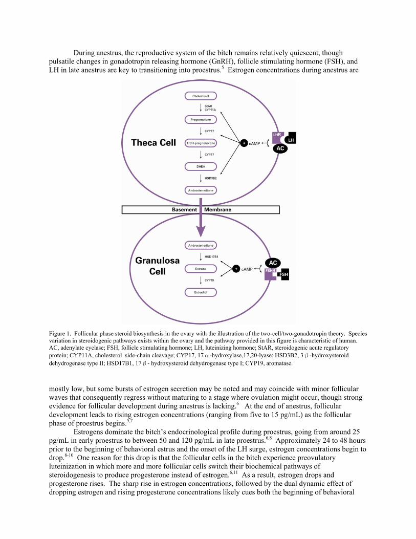
Veterinary Clinical Sciences, College of Veterinary Medicine, Iowa State University, Ames, IA Abstract Accurately timing breeding of a bitch is often done using vaginal cytology, vaginoscopy, and serum progesterone assays, none of which is a direct measure of ovulation or oocyte fertility. Despite the lack of direct connection to fertility, the indirect connections are strong and predictable and have to do with the effects of estrogen and progesterone on the bitch’s anatomy and physiology. This review discusses the effects of estrogen and progesterone on the bitch and how the changes we use to predict when to breed are related to the fluctuations in these hormones. Keywords: Breeding management, vaginal cytology, vaginoscopy, fertility, estrogen, progesterone Introduction Breeding management is probably the most common reason for evaluating a bitch in the context of reproduction and fertility. This is appropriate, as many bitches presenting for perceived subfertility have merely not had adequate monitoring and management of their previous estrous cycles.1,2 Conducting a thorough breeding management on the next estrous cycle of these bitches will often result in pregnancy. The variable lengths of both proestrus and estrus in the bitch account for the high number of miscalculated breeding dates (five–20 days and six–11 days, respectively).3 The actual fertile period, when oocytes are at a stage where they are fertile and accessible by vaginal deposition of semen (as is the case in natural mating and traditional artificial insemination methods), is much shorter, usually a two-three day window of time within the estrous period.4 Many breeders will note the swelling of the vulva and the serosanguinous discharge, and then count approximately nine days from this point and breed once or twice within a day or two. If the beginning of proestrus has been accurately determined, if the bitch has about a seven day proestrous period, if ovulation follows shortly thereafter, and if the stud dog’s sperm is relatively long-lived, then this plan may likely result in a pregnancy. But if the bitch has a short proestrous period, or a very long one, the bitch will likely be bred too late or too soon, respectively, and be unlikely to conceive. Using the tools of vaginal cytology, vaginoscopy, and serum progesterone assays, a clinician can accurately determine the date of the luteinizing hormone (LH) surge, and further estimate dates for ovulation and the fertile period. Use and interpretation of these tools is relatively well known among veterinarians offering reproductive services for dogs. Much of this material has been recently reviewed.3 This presentation will focus on explaining the physiological reasons behind the diagnostic tools of vaginal cytology, vaginoscopy, and serum progesterone monitoring. Vaginal cytology Endocrinology All of the changes noted on vaginal cytology through the estrous cycle of the bitch are due to changes in estrogen concentrations in the body. Understanding how and why those changes occur and how their consequent effects appear as cytological changes observable in a clinical setting will help the clinician to better chart normal and abnormal cycles. Estrogen is produced through a steroidogenic pathway starting in the thecal cells where various enzymes convert cholesterol into different pregnanes and eventually the androgen hormone androstenedione, which is then transported across the basement membrane into granulosa cells (Figure 1). The enzyme aromatase in the granulosa cells converts androstenedione into estrone and then another enzyme (17β-hydroxysteroid dehydrogenase type I) further converts estrone into estradiol. This steroidogenic pathway functions during active ovarian follicular activity.
During anestrus, the reproductive system of the bitch remains relatively quiescent, though pulsatile changes in gonadotropin releasing hormone (GnRH), follicle stimulating hormone (FSH), and LH in late anestrus are key to transitioning into proestrus.5 Estrogen concentrations during anestrus are
Figure 1. Follicular phase steroid biosynthesis in the ovary with the illustration of the two-cell/two-gonadotropin theory. Species variation in steroidogenic pathways exists within the ovary and the pathway provided in this figure is characteristic of human. AC, adenylate cyclase; FSH, follicle stimulating hormone; LH, luteinizing hormone; StAR, steroidogenic acute regulatory protein; CYP11A, cholesterol side-chain cleavage; CYP17, 17α-hydroxylase,17,20-lyase; HSD3B2, 3β-hydroxysteroid dehydrogenase type II; HSD17B1, 17β- hydroxysteroid dehydrogenase type I; CYP19, aromatase.
mostly low, but some bursts of estrogen secretion may be noted and may coincide with minor follicular waves that consequently regress without maturing to a stage where ovulation might occur, though strong evidence for follicular development during anestrus is lacking.6 At the end of anestrus, follicular development leads to rising estrogen concentrations (ranging from five to 15 pg/mL) as the follicular phase of proestrus begins.5,7 Estrogens dominate the bitch’s endocrinological profile during proestrus, going from around 25 pg/mL in early proestrus to between 50 and 120 pg/mL in late proestrus.6,8 Approximately 24 to 48 hours prior to the beginning of behavioral estrus and the onset of the LH surge, estrogen concentrations begin to drop.8-10 One reason for this drop is that the follicular cells in the bitch experience preovulatory luteinization in which more and more follicular cells switch their biochemical pathways of steroidogenesis to produce progesterone instead of estrogen.6,11 As a result, estrogen drops and progesterone rises. The sharp rise in estrogen concentrations, followed by the dual dynamic effect of dropping estrogen and rising progesterone concentrations likely cues both the beginning of behavioral
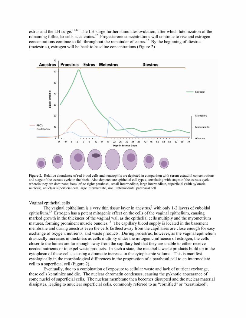
estrus and the LH surge.11,12 The LH surge further stimulates ovulation, after which luteinization of the remaining follicular cells accelerates.12 Progesterone concentrations will continue to rise and estrogen concentrations continue to fall throughout the remainder of estrus.12 By the beginning of diestrus (metestrus), estrogen will be back to baseline concentrations (Figure 2).
Figure 2. Relative abundance of red blood cells and neutrophils are depicted in comparison with serum estradiol concentrations and stage of the estrous cycle in the bitch. Also depicted are epithelial cell types, correlating with stages of the estrous cycle wherein they are dominant; from left to right: parabasal, small intermediate, large intermediate, superficial (with pyknotic nucleus), anuclear superficial cell, large intermediate, small intermediate, parabasal cell. Vaginal epithelial cells The vaginal epithelium is a very thin tissue layer in anestrus,3 with only 1-2 layers of cuboidal epithelium.13 Estrogen has a potent mitogenic effect on the cells of the vaginal epithelium, causing marked growth in the thickness of the vaginal wall as the epithelial cells multiply and the myometrium matures, forming prominent muscle bundles.13 The capillary blood supply is located in the basement membrane and during anestrus even the cells farthest away from the capillaries are close enough for easy exchange of oxygen, nutrients, and waste products. During proestrus, however, as the vaginal epithelium drastically increases in thickness as cells multiply under the mitogenic influence of estrogen, the cells closer to the lumen are far enough away from the capillary bed that they are unable to either receive needed nutrients or to expel waste products. In such a state, the metabolic waste products build up in the cytoplasm of these cells, causing a dramatic increase in the cytoplasmic volume. This is manifest cytologically in the morphological differences in the progression of a parabasal cell to an intermediate cell to a superficial cell (Figure 2). Eventually, due to a combination of exposure to cellular waste and lack of nutrient exchange, these cells keratinize and die. The nuclear chromatin condenses, causing the pyknotic appearance of some nuclei of superficial cells. The nuclear membrane then becomes disrupted and the nuclear material dissipates, leading to anuclear superficial cells, commonly referred to as “cornified” or “keratinized”.
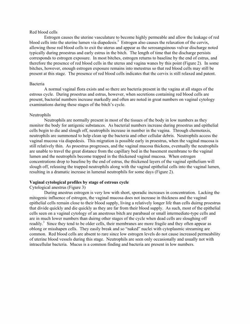
Red blood cells Estrogen causes the uterine vasculature to become highly permeable and allow the leakage of red blood cells into the uterine lumen via diapedesis.3 Estrogen also causes the relaxation of the cervix, allowing those red blood cells to exit the uterus and appear as the serosanguinous vulvar discharge noted typically during proestrus and early estrus in the bitch. The length of time that the discharge persists corresponds to estrogen exposure. In most bitches, estrogen returns to baseline by the end of estrus, and therefore the presence of red blood cells in the uterus and vagina wanes by this point (Figure 2). In some bitches, however, enough estrogen exposure remains into metestrus so that red blood cells may still be present at this stage. The presence of red blood cells indicates that the cervix is still relaxed and patent. Bacteria A normal vaginal flora exists and so there are bacteria present in the vagina at all stages of the estrous cycle. During proestrus and estrus, however, when secretions containing red blood cells are present, bacterial numbers increase markedly and often are noted in great numbers on vaginal cytology examinations during these stages of the bitch’s cycle. Neutrophils Neutrophils are normally present in most of the tissues of the body in low numbers as they monitor the body for antigenic substances. As bacterial numbers increase during proestrus and epithelial cells begin to die and slough off, neutrophils increase in number in the vagina. Through chemotaxis, neutrophils are summoned to help clean up the bacteria and other cellular debris. Neutrophils access the vaginal mucosa via diapedesis. This migration is possible early in proestrus, when the vaginal mucosa is still relatively thin. As proestrus progresses, and the vaginal mucosa thickens, eventually the neutrophils are unable to travel the great distance from the capillary bed in the basement membrane to the vaginal lumen and the neutrophils become trapped in the thickened vaginal mucosa. When estrogen concentrations drop to baseline by the end of estrus, the thickened layers of the vaginal epithelium will slough off, releasing the trapped neutrophils along with the vaginal epithelial cells into the vaginal lumen, resulting in a dramatic increase in lumenal neutrophils for some days (Figure 2). Vaginal cytological profiles by stage of estrous cycle Cytological anestrus (Figure 3) During anestrus estrogen is very low with short, sporadic increases in concentration. Lacking the mitogenic influence of estrogen, the vaginal mucosa does not increase in thickness and the vaginal epithelial cells remain close to their blood supply, living a relatively longer life than cells duirng proestrus that divide quickly and die quickly as they are far from their blood supply. As such, most of the epithelial cells seen on a vaginal cytology of an anestrous bitch are parabasal or small intermediate-type cells and are in much lower numbers than duirng other stages of the cycle when dead cells are sloughing off readily.3 Since they tend to be older cells, their membranes are more fragile and they often appear as oblong or misshapen cells. They easily break and so “naked” nuclei with cytoplasmic streaming are common. Red blood cells are absent to rare since low estrogen levels do not cause increased permeability of uterine blood vessels during this stage. Neutrophils are seen only occasionally and usually not with intracellular bacteria. Mucus is a common finding and bacteria are present in low numbers.
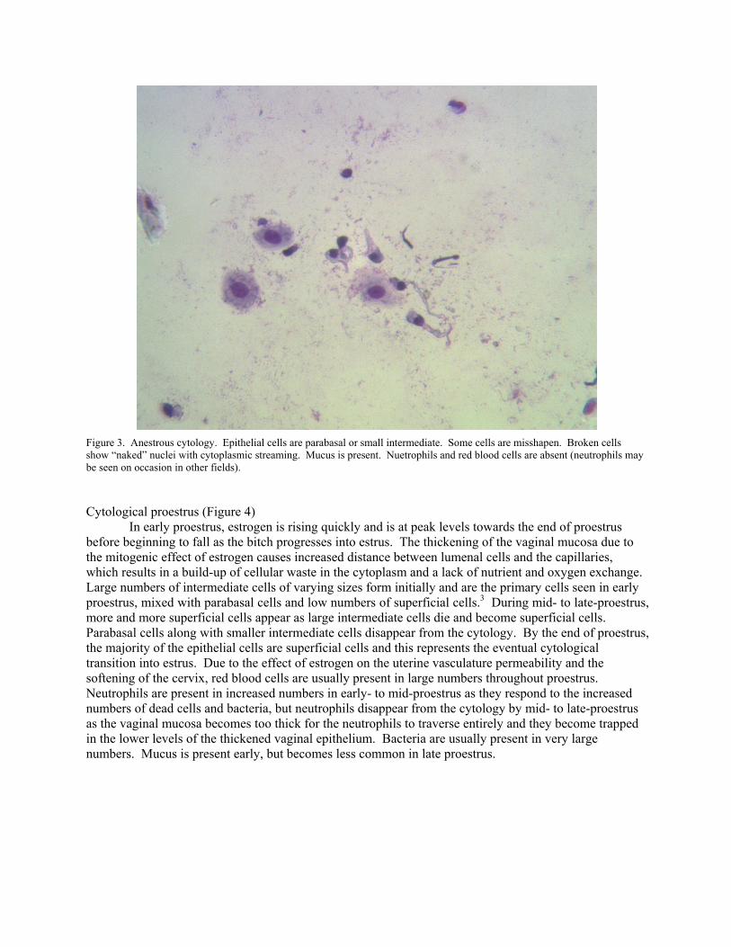
Figure 3. Anestrous cytology. Epithelial cells are parabasal or small intermediate. Some cells are misshapen. Broken cells show “naked” nuclei with cytoplasmic streaming. Mucus is present. Nuetrophils and red blood cells are absent (neutrophils may be seen on occasion in other fields). Cytological proestrus (Figure 4) In early proestrus, estrogen is rising quickly and is at peak levels towards the end of proestrus before beginning to fall as the bitch progresses into estrus. The thickening of the vaginal mucosa due to the mitogenic effect of estrogen causes increased distance between lumenal cells and the capillaries, which results in a build-up of cellular waste in the cytoplasm and a lack of nutrient and oxygen exchange. Large numbers of intermediate cells of varying sizes form initially and are the primary cells seen in early proestrus, mixed with parabasal cells and low numbers of superficial cells.3 During mid- to late-proestrus, more and more superficial cells appear as large intermediate cells die and become superficial cells. Parabasal cells along with smaller intermediate cells disappear from the cytology. By the end of proestrus, the majority of the epithelial cells are superficial cells and this represents the eventual cytological transition into estrus. Due to the effect of estrogen on the uterine vasculature permeability and the softening of the cervix, red blood cells are usually present in large numbers throughout proestrus. Neutrophils are present in increased numbers in early- to mid-proestrus as they respond to the increased numbers of dead cells and bacteria, but neutrophils disappear from the cytology by mid- to late-proestrus as the vaginal mucosa becomes too thick for the neutrophils to traverse entirely and they become trapped in the lower levels of the thickened vaginal epithelium. Bacteria are usually present in very large numbers. Mucus is present early, but becomes less common in late proestrus.
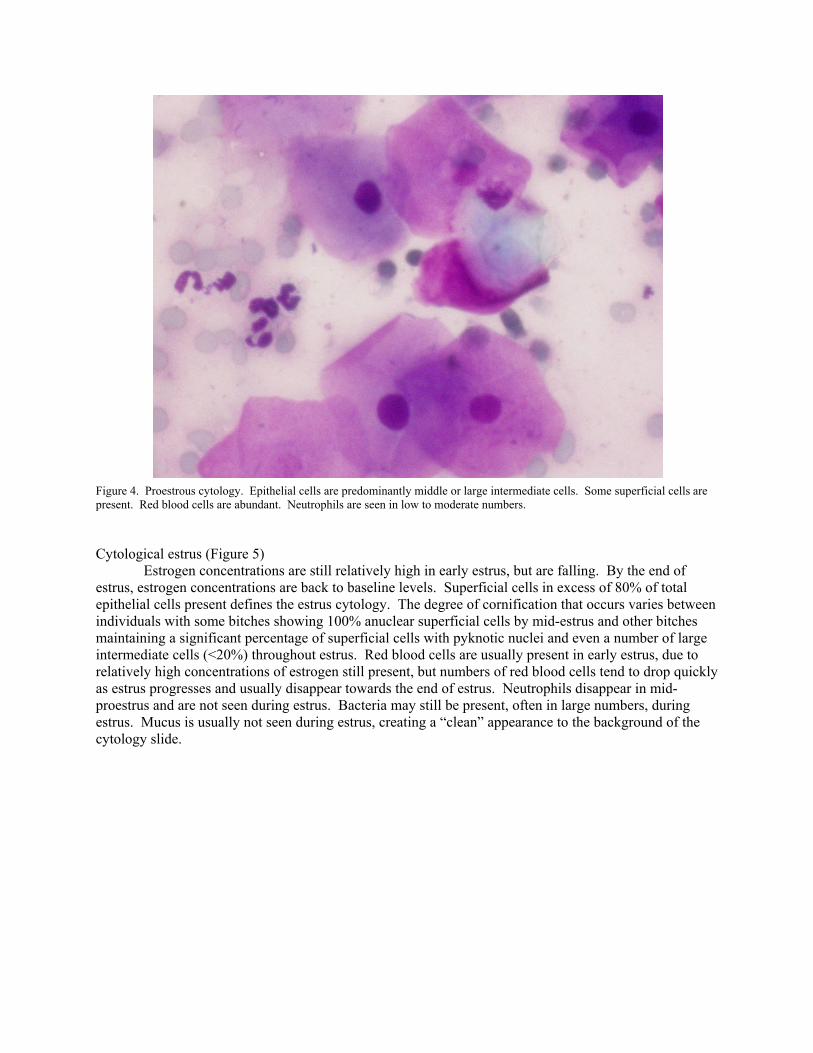
Figure 4. Proestrous cytology. Epithelial cells are predominantly middle or large intermediate cells. Some superficial cells are present. Red blood cells are abundant. Neutrophils are seen in low to moderate numbers. Cytological estrus (Figure 5) Estrogen concentrations are still relatively high in early estrus, but are falling. By the end of estrus, estrogen concentrations are back to baseline levels. Superficial cells in excess of 80% of total epithelial cells present defines the estrus cytology. The degree of cornification that occurs varies between individuals with some bitches showing 100% anuclear superficial cells by mid-estrus and other bitches maintaining a significant percentage of superficial cells with pyknotic nuclei and even a number of large intermediate cells (<20%) throughout estrus. Red blood cells are usually present in early estrus, due to relatively high concentrations of estrogen still present, but numbers of red blood cells tend to drop quickly as estrus progresses and usually disappear towards the end of estrus. Neutrophils disappear in mid-proestrus and are not seen during estrus. Bacteria may still be present, often in large numbers, during estrus. Mucus is usually not seen during estrus, creating a “clean” appearance to the background of the cytology slide.
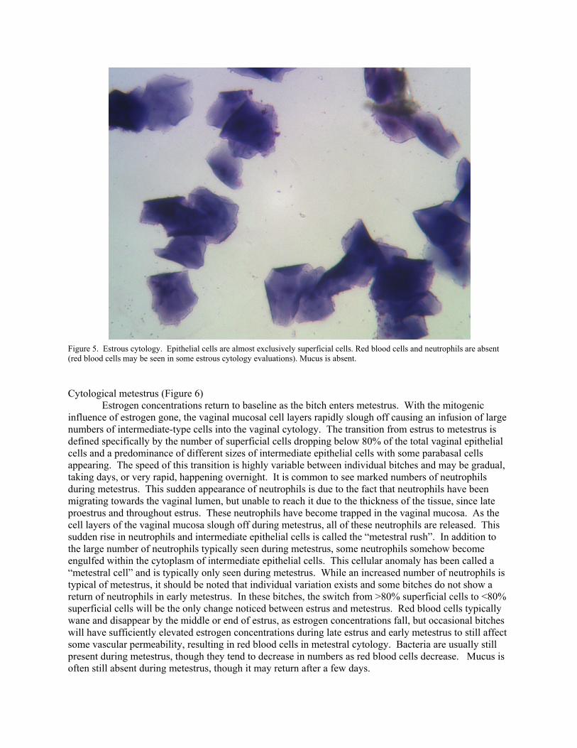
Figure 5. Estrous cytology. Epithelial cells are almost exclusively superficial cells. Red blood cells and neutrophils are absent (red blood cells may be seen in some estrous cytology evaluations). Mucus is absent. Cytological metestrus (Figure 6) Estrogen concentrations return to baseline as the bitch enters metestrus. With the mitogenic influence of estrogen gone, the vaginal mucosal cell layers rapidly slough off causing an infusion of large numbers of intermediate-type cells into the vaginal cytology. The transition from estrus to metestrus is defined specifically by the number of superficial cells dropping below 80% of the total vaginal epithelial cells and a predominance of different sizes of intermediate epithelial cells with some parabasal cells appearing. The speed of this transition is highly variable between individual bitches and may be gradual, taking days, or very rapid, happening overnight. It is common to see marked numbers of neutrophils during metestrus. This sudden appearance of neutrophils is due to the fact that neutrophils have been migrating towards the vaginal lumen, but unable to reach it due to the thickness of the tissue, since late proestrus and throughout estrus. These neutrophils have become trapped in the vaginal mucosa. As the cell layers of the vaginal mucosa slough off during metestrus, all of these neutrophils are released. This sudden rise in neutrophils and intermediate epithelial cells is called the “metestral rush”. In addition to the large number of neutrophils typically seen during metestrus, some neutrophils somehow become engulfed within the cytoplasm of intermediate epithelial cells. This cellular anomaly has been called a “metestral cell” and is typically only seen during metestrus. While an increased number of neutrophils is typical of metestrus, it should be noted that individual variation exists and some bitches do not show a return of neutrophils in early metestrus. In these bitches, the switch from >80% superficial cells to <80% superficial cells will be the only change noticed between estrus and metestrus. Red blood cells typically wane and disappear by the middle or end of estrus, as estrogen concentrations fall, but occasional bitches will have sufficiently elevated estrogen concentrations during late estrus and early metestrus to still affect some vascular permeability, resulting in red blood cells in metestral cytology. Bacteria are usually still present during metestrus, though they tend to decrease in numbers as red blood cells decrease. Mucus is often still absent during metestrus, though it may return after a few days.
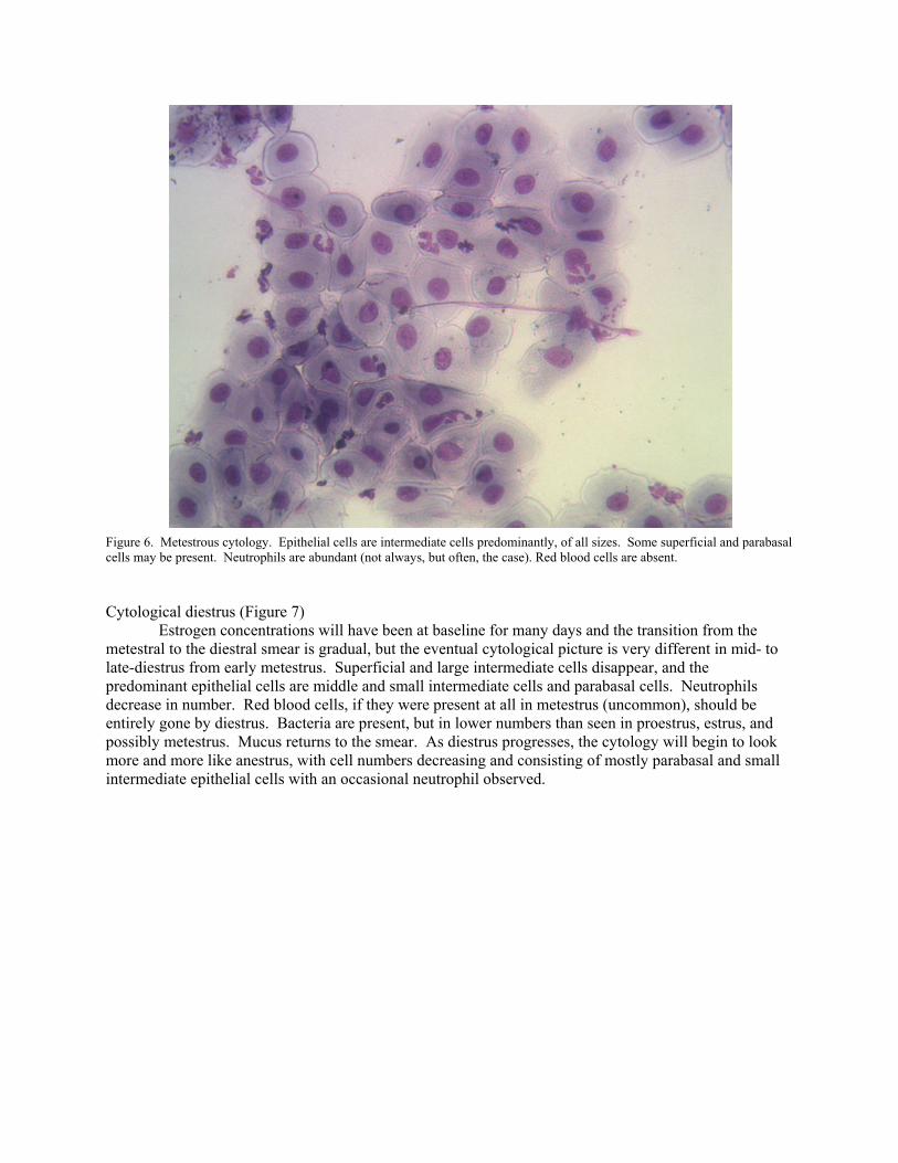
Figure 6. Metestrous cytology. Epithelial cells are intermediate cells predominantly, of all sizes. Some superficial and parabasal cells may be present. Neutrophils are abundant (not always, but often, the case). Red blood cells are absent. Cytological diestrus (Figure 7) Estrogen concentrations will have been at baseline for many days and the transition from the metestral to the diestral smear is gradual, but the eventual cytological picture is very different in mid- to late-diestrus from early metestrus. Superficial and large intermediate cells disappear, and the predominant epithelial cells are middle and small intermediate cells and parabasal cells. Neutrophils decrease in number. Red blood cells, if they were present at all in metestrus (uncommon), should be entirely gone by diestrus. Bacteria are present, but in lower numbers than seen in proestrus, estrus, and possibly metestrus. Mucus returns to the smear. As diestrus progresses, the cytology will begin to look more and more like anestrus, with cell numbers decreasing and consisting of mostly parabasal and small intermediate epithelial cells with an occasional neutrophil observed.
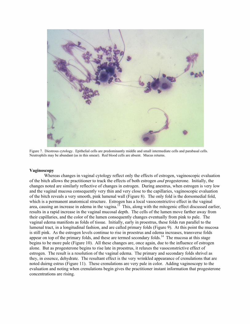
Figure 7. Diestrous cytology. Epithelial cells are predominantly middle and small intermediate cells and parabasal cells. Neutrophils may be abundant (as in this smear). Red blood cells are absent. Mucus returns. Vaginoscopy Whereas changes in vaginal cytology reflect only the effects of estrogen, vaginoscopic evaluation of the bitch allows the practitioner to track the effects of both estrogen and progesterone. Initially, the changes noted are similarly reflective of changes in estrogen. During anestrus, when estrogen is very low and the vaginal mucosa consequently very thin and very close to the capillaries, vaginoscopic evaluation of the bitch reveals a very smooth, pink lumenal wall (Figure 8). The only fold is the dorsomedial fold, which is a permanent anatomical structure. Estrogen has a local vasoconstrictive effect in the vaginal area, causing an increase in edema in the vagina.13 This, along with the mitogenic effect discussed earlier, results in a rapid increase in the vaginal mucosal depth. The cells of the lumen move farther away from their capillaries, and the color of the lumen consequently changes eventually from pink to pale. The vaginal edema manifests as folds of tissue. Initially, early in proestrus, these folds run parallel to the lumenal tract, in a longitudinal fashion, and are called primary folds (Figure 9). At this point the mucosa is still pink. As the estrogen levels continue to rise in proestrus and edema increases, transverse folds appear on top of the primary folds, and these are termed secondary folds.14 The mucosa at this stage begins to be more pale (Figure 10). All these changes are, once again, due to the influence of estrogen alone. But as progesterone begins to rise late in proestrus, it relaxes the vasoconstrictive effect of estrogen. The result is a resolution of the vaginal edema. The primary and secondary folds shrivel as they, in essence, dehydrate. The resultant effect is the very wrinkled appearance of crenulations that are noted duirng estrus (Figure 11). These crenulations are very pale in color. Adding vaginoscopy to the evaluation and noting when crenulations begin gives the practitioner instant information that progesterone concentrations are rising.
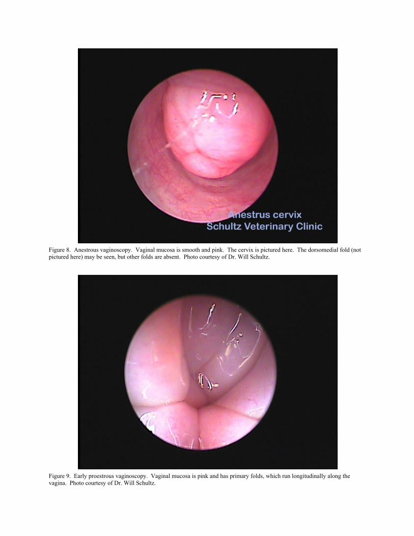
Figure 8. Anestrous vaginoscopy. Vaginal mucosa is smooth and pink. The cervix is pictured here. The dorsomedial fold (not pictured here) may be seen, but other folds are absent. Photo courtesy of Dr. Will Schultz.
Figure 9. Early proestrous vaginoscopy. Vaginal mucosa is pink and has primary folds, which run longitudinally along the vagina. Photo courtesy of Dr. Will Schultz.
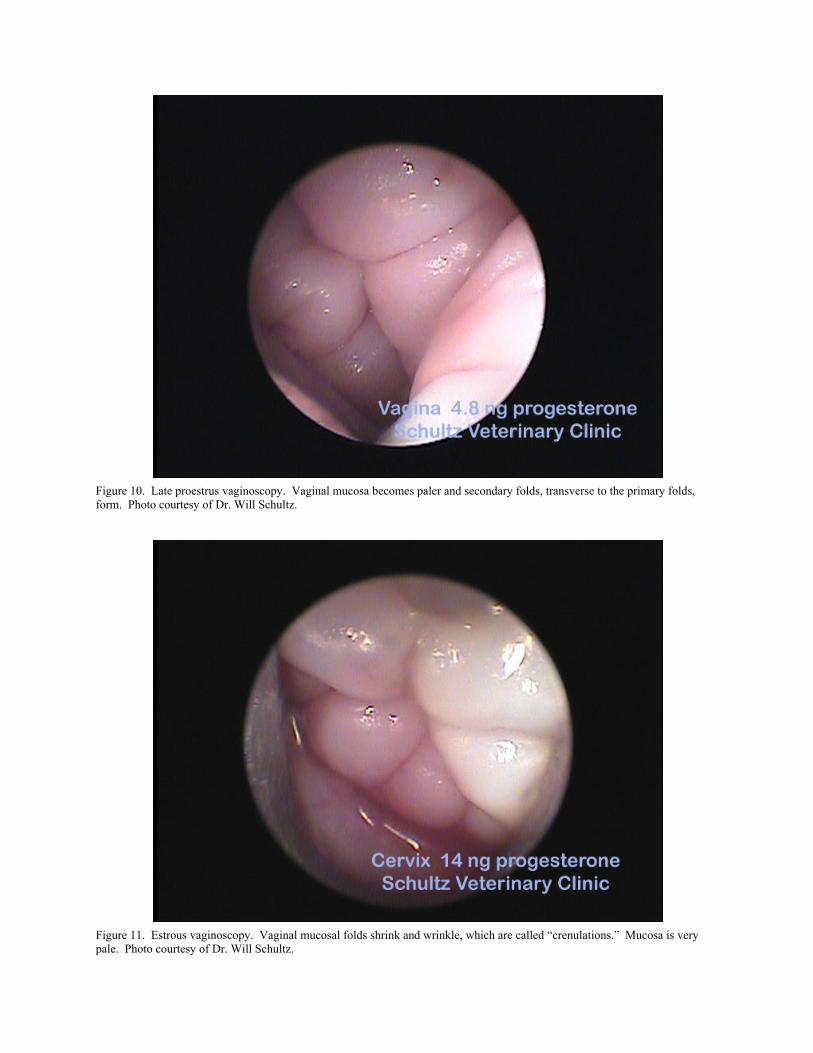
Figure 10. Late proestrus vaginoscopy. Vaginal mucosa becomes paler and secondary folds, transverse to the primary folds, form. Photo courtesy of Dr. Will Schultz.
Figure 11. Estrous vaginoscopy. Vaginal mucosal folds shrink and wrinkle, which are called “crenulations.” Mucosa is very pale. Photo courtesy of Dr. Will Schultz.
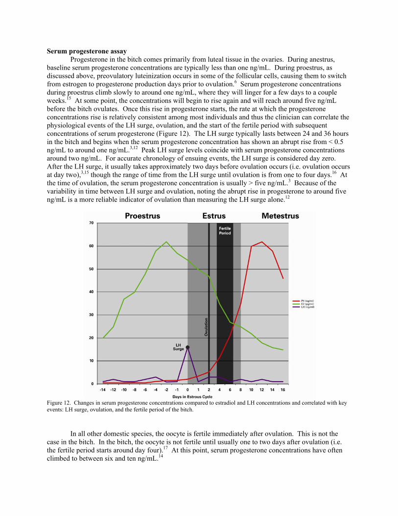
Serum progesterone assay Progesterone in the bitch comes primarily from luteal tissue in the ovaries. During anestrus, baseline serum progesterone concentrations are typically less than one ng/mL. During proestrus, as discussed above, preovulatory luteinization occurs in some of the follicular cells, causing them to switch from estrogen to progesterone production days prior to ovulation.6 Serum progesterone concentrations during proestrus climb slowly to around one ng/mL, where they will linger for a few days to a couple weeks.15 At some point, the concentrations will begin to rise again and will reach around five ng/mL before the bitch ovulates. Once this rise in progesterone starts, the rate at which the progesterone concentrations rise is relatively consistent among most individuals and thus the clinician can correlate the physiological events of the LH surge, ovulation, and the start of the fertile period with subsequent concentrations of serum progesterone (Figure 12). The LH surge typically lasts between 24 and 36 hours in the bitch and begins when the serum progesterone concentration has shown an abrupt rise from < 0.5 ng/mL to around one ng/mL.3,12 Peak LH surge levels coincide with serum progesterone concentrations around two ng/mL. For accurate chronology of ensuing events, the LH surge is considered day zero. After the LH surge, it usually takes approximately two days before ovulation occurs (i.e. ovulation occurs at day two),3,15 though the range of time from the LH surge until ovulation is from one to four days.16 At the time of ovulation, the serum progesterone concentration is usually > five ng/mL.3 Because of the variability in time between LH surge and ovulation, noting the abrupt rise in progesterone to around five ng/mL is a more reliable indicator of ovulation than measuring the LH surge alone.12
Figure 12. Changes in serum progesterone concentrations compared to estradiol and LH concentrations and correlated with key events: LH surge, ovulation, and the fertile period of the bitch. In all other domestic species, the oocyte is fertile immediately after ovulation. This is not the case in the bitch. In the bitch, the oocyte is not fertile until usually one to two days after ovulation (i.e. the fertile period starts around day four).17 At this point, serum progesterone concentrations have often climbed to between six and ten ng/mL.14
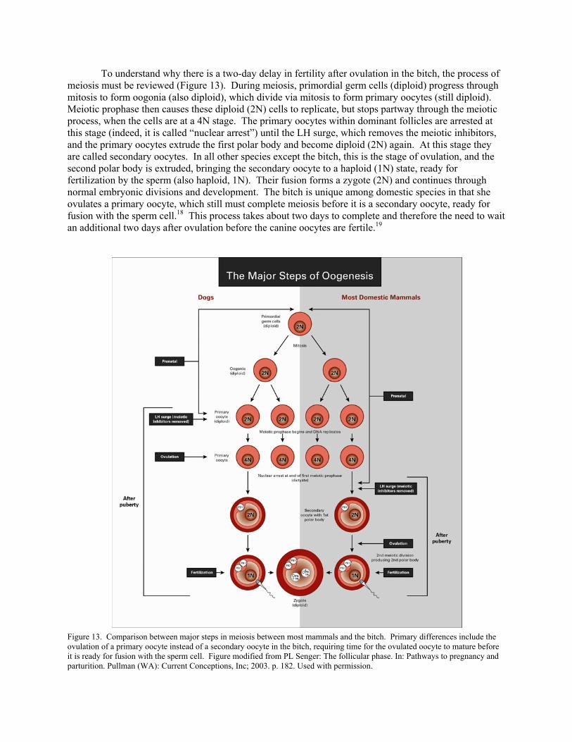
To understand why there is a two-day delay in fertility after ovulation in the bitch, the process of meiosis must be reviewed (Figure 13). During meiosis, primordial germ cells (diploid) progress through mitosis to form oogonia (also diploid), which divide via mitosis to form primary oocytes (still diploid). Meiotic prophase then causes these diploid (2N) cells to replicate, but stops partway through the meiotic process, when the cells are at a 4N stage. The primary oocytes within dominant follicles are arrested at this stage (indeed, it is called “nuclear arrest”) until the LH surge, which removes the meiotic inhibitors, and the primary oocytes extrude the first polar body and become diploid (2N) again. At this stage they are called secondary oocytes. In all other species except the bitch, this is the stage of ovulation, and the second polar body is extruded, bringing the secondary oocyte to a haploid (1N) state, ready for fertilization by the sperm (also haploid, 1N). Their fusion forms a zygote (2N) and continues through normal embryonic divisions and development. The bitch is unique among domestic species in that she ovulates a primary oocyte, which still must complete meiosis before it is a secondary oocyte, ready for fusion with the sperm cell.18 This process takes about two days to complete and therefore the need to wait an additional two days after ovulation before the canine oocytes are fertile.19
Figure 13. Comparison between major steps in meiosis between most mammals and the bitch. Primary differences include the ovulation of a primary oocyte instead of a secondary oocyte in the bitch, requiring time for the ovulated oocyte to mature before it is ready for fusion with the sperm cell. Figure modified from PL Senger: The follicular phase. In: Pathways to pregnancy and parturition. Pullman (WA): Current Conceptions, Inc; 2003. p. 182. Used with permission.
Often bitches may be bred a few days before the recognized start of the fertile period and still become pregnant. This is possible because the sperm from fertile male dogs usually remain viable inside the female tract for at least a couple of days and in some cases up to a week.20 On the other side of the equation, once the oocyte matures to a secondary oocyte, it likewise maintains its fertility for at least a couple of days (up to over 200 hours after ovulation, in some cases).4 In fact, studies have shown that it is usually not the oocyte losing fertility that causes a cessation to the fertile period, but rather the fact that rising progesterone concentrations cause the cervix to close,4 thus effectively shutting off access for vaginally deposited sperm to reach otherwise fertile oocytes in bitches bred longer than two to four days after the start of the fertile period (i.e. those bred after days six to eight, varying by individuals). Serum progesterone concentrations are usually around 22.5 ng/mL (± 3.4 ng/mL) at the end of the fertile period.4 This end to the fertile period, however, is most applicable to natural mating and vaginal artificial insemination methods, since these methods deposit the semen in the cranial vagina, for which a closed cervix would be an obstacle. Any method that deposits the semen into the uterus directly, such as transcervical insemination (TCI) or surgical insemination, can extend this fertile period for another couple of days.4 Anecdotally there are many reports of successful inseminations using TCI or surgical insemination when progesterone concentrations were up to 25 ng/mL, and some reports of even higher, though fertility seems to wane after that. The timeline outlined in Figure 12 pertains to the average cycle. Not every bitch in every cycle will follow this exact pattern. It is beyond the scope of this presentation to cover such events as delayed ovulation and split heats. It is worth pointing out, however, that it is a responsible and wise decision to monitor progesterone until at least ovulation, or ideally until the fertile period is reached. This will ensure that individual variation between bitches and cycles is noted and appropriate compensations are made. In some cycles, the progesterone concentrations will rise faster than the timeline in Figure 12, and insemination will need to be done sooner than anticipated. Other cycles will demonstrate that the progesterone lingers around two ng/mL for many days before climbing, indicating that ovulation is delayed longer than anticipated and insemination should be delayed accordingly. Summary The diagnostic tools used for breeding management in the bitch, vaginal cytology, vaginoscopy, and serum progesterone concentrations, are widely used. Understanding the endocrinology and physiology behind these tools helps to better interpret both normal and abnormal cycles. References 1. Zoldag L, Kacskemethy S, Nagy P: Heat progesterone profiles of bitches with ovulation failure. J Reprod Fertil
1993;Suppl 47:561-562. 2. Johnston SD, Olson PN, Root MV: Clinical approach to infertility in the bitch. Semin Vet Med Surg (Small Animal)
1994;9:2-6. 3. Concannon PW: Reproductive cycles of the domestic bitch. Anim Reprod Sci 2010;
doi:10.1016/janireprosci.2010.08.028. 4. Verstegen JP, Silva LD, Onclin K: Determination of the role of cervical closure in fertility regulation after mating or
artificial insemination in beagle bitches. J Reprod Fertil Suppl 2001;57:31-34. 5. Okkens AC, Kooistra HS: Anoestrus in the dog: a fascinating story. Reprod Domest Anim 2006;41:291-296. 6. Concannon PW: Endocrinologic control of normal canine ovarian function. Reprod Domest Anim 2009;44:3-15. 7. Jeffcoate IA: Endocrinology of anoestrous bitches. J Reprod Fertil Suppl 1993;47:69-76. 8. Concannon PW, Hansel W, Visek WJ: The ovarian cycle of the bitch: plasma estrogen, LH and progesterone. Biol
Reprod 1975;13:112-121. 9. Olson PN, Bowen RA, Behrendt MD, et al: Concentrations of reproductive hormones in canine serum throughout late
anestrus, proestrus and estrus. Biol Reprod 1982;27:1196-1206. 10. Wildt DW, Panko B, Chakraborty P, et al: Relationship of serum estrone, estradiol-17 and progesterone to LH sexual
behavior and time of ovulation in the bitch. Biol Reprod 1979;20:648-658. 11. Concannon P: Biology of gonadotripin secretion in adult and prepubertal female dogs. J Reprod Fertil Suppl
1993;47:3-27.
12. de Gier J, Kooistra HS, Djajadiningrat-Laanen SC, et al: Temporal relations between plasma concentrations of luteinizing hormone, follicle-stimulating hormone, estradiol-17[beta], progesterone, prolactin, and [alpha]-melanocyte-stimulating hormone during the follicular, ovulatory, and early luteal phase in the bitch. Theriogenology 2006;65:1346-1359.
13. Rehm S, Stanislaus DJ, Williams AM: Estrous cycle-dependent histology and review of sex steroid receptor expression in dog reproductive tissues and mammary gland and associated hormone levels. Birth Defects Res B Dev Reprod Toxicol 2007;80:233-245.
14. Jeffcoate IA, Lindsay FEF: Ovulation detection and timing of insemination based on hormone concentrations, vaginal cytology and the endoscopic appearance of the vagina in domestic bitches. J Reprod Fertil Suppl 1989;39:277-287.
15. Concannon PW, Hansel W, McEntee K: Changes in LH, progesterone and sexual behavior associated with preovulatory luteinization in the bitch. Biol Reprod 1977;17:604-613.
16. Wildt DE, Chakraborty PK, Panko WB, et al: Relationship of reproductive behavior, serum luteinizing hormone and time of ovulation in the bitch. Biol Reprod 1978;18:561-570.
17. Badinand F, Fontbonne A, Maurel MC, et al: Fertilization time in the bitch in relation to plasma concentration of oestradiol, progesterone and luteinizing hormone and vaginal smears. J Reprod Fertil Suppl 1993;47:63-67.
18. Holst PA, Phemister RD: The prenatal development of the dog: preimplantation events. Biol Reprod 1971;5:194-206. 19. Concannon PW, McCann JP, Temple M:. Biology and endocrinology of ovulation, pregnancy and parturition in the
dog. J Reprod Fertil Suppl 1989;39:3-25. 20. Concannon PW, Whaley S, Lein D, et al: Canine gestation length: variation related to time of mating and fertile life of
sperm. Am J Vet Res 1983;44:1819-1821.