Embed Size (px)
Citation preview

The Physiological Role of 5-HT2A Receptors in Working Memory
Graham V. Williams, Srinivas G. Rao, and Patricia S. Goldman-Rakic
Section of Neurobiology, Yale University School of Medicine, New Haven, Connecticut 06510
Dorsolateral prefrontal cortex has an essential role in the cog-nitive process of working memory, dysfunction of which isconsidered to be a core deficit in schizophrenia. Although thiscortical region is densely innervated with 5-HT2A receptors towhich atypical antipsychotic drugs bind with high affinity, little isknown of the influence of this serotonin receptor subtype onprefrontal function. We addressed this issue by examining theeffects of iontophoresis of selective receptor ligands on pre-frontal neurons possessing spatially tuned delay activity, or“memory fields,” in monkeys performing a delayed-responsetask. Memory fields of putative pyramidal cells were attenuatedby iontophoresis of 5-HT2A antagonists, which primarily pro-duced a reduction in delay activity for preferred target locations.Conversely, 5-HT2A stimulation by �-methyl-5-HT or 5-HT itself,accentuated the spatial tuning of these neurons by producing a
modest increase in activity for preferred target locations and/ora reduction in activity for nonpreferred locations. The agonisteffects could be reversed by the selective antagonistMDL100,907, and were dose-dependent, such that high levelsattenuated spatial tuning by profoundly reducing delay activity.A role for feedforward inhibitory circuitry in these effects wassupported by the finding that 5-HT2A blockade also attenuatedthe memory fields of putative interneurons. We conclude thatprefrontal 5-HT2A receptors have a hitherto unrecognized role inthe cognitive function of working memory, which involves ac-tions at both excitatory and inhibitory elements within localcircuitry.
Key words: prefrontal cortex; monkey; iontophoresis; 5-HT2A
receptor; working memory; single unit; spatial tuning; fast-spiking; interneuron; pyramidal cell; schizophrenia
The prefrontal cortex is substantially innervated by serotonergicfibers from the dorsal raphe nucleus in both primates and rodents(Porrino and Goldman-Rakic, 1982; Morrison et al., 1982; Takeu-chi and Sano, 1983; Smiley and Goldman-Rakic, 1996). Of all the5-HT receptor subtypes found in cortex, the G-protein-coupled5-HT2A receptor has received extensive attention in both physi-ological and pharmacological experiments. Immunocytochemis-try has revealed abundant 5-HT2A receptors on the dendrites ofprefrontal pyramidal cells as well as in large and medium-sizedcalbindin- and parvalbumin-positive interneurons (Jakab andGoldman-Rakic, 1998, 2000). These receptors have been shownto have a facilitatory action on cortical pyramidal cells (Aranedaand Andrade, 1991; Tanaka and North, 1993), which includes apresynaptic action on glutamate release (Aghajanian and Marek,1997). In primate prefrontal cortex, serotonergic fibers terminatemainly in association with the smooth dendrites of putative in-terneurons (Smiley and Goldman-Rakic, 1996). Accordingly,5-HT2A facilitation of this cell type (Gellman and Aghajanian,1993, 1994) has been shown to produce considerable IPSCs inneighboring pyramidal cells (Zhou and Hablitz, 1999). There-fore, the 5-HT2A-mediated influence of serotonin on corticalfunction would be expected to involve an interaction of facilita-tory and feedforward inhibitory components of intrinsic circuitry.Despite these insights into the physiology of cortical 5-HT2A
receptors, the consequences of serotonergic stimulation or deple-tion for cognitive function remains unclear. Although the study ofLuciana et al. (1998) suggests that supranormal levels of seroto-nin may be deleterious for spatial working memory, others havedemonstrated that depletion of serotonin leads to deficits in thecognitive process of decision-making, similar to that seen afterdamage to orbitofrontal cortex in humans (Rogers et al., 1999).The 5-HT2A receptor has also been shown to be particularlyinvolved in the action of hallucinogens such as lysergic aciddiethylamide (LSD) in the cortex (Marek and Aghajanian, 1996)as well as in the therapeutic efficacy of antipsychotic medications(Meltzer, 1989, 1999).
The circuitry of the dorsolateral prefrontal cortex has an es-tablished role in the working memory processes essential tohuman cognition (Goldman-Rakic, 1987; McCarthy et al., 1994,1996). Deficiency in these processes has been associated not onlywith the negative symptoms and cognitive deficits that are prom-inent in schizophrenia (Weinberger et al., 1986; Goldman-Rakic,1991, 1994; Liddle, 1987; Liddle and Morris, 1991; Park andHolzman, 1992), but also with the positive symptoms of thedisorder (Andreasen et al., 1997; Sabri et al., 1998; Lennox et al.,2000). Single cell recordings of dorsolateral prefrontal neurons innonhuman primates have revealed profiles of neuronal activationcorrelated with sensory, mnemonic, and response processes inboth manual (Fuster, 1973) and oculomotor delayed-responsetasks (ODRs) (Funahashi et al., 1989). The delay activity ob-served in many prefrontal neurons has been shown to be spatiallydependent, giving rise to “memory fields” with excitatory re-sponses to targets in preferred directions and null or inhibitoryresponses to other targets in nonpreferred directions. A recentstudy of regular-spiking (RS) and fast-spiking (FS) neurons inprefrontal cortex has indicated that putative interneurons can alsoexhibit memory fields, which have similar tuning properties to
Received Sept. 12, 2001; revised Dec. 11, 2001; accepted Dec. 28, 2001.This work was supported by National Institute of Mental Health Grants P50
MH44866 and R37 MH38546 (P.S.G.-R.). Further support was provided by theMedical Scientist Training Program of the National Institute of Health (S.G.R.). Wethank Chris Muly for helpful discussion, Susheel Vijayraghavan and Peter Vosler forassistance in data analysis, and Gary Leydon for his help in developing Spike2scripts.
Correspondence should be addressed to Dr. Graham V. Williams, Section ofNeurobiology, Yale University Medical School, 333 Cedar Street, New Haven, CT06510. E-mail: [email protected] © 2002 Society for Neuroscience 0270-6474/02/222843-12$15.00/0
The Journal of Neuroscience, April 1, 2002, 22(7):2843–2854

those of neighboring pyramidal cells (Rao et al., 1999). Thiscellular basis of working memory in prefrontal cortex provides anideal model for testing the influence of different neurotransmit-ters on cognitive function at the cortical level. A previous study,using iontophoresis of selective dopamine D1 receptor agonistsand antagonists, revealed an important relationship between thelevel of D1 receptor occupancy and the strength of pyramidal cellmemory fields (Williams and Goldman-Rakic, 1995). Here, weused a similar approach to investigate 5-HT2A receptor modula-tion of delay activity of both RS and FS units in the ODR task, todetermine the contribution of this particular receptor subtype tothe influence of prefrontal serotonergic input on cognitivefunction.
MATERIALS AND METHODSUse of animals. Studies were performed on two adult male rhesus mon-keys (Macaca mulatta), which were cared for under the guidelines of theNational Institutes of Health and Yale Animal Care Committee. Theywere prepared for chronic daily recording as previously described (Fu-nahashi et al., 1989). The recording cylinders were centered over thecaudal principal sulcus using x-ray imaging and stereotaxic coordinates(Fig. 1 A) (Rao et al., 1999). Magnetic resonance imaging in a number ofrecent monkeys has shown a high level of concordance in the coordinatesused. Single-unit recordings at this site (Fig. 1 B, central position) fre-quently reveal spatially tuned delay activity: the hallmark of responseproperties in caudal area 46 and rostral area 8a of Walker (1940).
ODR task. The animals were trained in a spatial ODR task shown inFigure 1C (Funahashi et al., 1989). In this task the monkey commenceseach trial by fixating a central stimulus (small square on monitor, or alight-emitting diode) within 2° for 0.5 sec and must continue to fixatewhile one of 8 peripheral stimuli (45° separation in circumference, 13°eccentricity) is illuminated for 0.5 sec (cue period). There then follows adelay period of 2.5 or 3.0 sec during which the monkey must maintainfixation. At the end of this time the central stimulus is extinguished, and
the monkey must make a saccade, within 0.5 sec (response period), to theposition of the peripheral stimulus shown earlier (again within 2°) to berewarded with fruit juice. The peripheral cues were presented in asemirandom order across trials such that, during the delay period, themonkey had to remember the cue location shown within the present trialto make the correct response. Note that the response period can bedivided into a presaccadic epoch (lasting 250 msec from the end of thedelay) because this incorporates the typical time required to initiate asaccade in this task (Funahashi et al., 1991), and a postsaccadic epoch(lasting 500 msec) starting 500 msec after the end of the delay andtherefore, by definition, after the performance of a successful saccade.
Electrode design and construction. Electrodes were constructed eitherfrom 33 �m carbon fiber (AVCO, Lowell MA) inside quad-barreledglass (Clark Electromedical Instruments, Reading, UK) or, more com-monly, from 20 �m pitch carbon fiber (ELSI, San Diego, CA) insertedinto the center of seven barrel nonfilamented capillary glass (Friedrichand Dimmock, Millville, NJ). The assembly was pulled through a heatingcoil element (nichrome wire; Narashige, Tokyo, Japan) using a propri-etary, computer-controlled electrode puller. The latter allowed for theprecise control of the heating-coil temperature, the time and velocityrequired to pull the shaft of the electrode (60–62 mm in length), and thetiming of the solenoid used to pull the tip of the electrode. The tip wasfurther fashioned by a combination of spark etching and beveling ondiamond (Stoelting, Wood Dale, IL) and stone (polishing) wheels. Thishelped to produce sharp tips, 20–40 �m, in length with impedancesranging from 250 k� to 1.2 M� (at 1 kHz) and a noise level of �15 �Vpeak to peak. Extracellular voltage was recorded with a custom (SKY-LAB) low-noise preamplifier and was bandpass filtered between 180 Hzand 8 kHz (four pole Butterworth; Krohn-Hite, Avon, MA). At thebeginning of each track, the dura was punctured with a 25 gauge hypo-dermic stainless steel guide tube (projecting from a 21 gauge guide tube),within which the electrode was lowered slowly into the brain using aMD-2 motorized hydraulic drive mounted on an MO-95 micromanipu-lator (Narishige).
Pharmacological agents and iontophoresis. In these and previous stud-ies, we have focused on the effects of ligands that act as antagonists atparticular receptors rather than agonist drugs. This is because the phys-
Figure 1. Region of recording and exper-imental paradigm. A, Left hemisphereview of the macaque brain showing thecalculated position of the recording cham-ber ( gray circle) over caudal areas 46 and8a (as, arcuate sulcus; ps, principal sulcus).B, Anatomical MRI at the same rostrocau-dal level as the center of the circle shownin A (� 27 mm anterior to ear, bar zero).The estimated lateral position of thechamber is shown in white with the centermarked by a dashed line. C, Schematicview of the ODR task. Top lef t panel showsthe position of the central fixation pointand the possible position of the eight pe-ripheral cues. One trial is depicted belowand to the right, where the target at 135° isdisplayed during the cue period and thecorrect response is portrayed by the arrowin the response (Resp.) epoch (Pre, presac-cadic epoch; Post, postsaccadic epoch).
2844 J. Neurosci., April 1, 2002, 22(7):2843–2854 Williams et al. • 5-HT2A Receptor Modulation in Working Memory
iological activation by neurotransmitters at their designated receptors atparticular locations on different neurons, and with a particular timecourse, can be best studied by examining the consequences of removingthat action. Iontophoresis of an agonist may level the normal spatial andtemporal profile of receptor action and, consequently, could obscure itsfunctional significance. The drugs used in this study have well docu-mented affinities at 5-HT2A receptors. MDL 100,907 (Aventis Pharma-ceuticals, Bridgewater, NJ) has high affinity (�1 nM) at 5-HT2A receptors(Johnson et al., 1996), whereas ritanserin (Janssen Pharmaceutical, Ti-tusville, NJ) and LY53857 (Sigma/RBI, Natick, MA) have an order ofmagnitude higher affinity at 5-HT2A receptors (�10 nM) than at 5-HT2Creceptors (Schreiber et al., 1995; Mazzola-Pomietto et al., 1996). Thepartial agonist �-methyl-5-HT (Sigma/RBI), which has high affinity at5-HT2A receptors and moderate affinity at 5-HT2C receptors (Gar-novskaya et al., 1995), was used to compare with, or reverse, the action ofthe antagonists. Serotonin (HCl salt; Sigma/RBI) was also used in someexperiments to compare the actions of endogenous and exogenous ago-nists. The drugs were dissolved in 1 ml of triple distilled water (adjustedwith HCl to pH 3.5–4.0) at a concentration of � 0.01 M and stored inaliquots of 50 �l at �70°C. Immediately before use, the drugs weresonicated briefly and drawn up into fine, fused-silica glass pipette fillers(WPI, Sarasota, FL), each instilled into one barrel of quad electrodes ortwo adjacent barrels of seven barrel electrodes, and forced to the tip bycompressed air. Thus, three drugs could be tested with one electrode,typically one agonist and two antagonists. Teflon-coated platinum–irid-ium wires (Medwire, Mt. Vernon, NY) were then fitted inside each drugbarrel and connected to a Neurophore BH2 iontophoretic system (Med-ical Systems Corp., Greenvale, NY) such that one channel (IP-2) of thedevice controlled the delivery of one drug. The results presented here aretaken from findings with ejection currents ranging from 5 to 100 nA.Retaining currents of �3 to �5 nA were used in a cycled manner (1 secon, 1 sec off) when not applying drugs, and current balancing was notrequired because of the low impedance of the electrode. Drug ejectiondid not create noise in the recording, and there was no systematic changein either spike amplitude or time course at any ejection current. Ionto-phoresis was started after a sufficient number of trials (�8) had beencollected for each target position in the task under the control condition.
Data acquisition and analysis. Eye movements were monitored with amagnetic search coil system (CNC Engineering, Seattle, WA) or byinfrared pupil tracking (ISCAN, Burlington, MA). These data wereincorporated into task control, performed by a PDP-11 running MONKsoftware or by a personal computer running TEMPO (Reflective Com-puting, St. Louis, MO). Spike waveform-sorting and data acquisition wasrun on a micro1401 using Spike2 software (Cambridge Electronic De-sign, Cambridge, UK). Waveform sorting (template matching algorithm)made it possible to isolate up more than one unit at the same recordingsite. The waveform templates constructed in the sorting were of sufficientrange in amplitude that they could incorporate any moment by momentchange in the magnitude of the spikes or slow drift in spike amplitudeover time. The data collected from each unit was time-stamped (and thespike waveform stored) to precisely determine when each spike occurredrelative to task events, and output via a text file for subsequent analysis.Unit activity was measured in spikes per second during each epoch of thetrial (Fig. 1C). Data were first collected from the cell under a controlcondition, followed by a drug condition in which one of the 5-HT2Aligands was applied, and then typically a recovery condition after drugapplication had been terminated. Because the synthetic, high-affinitydrugs used in these experiments take many seconds to act, they can alsotake a long time to wear off. Thus, although the general activity level ofthe cell may fully return to normal by the end of the recovery condition,the value obtained for spatial tuning over the entire condition can onlyapproach that in control (because multiple trials for each target directionare required in the analysis). However, in some recordings we used apost-drug condition immediately after drug application to ensure anoptimal recovery condition afterwards. Occasionally dose-dependent ef-fects of the drug were tested in two or more consecutive conditions, or anagonist was applied in the condition immediately after application of anantagonist (and vice versa) to detect reversal or opposing effects. Datawere obtained from at least five trials (typically 10 or more) at each cuelocation for each condition. The first 30–90 sec of data (or originally datafrom the first trial at each cue location) from noncontrol conditions wasomitted from the text file to allow for the time taken for drug action. Thetext file data were processed by a proprietary C�� program for statis-tical analysis using the Student’s two-tailed t test with unequal varianceand an � of 5%. In this way, data with a statistical probability level of �
0.05 were obtained for neuronal activity within each epoch of the task incomparison to baseline activity, and the effects of drug application onunit activity within each period of the task in comparison with theprevious control condition. Population analysis was performed on nor-malized data, derived by first aligning the preferred directions of thepopulation of tuned cells to 0°. The mean firing rate for each targetlocation for each unit was then taken as a ratio to the mean activity overall target locations and plotted relative to the preferred direction. Asimilar analysis was performed for the percentage change in activitybetween drug and control conditions for each target location. One-wayand two-way ANOVAs were performed on this normalized data, and theresults were analyzed with post hoc Scheffe tests.
Identification of single units. As previously reported (Mountcastle et al.,1969; Wilson et al., 1994; Rao et al., 1999, 2000), it was possible toidentify two distinct putative cell types in vivo by measuring the timecourse of their spike waveforms. Fast-spiking (FS) neurons had relativelylow-amplitude spikes (typically �50 �V), biphasic action potentials,relatively high firing rates, and short spike durations of � 0.9 msec.Regular-spiking (RS) neurons typically had more complex triphasicwaveforms with a larger initial negative deflection, relatively low basalfiring rates, and long durations of typically �1 msec. FS units could onlybe tracked for typically �20 �m, whereas RS units could often be trackedfor �100 �m, a distinction that probably arises from the larger dendriticfield of pyramidal cells. Using a cutoff point of 0.9 msec spike-basedwidth (the extracellular impulse being a close corollary to the first orderdifferential of the action potential recorded intracellularly) (McCormicket al., 1985; Kawaguchi, 1993, 1995), the two cell types could be readilysegregated, in accordance with their other spike properties. A recentreport by Gur et al. (1999), recording from macaque V1 neurons, pro-vides support for the assumption that units with different spike propertiesare likely to originate from cells of different types, which also showdifferent physiological properties, as recognized in many previous in vivorodent studies (Simons, 1978; Swadlow et al., 1998; Shimegi et al., 1999;Dantzker and Callaway, 2000; Morris and Henderson, 2000; Baeg et al.,2001; Timofeev et al., 2001). For further details on the unit isolation andspike segregation used, see Rao et al. (1999).
Assessment of spatial tuning. Spatial signal strength and direction inneuronal response was analyzed by a vector algorithm (Rao et al., 1999).Briefly, vectors were computed for loops constructed from firing rates foreach target direction in order of occurrence (for five or more trials), andtheir dot-products were determined, relative to the resultant vector. Astatistical comparison (Wilcoxon signed-rank test, p � 0.05; Conover,1971) of these scalar values was then made with arbitrary thresholds toyield an integer tuning factor (TF) ranging from 0 (untuned) to 10. Theeffect of a drug on this tuning was assessed by a statistical comparison(Wilcoxon sum-rank test, p � 0.05) of the final scalar values between thedrug and control conditions. The angle of tuning, �, varying continuously
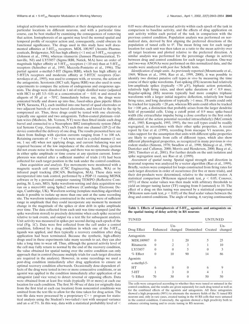
Table 1. Effects of iontophoresis of 5-HT2A agonists and antagonists onthe spatial tuning of delay activity in RS neurons
TUNED UNTUNED
Drug Effect EnhancedUn-changed Diminished Created
Un-changed
AntagonistsMDL100907 0 1 20 5 25Ritanserin 0 2 3 1 10LY53857 0 0 5 0 3% Effect 0 10 90 14 86
Agonists�-methyl-5-HT 6 0 0 6 95-HT 1 3 0 4 1% Effect 70 30 0 50 50
The cells were categorized according to whether they were tuned or untuned in thecontrol condition, and the results are given separately for each drug tested as well asfor the combined effects of the agonists and antagonists. All three antagonistsshowed a high propensity (90%) to attenuate the memory fields of the 31 tuned RSneurons and, only in rare cases, created tuning in the 44 RS cells that were untunedin the control condition. Conversely, the agonists showed a high proclivity both toenhance existing tuning and to create tuning in RS neurons.
Williams et al. • 5-HT2A Receptor Modulation in Working Memory J. Neurosci., April 1, 2002, 22(7):2843–2854 2845

Figure 2. Effects of 5-HT2A blockade on RS neurons. A, Rastergrams and average histograms of the activity of one RS neuron (C, cue; D, delay; R,response period; bin � 50 msec). The top panel shows activity during the control condition in which an elevation of activity can be seen during the delayand early response period for the preferred direction at 315° (lef t), and only a postsaccadic response for the nonpreferred location at 135° (right).Iontophoresis of MDL100,907 (middle panel ) attenuated the delay and presaccadic activity at 315° and the postsaccadic activity for the opposite targetlocation. After drug application was ceased (bottom panel ), the delay activity partially returns toward that in control. B, Mean and SE polar plots of thefiring rate of the same cell during the delay period for each target location (arrow: vector angle of tuning; inner circle indicates background activity). Amemory field can be seen in the control condition (top) that is diminished by the 5-HT2A antagonist (middle). In recovery (bottom), the memory fieldreturns in a similar shape, although smaller in size. C, Population analysis of the delay activity of 28 tuned RS cells tested with 5-HT2A antagonists inthe control (lef t) and drug (right) conditions. As described in Materials and Methods, the cells are first normalized for their preferred target locationwhich is set to 0° and then the activity for each target location, relative to the preferred location is taken as a ratio to the (Figure legend continues)
2846 J. Neurosci., April 1, 2002, 22(7):2843–2854 Williams et al. • 5-HT2A Receptor Modulation in Working Memory

between 0° and 360°,was determined by taking the median angle of theindividual loop vectors (Fisher, 1993).
RESULTSEffects of 5-HT2A receptor blockade on the memoryfields of RS unitsOf 75 RS units tested with 5-HT2A antagonists, 31 (41%) dis-played spatially tuned delay activity, firing maximally for one ortwo preferred directions and minimally for nonpreferred targetsin the opposite region of space. When examined for their effectson the spatial-tuning of delay activity in RS units, iontophoresisof 5-HT2A antagonists, ritanserin, LY53857, and MDL100,907, at15–50 nA ejection currents, attenuated tuning in nearly all (28 of31; 90%) cells that were tuned under the control condition (Table1). Attenuation of tuning between the drug and control conditionswas detected by a significant reduction ( p � 0.05, Wilcoxonrank-sum Test) in the vectors derived from the delay activity ofthe cell over all eight target directions (see Materials and Meth-ods). Data are presented for one cell in Figure 2A as rastergrams(top) and sum-histograms (below) for the preferred (315°) andnonpreferred (135°) target locations. In the control condition, itcan be seen that the cell fired consistently for trials at 315°throughout the delay and the presaccadic epochs and showed aconcomitant reduction in activity for trials at 135°. Iontophoresisof MDL100,907 at 25 nA produced a steady decline in theresponse of the cell for its preferred direction, which returnedslowly during the recovery period after drug application. Notethat the antagonist also reduced presaccadic activity (early re-sponse period) for the preferred direction and postsaccadic ac-tivity (late response period) for the nonpreferred direction of thecell (Fig. 2A, right panel). The polar plots of mean and SE of delayactivity for all target locations in Figure 2B shows the full extentof the memory field (TF � 5; � � 319°; see arrow) in the controlcondition, its destruction by the 5-HT2A antagonist, and its partialre-emergence (TF � 6) in recovery at a similar angle of tuning(� � 327°).
The attenuation of spatial tuning in delay activity could also beseen at the population level by normalizing both the preferreddirection of each cell to 0° and the activity for each target locationrelative to the mean activity over all target locations. The resultsare illustrated for 28 RS neurons in Figure 2C, which show that,in the control condition, there is a distinct elevation of activity forthe preferred target location above the mean (scaled to unity),less elevation for the adjacent directions (�45°), and a cleardepression below the mean for targets separated by 135–180° fromthe preferred location. ANOVA revealed a highly significanteffect of location (F � 21.87; p � 0.0001) with significant differ-ences between activity for the preferred direction and all loca-tions separated by �90° ( p � 0.0001; Scheffe post hoc test). Ahighly significant drug condition by direction interaction (F �4.17; p � 0.0002) was found between the control and drug con-ditions. The effect of direction still remained ( p � 0.0001), but itsmagnitude (F � 4.63) and the post hoc differences betweenpreferred and nonpreferred directions were much reduced in the
presence of 5-HT2A blockade ( p � 0.05 at �135 and 180°; p �0.01 at �135°). In rare instances (6 of 44 U), 5-HT2A antagonistsinduced tuning in RS cells that were not previously tuned in thecontrol condition. However, these drugs never improved tuning inthose neurons that were already tuned in the control condition.
To analyze further how delay activity was changed by 5-HT2A
blockade, we examined the percentage change in activity fromcontrol for each target direction for the same population of tunedRS units tested with 5-HT2A antagonists. From the results pre-sented in Figure 2D it can be seen that the deleterious effect of5-HT2A blockade was produced by an overall selective reductionin activity within the memory field of the cell with a greaterattenuation for the preferred target location (again normalizedfor each cell to 0°) than the two adjacent targets (�45°, p �0.0001; �45°, p � 0.0001). This effect would be expected toproduce a profound attenuation of spatial tuning in RS cells.
Effects of 5-HT2A receptor stimulation on the memoryfields of RS unitsIf the effect observed on the spatially tuned delay activity of RScells was, indeed, a direct effect of 5-HT2A blockade, then wewould expect that the agonist would produce an enhancement ofthe memory fields of these neurons. However, it should be rec-ognized that iontophoretic application of the agonist onto aneuron under its natural conditions with an operational level ofendogenous serotonin will not always produce a functionallysignificant influence (as discussed in Materials and Methods). Incontrast to the effect of the 5-HT2A antagonists, we saw no effecton most of the 21 RS cells tested with application of �-methyl-5-HT at 20–50 nA (Table 1). Nevertheless, 12 RS cells (57% ofthose tested) showed an elevated spatial tuning in the delayperiod with iontophoresis of the drug. Attenuation of the memoryfield was never found in RS units that previously showed anytuning in the control condition (n � 6; 29%). The enhancementof spatial tuning in delay activity by the agonist is illustrated forone RS cell in Figure 3A. Here it can be seen that there is amodest differentiation of delay period activity between the pre-ferred target location at 135° and the nonpreferred location at315°. The relationship of this activity to the memory field of thecell is apparent from the adjacent polar plot in Figure 3B. Thisneuron had a TF � 2 and a � � 124° (see arrow) in the controlcondition. Iontophoresis of �-methyl-5-HT at 40 nA increaseddelay activity in trials with the target at 135° but actually reducedit for those at 315°, increasing TF to 3, with � remaining at 127°.When MDL100,907 was coapplied at 25 nA subsequently, theprevious agonist-induced enhancement was reversed and the TFwas reduced to 1, with � at 130° (data not shown). Finally, whenagonist application was terminated, continued iontophoresis ofthe antagonist destroyed the memory field completely, withoutany overall decrease in activity of the cell (bottom panel). Rever-sal of the deleterious effect of MDL100,907 on the memory fieldby the agonist was also observed in three additional RS cells.Population analysis of the agonist effect on 11 RS neurons thatwere tuned in the control condition, or became tuned in the drug
4
(Figure legend continues) mean for the delay activity of the cell across all target directions (shown by line). The histogram therefore depicts the dispersionof delay activity between preferred and nonpreferred target locations. A clear spatial profile can be seen in the control condition, which is highlydiminished under 5-HT2A blockade (asterisks denote significant differences from the preferred direction). D, Histogram showing the percentage changein delay activity produced by 5-HT2A blockade (for the same neuronal population), relative to the activity in the control condition for each target location(preferred direction again normalized to 0°). A reduction in activity can be seen for the preferred direction, greater than that for the twoadjacentlocations, and a small increase in activity is evident for opposite locations in space.
Williams et al. • 5-HT2A Receptor Modulation in Working Memory J. Neurosci., April 1, 2002, 22(7):2843–2854 2847

Figure 3. Effects of 5-HT2A stimulation on RS neurons. A, Neuronal activity of an RS neuron showing a small response during the delay period fortargets at 135° but not at 315° in the control condition. Iontophoresis of �-methyl-5-HT boosts the delay activity for the preferred direction while, at thesame time, it depresses activity for the opposite location. The same cell tested with MDL100,907 (bottom panel ) shows a complete abolition of its previousresponse. B, The memory field of this cell exhibits modest tuning during the control condition (top), which is sharpened by application of the agonist(middle), but delay activity loses spatial specificity altogether after application of MDL100,907. C, Population analysis for 11 RS cells tested with theagonist reveals signs of a spatial profile in response in control (lef t) that is dramatically augmented by the agonist (right). Note that one cell was excludedfrom this analysis because it showed changes in activity for opposite directions in space between the first and second half of the delay period. D, Overall,the agonist produces a larger reduction in the delay activity for nonpreferred target locations than that for the preferred location in this population ofcells. Conventions as in previous figure.
2848 J. Neurosci., April 1, 2002, 22(7):2843–2854 Williams et al. • 5-HT2A Receptor Modulation in Working Memory

condition, revealed a highly significant effect of the drug on thedifferentiation between activity for the preferred and nonpre-ferred target locations (Fig. 3C). The preferred target location(again normalized to 0°) shows a small elevation of activity abovemean, whereas the activity of nonpreferred target locations ismoderately submerged. Even so, ANOVA revealed a distincteffect of direction (F � 7.09; p � 0.0001) with significant post hocdifferences at �90°, �135°, and 180°. Application of the agonistdramatically increased the effect of direction (F � 29.32; p �0.0001) with a significant drug by direction interaction (F � 4.72;p � 0.0001). This effect was particularly prominent for the pre-ferred target location of the cell, which created large post hocdifferences not only with target locations 135° and 180° distant butalso with the adjacent target locations (�45°). In this way, thespatial tuning of this population was considerably sharpened.Note that the distribution of activity about the mean for theagonist condition (Fig. 3C) is even more polarized betweenpreferred and nonpreferred directions than the population oftuned cells in the control condition that were tested with theantagonist (Fig. 2C). Thus, not only is it possible for a populationof cells with moderate or no tuning to become considerably tunedbecause of increased 5-HT2A stimulation (above the endogenouslevel), but these cells appear to be more tuned than a separatesample of neurons recorded in the control condition. To investi-gate further the mechanism by which the agonist may exert itsbeneficial effects, we analyzed the percentage change in activityfrom the control condition at each target location for the same
population of RS units tested with the agonist. As shown inFigure 3D, the agonist produced an overall decrease in delayactivity which was significant for nonpreferred target locations( p � 0.001; two-tailed t test) and significantly less for the pre-ferred target location (0°) than the adjacent (�45°) locations ( p �0.003, p � 0.001; two-tailed paired t test). Thus, it appears thatincreasing 5-HT2A receptor stimulation can enhance tuning in RScells, primarily by producing a net reduction in their activity, bothfor the opponent, nonpreferred target locations and the twolocations adjacent to the preferred target.
Given the above results for the effect of an agonist with someselectivity for the 5-HT2A receptor, we were interested to seewhether elevation of serotonergic stimulation, by iontophoresis of5-HT itself, could also have advantageous effects on the memoryfields of putative pyramidal cells. Serotonin at 4–10 nA improvedtuning in five of nine (56%) RS cells, in three cases by signifi-cantly increasing the delay activity of the cell (Table 1). Asillustrated in Figure 4, 5-HT, at just 10 nA, produced a dramatic,spatially dependent increase in the delay activity of an RS cellexhibiting no apparent tuning in control. However, the modestincrease in activity for the nonpreferred target locations partiallyoffset this effect, such that the cell becomes only weakly tuned(TF � 1; � � 11°). Although coapplication of MDL100,907clearly reverses this increase in delay activity, its combined effectis distributed over all target locations such as to improve thesignal to noise in the spatially-dependent firing of the cell andsignificantly enhance tuning (TF � 2; � � 14°). Thus, 5-HT is
Figure 4. Effect of serotonin on delay ac-tivity in an RS cell. A, In the controlcondition the cell shows barely any distinc-tion in its response for the 0° and 180°targets. However, on application of 5-HTat just 10 nA there is a marked enhance-ment of the firing of the cell that particu-larly accentuates the delay activity on 0°trials. Subsequent coapplication ofMDL100,907 (bottom panel ) dramaticallyreduces the firing rate and attenuates theprevious selective response in the delayperiod. B, The delay activity of this RScell does not show any spatial specificity incontrol but it develops into a significantmemory field (TF � 1) when 5-HT isapplied (middle panel ). Coapplication ofMDL100,907 produces a substantial re-duction in the delay activity but sharplylimits firing to a small region of space and,as a consequence, improves spatial tuning(TF � 2). Conventions as in previousfigures.
Williams et al. • 5-HT2A Receptor Modulation in Working Memory J. Neurosci., April 1, 2002, 22(7):2843–2854 2849
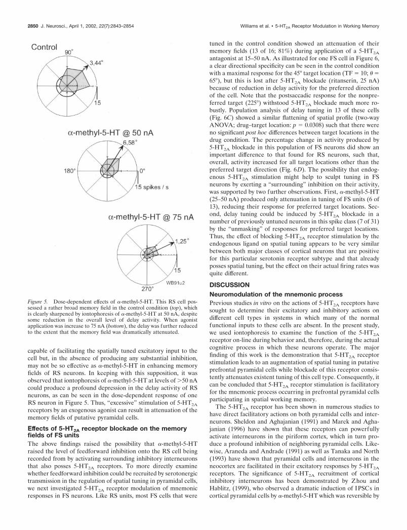
capable of facilitating the spatially tuned excitatory input to thecell but, in the absence of producing any substantial inhibition,may not be so effective as �-methyl-5-HT in enhancing memoryfields of RS neurons. In keeping with this supposition, it wasobserved that iontophoresis of �-methyl-5-HT at levels of �50 nAcould produce a profound depression in the delay activity of RSneurons, as can be seen in the dose-dependent response of oneRS neuron in Figure 5. Thus, “excessive” stimulation of 5-HT2A
receptors by an exogenous agonist can result in attenuation of thememory fields of putative pyramidal cells.
Effects of 5-HT2A receptor blockade on the memoryfields of FS unitsThe above findings raised the possibility that �-methyl-5-HTraised the level of feedforward inhibition onto the RS cell beingrecorded from by activating surrounding inhibitory interneuronsthat also posses 5-HT2A receptors. To more directly examinewhether feedforward inhibition could be recruited by serotonergictransmission in the regulation of spatial tuning in pyramidal cells,we next investigated 5-HT2A receptor modulation of mnemonicresponses in FS neurons. Like RS units, most FS cells that were
tuned in the control condition showed an attenuation of theirmemory fields (13 of 16; 81%) during application of a 5-HT2A
antagonist at 15–50 nA. As illustrated for one FS cell in Figure 6,a clear directional specificity can be seen in the control conditionwith a maximal response for the 45° target location (TF � 10; � �65°), but this is lost after 5-HT2A blockade (ritanserin, 25 nA)because of reduction in delay activity for the preferred directionof the cell. Note that the postsaccadic response for the nonpre-ferred target (225°) withstood 5-HT2A blockade much more ro-bustly. Population analysis of delay tuning in 13 of these cells(Fig. 6C) showed a similar flattening of spatial profile (two-wayANOVA; drug–target location: p � 0.0308) such that there wereno significant post hoc differences between target locations in thedrug condition. The percentage change in activity produced by5-HT2A blockade in this population of FS neurons did show animportant difference to that found for RS neurons, such that,overall, activity increased for all target locations other than thepreferred target direction (Fig. 6D). The possibility that endog-enous 5-HT2A stimulation might help to sculpt tuning in FSneurons by exerting a “surrounding” inhibition on their activity,was supported by two further observations. First, �-methyl-5-HT(25–50 nA) produced only attenuation in tuning of FS units (6 of13), reducing their response for preferred target locations. Sec-ond, delay tuning could be induced by 5-HT2A blockade in anumber of previously untuned neurons in this spike class (7 of 31)by the “unmasking” of responses for preferred target locations.Thus, the effect of blocking 5-HT2A receptor stimulation by theendogenous ligand on spatial tuning appears to be very similarbetween both major classes of cortical neurons that are positivefor this particular serotonin receptor subtype and that alreadyposses spatial tuning, but the effect on their actual firing rates wasquite different.
DISCUSSIONNeuromodulation of the mnemonic processPrevious studies in vitro on the actions of 5-HT2A receptors havesought to determine their excitatory and inhibitory actions ondifferent cell types in systems in which many of the normalfunctional inputs to these cells are absent. In the present study,we used iontophoresis to examine the function of the 5-HT2A
receptor on-line during behavior and, therefore, during the actualcognitive process in which these neurons operate. The majorfinding of this work is the demonstration that 5-HT2A receptorstimulation leads to an augmentation of spatial tuning in putativeprefrontal pyramidal cells while blockade of this receptor consis-tently attenuates existent tuning of this cell type. Consequently, itcan be concluded that 5-HT2A receptor stimulation is facilitatoryfor the mnemonic process occurring in prefrontal pyramidal cellsparticipating in spatial working memory.
The 5-HT2A receptor has been shown in numerous studies tohave direct facilitatory actions on both pyramidal cells and inter-neurons. Sheldon and Aghajanian (1991) and Marek and Agha-janian (1996) have shown that these receptors can powerfullyactivate interneurons in the piriform cortex, which in turn pro-duce a profound inhibition of neighboring pyramidal cells. Like-wise, Araneda and Andrade (1991) as well as Tanaka and North(1993) have shown that pyramidal cells and interneurons in theneocortex are facilitated in their excitatory responses by 5-HT2A
receptors. The significance of 5-HT2A recruitment of corticalinhibitory interneurons has been demonstrated by Zhou andHablitz, (1999), who observed a dramatic induction of IPSCs incortical pyramidal cells by �-methyl-5-HT which was reversible by
Figure 5. Dose-dependent effects of �-methyl-5-HT. This RS cell pos-sessed a rather broad memory field in the control condition (top), whichis clearly sharpened by iontophoresis of �-methyl-5-HT at 50 nA, despitesome reduction in the overall level of delay activity. When agonistapplication was increase to 75 nA (bottom), the delay was further reducedto the extent that the memory field was dramatically attenuated.
2850 J. Neurosci., April 1, 2002, 22(7):2843–2854 Williams et al. • 5-HT2A Receptor Modulation in Working Memory

coapplication of the 5-HT2A antagonist ketanserin. Finally, Agha-janian and Marek (1997) have recently shown that 5-HT2A re-ceptors directly facilitate pyramidal neurons in prefrontal cortexby a powerful effect at the “trigger zone” on their primary apicaldendrites, which has now been shown to contain intense immu-noreactivity for 5-HT2A receptors (Jakab and Goldman-Rakic,1998, 2000). These findings, from work in vitro, point to directfacilitation and indirect feedforward inhibition of pyramidal cellsby 5-HT2A receptor activation. Our present work demonstratesthat these mechanisms of facilitation and feedforward inhibitionare operative in vivo, where they are integral to the constructionof spatially tuned memory fields in putative pyramidal cells.
Mechanisms of drug actionThree distinctly different antagonists were used to demonstratethat 5-HT2A blockade attenuated memory fields in prefrontal
cortex. However, despite their differences in chemical structureand receptor affinity, all three compounds produced the sameoverall effect on putative pyramidal neurons with spatially tuneddelay activity, as evidenced by the data from both RS and FSpopulation analysis. Not only did the agonist produce the oppo-site effects on the spatial tuning of delay activity to the antagonistsat the population level, but it was possible to see the competitiveaction of both drug classes on the memory fields of individualcells. As expected, the antagonist effects, when looked at in termsof actual changes in activity for preferred and nonpreferred targetlocations, are primarily consistent with an attenuation of responseof the neuron to its spatially tuned excitatory inputs as well assome minor reduction in inhibition for nonpreferred target loca-tions. The effects of the agonist are not so directly interpretable,as the primary effect appears to be an overall reduction in activity
Figure 6. Effects of 5-HT2A blockade on FS neurons. A, An FS neuron which, in the control condition (top panel ), fired during the delay period on trialsin which the target was at 45°, and in the postsaccadic epoch for the opponent target location (225°). Application of ritanserin gradually abolished thedelay activity for the preferred target location but cue and presaccadic activity for this location, as well as the postsaccadic activity for the opposite targetlocation, persisted longer during drug application. B, The memory field of this FS cell included responses to two adjacent targets at 45° and 90° that wereboth diminished after the application of ritanserin. C, A population of 13 FS cells that were tuned in the control condition showed a clear spatial profilein their delay activity that was practically abolished by 5-HT2A blockade. D, Rather than generally reducing the activity of this cell type, the 5-HT2Aantagonists produced an overall increase in delay activity for all targets other than the preferred location in this cell population. Conventions as inprevious figures.
Williams et al. • 5-HT2A Receptor Modulation in Working Memory J. Neurosci., April 1, 2002, 22(7):2843–2854 2851

of RS neurons, primarily for their nonpreferred target locations.This reduction in activity is reasonably explained by the findingsof Zhou and Hablitz (1999) that �-methyl-5-HT drives substan-tial feedforward inhibition in neocortex, just as 5-HT2A stimula-tion does in piriform cortex (Gellman and Aghajanian, 1994).Thus, the agonist may facilitate excitatory spatially tuned inputsto pyramidal cells while at the same time activating inhibitorymechanisms that preserve the spatial resolution of their mne-monic response. Moreover, we now show that the memory fieldsof these putative parvalbumin-containing interneurons are also5-HT2A-dependent. We therefore postulate that �-methyl-5-HTdiffuses a sufficient distance to facilitate multiple surroundinginterneurons with similar spatial tuning, which feedforward ontothe pyramidal cells from which we recorded. This hypothesis issupported by the finding that iontophoresis of 5-HT itself at verylow ejection currents boosted the delay activity of the cells with-out producing pronounced enhancement of spatial tuning. In thiscase, we would not expect the endogenous ligand to diffuse asignificant distance from the recording site (because of selectiveprocesses of metabolism and reuptake), so low-level applicationof serotonin should not evoke considerable feedforward inhibi-tion. Therefore, it can be postulated that the synergistic action ofthe 5-HT2A receptor on both pyramidal cells, and the interneu-rons which innervate them, may be important for the expressionof significant spatially tuned delay activity in prefrontal cortex.The outcome of this interaction would obviously depend on thelevel of serotonin release, reuptake and the sensitivity of the5-HT2A receptor on the two cell types. Despite this complexity, itis clear that serotonin recruits inhibitory networks that are inte-gral components of the local circuits involved in modulating theconstruction of spatial tuning by excitatory afferents in prefrontalpyramidal cells.
Comparison of 5-HT2A and D1 receptor effectsIn a previous study, we have shown that D1 receptor blockade candramatically enhance the tuning of prefrontal pyramidal cellsduring the delay period by directly boosting the strength of theirmemory fields, and in some cases, reducing activity even furtherin the opponent memory field (Williams and Goldman-Rakic,1995). This was suggested to be a direct action at the level of thespines on the distal dendrites of pyramidal cells where the ma-jority of D1 receptors are located. Thus, there appears to be acritical concentration range of cortical dopamine required forcellular function in working memory (Arnsten et al., 1994; Mur-phy et al., 1996; Zahrt et al., 1997; Lidow et al., 1998; Castner etal., 2000). In contrast, the 5-HT2A receptor appears to operate ina more linear range in the enhancement of prefrontal memoryfields than the D1 receptor. Stimulation of this receptor would beexpected to increase the ability of EPSPs arriving at the proximaldendrites to reach sufficient magnitude for action potential gen-eration. As such, it could preferentially increase the response ofthe cell to weaker excitatory inputs and in theory reduce thespatial tuning of its activity. Why this does not happen in pyra-midal cells is most likely attributable to the strength of theirexcitatory input related to the preferred target direction (Fu-nahashi et al., 1989) and the counteractive effect of increasedinhibitory input to the cell for nonpreferred directions. There-fore, serotonin acting at 5-HT2A receptors might provide a tonicfacilitation of cortical pyramidal cells and interneurons that setstheir level of responsiveness to their direct excitatory inputs aswell as the degree to which they are held under the influence ofinhibitory local circuits. Preliminary data indicate that this tonic
facilitation appears to be consistent for neuronal responses in allepochs of the task, in contrast to the apparently selective suppres-sion of mnemonic activity ascribed to D1 receptor stimulation inour previous report.
Functional and clinical relevanceFrom the evidence above it would be expected that increasedserotonin release might unilaterally benefit working memory per-formance. However there is little or no data to support this case(Jakala et al., 1993; Curran and Travill, 1997; Ruotsalainen et al.,1997), and the physiological findings from the present study mightappear to be inconsistent with those from behavioral studies. Onepossible explanation for this is that under most normal conditions,the effects of 5-HT2A receptor activation interact strongly withthe effects of dopamine receptor activation, as suggested by anumber of clinical and experimental studies (Kuroki et al., 1999;Ichikawa et al., 2001). We propose an alternative hypothesis thatmay provide a better insight into the functions of serotonin inprefrontal cortex. In our experiments, only the stimuli relevant tothe spatial working memory task are present, and the animal ishighly motivated to engage in this task rather than any otherbehavior. When 5-HT2A receptor activation results in facilitationof the inputs to the prefrontal cortex, only the relevant inputs areboosted, and therefore the signal-to-noise ratio in the system canonly improve. However, in the presence of real world environ-mental stimuli, when there is motivation to engage in multiplebehaviors, 5-HT2A receptor activation of prefrontal neurons maycause the contents of working memory to become submerged in“noise” related to many alternative interoceptive and exterocep-tive stimuli. Accordingly, a recent fMRI study has shown thatincreasing task load on human cognition leads to increasingactivation in dorsolateral prefrontal cortex as more and moreinformation is required to be held on-line (Manoach et al., 1997).Secondly, hallucinogens have high affinities at the 5-HT2A recep-tor (Aghajanian and Marek, 1999) and, although they may havetheir major actions in sensory systems, they may also have similaractions on cognitive systems. In a recent positron emission to-mography (PET) study the 5-HT2/5-HT1 agonist psilocybin wasfound to produce marked increases in cerebral metabolism infrontomedial and frontolateral cortex (Vollenweider et al., 1997),which correlated positively with psychotic symptom formation.These effects could be reversed by the 5-HT2A antagonist ketan-serin, suggesting that sufficient activation of this hallucinoceptorcan disrupt prefrontal function.
In clinical studies, there is accumulating evidence that 5-HT2A
receptor blockade may help to ameliorate both the positive andnegative symptoms, and to some extent, the cognitive deficits inschizophrenia (Meltzer, 1999; Meltzer and McGurk, 1999). Clo-zapine and other atypical neuroleptics have been shown to oc-cupy 5-HT2A receptors considerably more than D2 receptors inPET studies of patients with schizophrenia (Farde et al., 1994,1995; Lundberg et al., 1996). Although emphasis has been placedon the ability of 5-HT2A antagonists to enhance dopamine releasein prefrontal cortex as a possible antipsychotic mechanism (Iyerand Bradberry, 1996), there is obviously a case for the directinvolvement of these receptors in the manifestation of cognitivedisorder in schizophrenia (Aghajanian and Marek, 2000). Pre-frontal dysfunction is also implicated in depression, in whichthere is evidence that stimulation of 5-HT receptors may be solow as to result in reduced cerebral blood flow in prefrontal cortex(Bremner et al., 1997; Smith et al., 1997). Treatment of thisinsufficiency by serotonin reuptake blockers can reinstate normal
2852 J. Neurosci., April 1, 2002, 22(7):2843–2854 Williams et al. • 5-HT2A Receptor Modulation in Working Memory
blood flow in the frontal lobes, indicating the requirement forserotonergic facilitation of neuronal activity for proper functionof this brain region. Therefore, our results support the proposalthat 5-HT2A signaling may also play an important role in theamelioration of cognitive function in this mental disorder(Degl’Innocenti et al., 1999; Hindmarch et al., 2000; Rajkowska,2000)
The present findings point to a beneficial role for 5-HT2A
receptors in the working memory process in primates performinga well learned task, although it remains to be seen whetherincreased activation of this serotonin receptor subtype couldactually lead to disruption of mnemonic processing when taskdemands increase. Hence, our results support the assertion thatalterations in 5-HT2A receptor signaling may be a contributingfactor to the development of cognitive dysfunction in mentaldisorders such as schizophrenia and depression, and thus, mayprovide an important target for drug therapy.
REFERENCESAghajanian GK, Marek GJ (1997) Serotonin induces excitatory postsyn-
aptic potentials in apical dendrites of neocortical pyramidal cells. Neu-ropharmacology 36:589–599.
Aghajanian GK, Marek GJ (1999) Serotonin and hallucinogens. Neuro-psychopharmacology 21:16.S-23S.
Aghajanian GK, Marek GJ (2000) Serotonin model of schizophrenia:emerging role of glutamate mechanisms. Brain Res Brain Res Rev31:302–312.
Andreasen NC, O’Leary DS, Flaum M, Nopoulos P, Watkins GL, BolesPonto LL, Hichwa RD (1997) Hypofrontality in schizophrenia: dis-tributed dysfunctional circuits in neuroleptic-naive patients. Lancet349:1730–1734.
Arnsten AF, Cai JX, Murphy BL, Goldman-Rakic PS (1994) DopamineD1 receptor mechanisms in the cognitive performance of young adultand aged monkeys. Psychopharmacology 116:143–151.
Araneda R, Andrade R (1991) 5-Hydroxytryptamine2 and5-hydroxytryptamine 1A receptors mediate opposing responses onmembrane excitability in rat association cortex. Neuroscience40:399–412.
Baeg EH, Kim YB, Jang J, Kim HT, Mook-Jung I, Jung MW (2001)Fast spiking and regular spiking neural correlates of fear conditioningin the medial prefrontal cortex of the rat. Cereb Cortex 11:441–451.
Bremner JD, Innis RB, Salomon RM, Staib LH, Ng, CK, Miller HL,Bronen RA, Krystal JH, Duncan J, Rich D, Price LH, Malison R, DeyH, Soufer R, Charney DS (1997) Positron emission tomography mea-surement of cerebral metabolic correlates of tryptophan depletion-induced depressive relapse. Arch Gen Psychiatry 54:364–374.
Castner SA, Williams GV, Goldman-Rakic PS (2000) Reversal ofantipsychotic-induced working memory deficits by short-term dopa-mine D1 receptor stimulation. Science 287:2020–2022.
Conover WJ (1971) Practical nonparametric statistics. New York: Wiley.Curran HV, Travill RA (1997) Mood and cognitive effects of �/�3,4-
methylene-dioxymethamphetamine (MDMA, ecstasy): weekend highfollowed by mid-week low. Addiction 92:821–831.
Dantzker JL, Callaway EM (2000) Laminar sources of synaptic input tocortical inhibitory interneurons and pyramidal neurons. Nat Neurosci3:701–707.
Degl’Innocenti A, Agren H, Zachrisson O, Backman L (1999) Theinfluence of prolactin response to D-fenfluramine on executive func-tioning in major depression. Biol Psychiatry 46:512–517.
Farde, L, Nordstrom AL, Nyberg, S, Halldin, C, Sedvall G (1994) D1-,D2-, and 5-HT2-receptor occupancy in clozapine-treated patients.J Clin Psychiatry 55:67–69.
Farde L, Nyberg S, Oxenstierna G, Nakashima Y, Halldin C, Ericsson B(1995) Positron emission tomography studies on D2 and 5-HT2 recep-tor binding in risperidone-treated schizophrenic patients. J Clin Psy-chopharmacol 15:19S–23S.
Fisher NI (1993) Statistical analysis of circular data. Cambridge: Cam-bridge UP.
Fuster JM (1973) Unit activity in prefrontal cortex during delayed-response performance: neuronal correlates of transient memory. J Neu-rophysiol 36:61–78.
Funahashi S, Bruce CJ, Goldman-Rakic PS (1989) Mnemonic coding ofvisual space in the monkey’s dorsolateral prefrontal cortex. J Neuro-physiol 61:331–349.
Funahashi S, Bruce CJ, Goldman-Rakic PS (1991) Neuronal activityrelated to saccadic eye movements in the monkey’s dorsolateral pre-frontal cortex. J Neurophysiol 65:1464–1483.
Garnovskaya MN, Nebigil CG, Arthur JM, Spurney RF, Raymond JR
(1995) 5-Hydroxytryptamine2A receptors expressed in rat renal mes-angial cells inhibit cyclic AMP accumulation. Mol Pharmacol 48:30–37.
Gellman RL, Aghajanian GK (1993) Pyramidal cells in piriform cortexreceive a convergence of inputs from monoamine activated GABAergicinterneurons. Brain Res 600:63–73.
Gellman RL, Aghajanian GK (1994) Serotonin2 receptor-mediated ex-citation of interneurons in piriform cortex: antagonism by atypicalantipsychotic drugs. Neuroscience 58:515–525.
Goldman-Rakic PS (1987) Circuitry of the prefrontal cortex and theregulation of behavior by representational knowledge. In: Handbook ofphysiology, Vol 5 (Plum F, Mountcastle V, eds), pp 373. Bethesda, MD:American Physiological Society.
Goldman-Rakic PS (1991) Prefrontal cortical dysfunction in schizophre-nia: the relevance of working memory. In: Psychopathology and thebrain (Carroll BJ, Barrett JE, eds), pp 1–23. New York: Raven.
Goldman-Rakic PS (1994) Working memory dysfunction in schizophre-nia. J Neuropsychiatry Clin Neurosci 6:348–357.
Goldman-Rakic PS (1995) Cellular basis of working memory. Neuron14:477–485.
Gur M, Beylin A, Snodderly DM (1999) Physiological properties ofmacaque V1 neurons are correlated with extracellular spike amplitude,duration, and polarity. J Neurophysiol 82:1451–1464.
Hindmarch I, Kimber S, Cockle SM (2000) Abrupt and brief discontin-uation of antidepressant treatment: effects on cognitive function andpsychomotor performance. Int Clin Psychopharmacol 15:305–318.
Ichikawa J, Ishii H, Bonaccorso S, Fowler WL, O’Laughlin IA, MeltzerHY (2001) 5-HT2A and D-2 receptor blockade increases cortical DArelease via 5-HT1A receptor activation: a possible mechanism of atyp-ical antipsychotic-induced cortical dopamine release. J Neurochem76:1521–1531.
Iyer RN, Bradberry CW (1996) Serotonin-mediated increase in prefron-tal cortex dopamine release: pharmacological characterization. J Phar-macol Exp Ther 277:40–47.
Jakab RL, Goldman-Rakic PS (1998) 5-hydroxytryptamine2A serotoninreceptors in the primate cerebral cortex: Possible site of action ofhallucinogenic and antipsychotic drugs in pyramidal cell apical den-drites. Proc Natl Acad Sci USA 95:735–740.
Jakab RL, Goldman-Rakic PS (2000) Segregation of serotonin 5-HT2Aand 5-HT3 receptors in inhibitory circuits of the primate cerebralcortex. J Comp Neurol 417:337–348.
Jakala P, Sirvio J, Riekkinen Jr P, Riekkinen Sr PJ (1993) Effects ofp-chlorophenylalanine and methysergide on the performance of a work-ing memory task. Pharmacol Biochem Behav 44:411–418.
Johnson MP, Siegel BW, Carr AA (1996) [3H]MDL 100,907: a novelselective 5-HT2A receptor ligand. Naunyn Schmiedebergs Arch Phar-macol 354:205–209.
Kawaguchi Y (1993) Groupings of nonpyramidal and pyramidal cellswith specific physiological and morphological characteristics in ratfrontal cortex. J Neurophysiol 69:416–431.
Kawaguchi Y (1995) Physiological subgroups of nonpyramidal cells withspecific morphological characteristics in layer II /III of rat frontal cor-tex. J Neurosci 15:2638–2655.
Kuroki T, Meltzer HY, Ichikawa J (1999) Effects of antipsychotic drugson extracellular dopamine levels in rat medial prefrontal cortex andnucleus accumbens. J Pharmacol Exp Ther 288:774–781.
Lennox BR, Park SBG, Medley I, Morris PG, Jones PB (2000) Thefunctional anatomy of auditory hallucinations in schizophrenia. Psychi-atry Res: Neuroimaging 100:13–20.
Liddle PF (1987) Schizophrenic syndromes, cognitive performance andneurological dysfunction. Psychol Med 17:49–57.
Liddle PF, Morris DL (1991) Schizophrenic syndromes and frontal lobeperformance. Br J Psychiatry 158:340–345.
Lidow MS, Williams GV, Goldman-Rakic PS (1998) The cerebral cor-tex: a case for a common site of action of antipsychotics. TrendsPharmacol Sci 19:136–140.
Luciana M, Collins PF, Depue RA (1998) Opposing roles for dopamineand serotonin in the modulation of human spatial working memoryfunctions. Cereb Cortex 8:218–226.
Lundberg T, Lindstrom L, Hartvig P, Reibring L, Agren H, Lundqvist H,Fasth KJ, Antoni G, Langstrom B (1996) Serotonin-2 and dopamine-1binding components of clozapine in frontal cortex and striatum in thehuman brain visualized by positron emission tomography. PsychiatryRes 67:1–10.
Manoach DS, Schlaug G, Siewert B, Darby DG, Bly BM, Benfield A,Edelman RR, Warach S (1997) Prefrontal cortex fMRI signal changesare correlated with working memory load. NeuroReport 8:545–549.
Marek GJ, Aghajanian GK (1996) LSD and the phenethylamine hallu-cinogen DOI are potent partial agonists at 5-HT2A receptors on inter-neurons in rat piriform cortex. J Pharmacol Exp Ther 278:1373–1382.
Mazzola-Pomietto P, Aulakh CS, Wozniak KM, Murphy DL (1996)Evidence that m-chlorophenylpiperazine-induced hyperthermia in ratsis mediated by stimulation of 5-HT2C receptors. Psychopharmacology(Berl ) 123:333–339.
McCarthy G, Blamire AM, Puce A, Nobre AC, Bloch G, Hyder F,Goldman-Rakic PS, Shulman RG (1994) Functional magnetic reso-
Williams et al. • 5-HT2A Receptor Modulation in Working Memory J. Neurosci., April 1, 2002, 22(7):2843–2854 2853

nance imaging of human prefrontal cortex activation during a spatialworking memory task. Proc Natl Acad Sci USA 91:8690–8694.
McCarthy G, Puce A, Constable RT, Krystal JH, Gore JC, Goldman-Rakic P (1996) Activation of human prefrontal cortex during spatialand nonspatial working memory tasks measured by functional MRI.Cereb Cortex 6:600–611.
McCormick DA, Connors BW, Lighthall JW, Prince DA (1985) Com-parative electrophysiology of pyramidal and sparsely spiny stellateneurons of the neocortex. J Neurophysiol 54:782–806.
Meltzer HY (1989) Clinical studies on the mechanism of action ofclozapine: the dopamine-serotonin hypothesis of schizophrenia. Psy-chopharmacology [Suppl] 99:S18–S27.
Meltzer HY (1999) The role of serotonin in antipsychotic drug action.Neuropsychopharmacology 21:106.S-115S.
Meltzer HY, McGurk SR (1999) The effects of clozapine, risperidone,and olanzapine on cognitive function in schizophrenia. Schizophr Bull25:233–255.
Morris NP, Henderson Z (2000) Perineuronal nets ensheath fast spik-ing, parvalbumin-immunoreactive neurons in the medial septum/diag-onal band complex. Eur J Neurosci 12:828–838.
Morrison JH, Foote SL, Molliver ME, Bloom FE, Lidow GW (1982)Noradrenergic and serotonergic fibers innervate complementary layersin monkey visual cortex: an immunohistochemical study. Proc NatlAcad Sci USA 79:2401–2405.
Mountcastle VB, Talbot WH, Sakata H, Hyvarinen J (1969) Corticalneuronal mechanisms in flutter-vibration studied in unanesthetizedmonkeys. Neuronal periodicity and frequency discrimination. J Neuro-physiol 32:452–484.
Murphy BL, Arnsten AF, Goldman-Rakic PS, Roth RH (1996) In-creased dopamine turnover in the prefrontal cortex impairs spatialworking memory performance in rats and monkeys. Proc Natl Acad SciUSA 93:1325–1329.
Porrino L, Goldman-Rakic PS (1982) Brainstem innervation of prefron-tal and anterior cingulate cortex in the rhesus monkey revealed byretrograde transport of HRP. J Comp Neurol 205:63–76.
Park S, Holzman PS (1992) Schizophrenics show working memory def-icits. Arch Gen Psychiat 49:975–982.
Rajkowska G (2000) Histopathology of the prefrontal cortex in majordepression: what does it tell us about dysfunctional monoaminergiccircuits? Prog Brain Res 126:397–412.
Rao SG, Williams GV, Goldman-Rakic PS (1999) Isodirectional tuningof adjacent interneurons and pyramidal cells during working memory:evidence for microcolumnar organization in PFC. J Neurophysiol81:1903–1916.
Rao SG, Williams GV, Goldman-Rakic PS (2000) Destruction and cre-ation of spatial tuning by disinhibition: GABA(A) blockade of prefrontalcortical neurons engaged by working memory. J Neurosci 20:485–494.
Rogers RD, Everitt BJ, Baldacchino A, Blackshaw AJ, Swainson R,Wynne K, Baker NB, Hunter J, Carthy T, Booker E, London M,Deakin JF, Sahakian BJ, Robbins TW (1999) Dissociable deficits inthe decision-making cognition of chronic amphetamine abusers, opiateabusers, patients with focal damage to prefrontal cortex, andtryptophan-depleted normal volunteers: evidence for monoaminergicmechanisms. Neuropsychopharmacology 20:322–339.
Ruotsalainen S, Sirvio J, Jakala P, Puumala T, Macdonald E, RiekkinenP (1997) Differential effects of three 5-HT receptor antagonists on theperformance of rats in attentional and working memory tasks. EurNeuropsychopharmacol 7:99–108.
Sabri O, Hellwig D, Schreckenberger M, Cremerius U, Schneider R,Kaiser HJ, Doherty C, Mull M, Ringelstein EB, Buell U (1998) Cor-relation of neuropsychological, morphological and functional (regionalcerebral blood flow and glucose utilization) findings in cerebral mi-croangiopathy. J Nucl Med 39:147–154.
Schreiber R, Brocco M, Audinot V, Gobert A, Veiga S, Millan MJ (1995)(1-(2,5-dimethoxy-4 iodophenyl)-2-aminopropane)-induced head-twitches in the rat are mediated by 5-hydroxytryptamine (5-HT) 2Areceptors: modulation by novel 5-HT2A/2C antagonists, D1 antagonistsand 5-HT1A agonists. J Pharmacol Exp Ther 273:101–112.
Sheldon PW, Aghajanian GK (1991) Excitatory responses to serotonin(5-HT) in neurons of the rat piriform cortex: evidence for mediation by5-HT1C receptors in pyramidal cells and 5-HT2 receptors in interneu-rons. Synapse 9:208–218.
Shimegi S, Ichikawa T, Akasaki T, Sato H (1999) Temporal characteris-tics of response integration evoked by multiple whisker stimulations inthe barrel cortex of rats. J Neurosci 19:10164–10175.
Simons DJ (1978) Response properties of vibrissa units in rat SI somato-sensory neocortex. J Neurophysiol 41:798–820.
Smiley JF, Goldman-Rakic PS (1996) Serotonergic axons in monkeyprefrontal cerebral cortex synapse predominantly on interneurons asdemonstrated by serial section electron microscopy. J Comp Neurol367:431–443.
Smith KA, Fairburn CG, Cowen PJ (1997) Relapse of depression afterrapid depletion of tryptophan. Lancet 349:915–919.
Swadlow HA, Beloozerova IN, Sirota MG (1998) Sharp, local synchronyamong putative feed-forward inhibitory interneurons of rabbit somato-sensory cortex. J Neurophysiol 79:567–582.
Takeuchi Y, Sano Y (1983) Immunohistochemical demonstration of se-rotonin nerve fibers in the neocortex of the monkey (Macaca fuscata).Anat Embryol 166:155–168.
Tanaka E, North RA (1993) Actions of 5-hydroxytryptamine on neuronsof the rat cingulate cortex. J Neurophysiol 69:1749–1757.
Timofeev I, Grenier F, Steriade M (2001) Disfacilitation and activeinhibition in the neocortex during the natural sleep-wake cycle: anintracellular study. Proc Natl Acad Sci USA 98:1924–1929.
Vollenweider FX, Leenders KL, Scharfetter C, Maguire P, StadelmannO, Angst J (1997) Positron emission tomography and fluorodeoxyglu-cose studies of metabolic hyperfrontality and psychopathology in thepsilocybin model of psychosis. Neuropsychopharmacology 16:357–372.
Walker AE (1940) A cytoarchitectural study of the prefrontal area of themacaque monkey. J Comp Neurol 73:59–86.
Weinberger DR, Berman KF, Zec RF (1986) Physiologic dysfunction ofdorsolateral prefrontal cortex in schizophrenia. I: Regional cerebralblood flow (rCBF) evidence. Arch Gen Psychiatry 43:114–125.
Williams GV, Goldman-Rakic PS (1995) Modulation of memory fieldsby dopamine D1 receptors in prefrontal cortex. Nature 376:572–575.
Wilson FA, O’ Scalaidhe SP, Goldman-Rakic PS (1994) Functional syn-ergism between putative gamma-aminobutyrate-containing neuronsand pyramidal neurons in prefrontal cortex. Proc Natl Acad Sci USA91:4009–4013.
Zahrt J, Taylor JR, Mathew RG, Arnsten AFT (1997) Supranormal stim-ulation of D-1 dopamine receptors in the rodent prefrontal cortex im-pairs spatial working memory performance. J Neurosci 17:8528–8535.
Zhou FM, Hablitz JJ (1999) Activation of serotonin receptors modu-lates synaptic transmission in rat cerebral cortex. J Neurophysiol 82:2989–2999.
2854 J. Neurosci., April 1, 2002, 22(7):2843–2854 Williams et al. • 5-HT2A Receptor Modulation in Working Memory


![d-Lysergic Acid Diethylamide (LSD) as a Model of …...D-Lysergic Acid Diethylamide (LSD) was first synthesized in 1937 by Albert Hoffman [1]. LSD produces changes in body perception,](https://img.pdfslide.us/doc/110x75/5f4babfd54b68b08552feaf7/d-lysergic-acid-diethylamide-lsd-as-a-model-of-d-lysergic-acid-diethylamide.jpg)