Embed Size (px)
Citation preview

REVIEW
The physiological impact of microRNA gene regulationin the retina
Thomas R. Sundermeier • Krzysztof Palczewski
Received: 18 January 2012 / Revised: 22 February 2012 / Accepted: 15 March 2012 / Published online: 30 March 2012
� Springer Basel AG 2012
Abstract microRNAs (miRNAs) are small, stable RNA
molecules that post-transcriptionally regulate gene
expression in plants and animals by base pairing to par-
tially complementary sequences on target mRNAs to
inhibit protein synthesis. More than 250 miRNAs are
reportedly expressed in the retina, and miRNA gene reg-
ulation has been shown to affect retinal development,
function, and disease. Here we highlight recent advances in
understanding the functional roles of vertebrate retinal
miRNAs. Details are emerging about the physiological
impact of specific miRNAs in the developing and mature
retina, and we discuss a group of emerging technologies for
studying miRNAs, which can be employed to yield a
deeper understanding of retinal miRNA gene regulation.
Keywords microRNA � Retina � Dicer � Photoreceptor �Argonaute
Abbreviations
AGO Argonaute
CKO Conditional knockout
ERG Electroretinogram
CLIPseq Crosslinking immunoprecipitation
followed by next-generation sequencing
L-VGCCa1C a1C subunit of the photoreceptor L-type
voltage-gated calcium channel
NGS Next-generation sequencing
miRISC microRNA-induced silencing complex
miRNA microRNA
pre-miRNA Precursor microRNA
pri-miRNA Primary microRNA
Introduction
The retina is a multi-layered sensory organ responsible for
transducing light into a pattern of electrophysiological
signals ultimately interpreted as ‘vision’ by the brain. The
sensitivity of the phototransduction cascade is tightly reg-
ulated to adjust to a wide variety of stimuli, including
levels of ambient light and circadian rhythm [1–3]. In
addition, the retina must rapidly cope with toxic byproducts
of phototransduction, maintain cell viability under highly
oxidizing conditions, and rapidly regenerate isomerized
visual chromophore [1–3]. A complex gene regulation
network simultaneously implements the many layers of
regulation required to achieve normal vision, and miRNAs
are an indispensable component of that regulation.
miRNAs are small, stable RNA molecules that post-
transcriptionally regulate gene expression by binding to
imperfectly complementary sites on target mRNAs, to
effect transcript-specific translational repression and
mRNA destabilization [4–6]. First discovered in C. ele-
gans, [7–9], miRNAs are ubiquitously expressed in plants
and animals and constitute an essential component of gene
regulation [10–13]. As summarized in Fig. 1, miRNAs are
transcribed in the nucleus, either independently or as part
of introns of protein-coding genes [14, 15]. Genes of
functionally related miRNAs are often clustered on the
same chromosome, and expressed as a single primary
transcript (pri-miRNA). Short hairpin structures within
these pri-miRNAs are recognized in the nucleus by a
T. R. Sundermeier � K. Palczewski (&)
Department of Pharmacology, School of Medicine, Case
Western Reserve University, 10900 Euclid Ave, Cleveland, OH
44106-4965, USA
e-mail: [email protected]
T. R. Sundermeier
e-mail: [email protected]
Cell. Mol. Life Sci. (2012) 69:2739–2750
DOI 10.1007/s00018-012-0976-7 Cellular and Molecular Life Sciences
123

protein complex known as the microprocessor, resulting in
endonucleolytic cleavage by the RNAse III-type enzyme
Drosha, and release of *70 nt precursor miRNAs (pre-
miRNAs). These precursors are exported to the cytosol by
exportin5 where they undergo a final endonucleolytic
cleavage catalyzed by another RNAse III-type enzyme
called Dicer.
The *22-nt mature miRNA is then assembled into a
ribonucleoprotein effector complex known as the miRNA-
induced silencing complex (miRISC). Core components of
the miRISC include proteins of the argonaute (AGO)
family that directly bind the miRNA and GW182 family
proteins which mediate translational repression and mRNA
decay [16–19]. The miRISC is recruited to specific tran-
scripts through base pairing between the mature miRNA
and imperfectly complementary target sites (usually in the
30 UTR) on cellular mRNAs. Though a consensus has yet
to be reached regarding the primary mechanism of miRNA
action, most recent reports favor a model whereby miRNAs
repress initiation of translation and facilitate target mRNA
decay (Fig. 1).
This review will focus on the substantial progress made
recently toward assessing the role of miRNAs in the ver-
tebrate visual system, with particular emphasis on work in
mammalian model species. Detailed reviews of advances in
understanding miRNA-mediated regulation of insect eye
development are available elsewhere [20–22]. Consistent
with the complexity of visual system physiology, miRNA
transcriptome analyses have revealed a remarkable diver-
sity of miRNAs expressed in the vertebrate retina [23–30].
Moreover, miRNA dysregulation has been demonstrated in
models of blinding diseases. Conditional Dicer knockout
has revealed roles for miRNAs both in retinal development
and in the physiology and survival of mature retinal neu-
rons [31–34]. Finally, genetic loss-of-function studies are
beginning to disclose the physiological roles of specific
miRNAs or miRNA clusters in the mammalian retina. The
recent dramatic increase in interest in retinal miRNAs
Fig. 1 miRNA biogenesis and mechanism of action. miRNAs are
transcribed in the nucleus, either independently or as introns of
protein coding genes, by RNA Polymerase II. Functionally related
miRNAs are often clustered on a chromosome and co-transcribed.
miRNA primary transcripts (pri-miRNAs) fold into hairpin structures,
which are recognized by the nuclear microprocessor complex and
cleaved by the RNAse III-type enzyme Drosha, generating *70 nt
precursor miRNAs (pre-miRNAs). Pre-miRNAs are exported to the
cytoplasm by exportin-5 and further processed into *22 nt double-
stranded RNAs. One strand is selected and bound by AGO family
proteins, then assembled into a large ribonucleoprotein effector
complex known as the miRISC. Imperfect base pairing between the
miRNA and target mRNAs recruits the miRISC to target transcripts
where it represses their expression either by repressing translation, or
enhancing the rate of mRNA decay [4–6]
2740 T. R. Sundermeier, K. Palczewski
123

comes as no surprise considering both our greater aware-
ness of their roles in cellular physiology and disease, and
the inherent advantages of the visual system as a model for
genetic research. Indeed the number of published reports
dealing with miRNA gene regulation has increased by
greater than tenfold over the past 6 years, bolstered by
discovery of miRNAs involved in a myriad of human
diseases together with substantial progress toward targeting
miRNAs for therapeutic intervention [35–39]. Addition-
ally, the visual system provides an advantageous model for
genetic investigation for several reasons. The laminar
architecture and cell-type functionalities of retinal neurons
have been clearly defined through decades of investigation
[1]. Genes essential for fundamental processes such as
phototransduction and the retinoid cycle are generally non-
redundant. Furthermore, visual system-inactivating muta-
tions are non-lethal under laboratory conditions, permitting
detailed investigation of their corresponding phenotypes.
Finally, the retina is readily amenable to genetic manipu-
lation by well-established techniques including virus-
mediated gene transfer, injection of nanoparticle-packaged
DNA, and retinal lipofection or electroporation of trans-
gene vectors [40–44].
The aims of this review are threefold, to: (a) summarize
recent progress toward understanding the physiological
impact of miRNAs in the vertebrate retina, (b) highlight
recent insights regarding the nature of miRNA gene regu-
lation gained by studying retinal miRNAs, and (c) suggest
a set of emerging technologies for studying miRNA gene
regulation that can be readily adapted to fill gaps in our
current understanding of retinal miRNA function.
miRNAs in the retina
Expression profiling
miRNA transcriptome analyses provided the first clues
about the importance of miRNA gene regulation in the
retina. About 80 different miRNAs were initially identified
in adult mouse retina by microarray, 23 of which were
preferentially expressed in retina as compared to other
tissues [28, 30, 45]. Recently, the increased sensitivity
afforded by next-generation sequencing (NGS) has dra-
matically expanded the known number of retinal miRNAs
to more than 250 [26]. Expression of retinal miRNAs is
also subject to precise regulation, varying dramatically by
cell type, developmental stage, environmental conditions,
and disease pathology. Laser capture microdissection
techniques have established significant retina layer-depen-
dent variations in the expression of a limited set of
miRNAs [24, 26], results confirmed in some cases by small
RNA in situ hybridization approaches [26]. Hackler and
colleagues monitored miRNA expression throughout reti-
nal development in mouse eye using locked nucleic acid
microarrays and found that of the 138 miRNAs detected in
the retina at some point between embryonic day 5 and
adult, 50 and 23 varied developmentally by at least 4- or
8-fold, respectively [24]. In a remarkably ambitious study,
investigators in the Banfi laboratory performed in situ
hybridization analysis to yield a high-resolution map of the
expression of over 200 miRNAs in the developing and
adult mouse eye [23]. Of miRNAs found in the retina, most
displayed variability of expression across retinal layers that
generally arose during development [23]. Diurnal variation
in expression also was reported for a set of retina-specific
miRNAs [28], results later confirmed in a larger study. The
latter investigation reported by the Filipowicz laboratory
revealed that in contrast to circadian rhythm, these miR-
NAs were regulated directly by light [26]. The same
elegant work combined miRNA transcriptome data from
three independent platforms (454 sequencing, Illumina
sequencing and microarray analysis) to identify a high-
confidence group of five retinal miRNAs (miRs -96, -182,
-183, -204, and -211) exhibiting this property [26].
miRNA expression levels also varied in rodent models
of human retinal pathologies. Importantly, four different
mouse models of retinitis pigmentosa (including both
autosomal dominant and autosomal recessive forms) evi-
denced changes in the expression of six different retinal
miRNAs [45, 46]. In these models, miRs -96, -182, and
-183 were consistently downregulated and miRs -1, -133,
and -142 were consistently upregulated, as compared to
wild-type mice. This result suggests involvement of some
or all of these miRNAs in the pathology of retinitis pig-
mentosa. Consistent with this notion, miRs -96, -182, and
-183 were later shown to promote survival of rods in the
light-damage mouse model of photoreceptor degeneration
(see below) [47]. In addition, altered miRNA expression
has been demonstrated in experimentally induced uveo-
retinitis, retinoblastoma, and in rodent models of diabetic
retinopathy [48–55] (Table 1).
Relevance to retinal diseases
Studies of retinal pathologies in animal models suggest that
miRNAs can be involved in disease progression, and recent
findings also suggest that miRNAs will be useful targets for the
prevention or treatment of retinal degenerative disorders. For
example, miRNAs have been shown to promote the survival of
both rod and cone photoreceptors, a crucial finding as photo-
receptor cell death is the primary cause of blindness in retinal
degenerative diseases such as Stargardt disease, retinitis pig-
mentosa, and age-related macular degeneration [56–58].
Knockout of one of the genes encoding miR-124a led to pre-
mature apoptosis of developing cones, whereas simultaneous
microRNAs in the vertebrate retina 2741
123

disruption of miR -96, -182, and -183 activities resulted in
dramatically enhanced sensitivity to light-induced degenera-
tion of rods [47, 59]. These results make these miRNAs and the
pathways they control attractive targets for the rational design
of therapeutics to prevent or ameliorate various retinal degen-
erative disorders. In addition, the let-7 miRNA has recently
been shown to play a role in Muller glia dedifferentiation in
zebrafish [60]. Although mammals are unable to regenerate
retinal neurons in response to injury, teleost fish retinas can
restore visual function through dedifferentiation of Muller glia
cells, followed by subsequent maturation of these restored
retinal precursors into any type of neuroretinal cell [61].
Mammalian Muller glia lacks this dedifferentiation property,
making retinal degenerative vision loss permanent. Rama-
chandran and colleagues [60] recently demonstrated that let-7
miRNA inhibits zebrafish Muller glia dedifferentiation by
repressing the expression of many regeneration-associated
target genes. Downregulation of let-7 after retinal injury per-
mits expression of these factors, facilitating glial
dedifferentiation and retinal repair. These results identify the
let-7 pathway as an intriguing therapeutic target to promote
mammalian retinal self-renewal.
The physiological roles of retinal miRNAs
Impact of global loss of miRNA regulation in the retina
Disruption of the pre-miRNA processing enzyme Dicer in
mice leads to death early in embryonic development.
However, conditional knockout (CKO) of Dicer has
become the most common technique used to assess the
phenotypic consequences of miRNA gene regulation loss
in selected tissues. Dicer CKO tends to yield more severe
phenotypes than disruption of other miRNA pathway
components for several reasons [62, 63]. First, mammalian
genomes encode only one Dicer protein, in contrast to four
argonaute family proteins. Furthermore, many miRNAs do
not require microprocessor activity for pri-miRNA matu-
ration. For example, mirtrons are short introns of protein
coding genes whose pre-miRNA is directly released by the
splicing machinery [64–68]. Dicer CKO is the most reli-
able method for tissue-specific disruption of miRNA
responses. However, as there are rare examples of Dicer-
independent production of mature miRNAs, complete loss
of Dicer activity in a given tissue results only in loss of
most mature miRNAs [62, 63]. In addition, a recent report
has implicated dicer in the processing of Alu RNAs in the
retinal pigmented epithelium [69]. This study concluded
that loss of dicer-mediated cleavage of toxic Alu RNA
transcripts (and not loss of mature miRNAs) in these cells
led to the phenotype of retinal pigmented epithelium-spe-
cific Dicer CKO. Therefore, the possible impact of loss of
miRNA-independent dicer RNA processing activities must
also be considered when evaluating the phenotype of Dicer
CKO models.
Four different recently developed retinal Dicer CKO
mouse models suggest that miRNAs play diverse roles in
the development and physiology of the mammalian retina.
Cre-mediated Dicer excision in retinal progenitors resulted
Table 1 Modulation of miRNA expression in models of retinal pathologies
Disease model miRNAs References
Diabetic retinopathy: Streptozotocin
injected rat model
miR-10a, miR-10b, miR-29b, miR-96,
miR-124a, miR-144, miR-146a, miR-182,
miR-183, miR-199-3p, miR-200b,
miR-204, miR-211, miR-219-2-3p, miR-338
[48, 51–54]
Experimental autoimmune uveoretinitis miR-21, miR-142-5p, miR-182 [49]
Neovascularization—oxygen-induced
retinopathy mouse model
miR-126 [119]
Photoreceptor degeneration: light
damage model (mouse)
miR-96, miR-182, miR-183 [47]
Retinitis pigmentosa:
opsin-/- mice
P347S opsin transgenic mice,
rds-/- mice
rds D307 transgenic mice,
miR-1, miR-96, miR-133, miR-142, miR-182,
and miR-183 (common miRNA signature
across RP models)
[45, 46]
Retinoblastoma:
Rb/p107DKO mouse model
miR-17, miR-18a, miR-19a, miR-20a,
miR-19b1, miR-92-1
[50]
Retinoblastoma:
Human patients
let-7 [55]
2742 T. R. Sundermeier, K. Palczewski
123

in phenotypes of variable severity, likely dependent on the
extent of Dicer deletion across the developing retina
(Table 2). More severe phenotypes resulted when Dicer
excision began earlier in retinal development, or when Cre
was more uniformly expressed throughout the developing
retina. Dicer CKO driven by the Chx10-cre transgene led
to decreased electroretinogram (ERG) responses, morpho-
logical anomalies, and progressive retinal degeneration
[31]. However, retinal development was essentially normal
in these mice. By contrast, aPax6cre-driven Dicer CKO
resulted in abnormal differentiation of retinal cell types
[32], and Dkk3-cre or Rx-cre driven Dicer deletion led to
widespread apoptosis of retinal progenitors [33, 34], a
phenotype consistent with the effect of Dicer disruption in
Xenopus [70]. Due to the well-defined laminar architecture
and cell type organization of the retina, it should be pos-
sible to define cell type-dependent roles of miRNA gene
regulation through cell type-specific Dicer CKO. It will be
interesting to discover which cell types require miRNAs
for proper function in the adult retina. In summary, a
wealth of evidence from Dicer disruption studies suggests
that miRNA gene regulation is essential for both retinal
development and mature retinal neuronal survival and
function. These findings have fueled further studies of the
functional impact of specific miRNAs or groups of miR-
NAs on retinal development and physiology.
Roles of specific miRNAs in the retina
Robust defects in retinal development reported in Dicer
CKO studies suggest that miRNAs are important both for
proper retinal neuronal differentiation and also for survival
of retinal precursors during development. Studies per-
formed in frogs, fish, and mice are beginning to identify
specific miRNAs and target genes that regulate these global
miRNA loss-of-function phenotypes (Table 3). Walker and
Harland reported that inhibition of miR-24a in Xenopus
resulted in increased apoptosis of retinal precursors, lead-
ing to reductions in eye size [71]. miR-24a is predicted to
target the pro-apoptotic factors caspase-9 and apoptosis
protease-activating factor 1 (apaf1). Both these factors
were upregulated by inhibition of miR-24a which also
repressed expression of reporter genes bearing either the
caspase-9 or apaf1 30UTR. These results suggest that miR-
24a targeting of these cell death genes is important for the
survival of neuroretinal progenitors. As the timing of
differentiation of retinal precursors is a key determinant of
their developmental cell fate, Decembrini et. al. hypothe-
sized that miRNAs exhibiting differential expression
during retinal patterning might play a role in regulating the
differentiation of retinal cell types [72]. They demonstrated
that simultaneous inhibition of a set of miRNAs (miRs
-129, -155, -214, and -222) expressed early in Xenopus
retinal development could, through de-repression of the
homeobox genes otx2, and vsx1, promote differentiation of
additional retinal bipolar cells, a late-developing retinal
cell type. Thus, in addition to supporting the survival of
retinal progenitors, miRNAs also regulate cell fate in the
developing visual system. Using the medaka fish as a
model system, investigators in the Banfi laboratory recently
demonstrated that loss of miR-204 function resulted in
gross abnormalities in eye development, an effect mediated
in part by dysregulation of the transcription factor Meis2
[73]. In both mice and humans, there are three genetic
sources of mature miR-124a (miR-124a-1, miR-124a-2,
and miR-124a-3), a highly abundant, brain-enriched
miRNA implicated in regulation of neuronal development
[74–78]. Knockout of one of these genes in mice resulted in
specific apoptosis of newly differentiated cone photore-
ceptors (along with pronounced effects elsewhere in the
CNS, including microencephaly and defects in hippocam-
pal axogenesis) [59]. This cone cell death was partially
rescued by shRNA-mediated reduction of the miR-124a
target gene, Lhx2, which encodes a homeobox transcription
factor required for eye development. This study is the first
report of a robust retinal phenotype for knockout of a single
miRNA in a mammalian model species. Interestingly, this
model represents only partial loss of miR-124a in the
Table 2 Functional consequences of retinal Dicer CKO in mice
Cre transgene Pattern of cre-recombination Phenotype Reference
Chx10-cre Mosaic pattern, beginning before embryonic day 14.5 in
precursors of all retinal layers
Decreased ERG responses, retinal disorganization,
progressive retinal degeneration
[31]
aPax6cre Expressed in peripheral regions of developing retina,
beginning at embryonic day 10.5; also expressed in
differentiated amacrine cells, beginning around
embryonic day 14.5
Impaired patterning of retinal cells. Overproduction
of ganglion cells, failure to generate late cell types
such as Muller glia and rod photoreceptors
[32]
Dkk3-cre Ubiquitously expressed in precursors of all neuroretinal
cell types beginning at embryonic day 10.5
Microphthalmia, massive apoptosis of retinal
progenitor cells
[33]
Rx-cre Ubiquitously expressed in the developing neuroretina,
optic stalk, and later in the optic chiasm. Some
expression also in the lens
Microphthalmia, massive apoptosis of retinal
progenitors, additional defects in retinal ganglion
cell axon pathfinding
[34]
microRNAs in the vertebrate retina 2743
123
retina, as significant retinal expression of pre-miR-124a-2
was observed in the retinas of knockout mice [59]. Con-
sidering the paucity of strong reported phenotypes for
individual miRNA disruptions in mammals, the severity of
the impact of partial loss of miR-124a is surprising and
highlights the importance of this miRNA in the neuroreti-
na, and for neurons in general.
To begin assessing the physiological impact of specific
groups of miRNAs in photoreceptors, our laboratory recently
evaluated the role of the miR-183/96/182 cluster (hereafter
referred to as the miR-183 cluster) in rods. The miR-183
cluster is an evolutionarily conserved, paralogous miRNA
gene cluster. Cluster miRNA components exhibit similar
seed region sequences and are predicted to share common
target mRNAs. Enriched in rod and cone photoreceptors,
these miRNAs are regulated by light, suggesting they might
play a role in phototransduction or the visual cycle [26, 28].
To test this hypothesis, we developed a miR-183 cluster
sponge transgenic mouse model (Fig. 2b) [47]. Briefly, we
engineered a transgene driving rod-specific expression of a
reporter mRNA bearing ten target sites for each cluster
miRNA, inactivating these miRNAs by sequestration. The
retinas of transgenic mice were morphologically indistin-
guishable from wild-type mice when kept under normal
laboratory lighting conditions, However, when the visual
system of transgenic mice was stressed with intense light,
they evidenced a dramatically increased sensitivity to light-
induced retinal degeneration. A 30-min light exposure
caused the death of *80 % of rods in the superior retina,
whereas similar light exposure led to no discernable changes
in wild-type mice. Though there are many examples of
miRNA-dependent defects in development, this light-
induced retinal degeneration is a rare example of a robust
miRNA loss-of-function phenotype in a mature tissue.
Moreover, we identified Casp2 as a direct target of the miR-
183 cluster. Expression of both the cluster miRNAs and
Casp2 increased in response to light in wild-type mice, and
pharmacological inhibition of caspase-2 partially rescued
light-induced retinal degeneration in transgenic mice.
Therefore, miR-183 cluster-mediated repression of this
apoptotic gene is at least partially responsible for the
observed miRNA-driven rod photoreceptor protection.
However, miRNAs are generally thought to ‘fine tune’ gene
expression, by effecting only minor changes in the expres-
sion of multiple genes. Therefore, it is likely that
dysregulation of other direct miRNA target genes contrib-
utes to the enhanced light sensitivity of these transgenic
mice. Clarification of this point will require systematic,
biochemical identification of these miRNAs’ physiologi-
cally relevant targets (see emerging technologies below).
Other than our ‘sponge’ transgenic mice, two additional
mouse models exhibiting disrupted miR-183/96/182 cluster
activity have been characterized. An N-ethyl-N-nitrosurea
(ENU)-induced mutation leading to a dominant auditory
system developmental defect was mapped to a seed region
substitution mutation in miR-96 [79], and a miR-182
knockout mouse model was recently generated [80]. Con-
sistent with the phenotype of miR-183 cluster sponge
transgenic mice, knockout of miR-182 resulted in no dis-
cernible change in retinal morphology under normal
laboratory lighting conditions and visual system function
has yet to be systematically characterized in miR-96
mutant mice. Evaluating these two mouse models for
sensitivity to light induced retinal degeneration will pro-
vide insight into the relative importance of each cluster
miRNA in supporting photoreceptor survival upon light
stress. However, it is quite possible that disruption of all
three miRNAs will be required to elicit a robust phenotype
due to functional redundancy. The seed region sequences
of these three miRNAs are quite similar and target pre-
diction algorithms generate overlapping lists of putative
target genes for them. Importantly, functional redundancy
of the three miRNAs has been directly established in the
zebrafish auditory system. Morpholino-induced knock-
down of all three cluster miRNAs produced auditory
system morphological defects that were more severe than
those resulting from knockdown of miR-96 alone, or miR-
182 and -183 together [81]. In addition, transgenic
expression of miR-182 partially rescued the phenotype of
miR-96 knockdown in this system [81].
In addition to the miR-183 cluster, miR-26a has also
been shown to be important for the physiology of mature
photoreceptors (Table 3). A study demonstrated that
miR-26a in chickens directly regulates the expression of
Table 3 Impact of specific miRNA loss of function on retina
miRNA Impact of loss in retina Reference
miR-24a Morpholino-induced inhibition in Xenopus leads to apoptosis of retinal progenitors [71]
miR-124a Knockout of one of the miR-124a genes (miR-124a-1) leads to apoptosis of newly differentiated cone
photoreceptors in mice
[59]
miRs -129, -155,
-214, and -222
Simultaneous inhibition of these miRNAs in Xenopus leads to abberrant timing of retinal precursor
differentiation
[72]
miR-183/96/182 cluster Simultaneous, sponge-driven inactivation in mouse rods leads to light-induced retinal degeneration [47]
miR-204 Inhibition in the medaka fish leads to gross defects in eye development [73]
2744 T. R. Sundermeier, K. Palczewski
123
the a1C subunit of the photoreceptor L-type voltage-gated
calcium channel (L-VGCCa1C) [82]. Circadian rhythm-
dependent fluctuations in the expression of this miRNA led
to changes in the rate of L-VGCCa1C translation, con-
tributing to the circadian regulation of light-dependent
neurotransmitter release in these dynamic sensory neurons.
Mammalian retina as a model for studying miRNA gene
regulation
Despite the expenditure of much energy and resources over
recent years, the precise mechanism of miRNA gene reg-
ulation remains unclear. Many possible mechanisms of
miRNA action have been proposed, including miRNA-
mediated repression of translation initiation, post-initiation
translational repression, destabilization of nascent poly-
peptides, facilitated decay of target mRNAs, and even
miRNA-mediated enhancement of gene expression under
certain circumstances [5, 6]. In addition, we are now only
beginning to understand the cellular regulation of the
miRNA response. There are at least two possible expla-
nations for the disparity among mechanisms advocated for
the miRNA response. First, most studies dealing with
miRNA gene regulation were performed with cultured cell
lines or in vitro extracts derived from immortalized cells.
These systems provide invaluable experimental flexibility
and have greatly advanced our knowledge of miRNA
action. However, miRNA dysregulation is a hallmark of
transformed cells [39, 83, 84]. Therefore, it is unclear
whether the insights gleaned from these model systems are
relevant to miRNA action in animals. Alternatively, all the
proposed mechanisms might be relevant to the biology of
different tissues under different physiological conditions.
Targeting of a particular transcript by a given miRNA, as
well as the primary mode of miRNA gene regulation, could
depend on multiple factors such as the presence of other
miRNAs, the abundance of other target transcripts, proteins
associated with the miRISC complex, the presence of other
mRNA-binding proteins that bind near the miRNA target
site, etc. In either case, it has become increasingly apparent
that studying the nature of the miRNA response in animal
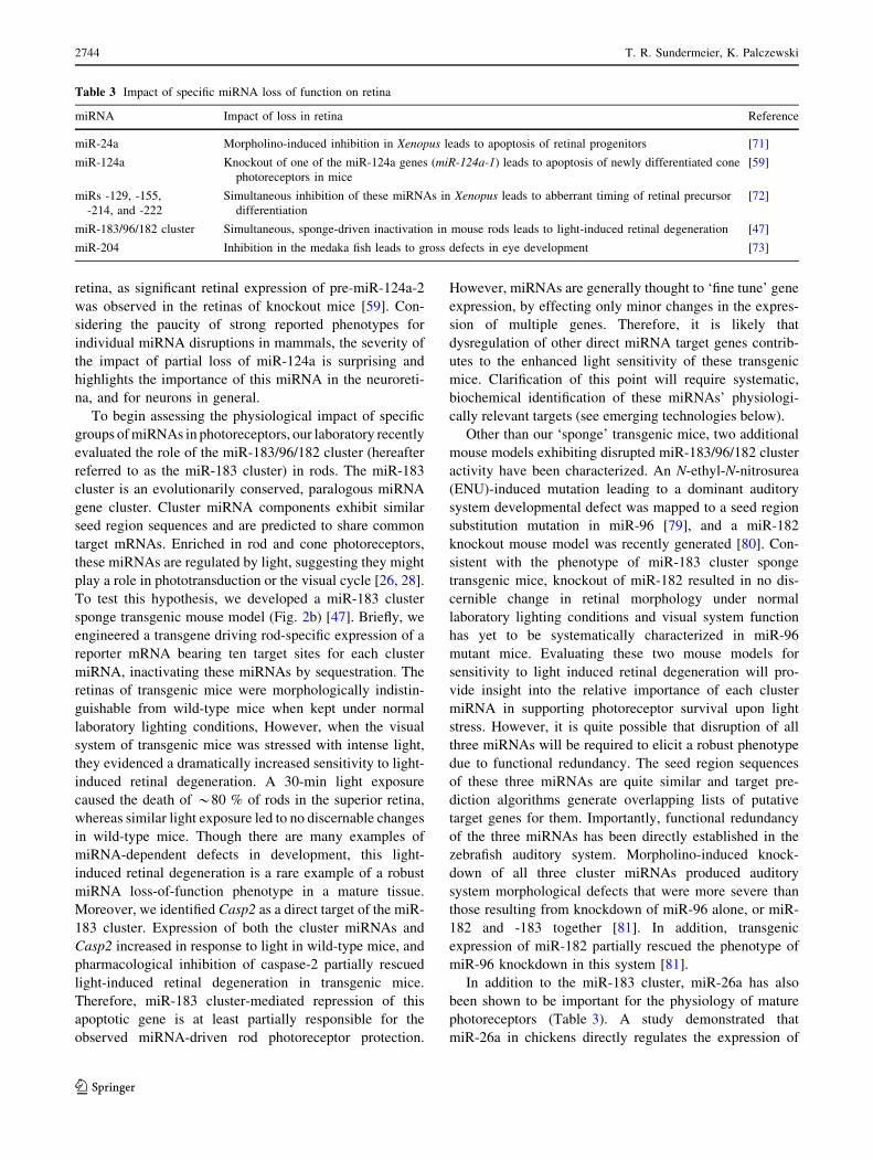
Fig. 2 Emerging technologies for the study of miRNA gene regu-
lation. a AGO CLIPseq permits systematic biochemical identification
of miRNA targets directly in selected tissues [109]. AGO proteins are
crosslinked in vivo to both miRNA and target mRNA. Limited
RNAse digestions followed by high stringency immunoprecipitation
with AGO-specific antibodies results in purification of AGO proteins
covalently linked to either miRNAs or short fragments of target
mRNAs. These co-purified RNAs are then identified by NGS. b A
miRNA sponge is a synthetic gene bearing multiple target sites for
selected miRNA(s) in its 30UTR. Sponges sequester miRISCs bound
to a miRNA, de-repressing synthesis of that miRNA’s target genes.
c Target protectors are oligonucleotides that interact with target
mRNAs by base-pairing, thus inhibiting interaction of the miRISC
with overlapping target sites, de-repressing the expression of specific
target transcripts
microRNAs in the vertebrate retina 2745
123

models promises to yield more physiologically relevant
insights.
Thus, many groups have turned to the retina as a model
system for studying miRNA gene regulation. The retina has
several advantages including: (a) its laminar architecture
and cell type functionalities are well characterized, (b) as a
central nervous system component, the retina is remarkably
amenable to experimental manipulation, and (c) genetic
manipulations that disrupt visual system development or
physiology do not cause premature death, permitting
detailed analysis of phenotypic consequences. Hence,
several significant insights regarding the regulation of the
miRNA response and its manipulation for therapeutic
purposes have recently been gained from studies of the
vertebrate retina.
In a study of broad scope and implications, investigators
in the Filipowicz laboratory set out to determine whether
retinal miRNAs are important for adaptation to variable
levels of illumination [26]. While examining the dynamic
expression levels of a set of light-regulated miRNAs, they
noticed an unexpectedly sharp decline in the levels of these
miRNAs upon dark adaptation. Based on this initial
observation, they went on to demonstrate that dynamic
activity-dependent enhancement in the rate of miRNA
decay is a common feature of regulation of the miRNA
response, especially in neurons. Such rapid, activity-
dependent miRNA turnover could enable more dynamic
modification of cellular miRNA composition, facilitating
miRNA regulation of a rapidly changing transcript pool in
active neurons. In addition, knockout of Dicer in the mouse
retinal pigmented epithelium revealed that this pre-miRNA
processing enzyme might also be involved in cleavage of
other cellular RNAs [69]. Finally, Karali and colleagues
[85] recently showed that engineering miRNA target sites
into the 30 UTR of virally delivered transgenes could
effectively modify the pattern of transgene expression in
the mammalian retina. The last result has interesting
implications for the design of gene delivery vectors, not
only for the visual system, but other organs and tissues as
well. Hence, studies in retina are beginning to yield useful
knowledge about regulation of the miRNA response, as
well as ways to hijack this gene regulatory mechanism to
facilitate eventual gene therapy.
Targeting miRNA gene regulation for treatment
of ocular diseases
Retinal pathologies are attractive candidates for gene
therapy approaches based on the relative accessibility of
this tissue for targeted gene delivery. Indeed, gene therapy
approaches for the treatment of ocular pathologies have
met with much success in recent years. Adeno-associated
virus-mediated gene replacement has proven both safe and
effective for treating Leber congenital amaurosis in clinical
trials with patients carrying mutations in the Rpe65 gene
[86–92]. In addition, gene therapy for neovascular age-
related macular degeneration, as well as retinoblastoma is
undergoing clinical trials [93, 94]. miRNA-based gene
therapeutic approaches can offer enhanced efficacy based
on targeting multiple genes in the same pathway, and
substantial progress has been made recently in developing
efficient delivery systems for targeting miRNA pathways
therapeutically [35–39]. Multiple strategies both for aug-
menting and repressing the activity of specific miRNAs
have been reported, including delivery of synthetic miRNA
mimics, miRNA-encoding transgenes, competitive miRNA
inhibitory antisense oligonucleotides (known as antagom-
irs) and miRNA sponges (Fig. 2b) [39]. The relative ease
of gene delivery to the retina also promises to facilitate the
introduction of small RNAs for therapeutic purposes, as
evidenced by recent clinical trials involving introduction of
siRNAs to the retina for treatment of neovascular age-
related macular degeneration [95–97]. Thus miRNA gene
therapy for retinal degenerative disorders melds two suc-
cessful, recently developed approaches (i.e., gene therapy
in the eye and gene therapy targeting miRNA genes). In
light of recent results which clearly demonstrate that spe-
cific miRNAs play a role in photoreceptor survival [47,
59], manipulation of miRNA activity constitutes an
attractive strategy to treat retinal degenerative disorders
such as retinitis pigmentosa, and age-related macular
degeneration.
Future directions
Recent findings have been discussed that have dramatically
expanded our understanding of the physiological impact
of miRNA gene regulation in the retina. Dicer CKO studies
have clearly demonstrated that global loss of retinal
miRNAs has markedly detrimental effects on retinal
development and physiology. Loss-of-function studies with
lower vertebrate model species have identified fundamental
roles for miRNAs in regulating the differentiation and pro-
moting the survival of retinal neuronal progenitors. Finally,
we are beginning to discern roles for specific miRNAs in
adult retina. Some remaining challenges toward under-
standing the roles of retinal miRNAs are to: (a) assess the
specific physiological roles of each retinal miRNA, (b) sys-
tematically identify genes directly targeted by miRNAs in
the retina, and (c) determine specific target gene(s) whose
dysregulation in the absence of miRNAs results in abnormal
retinal phenotypes. Though this list seems daunting,
emerging technologies developed for studying miRNAs in
other systems have the potential to dramatically accelerate
2746 T. R. Sundermeier, K. Palczewski
123
progress. It is possible to envision an experimental scheme
wherein the direct targets of a retinal miRNA are identified
biochemically by using AGO crosslinking immunoprecipi-
tation followed by next generation sequencing (CLIPseq),
the physiological impact of that miRNA determined using a
miRNA sponge, and the targets whose de-repression is
responsible for the phenotype identified utilizing target
protector oligonucleotides (Fig. 2). In this way, a complete,
detailed picture of the physiological impact of each retinal
miRNA could be revealed.
AGO CLIPseq
Reliable identification of genes targeted by miRNAs is of
paramount importance to understanding the physiological
impact of miRNA gene regulation. Until recently, targets
of a given miRNA were identified by using one of several
bioinformatic miRNA target prediction algorithms (e.g.,
TargetScan [98], PicTar [99], Microcosm [100], etc.).
Selected target genes were then validated by demonstrating
that their 30UTR could be directly targeted in a reporter-
based, cell culture transfection assay. However, this
approach suffers from several inherent limitations. First,
target prediction algorithms suffer from a high rate of false
positives, requiring potentially biased selection of putative
targets for further analyses. Second, constraints inherent in
each of the different target prediction algorithms render
them unlikely to identify certain classes of physiologically
relevant target genes. For example, reliance on perfect seed
region complementarity causes certain algorithms to ignore
targets exhibiting non-canonical (i.e., G-U wobble) base
pairing or seed region mismatches. Similarly, analyses
limited to the mRNA 30UTR miss a well-characterized
class of target sites residing within the coding sequence
[101–103]. Finally, cell type and tissue-specific factors
such as expression of other miRNA target transcripts,
mRNA binding proteins or target site-bearing non-coding
RNAs can influence miRISC interaction with a given
putative target transcript [84, 104–108]. Therefore, reli-
able, non-biased miRNA target identification requires a
systematic, biochemical assay of miRISC-target mRNA
interactions directly in the cell-type or tissue of interest.
The recently developed technology of CLIP-seq offers an
elegant solution to this problem (Fig. 2a) [109, 110]. Con-
veniently, miRNA-interacting proteins of the AGO family
exist in sufficiently close proximity to both the miRNA guide
strand and its bound target mRNA to permit in vivo
UV-crosslinking to both RNAs [109, 111]. The resulting
covalently linked protein-RNA complexes then can be
purified through high-stringency immunoprecipitation, and
the RNA sequences identified by NGS. First reported in
2009, the utility of AGO CLIP-seq is now well established in
a variety of systems including HeLa cells, mouse embryonic
stem cells, C. elegans, and mouse neocortex [109, 112–114].
However, no systematic, biochemical analysis of miRNA
targets in the retina has yet been reported. CLIPseq analysis
represents a dramatic improvement over earlier AGO
immunoprecipitation-based miRNA target analyses which
lack crosslinking, because in vivo crosslinking stabilizes the
AGO-RNA complexes such that the resulting genome-wide
map of retinal miRNA regulation represents a snapshot of
RNAs bound by AGO in living cells. In addition, covalent
attachment of AGO to bound RNAs allows more stringent
immunoprecipitation, reducing background, and identifica-
tion of RNAs whose interaction with the miRISC might be
too transient to survive immunoprecipitation and washing.
The miRNA sponge
With reported mammalian retinal miRNAs numbering in
the hundreds, it is impractical to knockout these miRNAs
individually in mice to evaluate their physiological func-
tion (even though a resource of mouse embryonic stem
cells bearing deletion of most individual miRNAs has
recently been reported) [115]. Inhibition of miRNA activity
with synthetic antagomirs is another option, but though the
retina is more amenable to this type of manipulation than
other tissues, the extent and duration of this effect is lim-
ited. A useful alternative is to express a sponge transgene
to inactivate one or a group of miRNAs by sequestration
(Fig. 2b). This approach has the advantages of decreased
model development time along with control over cell-type
specificity of miRNA inactivation through selection of an
appropriate promoter to drive sponge transgene expression.
miRNA target protectors
Once the impact of loss of a specific miRNA has been
established and its direct targets biochemically validated, the
next challenge is to establish which of the identified target
genes mediates the observed phenotype. The ideal way to
address this question would be to knock-in mutation to the
target site(s) to see if this disruption has an effect similar to
miRNA inactivation. However, as miRNAs tend to target
many genes, this approach is likely to be prohibitively
laborious. An alternative has recently been developed
involving the use of antisense oligonucleotides termed ‘tar-
get protectors’ (Fig. 2c). First developed in zebrafish [116],
this strategy has since been used to inhibit miRNA-target
interactions in Drosophila [117], and more recently in mice
[118]. Briefly, a target protector is an antisense oligonu-
cleotide that binds to a region of a given mRNA overlapping
the miRNA target site, acting as a competitive inhibitor of
miRISC-target interaction that is specific for the selected
target gene. Zovoilis and colleagues [118] used hippocampal
injection of target protector oligonucleotides to demonstrate
microRNAs in the vertebrate retina 2747
123

that miR-34c mimic-dependent learning impairment is
mediated through targeting of the SIRT1 mRNA in mice.
One can easily envision application of this approach to study
retinal miRNA-target interactions by subretinal injection of
precisely designed target protectors, or through cell type-
specific expression of target protector transgenes.
Conclusions
A wealth of new information has evolved recently regarding
the functional significance of miRNAs in the vertebrate
retina. Global as well as specific miRNA loss-of-function
studies have revealed important roles for miRNAs in retinal
development, physiology and disease. Furthermore, the
vertebrate retina has served as an attractive model system for
studying both the regulation and experimental manipulation
of the miRNA response. New technologies for studying
miRNA gene regulation now make it possible to evaluate
precisely the impact of loss of specific miRNAs or miRNA-
target mRNA interactions in selected tissues or cell types.
Moreover, technology permitting tissue-specific, biochemi-
cal identification of miRNA targets will allow systematic
analysis of downstream factors that mediate loss-of-function
phenotypes. Though much progress has recently been made
in this field, the pace of discovery is certain to increase
greatly in the near future.
Acknowledgments We thank Dr. Leslie T. Webster, Jr. for critical
comments on the manuscript. This work was supported, in whole or in
part, by National Institutes of Health Grants EY009339, and P30
EY11373. KP is John H. Hord Professor of Pharmacology.
References
1. Rodieck RW (1998) The first steps in seeing. Sinauer Associ-
ates, Sunderland
2. Palczewski K (2011) Focus on vision: 3 decades of remarkable
contributions to biology and medicine. FASEB J 25(2):439–443
3. Ridge KD, Palczewski K (2007) Visual rhodopsin sees the light:
structure and mechanism of G protein signaling. J Biol Chem
282(13):9297–9301
4. Huntzinger E, Izaurralde E (2011) Gene silencing by microR-
NAs: contributions of translational repression and mRNA decay.
Nat Rev Genet 12(2):99–110
5. Fabian MR, Sonenberg N, Filipowicz W (2010) Regulation of
mRNA translation and stability by microRNAs. Annu Rev
Biochem 79:351–379
6. Fabian MR, Sundermeier TR, Sonenberg N (2010) Under-
standing how miRNAs post-transcriptionally regulate gene
expression. Prog Mol Subcell Biol 50:1–20
7. Lee RC, Feinbaum RL, Ambros V (1993) The C. elegans het-
erochronic gene lin-4 encodes small RNAs with antisense
complementarity to lin-14. Cell 75(5):843–854
8. Wightman B, Burglin TR, Gatto J, Arasu P, Ruvkun G (1991)
Negative regulatory sequences in the lin-14 30-untranslated region
are necessary to generate a temporal switch during Caenorhab-ditis elegans development. Genes Dev 5(10):1813–1824
9. Wightman B, Ha I, Ruvkun G (1993) Posttranscriptional regu-
lation of the heterochronic gene lin-14 by lin-4 mediates
temporal pattern formation in C. elegans. Cell 75(5):855–862
10. Lee RC, Ambros V (2001) An extensive class of small RNAs in
Caenorhabditis elegans. Science 294(5543):862–864
11. Lau NC, Lim LP, Weinstein EG, Bartel DP (2001) An abundant
class of tiny RNAs with probable regulatory roles in Caeno-rhabditis elegans. Science 294(5543):858–862
12. Lagos-Quintana M, Rauhut R, Lendeckel W, Tuschl T (2001)
Identification of novel genes coding for small expressed RNAs.
Science 294(5543):853–858
13. Pasquinelli AE et al (2000) Conservation of the sequence and
temporal expression of let-7 heterochronic regulatory RNA.
Nature 408(6808):86–89
14. Newman MA, Hammond SM (2010) Emerging paradigms of
regulated microRNA processing. Genes Dev 24(11):1086–1092
15. Siomi H, Siomi MC (2010) Posttranscriptional regulation of
microRNA biogenesis in animals. Mol Cell 38(3):323–332
16. Fabian MR et al (2011) miRNA-mediated deadenylation is
orchestrated by GW182 through two conserved motifs that
interact with CCR4-NOT. Nat Struct Mol Biol 18(11):1211–1217
17. Chekulaeva M et al (2011) miRNA repression involves GW182-
mediated recruitment of CCR4-NOT through conserved W-con-
taining motifs. Nat Struct Mol Biol 18(11):1218–1226
18. Braun JE, Huntzinger E, Fauser M, Izaurralde E (2011) GW182
proteins directly recruit cytoplasmic deadenylase complexes to
miRNA targets. Mol Cell 44(1):120–133
19. Tritschler F, Huntzinger E, Izaurralde E (2010) Role of GW182
proteins and PABPC1 in the miRNA pathway: a sense of deja
vu. Nat Rev Mol Cell Biol 11(5):379–384
20. Rapicavoli NA, Blackshaw S (2009) New meaning in the
message: noncoding RNAs and their role in retinal development.
Dev Dyn 238(9):2103–2114
21. Xu S (2009) microRNA expression in the eyes and their signifi-
cance in relation to functions. Prog Retin Eye Res 28(2):87–116
22. Li X, Jin P (2010) Roles of small regulatory RNAs in deter-
mining neuronal identity. Nat Rev Neurosci 11(5):329–338
23. Karali M et al (2010) miRNeye: a microRNA expression atlas of
the mouse eye. BMC Genomics 11:715
24. Hackler L Jr, Wan J, Swaroop A, Qian J, Zack DJ (2010)
MicroRNA profile of the developing mouse retina. Invest
Ophthalmol Vis Sci 51(4):1823–1831
25. Karali M, Peluso I, Marigo V, Banfi S (2007) Identification and
characterization of microRNAs expressed in the mouse eye.
Invest Ophthalmol Vis Sci 48(2):509–515
26. Krol J et al (2010) Characterizing light-regulated retinal mi-
croRNAs reveals rapid turnover as a common property of
neuronal microRNAs. Cell 141(4):618–631
27. Makarev E, Spence JR, Del Rio-Tsonis K, Tsonis PA (2006)
Identification of microRNAs and other small RNAs from the
adult newt eye. Mol Vis 12:1386–1391
28. Xu S, Witmer PD, Lumayag S, Kovacs B, Valle D (2007)
MicroRNA (miRNA) transcriptome of mouse retina and iden-
tification of a sensory organ-specific miRNA cluster. J Biol
Chem 282(34):25053–25066
29. Wang FE et al (2010) MicroRNA-204/211 alters epithelial
physiology. FASEB J 24(5):1552–1571
30. Ryan DG, Oliveira-Fernandes M, Lavker RM (2006) MicroR-
NAs of the mammalian eye display distinct and overlapping
tissue specificity. Mol Vis 12:1175–1184
31. Damiani D et al (2008) Dicer inactivation leads to progressive
functional and structural degeneration of the mouse retina.
J Neurosci 28(19):4878–4887
2748 T. R. Sundermeier, K. Palczewski
123

32. Georgi SA, Reh TA (2010) Dicer is required for the transition
from early to late progenitor state in the developing mouse
retina. J Neurosci 30(11):4048–4061
33. Iida A, Shinoe T, Baba Y, Mano H, Watanabe S (2011) Dicer
plays essential roles for retinal development by regulation of
survival and differentiation. Invest Ophthalmol Vis Sci 52(6):
3008–3017
34. Pinter R, Hindges R (2010) Perturbations of microRNA function
in mouse dicer mutants produce retinal defects and lead to
aberrant axon pathfinding at the optic chiasm. PLoS One 5(4):
e10021
35. Kota J et al (2009) Therapeutic microRNA delivery suppresses
tumorigenesis in a murine liver cancer model. Cell 137(6):
1005–1017
36. Trang P et al (2010) Regression of murine lung tumors by the
let-7 microRNA. Oncogene 29(11):1580–1587
37. Lanford RE et al (2010) Therapeutic silencing of microRNA-
122 in primates with chronic hepatitis C virus infection. Science
327(5962):198–201
38. Thum T et al (2008) MicroRNA-21 contributes to myocardial
disease by stimulating MAP kinase signalling in fibroblasts.
Nature 456(7224):980–984
39. Sayed D, Abdellatif M (2011) MicroRNAs in development and
disease. Physiol Rev 91(3):827–887
40. Liu MM, Tuo J, Chan CC (2011) Gene therapy for ocular dis-
eases. Br J Ophthalmol 95(5):604–612
41. Conley SM, Cai X, Naash MI (2008) Nonviral ocular gene
therapy: assessment and future directions. Curr Opin Mol Ther
10(5):456–463
42. Chalberg TW, Genise HL, Vollrath D, Calos MP (2005) phiC31
integrase confers genomic integration and long-term transgene
expression in rat retina. Invest Ophthalmol Vis Sci 46(6):2140–2146
43. Farjo R, Skaggs J, Quiambao AB, Cooper MJ, Naash MI (2006)
Efficient non-viral ocular gene transfer with compacted DNA
nanoparticles. PLoS One 1:e38
44. Diebold Y, Calonge M (2010) Applications of nanoparticles in
ophthalmology. Prog Retin Eye Res 29(6):596–609
45. Loscher CJ et al (2007) Altered retinal microRNA expression
profile in a mouse model of retinitis pigmentosa. Genome Biol
8(11):R248
46. Loscher CJ et al (2008) A common microRNA signature in
mouse models of retinal degeneration. Exp Eye Res 87(6):
529–534
47. Zhu Q et al (2011) Sponge transgenic mouse model reveals
important roles for the microRNA-183 (miR-183)/96/182 cluster
in postmitotic photoreceptors of the retina. J Biol Chem
286(36):31749–31760
48. Kovacs B, Lumayag S, Cowan C, Xu S (2011) MicroRNAs in
early diabetic retinopathy in streptozotocin-induced diabetic
rats. Invest Ophthalmol Vis Sci 52(7):4402–4409
49. Ishida W et al (2011) Dynamic changes of microRNAs in the
eye during the development of experimental autoimmune uve-
oretinitis. Invest Ophthalmol Vis Sci 52(1):611–617
50. Conkrite K et al (2011) miR-17 *92 cooperates with RB path-
way mutations to promote retinoblastoma. Genes Dev 25(16):
1734–1745
51. Silva VA et al (2011) Expression and cellular localization of
microRNA-29b and RAX, an activator of the RNA-dependent
protein kinase (PKR), in the retina of streptozotocin-induced
diabetic rats. Mol Vis 17:2228–2240
52. McArthur K, Feng B, Wu Y, Chen S, Chakrabarti S (2011) Micr-
oRNA-200b regulates vascular endothelial growth factor-mediated
alterations in diabetic retinopathy. Diabetes 60(4):1314–1323
53. Feng B et al (2011) miR-146a-Mediated extracellular matrix
protein production in chronic diabetes complications. Diabetes
60(11):2975–2984
54. Wu JH et al (2011) Altered microRNA expression profiles in
retinas with diabetic retinopathy. Ophthalmic Res 47(4):
195–201
55. Mu G et al (2010) Correlation of overexpression of HMGA1 and
HMGA2 with poor tumor differentiation, invasion, and prolifer-
ation associated with let-7 down-regulation in retinoblastomas.
Hum Pathol 41(4):493–502
56. Zack DJ et al (1999) What can we learn about age-related
macular degeneration from other retinal diseases? Mol Vis 5:30
57. Sun H, Nathans J (2001) The challenge of macular degeneration.
Sci Am 285(4):68–75
58. Organisciak DT, Vaughan DK (2010) Retinal light damage:
mechanisms and protection. Prog Retin Eye Res 29(2):113–
134
59. Sanuki R et al (2011) miR-124a is required for hippocampal
axogenesis and retinal cone survival through Lhx2 suppression.
Nat Neurosci 14(9):1125–1134
60. Ramachandran R, Fausett BV, Goldman D (2010) Ascl1a reg-
ulates Muller glia dedifferentiation and retinal regeneration
through a Lin-28-dependent, let-7 microRNA signalling path-
way. Nat Cell Biol 12(11):1101–1107
61. Lamba DA, Karl MO, Reh TA (2009) Strategies for retinal
repair: cell replacement and regeneration. Prog Brain Res 175:
23–31
62. Yang JS, Lai EC (2011) Alternative miRNA biogenesis path-
ways and the interpretation of core miRNA pathway mutants.
Mol Cell 43(6):892–903
63. Yang JS, Lai EC (2010) Dicer-independent, Ago2-mediated
microRNA biogenesis in vertebrates. Cell Cycle 9(22):4455–4460
64. Sibley CR et al (2011) The biogenesis and characterization of
mammalian microRNAs of mirtron origin. Nucleic Acids Res
40(1):438–448
65. Flynt AS, Greimann JC, Chung WJ, Lima CD, Lai EC (2010)
MicroRNA biogenesis via splicing and exosome-mediated
trimming in Drosophila. Mol Cell 38(6):900–907
66. Berezikov E, Chung WJ, Willis J, Cuppen E, Lai EC (2007)
Mammalian mirtron genes. Mol Cell 28(2):328–336
67. Okamura K, Hagen JW, Duan H, Tyler DM, Lai EC (2007) The
mirtron pathway generates microRNA-class regulatory RNAs in
Drosophila. Cell 130(1):89–100
68. Ruby JG, Jan CH, Bartel DP (2007) Intronic microRNA pre-
cursors that bypass Drosha processing. Nature 448(7149):83–86
69. Kaneko H et al (2011) DICER1 deficit induces Alu RNA toxicity
in age-related macular degeneration. Nature 471(7338):325–330
70. Decembrini S, Andreazzoli M, Barsacchi G, Cremisi F (2008)
Dicer inactivation causes heterochronic retinogenesis in Xeno-pus laevis. Int J Dev Biol 52(8):1099–1103
71. Walker JC, Harland RM (2009) microRNA-24a is required to
repress apoptosis in the developing neural retina. Genes Dev
23(9):1046–1051
72. Decembrini S et al (2009) MicroRNAs couple cell fate and
developmental timing in retina. Proc Natl Acad Sci USA 106(50):
21179–21184
73. Conte I et al (2010) miR-204 is required for lens and retinal
development via Meis2 targeting. Proc Natl Acad Sci USA
107(35):15491–15496
74. Baudet ML et al (2012) miR-124 acts through CoREST to
control onset of Sema3A sensitivity in navigating retinal growth
cones. Nat Neurosci 15(1):29–38
75. Visvanathan J, Lee S, Lee B, Lee JW, Lee SK (2007) The
microRNA miR-124 antagonizes the anti-neural REST/SCP1
pathway during embryonic CNS development. Genes Dev 21(7):
744–749
76. Maiorano NA, Mallamaci A (2010) The pro-differentiating role
of miR-124: indicating the road to become a neuron. RNA Biol
7(5):528–533
microRNAs in the vertebrate retina 2749
123
77. Makeyev EV, Zhang J, Carrasco MA, Maniatis T (2007) The
microRNA miR-124 promotes neuronal differentiation by trig-
gering brain-specific alternative pre-mRNA splicing. Mol Cell
27(3):435–448
78. Cheng LC, Pastrana E, Tavazoie M, Doetsch F (2009) miR-124
regulates adult neurogenesis in the subventricular zone stem cell
niche. Nat Neurosci 12(4):399–408
79. Lewis MA et al (2009) An ENU-induced mutation of miR-96
associated with progressive hearing loss in mice. Nat Genet
41(5):614–618
80. Jin ZB et al (2009) Targeted deletion of miR-182, an abundant
retinal microRNA. Mol Vis 15:523–533
81. Li H, Kloosterman W, Fekete DM (2010) MicroRNA-183
family members regulate sensorineural fates in the inner ear.
J Neurosci 30(9):3254–3263
82. Shi L, Ko ML, Ko GY (2009) Rhythmic expression of micr-
oRNA-26a regulates the L-type voltage-gated calcium channel
alpha1C subunit in chicken cone photoreceptors. J Biol Chem
284(38):25791–25803
83. Kasinski AL, Slack FJ (2011) Epigenetics and genetics. MicroR-
NAs en route to the clinic: progress in validating and targeting
microRNAs for cancer therapy. Nat Rev Cancer 11(12):849–864
84. van Kouwenhove M, Kedde M, Agami R (2011) MicroRNA
regulation by RNA-binding proteins and its implications for
cancer. Nat Rev Cancer 11(9):644–656
85. Karali M et al (2011) MicroRNA-restricted transgene expression
in the retina. PLoS One 6(7):e22166
86. Bainbridge JW et al (2008) Effect of gene therapy on visual
function in Leber’s congenital amaurosis. N Engl J Med
358(21):2231–2239
87. Cideciyan AV et al (2009) Vision 1 year after gene therapy for
Leber’s congenital amaurosis. N Engl J Med 361(7):725–727
88. Cideciyan AV et al (2009) Human RPE65 gene therapy for Leber
congenital amaurosis: persistence of early visual improvements
and safety at 1 year. Hum Gene Ther 20(9):999–1004
89. Cideciyan AV et al (2008) Human gene therapy for RPE65
isomerase deficiency activates the retinoid cycle of vision but
with slow rod kinetics. Proc Natl Acad Sci USA 105(39):
15112–15117
90. Maguire AM et al (2009) Age-dependent effects of RPE65 gene
therapy for Leber’s congenital amaurosis: a phase 1 dose-esca-
lation trial. Lancet 374(9701):1597–1605
91. Maguire AM et al (2008) Safety and efficacy of gene transfer for
Leber’s congenital amaurosis. N Engl J Med 358(21):2240–
2248
92. Simonelli F et al (2010) Gene therapy for Leber’s congenital
amaurosis is safe and effective through 1.5 years after vector
administration. Mol Ther 18(3):643–650
93. Campochiaro PA et al (2006) Adenoviral vector-delivered pig-
ment epithelium-derived factor for neovascular age-related
macular degeneration: results of a phase I clinical trial. Hum Gene
Ther 17(2):167–176
94. Ildefonso CJ et al (2010) Absence of systemic immune response
to adenovectors after intraocular administration to children with
retinoblastoma. Mol Ther 18(10):1885–1890
95. Kaiser PK, et al. (2010) RNAi-based treatment for neovascular
age-related macular degeneration by Sirna-027. Am J Ophthal-
mol 150(1):33-39 e32
96. Bumcrot D, Manoharan M, Koteliansky V, Sah DW (2006)
RNAi therapeutics: a potential new class of pharmaceutical
drugs. Nat Chem Biol 2(12):711–719
97. Campa C, Harding SP (2011) Anti-VEGF compounds in the
treatment of neovascular age related macular degeneration. Curr
Drug Targets 12(2):173–181
98. Lewis BP, Burge CB, Bartel DP (2005) Conserved seed pairing,
often flanked by adenosines, indicates that thousands of human
genes are microRNA targets. Cell 120(1):15–20
99. Krek A et al (2005) Combinatorial microRNA target predic-
tions. Nat Genet 37(5):495–500
100. Griffiths-Jones S, Saini HK, van Dongen S, Enright AJ (2008)
miRBase: tools for microRNA genomics. Nucleic Acids Res
36:D154–D158
101. Kang JG et al (2011) Kaposi’s sarcoma-associated herpesvirus
ORF57 promotes escape of viral and human interleukin-6 from
microRNA-mediated suppression. J Virol 85(6):2620–2630
102. Moretti F, Thermann R, Hentze MW (2010) Mechanism of
translational regulation by miR-2 from sites in the 50 untrans-
lated region or the open reading frame. RNA 16(12):2493–2502
103. Schnall-Levin M, Zhao Y, Perrimon N, Berger B (2010) Con-
served microRNA targeting in Drosophila is as widespread in
coding regions as in 30UTRs. Proc Natl Acad Sci USA 107(36):
15751–15756
104. Karreth FA et al (2011) In vivo identification of tumor- sup-
pressive PTEN ceRNAs in an oncogenic BRAF-induced mouse
model of melanoma. Cell 147(2):382–395
105. Cesana M et al (2011) A long noncoding RNA controls muscle
differentiation by functioning as a competing endogenous RNA.
Cell 147(2):358–369
106. Tay Y et al (2011) Coding-independent regulation of the tumor
suppressor PTEN by competing endogenous mRNAs. Cell
147(2):344–357
107. Salmena L, Poliseno L, Tay Y, Kats L, Pandolfi PP (2011) A
ceRNA hypothesis: the Rosetta Stone of a hidden RNA lan-
guage? Cell 146(3):353–358
108. Mukherji S et al (2011) MicroRNAs can generate thresholds in
target gene expression. Nat Genet 43(9):854–859
109. Chi SW, Zang JB, Mele A, Darnell RB (2009) Argonaute HITS-
CLIP decodes microRNA-mRNA interaction maps. Nature
460(7254):479–486
110. Licatalosi DD et al (2008) HITS-CLIP yields genome-wide
insights into brain alternative RNA processing. Nature 456(7221):
464–469
111. Wang Y et al (2008) Structure of an argonaute silencing com-
plex with a seed-containing guide DNA and target RNA duplex.
Nature 456(7224):921–926
112. Leung AK et al (2011) Genome-wide identification of Ago2
binding sites from mouse embryonic stem cells with and without
mature microRNAs. Nat Struct Mol Biol 18(2):237–244
113. McKenna LB et al (2010) MicroRNAs control intestinal epithelial
differentiation, architecture, and barrier function. Gastroenterol-
ogy 139(5):1654–1664 1664 e1651
114. Zisoulis DG et al (2010) Comprehensive discovery of endoge-
nous Argonaute binding sites in Caenorhabditis elegans. Nat
Struct Mol Biol 17(2):173–179
115. Prosser HM, Koike-Yusa H, Cooper JD, Law FC, Bradley A
(2011) A resource of vectors and ES cells for targeted deletion
of microRNAs in mice. Nat Biotechnol 29(9):840–845
116. Choi WY, Giraldez AJ, Schier AF (2007) Target protectors
reveal dampening and balancing of Nodal agonist and antagonist
by miR-430. Science 318(5848):271–274
117. Gehrke S, Imai Y, Sokol N, Lu B (2010) Pathogenic LRRK2
negatively regulates microRNA-mediated translational repres-
sion. Nature 466(7306):637–641
118. Zovoilis A et al (2011) microRNA-34c is a novel target to treat
dementias. EMBO J 30(20):4299–4308
119. Bai Y et al (2011) MicroRNA-126 inhibits ischemia-induced
retinal neovascularization via regulating angiogenic growth
factors. Exp Mol Pathol 91(1):471–477
2750 T. R. Sundermeier, K. Palczewski
123