Embed Size (px)
Citation preview

!
Medical Science Series
THE PHYSICS OFMEDICAL IMAGING
Edited by
Steve Webb
Joint Department of Physics,Institute of Cancer Research and
Royal Marsden Hospital, Sutton, Surrey
Adam Hilger, Bristol and Philadelphia

'\
6.4 RADIONUCLIDES FOR IMAGING
One of the primary advantages associated with the use of radionuclidesin medicine is the large signal (in this case the emitted radiation)obtained from the relatively small mass of radionuclide ~J1lployed for agiven study, Nuclear medicine takes advantage of this physical charac-teristic by using various radioisotope-tagged compounds (radio-pharmaceuticals) in order to 'trace' various functions of the body(McAfee and Thakur 1977). The minute mass of radio labelled materialallows for non-invasive observation without disturbance of the systemunder study through pharmacological or toxicological effect. For mostnuclear medicine studies, the mass of tracer used is in the range ofnanograms (see table 6.4), and no other physical technique could beemployed to measure mass at these levels. Therefore, the sensitivemeasurement of biochemical and physiological processes through the useof radioactivity and its detection comprise the fundamental basis ofnuclear medicine and is the key to its future growth.

182 The physics of radioisotope imaging
Table 6.4 Approximate mass (g) of 37 GBq (1 Ci) of radionuclidefor a given half-life and atomic weight.
Atomic weight of atom (amu)TI/2 -"
18 99 201
15 s 2.4 x 10⁻¹¹ 1.3 X 10⁻¹⁰ 2.7 X 10-1015 min 1.4 x 10⁻⁹ 7.7 X 10⁻⁹ 1.6 X 10-86 h 3.5 X 10⁻⁸ 1.9 X 10⁻⁷ 3.8 X 10-78 d 1.1 X 10⁻⁶ 6.3 X 10⁻⁶ 1.2 X 10-515 a 7.7 X 10⁻⁴ 4.2 X 10⁻³ 8.3 X 10⁻³
6.4.1 Radioactive decay
The radioactivity Q of a nu,!Ilber (N) of nuclei is given by
Q = - AN = dN Idt (6.13)
where A is defined as the decay copstant for the radioisotope. We cansee that the rate of decay of nuclei depends only upon A and the numberof nuclei, N. The solution to equation (6.13) is
N=Noexp(-At) (6.14)where No is the number of nuclei at some reference time t = O. If T 1/2is the time for half the nuclei to decay, the so-called 'half-life', then
TI/2 = (In2)/A. (6.15)
An alternative form of equation (6.14) isN = No(~)m (6.16)
where m is the number of half-lives since the reference time t = O.Using these simple equations, it is possible to calculate the radioactiv-
ity of any mass of nuclei at any time subsequent to a measurement at areference-zero time.
6.4.2 The production of radionuclides
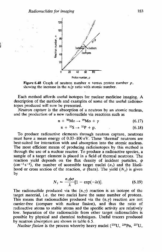
The fundamental property of all radioactive elements is the imbalance ofthe proton-to-neutron ratio of the nucleus. A proper balance of proton~and neutrons is essential for maintaining a stable atomic nucleus. Thebalance must be maintained to overcome electrostatic repulsion of thecharged protons, and figure 6.48 shows how the neutr{)n-to-proton (nip)ratio changes with increasing mass. There are four ways by whichradionuclides are produced:
(a) neutron capture (also known as neutron activation);(b) nuclear fission;(c) charged-particle bombardment; and(d) radionuclide generator.
Radionuclides for imaging 183
120 "f't,;-
.".,100 ~~:"
c: ,;;"!, ~- 80 .li:";;::-
Ii: ~ 60 lso~ lsoba~
E .{r"'5 40 .i'- Iso1upes~ .it"i'
2 ,-' ,
40 60 80
Proton number, p
Figure 6.48 Graph of neutron number n versus proton number p,showing the increase in the nip ratio with atomic number.
Each method affords useful isotopes for nuclear medicine imaging. Adescription of the methods and examples of some of the useful radioiso-topes produced will now be presented.
Neutron capture is the absorption of a neutron by an atomic nucleus,and the production of a new radionuclide via reactions such as
n + 98Mo - 99Mo + y (6.17)
n + 32S - 32p + p. (6.18)
To produce radioactive elements through neutron capture, neutrons
must have a mean energy of 0.03-100 eV. These 'thermal' neutrons are
best-suited for interaction with and absorption into the atomic nucleus.
The most efficient means of producing radioisotopes 'by this method is
through the use of a nuclear reactor. To produce a radioactive species, a
sample of a target element is placed in a field of thermal neutrons. The
reaction yield depends on the flux density of incident particles, cJ>
(cm-2s-1), the number of accessible target nuclei (nJ and the likeli-hood or cross section of the reaction, a (barn). The yield (Ny) is givenby
ntcJ>aNy = -;:-[1 - exp( -At)]. (6.19)
The radionuclide produced via the (n,y) reaction is an isotope of thetarget material, i.e. the two nuclei have the same number of protons.This means that radionuclides produced via the (n,y) reaction are notcarrier-free (compare with nuclear fission), and thus the ratio ofradioactive atoms to stable atoms and the specific activity are relativelylow. Separation of the radionuclide from other target radio nuclides ispossible by physical and chemical techniques. Useful tracers producedby neutron absorption are shown in table 6.5.
Nuclear fission is the process whereby heavy nuclei (235U, 239pU, 237U,

184 The physics of radioisotope imaging
232Th) irradiated with thermal neutrons are rendered unstable due toabsorption of these neutrons. Consequently, these unstable nucleiundergo 'fission', the breaking up of the heavy nuclei into two lighternuclei of approximately similar atomic weight, for example
235U + !n ~ 236U ~ 99Mo + 133Sn + 41n (620)92 U'. 92 42 50 0 . .
As seen from equation (6.20), this reaction produces four more neut-rons, which may be absorbed by other heavy nuclei, and the fissionprocess can continue until the nuclear fuel is exhausted. Interaction suchas that in equation (6.20) must, of course, conserve Z and A.
Table 6.5 Radionuclides produced by neutron absorption.
Gamma-ray AbsorptionIsotope energy Half-life cross section
(keV) (barn)
51Cr 320 27.7d 17"59Fe ,1099 44.5d 1.1"99Mo 740 66.02 h 0.131311b 364 8.05 d 0.2
" From BRH (1970).b 130Te(n,y)131Te - 1311.
Nuclides produced by fission of heavy nuclei must undergo extensivepurification in order to harvest one particular radionuclide from themixture of fission products. The fission process affords high specificactivity due to the absence of carrier material (non-radioactive isotopeof the same element). However, fission products are usually rich inneutrons and therefore decay principally via {3- emission, a physic~lcharacteristic that is undesirable for medical imaging, but of interest intherapy. Useful nuclides produced by nuclear fission are shown in table6.6.
Table 6.6 Radionuclides produced by nuclear fission.
Gamma-ray FissionIsotope energy Half-life yield"
(keV) (%)
99Mo 740 66.02 h '6.11311 364 8.05 d 2.9133Xe 81 5.27 d 6.5137CS 662 30 a 5.9
" From BRH (1970).

Radionuclides for imaging 185
Charged-particle bombardment is the process of production ofradio nuclides through the interaction of charged particles (H:t, D + ,3He2+, 4He2+) with the nuclei af stable atoms. The particles must haveenough kinetic energy to overcome the electrostatic repulsion of thepositively charged nucleus. Two basic types of accelerator are used forthis purpose, the linear accelerator and the cyclotron. In both systems,charged particles are accelerated over a finite distance by the applicationof alternating electromagnetic potentials (figure 6.49). In both types ofmachine, particles can usually be accelerated to various energies.Examples of typical reactions in a target are
p + 68Zn ~ 67Ga + 2n (6.21)
a + 160 ~ 18F + P + n. (6.22)
(alIon source
acuumhamber
Accbeat 'II ~ AC voltage
) (b)
~
Drift tubesIon .,-c'I:J's"EZJ...Ei;J",='.=~"=""'1 Targetsource -+-+-+-+-+-+-+-+-+
~diofrequency cavities
Figure 6.49 Schematic diagrams of (a) a cyclotron and (b) a linearaccelerator for radionuclide production.
(' For the production of medically useful radionuclides, particle energies
per nucleon in the range 1-100 MeV are commonly used. One majoradvantage of producing isotopes through charged-particle bombardmentis that the desired isotope is almost always of different atomic numberto the target material. This theoretically allows for the production ofradio nuclides with very high specific activity and minimal radionuclide

186 The physics of radioisotope imaging
impurity. However, the actual activity and purity obtained is related tothe isotopic and nuclidic purity of the target material, the cross sectionof the desired reaction and the cross section of any secondary reaction.
Charged-particle reactions yield radionuclides that are predominantlyneutron-deficient and therefore decay by (3+ emission or electroncapture. The latter radioisotopes are particularly useful for clinicalimaging due to the lack of particulate emission. Examples ofaccelerator-produced radionuclides routinely used in nuclear medicineare shown in table 6.7.
Table 6.7 Radionuclides produced by charged-particle bombard-ment.
PrincipalIsotope gamma-ray energy Half-life Reaction
(keV)
tiC 511 (fJ..+) 20.4 min 14N(p,CX)IIC13N 511 (fJ+) 9.96 min 13C(p,n)13NISO 511 (fJ+) 2.07 min 15N(p,n)15018F 511 (fJ+) 109.7min 180(p,n)18F67Ga 93 78.3 h 68Zn(p,2n) 67Ga
184300
lllln 171 67.9 h 112Cd(p,2n)lllln245
1231 159 13 h 124Te(p,2n) 12311271(p,5n) 123Xe~ 1231
201Tl 68-80.3 73 h 203Tl (})',3n) 201 Ph 2olTI
Radioactive decay can lead to the generation of either a stable or aradioactive nuclide. In either case, the new nuclide may have the sameor different atomic number depending on the type of decay (see nextsection). Radioactive decay leading to the production of a radioactivedaughter with a different Z allows for the possibility of simple chemicalseparation of the parent-daughter combination. If the daughterradio nuclide has good physical characteristics compatible with medicalimaging and the parent has a sufficiently long half-life to allow forproduction, processing and shipment, then remote parent-daughterseparation means a potentially convenient source of a medically usefulshort-lived radionuclide. This type of radionuclide production system isknown as a radionuclide generator.
A radionuclide generator is a means of having 'on tap' a short-livedradionuclide. It is technically achieved by the chemical separation of thedaughter radionuclide from the parent. This can be accomplishedthrough the use of chromatographic techniques, distillation or phasepartitioning. However, chromatographic techniques have been the most

Radionuclides for imaging 187
widely explored and are the current state-of-the-art technology (Yano1975) for the majority of generator systems in use today (figure 6.50).
Eluting solution Eluant outj)Jtin~t needle needle
0.22 ~m filter
Leadshielding
ChromoiUQmphicadsorbenT Plastic
housing
Figure 6.50 Schematic of a radioisotope generator.
The equations governing generator syst~ms stem from the formula
AZ 0 ( )Az =, , A,j[exp( -A1t) - exp( -Azt)] 6.23/l.z - /1.1
where A Y is the parent activity at time t = 0, t is the time since the lastelution of the generator, A z is the activity of the daughter product(A~ = 0), and Al and AZ are the decay constants of parent and daughter
radioisotopes, respectively.For the special case of secular equilibrium, defined by AZ »A1, we
have-J Az = AY[exp(-A1t) - exp(-Azt)]. (6.24)
If t is much less than the half-life of the parent, In(2)/A1, and greaterthan approximately seven times the daughter half-life, In(2)/Az, then
Az = AY. (6.25)
This is the equilibrium condition. The growth of the daughter here is
given byAz = AY[1 - exp( -Azt)]. (6.26)
For transient equilibrium, defined by AZ > Al but AZ not very muchgreater than AI, we have
Az = AzA~/(AZ - AI). (6.27)
Figure 6.51 shows the growth of 99Tcm activity in a 99Mo - 99Tcm
generator.

188 The physics of radioisotope imaging
1. Elution timeParent
05 tivity
0.2
~0.10>=:;:~ 0.0
O.
0 12 24 36 48
Time (h)Figure 6.51 The build-ue of activity of the daughter product withtime for a typical (99Mo/99Tcm) radioisotope generator.
The most widely used generator-produced radionuclide in nuclearmedicine is 99Tcm. The parent, 99Mo, has a half-life of about 66 h, canbe produced through neutron activation or fission, can be chemicallyadsorbed onto an Al2O3 (alumina) column and decays to 99Tcm (85%)and 99Tc (15%). 99Tcm has a half-life of 6.02 h, decays to 99Tc byisomeric transition and emits a 140 ke V y-ray (98%) with no asspciatedparticulate radiations:
99Mo - 99Tcm - 99Tc + y. (6.28)p- IT
In the early days of their development (see Chapter 1), technetiumgenerators were sometimes referred to as 'radioactive cows'. The 99Tcmis 'milked' from the chromatographic column of alumina by passing asolution of isotonic saline through the column (0.9% NaCI). This salinesolution and the solid phase of Al2O3 allow for efficient separation of99Tcm from the 99Mo with only minute amounts of 99Mo breakthrough(less than 0.1%). The eluted 99Tcm can be chemically manipulated sothat it binds to a variety of compounds, which will then determine itsfate in vivo (see §6.9). Other generator systems exist producingradionuclides useful for gamma-camera imaging as well as for PET andexamples of these are given in table 6.8.
6.4.3 Types of radioactive decay
All radionuclides used in nuclear medicine are produced via one of thefour ways described above. Each of these radio nuclides has a uniqueprocess by which it decays. The decay scheme describes the type ofdecay, the energy associated with it and the probability for each type ofdecay. These decay schemes can be very complex since many of theradionuclides decay via multiple nuclear processes (figure 6.52).
c

w
Radionuclides for imaging 189
Table 6.8 Generator-produced radionuclides.
Gamma-rayParent Mode of Mode of Daughter energy from
Parent half- decay Daughter decay half- daughterP life P-+D D of D life (keV)
,
99Mo 2.7 d f3- ,99Tcm ( IT 6 h 140
82Sr 25 d EC 82Rb EC 1.3 min 777f3+ 511
68Ge 280 d EC 68Ga EC 68 min 511
f3+52Fe 8.2 h EC 52Mnm EC 21 min 511
f3+ f3+IT
81Rb 4.7 h EC 81Krm IT 13 s 19062Zn 9.1 h EC 62CU EC 9.8 min 511
f3+ f3+178W 21.5 d EC 178Ta EC 9.5 min 93
1311
Emittedphotonenergies (MeV)
0.7230.6670,637
l11 18 17 15 13
~40.4050.3640.341
19 16 14 12
0.164
110 0.080
11Stable 131Xe
Figure 6.52 The nuclear decay process for 1311.
Alpha decay is the process of spontaneous emission of an a-particle (ahelium nucleus) in the decay of heavy radioisotopes, with a discreteenergy in the range 4-8 Me V. If a decay leaves the nucleus in an
L - " - ";i
~,

190 The physics of radioisotope imaging
excited state, the de-excitation will be via the emission of y-radiation.Most of the energy released in the transition is distributed between adaughter nucleus (as recoil energy) and the (X-particle (as kineticenergy). In the (X-decay process, the parent nucleus loses four units ofmass and two units of charge. As an example we show
2~Ra ~ 2~Rn + (x. (6.29)
Although (X-emitting nuclides have no use in medical imaging (sincethe (X-particles travel virtually no distance in tissue), there has been arenewed interest in their use for targeted (i.e. highly localised) therapy.
As discussed previously, many radionuclides are unstable due to theneutron/proton imbalance within the nucleus. The decay of neutron-richradionuclides involves the ejection of a {3--particle (e-), resulting in theconversion of a neutron into a proton. Decay via {3- emission results inthe atomic number of the atom changing but the atomic weightremaining the same. The energy of the emitted {3--particles is notdiscrete but a continuum (i.e. varies from zero to a maximum, Em) and,since the total energy lost by the nucleus during disintegration must bediscrete, an additional process must be responsible. Energy conservationof {3 decay is maintained by the emission of a third particle-theneutrino (v). The neutrino has no measurable mass nor charge, andinteracts weakly with matter. An example of {3- decay is
99Mo ~ 99Tcm + e- + v. (6.30)
Beta decay may be accompanied by y-ray emission if the daughternuclide is produced in an excited state. After {3- decay, the atomicnumber of the daughter nuclide is one more than the parent nuclide, butthe atomic mass remains the same.
Nuclei that are rich in protons or are neutron-deficient may decay bypositron emission from the nucleus. This decay is also accompanied bythe emission of an uncharged particle, the anti neutrino (v). Afterpositron decay, the daughter nuclide has an atomic number that is oneless than that of the parent, but again the atomic weight is the same.The range of a positron (e +) is short (of the order of 1 mm in tissue)and, when the particle comes to rest, it combines with an atomicelectron from a nearby atom, and is annihilated. Annihilation (thetr:;lnsformation of these two particles into pure energy) gives rise to twophotons both of energy 511 ke V emitted approximately antiparallel toeach other. These photons are referred to as annihilation radiation.Positron emission only takes place when the energy difference betweenthe parent and daughter nuclides is larger than 1.02 MeV. An exampleof positron decay is
68Ga ~ 68Zn + e+ + v. (6.31)An alternative to positron emission for nuclides with a proton-rich
nucleus is electron capture. Electron capture involves the absorptionwithin the nucleus of an atomic electron, transforming a proton into a

Radionuclides for imaging 191
neutron. For this process to occur, the energy difference between theparent and the daughter nuclides can be small, unlike positron emission.Usually the K-shell eleftrons are captured because of their closeness tothe nucleus. The vacancy created in the inner electron orbitals is filledby electrons from the outer orbitals. The difference in energy betweenthese electron shells appears as an x-ray that is characteristic of thedaughter radionuclide. The probability of electron capture increases withincreasing atomic number because electron shells in these nuclei arecloser to the nucleus. An example of electron capture is
1231 ~ 123Te + y. (6.32)
A nucleus produced in a radioactive decay can remain in an excitedstate for some time. Such states are referred to as isomeric states, anddecay to the ground state can take from fractions of a second to manyyears. A transition from the isomeric or excited state to the ground stateis accompanied by y emission. When the isomeric state is long-lived, thisstate is often referred to as a metastable state. 99Tcm is the mostcommon example of a metastable isotope encountered in nuclearmedicine (see equation (6.28».
Internal conversion is the process that can occur during y-ray emissionwhen a photon emitted from a nucleus may knock out one of the atomicelectrons from the atom. This particularly affects K-shell electrons, asthey are the nearest to the nucleus. The ejected electron is referred toas the conversion electron and will have a kinetic energy equal to theenergy of the y-ray minus the electron binding energy. The probabilityof internal conversion is highest for low-energy photon emission. Again,vacancies in the inner orbitals are filled by electrons from the outershells, leading to the emission of characteristic x-rays. Furthermore,characteristic x-rays produced during internal conversion may themselvesknock out other outer orbital electrons provided the x-rays have anenergy greater than the binding energy of the electron with which they-interact. This emitted electron is then referred to as an Auger electron.Again, vacancies in the electron shells due to Auger emission are filledby other electrons in outer orbitals leading to further x-ray emission.
6.4.4 Choice of radioisotope for imaging
The physical characteristics of radionuclides that are desirable fornuclear medicine imaging include:
(a) a suitable physi~~l half-life;(b) decay via photon emission;(c) associated photon energy high enough to penetrate the body
tissue with minimal tissue attenuation; but(d) low enough for minimal thickness of collimator septa; and(e) absence of particulate emission.
The effective half-life T E of a radiopharmaceutical is a combination of
192 The physics of radioisotope imaging
the physical half-life T p and the biological half-life T B, i.e.1 1 1-T =-T + -T . (6.33)E B P
Close matching of the effective half-life with the duration of the study isan important dosimetric as well as practical consideration in terms ofavailability and radiopharmaceutical synthesis.
The photon energy is critical, for various reasons. The photon mustbe able to escape from the body efficiently and it is desirable that thephotopeak should be easily separated from any scattered radiation.These two characteristics favour high-energy photons. However, at veryhigh energies, detection efficiency using a conventional gamma camerais poor (see figure 6.5) and the increased septal thickness required forcollimators decreases the sensitivity further. In addition, high-energyphotons are difficult to shield and present practical problems for staffhandling the isotope.
The radionuclide that fulfils most of the above criteria is technetium-99m (99Tcm), which is used in more than 90 % of all nuclear medicinestudies. It has a physical half-life of 6.02 h, is produced via decay of along-lived (T 1/2 = 66 h) parent 99Mo, and decays via isomeric transitionto 99Tc emitting a 140 keY y-ray. The short half-life and absence of f3:!:
emission results in a low radiation dose to the patient. The 140 keY yemission allows for 50% penetration of tissue at a thickness of 4.6 cmbut is easily collimated by lead. Most importantly, the radioisotope canbe produced from a generator lasting the best part of a week, supplyingimaging agents 'on tap'.
Other radioisotopes in common use in nuclear medicine include 1231,1111n, 67Ga, 2olTI and 8lKrm. 1231 has proved a valuable replacement for1311 as it decays via electron capture emitting a y-ray of energy 159 keYand has a 13 h half-life. It is easily bonded to proteins and phar-maceuticals that can be iodinated. However, like most of the otherradioisotopes in this list, it is cyclotron-produced (table 6.7) andpresently still very expensive in a form free of other iodine isotopes.
The radionuclides 1IIIn and 67Ga are very similar chemically and bothdecay via electron capture (table 6.9). Most recently, there has been anincreased interest in their use as antibody labels via bifunctionalchelates. III In is the superior imaging isotope, emitting acceptablephoton energies for gamma-camera studies, but again it is expensive asit is produced by charged-particle bombardment (table 6.7). 67Ga haslong been used as a tumour localising agent in the form of galliumcitrate and has also proved useful in the same form in the detection ofabcesses. 2OlTI is utilised by the cardiac muscle in a similar fashion topotassium and is in widespread use for imaging myocardial perfusion.However, the photon emissions used in myocardial imaging are the80 keY x-rays, which are close in energy to lead x-rays produced by thecollimator, and this, together with the long half-life (73 h), makes theisotope a poor imaging agent. Hence there is continued search for a99Tcm-labelled myocardial perfusion agent.

Radionuclides for imaging 193
Table 6.9 Main gamma emissions of 67Ga and IIIIn.
Gamma-ray MeanRadionuclide Gamma-ray energy number per
emission (ke V) disintegration~
67Ga Y2 93 0.38Y3 185 0.24Ys 300 0.16Y6 394 0.043
IllIn YI 171 0.90Y2 245 0.94
Radioisotopes emitting positrons (table 6.7) have been used extensive-ly for physiological research, in the main, rather than clinical nuclearmedicin~ because of the need for an on-site or nearby cyclotron toproduce them in view of~their relatively short half-lives. The radionuc-lides 15O, 13N, llC and 18F have many applications in the field offunctional imaging but have had little impact, as yet, in routine nuclearmedicine because of lack of availability. 68Ga and 82Rb, however, aretwo generator-produced positron-emitting nuclides that could provideinvaluable radiopharmaceuticals for clinical PET. In particular, 68Ga canbe used to label many agents in a manner similar to 99Tcm, and 82Rb isa greatly superior myocardial perfusion agent to 201TI. These radioiso-topes, coupled with the development of low-cost PET cameras (§6.3.7),may bring the much-needed advantages of high sensitivity and spatialresolution to clinical nuclear medicine.
ao- , - - -,
r


![Chapter Five 1850 Virginia Census Webb Families · Chapter Five 1850 Virginia Census Webb Families Carroll County Abner Webb .....29 [s/o John & Hannah (Cock) Webb] Sarah .....25](https://img.pdfslide.us/doc/110x75/5b04c8997f8b9a3c378e25c5/chapter-five-1850-virginia-census-webb-five-1850-virginia-census-webb-families-carroll.jpg)