Embed Size (px)
Citation preview
The phospho-occupancy of an atypicalSLIMB-binding site on PERIODthat is phosphorylated by DOUBLETIMEcontrols the pace of the clockJoanna C. Chiu,1 Jens T. Vanselow,2 Achim Kramer,2 and Isaac Edery3,4
1Rutgers University, Center for Advanced Biotechnology and Medicine, Piscataway, New Jersey 08854, USA; 2Laboratoryof Chronobiology, Charité Universitätsmedizin Berlin, 10115 Berlin, Germany; 3Department of Molecular Biologyand Biochemistry, Rutgers University, Center for Advanced Biotechnology and Medicine, Piscataway, New Jersey 08854,USA
A common feature of animal circadian clocks is the progressive phosphorylation of PERIOD (PER) proteins,which is highly dependent on casein kinase I�/� (CKI�/�; termed DOUBLETIME [DBT] in Drosophila) andultimately leads to the rapid degradation of hyperphosphorylated isoforms via a mechanism involving theF-box protein, �-TrCP (SLIMB in Drosophila). Here we use the Drosophila melanogaster model system, andshow that a key step in controlling the speed of the clock is phosphorylation of an N-terminal Ser (S47) byDBT, which collaborates with other nearby phosphorylated residues to generate a high-affinity atypicalSLIMB-binding site on PER. DBT-dependent increases in the phospho-occupancy of S47 are temporally gated,dependent on the centrally located DBT docking site on PER and partially counterbalanced by proteinphosphatase activity. We propose that the gradual DBT-mediated phosphorylation of a nonconsensusSLIMB-binding site establishes a temporal threshold for when in a daily cycle the majority of PER proteins aretagged for rapid degradation. Surprisingly, most of the hyperphosphorylation is unrelated to direct effects onPER stability. We also use mass spectrometry to map phosphorylation sites on PER, leading to theidentification of a number of “phospho-clusters” that explain several of the classic per mutants.
[Keywords: Circadian rhythms; Drosophila; PER; �-TrCP/SLIMB; CK1�/DBT; F-box protein; phosphorylation]
Supplemental material is available at http://www.genesdev.org.
Received April 9, 2008; revised version accepted May 9, 2008.
Circadian (≅24 h) rhythms are widespread in all king-doms of life and are driven by cellular “clocks” or pace-makers (for review, see Roenneberg and Merrow 2005).These pacemakers are based on the expression of a coreset of species or tissue specific “clock” genes that arecentral to rhythm generation. Much of the earlier workled to the realization that a major feature of intracellularclock mechanisms involves interconnected positive andnegative translational–transcriptional feedback loops thatyield cyclical gene expression in one or more of the coreclock genes (Dunlap 1999).
Despite the overwhelming success of this molecularframework featuring rhythmic gene expression, work inthe last several years has demonstrated the importanceof post-transcriptional regulation, especially protein phos-phorylation, in generating cellular oscillators with circa-dian properties (Bae and Edery 2006; Merrow et al. 2006;
Gallego and Virshup 2007). Most notably, it appears thata shared feature of all circadian systems analyzed to dateis that a major protein component functioning within thenegative limb of circadian transcription undergoes dailycycles in phosphorylation that are central to clock pro-gression. This was first observed for the Drosophila mel-anogaster PERIOD (dPER) protein (Edery et al. 1994). Be-sides dPER, major components of the clock in this sys-tem include TIMELESS (TIM), CLOCK (dCLK), andCYCLE (CYC) (Hardin 2005). dCLK and CYC are tran-scription factors of the basic helix–loop–helix (bHLH)/PAS (Per-Arnt-Sim) superfamily that heterodimerize tostimulate the daily transcription of dper and tim, in ad-dition to other clock and downstream genes.
Phosphorylation has been linked to regulation of dPERstability, nucleocytoplasmic distribution, and transcrip-tional repressor potency (Bae and Edery 2006). Newly syn-thesized dPER is initially present as a hypophosphorylatedvariant(s) in the late day/early night, progressively in-creasing in extent of phosphorylation such that by thelate night/early day only hyperphosphorylated species
4Corresponding author.E-MAIL [email protected]; FAX (732) 235-5318.Article is online at http://www.genesdev.org/cgi/doi/10.1101/gad.1682708.
1758 GENES & DEVELOPMENT 22:1758–1772 © 2008 by Cold Spring Harbor Laboratory Press ISSN 0890-9369/08; www.genesdev.org
Cold Spring Harbor Laboratory Press on June 17, 2018 - Published by genesdev.cshlp.orgDownloaded from
are detected (Edery et al. 1994). The DOUBLETIME (DBT)kinase (homolog of mammalian CKI�/�) is a key kinasecontrolling the temporal program underlying dPER phos-phorylation and stability (Kloss et al. 1998; Price et al.1998). As the levels of TIM increase during the day itbinds dPER and somehow protects it against DBT-medi-ated degradation (Price et al. 1995; Rothenfluh et al.2000; Ko et al. 2002). The interaction of dPER with TIMalso stimulates but is not obligatory for nuclear localiza-tion of both proteins (Shafer et al. 2002; Meyer et al.2006). In the nucleus, dPER binds to dCLK and blocks itstrans-activation potential (Darlington et al. 1998; Lee etal. 1998, 1999). During the late-night/early morning de-clining levels of nuclear TIM somehow accelerate hyper-phosphorylation of dPER, whereby highly phosphorylat-ed isoforms are targeted to the 26S proteasome by theF-box protein SLIMB (Drosophila homolog of �-TrCP)(Grima et al. 2002; Ko et al. 2002). In general, F-box pro-teins recognize phosphorylated substrates and are part oflarger SCF (Skp1, Cullin, F-box) E3 ligases that targetsubstrates to the ubiquitin–proteasome pathway (Ho etal. 2008). Rapidly declining levels of dPER in the nucleusseverely diminish or terminate its repressor function,initiating another round of dCLK–CYC-mediated tran-scription. Thus, a key aspect of the temporal regulationin dCLK–CYC activity involves the phase-specific accu-mulation and duration of dPER in the nucleus.
The progressive phosphorylation of dPER throughout adaily cycle and its ability to act as a transcriptional repres-sor are dependent on a small DBT-binding domain (dPERDBT-binding domain; dPDBD) in the central region ofdPER (Kim et al. 2007; Nawathean et al. 2007). In theabsence of the dPDBD, dPER is very stable, mainly hy-pophosphorylated, and has weak ability to inhibit dCLK,suggesting that the phosphorylated state of dPER alsomodulates its potency as a transcriptional repressor (Na-wathean and Rosbash 2004; Kim et al. 2007; Nawatheanet al. 2007). CKII and possibly other kinases also regulatedPER levels, although they appear to play less significantroles compared with DBT and might contribute in sec-ondary pathways such as via regulating subcellular dis-tribution. In addition to kinases, protein phosphatase 1(PP1) and 2A (PP2A) regulate the phosphorylated state ofdPER and other clock proteins, presumably antagonizingthe destabilizing effects of phosphorylation on clock pro-tein stability (Sathyanarayanan et al. 2004; Kim and Ed-ery 2006; Fang et al. 2007).
A strikingly similar scenario also occurs in mammals,whereby CKI�/� plays a major role in regulating dailycycles in the phosphorylation and abundance of mam-malian PERs (mPER1–3) (Gallego and Virshup 2007). Theimportance of PER phosphorylation is highlighted bystudies showing that mutations in either a phosphoryla-tion site on human PER2 or CKI� underlie several famil-ial advanced sleep phase syndromes (FASPS) (Toh et al.2001; Xu et al. 2005, 2007; Vanselow et al. 2006). Despitemuch progress, a detailed understanding of of the phos-phorylation events and the role of CKI�/DBT in thephase-specific degradation of PER proteins is lacking.Herein, we use the D. melanogaster system to deter-
mine how phosphorylation regulates the daily decline inPER levels. Our results indicate that gradual DBT-medi-ated increases in the phospho-occupancy of Ser47 ondPER acts like a “throttle” by timing when in the daythe majority of dPER proteins are tagged for efficientrecognition by SLIMB, a critical event in setting the paceof the clock. In addition, it appears that much, if not allof the more centrally localized phosphorylation on dPERis not directly involved in regulating stability, suggestingthat the majority of the hyperphosphorylation observedfor PER proteins is involved in other biochemical func-tions. Finally, using mass spectrometry we show thatmany of the phosphorylation sites on dPER are organizedin local “clusters” and that the classic perS(=Short) muta-tion (Konopka and Benzer 1971) abolishes a phosphory-lation site.
Results
Ser47 is a key determinant regulating dPER stability
We previously showed that the DBT-dependent progres-sive phosphorylation and subsequent SLIMB-mediateddegradation of dPER can be recapitulated in culturedDrosophila Schneider (S2) cells by expressing recombinantdper and dbt, whereby expression of dbt is controlled bythe copper-inducible metallothionein promoter (pMT)and that of dper by the constitutive actin5C promoter(pAct) (Ko et al. 2002). To identify regions regulatingdPER stability we analyzed a range of deletion mutantsusing this system. In one such series, we generated fiveV5-tagged dPER mutants, each containing nonoverlap-ping 100-amino-acid deletions that span the first 500amino acid residues. Although all five dPER deletionmutants exhibit dbt-induced increases in phosphoryla-tion (indicated by temporal decreases in electrophoreticmobility) that are similar to those observed for wild-typedPER, only dPER(�2–100) proteins remained stable (Fig.1A, lanes 5–8; data not shown; in the experiment shown,dPER levels exhibit a noticeable decline at 12 h post-dbtinduction, whereas in most experiments this occurslater). Our results not only indicate that amino acids2–100 are critical for DBT-mediated degradation but alsodemonstrate that global hyperphosphorylation per se isnot sufficient to trigger the rapid proteolysis of dPER.
One possibility explaining the enhanced stability ofdPER(�2–100) proteins is that amino acids 2–100 containsequence elements that mediate the interaction of dPERwith SLIMB. Indeed, amino acids 17–23 and amino acids47–55 show loose similarity to the standard SLIMB/�-TrCP consensus recognition motif of DpSG�X1 + npS(pS, phosphorylated Ser; �, any hydrophobic amino acid;X, any amino acid) (Supplemental Fig. S1; e.g., Fuchs etal. 2004). A series of smaller deletions targeting theseregions focused our attention on amino acids 44–51(Supplemental Fig. S2A). This region contains four serineresidues—S44, S45, S47, and S48—that are highly con-served in per genes from insects, raising the possibilitythat one or more of these is a phospho-determinant inregulating dPER stability. Indeed, a dPER variant in which
SLIMB–PER interaction sets clock speed
GENES & DEVELOPMENT 1759
Cold Spring Harbor Laboratory Press on June 17, 2018 - Published by genesdev.cshlp.orgDownloaded from

all four Ser were substituted with Ala, dPER(S44–48A),phenocopied the enhanced stability of dPER(�44–51) (cf.Fig. 1B and Supplemental Fig. S2A).
Single Ser-to-Ala mutants indicate that S47 and S48play a major role in DBT-mediated dPER turnover (Fig.1C; Supplemental Fig. S2B). In the case of S44 and S45,we noted a small but reproducible increase in dPER sta-bility for the double mutant (S44/45A) but not when sin-gly evaluated (Supplemental Fig. S2B; data not shown),suggesting minor roles. Replacing S47 with Asp (S47D)to mimic phosphorylation accelerates the kinetics ofdPER disappearance following induction of dbt (Fig. 1D).However, the dPER(S48D) variant is stable (Fig. 1D), sug-gesting that S48 modulates dPER stability in a mannerindependent of phosphorylation at this site. The in-creased stability of the different dPER mutants we ana-lyzed were confirmed by measuring degradation ratesafter dbt induction by treating cells with cycloheximideto block de novo protein synthesis (data not shown).Although S47 is a key DBT-dependent phospho-determi-nant in regulating dPER stability, other regions withinthe first 100 amino acids make contributions. Together,the results strongly suggest the following ranking,beginning with the most stable dPER variants; dPER(�2–100) > dPER(�44–51), dPER(S44–48A) > dPER(S47/48A) >dPER(S47A) > dPER(S44/45A) > dPER(WT).
Using a TEV/TAG strategy to examinephosphorylation in the first 100 amino acids of dPER
To enhance the detection of phosphorylation-dependentelectrophoretic mobility changes in the first 100 aminoacids of dPER we devised a strategy, which we call TEV/TAG (Fig. 2A), whereby we expressed a dPER versionwith a TEV protease site inserted at amino acid 100(dPER/TEV100). To visualize the amino acid 1–100 frag-ment after TEV cleavage, we used 16% Tris-glycine gelsand blotted with �-Flag antibodies, whereas 6% gels and�-c-myc antibodies were used to monitor the largeramino acid 101–1224 fragment. We placed TEV/TAGsites in several different positions on dPER and other testproteins and routinely obtain near total cleavage at thedesired site (e.g., Fig. 3; data not shown). The location forTEV site insertion is chosen based on the prediction byprotein secondary structure analysis, suggesting a lowprobability of disrupting important protein structural el-ements and high probability of access by the TEV prote-ase (data not shown). For dPER, we ensured that anyTEV/TAG derivative meets certain functional criteria,like DBT-dependent hyperphosphorylation, ability to in-hibit dCLK-dependent transcription, and interactionwith TIM (Fig. 2; data not shown).
Figure 1. Identification of N-terminal sequences required forDBT-mediated degradation of dPER in cultured S2 cells. S2 cellswere cotransfected with wild-type or mutant variants of dpercontaining plasmids (pAc-dper-V5-His [A–C] or pAc-3XFlag-His-dper/Tev100-6Xc-myc [D]) and pMT-dbt-V5-His, and col-lected at the indicated times (hours) post-dbt induction. dPERvariants were detected by immunoblotting in the presence of�-V5 (A–C) or �-Flag antibodies (D).
Figure 2. Using the TEV/TAG strategy to evaluate phosphor-ylation in the first 100 amino acids of dPER. (A) Schematicmodel showing the pAc-3XFlag-His-dper/Tev100-6Xc-myc con-struct and resulting cleavage products. (B) Extracts were pre-pared from S2 cells expressing pAc-3XFlag-His-dper/Tev100-6Xc-myc with (+) or without (−) coexpression of pMT-dbt-V5-His. (Right) Extracts were subjected to TEV cleavage, incubatedwith �-Flag beads, and immune complexes treated with �-phos-phatase (+) or mock treated (−). dPER(1–100) was detected using�-Flag antibodies. Hypo- and hyperphosphorylated isoforms areindicated. (*) Hyperphosphorylated isoforms only detected inthe presence of induced dbt. (C) S2 cells coexpressing pAc-3XFlag-His-dper/Tev100-6Xc-myc and pMT-dbt-V5-His wereharvested at the indicated times post-dbt induction and extractswere either TEV-treated or mock-treated. Mock-treated extractswere subjected to immunoblotting in the presence of �-c-mycantibodies to detect full-length dPER (top), and TEV-treated ex-tracts were used to detect dPER(1–100) using �-Flag antibodies(bottom).
Chiu et al.
1760 GENES & DEVELOPMENT
Cold Spring Harbor Laboratory Press on June 17, 2018 - Published by genesdev.cshlp.orgDownloaded from

Using the TEV/TAG strategy, we determined that inour S2 cell system there is some phosphorylation withinthe first 100 amino acids of dPER even in the absence ofinduced DBT, although the majority is hypo- or non-phosphorylated (Fig. 2B, lanes 1,2). The induction of DBTgreatly stimulates the “conversion” of non/hypo- to hy-perphosphorylated isoforms (Fig. 2B, lanes 1,3), and inaddition leads to the production of novel more highlyphosphorylated isoforms (Fig. 2B, lane 3, asterisk). Phos-phatase (�PP) treatment confirmed that the observedslower migrating �-Flag immunoreactive bands are dueto phosphorylation (Fig. 2B, lanes 2,4). The presence ofseveral mobility variants (e.g., Fig. 2B, lane 3) stronglysuggests that multiple residues are phosphorylated (seeTable 2, below). Following induction of dbt the ratio ofhighly phosphorylated isoforms to the non/hypophos-phorylated version progressively increases, peaking ∼18h post-dbt induction (Fig. 2C, cf. lanes 2 and 5). We didnot observe the accumulation of slower migrating iso-forms of the 1–100-amino-acid fragment with longer dbtinduction periods or in the presence of proteasome in-hibitors, suggesting that the hyperphosphorylated spe-cies detected are maximally phosphorylated (data notshown). As expected, decreases in the levels of the dPERfragment containing the first 100 amino acids coincidewith the degradation kinetics of the full-length protein,with substantial decreases by 25 h post-dbt induction(Fig. 2C, top panel, cf. lanes 2 and 7). Our results indicatethat the DBT-dependent transition in the first 100 aminoacids of dPER from mainly hypophosphorylated tolargely maximally phosphorylated is gradual and that therapid decline in dPER levels occurs several hours after a
substantial fraction of the molecules attain a highlyphosphorylated state in the first 100 amino acids.
Phosphorylation of S47 is critical for SLIMB bindingto dPER
We combined the TEV/TAG strategy with SLIMB fusedto glutathione-S-transferase (GST) to identify the SLIMB-binding region on dPER. The results clearly indicate thatamino acids 1–100 of dPER are sufficient and necessaryfor binding to SLIMB (Fig. 3A). No binding was observedwith a GST-control resin (data not shown). Binding ofthe 1–100 amino acid dPER fragment to SLIMB isstrongly enhanced by DBT-induced phosphorylation(Fig. 3A, lanes 9–12). The low level of dPER–SLIMB in-teraction detected at time 0 before dbt induction canpossibly be attributed to endogenous dbt expression orthe leakiness of the pMT promoter that drives the dbttransgene (Fig. 3A, lanes 1,9). Although the 101–1224amino acid fragment is responsible for much of the phos-phorylation-mediated mobility shifts in dPER (e.g., Fig.1A) and contains the DBT docking site (Kim et al. 2007;Nawathean et al. 2007), it showed no detectable bindingto SLIMB (Fig. 3A, lanes 5–8), indicating that the exten-sive global hyperphosphorylation of dPER is not suffi-cient for SLIMB binding. Rather, there is a clear linkbetween DBT-mediated phosphorylation in the first 100amino acids and the efficiency of dPER binding to SLIMB(Fig. 3A, lanes 9–12).
Mutations in the first 100 amino acids that stabilizedPER (e.g., S44–48A, S47A, S48A, and S48D) stronglyinhibit binding to SLIMB (Fig. 3B–D). Importantly, the
Figure 3. S47 is a key phospho-determi-nant mediating dPER–SLIMB interactions. (A)S2 cells coexpressing pAc-3XFlag-His-dper/Tev100-6Xc-myc and pMT-dbt-V5-His werecollected at the indicated times post-dbt in-duction. Extracts were either treated withTEV to yield amino acids 1–100 or amino ac-ids 101–1224, or mock-treated retaining thefull-length amino acids 1–1224. The top panelshows the input used in GST-SLIMB pull-down assays and the bottom panel shows thebound dPER proteins. The amino acid 1–100fragment was detected using �-Flag antibod-ies, whereas full-length dPER and the aminoacid 101–1224 fragment were detected using�-c-myc antibodies. Hyperphosphorylatedand hypophosphorylated dPER isoforms areindicated as Hyper-P and Hypo-P, respec-tively. (B–D) Extracts collected from S2 cellsexpressing wild-type (WT) or mutant deriva-tives of pAc-3XFlag-His-dper/Tev100-6Xc-myc with (+) or without (−) induced dbt weresubjected to TEV cleavage, subjected to GST-SLIMB pull-down assays and the amino acid1–100 dPER fragment detected with �-Flagantibodies. (B) Asterisk (*) denotes S44–48Amutant hyperphosphorylated isoforms withfaster mobility as compared with wild-typehyperphosphorylated isoforms.
SLIMB–PER interaction sets clock speed
GENES & DEVELOPMENT 1761
Cold Spring Harbor Laboratory Press on June 17, 2018 - Published by genesdev.cshlp.orgDownloaded from
S47D mutation, which renders the full-length proteinless stable (Fig. 1D), has a much higher affinity for SLIMB(Fig. 3D, lanes 1–4; cf. input levels to staining intensitiesof bound material). For example, in the absence of inducedDBT the “hypophosphorylated” first 100 amino acids ofthe dPER(S47D) variant exhibits strong binding to SLIMB,unlike the wild-type version (Fig. 3D, lanes 1,3). None-theless, the binding of the amino acid 1–100 fragmentfrom dPER(S47D) is further stimulated by exogenouslyexpressed DBT (Fig. 3D, lanes 3,4). Thus, although S47 islikely to be the key phosphodeterminant in mediatingdPER–SLIMB interactions, DBT-dependent phosphoryla-tion of other Ser/Thr residues in the first 100 amino ac-ids of dPER also contribute. For example, the double mu-tant S44/45A partially reduces binding to SLIMB (Fig. 3B,lanes 2,4), consistent with its smaller effect on dPERlevels (Supplemental Fig. S2B, lanes 3,7). That other resi-dues in the first 100 amino acids of dPER are phosphor-ylated besides those between amino acid 44 and aminoacid 48 is evident from the DBT-dependent shift in themobility of the dPER(S44–48A) fragment (Fig. 3B, lane 8),albeit resulting in isoforms with faster mobilities, al-most certainly due to blocking phosphorylation of Serresidues within amino acids 44–48 (see Table 2, below).However, unlike dPER(S44–48A) we did not observe no-ticeable alterations in the mobilities of the first 100-amino-acid fragment derived from the dPER(S47A) anddPER(S48A) versions following dbt induction (Fig. 3C),indicating that under the conditions used, single changesin the phosphorylated status of S47 do not yield notice-able electrophoretic mobility differences (although thestatus of S47 phosphorylation could affect mobility incombination with other mutations). We obtained similarresults with regards to mutant effects on the efficiency ofSLIMB binding when evaluating full-length versions ofdPER in the absence of TEV cleavage (data not shown).
Together, the results indicate that the dPER–SLIMBinteraction is highly dependent on phosphorylation ofS47 and stimulated by other determinants such as S48and additional phosphorylation events within the first100 amino acids of dPER, most likely including S44 and/or S45. As discussed below, mass spectrometry identifiednumerous phosphorylated residues on dPER and on go-ing efforts are aimed at determining the contributions ofall these phospho-residues to dPER stability. Based on thecentral importance of S47 in regulating dPER abundanceand binding to SLIMB in our S2 cell culture system, hereinwe mainly focused on the physiological role of this site inthe clockworks.
dPER S47 plays a key role in setting the paceof the clock
To investigate the physiological significance of our find-ings in whole animal systems, we generated transgenicflies that produce tagged versions of the dPER(�2–100),dPER(S44–48A), dPER(S47A), and dPER(S47D) proteins.The effects of the different transgenes were examined inthe per-null wper0 (Konopka and Benzer 1971), as well as
wper+ genetic backgrounds. In addition, transgenic fliesof the same genetic background but expressing a wild-type version of the dper transgene [herein referred to asp{dper(WT)}] were included as controls (Kim et al. 2007;Ko et al. 2007). At least three independent lines of eachgenotype were analyzed. Flies were entrained for 4 d in 12h:12 h LD (light/dark) cycle followed by 7 d in DD (con-stant dark) to determine their free-running period. Aspreviously shown, wper0 flies expressing the wild-typedper transgene exhibit strong rhythms with ∼24-h peri-ods (Table 1; Kim et al. 2007; Ko et al. 2007). Whereasp{dper(�2–100)} and p{dper(S44–48A)} transgenes failedto rescue the arrhythmic phenotype of the wper0 mu-tant, flies expressing p{dper(S47A)} or p{dper(S47D)}transgenes showed high levels of rhythmicity but mani-fested long (∼30.7 h) and short (∼22.1 h) periods, respec-tively (Table 1; Supplemental Fig. S3). This illustratesthe physiological importance of S47 as a key phospho-determinant in setting the pace of the Drosophila clock.In addition, the more severe behavioral phenotypes dis-played by p{dper(�2–100)} and p{dper(S44–48A)} fliescompared with the single S47A mutant are consistent withresults obtained in S2 cells (Figs. 1, 3), further supportingthe idea that residues nearby to S47 play supportive roles inregulating dPER metabolism.
The different transgenes manifested similar behavioralphenotypes in the per+ compared with those observed inthe per0 genetic background, indicating they behave in adominant manner (Table 1). The combined results indi-cate that when the stability of dPER increases, the periodof the clock lengthens and at some threshold value fur-ther increases in dPER stability are incompatible withoscillator function leading to arrhythmia. Interestingly,although the period is longer in the per0 compared withper+ background for p{dper(S47A)} flies, essentially iden-tical periods were observed in either genetic backgroundfor flies expressing p{dper(S47D)}. This suggests that de-spite the presence of wild-type dPER, most, if not all, theintracellular clocks are running at the accelerated paceset by dPER(S47D).
dPER protein and mRNA cycles reflect mutantcircadian phenotypes
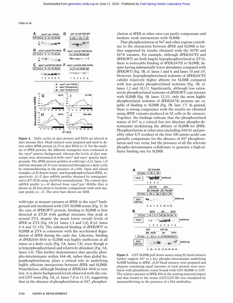
To examine the dPER protein profiles of the mutants,wper0 flies expressing wild-type or mutant dper trans-genes were collected at different times throughout adaily cycle and head extracts probed by immunoblotting.As shown previously, dPER is first observed as a newlysynthesized hypophosphorylated species (fastest migrat-ing species) at around ZT8, undergoes progressive in-creases in phosphorylation, peaks in abundance at aroundZT20 and attains the most highly phosphorylated isoformsaround ZT4, concomitant with rapid decreases in levels(Fig. 4A; Edery et al. 1994). As expected based on thebehavioral results, the dPER(S47A) and dPER(S47D) pro-teins showed daily rhythms in abundance and phosphor-ylation, but with some significant differences as com-pared with dPER(WT) (Fig. 4A–C). In the case of the
Chiu et al.
1762 GENES & DEVELOPMENT
Cold Spring Harbor Laboratory Press on June 17, 2018 - Published by genesdev.cshlp.orgDownloaded from
dPER(S47A) protein, hyperphosphorylated isoforms werefound to persist throughout the daily cycle (Fig. 4B, openarrow). This is in sharp contrast to hyperphosphorylatedisoforms of dPER(WT) proteins that undergo sharp de-creases in abundance beginning in the early day (e.g., Fig.4A,B, cf. ZT12). On the other hand, hyperphosphorylatedisoforms of dPER(S47D) protein were never detected (Fig.4C). Based on our results it is likely that hyperphos-phorylated isoforms of dPER(S47D) are more readily de-graded and never accumulate to detectable levels, al-though presently we cannot rule out the possibility thatdPER(S47D) does not proceed through the entire hyper-phosphorylation program in flies.
RNA levels of dper were attenuated in p{dper(S47A)}flies but enhanced in p{dper(S47D)} flies (Fig. 4G–H). A likelyreason is that the more rapid clearance of dPER(S47D) fromthe nucleus leads to decreased transcriptional repressorfunction and premature reactivation of dCLK-mediatedtranscription, whereas dPER(S47A) likely persists in thenucleus at elevated levels for longer, repressing dCLK-mediated transcription and hence attenuating the ampli-tude of the dper RNA rhythm. Also, it is possible thatphosphorylation might enhance the repressor ability ofdPER (Nawathean and Rosbash 2004; Kim et al. 2007;Nawathean et al. 2007). Similar effects on the dper RNAabundance cycles were also observed when the mutantswere analyzed in a per+ genetic background (Fig. 4G,H),consistent with the behavioral results (Table 1).
Neither dPER(�2–100) nor dPER(S44–48A) proteinsshowed daily changes in abundance or phosphorylated state(Fig. 4D–F). The electrophoretic mobility of dPER(S44–48A) ishigher than the most extensively phosphorylated iso-
forms that we can detect for wild-type dPER (Fig. 4D, cf.lanes 5,6 and 1,2). Phosphatase treatment demonstratedthat dPER(S44–48A) and wild-type dPER comigratewhen dephosphorylated (data not shown), indicating thatdPER(S44–48A) is very highly phosphorylated and thedecreased mobility is not due to secondary issues, suchas altered conformation. Although the electrophoreticmobility of dPER(�2–100) is faster because of the removalof 100 amino acids, it also is highly phosphorylated (datanot shown). dper mRNA levels in p{dper(�2–100)} andp{dper(S44–48A)} flies (in both wper0 and wper+ back-ground) were essentially pegged at wild-type trough val-ues, although some low amplitude cycling was observedfor the dper(S44–48A) RNA, especially in a per+ back-ground (Fig. 4I,J). Thus, in both S2 cells and flies,dPER(�2–100) and dPER(S44–48A) are very stable despitebeing highly phosphorylated, and exhibit more severephenotypes compared with the singly S47A and S47Dmutations. In addition, based on the RNA profiles wecan infer that dPER(�2–100) and dPER(S44–48A) are ef-fective in repressing dCLK-dependent transcriptional ac-tivation, consistent with prior work indicating thata more C-terminal region of dPER termed “CCID”(CLK–CYC inhibition domain) inhibits the trans-activa-tion potential of dCLK (Chang and Reppert 2003).
GST-SLIMB pull-down assays using fly head extractsconfirm results obtained in S2 cells
To study SLIMB–dPER interactions in flies, head ex-tracts were prepared from transgenic flies expressing
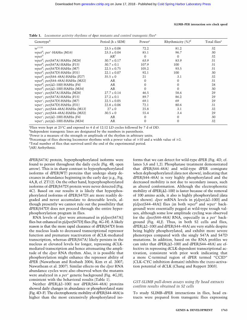
Table 1. Locomotor activity rhythms of dper mutants and control transgenic fliesa
Genotypeb Period (h ± SEM) Powerc Rhythmicity (%)d Total fliese
w1118 23.5 ± 0.08 72.2 81.2 32wper0; per+-HAHis (M16) 23.3 ± 0.04 85.1 96.7 30wper0 ARf 0 0 32wper0; per(S47A)-HAHis (M28) 30.7 ± 0.17 63.9 83.9 31wper0; per(S47A)-HAHis (F15) 30.7 ± 0.1 107.9 100 31wper0; per(S47D)-HAHis (M7) 22.5 ± 0.75 105.2 93.5 31wper0; per(S47D)-HAHis (F31) 22.1 ± 0.07 92.1 100 30wper0; per(S44–48A)-HAHis (F27) 35.5 ± 0 21 3.1 32wper0; per(S44–48A)-HAHis (M32) AR 0 0 31wper0; per(�2–100)-HAHis (F4) AR 0 0 28wper0; per(�2–100)-HAHis (M34) AR 0 0 30wper+; per(S47A)-HAHis (M28) 27.7 ± 0.14 46.5 58.6 29wper+; per(S47A)-HAHis (F15) 27.2 ± 0.1 89.7 86.2 29wper+; per(S47D)-HAHis (M7) 22.5 ± 0.05 69.1 69 29wper+; per(S47D)-HAHis (F31) 22.8 ± 0.06 72.1 80.6 31wper+; per(S44–48A)-HAHis (M13) 27 ± 0 25.8 3.1 32wper+; per(S44–48A)-HAHis (M32) 30.5 ± 0 55.4 3.4 29wper+; per(�2–100)-HAHis (F4) AR 0 0 30wper+; per(�2–100)-HAHis (M34) AR 0 0 32
aFlies were kept at 25°C and exposed to 4 d of 12:12 LD cycles followed by 7 d of DD.bIndependent transgenic lines are designated by the numbers in parenthesis.cPower is a measure of the strength or amplitude of the rhythm in arbitrary units.dPercentage of flies showing locomotor rhythms with a power value of �10 and a width value of �2.eTotal number of flies that survived until the end of the experimental period.f(AR) Arrhythmic.
SLIMB–PER interaction sets clock speed
GENES & DEVELOPMENT 1763
Cold Spring Harbor Laboratory Press on June 17, 2018 - Published by genesdev.cshlp.orgDownloaded from
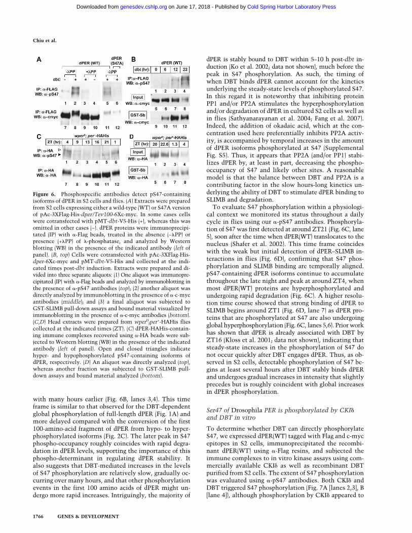
wild-type or mutant variants of dPER in the wper0 back-ground and incubated with GST-SLIMB resins (Fig. 5). Inthe case of dPER(WT) protein, binding to SLIMB is firstdetected at ZT20 with gradual increases that peak ataround ZT4, despite the much lower overall levels ofdPER at ZT4 (Fig. 5A [cf. lanes 1,2 and 5,6], B [cf. lanes3–6 and 12–15]). The enhanced binding of dPER(WT) toSLIMB at ZT4 is consistent with the accelerated degra-dation of dPER during the early day. Likewise, bindingof dPER(S44–48A) to SLIMB was highly inefficient at alltimes in a daily cycle (Fig. 5A, lanes 7,8), even though itis hyperphosphorylated and relatively abundant (Fig. 5A,lanes 3,4). This further demonstrates that specific phos-pho-determinants within S44–48, rather than global hy-perphosphorylation, plays a critical role in underlyinghighly efficient interactions between dPER and SLIMB.Nonetheless, although binding of dPER(S44–48A) is verylow, it is above background levels observed with the con-trol GST-resin (Fig. 5A, cf. lanes 7,8 to 11,12), suggestingthat in the absence of phosphorylation at S47, phosphor-
ylation of dPER at other sites can partly compensate andmediate weak interactions with SLIMB.
That phosphorylation at S47 and other regions contrib-ute to the interaction between dPER and SLIMB is fur-ther supported by results obtained with the S47D andS47A variants. For example, although dPER(S47D) anddPER(WT) are both largely hypophosphorylated at ZT16,there is noticeable binding of dPER(S47D) to SLIMB, de-spite having substantially lower abundance compared withdPER(WT) (Fig. 5B, cf. lanes 1 and 4, and lanes 10 and 13).However, hyperphosphorylated isoforms of dPER(S47D)exhibit relatively higher affinity for SLIMB comparedwith less grossly phosphorylated isoforms (Fig. 5B, cf.lanes 1,2 and 10,11). Significantly, although less exten-sively phosphorylated isoforms of dPER(WT) can interactwith SLIMB (Fig. 5B, lanes 12,15), only the most highlyphosphorylated isoforms of dPER(S47A) proteins are ca-pable of binding to SLIMB (Fig. 5B, lane 17). In general,there is strong congruence with the results we obtainedusing dPER variants produced in S2 cells or fly extracts.Together, the findings indicate that the phosphorylatedstatus of S47 is a critical but not absolute phospho-de-terminant modulating the affinity of SLIMB for dPER.Phosphorylation at other sites (including S44/45 and pos-sibly other S/T residues in the first 100 amino acids) canpartially compensate for the absence of S47 phosphory-lation and vice versa, but the presence of all the relevantphospho-determinants collaborate to generate a high-af-finity binding site for SLIMB.
Figure 5. GST-SLIMB pull-down assays using fly head extractsfurther support S47 as a key phospho-determinant underlyingSLIMB binding to dPER. (A,B) Head extracts were prepared andaliquots containing equal amounts of total protein were incu-bated with glutathione resins bound with GST-SLIMB or GST.The relative amount of dPER-HA in the starting material (input)and that bound to the resins (GST/GST-Sb) was visualized byimmunoblotting in the presence of �-HA antibodies.
Figure 4. Daily cycles in dper protein and RNA are altered indper mutant flies. Head extracts were prepared and used to de-tect either dPER protein (A–F) or dper RNA (G–J). For the analy-sis of dPER protein, the different transgenes were evaluated inthe wper0 genetic background, whereas the levels of dper tran-scripts were determined in both wper0 and wper+ genetic back-grounds. The dPER protein profiles of wild type (A,D, lanes 1,2)and four mutants (B–F) were monitored throughout a daily cycleby immunoblotting in the presence of �-HA. Open and closedtriangles (A,B) denote hyper- and hypophosphorylated dPER, re-spectively. (G–J) dper mRNA profiles obtained by semiquanti-tative RT–PCR using cbp20 for normalization. The control dpermRNA profile (n = 4) derived from wper0;per+-HAHis flies isshown in all four plots to facilitate comparisons with each mu-tant profile (n = 2). The error bars shown are SEM.
Chiu et al.
1764 GENES & DEVELOPMENT
Cold Spring Harbor Laboratory Press on June 17, 2018 - Published by genesdev.cshlp.orgDownloaded from

Mass spectrometric analysis identifies multiple‘phospho-clusters’ and suggests a molecular basisfor the classic perS mutant
To better understand the dPER phosphorylation programwe used a multiprotease approach in combination withmass spectrometry to comprehensively map dPER phos-phorylation sites. This protocol was recently used to iden-tify 21 serine/threonine phosphorylation sites on mousePER2 (mPER2) (Schlosser et al. 2005; Vanselow et al. 2006).To identify DBT-dependent dPER phosphorylation sites,we established a stable S2 cell line that coexpressed Flag-tagged dPER and untagged DBT under the inducible pMTpromoter. A stable cell line that expressed dPER alonewas also generated for the purpose of differentiatingDBT-dependent phosphorylation sites from those tar-geted by endogenous kinases in S2 cells. Nine phosphor-ylated Ser or Thr residues on dPER were identified in theabsence of exogenously expressed DBT (Table 2). Amongthem, five are proline-mediated S/T sites, suggesting arole for CMGC kinases (Kannan and Neuwald 2004) inthe Drosophila clock. Of note, S149, S151, and S153 arephosphorylated, consistent with prior work indicatingthat these sites are potential substrates of CKII and havea role in clock function (Lin et al. 2005). Eight of the ninesites targeted by endogenous kinases were also found tobe phosphorylated in dPER purified from S2 cells coex-pressing dPER and DBT. No phosphorylated tyrosineresidues, either in the absence or presence of induced
dbt, were detected, similar to mPER2 (Vanselow et al.2006).
We identified an additional 16 S/T residues in the pres-ence of induced recombinant DBT, strong candidates forDBT-dependent phosphorylation sites. S47 was amongpeptide fragments that were phosphorylated, but unfor-tunately, we could not unequivocally determine the phos-phorylated status of this residue. Nonetheless, massspectrometry indicates that two out of the six Ser resi-dues between S40 and S48 of dPER are phosphorylated inthe presence of DBT (Table 2). This is consistent withthe fact that in S2 cells and flies, dPER(S44–48A) exhibitsa more severe phenotype compared with dPER(S47A).Ongoing work is aimed at evaluating contributions fromother phospho-sites to dPER stability and binding to SLIMB.
Our mass spectral analysis also provides intriguing in-sights into the biochemical bases for several previouslycharacterized per mutants (Supplemental Fig. S4). Mostnotably, the classic perShort=S mutation, which yields∼19-h behavioral rhythms (Konopka and Benzer 1971), isan S589N mutation (Baylies et al. 1987; Yu et al. 1987).Phosphorylation of S589 was detected in the dPER + DBTsample but not dPER alone (Table 2). Finally, it is interest-ing to note that many of the phosphorylated residues ondPER are found in groups (Supplemental Fig. S4), sug-gesting distinct roles for the different phospho-clusters.Similar to mPER2 (Vanselow et al. 2006), most of the phos-phorylation sites are concentrated in the central and C-terminal half of the protein, and no sites are found in theconserved PAS (Per-Arnt-Sim) domain. However, we didnot detect specific phosphorylated residues that are con-served between dPER and mPER2, suggesting that al-though phosphorylation appears to regulate mammalianand insect PER proteins in similar manners, this mightbe attained by anchoring phospho-modules to conservedfunctional domains.
Daily cycles in the phospho-occupancy of S47
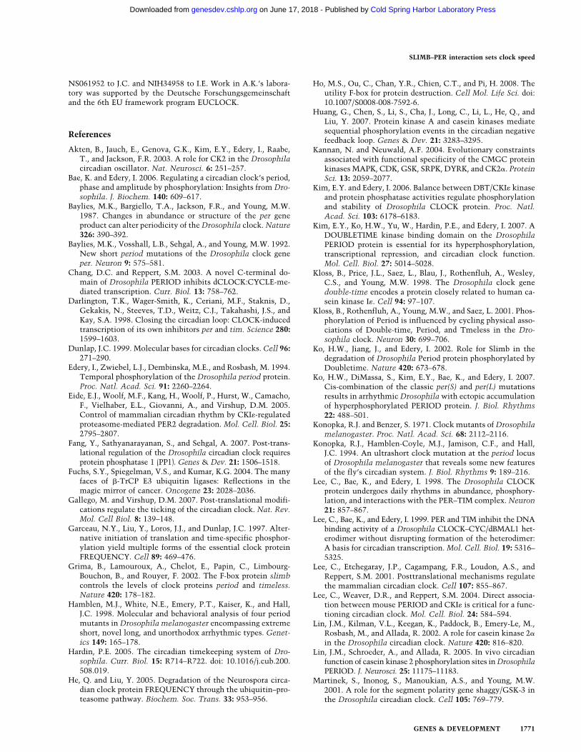
To better evaluate the phosphorylated state of S47, we gen-erated phospho-specific polyclonal antibodies (�-pS47) (Fig.6). The intensity of the immunoblotting signal obtainedusing �-pS47 antibodies is greatly increased when dPERis coexpressed with DBT (Fig. 6A, lanes 1,2), consistentwith the stimulatory effects of DBT on SLIMB binding todPER. The specificity of our �-pS47 antibodies was con-firmed by demonstrating that the signal intensity isstrongly attenuated by phosphatase treatment of dPER(Fig. 6A, lanes 2,3) and essentially abolished when prob-ing the dPER(S47A) mutant (Fig. 6A, lanes 5,6). dPERcontaining phosphorylated S47 is detected as early as 12h post-dbt induction, but steadily increases in intensitypeaking ∼10 h later, even though total dPER protein lev-els are lower at this later time point (Fig. 6B, cf. lanes 3,4and 7,8). The binding efficiency of dPER to SLIMB isroughly coincident with S47 phosphorylation (Fig. 6B),consistent with contributions from phosphorylation ofS47, in addition to other sites.
It is noteworthy that there is relatively more phos-phorylation of S47 at 22 h post-dbt induction compared
Table 2. Mapping phosphorylation sites on dPER producedin Drosophila S2 cells
dPERa,b dPER + DBTc
— [S40, S42, S44, S45, S47, S48]d,e
— S59, S60— S97— S132S149, S151f, S153 S149, S151, S153S596<P>g S585, S589, S596<P>T610<P> T610<P>S661<P> S661<P>— S773— S826, S828T883<P> S876, T883<P>, T889S981<P>, [T980, T983, S985]h T978<P>, S981<P>— S1103— [S1129, S1130<P>]h
aStable cell line expressing dper under the control of pMT-in-ducible promoter.bdPER residues phosphorylated by endogenous kinase(s) in S2cells.cStable cell line expressing dPER and DBT under the control ofpMT-inducible promoter.dTwo of the sites in the bracket are phosphorylated with S40and S42 being less likely phosphorylated.eAmino acids are numbered according to sequence ofdPER(amino acids 1–1224), GenBank accession number P07663.fS151 and S153 were identified previously as putative CKIIphosphorylation sites (Lin et al. 2005).g<P> denotes residues that are next to a proline.hOnly one site in the bracket is phosphorylated.
SLIMB–PER interaction sets clock speed
GENES & DEVELOPMENT 1765
Cold Spring Harbor Laboratory Press on June 17, 2018 - Published by genesdev.cshlp.orgDownloaded from
with many hours earlier (Fig. 6B, lanes 3,4). This timeframe is similar to that observed for the DBT-dependentglobal phosphorylation of full-length dPER (Fig. 1A) andmore delayed compared with the conversion of the first100-amino-acid fragment of dPER from hypo- to hyper-phosphorylated isoforms (Fig. 2C). The later peak in S47phospho-occupancy roughly coincides with rapid degra-dation in dPER levels, supporting the importance of thisphospho-determinant in regulating dPER stability. Italso suggests that DBT-mediated increases in the levelsof S47 phosphorylation are relatively slow, gradually oc-curring over many hours, and that other phosphorylationevents in the first 100 amino acids of dPER might un-dergo more rapid increases. Intriguingly, the majority of
dPER is stably bound to DBT within 5–10 h post-dbt in-duction (Ko et al. 2002; data not shown), much before thepeak in S47 phosphorylation. As such, the timing ofwhen DBT binds dPER cannot account for the kineticsunderlying the steady-state levels of phosphorylated S47.In this regard it is noteworthy that inhibiting proteinPP1 and/or PP2A stimulates the hyperphosphorylationand/or degradation of dPER in cultured S2 cells as well asin flies (Sathyanarayanan et al. 2004; Fang et al. 2007).Indeed, the addition of okadaic acid, which at the con-centration used here preferentially inhibits PP2A activ-ity, is accompanied by temporal increases in the amountof dPER isoforms phosphorylated at S47 (SupplementalFig. S5). Thus, it appears that PP2A (and/or PP1) stabi-lizes dPER by, at least in part, decreasing the phospho-occupancy of S47 and likely other sites. A reasonablemodel is that the balance between DBT and PP2A is acontributing factor in the slow hours-long kinetics un-derlying the ability of DBT to stimulate dPER binding toSLIMB and degradation.
To evaluate S47 phosphorylation within a physiologi-cal context we monitored its status throughout a dailycycle in flies using our �-pS47 antibodies. Phosphoryla-tion of S47 was first detected at around ZT21 (Fig. 6C, lane5), soon after the time when dPER(WT) translocates to thenucleus (Shafer et al. 2002). This time frame coincideswith the weak but initial detection of dPER–SLIMB in-teractions in flies (Fig. 6D), confirming that S47 phos-phorylation and SLIMB binding are temporally aligned.pS47-containing dPER isoforms continue to accumulatethroughout the late night and peak at around ZT4, whenmost dPER(WT) proteins are hyperphosphorylated andundergoing rapid degradation (Fig. 6C). A higher resolu-tion time course showed that strong binding of dPER toSLIMB begins around ZT1 (Fig. 6D, lane 7) as dPER pro-teins that are phosphorylated at S47 are also undergoingglobal hyperphosphorylation (Fig. 6C, lanes 5,6). Prior workhas shown that dPER is already associated with DBT byZT16 (Kloss et al. 2001; data not shown), indicating thatsteady-state increases in the phosphorylation of S47 donot occur quickly after DBT engages dPER. Thus, as ob-served in S2 cells, detectable phosphorylation of S47 be-gins at least several hours after DBT stably binds dPERand undergoes gradual increases in intensity that slightlyprecedes but is roughly coincident with global increasesin dPER phosphorylation.
Ser47 of Drosophila PER is phosphorylated by CKI�and DBT in vitro
To determine whether DBT can directly phosphorylateS47, we expressed dPER(WT) tagged with Flag and c-mycepitopes in S2 cells, immunoprecipitated the recombi-nant dPER(WT) using �-Flag resins, and subjected theimmune complexes to in vitro kinase assays using com-mercially available CKI� as well as recombinant DBTpurified from S2 cells. The extent of S47 phosphorylationwas evaluated using �-pS47 antibodies. Both CKI� andDBT triggered S47 phosphorylation (Fig. 7A [lanes 2,3], B[lane 4]), although phosphorylation by CKI� appeared to
Figure 6. Phosphospecific antibodies detect pS47-containingisoforms of dPER in S2 cells and flies. (A) Extracts were preparedfrom S2 cells expressing either a wild-type (WT) or S47A versionof pAc-3XFlag-His-dper/Tev100-6Xc-myc. In some cases cellswere cotransfected with pMT-dbt-V5-His (+), whereas this wasomitted in other cases (−). dPER proteins were immunoprecipi-tated (IP) with �-Flag beads, treated in the absence (−�PP) orpresence (+�PP) of �-phosphatase, and analyzed by Westernblotting (WB) in the presence of the indicated antibody (left ofpanel). (B, top) Cells were cotransfected with pAc-3XFlag-His-dper-6Xc-myc and pMT-dbt-V5-His and collected at the indi-cated times post-dbt induction. Extracts were prepared and di-vided into three separate aliquots: (1) One aliquot was immunopre-cipitated (IP) with �-Flag beads and analyzed by immunoblotting inthe presence of �-pS47 antibodies (top); (2) another aliquot wasdirectly analyzed by immunoblotting in the presence of �-c-mycantibodies (middle); and (3) a final aliquot was subjected toGST-SLIMB pull-down assays and bound material visualized byimmunoblotting in the presence of �-c-myc antibodies (bottom).(C,D) Head extracts were prepared from wper0;per+-HAHis fliescollected at the indicated times (ZT). (C) dPER-HAHis-contain-ing immune complexes recovered using �-HA beads were sub-jected to Western blotting (WB) in the presence of the indicatedantibody (left of panel). Open and closed triangles indicatehyper- and hypophosphorylated pS47-containing isoforms ofdPER, respectively. (D) An aliquot was directly analyzed (top),whereas another fraction was subjected to GST-SLIMB pull-down assays and bound material analyzed (bottom).
Chiu et al.
1766 GENES & DEVELOPMENT
Cold Spring Harbor Laboratory Press on June 17, 2018 - Published by genesdev.cshlp.orgDownloaded from

be more efficient and the majority of dPER was hyper-phosphorylated as readily observed from the slower elec-trophoretic mobility (Fig. 7A, lanes 6,7). Phosphatasetreatment of dPER did not abrogate the ability of DBT orCKI� to phosphorylate S47, strongly suggesting that prim-ing by other kinases is not a prerequisite for S47 phosphor-ylation (Fig. 7A,B). To the contrary, phosphatase-treateddPER was more efficiently phosphorylated in our in vitroassays using DBT or CKI�, perhaps because some dPERsites phosphorylated by endogenous kinases in S2 cellsantagonize DBT/CKI� kinase activity on S47. Otherclock relevant kinases such as GSK3�/SHAGGY andCKII (Martinek et al. 2001; Lin et al. 2002; Akten et al.2003) failed to stimulate S47 phosphorylation (Fig. 7B,lanes 1,2). Although we cannot rule out that priming atother sites by DBT is required for S47 phosphorylation,we tested a number of dPER variants with mutated Ser/Thr residues in the first 100 amino acids and the effi-ciency of in vitro phosphorylation of S47 by DBT wassimilar or better compared with the control situation(data not shown).
Phosphorylation at Ser47 requires the DBT-bindingdomain of Drosophila PER
Prior mutational analysis showed that the major DBTdocking site on dPER (dPDBD) does not contribute toDBT-dependent hyperphosphorylation by being a region
that is extensively phosphorylated (Kim et al. 2007), con-firmed here by mass spectrometry (Table 2). Rather, itappears that the main role of the dPDBD in regulatingdPER phosphorylation and stability is to serve as a dock-ing site that enables stable interactions with DBT andpossibly CKII or other kinases (Kim et al. 2007; Na-wathean et al. 2007). To explore the relationship of thedPDBD to S47 phosphorylation we analyzed the ability ofDBT/CKI� to phosphorylate the mutant dPER(�dPDBD)protein, using our in vitro kinase assay and extracts pre-pared from our previously characterized transgenic fliesexpressing dPER(�dPDBD) (Kim et al. 2007). Phosphory-lation of S47 was greatly attenuated in the mutant ver-sion of dPER (Fig. 7B, cf. lanes 8,9 and 4,5). In addition,we did not detect phosphorylation of S47 for the dPER-(�dPDBD) protein expressed in flies (Supplemental Fig.S6A), and it exhibits severely impaired ability to interactwith SLIMB despite its high levels (Supplemental Fig.S6B, lanes 7,12).
Discussion
To better understand the physiological role of phosphor-ylation in regulating PER stability we used D. melano-gaster as a model system. Using a range of strategies,including mutational analysis, mass spectrometry andphospho-specific antibodies we identified S47 as a keyphospho-determinant regulating the efficiency of SLIMBbinding to dPER. By evaluating the behavior of dPERmutants whereby amino acid 47 is constitutively “non-phosphorylated” (S47A) or “phosphorylated” (S47D), weshow that the phospho-occupancy of S47 is a key bio-chemical throttle adjusting the pace of the clock. How-ever, phosphorylation of S47 occurs within an atypicalSLIMB-binding site. Additional DBT-dependent phos-phorylated residues, which likely include one or morenearby Ser residues at amino acid 44/45 and possiblyothers within the first 100 amino acids, collaborate withpS47 to generate a high-affinity SLIMB-binding region ondPER. As such, the affinity of SLIMB for dPER is propor-tional to the degree of phospho-occupancy within an ex-tended phosphorylation network centered on S47 that asa unit yields a graded response in the affinity of SLIMB–dPER interactions. Attaining a high proportion of dPERmolecules that are phosphorylated at S47 and other keysites mediating SLIMB binding is progressive and occursseveral hours after DBT stably interacts with dPER viathe centrally located dPDBD, likely because DBT “activ-ity” is counterbalanced by TIM and protein phosphata-ses. We propose that the relatively slow assembly of ahigh-affinity SLIMB-binding site on dPER is at leastpartly “designed” to extend the time that dPER acts as atranscriptional repressor, critical in generating transcrip-tional feedback loops with daily time frames. Finally,our mass spectrometry analysis identify “hot spots” forphosphorylation, indicates that the majority of dPERphosphorylation is unrelated to direct effects on stabilityand sheds new insights into the underpinnings of severalpreviously characterized mutants, including the classicperS allele.
Figure 7. Ser47 of Drosophila PER is phosphorylated by DBTand CKI� in vitro. (A,B) Extracts were prepared from S2 cellsexpress-ing wild-type or �dPDBD versions of pAc-3XFlag-His-dper/Tev100-6Xc-myc. (Left) dPER proteins were immunopre-cipitated using �-Flag beads and subjected to in vitro kinaseassays, followed by Western blotting (WB) in the presence of theindicated antibody. dPER containing immune complexes wereeither mock-treated (−) or pretreated with �-phosphatase (+)prior to in vitro incubation in the absence (−) or presence of theindicated kinase.
SLIMB–PER interaction sets clock speed
GENES & DEVELOPMENT 1767
Cold Spring Harbor Laboratory Press on June 17, 2018 - Published by genesdev.cshlp.orgDownloaded from

Temporal regulation of dPER degradation by the slowDBT-dependent phosphorylation of a noncanonicalSLIMB-binding site
Early studies identified DpSG�X1+npS (pS, phosphorylatedSer; �, any hydrophobic amino acid; X, any amino acid) asthe consensus motif for recognition by the �-TrCP/SLIMBF-box protein (Fuchs et al. 2004). Phosphorylation at bothsites in this six amino acid consensus generally leads toa high-affinity �-TrCP/SLIMB-binding site. Indeed, thethree negatively charged residues (Asp/Glu and two phos-pho-S/T residues) are important binding contacts underly-ing �-TrCP/substrate interactions (Wu et al. 2003). Fur-thermore, it is thought that the presence of an Asp/Gluat position 2 of the canonical binding domain can cir-cumvent the need for a phospho-S/T at that position, asis the case for Wee1A (Watanabe et al. 2004). However,accumulating evidence indicates that �-TrCP-bindingsites can deviate from this consensus (Supplemental Fig.S1). For example, recent work on the Ci/Gli family oftranscription factors suggests a novel class of degenerateand weaker �-TrCP-binding sites that extend beyond thestandard six-amino-acid binding motif, especially forthose missing a Gly at the third position (Smelkinson etal. 2007). It was suggested that for these extended �-TrCP/SLIMB-binding motifs, significant contributionsare made by the local presence of nonpolar residues, suchas those found in the motifs for Ci, Gli, and Wee1A(Supplemental Fig. S1). Additional phosphorylation eventsat nearby regions are also thought to enhance the inher-ently weak binding affinities of extended �-TrCP-bind-ing sites, enabling a more graded response compared withthe standard sequence (Smelkinson et al. 2007).
The major SLIMB-dependent phospho-degron we iden-tified [44p*Sp*SGpSSGYGG52; where p* = possible phos-phorylation] seems to include signature elements foundin both the standard and extended �-TrCP-binding mo-tifs. A rather unique feature of the SLIMB-binding do-main on dPER is that it includes two SSG repeats. Weshow that S47 is phosphorylated, and based on mutationalanalysis and mass spectrometry, it is almost certain thateither S44 and/or S45 are phosphorylated. A physiologi-cal role for S45 is further indicated by the perSLIH mu-tant (S45Y) (Supplemental Fig. S4) that exhibits long pe-riods (Hamblen et al. 1998), which based on our findingsis likely due to reduced dPER–SLIMB interactions. Al-though changing S48 to Ala phenocopied the S47A mu-tation, the S48D mutation did not enhance binding toSLIMB, as was the case for S47D. Together with our re-sults showing that S48A did not modulate phosphoryla-tion of S47 (data not shown), the data strongly suggestthat S48 has a non-phosphorylation-dependent role as acrucial structural element. Mass spectrometry identifiedtwo phospho-residues in a dPER peptide from amino ac-ids 40–48 (Table 2). Thus, there might only be two nega-tively charged residues in the major SLIMB-binding siteon dPER. It is possible that the presence of a SSG tandemand a Tyr at position 50 can compensate for the lack of athird acidic residue normally found in �-TrCP-bindingsites. It is also highly probable that other, yet to be iden-
tified, DBT-dependent phosphorylation sites besidesthose within the atypical SLIMB-binding site identifiedhere contribute to enhancing SLIMB–dPER interactions.
The presence of numerous suboptimal phospho-deter-minants is thought to generate a graded response in thebinding efficiencies of F-box proteins to substrates (e.g.,Nash et al. 2001). The general molecular framework isthat progressive increases in the phospho-occupancy ofmultiple phosphorylated residues eventually reaches athreshold value that drives sufficient F-box protein/sub-strate interactions to yield desired outcomes. As such,regulating the kinetics of phosphorylation within the phos-pho-network mediating F-box recognition is a key deter-minant in the timing of substrate degradation. In the caseof animal PER proteins, they undergo progressive in-creases in global phosphorylation that occur over an∼10-h time frame, whereby highly phosphorylated iso-forms are associated with a rapid decline in levels (Ederyet al. 1994; Lee et al. 2001).
What accounts for the hours-long kinetics underlyingthe gradual increases in phosphorylation of S47 andlikely many other DBT-dependent sites on dPER? Basedon the in vitro ability of DBT to phosphorylate S47 de-spite phosphatase treatment of dPER (Fig. 7), we do notbelieve that hierarchical phosphorylation based on priorpriming is a major component in regulating the timing ofwhen S47 is phosphorylated in vivo. Rather, our findingsstrongly suggest that the gradual build-up in the phos-pho-occupancy of S47 and other sites is at least partlybased on a dynamic balance between DBT-mediated phos-phorylation and the opposing activities of TIM and proteinphosphatases. In agreement, blocking phosphatase activ-ity strongly enhanced the abundance of phosphorylatedS47 (Supplemental Fig. S5). Recent evidence suggeststhat the ability of TIM to stabilize dPER might be byacting as a bridge that facilitates the targeting of proteinphosphatase activity toward dPER (Fang et al. 2007). In-deed, it is likely that the strong protective function ofTIM on dPER partially overrides the destabilizing effectsof the S47D mutant as it attains peak levels comparablewith those of wild-type dPER (Fig. 4). Following this lineof reasoning, we suggest that a major reason for the ad-vanced dper RNA and protein cycles in the S47D mutantis that as TIM levels decline in the late night the “re-leased” dPER(S47D) protein is no longer protected (orless so) and undergoes accelerated nuclear clearance,leading to an earlier disengagement from transcriptionalrepression, which advances the subsequent dper RNAand protein cycles (Fig. 4). Likewise, while this manu-script was under review a recent report showed that theCKI� tau mutation, which shortens rhythms in mice,has an “asymmetrical” effect on PER protein stability,preferentially accelerating nuclear clearance and henceadvancing the molecular oscillations underpinning theclockworks (Meng et al. 2008). Thus, although differen-tial phosphorylation plays a major role in setting theintrinsic stabilites of PER proteins, additional variables,such as phase-specific protein–protein interactions, arecritical in the “readout” from these phospho-signals.
Many of the DBT-dependent phosphorylation sites
Chiu et al.
1768 GENES & DEVELOPMENT
Cold Spring Harbor Laboratory Press on June 17, 2018 - Published by genesdev.cshlp.orgDownloaded from

that we identified using mass spectrometry do not liewithin optimal CKI sites, suggesting that inefficient phos-phorylation by DBT might also contribute to the overallrate of progressive increases in dPER phosphorylation. Itis also possible that the strong binding of DBT to thecentrally located dPDBD, while increasing the local con-centration of DBT, could function as a slow “time-re-lease capsule” whereby the disengagement of DBT isfirst required prior to phosphorylation of dPER residuesat more distantly located sites.
Although the phosphorylation requirements and in vivosignificance of recently identified regions on mPER1 andmPER2 that interact with �-TrCP are not known, it islikely to also be based on noncanonical �-TrCP-bindingsites (Supplemental Fig. S1; Eide et al. 2005; Shirogane etal. 2005). In addition, hyperphosphorylation of mamma-lian PER proteins requires a centrally located CKI-bind-ing site (Lee et al. 2004; Eide et al. 2005; Shirogane et al.2005). Therefore, mammalian PER proteins, especiallymPER1 and mPER2, are likely to be targeted by �-TrCPto the 26S proteasome in a manner similar to that de-scribed here for dPER. This type of mechanism mightalso apply to other clock proteins such as FREQUENCY(FRQ) in Neurospora that undergoes daily changes inphosphorylation and stability that are remarkably simi-lar to those observed for PER proteins (Garceau et al.1997). In addition, the phosphorylated state of FRQ isregulated by casein kinases, protein phosphatases, andthe rapid degradation of highly phosphorylated isoformsis mediated by the F-box protein FWD1, a homolog of�-TrCP (Yang et al. 2004; He and Liu 2005; Huang et al.2007).
A systematic screen for dPER phosphorylationsites using mass spectrometry identifiesphospho-clusters and sets the stage for understandingsome of the classic clock mutants
An interesting feature of the distribution in phosphory-lation sites on dPER that we identified using mass spec-trometry is that they seem to concentrate in clusters,suggesting the presence of “phospho-modules” with dif-ferent functions. Most of these clusters appear anchoredby proline-directed phosphorylation sites, which are phos-phorylated by endogenous kinases expressed in S2 cells. Ofnote, one such cluster is located in the dPER “short do-main” (T585–T610). Mutations in this region result inanimals with short periods (Konopka and Benzer 1971;Baylies et al. 1992; Rutila et al. 1992; Konopka et al.1994). In fact, the mutated residues of two classic permutants that have short periods, perS (S589N, 19-h pe-riod) and perT (G593D, 16-h period), are right in the heartof this cluster. S589 is phosphorylated in a DBT-depen-dent manner; and G593, when mutated, may affect phos-phorylation at nearby S589 and/or S596. Although theperS mutants was isolated more than 35 years ago, ourresults provide the first biochemical understanding for theshort period phenotype, suggesting that phosphorylationevents in the “short domain,” some of which are DBT-dependent, may collaboratively function to slow down
the clock. It is now becoming apparent that phosphory-lation at different sites on PER proteins can result in dif-ferential effects on the pace of the clockworks, wherebysome lead to faster clocks while others slow it down(e.g., Vanselow et al. 2006). The presence of phosphory-lated residues with opposing outcomes on the speed ofthe clock can explain why mutations in CKI�/�/DBT canyield a variety of period-altering phenotypes from shortto long, despite the fact that overall enzymatic activity isgenerally reduced (Price et al. 1998; Suri et al. 2000;Vanselow et al. 2006; Muskus et al. 2007).
A rather unanticipated finding is that the majority ofdPER phosphorylation is unrelated to direct effects onstability. This is supported by the lack of detectableSLIMB binding to a dPER fragment only missing the first100 amino acids despite extensive phosphorylation asinferred from being the region underlying the majority ofphosphorylation-dependent electrophoretic mobility shiftsand confirmed by our mass spectrometric analysis (Table2; Supplemental Fig. S4). Other lines of evidence alsoimply that a significant amount of multiphosphorylationis not linked to direct effects on PER stability. For ex-ample, abolishing phosphorylation at many centrally lo-cated sites on mPER3 does not attenuate CKI-mediatedin vitro interactions with �-TrCP (Shirogane et al. 2005).Also, a trans-dominant version of CKII reduced globalhyperphosphorylation of dPER without major effects onits levels (Smith et al. 2008).
Thus, there are likely to be at least two functionallydistinct DBT-dependent phosphorylation programs regu-lating different aspects of PER metabolism and activity:one that controls �-TrCP/SLIMB binding, and anotherthat integrates with other kinases, such as CKII, to modu-late nuclear entry/accumulation and/or ability to functionas a transcriptional repressor. Indeed, our mass spectro-metric analysis of dPER identified numerous phosphor-ylation sites in a putative nuclear localization site andwithin the CCID mediating dPER inhibition of CLK-me-diated transcription (Supplemental Fig. S4; Chang andReppert 2003). Variants of dPER missing the major DBTdocking site are hypophosphorylated and weak repres-sors (Kim et al. 2007; Nawathean et al. 2007). However,the relationship between hyperphosphorylation and re-pressor potency is not clear, as the DBT docking site ondPER also functions as a molecular scaffold for DBT andperhaps CKII-mediated inhibition of CLK-dependenttranscription (Nawathean and Rosbash 2004; Yu et al.2006; Kim et al. 2007; Nawathean et al. 2007). Nonethe-less, it is clear that the DBT docking site is a criticalnexus for coordinating multiple phosphorylation pro-grams. A challenge is to examine the functions of thenewly identified phosphorylation sites and dissect themechanisms by which they regulate dPER metabolismand activity.
Materials and methods
Plasmids for S2 cell expression and site-directed mutagenesis
Plasmids expressing pAc-dper-V5-His and pMT-dbt-V5-Hiswere described in Kim et al. (2007) and Ko et al. (2002), respec-
SLIMB–PER interaction sets clock speed
GENES & DEVELOPMENT 1769
Cold Spring Harbor Laboratory Press on June 17, 2018 - Published by genesdev.cshlp.orgDownloaded from

tively. Procedures for generating pAc-3XFlag-His-dper-6Xc-mycand pAc-3XFlag-His-dper/Tev100-6Xc-myc are described in theSupplemental Material. QuikChange site-directed mutagenesiskit (Stratagene) was used to generate dper mutations using thevarious vectors described above as templates (see the Supple-mental Material for primer sequences). Procedures for generat-ing pMT-3XFlag-His-dper, pMT-dbt, pMT-gst, and pMT-gst-slimb plasmids are discussed in the Supplemental material.
S2 cell culture and transfection
S2 cells and DES expression medium were obtained from Invit-rogen, and transient transfections were performed using Effec-tene (Qiagen) according to the manufacturer’s instructions. Foreach transient transfection, 0.8 µg of different dper containingplasmids and 0.2 µg of pMT-dbt-V5-His or empty control pMT-V5-His plasmids were used. Expression of dbt was induced byadding 500 µM CuSO4 to the culture media 36 h after transfec-tion. Stable cell lines expressing pMT-3XFlag-His-dper alone orwith pMT-dbt, pMT-3XFlag-His-dbt, pMT-gst, and pMT-gst-slimb were generated using calcium phosphate transfection kit(Invitrogen), and induction of pMT-driven expression wasachieved using 500 µM CuSO4. For experiments in which theproteasome inhibitor MG132 (50 µM; Sigma) and cyclohexi-mide (10 µg/mL; Sigma) were used, they were added 4 h prior tocell harvest. These drugs were not used in the time-course ex-periments shown.
Phosphorylation site mapping
Hygromycin-resistant stable cell lines expressing pMT-3XFlag-His-dper alone or with pMT-dbt were established for dPER pu-rification (see Supplemental Material for cell culture conditionsand purification procedures). Phosphorylation site mapping us-ing mass spectrometry was performed as described in Schlosseret al. (2005). Data analysis was essentially performed as de-scribed previously (Schlosser et al. 2007).
Transgenic flies and locomotor activity assays
To generate transgenic flies carrying dper mutations, we used apreviously characterized vector that contains a 13.2-kb dper ge-nomic fragment tagged with the HA epitope and multiple his-tidine residues (10XHis) at the C-terminal (13.2per+-HAHis)(Lee et al. 1998). A XbaI–BamHI subfragment of this vector (in-cluding sequences encoding amino acids 1–870 of dPER) wassubcloned into pGEM7 vector (Promega). This was used as thetemplate for site-mutagenesis using the QuikChange Mutagen-esis Kit (Stratagene). See the Supplemental Material for primersequences. Mutated dper regions were confirmed by DNA se-quencing, and used to replace the corresponding fragment in the13.2per+-HAHis plasmid. Injection of w1118 embryos was per-formed by Genetic Services, Inc., and transgenic flies expressingdper (wild-type or mutant) transgenes in w1118per+ (referred toas wper+) and w1118per0 (wper0) backgrounds were generated asdescribed in Kim et al. (2007). Flies were kept at 25°C and en-trained for at least three cycles of 12-h light:12-h dark (12:12 LD,where zeitgeber time [ZT] 0 is the start of the light period),followed by at least 7 d in constant dark conditions (DD) fordetermination of free-running period. The measurement of flylocomotor activity rhythms was as previously described (Kim etal. 2007). To evaluate fly materials for Western blotting, GST-SLIMB pull-down, or immunoprecipitation, we entrained flies at25°C in 12:12 LD for 3 d, and collected them at the indicated ZTon the fourth day.
Generation of S47 phosphospecific antibodiesand immunobloting
Rabbits were immunized with a 15-amino-acid peptide (amino acid40—SGSHSSGpSSGYGGKP—amino acid 54; where p = phosphate)(Proteintech Group, Inc.). Immunoblotting analysis was performed asdescribed previously (Lee et al. 1998) with modifications detailed inthe Supplemental Material.
Immunoprecipitation (IP) and phosphatase (�-PP) treatment
IP and �-PP treatment were performed as described (Lee et al. 1998)with modifications detailed in the Supplemental Material.
GST pull-down assay and TEV enzyme cleavage
S2 cells expressing pAc-3XFlag-His-dper/Tev100-6Xc-myc vari-ants were expressed with or without pMT-dbt-V5-His. Cellswere treated with MG132 (50 µM) and cycloheximide (10 µg/mL) at 12 h (Fig. 3B) or at 16 h post-dbt induction (Fig. 3C,D),and harvested 4 h later. For the time course experiment (Fig.3A), cells were harvested at the indicated times without drugtreatment. dPER proteins were extracted using EB2 (see theSupplemental Material). TEV cleavage was performed overnightat 4°C according to the manufacturer’s instructions (Invitro-gen). Mock or TEV-treated extracts were incubated with gluta-thione beads (Pierce) bound with GST or GST-SLIMB (purifiedfrom stable cell lines) usually for 4 h (up to overnight) at 4°C.Bound dPER proteins were eluted with SDS sample buffer andresolved in 6% SDS-PAGE for dPER(1–1224) and dPER(101–1224), or 16% SDS-PAGE for dPER(1–100). To generate GST andGST-SLIMB proteins, expression was induced from stable celllines for 36 h, and cells were lysed in GST lysis buffer (see theSupplemental Material). The extracts were then incubated withglutathione beads for 4 h at 4°C to achieve binding. GST pull-down assays for fly extracts were performed as described for S2cell extracts (see the Supplemental Material for extract prepa-ration), and bound dPER was resolved using 6% SDS-PAGE.
In vitro kinase assay
S2 cells were transiently transfected with pAc-3XFlag-His-dper/Tev100-6Xc-myc wild-type or �dPDBD variants. S2 cells wereharvested around 48 h after transfection without any drug treat-ment and dPER proteins were extracted using EB2 (see theSupplemental Material). After IP (see the Supplemental Mate-rial) to obtain dPER proteins, samples were subjected to �-PP ormock treatment. Samples were subsequently washed three timeswith appropriate reaction buffers (CKI, CKII, or GSK3; New En-gland Biolabs). Kinase assays were performed for 30 min at 30°Cusing 500 U of CKI�, CKII, GSK3 (New England Biolabs), or 0.5µg of Flag-tagged DBT purified from a stable S2 cell line express-ing pMT-3XFlag-His-dbt (reaction were supplemented with 100µM ATP [New England Biolabs]). Purification procedures aredetailed in the Supplemental Material. All kinases used for invitro phosphorylation studies were active as assayed using con-trol substrates (data not shown).
RT–PCR
The relative dper mRNA levels were measured by semiquanti-tative RT–PCR as described previously (Kim et al. 2007). dpermRNA levels were normalized against noncycling cbp20 (cap-binding protein 20) mRNA.
Acknowledgments
We thank H. Krause (University of Toronto) for pMT-3XFLAG-His. This work was supported by NIH grants NS049862 and
Chiu et al.
1770 GENES & DEVELOPMENT
Cold Spring Harbor Laboratory Press on June 17, 2018 - Published by genesdev.cshlp.orgDownloaded from
NS061952 to J.C. and NIH34958 to I.E. Work in A.K.’s labora-tory was supported by the Deutsche Forschungsgemeinschaftand the 6th EU framework program EUCLOCK.
References
Akten, B., Jauch, E., Genova, G.K., Kim, E.Y., Edery, I., Raabe,T., and Jackson, F.R. 2003. A role for CK2 in the Drosophilacircadian oscillator. Nat. Neurosci. 6: 251–257.
Bae, K. and Edery, I. 2006. Regulating a circadian clock’s period,phase and amplitude by phosphorylation: Insights from Dro-sophila. J. Biochem. 140: 609–617.
Baylies, M.K., Bargiello, T.A., Jackson, F.R., and Young, M.W.1987. Changes in abundance or structure of the per geneproduct can alter periodicity of the Drosophila clock. Nature326: 390–392.
Baylies, M.K., Vosshall, L.B., Sehgal, A., and Young, M.W. 1992.New short period mutations of the Drosophila clock geneper. Neuron 9: 575–581.
Chang, D.C. and Reppert, S.M. 2003. A novel C-terminal do-main of Drosophila PERIOD inhibits dCLOCK:CYCLE-me-diated transcription. Curr. Biol. 13: 758–762.
Darlington, T.K., Wager-Smith, K., Ceriani, M.F., Staknis, D.,Gekakis, N., Steeves, T.D., Weitz, C.J., Takahashi, J.S., andKay, S.A. 1998. Closing the circadian loop: CLOCK-inducedtranscription of its own inhibitors per and tim. Science 280:1599–1603.
Dunlap, J.C. 1999. Molecular bases for circadian clocks. Cell 96:271–290.
Edery, I., Zwiebel, L.J., Dembinska, M.E., and Rosbash, M. 1994.Temporal phosphorylation of the Drosophila period protein.Proc. Natl. Acad. Sci. 91: 2260–2264.
Eide, E.J., Woolf, M.F., Kang, H., Woolf, P., Hurst, W., Camacho,F., Vielhaber, E.L., Giovanni, A., and Virshup, D.M. 2005.Control of mammalian circadian rhythm by CKI�-regulatedproteasome-mediated PER2 degradation. Mol. Cell. Biol. 25:2795–2807.
Fang, Y., Sathyanarayanan, S., and Sehgal, A. 2007. Post-trans-lational regulation of the Drosophila circadian clock requiresprotein phosphatase 1 (PP1). Genes & Dev. 21: 1506–1518.
Fuchs, S.Y., Spiegelman, V.S., and Kumar, K.G. 2004. The manyfaces of �-TrCP E3 ubiquitin ligases: Reflections in themagic mirror of cancer. Oncogene 23: 2028–2036.
Gallego, M. and Virshup, D.M. 2007. Post-translational modifi-cations regulate the ticking of the circadian clock. Nat. Rev.Mol. Cell Biol. 8: 139–148.
Garceau, N.Y., Liu, Y., Loros, J.J., and Dunlap, J.C. 1997. Alter-native initiation of translation and time-specific phosphor-ylation yield multiple forms of the essential clock proteinFREQUENCY. Cell 89: 469–476.
Grima, B., Lamouroux, A., Chelot, E., Papin, C., Limbourg-Bouchon, B., and Rouyer, F. 2002. The F-box protein slimbcontrols the levels of clock proteins period and timeless.Nature 420: 178–182.
Hamblen, M.J., White, N.E., Emery, P.T., Kaiser, K., and Hall,J.C. 1998. Molecular and behavioral analysis of four periodmutants in Drosophila melanogaster encompassing extremeshort, novel long, and unorthodox arrhythmic types. Genet-ics 149: 165–178.
Hardin, P.E. 2005. The circadian timekeeping system of Dro-sophila. Curr. Biol. 15: R714–R722. doi: 10.1016/j.cub.200.508.019.
He, Q. and Liu, Y. 2005. Degradation of the Neurospora circa-dian clock protein FREQUENCY through the ubiquitin–pro-teasome pathway. Biochem. Soc. Trans. 33: 953–956.
Ho, M.S., Ou, C., Chan, Y.R., Chien, C.T., and Pi, H. 2008. Theutility F-box for protein destruction. Cell Mol. Life Sci. doi:10.1007/S0008-008-7592-6.
Huang, G., Chen, S., Li, S., Cha, J., Long, C., Li, L., He, Q., andLiu, Y. 2007. Protein kinase A and casein kinases mediatesequential phosphorylation events in the circadian negativefeedback loop. Genes & Dev. 21: 3283–3295.
Kannan, N. and Neuwald, A.F. 2004. Evolutionary constraintsassociated with functional specificity of the CMGC proteinkinases MAPK, CDK, GSK, SRPK, DYRK, and CK2�. ProteinSci. 13: 2059–2077.
Kim, E.Y. and Edery, I. 2006. Balance between DBT/CKI� kinaseand protein phosphatase activities regulate phosphorylationand stability of Drosophila CLOCK protein. Proc. Natl.Acad. Sci. 103: 6178–6183.
Kim, E.Y., Ko, H.W., Yu, W., Hardin, P.E., and Edery, I. 2007. ADOUBLETIME kinase binding domain on the DrosophilaPERIOD protein is essential for its hyperphosphorylation,transcriptional repression, and circadian clock function.Mol. Cell. Biol. 27: 5014–5028.
Kloss, B., Price, J.L., Saez, L., Blau, J., Rothenfluh, A., Wesley,C.S., and Young, M.W. 1998. The Drosophila clock genedouble-time encodes a protein closely related to human ca-sein kinase I�. Cell 94: 97–107.
Kloss, B., Rothenfluh, A., Young, M.W., and Saez, L. 2001. Phos-phorylation of Period is influenced by cycling physical asso-ciations of Double-time, Period, and Tmeless in the Dro-sophila clock. Neuron 30: 699–706.
Ko, H.W., Jiang, J., and Edery, I. 2002. Role for Slimb in thedegradation of Drosophila Period protein phosphorylated byDoubletime. Nature 420: 673–678.
Ko, H.W., DiMassa, S., Kim, E.Y., Bae, K., and Edery, I. 2007.Cis-combination of the classic per(S) and per(L) mutationsresults in arrhythmic Drosophila with ectopic accumulationof hyperphosphorylated PERIOD protein. J. Biol. Rhythms22: 488–501.
Konopka, R.J. and Benzer, S. 1971. Clock mutants of Drosophilamelanogaster. Proc. Natl. Acad. Sci. 68: 2112–2116.
Konopka, R.J., Hamblen-Coyle, M.J., Jamison, C.F., and Hall,J.C. 1994. An ultrashort clock mutation at the period locusof Drosophila melanogaster that reveals some new featuresof the fly’s circadian system. J. Biol. Rhythms 9: 189–216.
Lee, C., Bae, K., and Edery, I. 1998. The Drosophila CLOCKprotein undergoes daily rhythms in abundance, phosphory-lation, and interactions with the PER–TIM complex. Neuron21: 857–867.
Lee, C., Bae, K., and Edery, I. 1999. PER and TIM inhibit the DNAbinding activity of a Drosophila CLOCK–CYC/dBMAL1 het-erodimer without disrupting formation of the heterodimer:A basis for circadian transcription. Mol. Cell. Biol. 19: 5316–5325.
Lee, C., Etchegaray, J.P., Cagampang, F.R., Loudon, A.S., andReppert, S.M. 2001. Posttranslational mechanisms regulatethe mammalian circadian clock. Cell 107: 855–867.
Lee, C., Weaver, D.R., and Reppert, S.M. 2004. Direct associa-tion between mouse PERIOD and CKI� is critical for a func-tioning circadian clock. Mol. Cell. Biol. 24: 584–594.
Lin, J.M., Kilman, V.L., Keegan, K., Paddock, B., Emery-Le, M.,Rosbash, M., and Allada, R. 2002. A role for casein kinase 2�
in the Drosophila circadian clock. Nature 420: 816–820.Lin, J.M., Schroeder, A., and Allada, R. 2005. In vivo circadian
function of casein kinase 2 phosphorylation sites in DrosophilaPERIOD. J. Neurosci. 25: 11175–11183.
Martinek, S., Inonog, S., Manoukian, A.S., and Young, M.W.2001. A role for the segment polarity gene shaggy/GSK-3 inthe Drosophila circadian clock. Cell 105: 769–779.
SLIMB–PER interaction sets clock speed
GENES & DEVELOPMENT 1771
Cold Spring Harbor Laboratory Press on June 17, 2018 - Published by genesdev.cshlp.orgDownloaded from

Meng, Q.J., Logunova, L., Maywood, E.S., Gallego, M., Lebiecki,J., Brown, T.M., Sladek, M., Semikhodskii, A.S., Glossop,N.R., Piggins, H.D., et al. 2008. Setting clock speed in mam-mals: The CK1� mutation in mice accelerates circadianpacemakers by selectively destabilizing PERIOD proteins.Neuron 58: 78–88.
Merrow, M., Mazzotta, G., Chen, Z., and Roenneberg, T. 2006.The right place at the right time: Regulation of daily timingby phosphorylation. Genes & Dev. 20: 2629–2633.
Meyer, P., Saez, L., and Young, M.W. 2006. PER–TIM interac-tions in living Drosophila cells: An interval timer for thecircadian clock. Science 311: 226–229.
Muskus, M.J., Preuss, F., Fan, J.Y., Bjes, E.S., and Price, J.L. 2007.Drosophila DBT lacking protein kinase activity produceslong-period and arrhythmic circadian behavioral and mo-lecular rhythms. Mol. Cell. Biol. 27: 8049–8064.
Nash, P., Tang, X., Orlicky, S., Chen, Q., Gertler, F.B., Menden-hall, M.D., Sicheri, F., Pawson, T., and Tyers, M. 2001. Mul-tisite phosphorylation of a CDK inhibitor sets a threshold forthe onset of DNA replication. Nature 414: 514–521.
Nawathean, P. and Rosbash, M. 2004. The doubletime and CKIIkinases collaborate to potentiate Drosophila PER transcrip-tional repressor activity. Mol. Cell 13: 213–223.
Nawathean, P., Stoleru, D., and Rosbash, M. 2007. A small con-served domain of Drosophila PERIOD is important for cir-cadian phosphorylation, nuclear localization, and transcrip-tional repressor activity. Mol. Cell. Biol. 27: 5002–5013.
Price, J.L., Dembinska, M.E., Young, M.W., and Rosbash, M.1995. Suppression of PERIOD protein abundance and circa-dian cycling by the Drosophila clock mutation timeless.EMBO J. 14: 4044–4049.
Price, J.L., Blau, J., Rothenfluh, A., Abodeely, M., Kloss, B., andYoung, M.W. 1998. double-time is a novel Drosophila clockgene that regulates PERIOD protein accumulation. Cell 94:83–95.
Roenneberg, T. and Merrow, M. 2005. Circadian clocks—Thefall and rise of physiology. Nat. Rev. Mol. Cell Biol. 6: 965–971.
Rothenfluh, A., Young, M.W., and Saez, L. 2000. A TIMELESS-independent function for PERIOD proteins in the Dro-sophila clock. Neuron 26: 505–514.
Rutila, J.E., Edery, I., Hall, J.C., and Rosbash, M. 1992. Theanalysis of new short-period circadian rhythm mutants sug-gests features of D. melanogaster period gene function. J.Neurogenet. 8: 101–113.
Sathyanarayanan, S., Zheng, X., Xiao, R., and Sehgal, A. 2004.Posttranslational regulation of Drosophila PERIOD proteinby protein phosphatase 2A. Cell 116: 603–615.
Schlosser, A., Vanselow, J.T., and Kramer, A. 2005. Mapping ofphosphorylation sites by a multi-protease approach with spe-cific phosphopeptide enrichment and NanoLC-MS/MS analy-sis. Anal. Chem. 77: 5243–5250.
Schlosser, A., Vanselow, J.T., and Kramer, A. 2007. Comprehen-sive phosphorylation site analysis of individual phosphopro-teins applying scoring schemes for MS/MS data. Anal. Chem.79: 7439–7449.
Shafer, O.T., Rosbash, M., and Truman, J.W. 2002. Sequentialnuclear accumulation of the clock proteins period and time-less in the pacemaker neurons of Drosophila melanogaster.J. Neurosci. 22: 5946–5954.
Shirogane, T., Jin, J., Ang, X.L., and Harper, J.W. 2005. SCF�–TRCP controls clock-dependent transcription via casein ki-nase 1-dependent degradation of the mammalian period-1(Per1) protein. J. Biol. Chem. 280: 26863–26872.
Smelkinson, M.G., Zhou, Q., and Kalderon, D. 2007. Regulationof Ci-SCFSlimb binding, Ci proteolysis, and hedgehog path-
way activity by Ci phosphorylation. Dev. Cell 13: 481–495.Smith, E.M., Lin, J.M., Meissner, R.A., and Allada, R. 2008. Dom-
inant-negative CK2� induces potent effects on circadian rhyth-micity. PLoS Genet. 4: e12. doi: 10.1371/journal.pgen.0040012.
Suri, V., Hall, J.C., and Rosbash, M. 2000. Two novel double-time mutants alter circadian properties and eliminate thedelay between RNA and protein in Drosophila. J. Neurosci.20: 7547–7555.
Toh, K.L., Jones, C.R., He, Y., Eide, E.J., Hinz, W.A., Virshup,D.M., Ptacek, L.J., and Fu, Y.H. 2001. An hPer2 phosphory-lation site mutation in familial advanced sleep phase syn-drome. Science 291: 1040–1043.
Vanselow, K., Vanselow, J.T., Westermark, P.O., Reischl, S.,Maier, B., Korte, T., Herrmann, A., Herzel, H., Schlosser, A.,and Kramer, A. 2006. Differential effects of PER2 phosphor-ylation: Molecular basis for the human familial advancedsleep phase syndrome (FASPS). Genes & Dev. 20: 2660–2672.
Watanabe, N., Arai, H., Nishihara, Y., Taniguchi, M., Watan-abe, N., Hunter, T., and Osada, H. 2004. M-phase kinasesinduce phospho-dependent ubiquitination of somatic Wee1by SCF�–TrCP. Proc. Natl. Acad. Sci. 101: 4419–4424.
Wu, G., Xu, G., Schulman, B.A., Jeffrey, P.D., Harper, J.W., andPavletich, N.P. 2003. Structure of a �-TrCP1–Skp1–�-catenincomplex: Destruction motif binding and lysine specificity ofthe SCF(�-TrCP1) ubiquitin ligase. Mol. Cell 11: 1445–1456.
Xu, Y., Padiath, Q.S., Shapiro, R.E., Jones, C.R., Wu, S.C.,Saigoh, N., Saigoh, K., Ptacek, L.J., and Fu, Y.H. 2005. Func-tional consequences of a CKI� mutation causing familial ad-vanced sleep phase syndrome. Nature 434: 640–644.
Xu, Y., Toh, K.L., Jones, C.R., Shin, J.Y., Fu, Y.H., and Ptacek,L.J. 2007. Modeling of a human circadian mutation yieldsinsights into clock regulation by PER2. Cell 128: 59–70.
Yang, Y., He, Q., Cheng, P., Wrage, P., Yarden, O., and Liu, Y.2004. Distinct roles for PP1 and PP2A in the Neurosporacircadian clock. Genes & Dev. 18: 255–260.
Yu, Q., Jacquier, A.C., Citri, Y., Hamblen, M., Hall, J.C., andRosbash, M. 1987. Molecular mapping of point mutations inthe period gene that stop or speed up biological clocks inDrosophila melanogaster. Proc. Natl. Acad. Sci. 84: 784–788.
Yu, W., Zheng, H., Houl, J.H., Dauwalder, B., and Hardin, P.E.2006. PER-dependent rhythms in CLK phosphorylation andE-box binding regulate circadian transcription. Genes &Dev. 20: 723–733.
Chiu et al.
1772 GENES & DEVELOPMENT
Cold Spring Harbor Laboratory Press on June 17, 2018 - Published by genesdev.cshlp.orgDownloaded from

10.1101/gad.1682708Access the most recent version at doi: 22:2008, Genes Dev.
Joanna C. Chiu, Jens T. Vanselow, Achim Kramer, et al.
clockthat is phosphorylated by DOUBLETIME controls the pace of the The phospho-occupancy of an atypical SLIMB-binding site on PERIOD
Material
Supplemental
http://genesdev.cshlp.org/content/suppl/2008/07/01/22.13.1758.DC1
Related Content
Genes Dev. July , 2008 22: 1737-1740
Justin BlauPERspective on PER phosphorylation
References
http://genesdev.cshlp.org/content/22/13/1758.full.html#related-urls
Articles cited in:
http://genesdev.cshlp.org/content/22/13/1758.full.html#ref-list-1This article cites 63 articles, 25 of which can be accessed free at:
License
ServiceEmail Alerting
click here.right corner of the article or
Receive free email alerts when new articles cite this article - sign up in the box at the top
Copyright © 2008, Cold Spring Harbor Laboratory Press
Cold Spring Harbor Laboratory Press on June 17, 2018 - Published by genesdev.cshlp.orgDownloaded from