Embed Size (px)
Citation preview
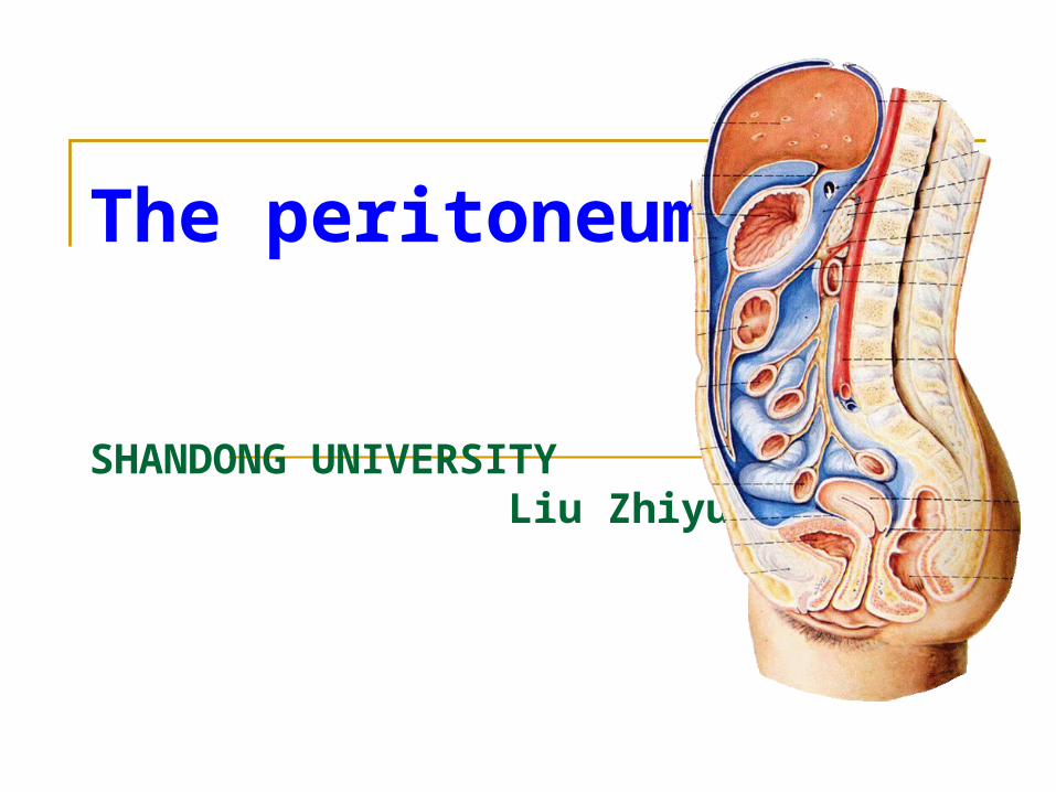
The peritoneum
SHANDONG UNIVERSITY Liu Zhiyu
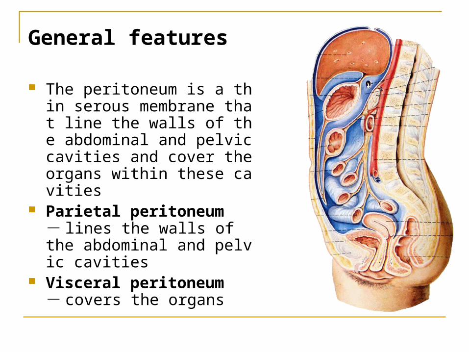
General features
The peritoneum is a thin serous membrane that line the walls of the abdominal and pelvic cavities and cover the organs within these cavities
Parietal peritoneum - lines the walls of the abdominal and pelvic cavities
Visceral peritoneum - covers the organs
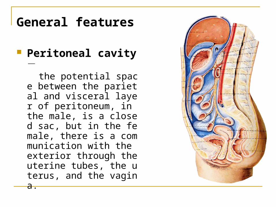
General features
Peritoneal cavity - the potential space betwee
n the parietal and visceral layer of peritoneum, in the male, is a closed sac, but in the female, there is a communication with the exterior through the uterine tubes, the uterus, and the vagina.
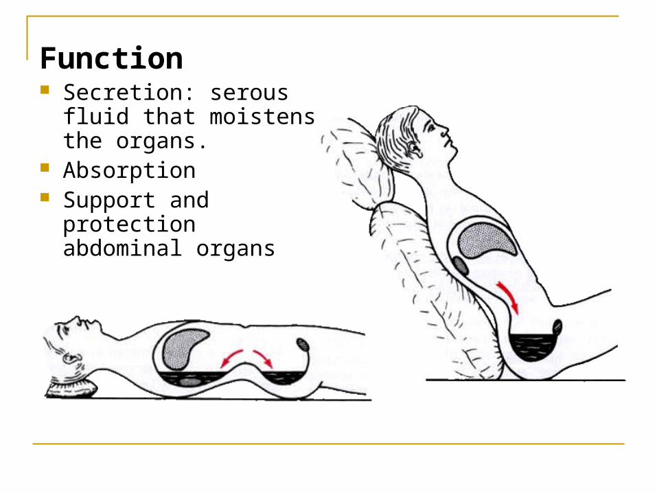
Function Secretion: serous fluid
that moistens the organs.
Absorption Support and protection
abdominal organs
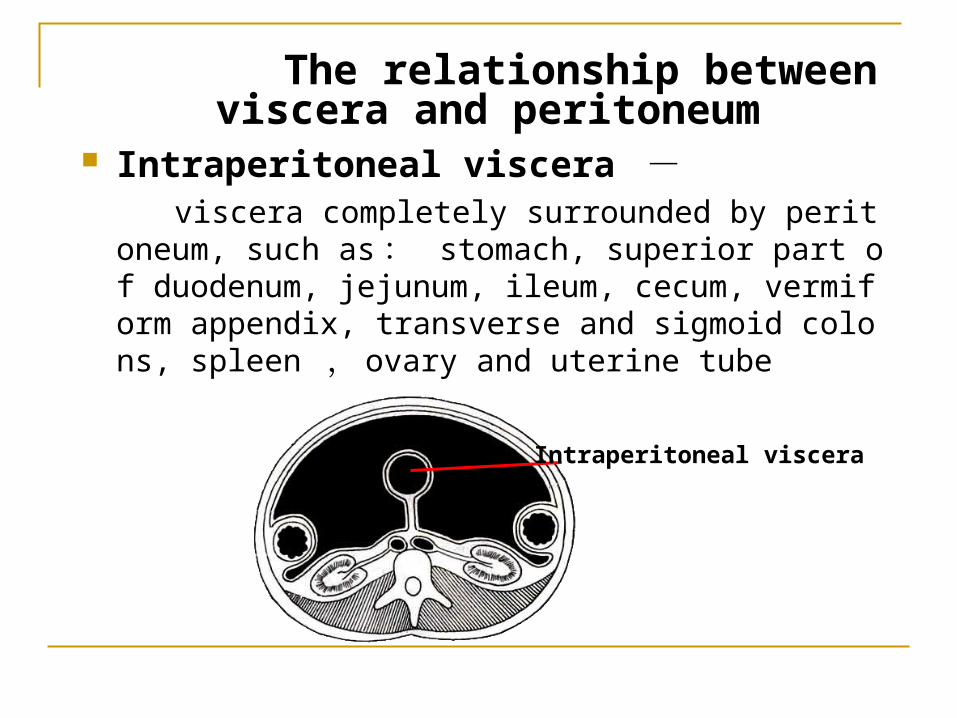
The relationship between viscera and peritoneum
Intraperitoneal viscera - viscera completely surrounded by peritoneum, such a
s : stomach, superior part of duodenum, jejunum, ileum, cecum, vermiform appendix, transverse and sigmoid colons, spleen , ovary and uterine tube
Intraperitoneal viscera
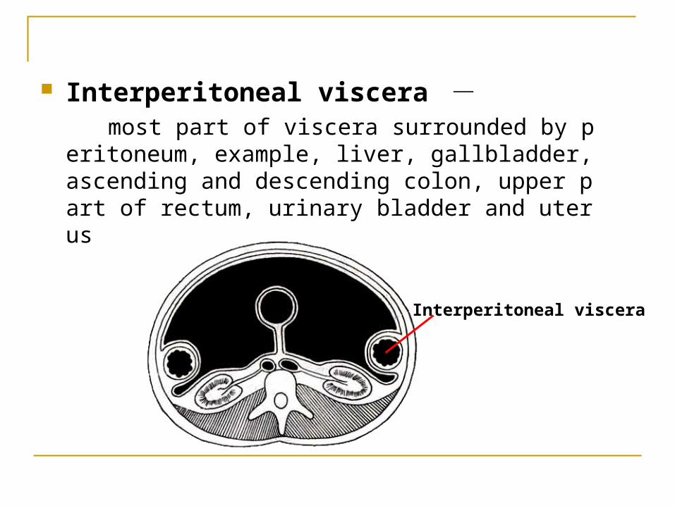
Interperitoneal viscera - most part of viscera surrounded by peritoneum, exa
mple, liver, gallbladder, ascending and descending colon, upper part of rectum, urinary bladder and uterus
Interperitoneal viscera
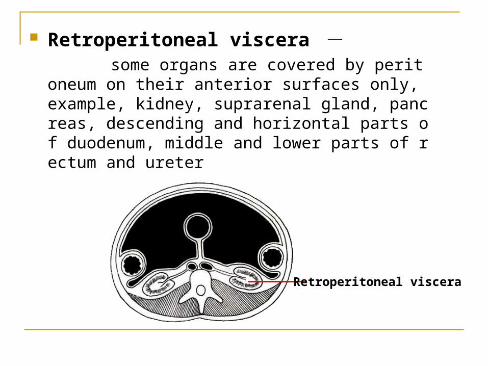
Retroperitoneal viscera - some organs are covered by peritoneum on their
anterior surfaces only, example, kidney, suprarenal gland, pancreas, descending and horizontal parts of duodenum, middle and lower parts of rectum and ureter
Retroperitoneal viscera
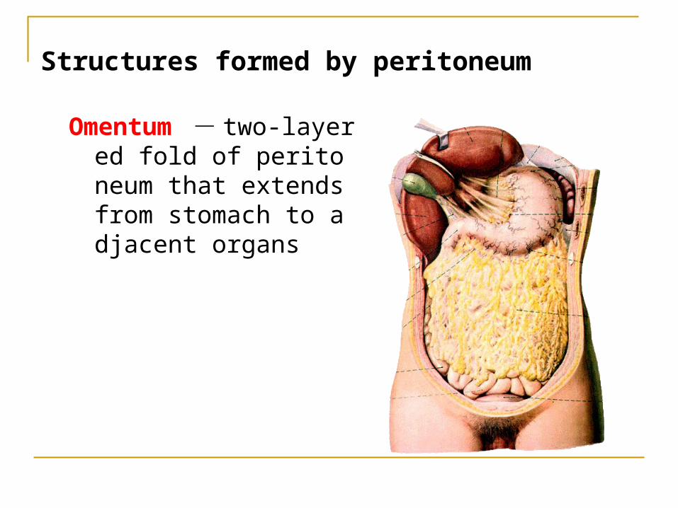
Structures formed by peritoneum
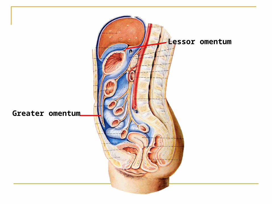
Omentum - two-layered fold of peritoneum that extends from stomach to adjacent organs
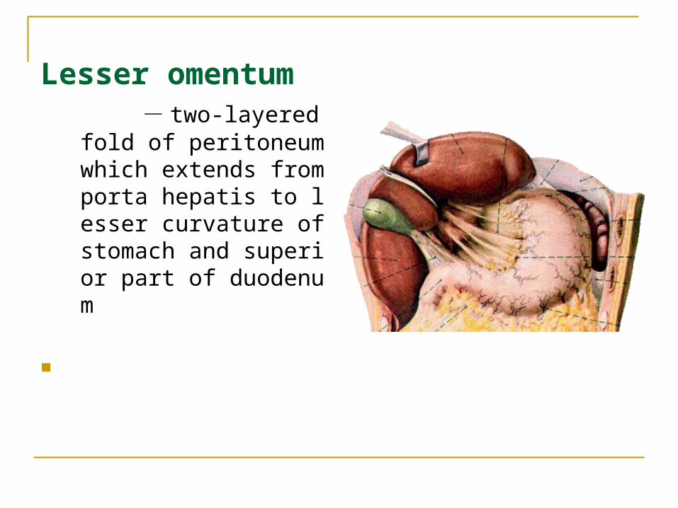
Lesser omentum - two-layered fold of p
eritoneum which extends from porta hepatis to lesser curvature of stomach and superior part of duodenum
Lessor omentum
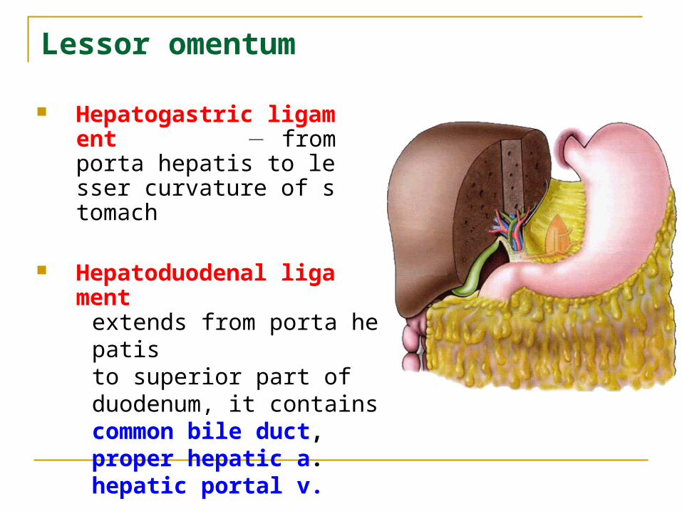
Hepatogastric ligament - from porta hepatis to lesser curvature of stomach
Hepatoduodenal ligament
extends from porta hepatis to superior part of duodenum, it contains common bile duct, proper hepatic a. hepatic portal v.
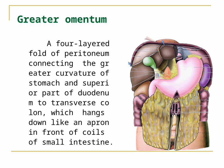
A four-layered fold of peritoneum connecting the greater curvature of stomach and superior part of duodenum to transverse colon, which hangs down like an apron in front of coils of small intestine.
Greater omentum
Lessor omentum
Greater omentum
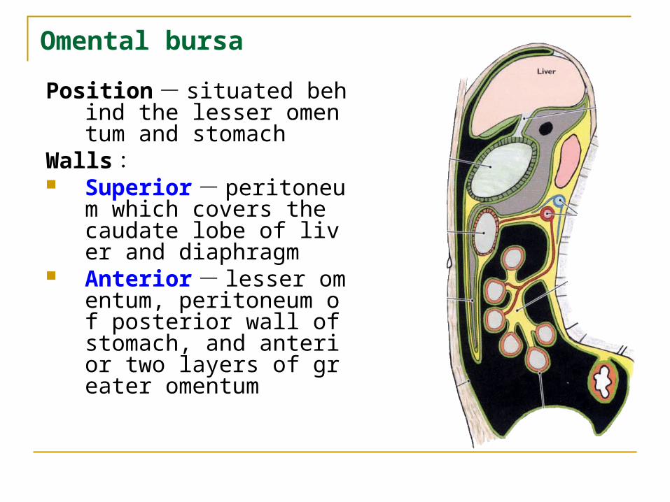
Omental bursa
Position - situated behind the lesser omentum and stomach
Walls : Superior - peritoneum w
hich covers the caudate lobe of liver and diaphragm
Anterior - lesser omentum, peritoneum of posterior wall of stomach, and anterior two layers of greater omentum
Omental bursa
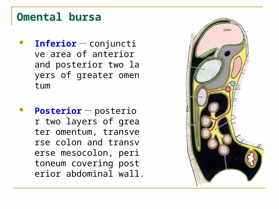
Inferior - conjunctive area of anterior and posterior two layers of greater omentum
Posterior - posterior two layers of greater omentum, transverse colon and transverse mesocolon, peritoneum covering posterior abdominal wall.
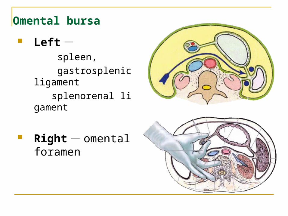
Left - spleen,
gastrosplenic ligament
splenorenal ligament
Right - omental foramen
Omental bursa
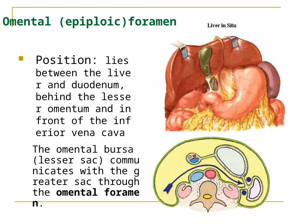
Position: lies between the liver and duodenum, behind the lesser omentum and infront of the inferior vena cava
Omental (epiploic)foramen
The omental bursa (lesser sac) communicates with the greater sac through the omental foramen.
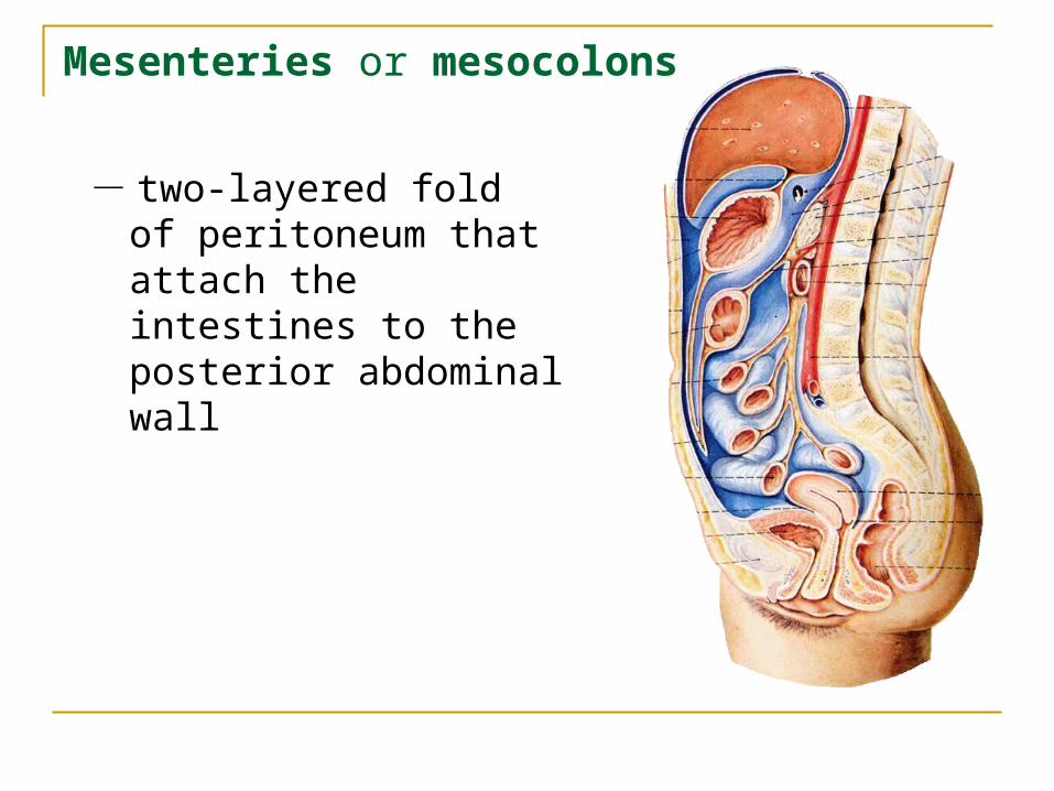
Mesenteries or mesocolons
- two-layered fold of peritoneum that attach the intestines to the posterior abdominal wall
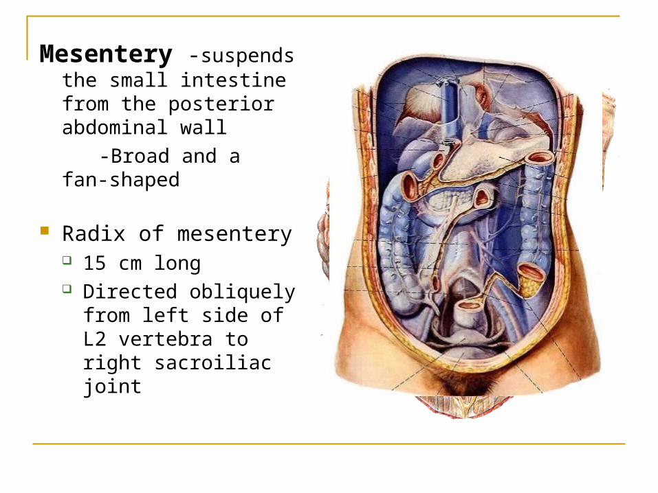
Mesentery -suspends the small intestine from the posterior abdominal wall
-Broad and a fan-shaped
Radix of mesentery 15 cm long Directed obliquely
from left side of L2 vertebra to right sacroiliac joint
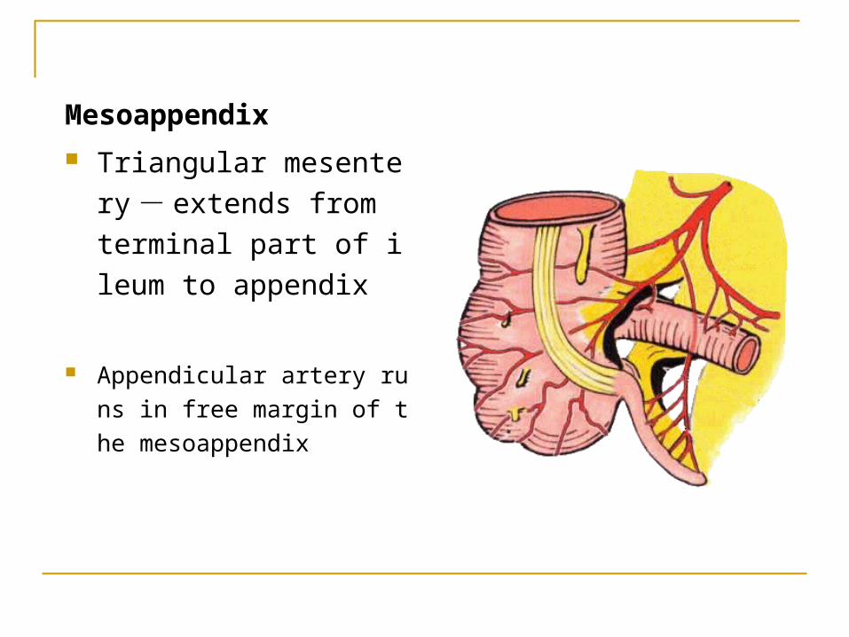
Mesoappendix
Triangular mesentery -extends from terminal pa
rt of ileum to appendix
Appendicular artery runs in fr
ee margin of the mesoappen
dix
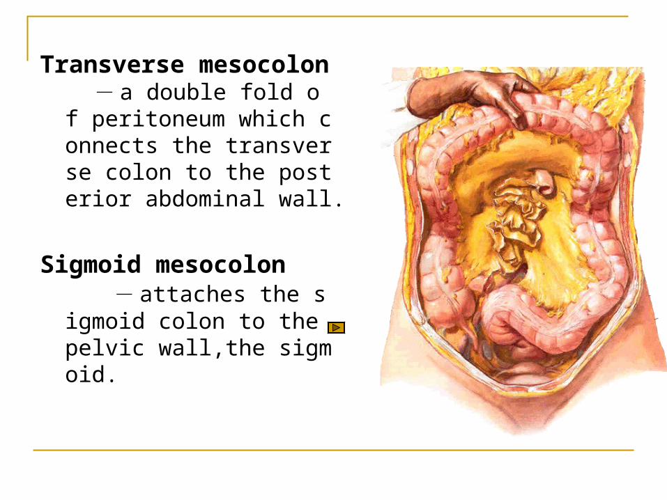
Transverse mesocolon - a double fold of peritoneum which connects the transverse colon to the posterior abdominal wall.
Sigmoid mesocolon - attaches the sigmoid colon to the pelvic wall,the sigmoid.
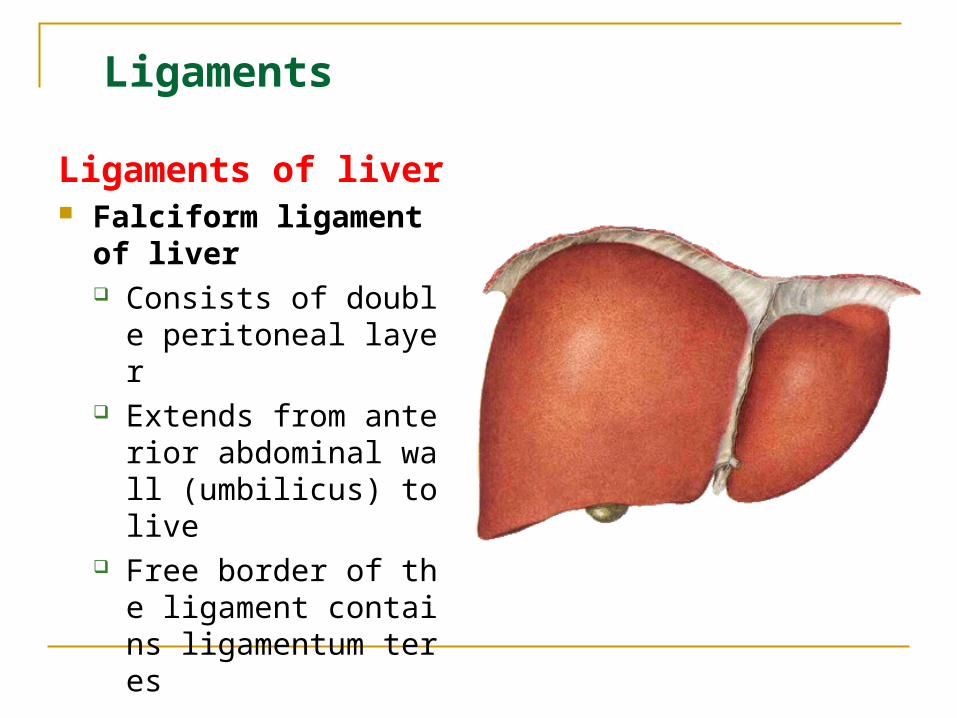
Ligaments of liver Falciform ligament of li
ver Consists of double per
itoneal layer Extends from anterior
abdominal wall (umbilicus) to live
Free border of the ligament contains ligamentum teres
Ligaments
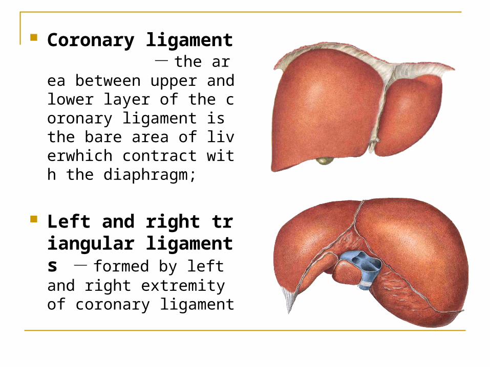
Coronary ligament - the area between upper and lower layer of the coronary ligament is the bare area of liverwhich contract with the diaphragm;
Left and right triangular ligaments - formed by left and right extremity of coronary ligament
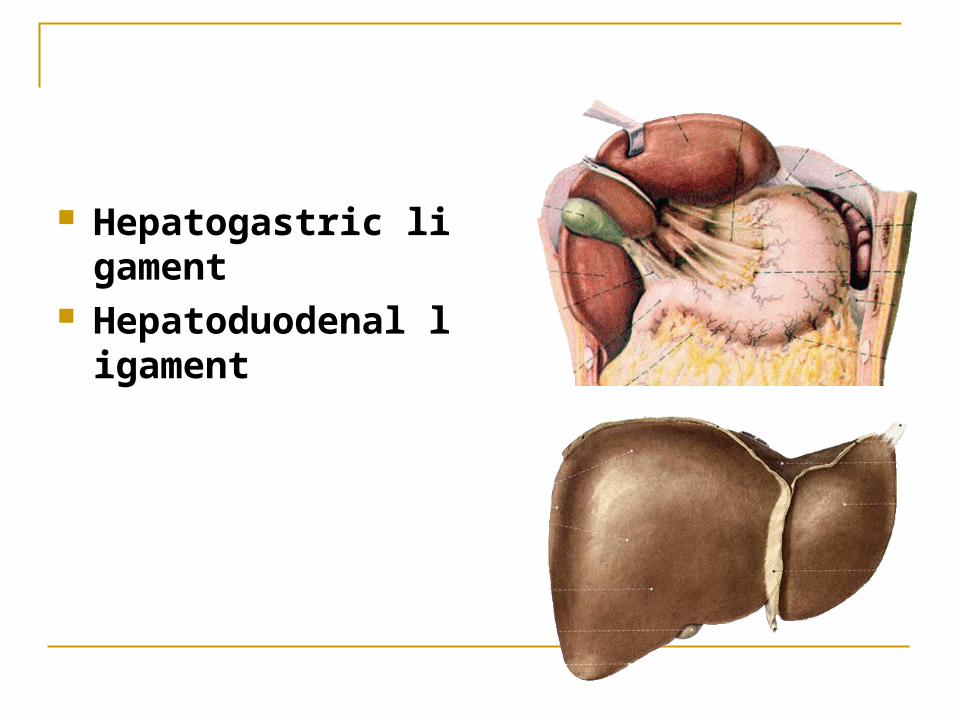
Hepatogastric ligament
Hepatoduodenal ligament
Ligaments of spleen
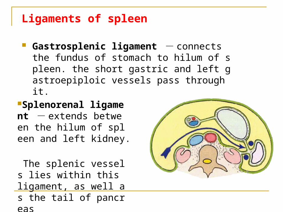
Gastrosplenic ligament - connects the fundus of stomach to hilum of spleen. the short gastric and left gastroepiploic vessels pass through it.
Splenorenal ligament - extends between the hilum of spleen and left kidney.
The splenic vessels lies within this ligament, as well as the tail of pancreas

Ligaments of spleen
Phrenicosplenic ligament Splenocolic ligament
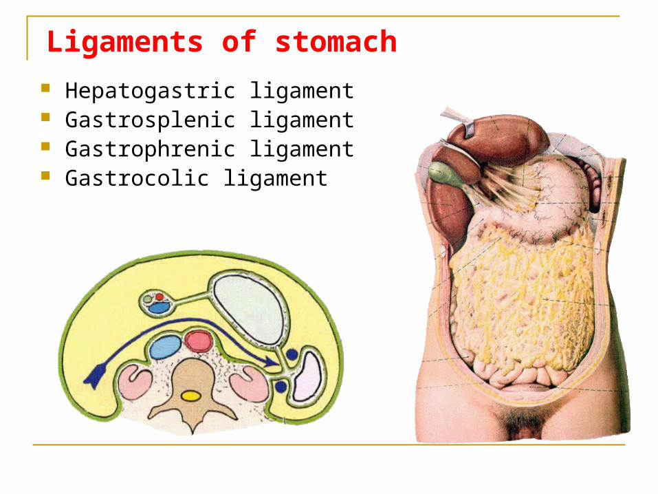
Hepatogastric ligament Gastrosplenic ligament Gastrophrenic ligament Gastrocolic ligament
Ligaments of stomach
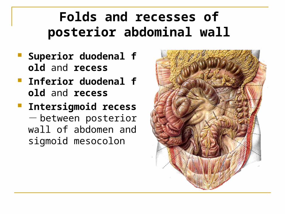
Folds and recesses of posterior abdominal wall
Superior duodenal fold and recess
Inferior duodenal fold and recess
Intersigmoid recess -between posterior wall of abdomen and sigmoid mesocolon
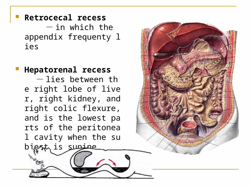
Retrocecal recess - in which the appendix frequenty lies
Hepatorenal recess - lies between the right lobe of liver, right kidney, and right colic flexure, and is the lowest parts of the peritoneal cavity when the subject is supine
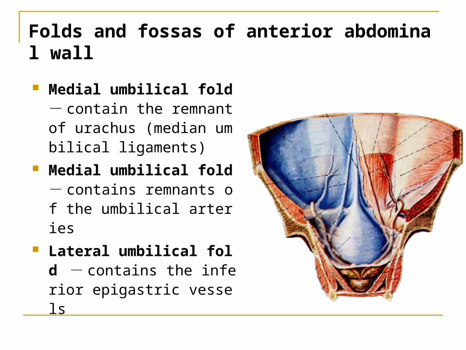
Folds and fossas of anterior abdominal wall
Medial umbilical fold - contain the remnant of urachus (median umbilical ligaments)
Medial umbilical fold - contains remnants of the umbilical arteries
Lateral umbilical fold -contains the inferior epigastric vessels
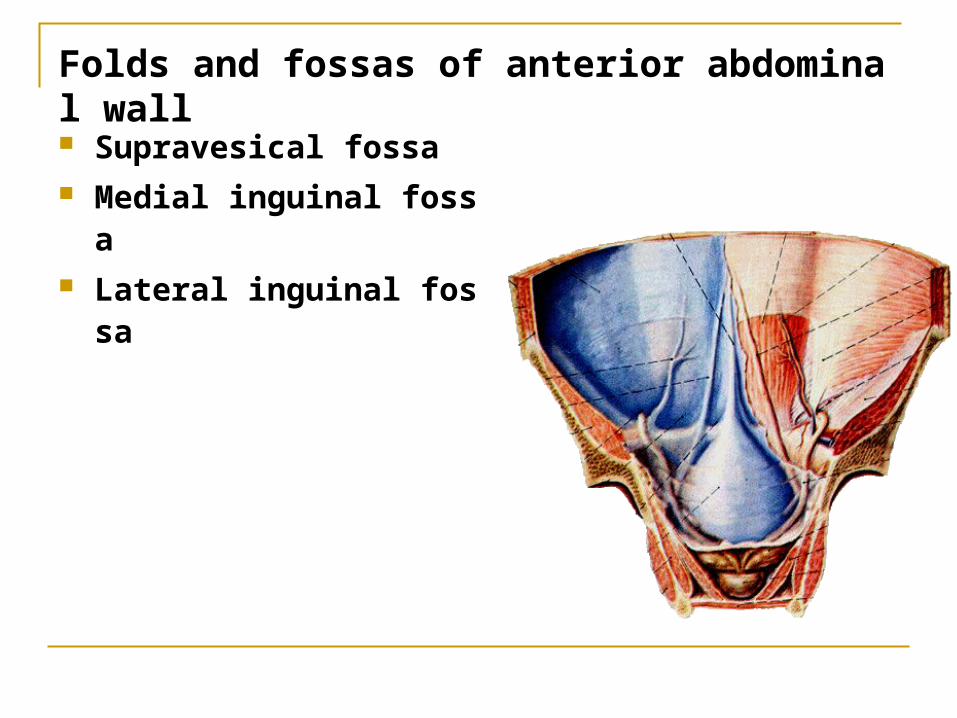
Folds and fossas of anterior abdominal wall
Supravesical fossa Medial inguinal fossa
Lateral inguinal fossa
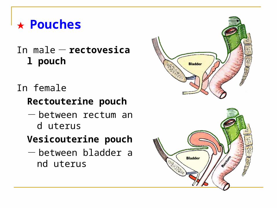
★ Pouches
In male - rectovesical pouch
In female
Rectouterine pouch
- between rectum and uterus
Vesicouterine pouch
- between bladder and uterus
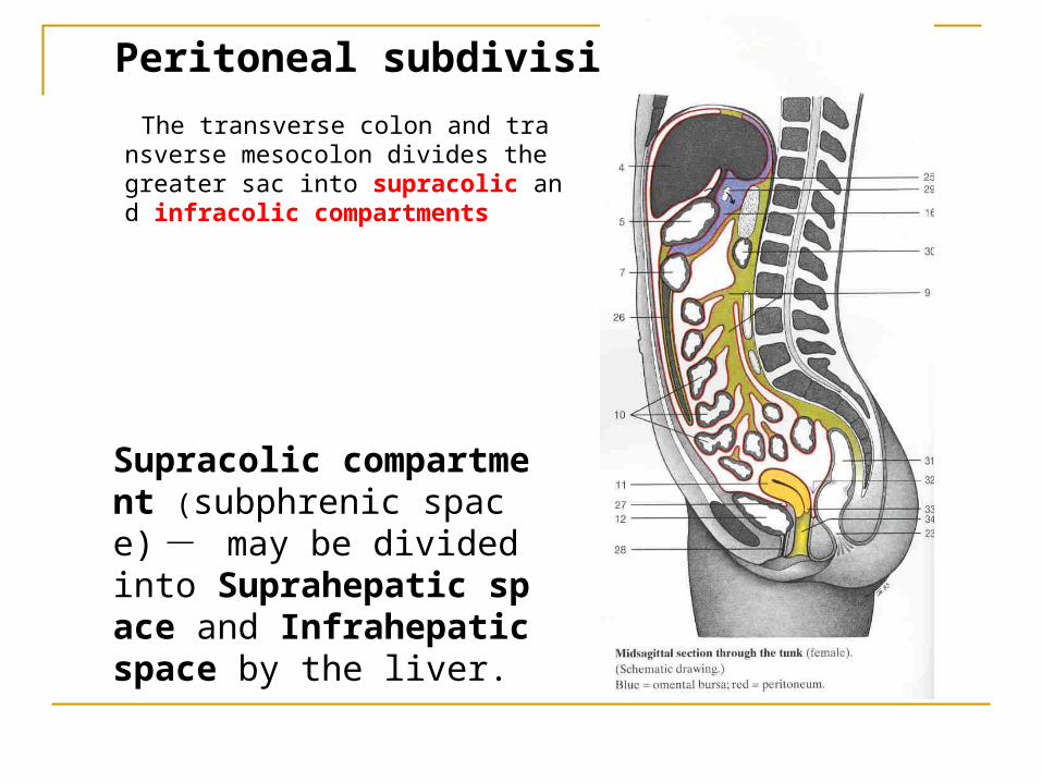
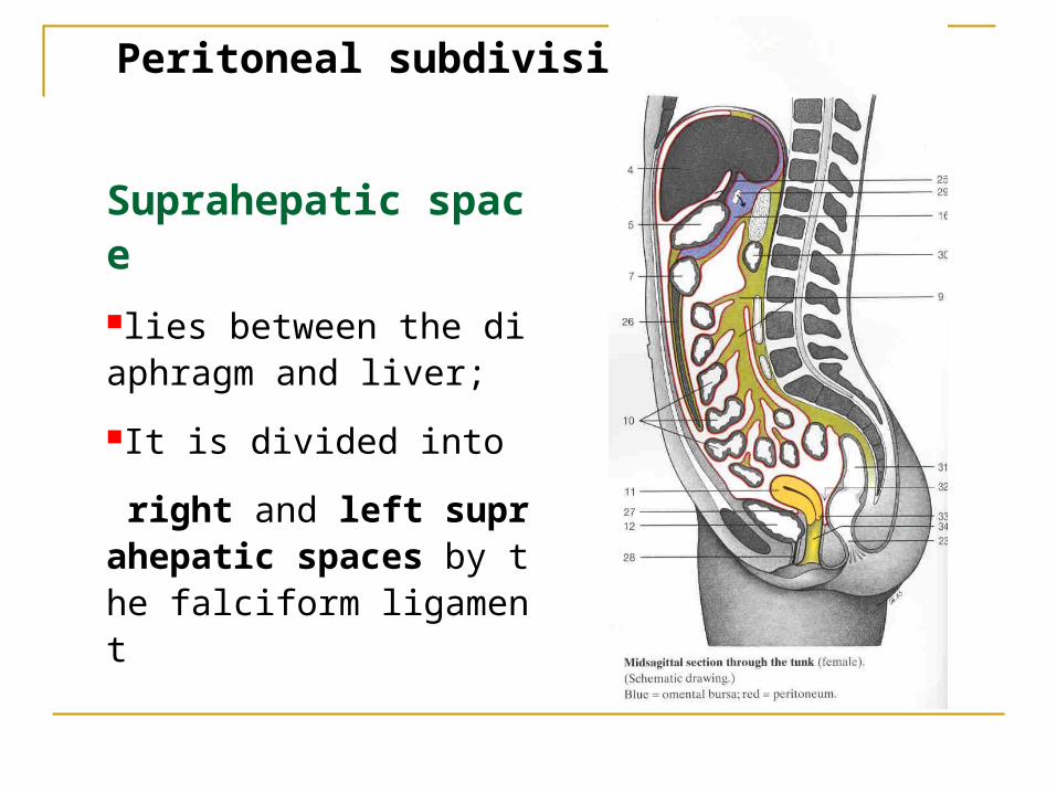
Peritoneal subdivisions
The transverse colon and transverse mesocolon divides the greater sac into supracolic and infracolic compartments
Supracolic compartment (subphrenic space) - may be divided into Suprahepatic space and Infrahepatic space by the liver.
Peritoneal subdivisions
Suprahepatic space
lies between the diaphragm and liver;It is divided into
right and left suprahepatic spaces by the falciform ligament
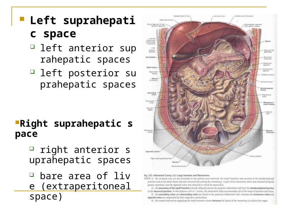
Left suprahepatic space left anterior suprahepat
ic spaces left posterior suprahep
atic spaces
Right suprahepatic space
right anterior suprahepatic spaces
bare area of live (extraperitoneal space)
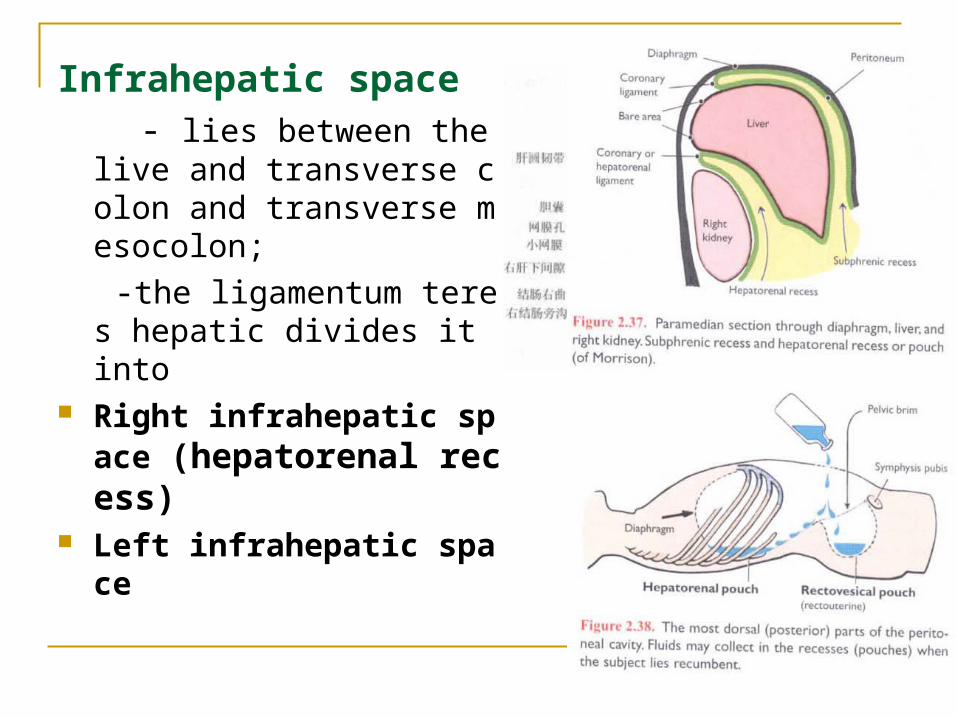
Infrahepatic space - lies between the live and tr
ansverse colon and transverse mesocolon;
-the ligamentum teres hepatic divides it into
Right infrahepatic space (hepatorenal recess)
Left infrahepatic space
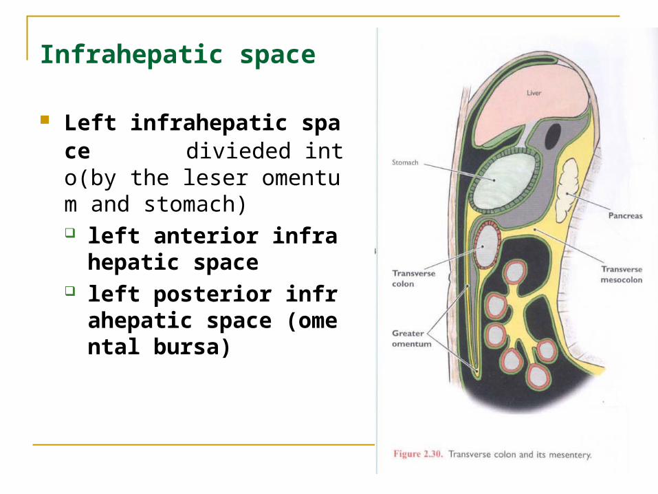
Infrahepatic space
Left infrahepatic space divieded into(by the leser omentum and stomach) left anterior infrahepatic
space left posterior infrahepati
c space (omental bursa)
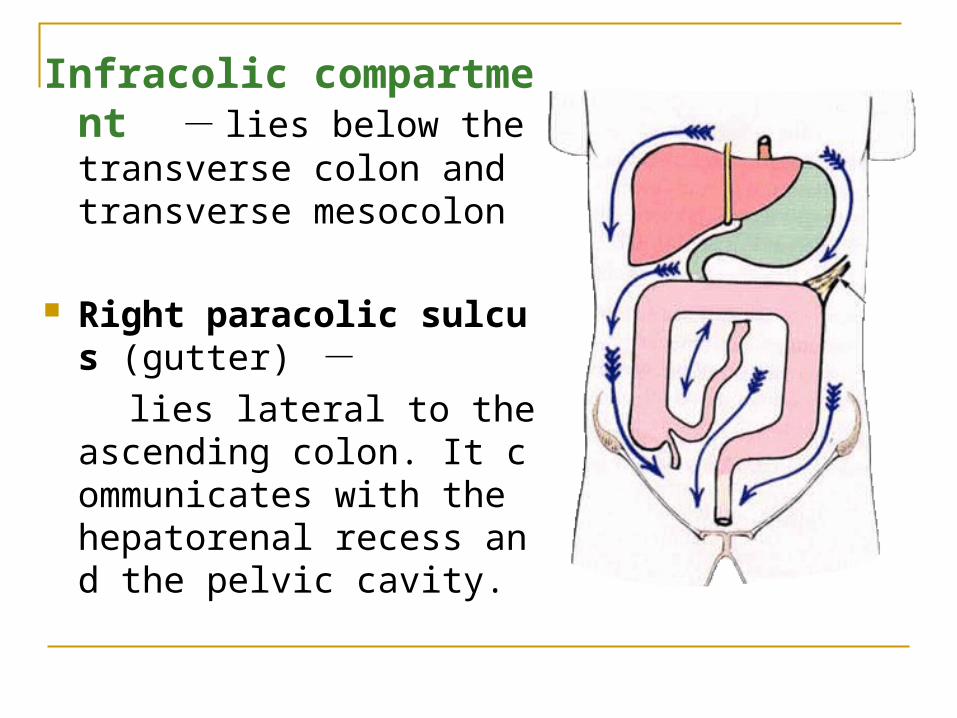
Infracolic compartment - lies below the transverse colon and transverse mesocolon
Right paracolic sulcus (gutter) -
lies lateral to the ascending colon. It communicates with the hepatorenal recess and the pelvic cavity.
Infracolic compartment
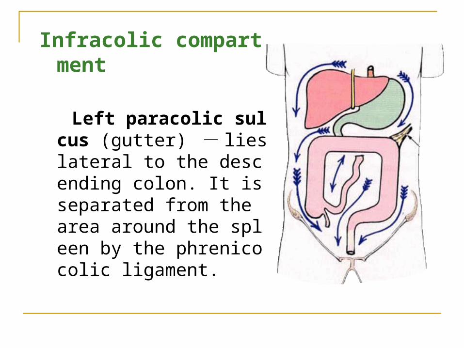
Left paracolic sulcus (gutter) - lies lateral to the descending colon. It is separated from the area around the spleen by the phrenicocolic ligament.
Infracolic compartment
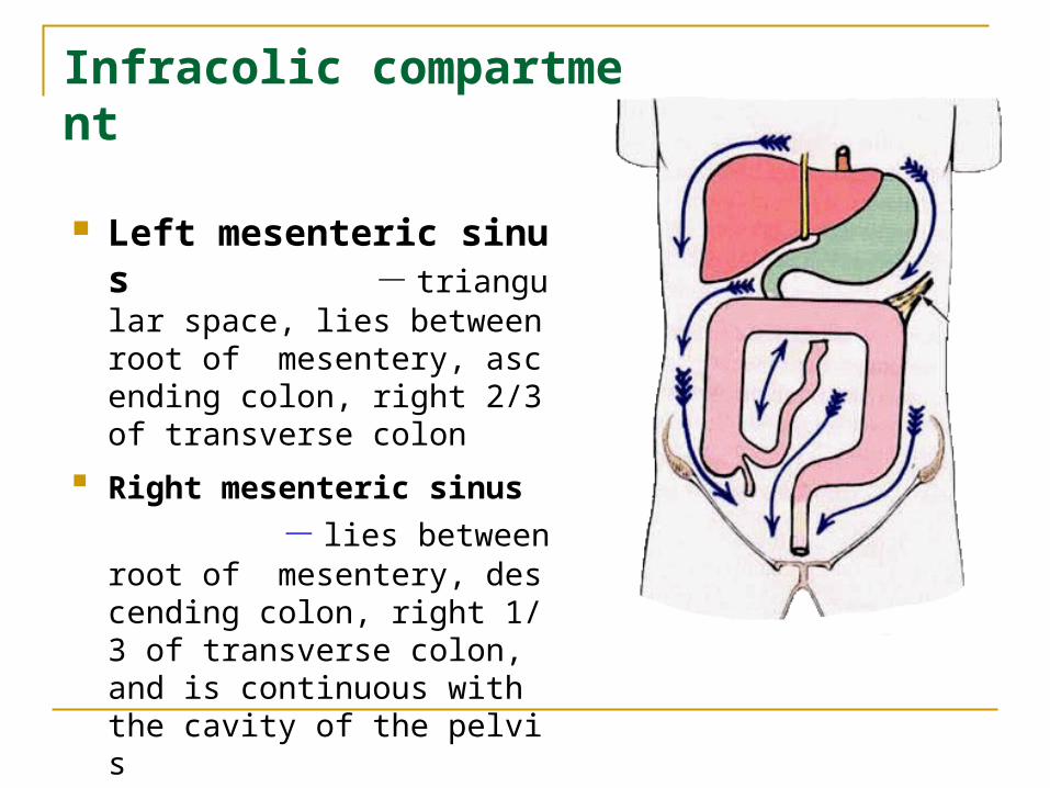
Left mesenteric sinus - triangular space, lies between root of mesentery, ascending colon, right 2/3 of transverse colon
Right mesenteric sinus - lies between root of mesentery, descending colon, right 1/3 of transverse colon, and is continuous with the cavity of the pelvis