Embed Size (px)
Citation preview

(NASA-CR-142192) THE PERFORMANCE AND T N75-17077CAPABILITIES OF, TERRESTRIAL ORGANISMS INEXTREME AND UNUSUAL GASEOUS AND LIQUIDENVIRONMENTS Ph.D. Thesis. Semiannual UnclasReport (Hawaii Univ.) 63 p i G3/51 10906
THE PERFORMANCE AND CAPABILITIES OF
TERRESTRIAL ORGANISMS IN EXTREME AND
UNUSUAL GASEOUS AND LIQUID ENVIRONMENTS
S. M. SIEGEL A
BOTANY DEPARTMENT, UNIVERSITY OF HA'--
PRICES SUBJECT TO CHANGE
Prepared under
Grant No. NGL 12-001-042with the
NATIONAL AERONAUTICS AND SPACE ADMINISTRATION
NATo AIL TE
u S..,; , - _ SER VICEV_,oA. 2ec

SEMI-ANNUAL REPORT
THE PERFORMANCE AND CAPABILITIES OF
TERRESTRIAL ORGANISMS IN EXTREME AND
UNUSUAL GASEOUS AND LIQUID ENVIRONMENTS
Halo- and Cryotolerance of theAlga DunaZiella
November 1974
University of HawaiiHawaii Botanical Science Paper
No. 38
Informzl technical report, neither to
be cited nor credited as a publication.
Submitted by:
Sanford M. S egelProfessor of Botany &Principal InvestigatorUniversity of HawaiiHonolulu, Hawaii 96822
NATIONAL AERONAUTICS AND SPACE ADMINISTRATION
Grant No. NGL 12-001-042

This dissertation is part of a research program
supported by National Aeronautics and Space
Administration Research Grant NGL 12-001-042.
THE CRYO- AND HALOBIOLOGY OF DUNALIELLA
Part I
A Dissertation submitted to the Graduate Division ofthe University of Hawaii in partial fulfillment
of the requirements for the degree of
Doctor of Philosophy
in
Botanical Sciences
By
Thomas W. Speitel

TABLE OF CONTENTS
Page
INTRODUCTION ............... 1
THE BIOLOGY OF DUNALIELLA TERTIOLECTA
AND DUNALIELLA SALINA . . . . . . . . . . 3
REVIEW OF THE LITERATURE ON HALOPHILISM . . . . . 9
HALOTOLERANCE - EXPERIMENTAL INVESTIGATIONS . . . 18
REVIEW OF THE LITERATURE ON CRYOTOLERANCE . . . . . 31
CRYOTOLERANCE - EXPERIMENTAL INVESTIGATIONS . . . 42
REFERENCES . . . . . . . . . . . ..... 47

INTRODUCTION
The accounts of physiological research on the active metabolism
and behavior of autotrophic eukaryotes at sub-zero temperatures are
relatively meagre. Possibly, this is because so few organisms remain
unfrozen at temperatures of OOC to -150 C. This report will deal with
the alga DunaZieZZlla, and its remarkable cryo- and halo-tolerance.
The genus Dunaliella, composed of green unicellular algae, is
in the family Polybelpharidaceae (phylum Chlorophyta) (24). The
organisms are motile, with two equal, unipolar flagella. They pos-
sess a thin, elastic cell envelope which follows changes in body
outline. Reproduction occurs by longitudinal fission or by fusion
of two motile, haploid cells to form a zygote (136). This genus
resembles ChZamydomonas, but differs in the nature of the cell enve-
lope. Chlamydomonas has not only a periplast but also a cell wall.
The movement of most species of Dunaliella is a constant, linear
motion. The algae rotate only slowly, and when quiescent, continue
to vibrate. Dunaliella is of commerical importance as a source of
vitamin A (105, 107, 108, 109).
Dunatiella have been recorded in oceans, salt marshes, inland
salt springs, lagoons and saline pools all over the world (24, 105,
170). Some specimens have been found at depths of 315 meters in
the Dead Sea (44).

2
The similarities and differences in salt and cold tolerance
mechanisms of DunalieZZa satina and DunalieZZa tertiolecta will
be under focus in this investigation.
The NASA Technical Officer for this grant is D. S. Geib,
Planetary Programs, NASA, Washington, D. C. 20546.

3
THE BIOLOGY OF DUNALIELLA TERTIOLECTA
AND DUNALIELLA SALINA
D. esaina and D. tertiolecta differ from each other in morpho-
logy. Although both species are ovoid or ellipsoid, mature D. salina
cells usually have at least twice the volume of D. tertiolecta cells.
The periplast of D. satina is less firm than that of D. tertiolecta.
Both species have companulate chromatophores, D. tertiolecta's being
more rugose. The pyrenoid of D. satina is sub-basal and D. tertio-
lecta's is central. D. satina contains a large number of refractive
granules arranged in a girdle just above the chromatophore. The gran-
ules of D. tertioZecta are distributed evenly throughout the cell.
D. saZina has a haploid number of chromosomes of about 10, while the
count for D. tertiolecta has not been recorded (24, 134).
Light optima for photosynthesis of both species have been re-
ported. D. saZina is positively phototaxic (17). Reports of optimal
light intensities for growth range from 600 lux (116) to 10,000 lux
(50, 170). D. salina's carotene composition (63) and accumulation
with higher salinities is documented (37, 38, 39, 40, 41, 42).
After high illumination, D. tertiolecta needs a short period
in the dark to develop maximum photosynthetic rate (64). Oxygen
evolution is higher with NH3 than NO3 as a nitrogen source (133).
Grant (55) gave evidence for the existence of two independent
systems of nitrate reduction, one within and the other outside

4
the chloroplast. The enzyme system which fixes CO2 is likely to be
the rate limiting step in photosynthesis. Glycerol is the major
soluble photosynthate (31). Photopigment extractions and concentra-
tions have been reported (34, 101, 138, 159, 163).
Redox enzymes of D. satina such as cytochrome oxidase, peroxi-
dase, catalase, reductase-ascorbate oxidase and polyphenol oxidase
have been studied. Its polyphenol oxidase is specific for tri-
phenols (122). For D. tertiolecta, nitrate reductase (56), glucose
6-phosphate dehydrogenase, pyrophosphatase, ATP-ase (75), hexokinase
(89), ribulose di-phosphate carboxylase and aldolase (64, 133) have
been studied.
The general mechanism of salt tolerance for D. salina and D.
tertioZecta is reported. D. satina is a "coper", whose cytoplasmic
salt concentration is at least as high as the ambient medium. D.
tertiolecta is an "avoider" and actively keeps salt out of the
cytoplasm.
D. salina is an inhabitant of ultra-haline, neutral-alkaline
waters (104, 171). Blooms in such places as the Krymsk Oblast
occur in summer, which coincides with a decrease in Ca2+ and an
increase in Mg2+ and S042- (103). Increasing NaCi concentration
is increased from 2 to 4 M; from 4 to 5 M it decreases (119).
Increasing NaCl results in disturbance of nucleic acid and pro-
tein metabolism. RNA per cell decreases and protein accumulates
5
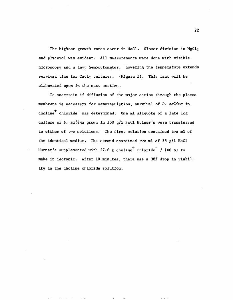
in large amounts (122). Marre and Servattaz (102) reported high
conductivity and osmotic pressure of the cell sap of D. salina grown
in 3.9 M NaCI in comparison to those grown in 1.5 M NaCi. Based on
24Na experiments, they also concluded that the cell membrane is
freely permeable to sodium.
Trezzi (161) looked at the ultrastructural changes in D. salina
following large increases and decreases in osmolarity of the culture
medium.
"When the tonicity of the nutrient medium is suddenly lowered
the whole cell and single cytoplasmic structure appear equally
swollen and the state of dispersion of the stroma of cellular
organelles is increased. A conspicuous amount of water pene-
trates in every part of the cell. The mitochondria show a
uniform type of swelling, while in the inner space of the
nuclear envelope wide sacs appear upon hypotonic shock. The
outer layer of the double membrance of the nucleus may be
more permeable to water than the inner one. In chloroplasts
the flattened sacs formed by paired lamellae having blind
ends in the peristromium swell up giving rise to conspicuous
vesicles. The permeability of water of the lamellae is
greater than that of the external plastid boundary. Sudden
and strong hypertonic stress causes great loss of water from
every part of the cell. Shrinking can be seen in all cyto-
plasmic organelles together with great electron density of

their stroma. The two layers of the nuclear membrane and
the lamellar pairs forming the succuli of the chloroplasts
stick closely together causing a nearly total disappearance
of the inner space. The formation of large vesicles after
sudden hypotonic stress is due to a localized osmotic gra-
dient. In both cases the conspicuous structural variations
are reversible and the alga returns to the normal state in
a short time. During the short time span necessary for the
reestablishment of a new osmotic equilibrium and of a normal
state of the plasmic structures, the cells suffer from a lose
of motility and from an altered metabolism. A specific re-
sistance of plasma proteins to denaturation may be involved."
Concommitant with high internal salt concentration is the depressed
freezing point of the cytoplasm. The organism remains motile at tem-
peratures as low as -150C (102). Low temperatures within the range
-150C to +110C depress the growth of D. satina and in the course of
eight days cause death of the cells. Some cells tolerated a tempo-
rary fall of the temperature to -300 C (170).
D. tertiolecta is a euryhaline organism growing in salinities
ranging from 3.75 to 150% (75, 114). The first detailed study of
the culture of D. tertiolecta was reported by MacLachlan (114). He
established its comparatively high sodium requirement as 10 mM.
Calcium and magnesium are inhibitory at high concentrations, but
the inhibition can be prevented if the Ca2+/Mg2+ ratio was maintained

7
at 4. Jokela (75) showed that the internal concentration of both
Na+ and K+ increase as the salinity of the medium increases. Never-
theless the internal Na concentration stayed considerably lower than
that in the culture medium at high salinities. For example, when the
NaC1 concentration of the medium is 15%, the intracellular concentra-
tion is 4.35%. The cell membrane is therefore not freely permeable
to sodium. The cell membrane is characterized by a high phosphatidic
acid content and high concentrations of C-16 saturated fatty acids.
It is also characterized by its divalent ion dependent stability, its
relatively high hydrophobic amino acid content and the presence of a
Na-K activated ATP-ase.
Both the soluble enzymes (as exemplified by glucose 6-phosphate
dehydrogenase) and particulate enzymes (pyrophosphatase and ATP-ase)
of D. tertiolecta were found to be severely inhibited by high Na and
K concentrations (above 6%) (75).
Increasing the concentration of osmotic substances causes a
decline in photosynthetic oxygen evolution, then oxygen consumption,
accompanied by CO2 evolution. Both show distinct maxima at 2.8 M
NaCl. Fermentation probably occurs at higher osmotic pressures as
indicated by respiratory quotients (165). Glycerol production is
considered to be a protective mechanism for osmoregulation in D.
tertiolecta. With increasing concentrations of NaCi, photosynthetic
incorporation of 14C declines, but the percentage incorporated into
glycerol increases to a distinct maximum at 2.8 M NaC1 (31, 47, 165).

8
When transferred from a medium of 15% NaCI to 4% NaCI, 15% of the
total cell glycerol is released instantaneously from the cells (75).
The upper temperature limit for growth of D. tertioZecta in-
creases with NaCl concentration. Greatest growth rates occur at
0.5 to 0.75 M NaCl at 33*C (46).

9
REVIEW OF THE LITERATURE ON HALOPHILISM
The term halophyte literally means salt plant, and is usually
used for plants that grow in the presence of high concentrations of
sodium salts. A halophyte can be halophilic, salt resistant, halo-
tolerant or haloavoidant. There is confusion in the literature as
to the proper usages of these terms. According to Levitt (97) the
following explanation would probably be proper. A halophilic plant
is one which grows best at high salinities. On the other hand, a
salt resistant plant can withstand high salinities, but grows best
at lower salinities. There are two major mechanisms utilized by
halotolerance and haloavoidance. The first, halotolerance, is the
same as ion accumulation. The second, haloavoidance, can be accom-
plished in any of three ways; salt can be excluded passively, ex-
truded actively, or can be diluted.
The salinity extremes that organisms can survive in or thrive
in are very varied. Some examples are given. Certain bacteria, such
as Halobacterium satinarium can grow in saturated NaCl (29). Selected
strains of Penicillium notatwnu grow on saturated calcium acetate (143).
The fungus Scrophulariopsis parvula grows in media with saturated NaC1.
The yeast Debaryomyces hansenii shows respiratory activity in 24% NaC1,
which is 10% of normal.
Species of algae have been reported from brine lakes, salterns,
salt springs and pools with saline contents 2 to 17 times that found

10
in the ocean. The algae are largely Chlorophyceae, although there
occur species of Myxophyceae, Bacillariophyceae and Euglenophyceae.
Dunaliella, reported from numerous brine waters, is probably the most
common of the green algae. ChZamydomonas has been found in the saline
lakes of the Crimea. Both Dunaliella and Stephanoptera form light
green areas on solid salt crusts (157). Elazari-Volcani (44) examined
mud sampels taken from the Dead Sea bed. These contained green and
blue green algae and diatoms. Most of the green forms resembled
Dunaliella, with lesser amounts of Scenedesmus bijugatus, Pediastrim
simplex and UZothrix. The blue greens resembled Aphanocapsa
littoraZis, Aphanothece halophytica or an Oscillatoria. Of the
diatoms, forms of Melosira, Navicula, Pinnutaria, Gomphonema,
Cymbella and Synedra occurred. Each liter of Dead Sea water con-
tains approximately: 143 g. magnesium chloride, 87 g. sodium chlo-
ride, 37 g. calcium chloride, 11.5 g. potassium chloride and 5 g.
magnesium bromide.
Species of Enteromorpha occur in the Great Salt Lake as well as
from fresh waters. Fluctuating amounts of salinity produce specimens
that have characteristics of several species in a single frond (157).
The blue green algae SpriruZina subsala, Phormidium tenue and others
grow and multiply in 3 M NaCI (65, 84).
A few examples of halophilism of higher plants are given. Seed-
lings of Atripeex vesicaria were established in 1 M NaCI (16). At

100-200 mEq/liter of Na+, K+ or Mg2+ Cl Atriplex nummularia grows
optimally. There is still good growth at 300 mEq/liter (57).
Halogeton glomeratus survives at 1.4 molal NaCi (137). The optimal
level of NaCl for growth of the mangrove Avicennia marina is 1.5%,
one half the concentration of sea water (30). Succulent halophytes
such as Sueda depressa, Salicornia europaea and Spergulia marina
germinate and develop in 5% NaCl (162). Leaves of Nitraria schoberi
(the most tolerant among woody plants) may contain 14% of their dry
matter in the form of NaCl, 57% as total salts (158).
Mechanisms of Osmotic Regulation
The following are mechanisms of osmotic regulation utilized by
halophilic plants and microorganisms:
A. Development of cell membranes highly permeable to ions.
A membrane which is highly permeable to sodium enables the cell
to adjust rapidly to high osmotic pressure changes. This requires
adaptation of a cell's enzyme system, nucleic acids, ribosomes, etc.,
to function at high as well as low ionic concentrations. This mech-
anism is suggested as being responsible for the osmotic regulation
of D. satina (102). Another species of the halophilic organism,
D. parva, is freely permeable to sucrose, inulin, starch and even
polyvinylpyrollidone with a molecular weight of 20,000 to 40,000
(53). It is impermeable however to dextran (mol. wt. 80,000). The

12
mycelia of a salt tolerant strain of Penicillium notatum accumulate
high intracellular concentrations of copper when grown on metallic
copper surfaces (154). Kylin and Gee (88) showed that a halophilic
mangrove (Avicennia nitrida) possesses a Na extrusion pump, but it is
inactivated at high concentrations of NaCI (0.2 to 0.4 M). Similar
inactivation also occurs with Atriplex subcordata, Suaeda marxtima
and Astes tripoliwn (168).
B. Avoidance, including active Na extrusion.
Many plant halophiles, including Spartina townsendii, Limoniwn
Latifolium (3) and the mangroves (6) posses salt glands which extrude
NaCI against a concentration gradient (144). In the case of Tamarix
aphylla, the glands show no apparent selectivity between Na+ and K
ions (14). Even Rb was taken up from the nutrient solution and
secreted by the glands (160). Single celled organisms may use
vacuoles to maintain a steady state low concentration of NaCl. The
non-photosynthetic flagellate Choanogaster plattneri is found in
water of 20% salinity. The cells have a complex vacuolar system,
producing smaller accessory vacuoles that empty outside the cells and
that decrease in activity when the salinity of the medium is lowered.
The vacuoles are said by Pochman (136) to contain a higher concentra-
tion of salt than the medium and serve to excrete salt. The contrac-
tile vacuoles of Chlamydomonas moewusii have an entirely different
function, the elimination of water. A mutant strain lacking contrac-

13
tile vacuoles survives and grows only if the osmotic pressure of
the medium exceeds 1.5 atm (59).
For algae, sodium pumps have been described since Scott and
Hayward (146) described a sodium excretion mechanism in UZa Zactuca.
The mechanism of this active transport was envisioned as involving
a cation sensitive ATP-ase system as demonstrated for erythrocytes
(36, 137). As far as completing the comparison, Eppley in 1962 (45)
stated that 'an experimental approach might be facilitated if an
algal equivalent of the erythrocyte ghost were available.; Jokela,
in 1969 (75) isolated naturally wall free, ghost forming membranes
of D. tertiolecta and detected Na-K ATP-ase activity.
C. Development of an active exchange-transport system.
Halobacteria avoid excessively high sodium content of the cell
by an active inward transport of potassium. K+ concentration is
much higher than in the medium, and in some cases the value found
intracellularly was close to the limit of solubility for KC1 (29).
Adaptation of the cell's enzyme structure is necessary for functioning
at high ionic strength. Many of the enzymes are reversibly inac-
tivated in the absence of high concentrations of neutral salts (91).
D. Utilization of inert neutral compounds as osmotic agents.
The maximum proline concentration that E. coli can concentrate
internally has been shown to be dependent on the osmolarity of the
suspension medium; the labeled amino acid pool in E. coli has been
shown to be proportionally released when the cells are washed with

14
low osmotic strength solutions (20). Oxalate synthesis in Atriplex
spp. has been shown to be related to the external osmotic pressure
(132). Polyalcohol production of yeast is influenced by the presence
of high salt concentrations (131). Glycerol production by D. tertio-
Zecta in response to increasing osmotic pressures has been mentioned.
E. Salt dilution.
Rhizophora mucronata does not excrete salt, yet its growing
leaves retain a constant concentration (510 to 560 mEq/liter) though
receiving 17 mEq Cl/day (6). During growth, sufficient water is
absorbed to prevent increase in concentration. This dilution of
cell sap during growth has been found in some moderately salt tol-
erant halophytes (97). Many other plants avoid increase in concentra-
tion by increasing succulence. Cells, especially parenchyma cells
enlarge, avoiding excessive salt concentrations (139).
High Salinities and Cellular Adaptations
The adaptations of some cellular constituents to high salt con-
centrations are related.
A. Cell envelopes.
Halobacteria do not have clearly separable wall and membrane
structures (142). The term cell envelope is used to designate struc-
tures enveloping the cytoplasm. The cell envelope requires a high
salt concentration for its maintenance; at low concentrations it loses

15
its rigidity and collapses. Chemicals which do not possess a strong
net charge in aqueous solutions do not protect the structure of the
envelope (123). The hypothesis of Abram and Gibbons (1) is that the
cell envelopes of the halobacteria "are held together rather loosely
by hydrogen bonds, Coulorb forces or 'salt' linkages, and in the pres-
ence of high concentrations of NaCl the electrostatic forces are
screened so that the bonds hold the organism together in a rod shape."
The cell envelopes of the halobacteria have very few or no higher
fatty acid residues, and in this way differ pronouncedly from other
bacteria (27, 91). The bulk of the lipid is an analogue of diphos-
phatidyl glycerophophate (147). The proteins of the cell envelope of
halobacteria are very acidic (21).
It is of interest that in the cell membrane of D. tertiolecta
long chain fatty acids are absent, at least when the organism is cul-
tured at high salinities. There is also a high phosphatidyl glycerol
content and a predominantly acidic amino acid composition (75).
B. Nucleic acids.
According to Kessler (83) high osmotic pressures induce changes
in copolyribonucleotides synthesized from equimolar concentrations of
ADP, GDP, CDP and UDP. He suggests that osmotic stress is coded into
a changed composition of polyribonucleotides which may lead to adaptive
reactions by serving as primers for RNA dependent RNA polymerase.
Siegel (152) has demonstrated the role of nucleotides, especially IMP,
in the recovery of Penicillium from extreme salt effects.

16
C. Ribosomes.
The ribosomes of H. cutirubrum are stable only in solutions
containing KC1 close to saturation (4M) and Mg2+ in the concentration
range 0.1-0.4 M. The KC1 requirement is quite specific, and cannot
be replaced by iso-osmolar NaCI (12). With regard to higher plants,
it has been shown that the ribosomes of salt sensitive citrus species
are less stable than those of more resistant species, but were able to
acquire an increase in stability (83).
D. Proteins and enzymes.
In a culture of callus tissues from cabbage leaves, it was found
that salts slow down amination and amidation, as well as the transfer
of sulfides to S-containing amino acids (148). Shevyokova (148)
suggests that an increase in derivatives of S-amino acids may be one
reason for the unfavorable effect of salts.
Baxter and Gibbons (11) made the important discovery that the
enzymes of extremely halophilic bacteria are adapted to function at
the very high salt concentrations found within the cells. Since then
about twenty different enzymes have been reported, and all data fit
the general contention that the enzymes of extremely halophilic bacte-
ria are extremely halotolerant, and in most cases even strikingly
halophilic (11, 29, 91). These enzymes are irreversibly inactivated
in the absence of high concentrations of salts. For example, Hato-
bacterium salinamr excretes a protease which is possible to chro-

17
motograph only in the presence of 3.4 M NaCI (127). Another specific
property of their proteins was the absence of half-cysteine residues,
in opposition to the non-halophilic E. coli which has 0.6 moles half-
cysteine per 100 moles of amino acid. Stroganov (158) suggests that
the bacteria adapt to high salinity by binding of the mineral element
(158). Haloavoidant organisms and non-halophiles usually have enzymes
that are salt sensitive, as is the case with D. tertiolecta (75).
The capabilities of some enzymes under stress are enormous. In
vitro horseradish peroxidase activity is present in 10 M lithium
chloride. Considering the coordination number of the ions, a 10 M
LiCl solution can have little or no uncoordinated water (153).

18
HALOTOLERANCE - EXPERIMENTAL INVESTIGATIONS
Growth of D. salina was reported by Marre (102) to occur in sa-
turated NaCi solutions. McLachlan (114) demonstrated D. tertioZecta
growth in 2.5 M NaCI, the highest concentration tested. This inves-
tigation corroborates these results. It was also found that D.
tertiolecta growth occurs in saturated NaCI, though greatly atten-
uated (1/4 the rate of growth in 2.5 M NaCI).
All cultures were grown in Hutner's Marine ChZarmdomonas medium,
with variation only in the major chloride (69) (See Table 1). The pH
was adjusted to 7.2 with KOH. This glass electrode measurement
proved to be optimum for increase in cell number for D. saZina cells
grown in 300 g/l NaCI (See Table 2). It must be kept in mind that
such pH measurements are inaccurate, due to dilution of water by the
concentrated salts. The light intensity of all experiments was a
continuous 3000 lux.
At room temperature the effect of various electrolytes and glyc-
erol on Dunaliella growth are shown in Table 3. NaCl, KC1, MgC1 2,
CaC12 and glycerol were added to the medium in concentrations yielding
an osmolarity equivalent to 550/00 NaC1.

19
Table 1. Hutner's marine chlamydomonas
medium (69), pH adjusted to 7.2 with KOH.
Metal chloride Xg/liter
EDTA 0.50 g
K2 HP 0 4 - 3H20 0.26 g
MgSO 4 1.22 g
Glycine 2.50 g
K-acetate 2.00 g
H 3BO3 244.00 mg
ZnC1 2 62.00 mg
Ca(N03) 2 °
4H20 147.00 mg
MnSO 4 ° H20 31.00 mg
Na2MoO4 * 2H20 25.00 mg
FeC1 3 ° 6H20 14.00 mg
CoC1 2 a 6H20 1.00 mg

20
Table 2. Effect of initial pH of Hutner's marine
chlamydomonas medium on growth of D. saZina. Cul-
tures initially contain 250 ml of 5 X 10⁴ cells/ml.
Glass electrode pH % increase in cell number after 3 weeks
5.0 all dead
6.0 300
6.8 1000
7.7 1300

21
Table 3. Change in cell number of 250 ml cultures in
Hutner's supplemented with chlorides and glycerol of
osmolarities comparable to 550/00 NaC1. Incubation
period is three weeks. The initial cell concentra-
tion (X) is 5 X 10⁴ cells/ml.
D. salina D. tertiolecta
NaCl 14X 31X2
MgC1 OX 5X
KC1 OX OX
CoC1 OX OX2
Glycerol OX lX
22
The highest growth rates occur in NaCI. Slower division in MgCl2
and glycerol was evident. All measurements were done with visible
microscopy and a Levy hemocytometer. Lowering the temperature extends
survival time for CaC1 2 cultures. (Figure 1). This fact will be
elaborated upon in the next section.
To ascertain if diffusion of the major cation through the plamsa
membrane is necessary for osmoregulation, survival of D. salina in
choline+ chloride- was determined. One ml aliquots of a late log
culture of D. salina grown in 150 g/l NaCl Hutner's were transferred
to either of two solutions. The first solution contained two ml of
the identical medium. The second contained two ml of 35 g/l NaCl
Hutner's supplemented with 27.6 g choline+ chloride- / 100 ml to
make it isotonic. After 10 minutes, there was a 38% drop in viabil-
ity in the choline chloride solution.

,q
4 '-
Figure 1. Duale~ saiai aurtdCC2
Optca reouin Cel dimtrlntws- s1m
OF Poop Q s-0
UA"r

24
Osmotic Regulation
The conclusion of Marre (102) that DunaZielZa salina plasma
membranes are freely permeable to sodium is based almost entirely on
his 2 4Na experiments. Our studies have shown that his interpretations
of the data were almost completely fallacious.
Marre suspended aliquots of algae in the original nedia indicated
below and dialyzed them for ten hours against 20 volumes of the final
medium containing 24NaHCO 3.
NaCl in medium Na 24CPM / cc
original final medium cell sap
2.50 M 7 M 165,000 81,000
1.25 M 7 M 163,000 112,000
The original medium did not contain the isotope. This was
introduced with a specific activity as high as practical without
altering the Na+ ion concentration of the medium. Before monitoring
radioactivity of the algae, Marre centrifuged the cells for 20,000 g
for 20 minutes and decanted the medium. He somehow assumed that the
intercellular volume of the packed cells was less than 3% of the
total volume. No basis for this assumption is given.

25
A follow-up experiment involved exclusive use of iso-osmotic
solutions.
Na24 cpm / cc 30 minutes afterNaC1 cone. of
after 10 hours in the transport of cellsthe medium
medium containing the from med. contain-isotope ing Na 2
4 to isotopefree med.
In medium In cell In cell
2.50 kM 129,000 71,000 360
3.75 M 129,000 94,000 150
On the basis of these two experiments, along with the obser-
vations of Trezzi (161) that the cells shrink in hypertonic solu-
tions and then regain their original shape--that all the subsequent
literature on the passive Na+ intracellular accumulation is based.
Assuming Marre's interpretation of his data was correct, we
proceeded to look for halophilic or halotolerant enzymes in D. salina.
This search seemed to be called for since there has never been
reported a halophilic or halotolerant enzyme isolated from an alga.
Both glucose-6-phosphate dehydrogenase and catalase had attenuated
activities with increasing NaCl concentrations. Increasing NaC1
concentration from 60/00oo to 100 O/00 resulted in a 90% decrease in
glucose-6-phosphate dehydrogenase activity. At 2500/o00 NaCl there
was another tenfold loss in activity.

26
To make precise estimates of osmotically active substances in
D. salina, the following determinations for cell volume, sodium,
potassium and glycerol were performed.
Determination of Intracellular Volume of D. saZina Cells
Using a Bluedextran Indicator Method
Cells were harvested at the end of their log phase of growth at
250 in 1500/00 NaCl Hutner's and resuspended in identical sterile
solution. Two hematocrit tubes were filled with aliquots of the
suspension and centrifuged at 2,500 g for 10 minutes. The ratio of
packed cell volume to total volume of liquid was then measured.
Four ml aliquots of the algal suspension were centrifuged under
the same conditions as the hematocrit tubes. The supernatants were
decanted, the sides of the centrifuge tubes carefully dried, and then
the packed cells were resuspended in a Blue Dextran solution of known
concentration and Optical Density at 580 mm. The cells were then
recentrifuged under the previous conditions (2,500 g for 10 minutes).
The supernatant was decanted, millipore filtered, and then the Optical
Density determined. By comparing standard Blue Dextran dilution plots
to the decrease in 0. D. of the dye solution caused by dilution of the
dye by intercellular space liquid, the volume of the intercellular
space was determined. The intercellular volume was 32% of the packed
volume, ten times Marre's figure.

27
Assay of Glycerol Using Glycerol Dehydrogenase
D. saZina cultures in late log phase were centrifuged at 2,500
for 10 minutes, and then the supernatant analyzed for glycerol.
1i, To each tube containing from 0 to .4 V moles of standard
glycerol solution, or similar amount of sample, or both, the following
solutions were added:
2. 1.9 ml of Sorensen's glycine II buffer, pH 9.5
3. 0.2 ml of glycerol dehydrogenase (Worthington, from Enter-
obacter aerogenes) (2mg/3 ml), prepared immediately before
use.
4. Distilled water to bring volume up to 3 ml.
5. 0.1 ml of DPN solution (6.6 mg/ml)
Optical density at 340 mu with appropriate standards recorded
for twenty minutes.
To determine intracellular glycerol concentrations, packed cells
were washed twice with 100 volumes of fresh medium at 40, then freeze-
thawed and centrifuged. The supernatant was analyzed in the manner
indicated above. Results are shown in Table 4.

28
Table 4. Intra and extracellular glycerol, Na and K in late log
D. satina culture grown in 150 g/l NaCl Hutner's medium. Values are
means of three determinations.
Intracellular Extracellular
Compound Molarity Osmolaritya Molarity Osmolaritya
Glycerol 1.640 ± 0.260 1.64 0.004 0.004
Na 0.534 + 0.237 1.07 2.560 5.120
K 0.084 + 0.034 0.16 0.044 0,088
2.87 5.212
,a Osmolarities include anion and assume (without justification)
a 1 1 anion-cation ratio, in a completely free liquid cell.

29
Determination of Intracellular Sodium and Potassium Content
Triplicate samples of packed cells were digested in 2 ml of 17:3
nitric-perchloric acid and heated until solutions were clear. Solu-
tions were then diluted with deionized water and analyzed with flame
atomic absorption apparatus. The absorbancy of sodium at 589 nm was
measured and the concentration determined by interpolating in the
standard curve of Na. Potassium absorbance was measured at 768 nm.
Results are summarized in Table 4.
Apparently glycerol is the principal osmotically active intra-
cellular substance in D. saZina. Sodium ion extrusion and potassium
ion accumulation is occurring. In light of these results investiga-
tions into glycerol-salt interaction with D. saZina enzyme are planned
for the near future.
The effect of salinity on mercury toxicity was also investigated.
Mid log cultures of both Dunaliella species were grown at 350/o00 or
1500/00 NaC1 and exposed to various concentrations of HgC1 for two
hours. The per cent of motile cells before and after treatment was
counted using light microscopy and a hemocytometer. Results are
plotted in Figure 3. Interesting to note is that Hg tolerance as
reflected by ID 0o's is roughly doubled at higher concentrations of
NaC1.

I.0
9 NaCI IDso0Hg
35 %o D. solino 82 ppm150 %o D. salino 175 ppm35 %o D. tertiolecto 47 ppm150 %o D. tertiolecta 92 ppm
.7
.6-
-o .5 --- --- ---------- ------------
0zz .4
.tertiolecta D. solina150%0 150%o
D. tertiolecta35 %o
.2-D. solina35 %o
O I I I I I
0 40 80 120 160 200 240 280
Hg (ppm)
Figure 3. Response of DunalieZa to HgC1 2 in different concentrations
of NaC1. Each determination represents a count of the number of motile
cells per 1000 cells before treatment and after two hours of incubation
with prescribed mercury levels.

31
REVIEW OF THE LITERATURE OF CRYOTOLERANCE
There is an apparent contradiction between the use of very low
temperatures to store cells and tissues for long periods of time and
the finding that living cells are usually destroyed by sub-zero tem-
peratures (48). With the exception of nuclear processes and combining
of free radicals (140), the rates of all physical, chemical, and
therefore biological processes are temperature dependent (110). When
processes depend solely on the mean velocity of molecular motion, the
decrease in rate with decrease in temperature is proportional to the
fractional change in absolute temperature, and is therefore small
over ten or twenty degree intervals. Diffusion is such a process.
But for reactions requiring collision of molecules having energies
over some threshold value, the effect of temperature on reaction
rate can be tremendous. Thousands of enzymatically catalyzed reac-
tions are occurring in living cells and most occur in the correct
temporal and spatial sequence to be effective. Since the temperature
coefficients (Q10 's) and activation energies of these reactions are
not identical, a decrease in temperature is likely to upset the
balance (74).
Lowering the body temperature 100C or 20*C is lethal to most
homoiothermic animals and profoundly alters the metabolic rates of all
poikilothermic organisms. These alterations aee reversible in most
poikilotherms as long as no water phase change is involved. Examples

32
of organisms that can withstand low temperature is given.
Bacteria
Psychrophiles are bacteria capable of growth at O*C, but subject
to enhanced rates at higher temperatures (153). The implication that
there is an actual preference at or near OOC is incorrect (72). It
has been suggested that they should be called psychrotrophic, and
that the term psychrophilic be reserved for organisms that grow
optimally below 30*C (43, 156). Psychrophiles are ubiquitous, found
not only in soils and oceans, but also in dairy products, meats and
fruits.
Mostly, these bacteria are gram-negative, non-spore forming
rods, including the following genera: Serratia (5, 34, 156), Flavo-
bacterium, Archromobacter, Pseudomonas (156), SalmoneZZa, Proteus,
Bacillus (52), and Lactobacillus, Cocci, namely Streptococcus (32),
Micrococcus (52, 117), and Rhodococcus have been reported. Strictly
anaerobic psychrophiles seem to be rare in most habitats, although
sulfate reducers occur in ocean deposits (159) and some spore
forming anaerobes such as Clostridium will grow well at 60 C (13).
Bacteriophage with growth temperature maxima only slightly above
that of the psychrophilic host have been reported (130). ZoBell
(173) has been able to culture 76 out of 88 kinds of marine bacteria
at sub-zero temperatures. This is significant since over 80% of
the ocean floor is 1700 m or more deep with temperatures < 3C (7).

33
Protozoa
Wolfson (165) was able to supercool suspensions of Parameciwn to
-14 0 C without completely stopping ciliary action. Viable cells of
Paramecium recovered and multiplied after exposure to liquid nitrogen
temperatures (155). Some amoebae survive supercooling to -50 C. (25).
There do not seem to be any reports of growth or multiplication of
protozoa in sub-zero temperatures.
Fungi
There exists a Penicilliwn mutant capable of growth in ordinary
broth or solid media saturated with acetates or chlorides of Na, K,
Mg, Ca or Sr that will grow at constant 40C or with 16 hours per 24
hours at -30 0 C just as well as at constant 250 C (150). Vital activ-
ity of this same organism was found in liquid ammonia-glycerol media
at -400 C (49). Cells of the yeast Sacaharomyces cerevisiae survive
in prolonged suspension supercooled to -160 C (108), and are capable
of surviving liquid N2 temperatures if slow cooled and fast warmed
(70, 100, 111). Isolated mitochondria of the same organism can be
preserved intact at -190*C without addition of cryoprotective agents
(8). The freeze-thaw response of Neurospora crassa is very similar
to S. cerevisiae (9). The list of psychrophilic fungi include
Candida, Rhodotorula, Aureobasidiwn, Geotrichum and Phoma (161).
Lichens such as Cladonia, Lobaria, and Umbilicaria are capable of
withstanding temperatures well below 00 C (66).

34
Algae
The plants of perpetual snow and ice are aften related to plank-
tonic algae of lakes and streams (157). The vegetation is largely
made up of algae, although moss protonema, fungi and bacteria also
occur. The algal forms generally grow close to the surface. Many
forms multiply and grow in the melted snow and are therefore living
at temperatures no lower than the freezing point. Oxygen is always
ample.
Cryophytes are frequently classified on the basis of color. The
green color of the snow of European and Arctic regions is associated
usually with limestone. Species of Chiamydomonas, Ankistrodesmus
and Mestaeniwn are common (157). EugZena also occur (82). Red snow
is common all over the world. The algae responsible for the color
are species of ChZamydomonas, ScotielZa (28), Chionaster, Raphidonema
GZeocapsa and some diatoms (90). Yellow or yellow-green snow is
caused by Protoderma, ScotieZla and ChZorosphaera (98, 157).
Active life for these forms poses two problems, the ability to
survive freezing during extremely cold nights and the capacity to
carry on metabolic reactions required for growth and assimilation when
the day temperature is in the neighborhood of OC. The first problem
is common to many plants and is usually defined as that of frost
resistance. One mechanism of defense against low temperatures can be
a high osmotic pressure of the cell sap, correlated with a lowering

35
of the freezing point. This is the case with DunaZieiZa salina.
However, high internal osmotic values do not always result in cold
tolerance; for example in Griffithsia, an intertidal red alga, the
higher osmotic pressure of the apical cells is associated with a
higher sensitivity to cold damage (15). Drying of intertidal algae
is sometimes related to freeze tolerance. For example, respiration
in Fucus has been measured at -150C. Fucus is a prominent Arctic
intertidal alga (77).
Just as an aside, at least 23 strains of green algae (Chlorella,
Ankistrodesmus, Coccomyxa, Scenedesmus, Euglena) and blue green
algae have been frozen in the laboratory to liquid nitrogen tem-
peratures and remained viable (23, 67, 70).
Higher Plants
Aside from conifers, winter evergreen species include Pterido-
phytes, and the angiosperms Aspleniwn, Dryopteris, Polypodiwn,
Rhododendron, ArctostaphyZos and Lauris (153). These forms maintain
some degree of metabolic activity at sub-zero temperatures.
Higher Invertebrates
Survival of intertidal mollusks of temperatures down to -5*C have
been recorded (74). Sea urchin eggs can survive in the -50C to -150C
range (4). The intrinsic nontoxicity of glycerol as a cryoprotectant
is supported by many observations of high concentrations of this com-
pound found in insects. The most dramatic example is of an Alaskan

36
insect which elaborates glycerol to a whole body concentration of 2.5 M
and is uninjured by months of freezing at -40*C, at which temperature
concentrations in excess of 6 M will be produced (10, 128).
Vertebrates
In fish and turtles, core temperatures of -10 C have been found
(35, 144). Human erythrocytes are routinely stored at liquid nitrogen
temperatures. Bare skinned mammals such as swine tolerate -50*C (153).
Recently mouse embryos were frozen to -2690C. When reimplanted after
thawing they developed into normal mice (165).

37
Effects of Cold and Frost on Cells
The effects of cold and frost on different cell organelles
including membranes (33, 49, 62, 87, 96, 109, 112, 116, 125, 135),
chromosomes (95, 96, 98, 126), chloroplasts (60, 61, 62), mitochondria
(8, 60, 87), ribosomes (73), vacuoles (61, 79) and lysosomes (135)
are well documented. This is also the case with chemical constituents
such as proteins (19, 23, 34, 48, 58, 60, 61, 62, 73, 78, 81, 92, 93,
95, 96, 98, 115, 126), lipoproteins (61), lipids (2, 84, 85, 87, 96),
sugars (60, 61, 79, 113) and nucleic acids (84, 96, 98, 126). Rather
than discussing each constituent individually, general theories of
cryodamage, tolerance and avoidance will be discussed.
Plants, as poikilotherms, are unable to develop low temperature
avoidance. They are either tolerant (hardy) or else they show damage.
A number of theories of frost injury and tolerance were compiled by
Levitt (94). They will be summarized.
A. Armchair theories
Perhaps the oldest is the "caloric theory" which proclaimed that
frost resistance results from the release by the plant of enough heat
to prevent freezing. This theory was disposed long ago since it
imples avoidance of both frost and low temperature injury, whereas
frost resistance is actually tolerance.
The second oldest is the 'rupture theory". According to this
theory, cellular expansion caused by ice formation resulted in cell

38
rupture. However, later investigations showed that frozen tissues
actually contract.
B. Frost precipitation theory
Gorke (54) in 1906 found that freezing plant juice precipitated
the proteins and that this precipitation was prevented by adding
sugar. He therefore explained frost injury as a precipitation of the
protoplasmic proteins caused by the concentration of the cell salts
that occurs on freezing, and frost resistance as a result of the
protection of the proteins by the sugars that accumulate on hardening.
Gorke's protective effect by sugars was corroborated using grains but
not in most observations. Furthermore, there are t.any plants that
cannot become resistant in spite of high sugar content, so his
explanation of frost resistance is inadequate. Finally, it has been
possible to increase frost resistance by inducing the uptake of salts
(141).
C. Iljin's mechanical strecs theory (71)
Iljin was struck by the pronounced cell collapse that occurs on
extracellular ice formation and concluded that the protoplasm of such
cells must be subjected to stresses and strains. He determined that
injury occurred only during the thawing, mainly because of his success
in preventing injury by simply thawing in strongly plasmolyzing solu-
tions. The factors associated with hardiness can be explained log-
ically by Iljin's theory. The smaller the cell size, the greater
the specific surface and therefore, the less the strain developed due

39
to any one degree of cell contraction. The greater the sugar
concentration, the smaller the water loss and therefore the less the
cell contraction and the stress at any one freezing temperature. An
increase in bound water produced by penetrating cryoprotective agents
would have the same effect. However, the theory gave no explanation
for the greater injury caused by intracellular than by extracellular
freezing, nor for the greater resistance of hardy protoplasm to
stresses and strains. Nor could it explain the injury to animal cells
without stiff cellulose walls.
D. Intracellular freezing theory
Intracellular freezing is consistently fatal at temperatures
that fail to injure when freezing is extracellular. Thus, rapid
cooling to -300C results in 99.9% killing of yeast cells; slow cooling
to the same temperature results in 50% kill (108). Such evidence has
frequently led to the suggestion that all frost resistance is due to
the avoidance of intracellular freezing. Scarth (143) was the first
to suggest that the higher permeability of hardy cells to water would
favor this avoidance, and to later produce evidence that hardy cells
do actually show an avoidance of intracellular freezing. For example,
sea urchin eggs are more resistant to rapid freezing injury when
fertilized than when unfertilized, and this is correlated with four
times greater membrane permeability when fertilized. Chambers and
Hale (25) first showed that cell membranes are essentially impermeable
barriers to ice seedings, except at temperatures below -400 C.

40
E. The sulfhydryl-disulfide theory
According to this theory of Levitt's (94) frost injury is the
result of unfolding and therefore the denaturation of the protoplasmic
proteins. This results from the mutual approach of the protein mol-
ecules (due to their dehydration during freezing) until they are
close enough for the formation of intermolecular SS bonds. Levitt
admits "although direct evidence in its favor is admittedly still
lacking, there is much to recommend it as a working hypothesis." The
hypothesis is supported by the following facts: 1) increase in pro-
tein SS is found when freezing does not result in injury; 2) the
attenuated effect of freezing if done under low 02 tensions (150);
3) general apparent increases in protein SH on hardening; 4) greater
injury during intracellular freezing may result from the compression
of the dehydrated molecules between expanding ice loci. This would
bring SH and SS groups closer together; 5) the protective effect of
glycerol in the case of animal cells can be explainable by the binding
of the glycerol molecules (by hydrogen bridges) to the SH groups of
the protein molecules (replacing removed water molecules), thus keeping
them too far apart for interaction; 6) the increase in GSH oxidizing
ability of the cell that occurs during hardening, which would tend to
eliminate GSH - a substance capable of triggering SH-SS interchange
reactions in protein; 7) enzymes are frequently prepared from tissues
killed by freezing and since their activity is retained, unlike other

41
proteins in the dead protoplasm, they cannot have been denatured. But
the enzymes so prepared are soluble ones which would not be held in
the protoplasmic framework, and therefore would remain too far apart
for SH-SS interchange.

42
CRYOTOLERANCE - EXPERIMENTAL INVESTIGATIONS
The premise of necessary Antarctic Dry Valley sterility as
promulgated by Horowitz and Cameron in 1972 (68) is contradicted by
the motility capabilities of D. saZina at -15*C. Studies reported by
this author concerning uptake of radioactively labelled organic com-
pounds by "greenhouse sludge" in a Don Juan Pond simulator were
reported in University of Hawaii Botanical Science Paper Number 31.
It is a fact that there is metabolism of autotrophic eukaryotes at
sub-zero temperatures. The high internal osmolarity and concommitant
extensive freezing point depression capabilities (3 M glycerol
depresses freezing point to -9*) were another reason for it being a
likely candidate for being a cryophile.
Azenic cultures of D. saZina and D. tertiolecta were grown at
room temperature in Hutner's medium supplemented with 2.5 molar
sodium chloride and adjusted to pH 7.2. A constant light intensity
of 3000 lux candles was maintained. At late log phase with approx-
imately 5 X 10⁷ cells/ml, aliquots of both cultures were equilibrated
to 25, 6, -1 and -8 degrees respectively under identical light inten-
sities. After two hours, each culture was innoculated with a sterile,
equimolar solution containing 40 pCi/ml 14C labelled sodium car-
bonate. After an additional 36 hours, aliquots were withdrawn from
each culture, millipore filtered (.22 M) and washed. Cells plus
filters were placed in scintillation cocktail. One day was allowed

43
for bleaching of cocktails, and then radioactivity was read using a
scintillation counter. Appropriate standards were used for calcula-
tion disintegrations per minute. As indicated by Figure 2, CO2 reduc-
tion is appreciable belno zero degrees for both species. Although
extrapolations to the baseline as drawn do not reach below minus ten
degrees, further experimentation is necessary to determine the actual
lower limit of carbon dioxide reduction.
In an attempt to isolate labeled photosynthates, produced at
-60 C, high specific activity 1 mCi/ 1.6 mg NaHC0 3 was utilized. A
few low molecular weight organic compounds have been isolated, but
not presently positively identified. They are very convincing ev-
idence that more than non-specific absorption is occurring at -60C.
For low temperature studies, both species of DunaZiella provide
excellent visual indicators of their osmotic state and viability.
These indicators are cell shape and motility. We have observed that
approximately nine times out of ten, structural integrity and motil-
ity of individual cells occur together. The value of viability
determinations for low temperatures is not mitigated if observations
are made after cultures are returned to room temperature, being that
no phase change of the salty medium is involved. The high correlation
of structure and function which can be observed using light microscopy
provides an efficient, yet non-complex system for evaluating the
effect of any sort of parameter on the low temperature viability

3- o D. solinaA D. tertiolecta
O 2-
0--
a
o 1 1 I I I-10 -5 0 5 10 15 20 25
TEMPERATURE, OC
Figure 2. 1"C0 2 reduction by Dunaliella in
150/..o NaC1 at temperatures approaching -10 0 C.

45
of Dunaliella.
Dunaliella cells in mid-log phase growing in 2.5 M NaCI Hutner's
divided into 12 aliquots per species and each of the below treatments
were performed in triplicate for three weeks:
250 constant 3000 lux illumination
25* dark
-60 constant 3000 lux illumination
-6* dark
The following table indicates cell number before and after three
week treatment.
Viable cells/.1mm3
Pretreatment Post-treatment % change
D. eatina
250 light 63 182.00 ± 28.00 288.8
25* dark 63 4.00 ± 3.00 6.6
-6o light 63 .55 ± 0.58 0.9
-60 dark 63 26.60 + 30.00 42.2
D. tertiolecta
250 light 31 63.00 ± 30.00 180.0
25" dark 31 11.00 ± 6.00 31.4
-60 light 31 13.00 ± 2.00 41.9
-6* dark 31 6.50 ± 4.30 20.3

46
The increased low temperature survival in the dark for both
species is most interesting. It was observed by R.T. Wilce (166)
and other investigators that the intensity of light available to
many Arctic deep water benthic marine algae is almost negligible
(1-30 candle power) for most of the year, yet they managed to survive.
He goes so far as to hypothesize facultative heterotrophy.
Viewing the Dunaliella low temperature survival findings, per-
haps low light intensities, though they are not conducive to active
metabolism (i.e., photosynthesis), do favor survival (preservation)
before spring thaw.

47
REFERENCES
1. Abram, D. and N. E. Gibbons. 1961. The effects of chlorides ofmonovalent cations, urea, detergents and heat on morphologyand turbidity of suspensions of red halophilic bacteria. Can.J. Microbiol. 7:741-750.
2. Adams, Bryant L., Vern McMahon and Joseph Seckbach. 1971. Pattyacids in the thermophilic alga, Cyanidium caldarium. Bio-chem. Biophy. Res. Commun. 42:359-365.
3. Ariszm W. H., I. J. Camphuis, H. Heikens and A. J. van Tooren.1965. The secretion of the salt glands of Limoniumlatifolium. Acta Bot. Neer. 4:322-338.
4. Asahina, E. 1961. Intracellular freezing and frost resistancein egg cells of the sea urchin. Nature (London) 191:1263-1265.
5. Ashwood-Smith, Michael J. and Carol Warby. 1971. A speciesof Pseudomonas, a most useful bacterium for cryobiology
8:208-210.
6. Atkinson, M. R., G. P. Findlay, A. B. Hope, M. G. Pitman, H. S.W. Saddler and K. R. West. 1967. Salt regulation inthe mangroves Rhizophora mucronata Lam. and Aegiolitisannulata R. Br.. Aust. J. Biol. Sce. 20:589-599.
7. Baas-Becking, L. J. 1M. 1930. Contributions to Marine Biology,Stanford University Press, Palo Alto.
8. Balcavage, Walter X., Jeanne C. Beck, David P. Beck, John W.Greenwalt, John H. Parker and James R. Matoon. 1970.Cryobiological studies of yeast mitochondria. Cryobio-logy 6:385-394.
9. Barnhart, E. R. and Claude E. Terry. 1971. Cryobiology ofNeurospora crassa I. Freeze response of Neurosporacrassa conidia. Cryobiology 8:323-327.
10. Baust, J. G. and L. K. Miller. 1970. Mechanisms of freezingtolerance in an Alaskan insect. Cryobiology 6:258.
11. Baxter, R. M. and N. E. Gibbons. 1954. The glycerol dehydro-genases of Pseudomonas salinaria, Vibrio costicolus andEscherechia coli in relation to bacterial halophilism.Can. J. Physiol. Pharmacol. 32:206-217.
12. Bayley, S. T. and D. J. Kushner. 1964. The ribosomes of theextremely halophilic bacterium, Halobacterium cutirubrum.J. Mol. Biol. 9:654-669.

48
13. Beerens, H., S. Sugama and M. Tahon-Castel. 1965. Psychro-trophic Clostridia. J. Appl. Bacteriol. 28:36-48.
14. Berry, W. L. and W. W. Thomson. 1967. Composition of saltsecreted by salt glands of Tamarix aphylla. Can. J. Bot.45:1774-1775.
15. Biebl, R. 1939. Protoplasmstische Oekologie der Meeresalgen.Ber. Deut. Bot. Ges. 57:79-90.
16. Black, R. F. 1960. Effects of NaCl on the ion uptake andgrowth of Atriplex vesicaria Heward. Aust. J. Biol. Sci.13:249-266.
17. Blum, H. F. 1933. Light responses in the brine flagellateD. salina with respect to wavelength. Univ. Calif. Publ.Physiol. 8:21-30.
18. Borgstrom, G. 1961. Unsolved problems in frozen food micro-biology. Proceedlings of the Low Temperature MicrobiologySymposium, Cambell Soup Co., pp. 197-250.
19. Brandts, John F., Joan Fu and John H. Nordin. 1970. The lowtemperature denaturation of chymotrypsinogen in aqueoussolutions and in frozen aqueous solutions. In: G. E. W.Wolstenholme and Maeve O'Connor, eds. The Frozen Cell.J. and A. Churchill, London. pp. 189-209.
20. Britten, R. J. and F. T. McClure. 1960. The amino acid poolof E. coli. Bacteriol. Rev. 26:292-330.
21. Brown, A. D. 1963. The peripheral structures of gram-negativebacteria IV. The cation-sensitive dissolution of the cellmembrane of the halophilic bacterium, Halobacterium halo-bium. Biochem. Biophys. Acta 75:425-435.
22. Brown, A. D. and C. D. Shorey. 1965. An alternative methodof isolating the membrane of a halophilic bacterium. J.Gen. Microbiol. 41:225-231.
23. Burns, M. E. 1964. Cryobiology as viewed by the microbiol-ogist. Cryobiology 1:18-39.
24. Butcher, R. W. 1959. An introductory account of the smalleralgae of British coastal waters. Fish. Invest. Min. Agr.Fish. Food (Gt Brit) Ser. IV. pp. 21-24.
25, Chambers, R. and H. P. Hale. 1932. The formation of ice inprotoplasm. Proc. Roy. Soc. London B Biol. Sci. 110:236-352.
26. Chen, A. W. 1964. Soil fungi with high salt resistance. Trans.Kans. Acad. Sci. 67:36-40.

49
27. Cho, K. Y. and M. R. J. Salton. 1966. Fatty acid compositionof bacterial membrane and wall lipids. Biochem. Biophys.Acta 116:73-79.
28. Chodat, R. 1922. Materiaux pour 1'histoire des algues de laSuisse. Bull. Soc. Botan. Geneve 13:66-114.
29. Christian, J. H. B. and Judith A. Waltho. 1962. Solute con-centrations within cells of halophilic and non-halophilicbacteria. Biochem. Biophys. Acta 65:506-508.
30. Connor, D. J. 1969. Growth of the grey mangrove, Avicenniamarina in nutrient culture. Biotropica 1:36-40.
31. Craigie, J. S. and J. McLachlan. 1964. Glycerol as a photo-synthetic product in Dunaliella tertiolecta Butcher. Can. J.Bot. 42:777-778.
32. Cowman, R. A. and M. L. Spock. 1969. Low temperature as anenvironmental stress on microbial enzymes. Cryobiology 5:291-299.
33. Davies, Anthony G. 1970. Iron, chelation and the growth ofmarine phytoplankton. I. Growth kinetics and chlorophyllproduction in cultures of the euryhaline flagellateDunaliella tertiolecta under iron limiting conditions. J.Mar. Biol. Ass. U. K. 50:65-86.
34. Davies, J. D. 1970. The role of peptides in preventing freeze-thaw injury. In: G. E. W. Wolstenholm and Maeve O'Connor,eds. The Frozen Cell. J. and A. Churchill, London. pp.213-233.
35. DeVries, Arthur L. 1971. Glycoproteins as biological anti-freeze agents in Antarctic fishes. Science (Washington)172:1152-1155.
36. Dodds, J. J. A. and R. J. Ellis. 1966. Cation stimulatedadenosine triphosphatase activity in plant cell walls.Biochem. J. 101: 31.
37. Drovka, I. H., R. T. Popova and N. D. Tupyk. 1964. Carotenecontent in the alga Dunaliella salina when grown underlaboratory conditions. Ukrayins'kyi Bot. Zh. 21:44-49.
38. Drovka, I. H. and S. I. Dovhoruka. 1966. Carotene formationin Dunaliella salina under the effect of some carbon sources.Ukrayins'kyi Bot. Zh. 23:59-62.
39. Drovka, I. H. and R. T. Popova. 1969. Pigment content of twostrains of D. salina Teod. Ukrayins'kyi Bot. Zh. 26:90-92.

50
40. Drovka, I. H. and R. T. Popova. 1969. Carotene content inDunaliella salina Teod. under mass culture conditions.Ukrayins'kyi Bot. Zh. 26:17-20.
41. Drovka, I. H. 1970. Some carotene containing strains of thealga Dunaliella salina Teod. Ukrayins'kyi Bot. Zh. 27:370-372.
42. Drovka, I. H. 1971. Stereoisomers of B-carotene of Dunaliellasalina Teod. Ukrayins'kyi Bot. Zh. 28:670-673.
43. Eddy, B. P. 1960. The use and meaning of the term psychrophilic.J. Appl. Bacteriol. 23:189-190.
44. Elazari-Volcani, B. 1943. A Dimastibamoeba in the bed of theDead Sea. Nature (London) 152:301-302.
45. Eppley, Richard W. 1962. Major cations. In: Ralph A. Lewin,ed. Physiology and Biochemistry of the Algae. Academic Press,New York and London. pp. 255-266.
46. Eppley, Richard W. and F. M. Maciasr. 1963. Temperature rela-tionships in the growth of Dunaliella tertiolecta and itsdependence upon salt concentration. Amer. J. Bot. 50:629-632.
47. Eppley, Richard W. and Phillip R. Sloan. 1965. Carbon balanceexperiments with marine phytoplankton. J. Fish. Res. BoardCan. 22:1083-1097.
48. Farrant, J. 1970. Mechanisms of injury and protection inliving cells and tissues at low temperatures. In: AudreyU. Smith, ed. Current Trends in Cryobiology. Plenum Press,New York and London. pp. 139-152.
49. Farrant, John and A. E. Woolgar. 1972. Human red cells underhypertonic stress; a model system for investigating freezingdamage 2. Sucrose. Cryobiology 9:16-21.
50. Fedorov, V. D., V. N. Maksimov and V. M. Khromov. 1968. Effectof light and temperature on the primary production of someunicellular green algae and diatoms. Fizol. Rast. 15:640-651.
51. Fritch, R. E. 1935. The Structure and Reproduction of theAlgae. Vol. 1. Cambridge University Press, London and NewYork.
52. Geiger, P. J., L. D. Jaffe and G. Mamikunian. 1965. Biologicalcontamination of the planets. In: G. Mamikunian and M. H.Briggs, eds. Current Aspects of Exobiology. Pergamon PressNew York.

53. Ginzburg, largaret. 1969. The unusual permeability of twohalophilic unicellular organisms. Biochim. Biophys. Acta173:370-376.
54. Gorke, H. 1906. Uber den Kalletod der Pflanyen und seineUrsachen. Landw. Vers. Sta. 65:149-160.
55. Grant, B. R. 1968. The effect of carbon dioxide concentra-tion and buffer system on nitrate and nitrite assimilationby Dunaliella tertiolecta. J. Gen Hicrobiol. 54:327-336.
56. Grant, Bruce R. 1970. Nitrite reductase in Dunaliellatertiolecta: isolation and properties. Plant. Cell. Physiol.11:55-64.
57. Greenway, H., B. Klepper and P. G. Hughes. 1968. Effects oflow water potential on ion uptake and loss for excisedroots. Planta Arch. Wiss. Bot. 80:129-141.
58. Greiff, Donald and Richard T. Kelly. 1966. Cryotolerance ofenzymes I. Freezing of lactic dehydrogenase. Cryobiology2:335-341.
59. Guillard, R. R. L. 1960. A mutant of Chlamydomonas moewusiilacking contractile vacucles. J. Protozool. 7:262-268.
60. Heber, U. W. and K. A. Santarius. 1964. Loss of adenosinetriphophate synthesis caused by freezing and its relation-ship to frost hardiness problems. Plant Physiol. 39:712-719.
61. Heber, U. and K. A. Santarius. 1967. Biochemical and Physiolo-gical aspects of plant frost-resistance. In: A. S. Troshined. The Cell and Environmental Temperature. PergamonPress, New York. pp.27-34.
62. Heber, U. 1970. Proteins capable of protecting chloroplastmembranes against freezing. In: G. E. W. Wolstenholme andMIaeve O'Connor. eds. The Frozen Cell. J. and A. Churchill,London. pp. 175-186.
63. Heleskul, Y. F. 1964. Chromatographic and spectrophotometricinvestigation of the carotenoide of the alga Dunaliellasalina and the determination of their biological activity.Ukrayins'kyi Biochim. h. 36:778-783.
64. Hellebust, Johen A. and John Terborgh. 1967. Effects of envi-ronmental conditions on the rate of photosynthesis and somephotosynthetic enzymens in Dunaliella tertiolecta Butcher.Limnol. Oceanogr. 12:559-567.

52
65. Hof, T. and P. Fremy. 1933. On Myxophyceae living in strongbrines. Rec. Travy. Bot. Neerland 30:140-162.
66. Holmes, P. K. and-H. 0. Halvorson. 1965. Purification of a'salt requiring enzyme from an obligately halophilic bacte-rium. J. Bacteriol. 90:312-315.
67. Holm-Hansen, 0. 1963. Viability of bluegreen and green
algae after freezing. Physiol. Plant. 16:530-540.
68. Horowitz, N. H., Roy E. Cameron, and Jerry S. Hubbard. 1972.Microbiology of the Dry Valleys of Antarctica: Studies inthe worlds coldest and driest desert have implications forthe Mars biological program. Science 176:242-245.
69. Hutner, S. F., L. Provasoli, Albert Schatz, and C. P. Haskins1950. Some approaches to the study of the role of metalsin the metabolism of microorganisms. Proceedings of theAmerican Philosophical Society 94 No. 2 pp. 152-170.
70. Hwang, Shush-Wei and Wanda Homeland. 1965. Survival ofalgal cultures after freezing by controlled and uncontrol-led cooling. Cryobiology 5:305-311.
71. Iljin, W. S. 1933. Protoplasma 20:105-124.
72. Ingraham, J. L. and J. L. Stokes. 1959. Psychrophilic bac-teria. Bacteriol. Rev. 23:97-108.
73. Ingraham, John L. 1969. Factors which preclude growth ofbacteria at low temperature. Cryobiology 6:188-193.
74. Johnson, F. H. 1954. The Kinetic Basis of Molecular Bio-
logy. Wiley, New York.
75. Jokela, Chung-Chau Tang. 1970. Outer membrane of Dunaliellatertiolecta: isolation and properties. Diss. Abstr. IntB Sci. Eng. 30:5122.
76. Kanwisher, J. W. 1955. Freezing in intertidal animals.Biol. Bull. (WoodsHole) 109:56-63.
77. Kanwisher, John. 1957. Freezing and drying in intertidalalgae. Biol. Bull. (Woods Hole) 113:275-285.
78. Kappen, Von Ludger. 1969. Frostresistenz einheimischer Halo-phyten in Beziehung zu ihrem Salz-, Zucker-, and Wasserge-halt im Sommer und Winter. Flora allg. Bot. Ztg. (Jena)158:232-260.
79. Kappen, Von Ludger. 1969. Kaltvertraglichkeit und Zuckerge-halt von Salzpflazen. Ber. Deut. Bot. Ges. 82:103-106.

53
80. Kappen, Von Ludger and Oto L. Lange. 1972. Die Kalter Resistenzeinger Makrolichenen. Flora Allg. Bot. Ztg. (Jena) 161:1-29.
81. Karush, Fred, Norman R. Kliman and Robert Marks. 1963. Anassay method for disulfide groups by fluorescense quenchingAnal. Biochem. 9:100-114.
82. Kiener, Walter. 1944. Green snow in Nebraska. Proc. NebraskaAcad. Sci. Affiliated Soc. 54:12.
83. Kessler, B., N. Engelberg, D. Chen and H. Greenspan. 1964.Studies on physiological and biochemical problems of stressin higher plants. Spec. Bul. Isr. Ministry Agr. Rehovot.64:74-81.
84. Kleinschmidt, M. G. and Vern McMahon. 1970. Effect of growthtemperature on the lipid concentration of Cyanidiumcaldarium. I. Class separation of lipids. Plant Physiol.46:286-289.
85. Kleinschmidt, M. G. and Vern McMahon. 1970. Effect of growthtemperature on the lipid composition of Cyanidium caldariumII. Glycolipid and phospholipid components. Plant Physiol.46:290-293.
86. Krishna, P. V. 1955. Observations on the ionic compositionof blue green algae growing in saline lagoons. Proc. Nat.Inst. Sci. India Part B, Biol. Sci. 21:90-102.
87. Kuiper, Pieter J. C. 1964. Inducing resistance to freezing anddessication in plants by decenylsuccinic acid. Science 146:544-546.
88. Kylin, A, and R, Gee. 1970. Adenosine triphosphate activitiesin leaves of the mangrove Avicennia nitrida Jacq. Influenceof sodium to potassium ratios and salt concentrations.Plant Physiol. 45:169-172.
89. Kwon, Y. M. and B. R. Grant. 1971. Assimilation and metabo-lism by cell free systems. Plant Cell. Physiol. 12:29-39.
90. Lagerheim, G. 1892. Die Schneeflora des Pichincha. Ber. Deut.Bot. Ges. 10:517-534.
91. Larsen, Helge. 1967. Biochemical aspects of extreme halo-philism. In: A. H. Rose and J. F. Wilkinson, eds. Advancesin Mycrobial Physiollogy, Vol. 1. Academic Press, Londonand New York. pp. 97-132.
92. Levitt, J. 1964. Cryobiology as viewed by a botanist. Cry-obiology 1:11-17.

54
93. Levitt, J. 1965. Thiogel-a model system for demonstratingintermolecular disulfide bond formation on freezing. Cryo-biology 1:312-316.
94. Levitt, J. 1966. Winter hardiness in plants. In: H. T. Mery-'man, ed. Cryobiology Academic Press, New York and London.pp. 495-563.
95. Levitt, J. 1966. Cryochemistry of plant tissue protein inter-actions. Cryobiology 3:243-251.
96. Levitt, J, and John Dear. 1970. The role of membrane proteinsin freezing injury and resistance. In: G. E. W. Wolstenholmeand Maeve O'Connor, eds. The Frozen Cell. J. and A.Churchill, London. pp. 149-173.
97. Levitt, J. 1972. Responses of Plants to Environmental StressesAcademic Press, New York and London.
98. Lewin, Ralph A. 1962. Physiology and Biochemistry of the AlgaeAcademic Press, New York. pp. 541-555.
99. Luyet, B. J. 1970. Physical changes occuring in frozen solu-tions during rewarming and melting. In: G. E. W. Wolstenholmeand Maeve O'Connor, eds. The Frozen Cell. J. and A.Churchill, London. pp. 27-64.
100. MacKenzie, A. P. 1970. Death of frozen yeast in the courseof slow warming. In: G. E. W. Wolstenholme and Maeve O'Connoreds. The Frozen Cell. J. and A. Churchill, London. pp.89-114.
101. Madgwick, J. C. 1965. Quantitative chromatography of algalchlorophylls (Nitzschia closterium and Dunaliella tertiolecta)on thin layers of glucose. Deep-Sea Res. Oceanogr. Abstr.12:233-236.
102. Marre, E. and 0. Servattaz. 1959. Sul meccanismo di adatta-mento a condizioni osmotische estreme. II. Concentrazionedel mezzo esterno e composizione del succo cellulare.Atti acad. nazl. Loncei Rend. Class sci. fis. mat. e nat.26:272-278.
103. Masyuk, N. P. 1961. Carotene producing alga Dunaliella salinaTeod. in salt water reservoirs of the Krymsk Oblast.Ukrayins'kyi Bot. Zh. 18:100-109.
104. Masyuk, N. P. 1965. Carbonates and bicarbonates as stimula-tors of growth and carotene production in Dunaliella salinaculture. Ukrayins'kyi Bot. Zh. 22:18-22.

55
105. Masyuk, N. P. 1966. Mass culture of the carotene-bearingalga Dunaliella salina. Ukrayins'kyi Bot. Zh. 23:12-19.
106. Masyuk, N. P. and Y. H. Abdulla. 1969. First experimentof growing carotene algae under semi-industrial conditions.
107. Masyuk, N. P., Y. H. Abdulla and M. I. Radchenko. 1970.Effect of some outside organisms on culture of Dunaliellasalina Teod. under semi-industrial conditions. Ukrayins'kyi Bot. Zh. 27:456-461.
108. Mazur, P. 1961. Physical and temporal factors involved inthe death of yeast at sub-zero temperatures. Biophys. J.1:247-264.
109. Mazur, Peter. 1963. Kinetics of water loss from cellsbrought to sub-zero temperatures and the likelihood ofintracellular freezing. J. Can. Physiol. 47:347-368.
110. Mazur, Peter. 1966. Physical and chemical basis of injuryin single celled micro-organisms subjected to freezing andthawing. In: H. T. Meryman, ed. Cryobiology. AcademicPress, New York. pp. 214-315.
111. Mazur, Peter and Janice Schmidt. 1968. Interactions of cool-ing velocity, temperature, and warming velocity on the sur-vival of frozen and thawed yeast. Cryobiology 5:1-17.
112. Mazur, Peter. 1970. Cryobiology: the freezing of biologicalsystems. Science (Washington) 168:939-949.
113. Mazur, P., S. P. Leibo and E. H. Y. Chu. 1972. The two-factor hypothesis of freezing injury. Evidence from Chinesehamster tissue culture cells. Exp. Cell Res. 71:345-355.
114. McLachlan, Jack. 1960. The culture of Dunaliella tertiolectaButcher - a euryhaline organism. Can. J. Microbiol.6:367-379.
115. Meryman, Harold T. 1966. Review of biological freezing.In: Harold T. Meryman, ed. Cryobiology. Academic Press.London and New York. pp. 2-81.
116. Meryman, H. T. 1971. Cryoprotective agents. Cryobiology8:173-183.
117. Meyer, George H., Marie B. Marrow, and Orville Wyss. 1962.Antarctica: the microbiology of an unfrozen saline pond.Science 138:1103-1104.
118. Mil'ko, E. S. 1963. Effect of illumination and temperatureon pigment formation in Dunaliella salina. Mikrobiologiya32:590-597.

56
119. Mironyuk, V. I. and L. 0. Einor. 1968. Oxygen exchange andthe content of pigments in various forms of Dunaliella salinaTeod. under conditions of increased NaCi content. Gidrobiol.Zh. 4:23-29.
120. Mironyuk, V. I. 1969. Some properties of the oxidation-re-duction systems of the unicellular alga Dunaliella salina Teod.Ukrayins'kyi Bot. Zh. 26:54-59.
121. Mironyuk, V. I. 1969. Catalase and peroxidase of Dunaliellasalina Teod. Ukrayins'kyi Bot. Zh. 26:92-95.
122. Mironyuk, V. I. and.L. 0. Einor. 1970. Effect of phenolderivatives on the oxygen metabolism of Dunaliella salinaTeod. Gidrobiol. Zh. 6:91-95.
123. Mohr, V. and H. Larsen. 1963. On the structural transforma-tions and lysis of Halobacterium salinarium in hypotonic andisotonic solutions. J. Gen. Microbiol. 31:267-280.
124. Mushyak, P. 0. 1968. Content and state of nucleic acids inthe alga Dunaliella salina Teod. depending on salt concentra-tions. Ukrayins'kyi Bot. Zh. 25:91-95.
125. Nei, Tokio. 1970. Mechanism of haemolysis of erythrocytesby freezing, with special reference to freezing at near zerotemperature. In: G. E. W. Wolstenholme and Maeve O'Connor,eds. The Frozen Cell. J. and A. Churchill, London. pp. 13-142.
126. Nemethy, George. 1966. Structure of water and of aqueoussolutions. Cryobiology 3:19-26.
127. Norberg, P. and B. V. Hofsten. 1970. Chromatography of ahalophilic enzyme on hydroxyapatite in 3.4 M sodium chloride.Biochem. Biophys. Acta 220:132-133.
128. Nordin, J. H., Richard Duffield, Nancy Freedman, William Gelband J. R. Brandts. 1970. Enzyme activity in cryobiologicalsystems. Studies on glycolytic enzymes and low temperature-induced accumulation in Hymenoptera. Cryobiology 6:373-384.
129. Norkrans, B. 1968. Studies on marine occurring yeasts:respiration, fermentation and salt tolerance. Arch. Mikro-biol. 62:358-372.
130. Olsen, R. H. 1967. Isolation and growth of psychrophilicbacteriophage. Appl. Microbiol. 15:198-200.
131. Onishi, H. 1963. Studies on osmophilic yeasts. XV. Theeffects of high concentrations of NaCI on polyalcohol produc-tion. Agri. Biol. Chem. 27:543-545.

57
132. Osmond, C. B. 1967. Acid metabolism in Atriplex. I.Regula-tion of oxalate synthesis by the apparent excess cationabsorption of leaf tissue. Aust. J. Biol. Sci. 20:575-587.
133. Paasche, E. 1971. Effect of ammonia and nitrate on growth
photosynthesis, and ribulose-diphosphate carboxylase contentof Dunaliella tertiolecta. Physiol. Plant. 25:294-299.
134. Penn, Amos Benkoo and Kuan Chin. 1937. Die Cytologie derZelltheilung von Dunaliella (Teodoresco). Arch. Protistenk90:162-164.
135. Peridsky, M. D. and M. H. Ellett. 1971. Lysosomes and cellcryoinjury. Cryobiology 8:345-349.
136. Pochman, A. 1959. Uber die Tatigkeit der nichtkontraktilenImportvacuole und den Modus der Osmoregulation bei demSalzflagellaten Choanogaster nevst Bemerkungen uber dieFunktion der Posulen. Ber Deut. Bot. Ges. 72:99-108.
137. Post, R. L. 1959. Relationship of an ATP-ase in human
erythrocyte membrane to the active transport of sodium
and potassium. Fed. Proc. 18:121-123.
138. Radchenko, M. L. and N. P. Masyuk. 1969. Chromatographicmethods for the study of the pigments of polyblepharidalgae. Gidrobiol. Zh. 5:130-132.
139. Repp, G. 1958. Die Salztoleranz der Pflanzen. I. Salzhau-shalt und Salzresistenz von Marschpflanzen der NordseekustrDanemarks in Beziehung zum Standort. Oesterr. Bot. Z.104:454-490.
140. Rice, F. 0. 1960. In: A. M. Bass and H. P. Broida, eds.Formation and Trapping of Free Radicals. Academic Press,New York. pp. 7-30.
141. Sakai, A. 1961. Effect of polyhydric alcohols on frosthardiness in plants. Nature (London) 189:416-417.
142. Salton, M. R. J. 1964. The Bacterial Cell Wall. Elsevier,Amsterdam.
143. Scarth, G. W. 1936. Trans. Roy. Soc. Can. 30:1-10.
144. Scholander, P. F. 1957. Supercooling and osmoregulation inArctic fish. J. Cell. Physiol. 49:5-24.
145. Scholander, P. F., H. R. Hammel, E. Hemmingsen and W. Garey.1962. Salt balance in mangroves. Plant Physiol. 37:722-729.

58
146. Scott, G. T. and H. R. Haywood. 1953. Metabolic factorsinfluencing the sodium and potassium distribution in Ulvalactuca. J. Gen Physiol. 36:659-671.
147. Sehgal, S. N., M. Kates and N. E. Gibbons. 1962. Lipidsof Halobacterium cutirubrum. Cand. J. Biochem. 40:69-80.
148. Shevyokova, N. I. and E. I. Komiyerko. 1969. Amino acid
metabolism in culture of callus tissue from cabbage leaves(Brassica oleraceae va. capitata) under salinization con-ditions. Dokl. Akad. Nauk. SSSR Bot. Sci. Sect. (Transl.)186:1441-1444.
149. Siegel, S. M., K. Roberts, M. Lederman and 0. Daly. 1967.Microbiology of saturated salt solutions and other harshenvironments. II. Ribonucleotide dependency in the growth
of a salt-habituated Penicillium notatum in salt-freemedia. Plant Physiol. 42:201-204.
150. Siegel, S. M. 1968. Elements of space biology. Advan.
Space Sci. Technol. 9:1-100.
151. Siegel, S. M., Henry C. Nathan, and Karen Roberts. 1968.Experimental biology of auaonia-rich environments: optical
and isotopic evidence for vital activity in Penicilliumin liquid awmonia-glucerol media at -40C. Proc. Nat.Aca. Sci. U. S. A. 60:505-508.
152. Siegel, S. M. 1969. Microbiology of saturated salt solutionsand other harsh environments. V. Relation of inosine-5-phos-
phate and carbohydrate to growth of wildtype and mutantPenicillium in boric acid and potassium chloride selectivemedia. Physiol. Plant. 22:1152-1157.
153. Siegel, S. M., Thomas Speitel and Roy Stoecker. 1969. Life
in earth extreme environments: a study in cryobiotic poten-tialities. Cryobiology 6:160-181.
154. Siegel, S, M. 1973. Solubilization and accumulation of copperfrom elementary surfaces by Penicillium notatum. Environ-men. Bio. Med. 2:19-22.
155. Simon, Ellen M. 1971. Paramecium aurelia: recovery from -196oC
Cryobiology 8:361-365.
156. Skinner, F. A. 1968. The limits of microbial existence.Proc. Roy. Socz London B Biol. Sci. 171:77-99.
157. Smith, G. M. 1951. Manual of Phycology. Ronald Press,New York.

59
158. Stroganov, B. P. and L. P. Lapina. 1964. A possible methodfor the separate study of toxic and osmotic action of saltson plants. Fiziol. Rast. 11:674-680.
159. Subba Rao, D. V. and T. Platt. 1969. Optimal extractionconditions for chlorophylls from cultures of five speciesof marine phytoplankton. J. Fish. Res. Board Can. 26:1625-1630.
160. Thomson, W. W., W. L. Berry and L. L. Liu. 1969. Localiza-tion and secretion of salt by the salt gland of Tamrixaphylla. Proc. Nat. Acad. Sci. U. S. A. 63:310-317.
161. Trezzi, F. M., M. G. Galli and E. Bellini. 1965. L'osmo-resistenza di Dunaliella salina ricerche ultrastruttarali.G. Bot. Ital. 72:255-263.
162. Ungar, I. A. 1962. The influence of salinity on seed germina-tion in succulent plants. Ecology 43:763-764.
163. Wallen. D. G. and G. H. Geen. 1971. Light quality and concen-tration of proteins, RNA, DNA, and photosynthetic pigmentsin two species of marine plankton algae. Mar. Biol,. (Berlin)10:44-51.
164. Wegman, Klaus. 1971. Osmotic regulation of photosyntheticglycerol production in Dunaliella. Biochem. Biophys. Acta234:317-323.
165. Whittingham, C. G., S. P. Leibo and P. Mazur. 1972. Survivalof mouse embryos frozen to -196 0 C. Science (Washington)178:411-414.
166. Wilce, R. T. 1960. Heterotrophy in Arctic Sublittoral Sea-weeds: An Hypothesis. Bot. Mar. 10:185-197.
167. Winter, J. E. 1969. On the influence of food concentrationand other factors on filtration rate and food utilizationin the mussels Antarctica islandica and Modiolus modiolus.Mar. Biol. (Berlin) 4:87-135,
168. Wolfson, C. 1935. Observations of Paramecium during exposureto subzero temperatures. Ecology 16:630-639.
169. Yabe, A., G. Kuse, T. Maruta and H. Takada. 1965. Physiologyof the halophytes. I. The osmotic value of leaves and theosmotic role of each ion in the cell sap. Physiol. Ecol.13:25-33.
170. Yurina, E. V. 1966. Experiment in the cultivation of thehalobiont algae Asteromonas gracilis and Dunaliella salina.Vestn. Mosk. Univ. 21:76-83.

60O
171. Yurkova, H. N. 1965. Effect of the temperature factor onDunaliella salina. Ukrayins'kyi Bot. Zh. 22:51-57.
172. Zanco, Fanny Fontana. 1931. Rapproti fra concentrazionidegli idrogenioni e possibilita di vita di organismianimalie vetetali nei bacini dell Saline di Cagliari.Publ. Staz. Biol. San Bartolomeo (Cagliari) 32:3-28.
173. ZoBell, C. E. 1957. Barophilic bacteria in some deep seasediments. J. Bacteriol. 73:563-568.