Embed Size (px)
Citation preview
The Pathology Imaging Engine
Histology Workflow with WSI
John Gilbertson MDAssociate Chief of Pathology
Director of Pathology InformaticsMassachusetts General Hospital
Associate Professor of Pathology
Harvard Medical School
HARVARDMEDICAL SCHOOL
Pathology Visions 2010October 25, 2010
San Diego, CA
John Gilbertson: Monday July 25, 2010 Pathology Visions 2010
• I have no personal financial relationship with any company*
• However, I am Associated Chief of Pathology and Director of Pathology Informatics in a Department that does significant research and co-development with industry
• Names that we work with and I might mention today include Kurabo, Sony, Olympus, 3D Histech, Corista, Bioimagene, Hamamatsu and Sunquest*
• I am overall PI on a collaboration agreement with Philips
• All conflicts are disclosed and compliant with Partners and Harvard ethics rules
John Gilbertson: Monday July 25, 2010 Pathology Visions 2010
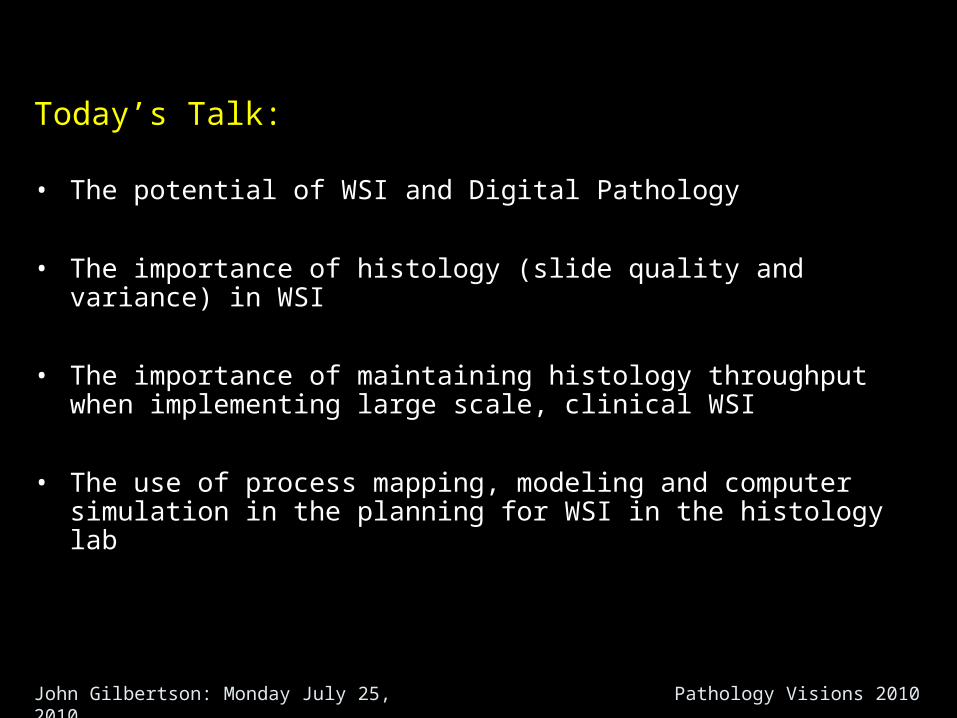
Today’s Talk:
• The potential of WSI and Digital Pathology
• The importance of histology (slide quality and variance) in WSI
• The importance of maintaining histology throughput when implementing large scale, clinical WSI
• The use of process mapping, modeling and computer simulation in the planning for WSI in the histology lab
John Gilbertson: Monday July 25, 2010 Pathology Visions 2010
John Gilbertson: Monday July 25, 2010 Pathology Visions 2010
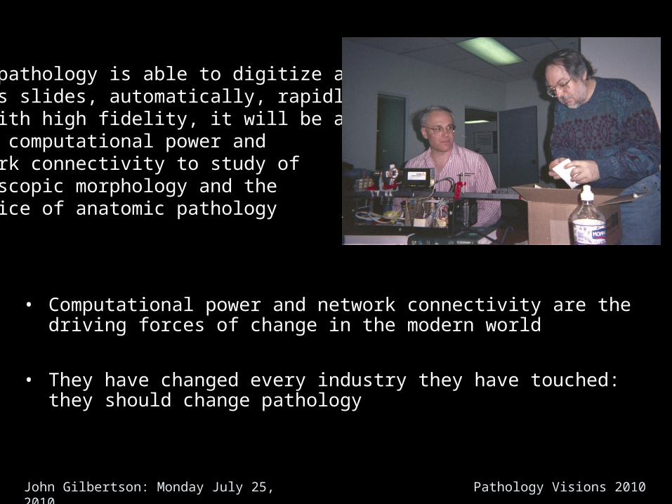
• Computational power and network connectivity are the driving forces of change in the modern world
• They have changed every industry they have touched: they should change pathology
When pathology is able to digitize allof its slides, automatically, rapidlyand with high fidelity, it will be able toapply computational power andnetwork connectivity to study ofmicroscopic morphology and thepractice of anatomic pathology
John Gilbertson: Monday July 25, 2010 Pathology Visions 2010
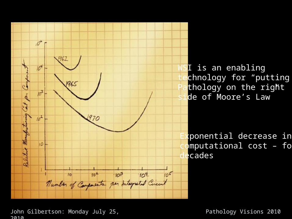
WSI is an enablingtechnology for “putting Pathology on the rightside of Moore’s Law”
Exponential decrease incomputational cost – fordecades
John Gilbertson: Monday July 25, 2010 Pathology Visions 2010
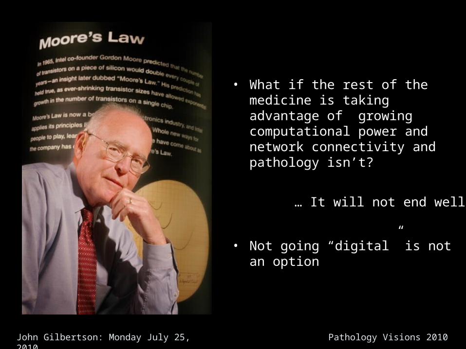
• What if the rest of the medicine is taking advantage of growing computational power and network connectivity and pathology isn’t?
… It will not end well
• Not going “digital” is not an option
John Gilbertson: Monday July 25, 2010 Pathology Visions 2010
But
John Gilbertson: Monday July 25, 2010 Pathology Visions 2010
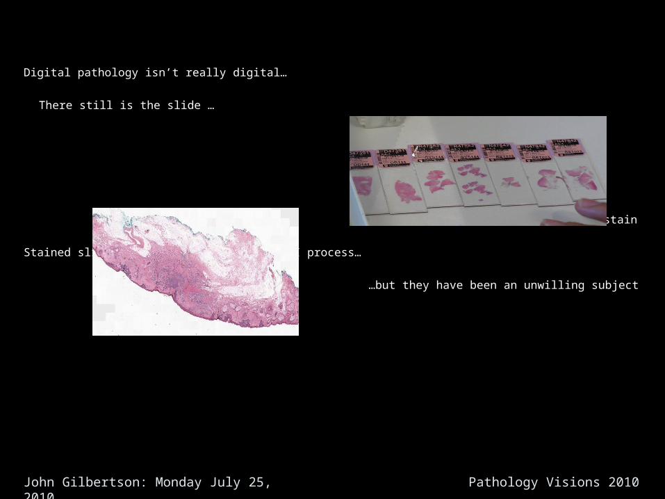
Digital pathology isn’t really digital…
There still is the slide …
…and the stain
Stained slides are the subject of the WSI process…
…but they have been an unwilling subject
John Gilbertson: Monday July 25, 2010 Pathology Visions 2010
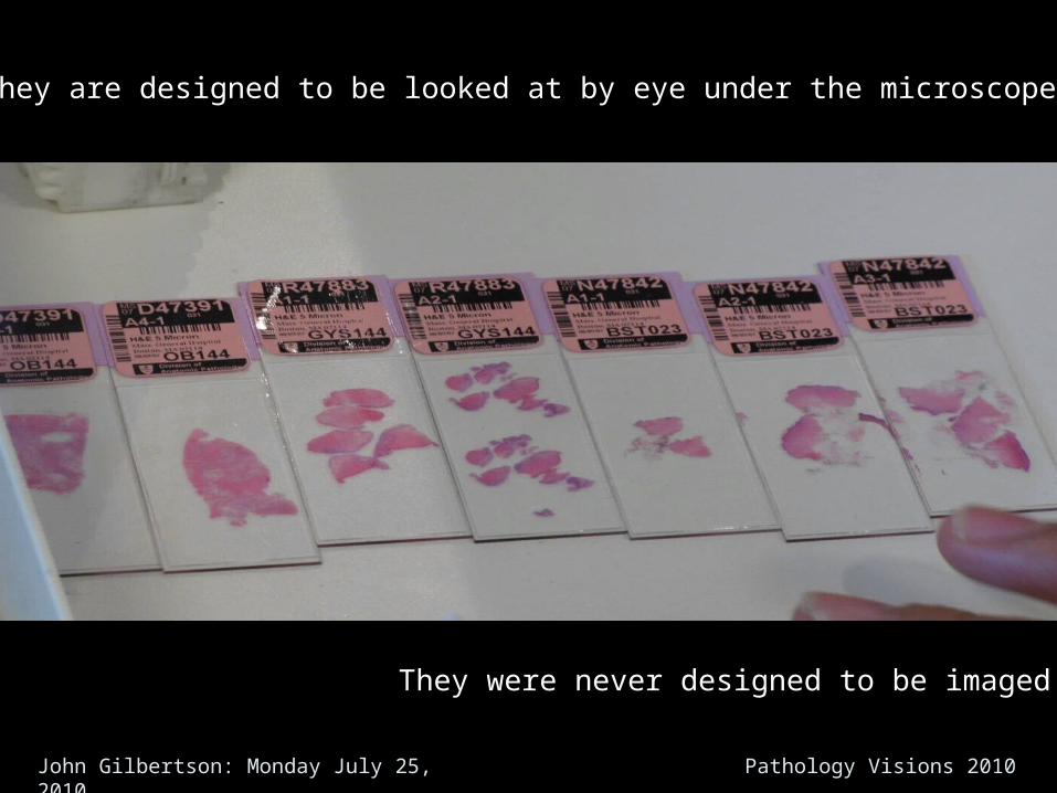
They are designed to be looked at by eye under the microscope
They were never designed to be imaged
John Gilbertson: Monday July 25, 2010 Pathology Visions 2010
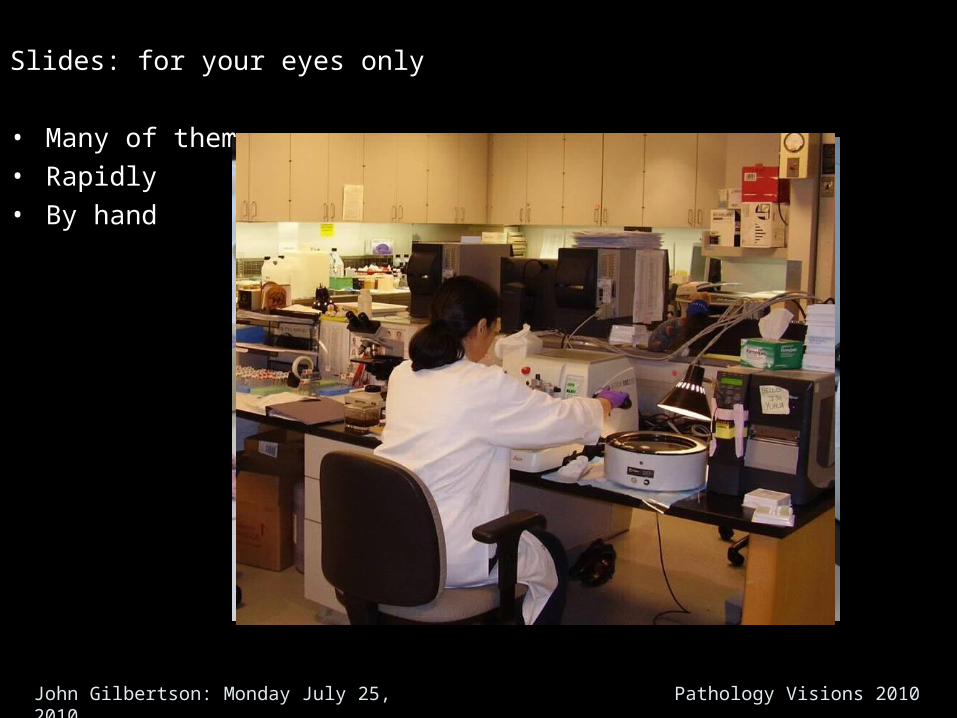
Slides: for your eyes only
• Many of them• Rapidly• By hand
John Gilbertson: Monday July 25, 2010 Pathology Visions 2010
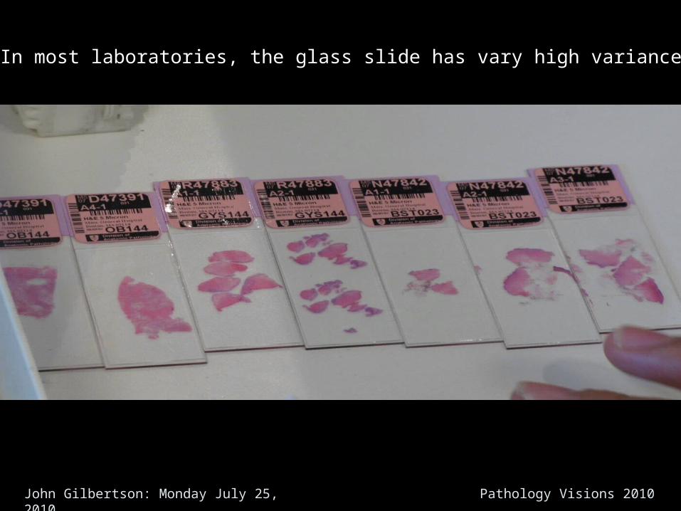
In most laboratories, the glass slide has vary high variance
John Gilbertson: Monday July 25, 2010 Pathology Visions 2010
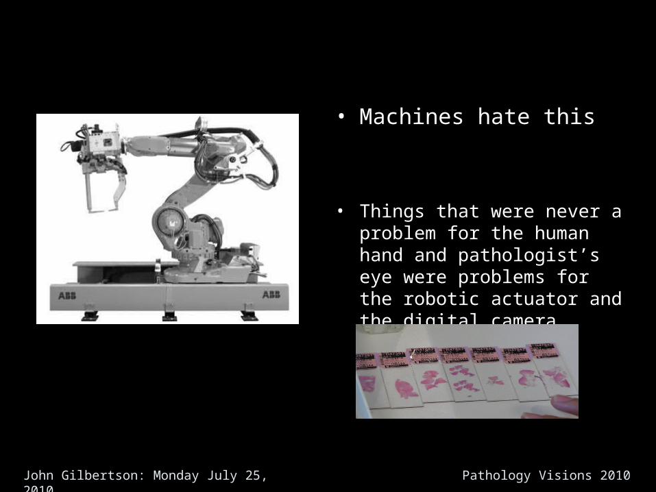
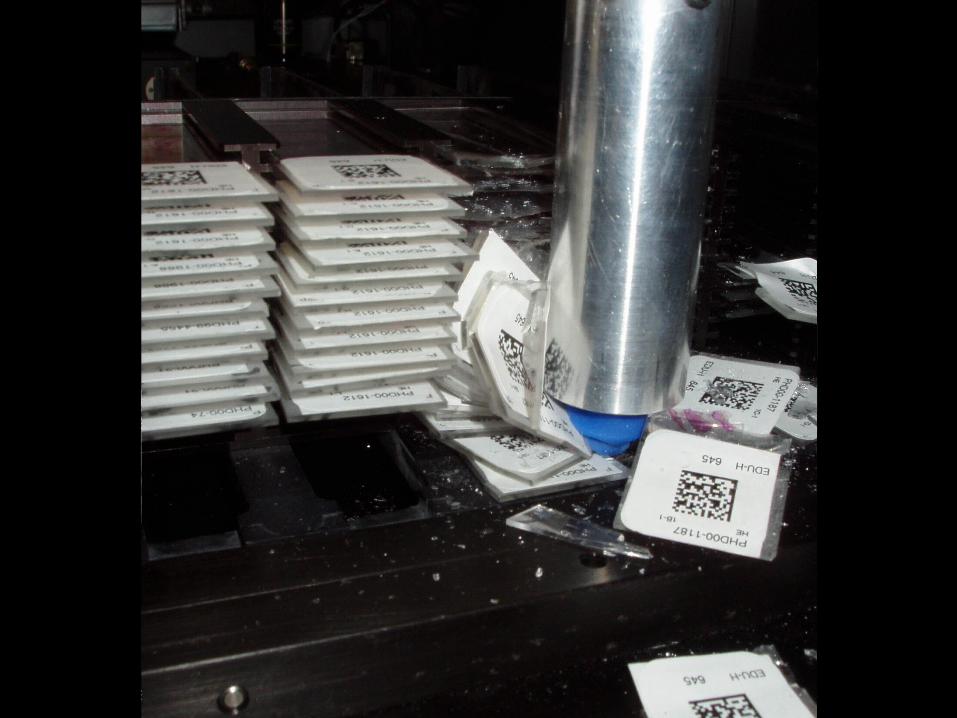
• Machines hate this
• Things that were never a problem for the human hand and pathologist’s eye were problems for the robotic actuator and the digital camera
John Gilbertson: Monday July 25, 2010 Pathology Visions 2010
John Gilbertson: Monday July 25, 2010 Pathology Visions 2010
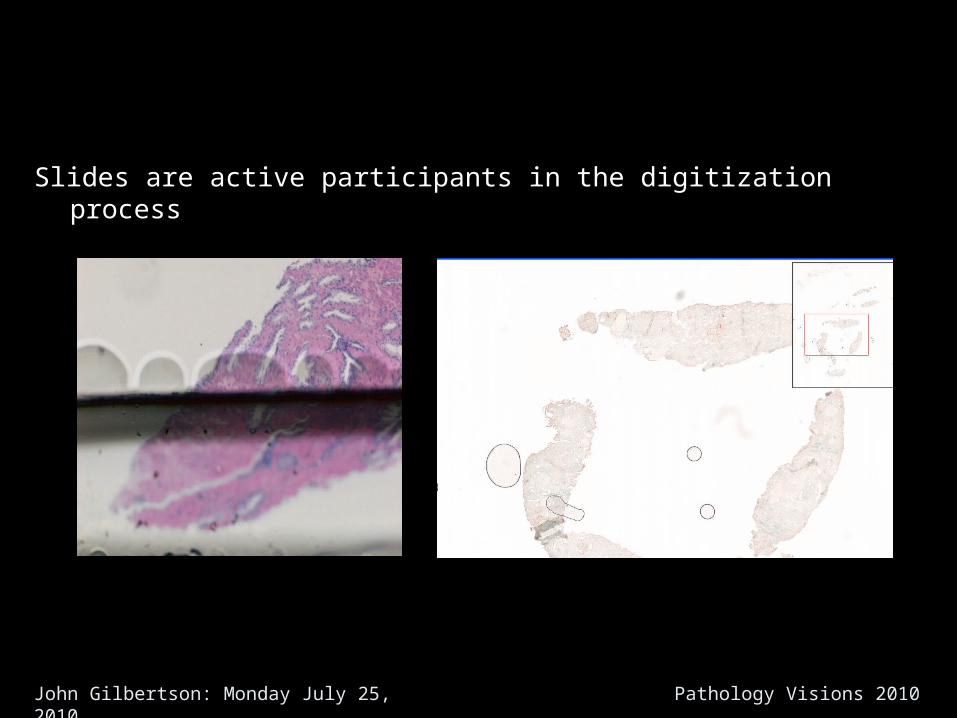
Slides are active participants in the digitization process
John Gilbertson: Monday July 25, 2010 Pathology Visions 2010
Slide variance is a major factor in imaging failures and imaging speed
John Gilbertson: Monday July 25, 2010 Pathology Visions 2010
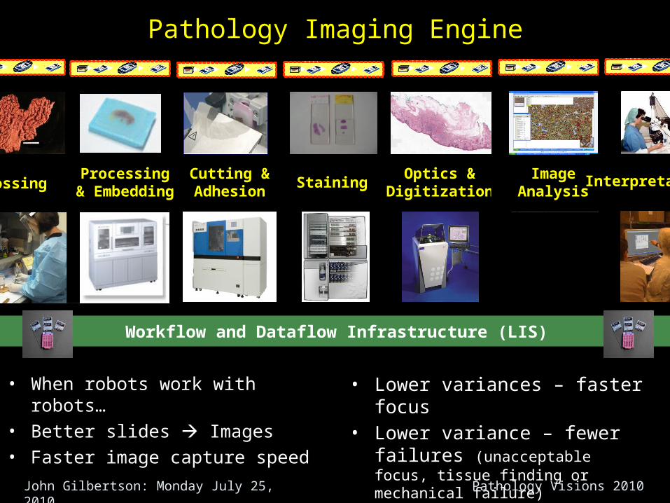
• When robots work with robots…• Better slides Images• Faster image capture speed
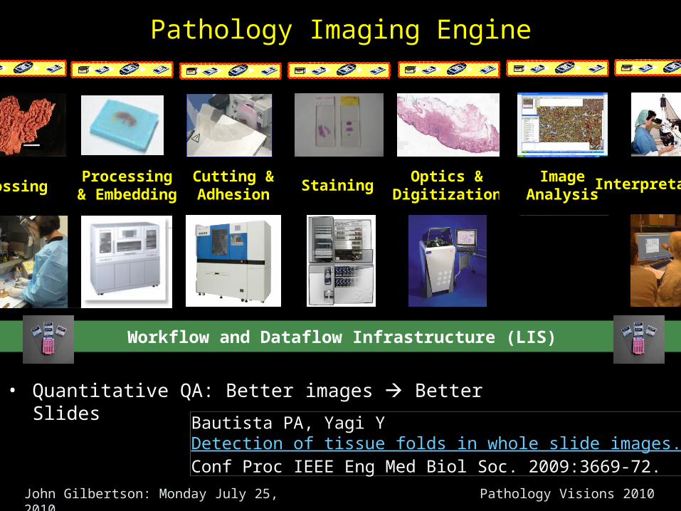
Staining Optics &
Digitization
Workflow and Dataflow Infrastructure (LIS)
Cutting &Adhesion
Processing& Embedding
ImageAnalysisGrossing Interpretation
• Lower variances – faster focus• Lower variance – fewer failures
(unacceptable focus, tissue finding or mechanical failure)
Pathology Imaging Engine
John Gilbertson: Monday July 25, 2010 Pathology Visions 2010
John Gilbertson: Monday July 25, 2010 Pathology Visions 2010
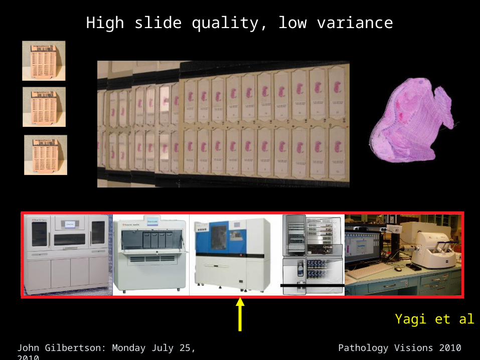
High slide quality, low variance
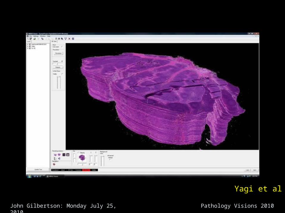
Yagi et al
John Gilbertson: Monday July 25, 2010 Pathology Visions 2010
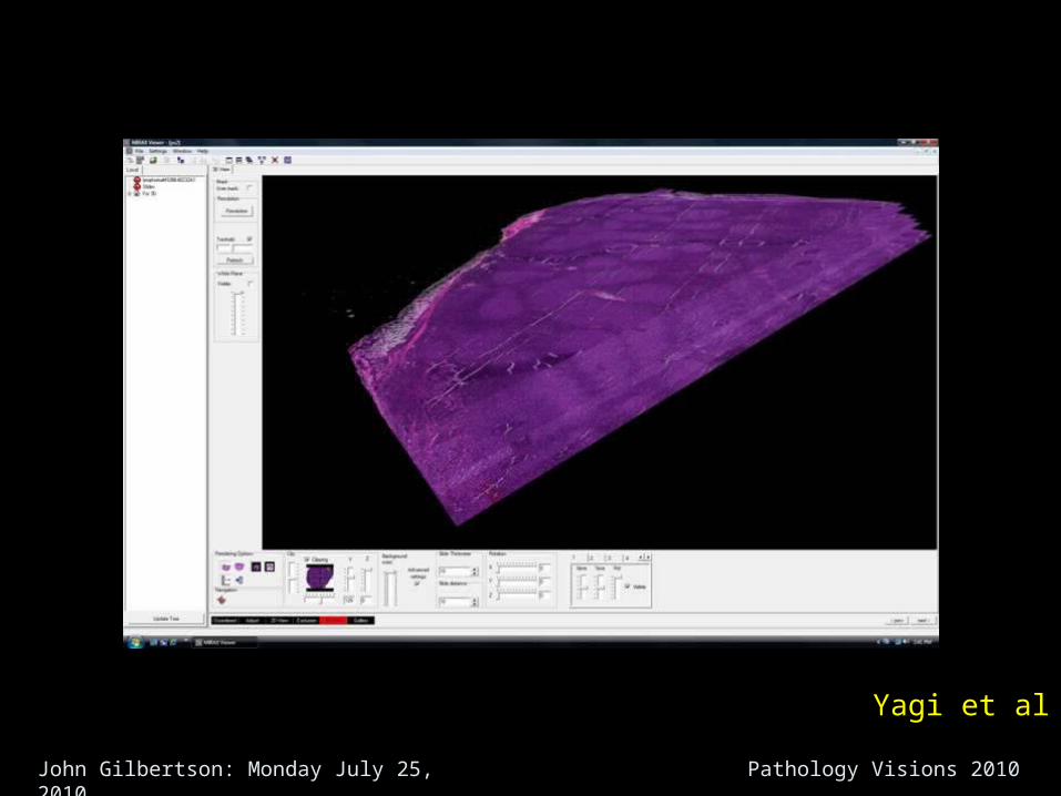
Yagi et al
John Gilbertson: Monday July 25, 2010 Pathology Visions 2010
Yagi et al
John Gilbertson: Monday July 25, 2010 Pathology Visions 2010
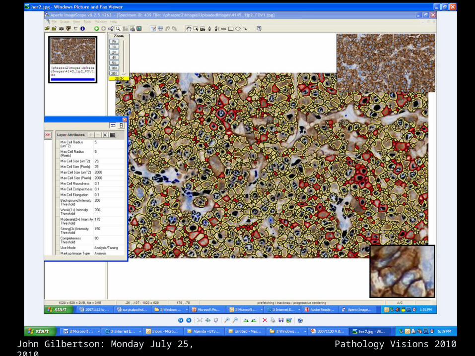
• Quantitative QA: Better images Better Slides
Staining Optics &
Digitization
Workflow and Dataflow Infrastructure (LIS)
Cutting &Adhesion
Processing& Embedding
ImageAnalysisGrossing Interpretation
Bautista PA, Yagi YDetection of tissue folds in whole slide images.Conf Proc IEEE Eng Med Biol Soc. 2009:3669-72.
Pathology Imaging Engine
John Gilbertson: Monday July 25, 2010 Pathology Visions 2010
There is a trend to incorporate robots and quantitative, Imaging-based QA at every step of the histology process
This is great for imaging, histology and anatomic pathology
John Gilbertson: Monday July 25, 2010 Pathology Visions 2010
Throughput
John Gilbertson: Monday July 25, 2010 Pathology Visions 2010
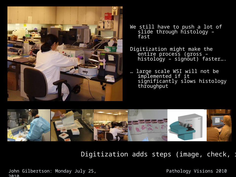
Digitization adds steps (image, check, redo)
We still have to push a lot of slide through histology – fast
Digitization might make the entire process (gross – histology – signout) faster….
… large scale WSI will not be implemented if it significantly slows histology throughput
John Gilbertson: Monday July 25, 2010 Pathology Visions 2010
Anatomic Pathology Workflow
• We have a real interest in AP productivity– Our pathologist’s time is the more valuable resource we have– This is true world wide– Potential Market is increasing…– Case complexity is increasing…– Need for documentation is increasing…– Research opportunities are increasing…– Pathology satisfaction is declining…
– High turnover in Histology Labs– Aging infrastructure
John Gilbertson: Monday July 25, 2010 Pathology Visions 2010
Anatomic Pathology Workflow
• We have a specific interest in AP productivity– Sunquest collaboration requires the implementation of a new AP
LIS– Axiom: Bad workflow eats good software for lunch– Axiom: Don’t prop up bad workflow with new software
• All these things give us a very good excuse to examine our workflow (and infrastructure)
John Gilbertson: Monday July 25, 2010 Pathology Visions 2010
Infrastructure to study/improve productivity
• Green belts, analysts, techs and engineers (Lean, six sigma, FMEA)
• Mapping: What is our baseline workflow’
• Modeling: Include rates, numbers, times (manually initially)
• Populated Models: Asset tracking (2011), routing and station protocols (process steps) (2012)
• Process Laboratory: “A laboratory to study the laboratory”
• Simulation Software: “What if we make a change”
• Clinical Laboratory: “Beta Testing”
• Compare: Scientifically compare the pre- and post states
John Gilbertson: Monday July 25, 2010 Pathology Visions 2010
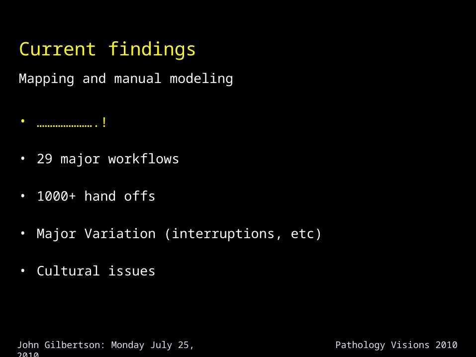
Current findings
Mapping and manual modeling
• ………………….!
• 29 major workflows
• 1000+ hand offs
• Major Variation (interruptions, etc)
• Cultural issues
John Gilbertson: Monday July 25, 2010 Pathology Visions 2010
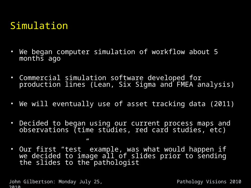
Simulation
• We began computer simulation of workflow about 5 months ago
• Commercial simulation software developed for production lines (Lean, Six Sigma and FMEA analysis)
• We will eventually use of asset tracking data (2011)
• Decided to began using our current process maps and observations (time studies, red card studies, etc)
• Our first “test” example, was what would happen if we decided to image all of slides prior to sending the slides to the pathologist
John Gilbertson: Monday July 25, 2010 Pathology Visions 2010
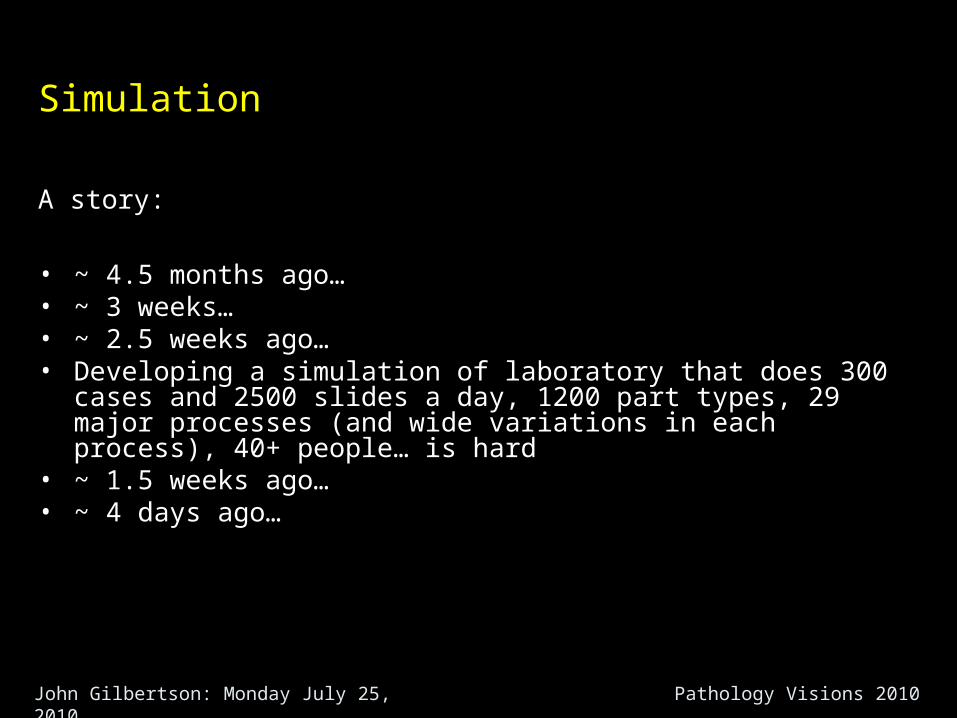
Simulation
A story:
• ~ 4.5 months ago…• ~ 3 weeks…• ~ 2.5 weeks ago…• Developing a simulation of laboratory that does 300 cases and 2500
slides a day, 1200 part types, 29 major processes (and wide variations in each process), 40+ people… is hard
• ~ 1.5 weeks ago…• ~ 4 days ago…
John Gilbertson: Monday July 25, 2010 Pathology Visions 2010
Simulation
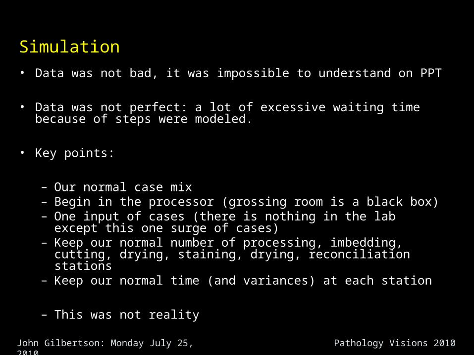
• Data was not bad, it was impossible to understand on PPT
• Data was not perfect: a lot of excessive waiting time because of steps were modeled.
• Key points:
– Our normal case mix– Begin in the processor (grossing room is a black box)– One input of cases (there is nothing in the lab except this one
surge of cases)– Keep our normal number of processing, imbedding, cutting,
drying, staining, drying, reconciliation stations– Keep our normal time (and variances) at each station
– This was not reality
John Gilbertson: Monday July 25, 2010 Pathology Visions 2010
Simulation
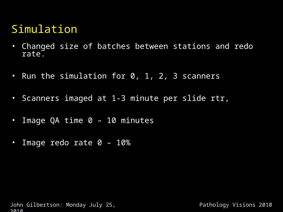
• Changed size of batches between stations and redo rate.
• Run the simulation for 0, 1, 2, 3 scanners
• Scanners imaged at 1-3 minute per slide rtr,
• Image QA time 0 – 10 minutes
• Image redo rate 0 – 10%
John Gilbertson: Monday July 25, 2010 Pathology Visions 2010
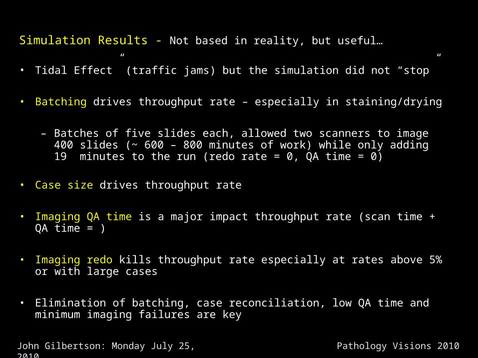
Simulation Results - Not based in reality, but useful…
• Tidal Effect” (traffic jams) but the simulation did not “stop”
• Batching drives throughput rate – especially in staining/drying
– Batches of five slides each, allowed two scanners to image 400 slides (~ 600 – 800 minutes of work) while only adding 19 minutes to the run (redo rate = 0, QA time = 0)
• Case size drives throughput rate
• Imaging QA time is a major impact throughput rate (scan time + QA time = )
• Imaging redo kills throughput rate especially at rates above 5% or with large cases
• Elimination of batching, case reconciliation, low QA time and minimum imaging failures are key
John Gilbertson: Monday July 25, 2010 Pathology Visions 2010
Conclusions
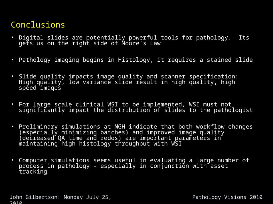
• Digital slides are potentially powerful tools for pathology. Its gets us on the right side of Moore’s Law
• Pathology imaging begins in Histology, it requires a stained slide
• Slide quality impacts image quality and scanner specification: High quality, low variance slide result in high quality, high speed images
• For large scale clinical WSI to be implemented, WSI must not significantly impact the distribution of slides to the pathologist
• Preliminary simulations at MGH indicate that both workflow changes (especially minimizing batches) and improved image quality (decreased QA time and redos) are important parameters in maintaining high histology throughput with WSI
• Computer simulations seems useful in evaluating a large number of process in pathology – especially in conjunction with asset tracking
John Gilbertson: Monday July 25, 2010 Pathology Visions 2010
Conclusions
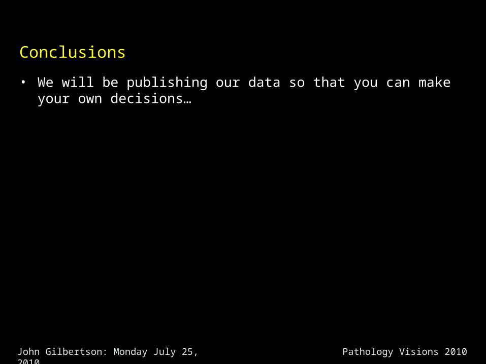
• We will be publishing our data so that you can make your own decisions…