Embed Size (px)
Citation preview
651
Review
ISSN 1758-427210.2217/IJR.10.62 © 2010 Future Medicine Ltd Int. J. Clin. Rheumatol. (2010) 5(6), 651–658
The pathogenic role of rheumatoid factor in rheumatoid arthritis
The first autoantibody in rheumatoid arthritis (RA), rheumatoid factor (RF), was described by Waaler in 1940, who reported the hemagglutinating activity of a serum from a patient with RA [1]. RFs were named by Pike in 1949 due to their association with RA [2] and were later found to be autoantibodies targeting the Fc region of IgG in the g2–3 cleft. Although frequently observed in diseased conditions including RA, RFs are also observed in normal subjects [3], which implies that they have both pathological and physiological roles that depend on the circumstances under which they are induced. In this article, we provide an overview of current knowledge regarding the induction of pathologic RFs and discuss the mechanisms by which RFs contribute to the RA disease process.
Characteristics of RFs & RF-producing B cells in normal individuals & in RA patientsRheumatoid factors are normally produced during secondary immune responses against infections or immunizations [4–6]. This finding is in line with the observation that RFs are frequently found in chronic infectious conditions [7] and that immunecomplexed rather than monomeric IgG is an efficient inducer of RF response [8]. In addition, RFs themselves bind with much greater affinity to aggregated IgG or immune complex than to monomeric IgG [9–11]. RFs produced during the secondary immune response are generally polyclonal IgMs of lowaffinity, which resemble the characteristics of
natural antibodies produced by CD5+ B1 cells (B1 cells). B1 cells are a subclass of B cells that spontaneously secrete IgM antibodies (natural antibodies) of lowaffinity and polyspecificity. They are different from conventional B2 cells in that they do not undergo somatic hypermutation and memory formation. Bcell subsets capable of secreting RFs are likely to include both B1 and B2 cells, but the culprit subset may be variable depending on the biological condition that induces RF production. B1 cells have been shown to compose a major fraction that secretes large amounts of IgM RFs against the stimulation with Staphylococcus aureus [12]; RF secretion from B1 cells was found to occur at comparable levels in RA patients and normal subjects. Thus, it seems that the production of polyclonal lowaffinity IgM RFs is not disease specific, but rather a physiological response that may serve in host defense by facilitating the form ation and clearance of immune complexes. However, larger numbers of B1 cells are present in RA patients compared with normal subjects [13], and a correlation between peripheral blood B1 cell frequency and RF titer has been reported in RA [14]. Therefore, the significance of these findings remains to be resolved, although these findings may mean that RA patients are more readily triggered to produce RF and, thus, prone to RA development. Alternatively, these could be secondary findings unrelated to disease pathogenesis, because in patients with infectious diseases, RF production is not correlated with the development of arthritis.
Rheumatoid factors (RFs) are the first autoantibodies described in rheumatoid arthritis (RA), which target the Fc region of IgG. Since their discovery, RFs have been the subject of extensive studies not just because of their association with RA, but also because they serve as an excellent model for the regulation and induction of disease-related autoantibodies. Although RFs are normally induced during secondary immune responses, isotype switching and affinity maturation are confined to RA patients and are not observed in normal individuals. Therefore, the emergence of high-affinity RFs, which are believed to depend on T-cell help, is under strict control in normal subjects, whereas these control mechanisms appear to be bypassed in RA. In this article, we provide an overview of the current knowledge of how pathologic RFs are induced and discuss the mechanisms by which RFs contribute to RA disease process.
keywoRds: immune complex n rheumatoid arthritis n rheumatoid factor n Toll-like receptor
Yeong Wook Song†1 & Eun Ha Kang2
1Division of Rheumatology, Department of Internal Medicine, Seoul National University College of Medicine, Seoul, Republic of Korea 2Division of Rheumatology, Department of Internal Medicine, Seoul National University Bundang Hospital, Seongnam-si, Republic of Korea †Author for correspondence: Department of Internal Medicine, Seoul National University Hospital, 28 Yongun-dong, Chongno-gu, Seoul, 110–744, Republic of Korea Tel.: +82 220 722 234 Fax: +822 762 9662 [email protected]
Int. J. Clin. Rheumatol. (2010) 5(6)652 future science group
Review Song & Kang Rheumatoid factors in rheumatoid arthritis Review
Nucleotide sequence ana lysis of RF genes from normal immunized hosts and R A patients has shown that RFs in normal individuals are frequently encoded by a limited set of IgV genes, do not appear to switch isotype and have few replacement mutations in complementary determinant regions, which results in little affinity maturation [15]. On the other hand, the synovial RFs of RA patients are encoded by a wider range of IgV genes, undergo isotype switching and exhibit extensive nucleotide changes in complementary determinant regions, which lead to affinity maturation [15]. Although the precise phenotypes of RFproducing B cells in lymph nodes and tissue germinal centers (GCs) have not been determined in RA patients, the presence of accumulated somatic mutations and of isotype switching makes it plausible that pathologic RFs are produced by Tcelldriven B2 cells in RA.
How are RFs produced?The mechanisms responsible for the activation of RFproducing B cells have been elusive for decades. In normal individuals, RFproducing B cells are not activated despite abundant self IgGs. The f inding that monomeric IgGs, unlike immunecomplexed IgGs, are poor inducers of RF production suggests that effective Bcell receptor (BCR) crosslinking is critical to transfer the Bcell activation signal. However, it has been shown that Bcell activating ability depends not only on the immune complex formation, but also on the nature of antigens contained in the complex in the sense that the ligation of Tolllike receptors (TLRs) is critical to trigger RF production in addition to BCR ligation [16]; both B1 and B2 cells express TLRs, particularly TLR1 and 9 [17,18]. This finding represents a good example of how TLRs provide the link between innate and adaptive immunity. The interesting aspects of the simultaneous coligation of BCR and TLR are that TLRs are expressed on memory B cells and RFs are produced via TLR ligation alone in memory B cells [19]. However, in order to generate highaffinity RFs, particularly of IgG or IgA isotypes, Tcell help might be required.
T-cell requirements for RF productionIt has long been believed that Tcell help is crucial for the production of pathologic RFs that have undergone isotype switching and aff inity maturation. Studies on transgenic
mice expressing human IgM demonstrated that RFproducing B cells are deleted in the absence of Tcell help and that complete Bcell activation involving GC formation only occurs with Tcell help [20]. Another finding that further supports the role of T cells in the production of pathologic RFs is that a critical contact residue of RF from a RA patient was found to be derived from somatic hypermutation [21]. To date, Tcell clones reactive to autologous IgG have not been detected in RA patients, most likely owing to clonal deletion. T cells that infiltrate RA synovium have been shown to be polyclonal and to lack specificity for any particular autoantigen [22,23]. Nevertheless, any T cells that recognize antigenderived peptides presented by RFproducing B cells have the potential to activate these B cells; the ability of RFexpressing B cells to take up immune complexes and to present trapped antigens to ubiquitous T cells [24] may enable these cells to bypass the need for specific Tcell help, and could, ultimately, lead to the emergence of autoreactive T cells that can trigger RF synthesis. On the contrary, recent studies on the role of TLRs in Bcell activation and differentiation harbor questions whether Tcell help is indispensible (or whether TLR signaling is vital) for the production of pathologic RFs. Emerging data suggest that TLR and BCR coactivation induces isotope switching without Tcell help [25–27], although little is known regarding the effect of TLR signaling in somatic hypermutation and affinity maturation. In addition, TLR engagement seems to provide an essential signal for optimal antibody response even in the presence of T cells [28,29]. Therefore, highaffinity RFs might be triggered in the absence of Tcell help in the initial phase of RA, but their production in established RA is likely to involve both Tcell help and TLR signaling in a synergistic manner.
How is tolerance to self IgG lost in RA?Evidence shows that RFproducing B cells are highly efficient antigenpresenting cells for multivalent, immunecomplexed antigens [24]. Although T cells reactive to human IgG are deleted by a Tcell tolerance mechanism [20], T cells that recognize antigenderived peptides presented by RFproducing B cells have the potential to activate these B cells. Therefore, the generation of predominantly IgM RFs during secondary immune responses (when Ig isotype switching usually occurs) suggests that
Review Song & Kang
www.futuremedicine.com 653future science group
Rheumatoid factors in rheumatoid arthritis Review
a strict control mechanism prevents the emergence of highaffinity RFs. Affinitydependent Bcell deletion has been reported to censor auto reactive B cells during GC reaction [30,31]. However, much remains to be determined regarding how highaff inity RFproducing B cells escape tolerance mechanisms.
It has been shown that the synovial membranes of RA patients are extrafollicular secondary lymphoid organs that provide a microenvironment for isotype switching and the somatic mutation of RFproducing B cells [32]. William et al. have reported that the somatic hypermutation in an autoimmune response could occur elsewhere to the conventional GCs in an autoimmune murine model [33]. Based on the concept that censoring mechanisms are unique to GCs, these authors proposed that B cells, which have undergone hypermutation outside GCs might escape the selfcensoring mechanism that selectively eliminates autoreactive Bcell clones and that they are more prone to be autoreactive. Highaffinity RFs are locally produced in RA by B cells in the inf lamed synovium [34,35] where lymphoid folliclelike structures are often found [36]. GCs have been found in approximately 25% of the RA synovial samples examined, with the remaining samples showing either diffuse or aggregated infiltrates of T and B cells [36]. Although a substantial amount of evidence indicates that GCs in RA synovium are the primary sites for the affinity maturation of autoantibodies [34,35,37], neither the clinical phenotype of RA nor the presence of RFs is associated with the presence of GCs [38,39]. Therefore, it remains to be resolved whether selfcensoring mechanisms in synovial GCs operate in the same manner as in nodal GCs.
Contributions of RFs to the disease process in RAIgM RFs are the major RF species in RA and are detected in 60–80% of RA patients [40]. Furthermore, RF specificity to RA is increased at high titers (e.g., IgM RF ≥50 IU/ml) and with IgA isotypes [40,41]. High titer RFs and IgA isotypes are also associated with radiologic erosion, extraarticular manifestations and, thus, poorer outcomes [40,42–45]. The association between high titer RF status and a poor prognosis indicates that RFs may have a role in the pathogenesis of RA. In addition, RF has proven to be a useful disease marker of RA, and is included in the 1987 American Rheumatism Association classification criteria for RA [46]
and in the recently proposed American College of Rheumatology (ACR)/European League Against Rheumatism (EULAR) criteria for RA [47].
The physiological roles of RFs under normal conditions have been shown [48–50]:
� To enhance immune complex clearance by increasing complex avidity and size;
� To help B cells uptake immune complex and, thereby, effectively present antigens to T cells;
� To facilitate complement fixation by binding to IgGcontaining immune complexes.
Thus, highaffinity and hightiter RFs in RA synovial fluid are believed to exert such functions in a pathologic manner; the capacity of RFs to enhance immune complex formation [48,51] may not only grant arthritogenicity to RF itself, but also potentiate the arthritogenicities of other autoantibodies, including anticitrullinated protein antibodies (ACPAs). Moreover, several studies have shown that immune complexes isolated from RA patients induce TNFa or other cytokines from peri pheral blood mononuclear cells via Fcg receptor IIa engagement [52–54]. In particular, Mathsson et al. have demonstrated that both serum and synovial fluid RF levels of RA patients are correlated with polyethylene glycolprecipitated synovial fluid IgG levels, the latter being correlated with TNFa levels of peripheral blood mononuclear cells induced by polyethylene glycolprecipitated synovial fluid immune complexes; the immune complexinduced TNFa levels were significantly higher in RFpositive patients than RFnegative patients [54]. They have also shown that serum/synovial fluid ACPA levels were not correlated with serum/synovial fluid polyethylene glycolprecipitated IgG levels or with in vitro induced TNFa levels. On the other hand, immune complexes formed with ACPAs and citrullinated proteins have been shown to induce TNFa from peripheral blood mononuclear cells or in vitro differentiated macrophages [52]. Therefore, both RFs and ACPAs seem to contribute to RA severity.
Although clinical as well as biological evidence indicate that RFs are important players in RA pathogenesis, there has been no clear evidence that RFs are involved in the initial events triggering the disease process of RA rather than themselves being triggered by RA. Unlike the relatively low disease specificity of RF for RA, it has been shown that ACPA production in RA patients is a diseasespecif ic humoral
Int. J. Clin. Rheumatol. (2010) 5(6)654 future science group
Review Song & Kang Rheumatoid factors in rheumatoid arthritis Review
auto immune response [55–57]. It has been demonstrated in studies that retrospectively utilized preclinical serum samples that both RF and ACPAs are present in the sera of RA patients months to years prior to disease onset [58–60]. Interestingly, the majority of RFpositive RA patients are also positive for ACPA [55], and considering that immune complexes are strong inducers of RFs, immune complexes containing citrullinated antigens and ACPA might facilitate RF production. In support of this notion, the onset of RF was found by Nielen et al. to follow the onset of ACPA by a few years when RF and ACPA titers were serially examined during the preclinical period [60]. Furthermore, RF titers in ACPApositive RA patients were found to be much higher than those in ACPAnegative patients in their study and vice versa. Therefore, a vicious cycle between ACPA and RF is expected; ACPAcontaining immune complexes induce RF production and RFs, in return, amplify the inflammatory response by
facilitating further immune complex formation, complement fixation, cytokine release and tissue damage. Ultimately, exaggerated inflammation will result in tissue citrullination and more ACPA production.
A number of studies have proved that effective diseasemodifying antirheumatic drug therapy can decrease serum RF levels [61]. In particular, reduction of IgM RF levels seems to be correlated with clinical improvement [62–64]. Moreover, TNFa inhibitors have been shown to decrease serum RF levels [65–68], which was correlated with clinical improvement [65,66,68]. However, pretreatment RF positivity was not different between responders and non responders [67]. Rather, high basal levels of IgA RFs but not of ACPAs were associated with poor response to TNFa inhibitors in a recent prospective study on 131 longstanding RA patients [67]. Unlike RF levels, reduction of anticyclic citrullinated peptide levels after diseasemodifying antirheumatic drug or
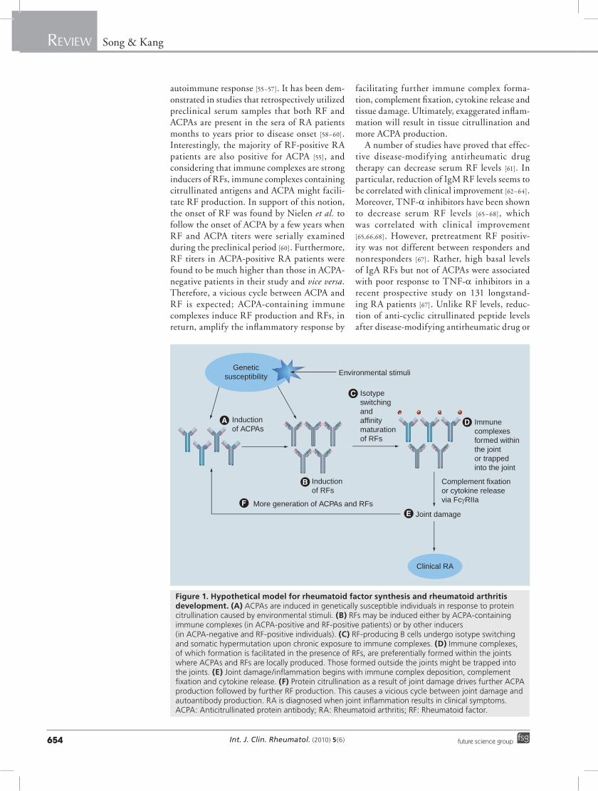
Environmental stimuli
Inductionof ACPAs
Isotypeswitchingandaffinitymaturationof RFs
Immunecomplexes formed within the jointor trappedinto the joint
Joint damageMore generation of ACPAs and RFs
Clinical RA
Geneticsusceptibility
Inductionof RFs
Complement fixation or cytokine release via FcγRIIa
Figure 1. Hypothetical model for rheumatoid factor synthesis and rheumatoid arthritis development. (A) ACPAs are induced in genetically susceptible individuals in response to protein citrullination caused by environmental stimuli. (B) RFs may be induced either by ACPA-containing immune complexes (in ACPA-positive and RF-positive patients) or by other inducers (in ACPA-negative and RF-positive individuals). (C) RF-producing B cells undergo isotype switching and somatic hypermutation upon chronic exposure to immune complexes. (d) Immune complexes, of which formation is facilitated in the presence of RFs, are preferentially formed within the joints where ACPAs and RFs are locally produced. Those formed outside the joints might be trapped into the joints. (e) Joint damage/inflammation begins with immune complex deposition, complement fixation and cytokine release. (F) Protein citrullination as a result of joint damage drives further ACPA production followed by further RF production. This causes a vicious cycle between joint damage and autoantibody production. RA is diagnosed when joint inflammation results in clinical symptoms. ACPA: Anticitrullinated protein antibody; RA: Rheumatoid arthritis; RF: Rheumatoid factor.
Review Song & Kang
www.futuremedicine.com 655future science group
Rheumatoid factors in rheumatoid arthritis Review
TNFa treatment has been controversial but reported to occur in early RA patients with less than 1 year of disease duration [64,69], which, however, was not correlated with clinical improvement. The close relationship between serum RF levels (but not ACPA levels) and clinical response to TNFa therapy might suggest that RFs play a distinguished pathologic role from that of ACPAs. Alternatively, it might suggest that autoantibodyproducing cells are differently regulated for RFs and ACPAs; a number of factors are known to be important in plasma cell survival, including cytokines such as TNFa and the cell adhesion molecule CD44 [70].
Recent advances in RA treatment include the introduction of rituximab, an antiCD20 monoclonal antibody. CD20 is expressed by Bcell precursors and mature B cells, but not by stem cells or endstage plasma cells. Rituximab is interesting in that it directly targets auto antibodyproducing B cells. Trials of rituximab have shown that the agent is highly effective at reducing RA activity [71–73]. Following Bcell depletion by rituximab, a large and rapid decrease in RF titers was observed, whereas immunoglobulin concentrations remained within normal ranges [73]. Decrease of ACPA levels tended to be delayed and modest compared with that of RF levels [74,75] and decreased levels of RFs and ACPAs were only observed in responders [74]. The different kinetics of RF and ACPA levels during rituximab treatment implies that the production of RFs is more dependent on shortlived plasma cells, while that of ACPAs on longlived plasma cells. In fact, spontaneous RF response has been shown to occur by continuous generation of shortlived plasmablasts in a transgenic animal model with autoimmune background [76]. The effect of rituximab has been more beneficial in RFpositive than in RFnegative
patients [77]. Moreover, RFs have been shown to be a better predictor of a good response to rituximab than ACPAs [78]. These findings provide sound information regarding the actual and critical roles of RFs in the patho physiology of RA and suggest that RFproducing B cells and their activation mechanisms may be important therapeutic targets in RA.
Conclusion & future perspectiveSince RF was first described, substantial evidence has been accumulated from both basic and clinical studies to suggest that RFs are key players in the pathogenesis of RA. This concept has been further corroborated by the results of recent clinical trials adopting Bcell depleting agents. The current hypothesis is that RFs are induced by immune complexes derived from diseasespecific autoantibodies, which, in turn, further potentiate immune complex formation, complement fixation, tissue damage and more diseasespecific autoantibody production (Figure 1). Since simultaneous TLR ligation is required in addition to BCR ligation to activate B cells to secrete RFs, the nature of auto antigens contained in the immune complex might be a critical factor to elicit RF production. Future studies on the mechanisms by which RFproducing B cells are activated and bypass tolerance mechanisms will help refine crucial therapeutic targets.
Financial & competing interests disclosureThis study was supported by a grant from the Korea Healthcare technology R&D Project, Ministry for Health and Welfare, Republic of Korea (A084794). The authors have no other relevant affiliations or financial involvement with any organization or entity with a financial interest in or financial conflict with the subject matter or materials discussed in the manuscript apart from those disclosed.
No writing assistance was utilized in the production of this manuscript.
executive summary
� The production of the polyclonal low-affinity IgM rheumatoid factor (RF) is not disease specific, but rather a physiological response that may serve host defenses by facilitating the formation and clearance of immune complexes.
� RF-producing B cells secrete RFs when co-ligated on Toll-like receptors together with B-cell receptors, reflecting the link between innate and adaptive immunity in the humoral immune response. T-cell help is believed to be required to enable RF-producing B cells to undergo isotype switching and somatic hypermutation, a process that is responsible for the production of high-affinity pathologic RFs in rheumatoid arthritis (RA). However, the emergence of such RFs is strictly prevented in normal individuals via tolerance mechanisms that are not completely understood.
� Although high-affinity RFs do not seem to trigger RA themselves, they are thought to contribute actively to disease severity and chronicity by enhancing immune complex formation and complement fixation; RFs induced by immune complexes derived from disease-specific autoantibodies further potentiate immune complex formation, complement fixation, tissue damage and more disease-specific autoantibody production. Therefore, RF-producing B cells and their activation mechanisms, including Toll-like receptor ligation may be important targets for RA treatment.
Int. J. Clin. Rheumatol. (2010) 5(6)656 future science group
Review Song & Kang Rheumatoid factors in rheumatoid arthritis Review
BibliographyPapers of special note have been highlighted as:n of interestnn of considerable interest
1 Waaler E: On the occurrence of a factor in human serum activating the specific agglutination of sheep red corpuscles. Acta Pathol. Microbiol. Scand. 17(2), 172–88 (1940).
2 Pike RM, Sulkin SE, Coggeshall HC: Serological reactions in rheumatoid arthritis; factors affecting the agglutination of sensitized sheep erythrocytes in rheumatidarthritis serum. J. Immunol. 63(4), 441–446 (1949).
3 Lawrence TG Jr, Williams RC Jr: Studies of human antigglobulin factors reacting with pepsindigested gglobulins. J. Exp. Med. 125(2), 233–248 (1967).
4 Welch MJ, Fong S, Vaughan J, Carson D: Increased frequency of rheumatoid factor precursor B lymphocytes after immunization of normal adults with tetanus toxoid. Clin. Exp. Immunol. 51(2), 299–304 (1983).
5 Slaughter L, Carson DA, Jensen FC, Holbrook TL, Vaughan JH: In vitro effects of Epstein–Barr virus on peripheral blood mononuclear cells from patients with rheumatoid arthritis and normal subjects. J. Exp. Med. 148(5), 1429–1434 (1978).
6 Izui S, Eisenberg RA, Dixon FJ: IgM rheumatoid factors in mice injected with bacterial lipopolysaccharides. J. Immunol. 122(5), 2096–2102 (1979).
7 Williams RC: Rheumatoid factors in subacute bacterial endocarditis and other infectious diseases. Scand. J. Rheumatol. (Suppl. 75), 300–308 (1988).
8 Nemazee DA: Immune complexes can trigger specific, T celldependent, autoantiIgG antibody production in mice. J. Exp. Med. 161(1), 242–256 (1985).
9 Johnson PM, Faulk WP: Rheumatoid factor: its nature, specificity, and production in rheumatoid arthritis. Clin. Immunol. Immunopathol. 6(3), 414–430 (1976).
10 Normansell DE: Antiglobulins in rheumatoid arthritis sera. II. The reactivity of antiglobulin rheumatoid factors with altered Gglobulin. Immunochemistry 8(7), 593–602 (1971).
11 Gorgani NN, Altin JG, Parish CR: Histidinerich glycoprotein prevents the formation of insoluble immune complexes by rheumatoid factor. Immunology 98(3), 456–463 (1999).
12 Hardy RR, Hayakawa K, Shimizu M, Yamasaki K, Kishimoto T: Rheumatoid factor secretion from human Leu1+ B cells. Science 236(4797), 81–83 (1987).
13 Moynier M, Abderrazik M, Didry C, Sany J, Brochier J: The B cell repertoire in rheumatoid arthritis. III. Preferential homing of rheumatoid factorproducing B cell precursors in the synovial fluid. Arthritis Rheum. 35(1), 49–54 (1992).
14 Youinou P, Mackenzie L, Katsikis P et al.: The relationship between CD5expressing B lymphocytes and serologic abnormalities in rheumatoid arthritis patients and their relatives. Arthritis Rheum. 33(3), 339–348 (1990).
15 Mageed RA, Børretzen M, Moyes SP, Thompson KM, Natvig JB: Rheumatoid factor autoantibodies in health and disease. Ann. NY Acad. Sci. 815, 296–311 (1997).
nn Excellent review that compares the structural, functional and genetic aspects of rheumatoid factors (RFs) from rheumatoid arthritis (RA) patients and from normal subjects, and presents a concept that strict control mechanisms operate to prevent the emergence of high-affinity RFs in normal individuals.
16 Leadbetter EA, Rifkin IR, Hohlbaum AM, Beaudette BC, Shlomchik MJ, MarshakRothstein A: ChromatinIgG complexes activate B cells by dual engagement of IgM and Tolllike receptors. Nature 416(6881), 603–607 (2002).
nn Establishes a critical link between the innate and adaptive immune systems by demonstrating that the effective activation of RF-producing B cells requires simultaneous engagement of Toll-like receptor and B-cell receptor.
17 Hornung V, Rothenfusser S, Britsch S et al.: Quantitative expression of Tolllike receptor 1–10 mRNA in cellular subsets of human peripheral blood mononuclear cells and sensitivity to CpG oligodeoxynucleotides. J. Immunol. 168(9), 4531–4537 (2002).
18 Dasari P, Nicholson IC, Hodge G, Dandie GW, Zola H: Expression of Tolllike receptors on B lymphocytes. Cell. Immunol. 236(1–2), 140–145 (2005).
19 Bernasconi NL, Traggiai E, Lanzavecchia A: Maintenance of serological memory by polyclonal activation of human memory B cells. Science 298(5601), 2199–2202 (2002).
20 Tighe H, Warnatz K, Brinson D et al.: Peripheral deletion of rheumatoid factor B cells after abortive activation by IgG. Proc. Natl Acad. Sci. USA 94(2), 646–651 (1997).
n Presents one of the mechanisms that prevent the emergence of high-affinity RFs in normal individuals; exposure of transgenic mice expressing human IgM RFs to soluble human IgG in the absence of T-cell help resulted in antigen-specific B-cell deletion.
21 Corper AL, Sohi MK, Bonagura VR et al.: Structure of human IgM rheumatoid factor Fab bound to its autoantigen IgG Fc reveals a novel topology of antibodyantigen interaction. Nat. Struct. Biol. 4(5), 374–381 (1997).
22 Schmidt D, Goronzy JJ, Weyand CM: CD4+ CD7 CD28 T cells are expanded in rheumatoid arthritis and are characterized by autoreactivity. J. Clin. Invest. 97(9), 2027–2037 (1996).
23 Ziff M: Role of endothelium in the pathogenesis of rheumatoid synovitis. Int. J. Tissue. React. 15(3), 135–137 (1993).
24 Roosnek E, Lanzavecchia A: Efficient and selective presentation of antigenantibody complexes by rheumatoid factor B cells. J. Exp. Med. 173(2), 487–489 (1991).
25 He B, Qiao X, Cerutti A: CpG DNA induces IgG class switch DNA recombination by activating human B cells through an innate pathway that requires TLR9 and cooperates with IL10. J. Immunol. 173(7), 4479–4491 (2004).
26 Groom JR, Fletcher CA, Walters SN et al.: BAFF and MyD88 signals promote a lupuslike disease independent of T cells. J. Exp. Med. 204(8), 1959–1971 (2007).
27 Poeck H, Wagner M, Battiany J et al.: Plasmacytoid dendritic cells, antigen, and CpGC license human B cells for plasma cell differentiation and immunoglobulin production in the absence of Tcell help. Blood 103(8), 3058–3064 (2004).
28 Quintana FJ, Solomon A, Cohen IR, Nussbaum G: Induction of IgG3 to LPS via Tolllike receptor 4 costimulation. PLoS ONE 3(10), e3509 (2008).
29 Ruprecht CR, Lanzavecchia A: Tolllike receptor stimulation as a third signal required for activation of human naive B cells. Eur. J. Immunol. 36(4), 810–816 (2006).
30 Pulendran B, Kannourakis G, Nouri S, Smith KG, Nossal GJ: Soluble antigen can cause enhanced apoptosis of germinalcentre B cells. Nature 375(6529), 331–334 (1995).
31 Shokat KM, Goodnow CC: Antigeninduced Bcell death and elimination during germinalcentre immune responses. Nature 375(6529), 334–338 (1995).
32 Williams DG, Moyes SP, Mageed RA: Rheumatoid factor isotype switch and somatic mutation variants within rheumatoid arthritis synovium. Immunology 98(1), 123–136 (1999).
33 William J, Euler C, Christensen S, Shlomchik MJ: Evolution of autoantibody responses via somatic hypermutation outside of germinal centers. Science 297(5589), 2066–2070 (2002).
Review Song & Kang
www.futuremedicine.com 657future science group
Rheumatoid factors in rheumatoid arthritis Review
657www.futuremedicine.com
34 Wernick RM, Lipsky PE, MarbanArcos E, Maliakkal JJ, Edelbaum D, Ziff M: IgG and IgM rheumatoid factor synthesis in rheumatoid synovial membrane cell cultures. Arthritis Rheum. 28(7), 742–752 (1985).
35 Jones V, Taylor PC, Jacoby RK, Wallington TB: Synovial synthesis of rheumatoid factors and immune complex constituents in early arthritis. Ann. Rheum. Dis. 43(2), 235–239 (1984).
36 Takemura S, Braun A, Crowson C et al.: Lymphoid neogenesis in rheumatoid synovitis. J. Immunol. 167(2), 1072–1080 (2001).
n Examined the microstructure of synovial lymphocyte aggregates and the expression of chemokines and cytokines in a series of 64 synovial tissue biopsies, thus presenting detailed information on the cellular participants in the lymphoid-like structure of RA synovium.
37 Humby F, Bombardieri M, Manzo A et al.: Ectopic lymphoid structures support ongoing production of classswitched autoantibodies in rheumatoid synovium. PLoS Med. 6(1), e1 (2009).
38 Cantaert T, Kolln J, Timmer T et al.: B lymphocyte autoimmunity in rheumatoid synovitis is independent of ectopic lymphoid neogenesis. J. Immunol. 181(1), 785–794 (2008).
39 Thurlings RM, Wijbrandts CA, Mebius RE et al.: Synovial lymphoid neogenesis does not define a specific clinical rheumatoid arthritis phenotype. Arthritis Rheum. 58(6), 1582–1589 (2008).
40 Nell VP, Machold KP, Stamm TA et al.: Autoantibody profiling as early diagnostic and prognostic tool for rheumatoid arthritis. Ann. Rheum. Dis. 64(12), 1731–1736 (2005).
41 Jónsson T, Steinsson K, Jónsson H, Geirsson AJ, Thorsteinsson J, Valdimarsson H: Combined elevation of IgM and IgA rheumatoid factor has high diagnostic specificity for rheumatoid arthritis. Rheumatol. Int. 18(3), 119–122 (1998).
42 Jónsson T, Arinbjarnarson S, Thorsteinsson J et al.: Raised IgA rheumatoid factor (RF) but not IgM RF or IgG RF is associated with extraarticular manifestations in rheumatoid arthritis. Scand. J. Rheumatol. 24(6), 372–375 (1995).
43 Jorgensen C, Legouffe MC, Bologna C, Brochier J, Sany J: IgA isotype rheumatoid factor in rheumatoid arthritis: clinical implications. Clin. Exp. Rheumatol. 14(3), 301–304 (1996).
44 Päi S, Päi L, Birkenfeldt R: Correlation of serum IgA rheumatoid factor levels with disease severity in rheumatoid arthritis. Scand. J. Rheumatol. 27(4), 252–256 (1998).
45 Berglin E, Johansson T, Sundin U et al.: Radiological outcome in rheumatoid arthritis is predicted by presence of antibodies against cyclic citrullinated peptide before and at disease onset, and by IgARF at disease onset. Ann. Rheum. Dis. 65(4),453–458 (2006).
46 Arnett FC, Edworthy SM, Bloch DA et al.: The American Rheumatism Association 1987 revised criteria for the classification of rheumatoid arthritis. Arthritis Rheum. 31(3), 315–324 (1988).
47 Aletaha D, Neogi T, Silman AJ et al.: 2010 rheumatoid arthritis classification criteria: an American College of Rheumatology/European League Against Rheumatism collaborative initiative. Ann. Rheum. Dis. 69(9), 1580–1588 (2010).
nn Revised criteria incorporating both RF and anticitrullinated protein antibodies (ACPAs) positivities in the diagnosis of RA.
48 Van Snick JL, Van Roost E, Markowetz B, Cambiaso CL, Masson PL: Enhancement by IgM rheumatoid factor of in vitro ingestion by macrophages and in vivo clearance of aggregated IgG or antigenantibody complexes. Eur. J. Immunol. 8(4), 279–285 (1978).
49 Brown PB, Nardella FA, Mannik M: Human complement activation by selfassociated IgG rheumatoid factors. Arthritis Rheum. 25(9), 1101–1107 (1982).
50 Tighe H, Chen PP, Tucker R et al.: Function of B cells expressing a human immunoglobulin M rheumatoid factor autoantibody in transgenic mice. J. Exp. Med. 177(1), 109–118 (1993).
51 Pope RM, Teller DC, Mannik M: The molecular basis of selfassociation of antibodies to IgG (rheumatoid factors) in rheumatoid arthritis. Proc. Natl. Acad. Sci. USA 71(2), 517–521 (1974).
52 Clavel C, Nogueira L, Laurent L et al.: Induction of macrophage secretion of tumor necrosis factora through Fcg receptor IIa engagement by rheumatoid arthritisspecific autoantibodies to citrullinated proteins complexed with fibrinogen. Arthritis Rheum. 58(3), 678–688 (2008).
53 Cassatella MA, PereiradaSilva G, Tinazzi I et al.: Soluble TNFlike cytokine (TL1A) production by immune complexes stimulated monocytes in rheumatoid arthritis. J. Immunol. 178(11), 7325–7333 (2007).
54 Mathsson L, Lampa J, Mullazehi M, Rönnelid J: Immune complexes from rheumatoid arthritis synovial fluid induce Fcg RIIa dependent and rheumatoid factor correlated production of tumour necrosis factora by peripheral blood mononuclear cells. Arthritis Res. Ther. 8(3), R64 (2006).
55 Schellekens GA, Visser H, de Jong BA et al.: The diagnostic properties of rheumatoid arthritis antibodies recognizing a cyclic citrullinated peptide. Arthritis Rheum. 43(1), 155–163 (2000).
56 Avouac J, Gossec L, Dougados M: Diagnostic and predictive value of anticyclic citrullinated protein antibodies in rheumatoid arthritis: a systematic literature review. Ann. Rheum. Dis. 65(7), 845–51 (2006).
57 Nishimura K, Sugiyama D, Kogata Y et al.: Metaanalysis: diagnostic accuracy of anticyclic citrullinated peptide antibody and rheumatoid factor for rheumatoid arthritis. Ann. Intern. Med. 146(11), 797–808 (2007).
58 Aho K, Heliövaara M, Maatela J, Tuomi T, Palosuo T: Rheumatoid factors antedating clinical rheumatoid arthritis. J. Rheumatol. 18(9), 1282–1284 (1991).
59 RantapääDahlqvist S, de Jong BA, Berglin E et al.: Antibodies against cyclic citrullinated peptide and IgA rheumatoid factor predict the development of rheumatoid arthritis. Arthritis Rheum. 48(10), 2741–2749 (2003).
60 Nielen MM, van Schaardenburg D, Reesink HW et al.: Specific autoantibodies precede the symptoms of rheumatoid arthritis: a study of serial measurements in blood donors. Arthritis Rheum. 50(2), 380–386 (2004).
nn Serially measured IgM RF and ACPA in RA patients who had been blood donors before the disease onset and shows the titer change during the preclinical period. It provides a longitudinal picture on IgM RF levels in relation to ACPA levels.
61 BobbioPallavicini F, Caporali R, Alpini C, Moratti R, Montecucco C: Predictive value of antibodies to citrullinated peptides and rheumatoid factors in antiTNFa treated patients. Ann. NY Acad. Sci. 1109, 287–295 (2007).
62 Olsen NJ, Teal GP, Brooks RH: IgMrheumatoid factor and responses to secondline drugs in rheumatoid arthritis. Agents Actions 34(1–2), 169–171 (1991).
63 Alarcón GS, Schrohenloher RE, Bartolucci AA, Ward JR, Williams HJ, Koopman WJ: Suppression of rheumatoid factor production by methotrexate in patients with rheumatoid arthritis. Evidence for differential influences of therapy and clinical status on IgM and IgA rheumatoid factor expression. Arthritis Rheum. 33(8), 1156–1161 (1990).
64 Mikuls TR, O’Dell JR, Stoner JA et al.: Association of rheumatoid arthritis treatment response and disease duration with declines in serum levels of IgM rheumatoid factor and anticyclic citrullinated peptide antibody. Arthritis Rheum. 50(12), 3776–3782 (2004).
Int. J. Clin. Rheumatol. (2010) 5(6)658 future science group
Review Song & Kang
65 Chen HA, Lin KC, Chen CH et al.: The effect of etanercept on anticyclic citrullinated peptide antibodies and rheumatoid factor in patients with rheumatoid arthritis. Ann. Rheum. Dis. 65(1), 35–39 (2006).
66 Alessandri C, Bombardieri M, Papa N et al.: Decrease of anticyclic citrullinated peptide antibodies and rheumatoid factor following antiTNFa therapy (infliximab) in rheumatoid arthritis is associated with clinical improvement. Ann. Rheum. Dis. 63(10), 1218–1221 (2004).
67 BobbioPallavicini F, Caporali R, Alpini C et al.: High IgA rheumatoid factor levels are associated with poor clinical response to tumour necrosis factora inhibitors in rheumatoid arthritis. Ann. Rheum. Dis. 66(3), 302–307 (2007).
68 De Rycke L, Verhelst X, Kruithof E et al.: Rheumatoid factor, but not anticyclic citrullinated peptide antibodies, is modulated by infliximab treatment in rheumatoid arthritis. Ann. Rheum. Dis. 64(2), 299–302 (2005).
69 Rönnelid J, Wick MC, Lampa J et al.: Longitudinal analysis of citrullinated protein/peptide antibodies (antiCP) during 5 year follow up in early rheumatoid arthritis: antiCP status predicts worse disease activity and greater radiological progression. Ann. Rheum. Dis. 64(12), 1744–1749 (2005).
70 Cassese G, Arce S, Hauser AE et al.: Plasma cell survival is mediated by synergistic effects of cytokines and adhesiondependent signals. J. Immunol. 171(4), 1684–1690 (2003).
71 Vita S, Zaja F, Sacco S, De Candia A, Fanin R, Ferraccioli G: Efficacy of selective B cell blockade in the treatment of rheumatoid arthritis: evidence for a pathogenetic role of B cells. Arthritis Rheum. 46(8), 2029–2033 (2002).
72 Leandro MJ, Edwards JC, Cambridge G: Clinical outcome in 22 patients with rheumatoid arthritis treated with B lymphocyte depletion. Ann. Rheum. Dis. 61(10), 883–888 (2002).
73 Edwards JC, Szczepanski L, Szechinski J et al.: Efficacy of Bcelltargeted therapy with rituximab in patients with rheumatoid arthritis. N. Engl. J. Med. 350(25), 2572–2581 (2004).
nn One of the largest studies that investigated the therapeutic effect of rituximab in RA patients. Significant improvement was observed in clinical parameters, which proves that B cells are critical players in RA pathogenesis.
74 Cambridge G, Leandro MJ, Edwards JC et al.: Serologic changes following B lymphocyte depletion therapy for rheumatoid arthritis. Arthritis Rheum. 48(8), 2146–2154 (2003).
75 Thurlings RM, Vos K, Wijbrandts CA, Zwinderman AH, Gerlag DM, Tak PP: Synovial tissue response to rituximab:
mechanism of action and identification of biomarkers of response. Ann. Rheum. Dis. 67(7), 917–925 (2008).
n Investigated the kinetics of changes of different subsets of immune cells in the peripheral blood and in the synovial of RA patients during rituximab treatment. The result of this study demonstrated that synovial B-cell depletion can occur with rituximab treatment. Persistence of synovial plasma cells was related to persistence of synovial B cells and the reduction of synovial plasma cells was associated with clinical improvement.
76 William J, Euler C, Shlomchik MJ: Shortlived plasmablasts dominate the early spontaneous rheumatoid factor response: differentiation pathways, hypermutating cell types, and affinity maturation outside the germinal center. J. Immunol. 174(11), 6879–6887 (2005).
77 Edwards JC, Cambridge G: Prospects for Bcelltargeted therapy in autoimmune disease. Rheumatology (Oxford) 44(2), 151–156 (2005).
78 Quartuccio L, Fabris M, Salvin S et al.: Rheumatoid factor positivity rather than antiCCP positivity, a lower disability and a lower number of antiTNF agents failed are associated with response to rituximab in rheumatoid arthritis. Rheumatology (Oxford) 48(12), 1557–1559 (2009).