Embed Size (px)
Citation preview
The particles of the urinary sediment and their clinical
meaning
Cecilia Bellincioni and Giovanni B. Fogazzi
Clinical and Research Laboratory on Urinary Sediment U.O. di Nefrologia
Fondazione IRCCS Ca’ Granda Ospedale Maggiore Policlinico
Milano, Italy
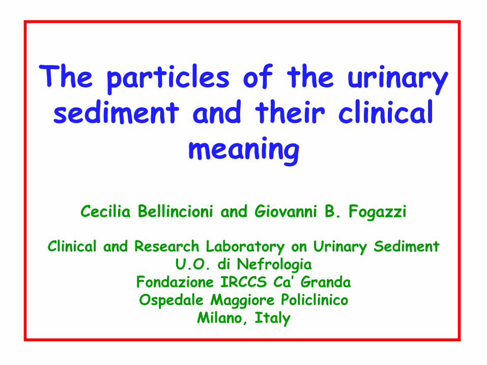
THE PARTICLES OF THE URINARY SEDIMENT
• Cells
• Lipids
• Casts
• Crystals
• Microorganisms
• Contaminants
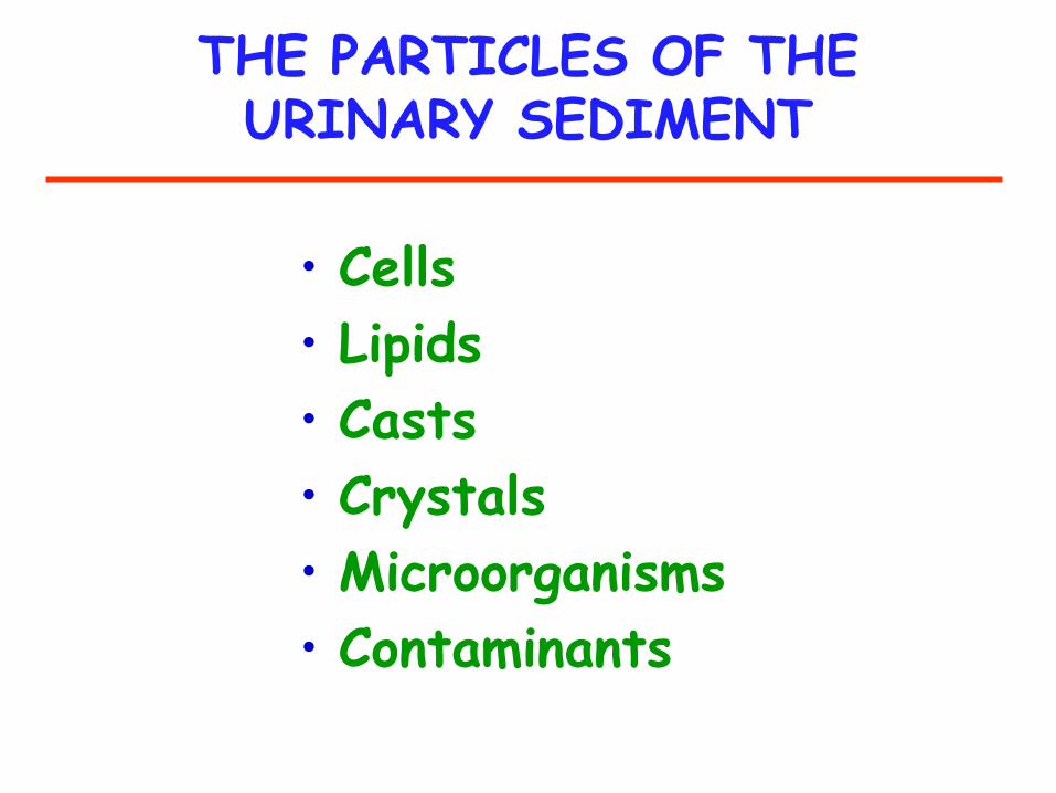
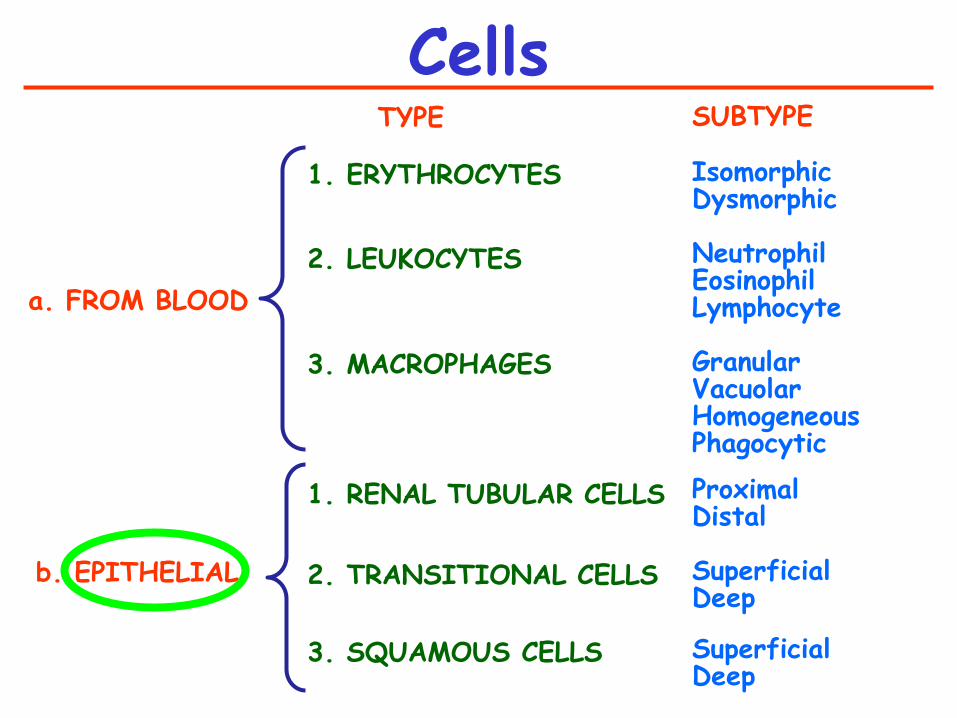
Cells
TYPE
1. ERYTHROCYTES
2. LEUKOCYTES 3. MACROPHAGES
1. RENAL TUBULAR CELLS
2. TRANSITIONAL CELLS
3. SQUAMOUS CELLS
SUBTYPE Isomorphic Dysmorphic Neutrophil Eosinophil Lymphocyte Granular Vacuolar Homogeneous Phagocytic
Proximal Distal Superficial Deep Superficial Deep
a. FROM BLOOD
b. EPITHELIAL
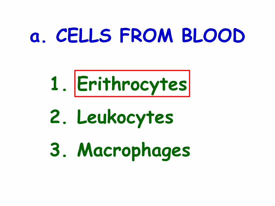
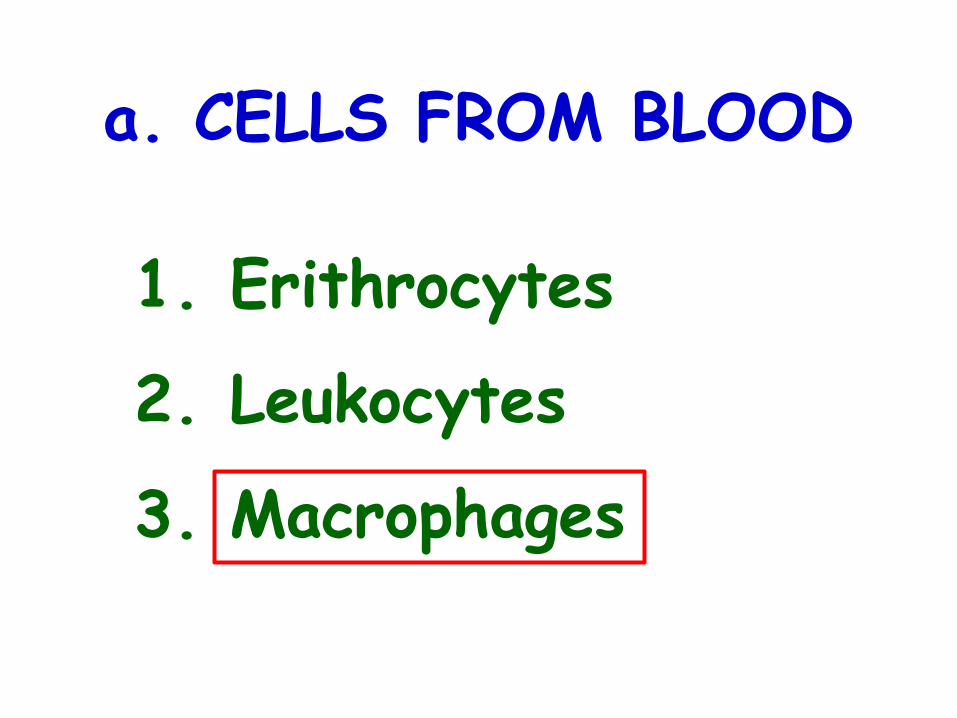
a. CELLS FROM BLOOD
1. Erithrocytes
2. Leukocytes
3. Macrophages
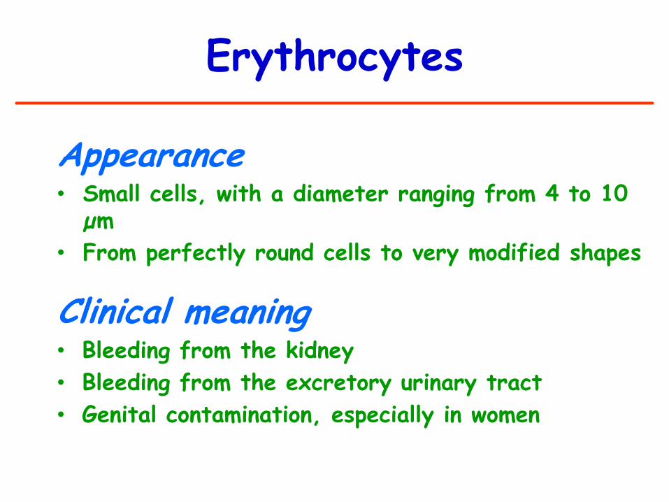
Erythrocytes
Appearance • Small cells, with a diameter ranging from 4 to 10
µm
• From perfectly round cells to very modified shapes
Clinical meaning • Bleeding from the kidney
• Bleeding from the excretory urinary tract
• Genital contamination, especially in women
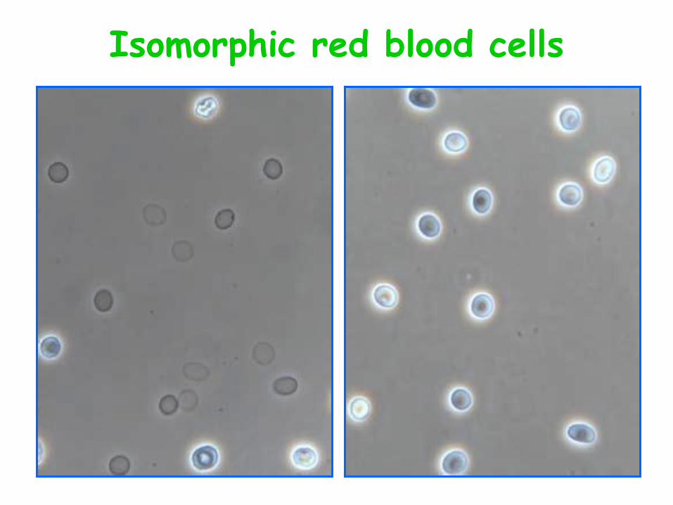
Isomorphic red blood cells
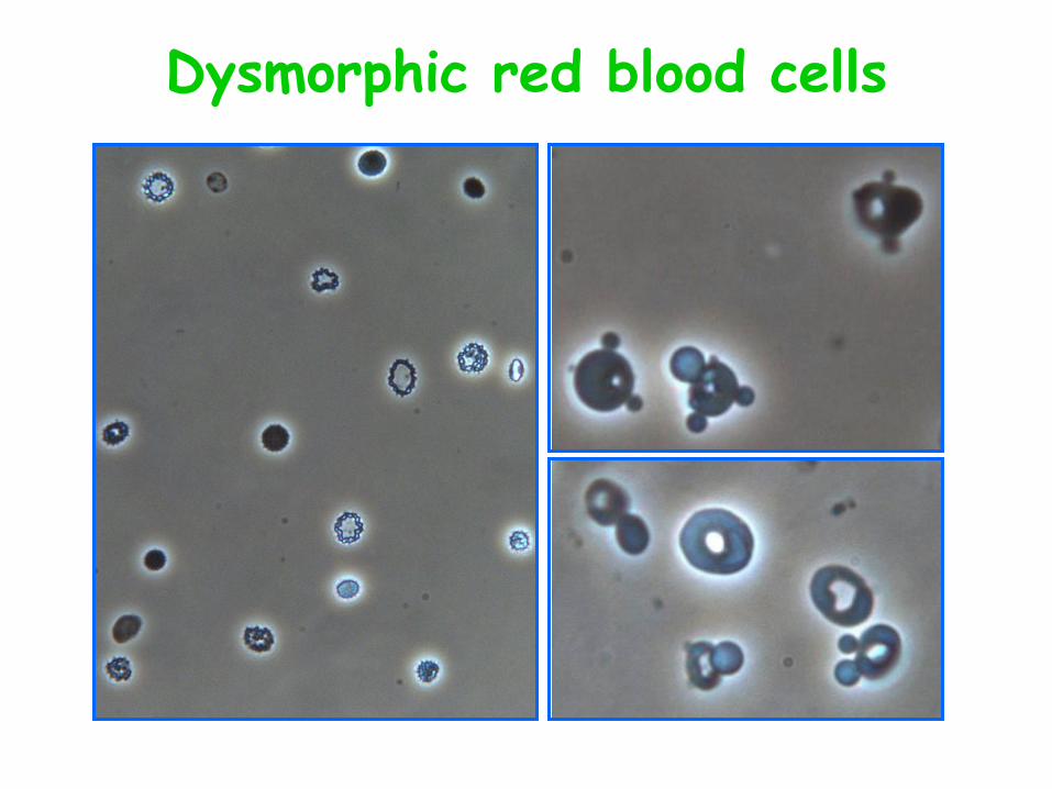
Dysmorphic red blood cells
Erythrocyte morphology (isomorphic and dysmorphic)
See presentation by Dr A. Skoberne:
“Estimating erythroctye
shape-dysmorphism”

a. CELLS FROM BLOOD
1. Erithrocytes
2. Leukocytes
3. Macrophages
3 types:
1.Polymorphonuclear
2.Lymphocytes
3.Eosinophils
Leukocytes
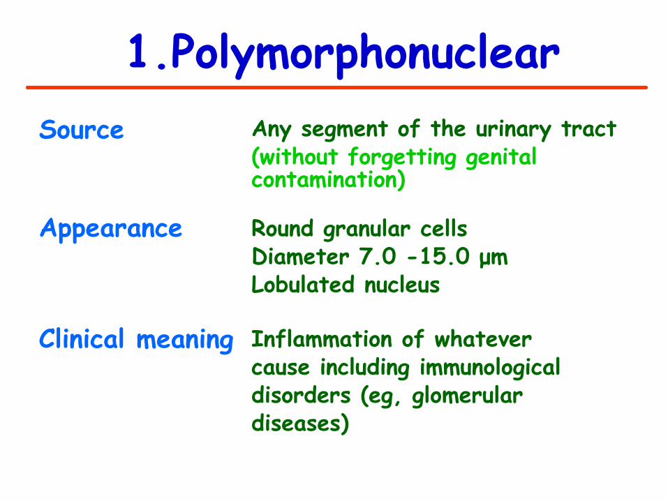
1.Polymorphonuclear Source Appearance
Clinical meaning
Any segment of the urinary tract (without forgetting genital contamination)
Round granular cells Diameter 7.0 -15.0 μm Lobulated nucleus
Inflammation of whatever cause including immunological disorders (eg, glomerular diseases)
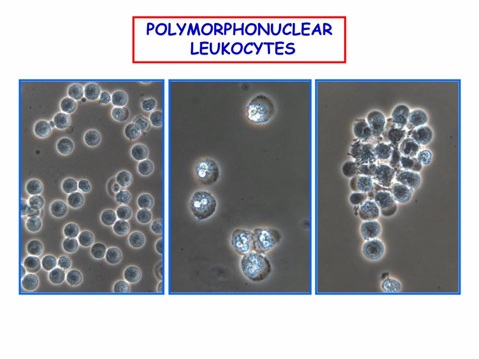
POLYMORPHONUCLEAR LEUKOCYTES
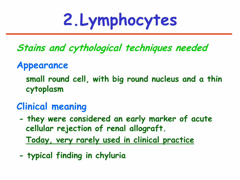
2.Lymphocytes
Stains and cythological techniques needed
Appearance
small round cell, with big round nucleus and a thin cytoplasm
Clinical meaning - they were considered an early marker of acute
cellular rejection of renal allograft.
Today, very rarely used in clinical practice
- typical finding in chyluria

See presentation by
Dr G.B. Fogazzi:
“Urinary profiles”
3.Eosinophils
a. CELLS FROM BLOOD
1. Erithrocytes
2. Leukocytes
3. Macrophages
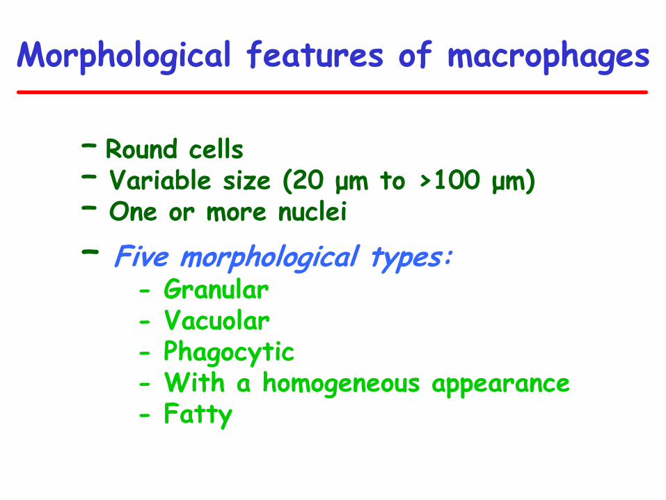
Morphological features of macrophages
– Round cells – Variable size (20 μm to >100 μm) – One or more nuclei
– Five morphological types: - Granular - Vacuolar - Phagocytic - With a homogeneous appearance - Fatty
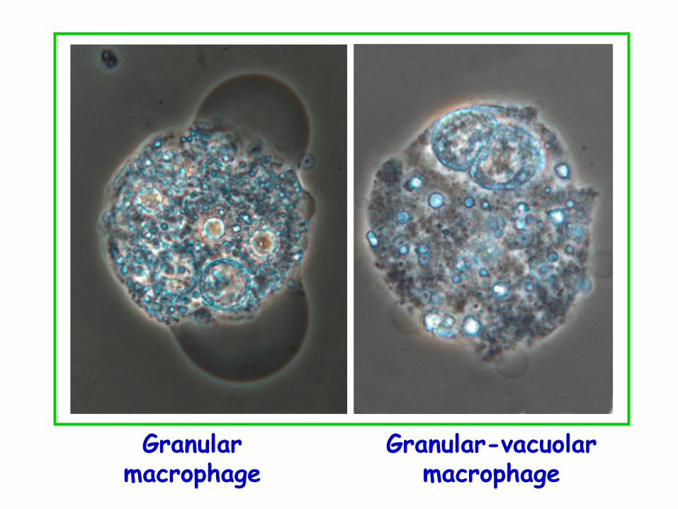
Granular macrophage
Granular-vacuolar macrophage
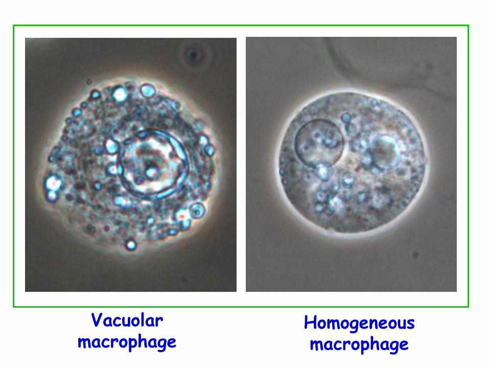
Homogeneous macrophage
Vacuolar macrophage
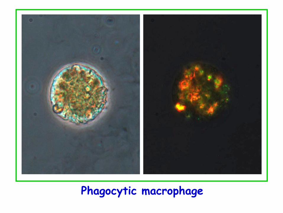
Phagocytic macrophage
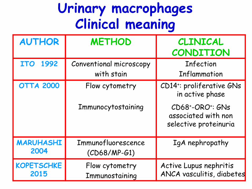
Urinary macrophages Clinical meaning
IgA nephropathy Immunofluorescence
(CD68/MP-G1)
MARUHASHI 2004
CD14+: proliferative GNs in active phase
CD68+-ORO+: GNs associated with non selective proteinuria
Flow cytometry
Immunocytostaining
OTTA 2000
Infection
Inflammation
Conventional microscopy
with stain
ITO 1992
CLINICAL CONDITION
METHOD AUTHOR
KOPETSCHKE 2015
Flow cytometry
Immunostaining
Active Lupus nephritis ANCA vasculitis, diabetes
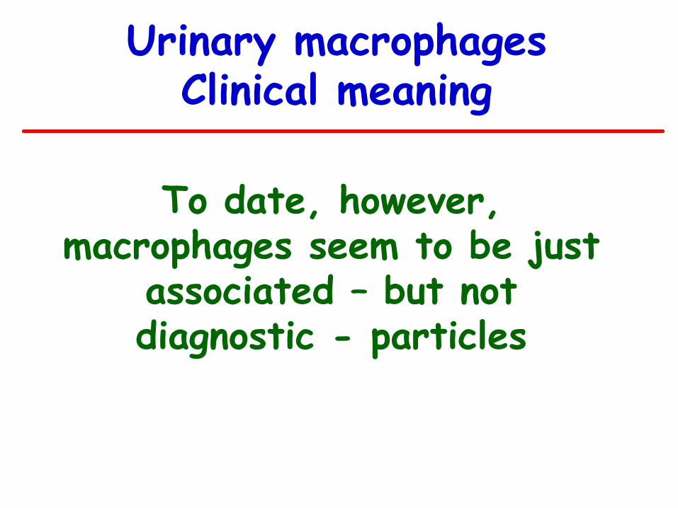
Urinary macrophages Clinical meaning
To date, however, macrophages seem to be just
associated – but not diagnostic - particles
Cells
TYPE
1. ERYTHROCYTES
2. LEUKOCYTES 3. MACROPHAGES
1. RENAL TUBULAR CELLS
2. TRANSITIONAL CELLS
3. SQUAMOUS CELLS
SUBTYPE Isomorphic Dysmorphic Neutrophil Eosinophil Lymphocyte Granular Vacuolar Homogeneous Phagocytic
Proximal Distal Superficial Deep Superficial Deep
a. FROM BLOOD
b. EPITHELIAL
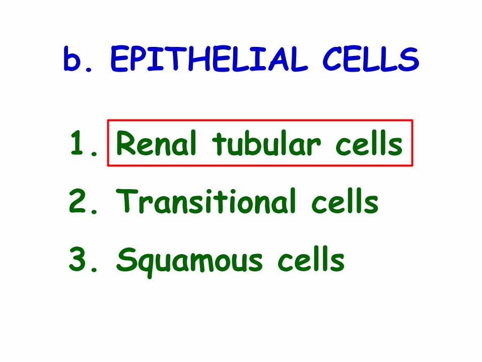
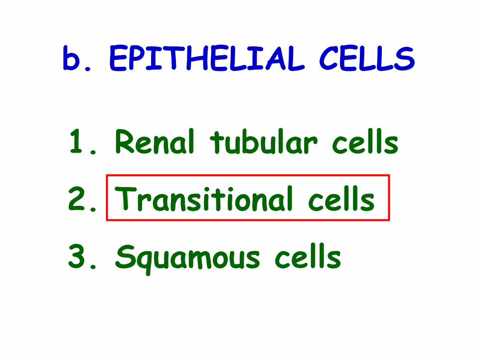
b. EPITHELIAL CELLS
1. Renal tubular cells
2. Transitional cells
3. Squamous cells
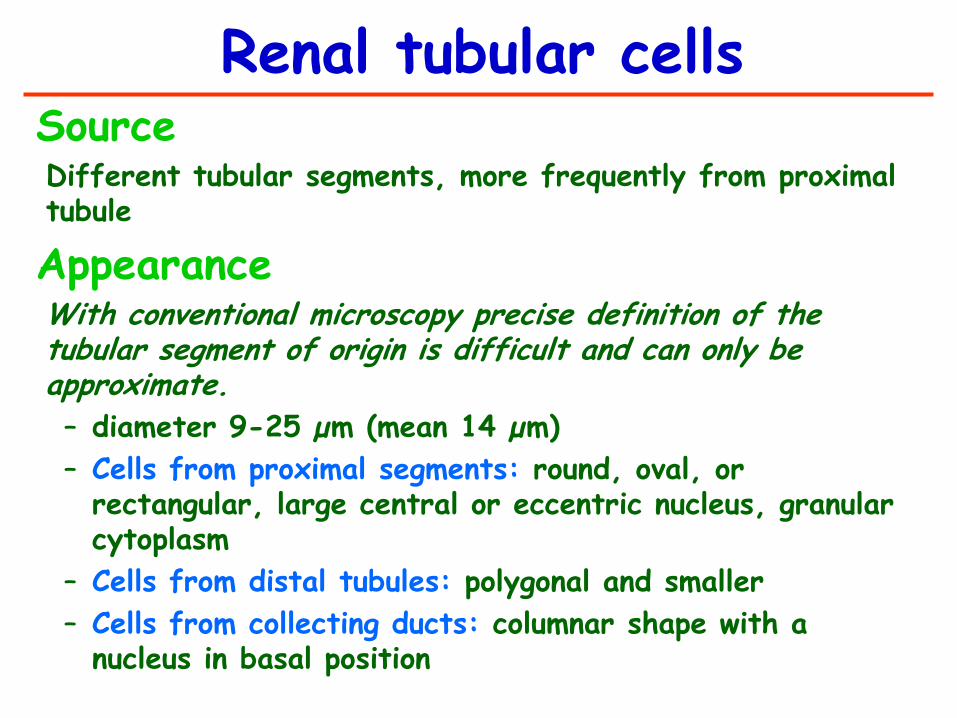
Source Different tubular segments, more frequently from proximal tubule
Appearance With conventional microscopy precise definition of the tubular segment of origin is difficult and can only be approximate.
– diameter 9-25 µm (mean 14 µm)
– Cells from proximal segments: round, oval, or rectangular, large central or eccentric nucleus, granular cytoplasm
– Cells from distal tubules: polygonal and smaller
– Cells from collecting ducts: columnar shape with a nucleus in basal position
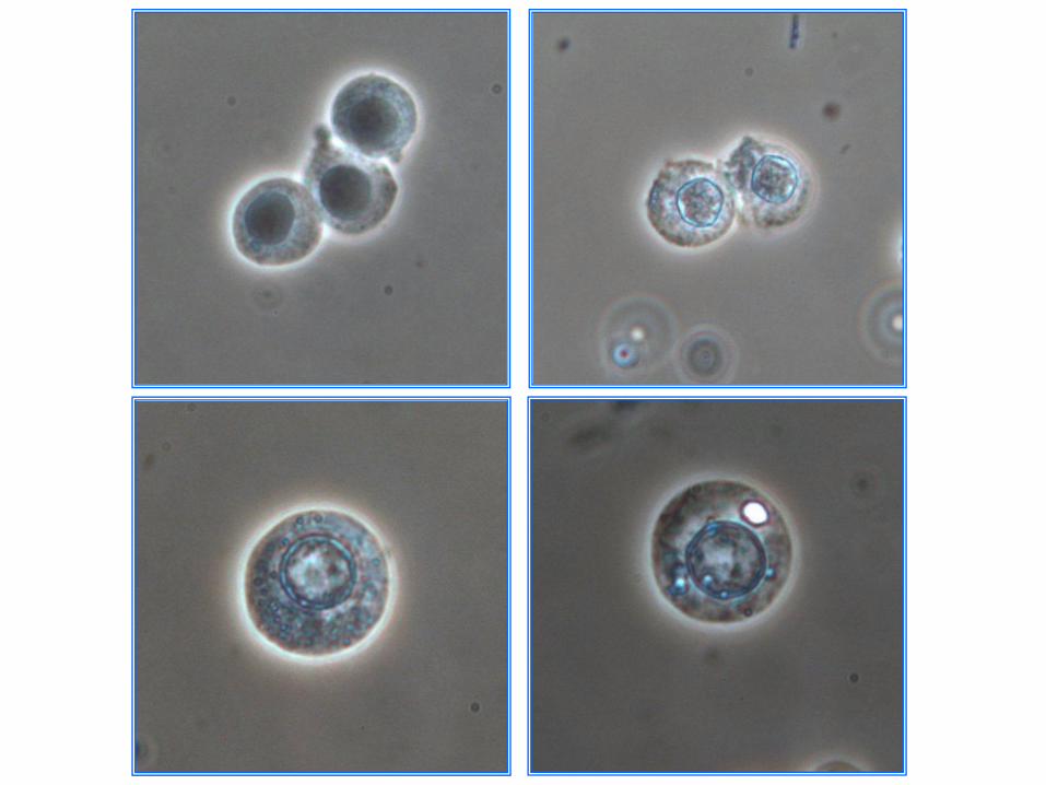
Renal tubular cells

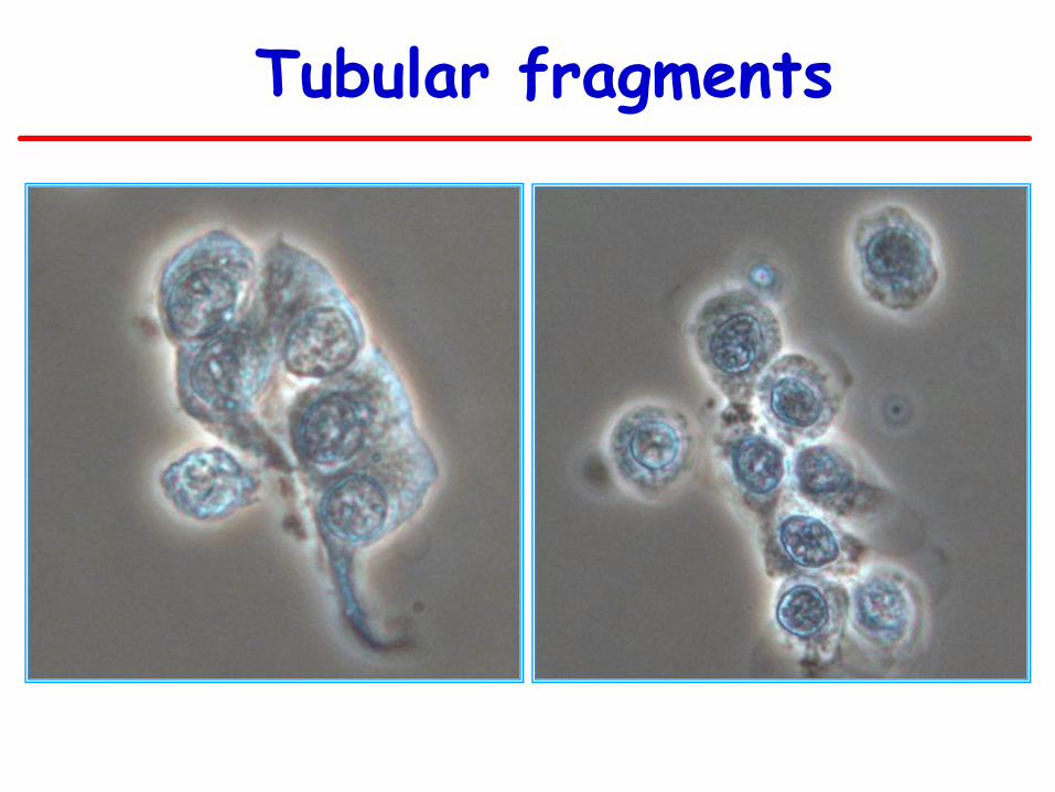
Tubular fragments

Acute tubular necrosis Clinical Acute interstitial meaning nephritis Glomerular diseases (especially proliferative types)
Renal tubular cells
b. EPITHELIAL CELLS
1. Renal tubular cells
2. Transitional cells
3. Squamous cells
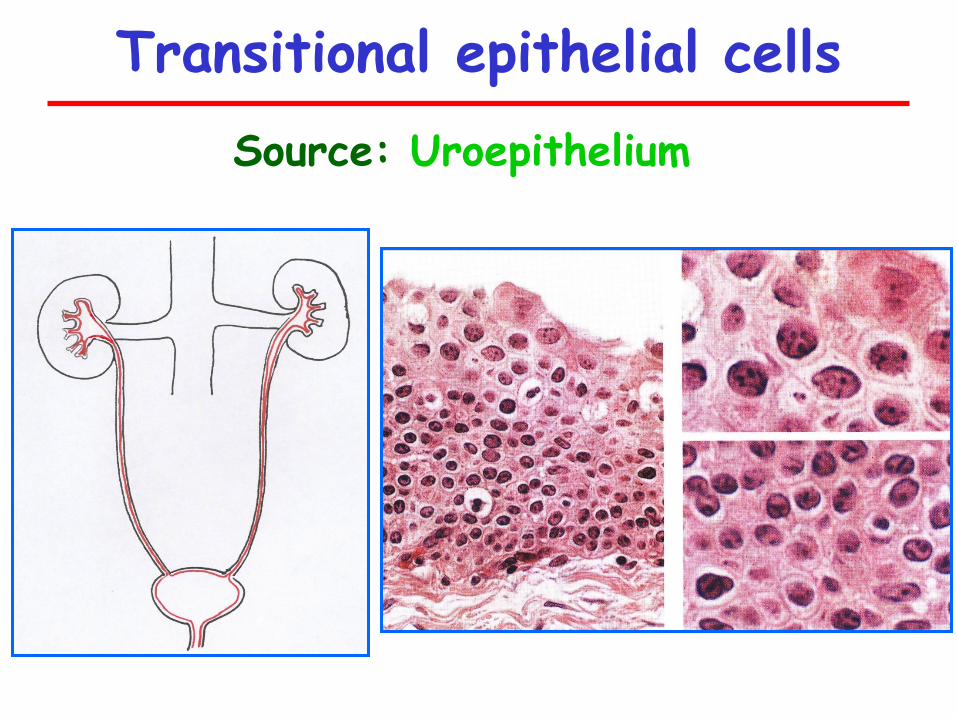
Transitional epithelial cells
Source: Uroepithelium
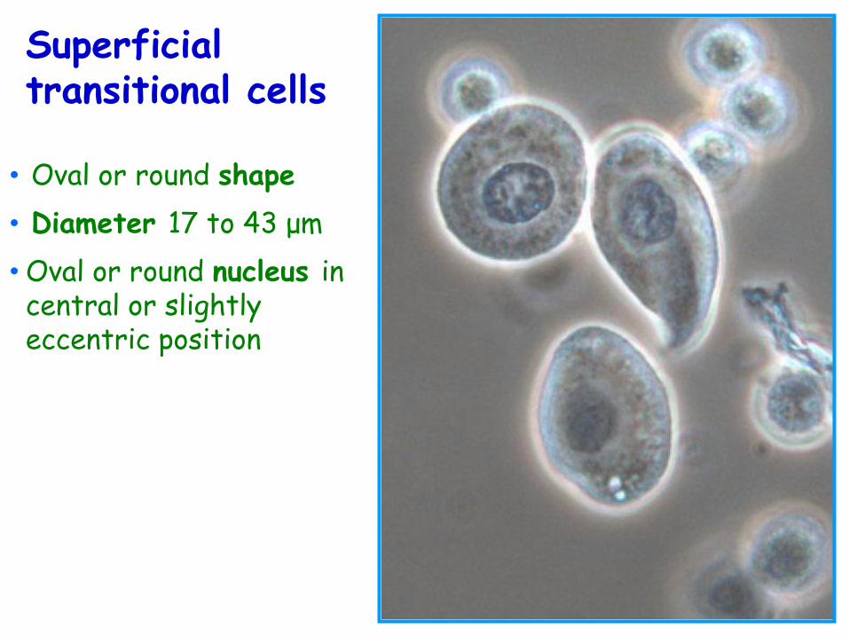
Superficial transitional cells
• Oval or round shape
• Diameter 17 to 43 μm
• Oval or round nucleus in central or slightly eccentric position
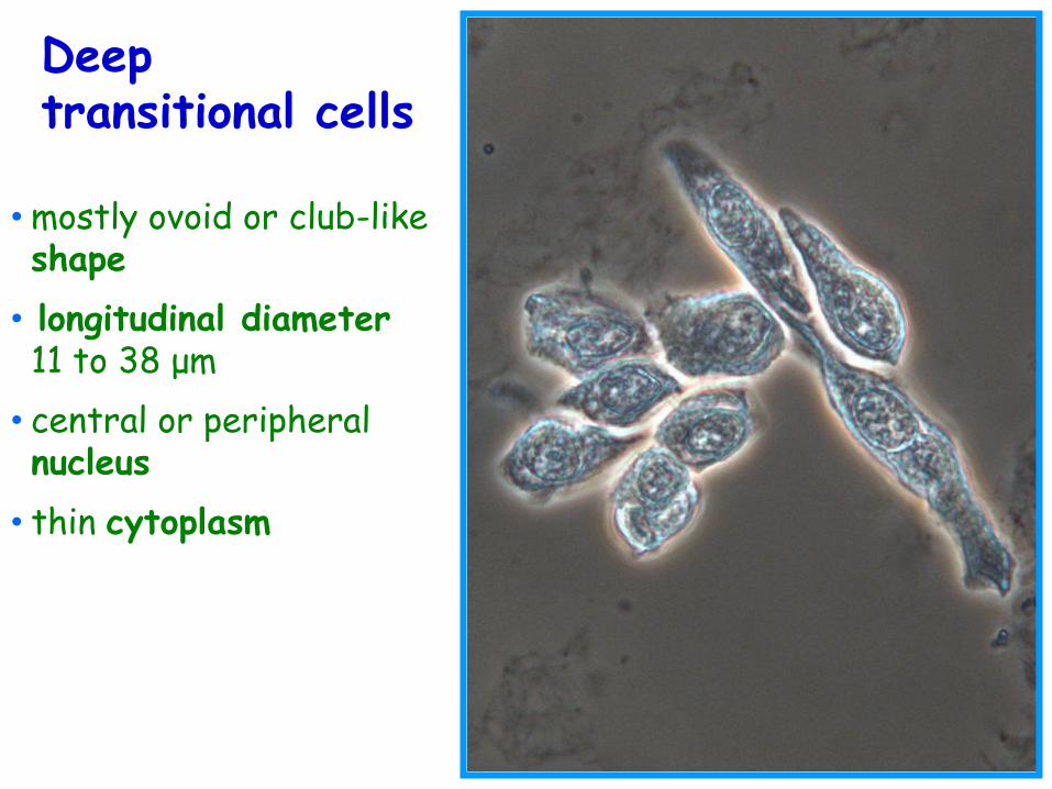
Deep transitional cells
• mostly ovoid or club-like shape
• longitudinal diameter 11 to 38 μm
• central or peripheral nucleus
• thin cytoplasm
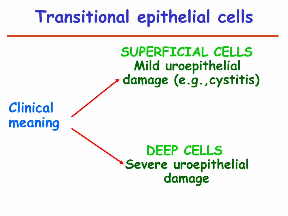
SUPERFICIAL CELLS Mild uroepithelial damage (e.g.,cystitis) Clinical meaning DEEP CELLS Severe uroepithelial damage
Transitional epithelial cells
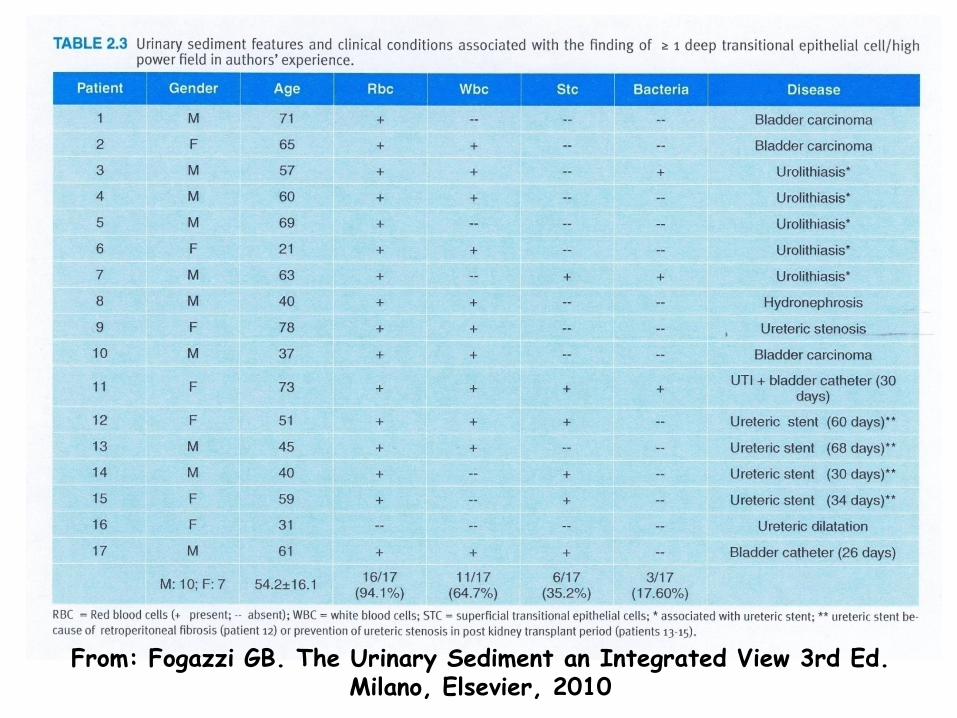
From: Fogazzi GB. The Urinary Sediment an Integrated View 3rd Ed. Milano, Elsevier, 2010
b. EPITHELIAL CELLS
1. Renal tubular cells
2. Transitional cells
3. Squamous cells
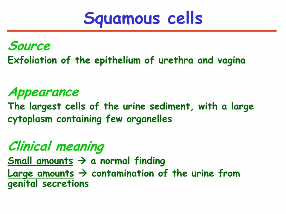
Squamous cells
Source Exfoliation of the epithelium of urethra and vagina
Appearance The largest cells of the urine sediment, with a large cytoplasm containing few organelles
Clinical meaning Small amounts a normal finding Large amounts contamination of the urine from genital secretions

Squamous epithelial cells
LIPIDS
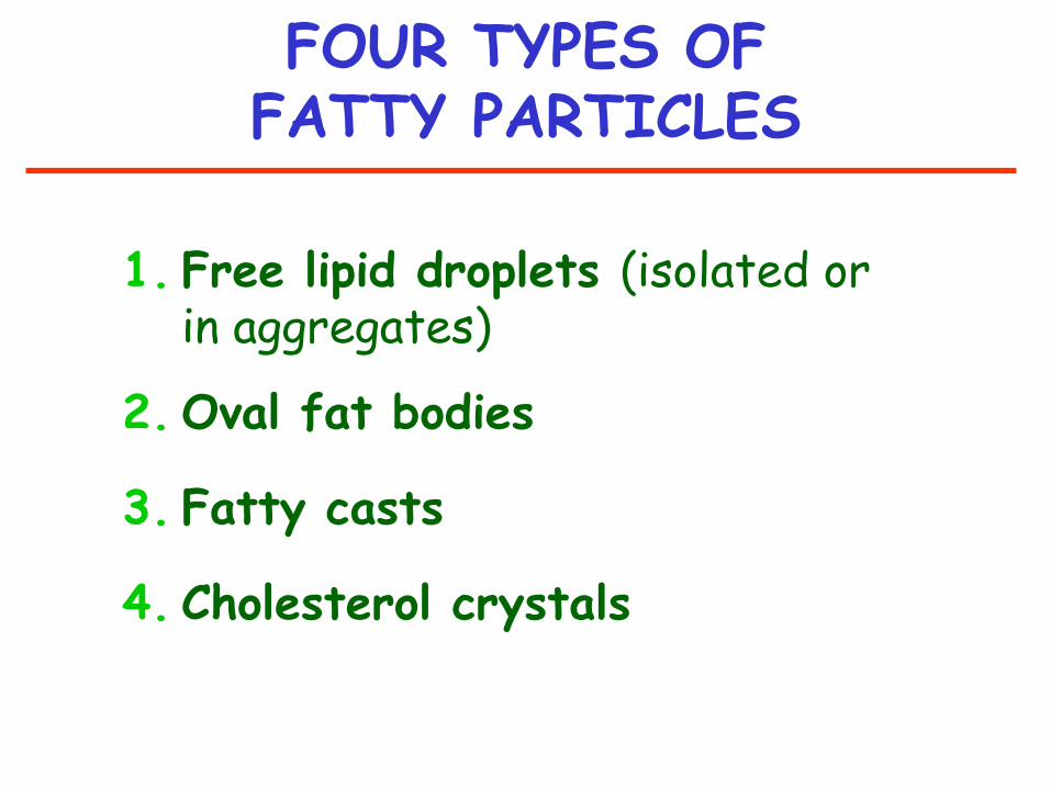
FOUR TYPES OF FATTY PARTICLES
1. Free lipid droplets (isolated or in aggregates)
2. Oval fat bodies
3. Fatty casts
4. Cholesterol crystals
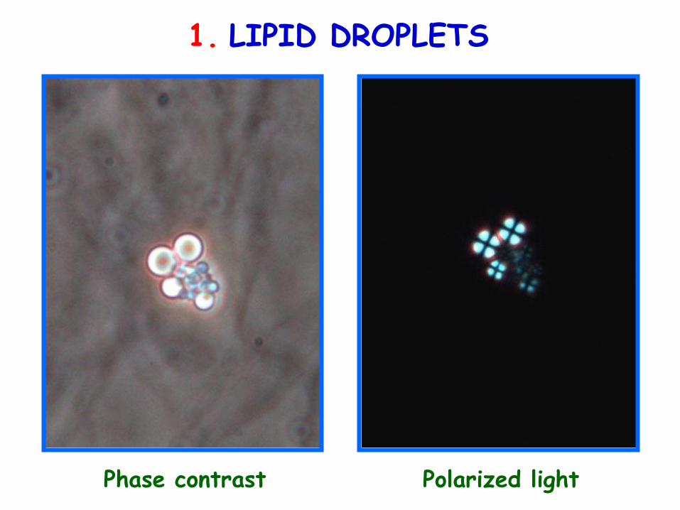
1. LIPID DROPLETS
Phase contrast Polarized light
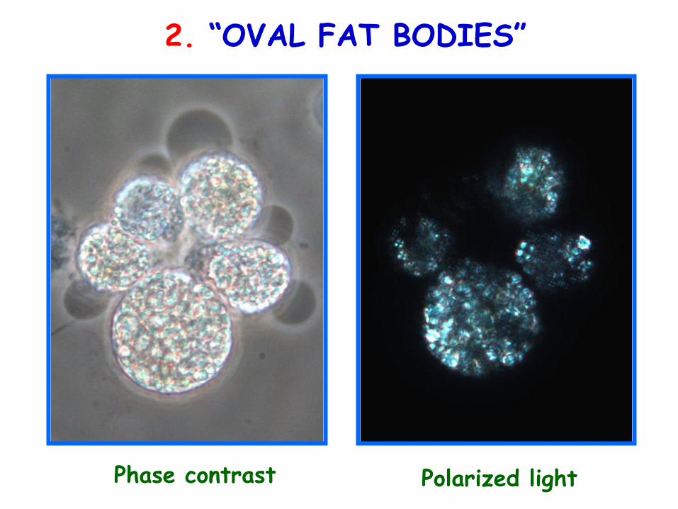
2. “OVAL FAT BODIES”
Phase contrast Polarized light
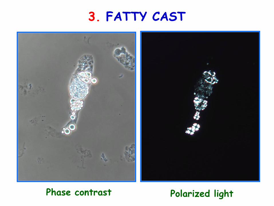
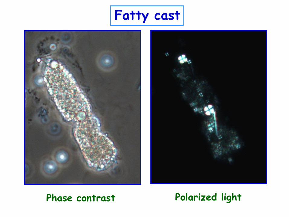
3. FATTY CAST
Polarized light Phase contrast
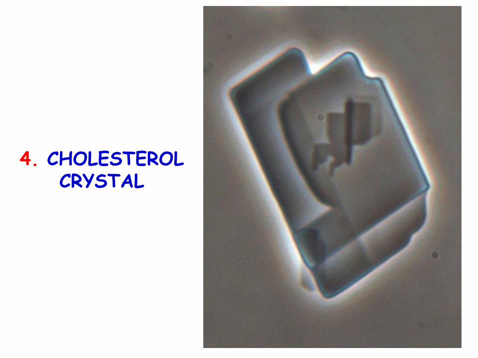
4. CHOLESTEROL CRYSTAL
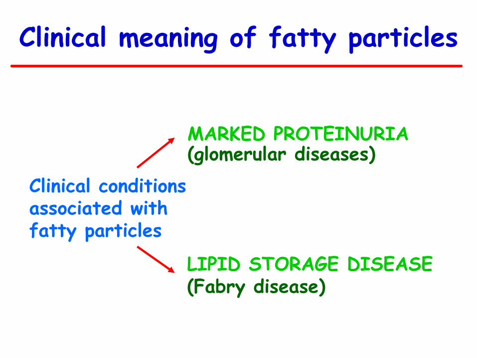
Clinical meaning of fatty particles
Clinical conditions associated with fatty particles
LIPID STORAGE DISEASE (Fabry disease)
MARKED PROTEINURIA (glomerular diseases)
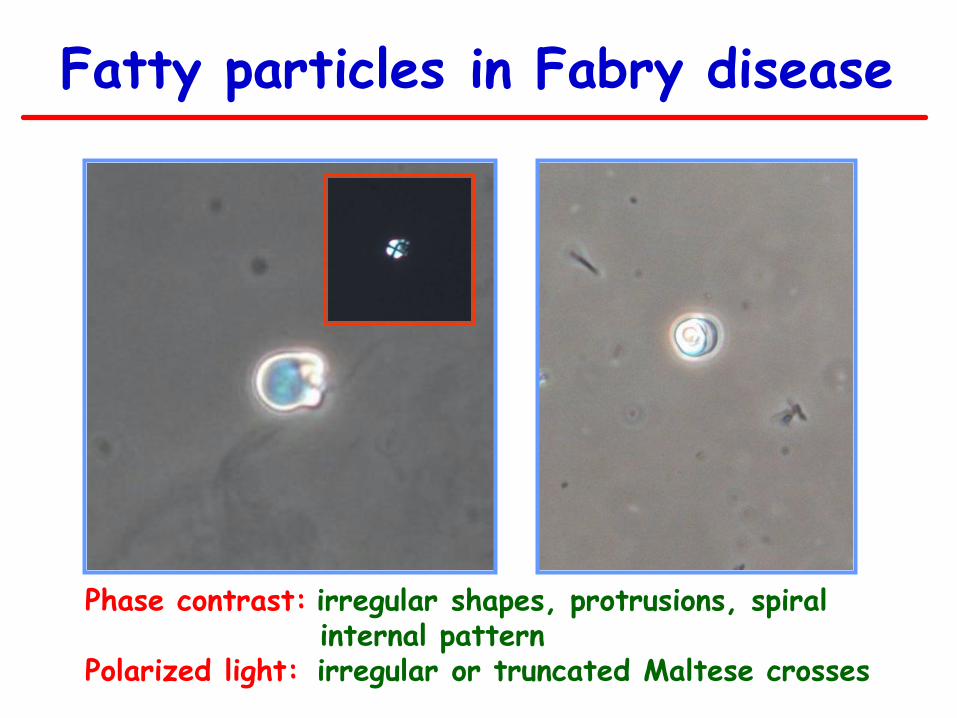
Fatty particles in Fabry disease
Phase contrast: irregular shapes, protrusions, spiral internal pattern
Polarized light: irregular or truncated Maltese crosses
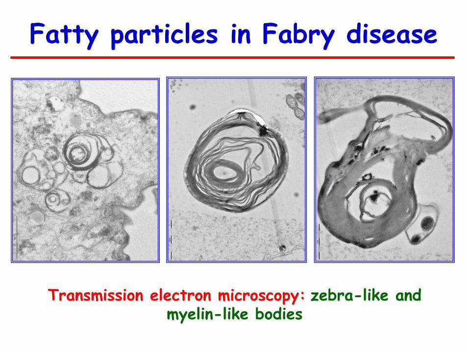
Transmission electron microscopy: zebra-like and myelin-like bodies
Fatty particles in Fabry disease

CASTS
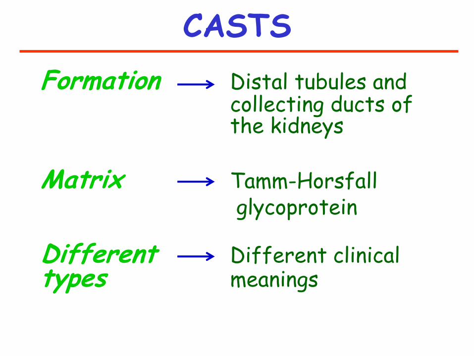
CASTS
Formation Distal tubules and collecting ducts of the kidneys
Matrix Tamm-Horsfall glycoprotein Different Different clinical types meanings
IMPORTANT TAKE HOME MESSAGE ABOUT CASTS
Since casts form
WITHIN THE KIDNEYS
whatever particle is contained
in a cast
COMES
FROM THE KIDNEYS!!!
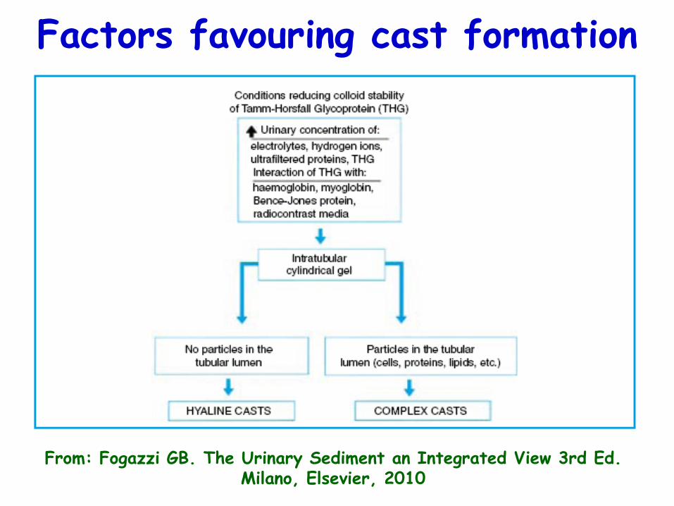
Factors favouring cast formation
From: Fogazzi GB. The Urinary Sediment an Integrated View 3rd Ed. Milano, Elsevier, 2010
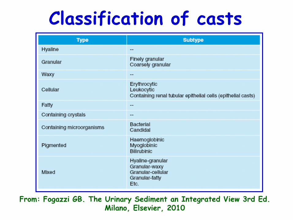
Classification of casts
From: Fogazzi GB. The Urinary Sediment an Integrated View 3rd Ed. Milano, Elsevier, 2010
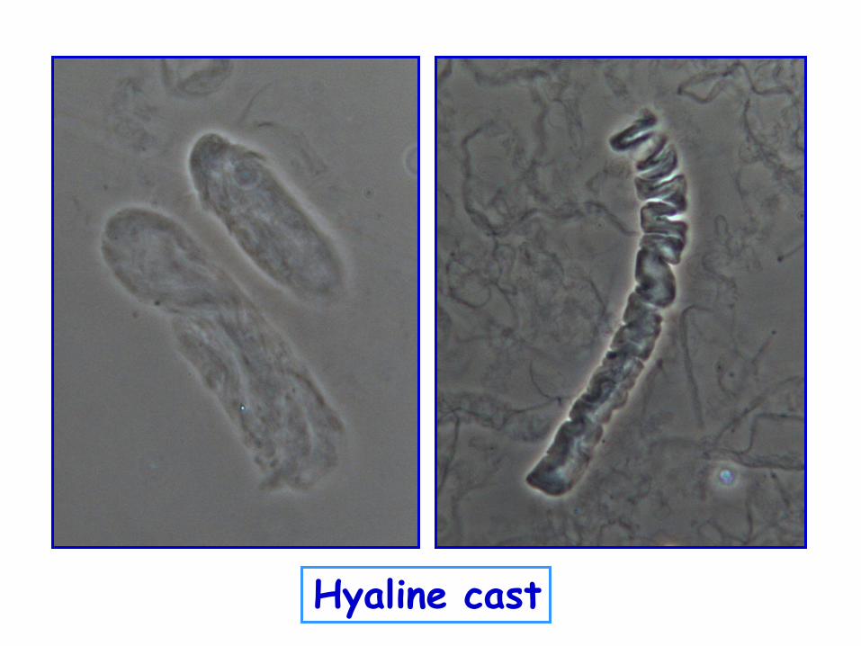
Hyaline cast
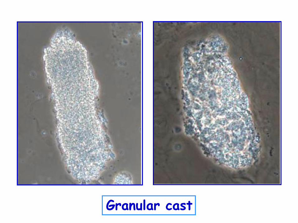
Granular cast
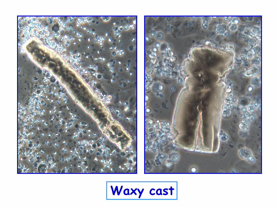
Waxy cast
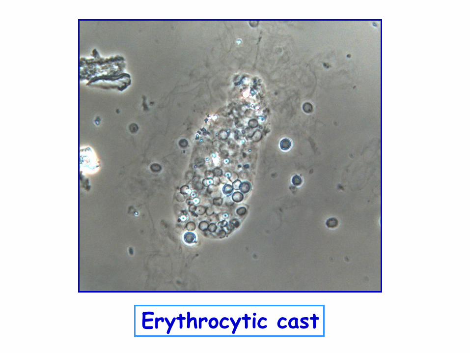
Erythrocytic cast
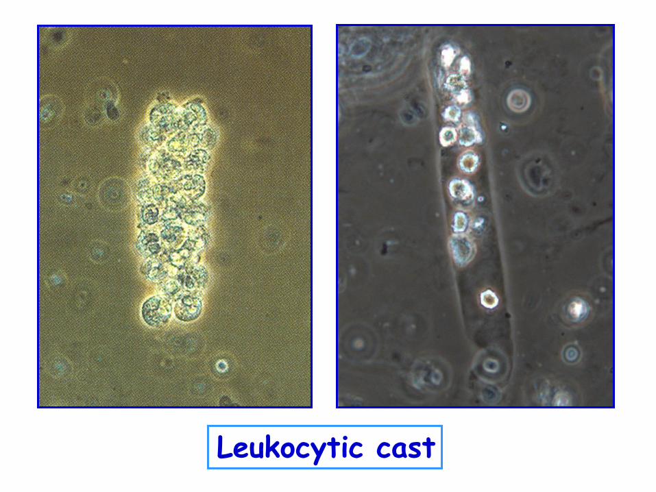
Leukocytic cast
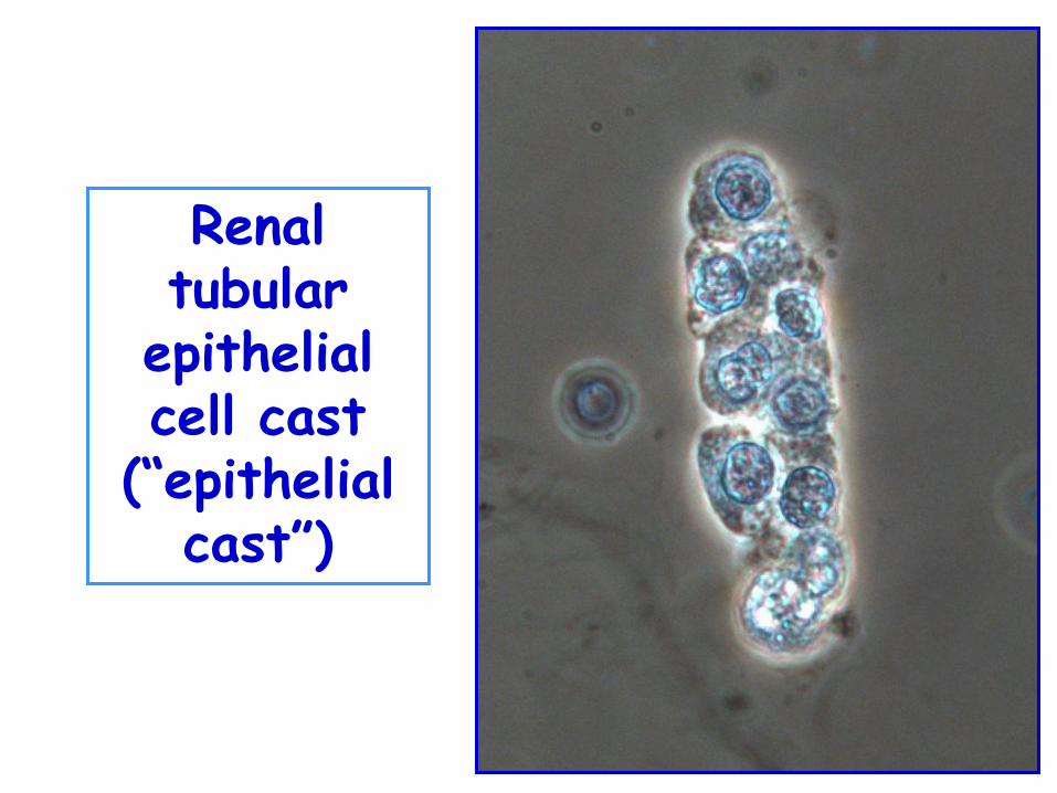
Renal tubular
epithelial cell cast
(“epithelial cast”)
Fatty cast
Phase contrast Polarized light
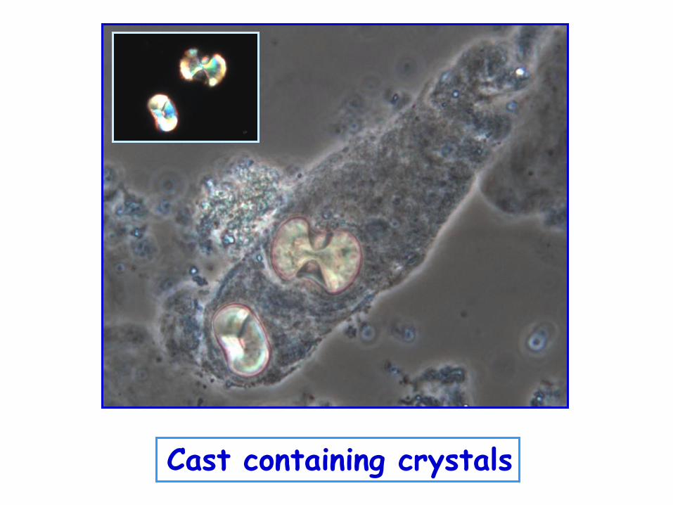
Cast containing crystals
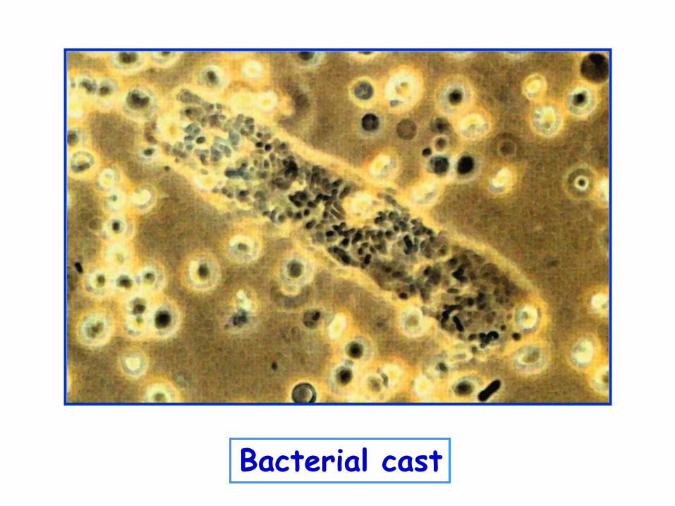
Bacterial cast
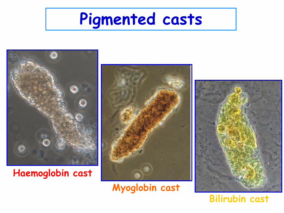
Haemoglobin cast
Myoglobin cast Bilirubin cast
Pigmented casts
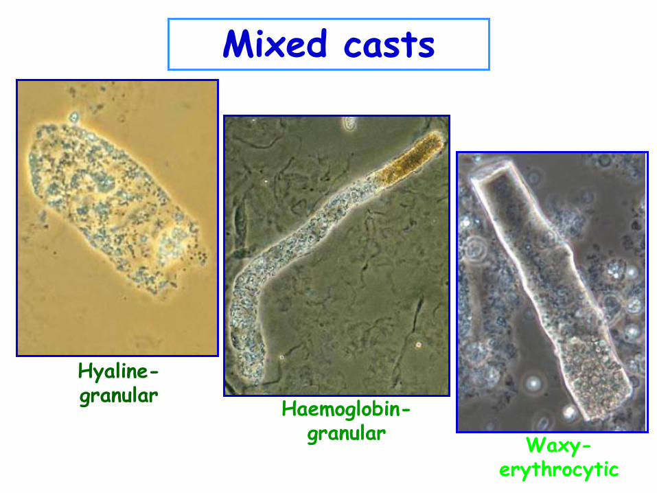
Hyaline-granular
Haemoglobin-granular
Waxy-erythrocytic
Mixed casts
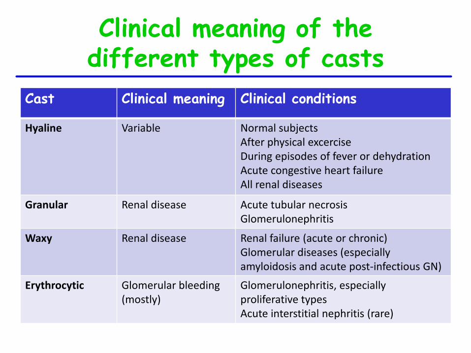
Cast Clinical meaning Clinical conditions
Hyaline Variable Normal subjects After physical excercise During episodes of fever or dehydration Acute congestive heart failure All renal diseases
Granular Renal disease Acute tubular necrosis Glomerulonephritis
Waxy Renal disease Renal failure (acute or chronic) Glomerular diseases (especially amyloidosis and acute post-infectious GN)
Erythrocytic Glomerular bleeding (mostly)
Glomerulonephritis, especially proliferative types Acute interstitial nephritis (rare)
Clinical meaning of the different types of casts
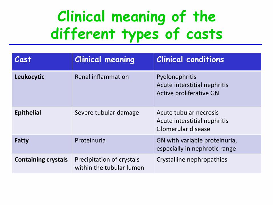
Cast Clinical meaning Clinical conditions
Leukocytic Renal inflammation Pyelonephritis Acute interstitial nephritis Active proliferative GN
Epithelial Severe tubular damage Acute tubular necrosis Acute interstitial nephritis Glomerular disease
Fatty Proteinuria GN with variable proteinuria, especially in nephrotic range
Containing crystals Precipitation of crystals within the tubular lumen
Crystalline nephropathies
Clinical meaning of the different types of casts
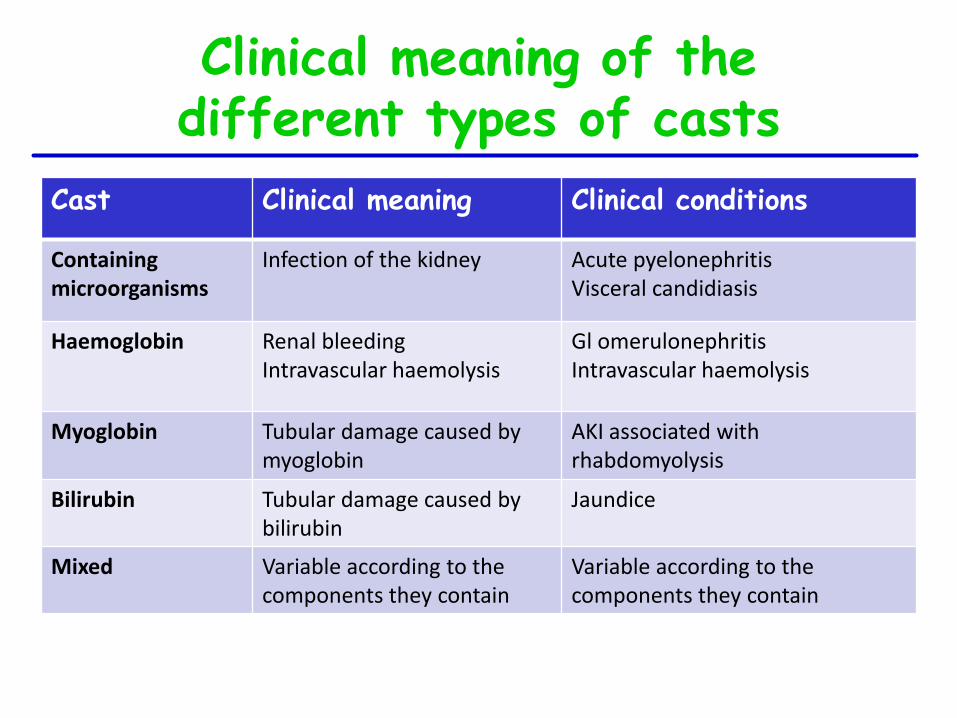
Cast Clinical meaning Clinical conditions
Containing microorganisms
Infection of the kidney Acute pyelonephritis Visceral candidiasis
Haemoglobin Renal bleeding Intravascular haemolysis
Gl omerulonephritis Intravascular haemolysis
Myoglobin Tubular damage caused by myoglobin
AKI associated with rhabdomyolysis
Bilirubin Tubular damage caused by bilirubin
Jaundice
Mixed Variable according to the components they contain
Variable according to the components they contain
Clinical meaning of the different types of casts
CRYSTALS
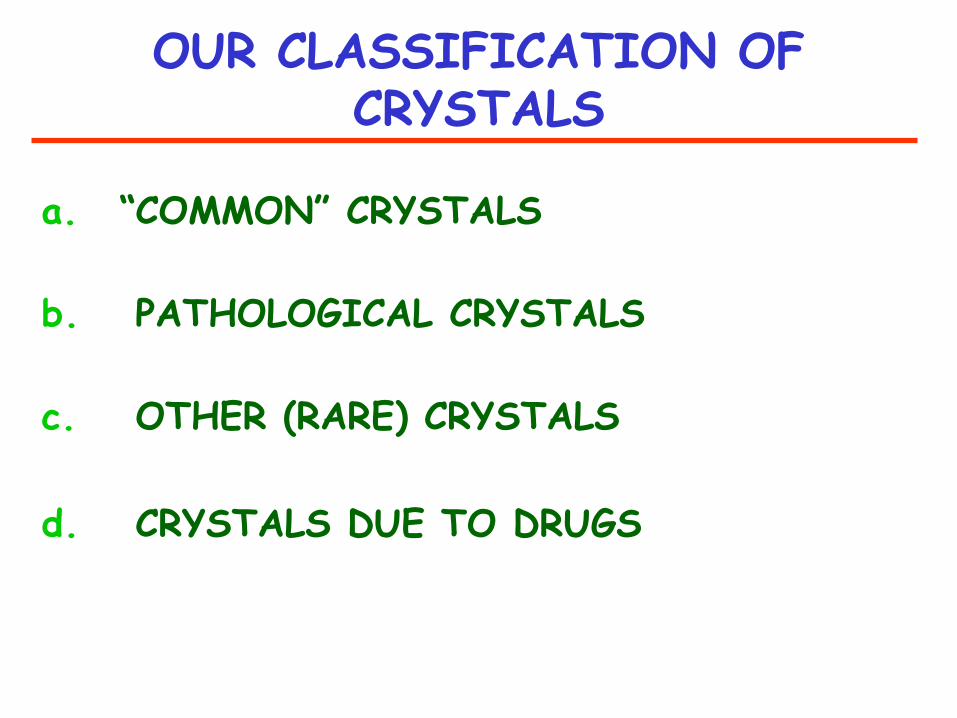
OUR CLASSIFICATION OF CRYSTALS
a. “COMMON” CRYSTALS
b. PATHOLOGICAL CRYSTALS
c. OTHER (RARE) CRYSTALS
d. CRYSTALS DUE TO DRUGS
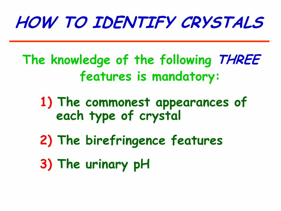
HOW TO IDENTIFY CRYSTALS
The knowledge of the following THREE features is mandatory:
1) The commonest appearances of each type of crystal
2) The birefringence features
3) The urinary pH
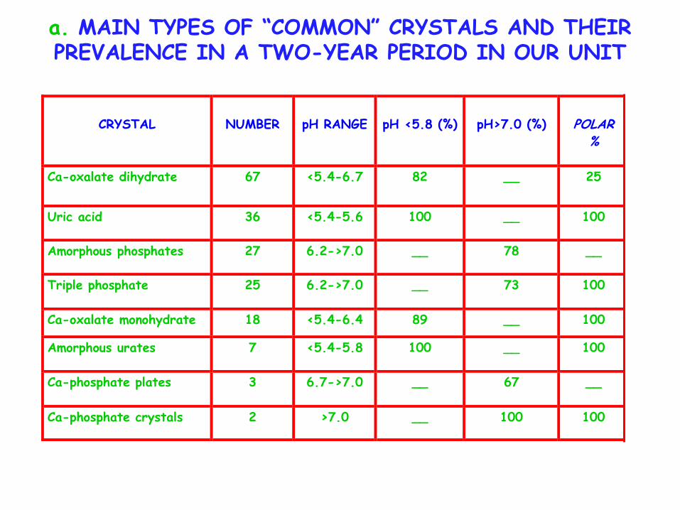
a. MAIN TYPES OF “COMMON” CRYSTALS AND THEIR PREVALENCE IN A TWO-YEAR PERIOD IN OUR UNIT
CRYSTAL
NUMBER
pH RANGE
pH <5.8 (%)
pH>7.0 (%)
POLAR
%
Ca-oxalate dihydrate 67 <5.4-6.7 82 __ 25
Uric acid 36 <5.4-5.6 100 __ 100
Amorphous phosphates 27 6.2->7.0 __ 78 __
Triple phosphate 25 6.2->7.0 __ 73 100
Ca-oxalate monohydrate 18 <5.4-6.4 89 __ 100
Amorphous urates 7 <5.4-5.8 100 __ 100
Ca-phosphate plates 3 6.7->7.0 __ 67 __
Ca-phosphate crystals 2 >7.0 __ 100 100

URIC ACID (U-pH <5.8)
Phase contrast Polarized light
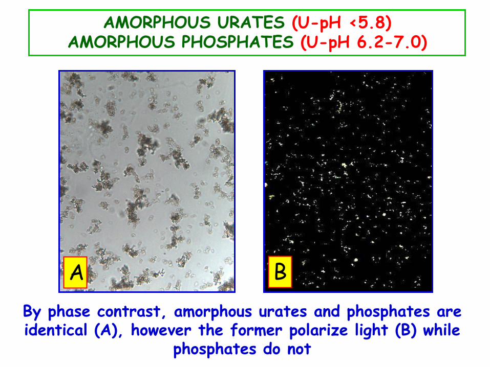
AMORPHOUS URATES (U-pH <5.8) AMORPHOUS PHOSPHATES (U-pH 6.2-7.0)
By phase contrast, amorphous urates and phosphates are identical (A), however the former polarize light (B) while
phosphates do not
A B
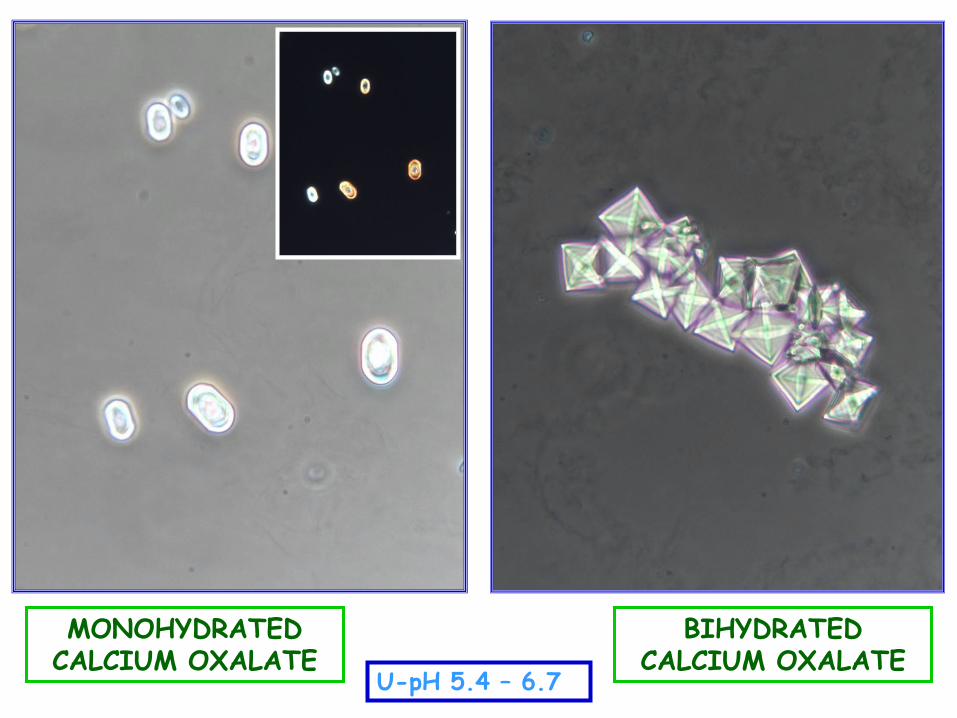
U-pH 5.4 – 6.7
MONOHYDRATED CALCIUM OXALATE
BIHYDRATED CALCIUM OXALATE
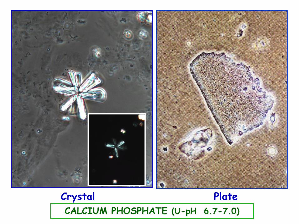
CALCIUM PHOSPHATE (U-pH 6.7-7.0)
Crystal Plate
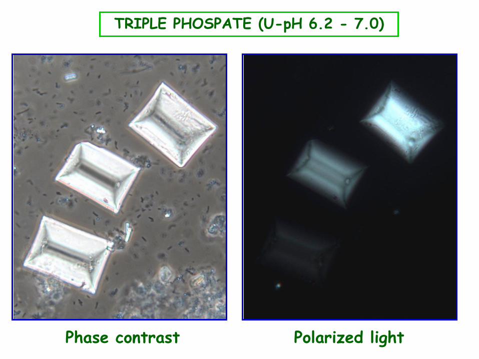
TRIPLE PHOSPATE (U-pH 6.2 - 7.0)
Phase contrast Polarized light
CLINICAL IMPORTANCE OF “COMMON” CRYSTALS (I)
• In most instances, UA, Ca-Ox, and Ca-P crystals are due to a transient supersaturation of the urine caused by foods, dehydration, or changes of urine pH and/or temperature upon standing
• However, especially when they are persistent, large and in aggregates they may be associated with metabolic disorders such as hyper-calciuria, -oxaluria, or -uricosuria
• Triple phosphate crystals are associated with UTI caused by urea-splitting bacteria (eg, Ureaplasma or Corynebacterium urealyticum)
Acute kidney injury associated with crystalluria:
Acute urate nephropathy
(uric acid)
Ethylene glycol poisoning (calcium oxalate)
CLINICAL IMPORTANCE OF “COMMON” CRYSTALS (II)
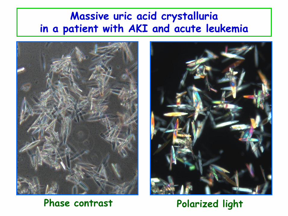
Massive uric acid crystalluria in a patient with AKI and acute leukemia
Phase contrast Polarized light
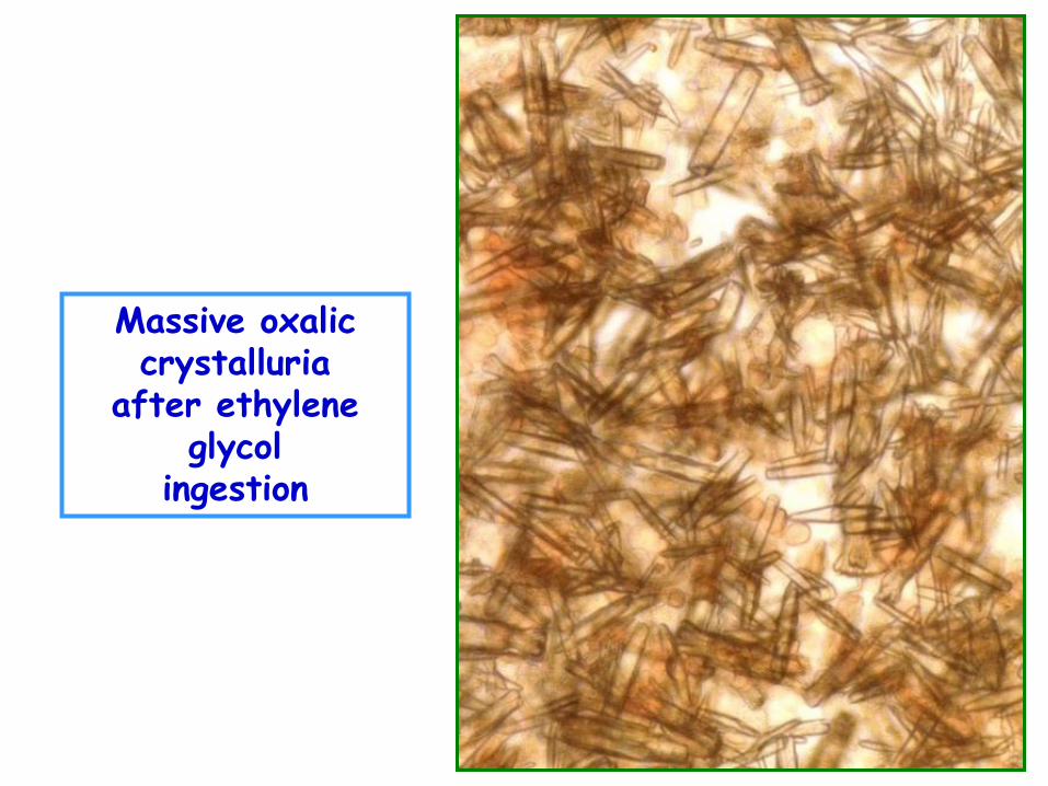
Massive oxalic crystalluria
after ethylene glycol
ingestion
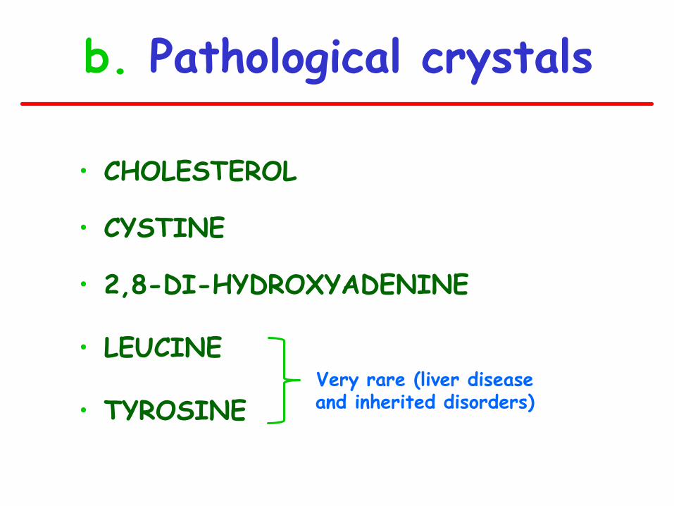
b. Pathological crystals
• CHOLESTEROL
• CYSTINE
• 2,8-DI-HYDROXYADENINE
• LEUCINE
• TYROSINE
Very rare (liver disease and inherited disorders)
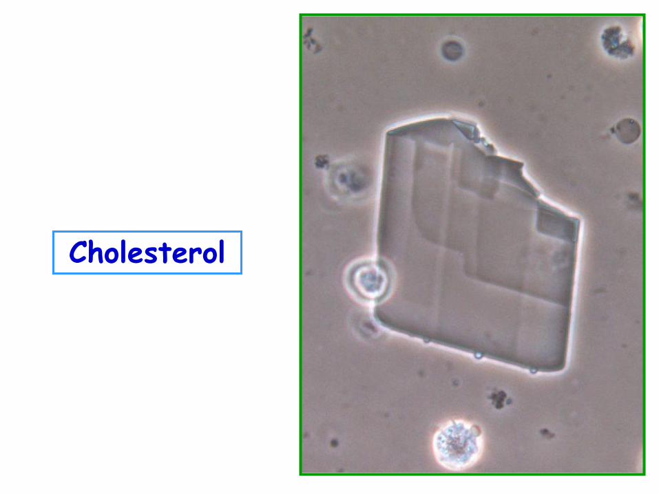
Cholesterol
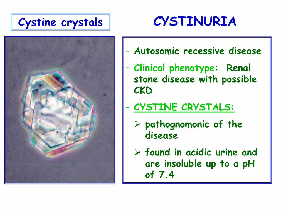
Cystine crystals
– Autosomic recessive disease
– Clinical phenotype: Renal stone disease with possible CKD
– CYSTINE CRYSTALS:
pathognomonic of the disease
found in acidic urine and are insoluble up to a pH of 7.4
CYSTINURIA
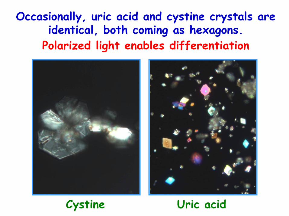
Occasionally, uric acid and cystine crystals are identical, both coming as hexagons.
Polarized light enables differentiation
Cystine Uric acid
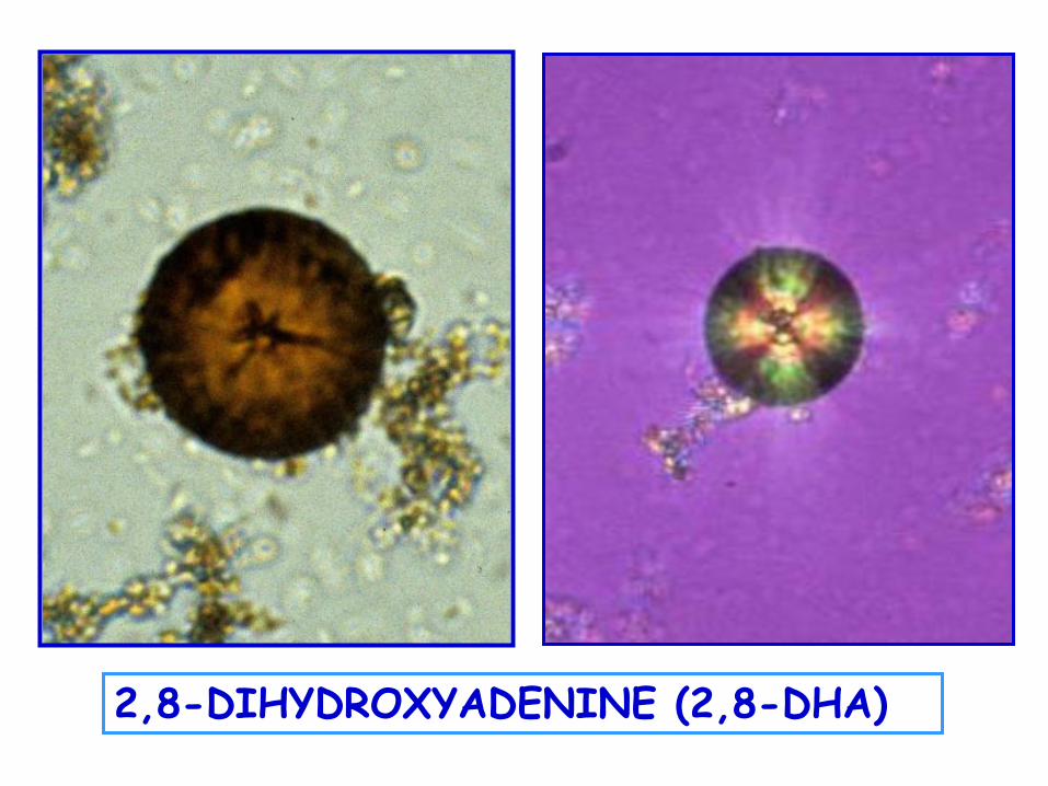
2,8-DIHYDROXYADENINE (2,8-DHA)
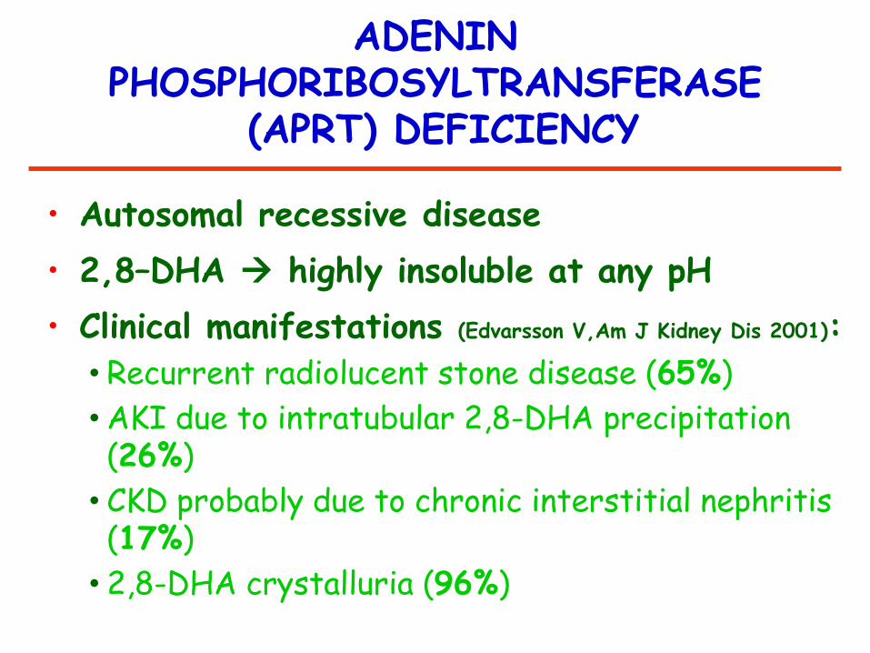
ADENIN PHOSPHORIBOSYLTRANSFERASE
(APRT) DEFICIENCY
• Autosomal recessive disease
• 2,8–DHA highly insoluble at any pH
• Clinical manifestations (Edvarsson V,Am J Kidney Dis 2001):
• Recurrent radiolucent stone disease (65%)
• AKI due to intratubular 2,8-DHA precipitation (26%)
• CKD probably due to chronic interstitial nephritis (17%)
• 2,8-DHA crystalluria (96%)
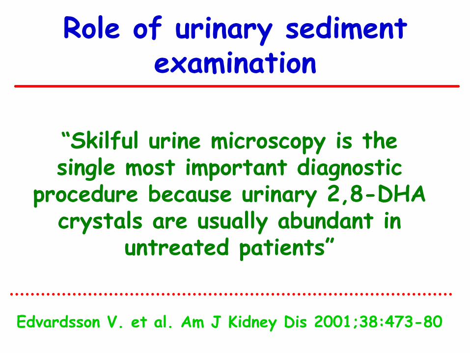
Role of urinary sediment examination
“Skilful urine microscopy is the single most important diagnostic
procedure because urinary 2,8-DHA crystals are usually abundant in
untreated patients”
Edvardsson V. et al. Am J Kidney Dis 2001;38:473-80
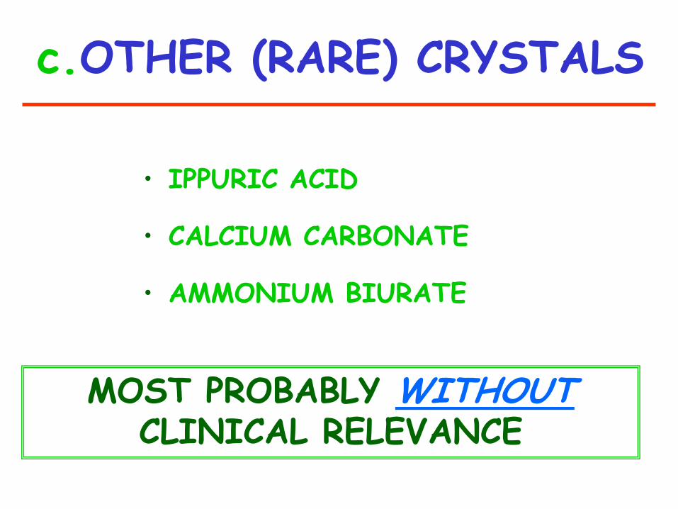
c.OTHER (RARE) CRYSTALS
• IPPURIC ACID
• CALCIUM CARBONATE
• AMMONIUM BIURATE
MOST PROBABLY WITHOUT CLINICAL RELEVANCE
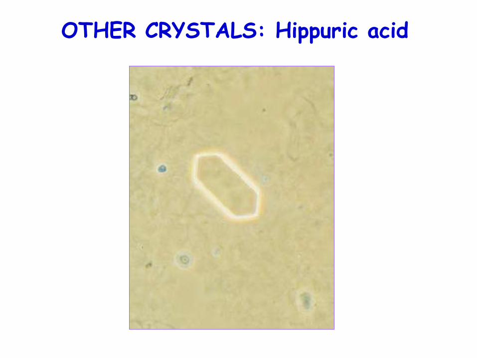
OTHER CRYSTALS: Hippuric acid
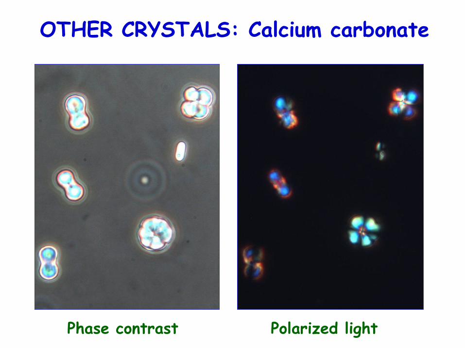
OTHER CRYSTALS: Calcium carbonate
Phase contrast Polarized light
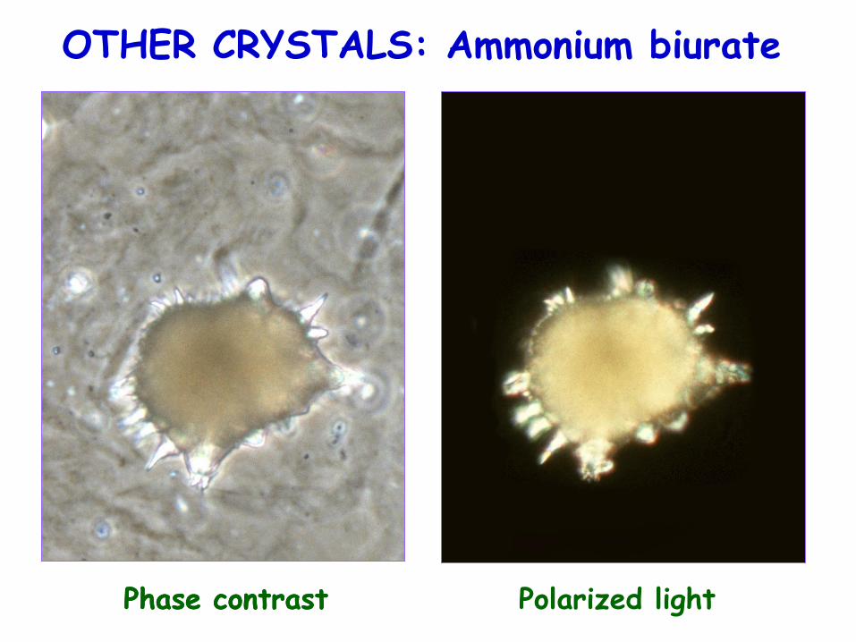
OTHER CRYSTALS: Ammonium biurate
Phase contrast Phase contrast Polarized light
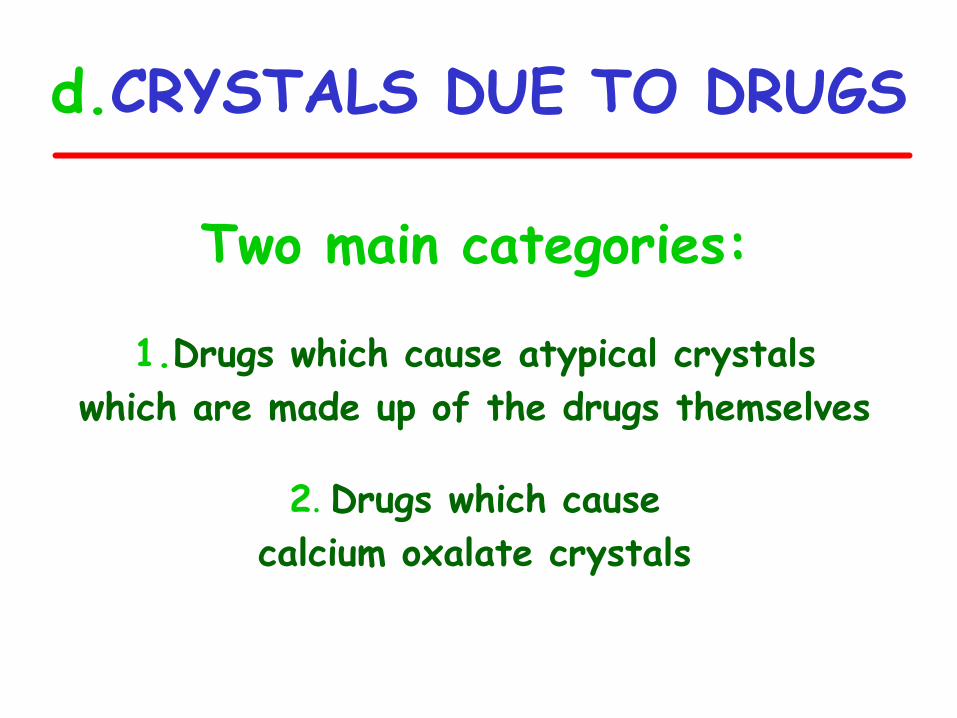
d.CRYSTALS DUE TO DRUGS
Two main categories:
1.Drugs which cause atypical crystals
which are made up of the drugs themselves
2. Drugs which cause
calcium oxalate crystals
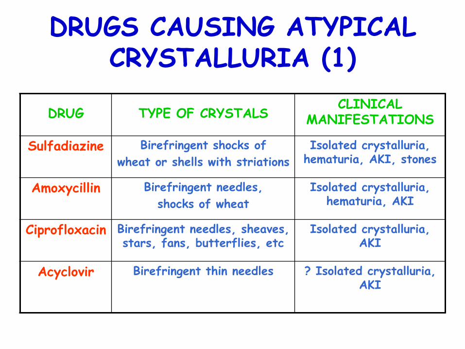
DRUGS CAUSING ATYPICAL CRYSTALLURIA (1)
DRUG
TYPE OF CRYSTALS CLINICAL
MANIFESTATIONS
Sulfadiazine Birefringent shocks of
wheat or shells with striations
Isolated crystalluria, hematuria, AKI, stones
Amoxycillin Birefringent needles,
shocks of wheat
Isolated crystalluria, hematuria, AKI
Ciprofloxacin Birefringent needles, sheaves, stars, fans, butterflies, etc
Isolated crystalluria, AKI
Acyclovir Birefringent thin needles ? Isolated crystalluria, AKI
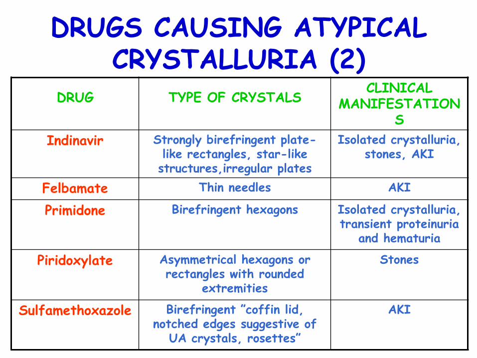
DRUGS CAUSING ATYPICAL CRYSTALLURIA (2)
DRUG
TYPE OF CRYSTALS CLINICAL
MANIFESTATIONS
Indinavir Strongly birefringent plate-like rectangles, star-like structures,irregular plates
Isolated crystalluria, stones, AKI
Felbamate Thin needles AKI
Primidone Birefringent hexagons Isolated crystalluria, transient proteinuria
and hematuria
Piridoxylate
Asymmetrical hexagons or rectangles with rounded
extremities
Stones
Sulfamethoxazole Birefringent ”coffin lid, notched edges suggestive of
UA crystals, rosettes”
AKI
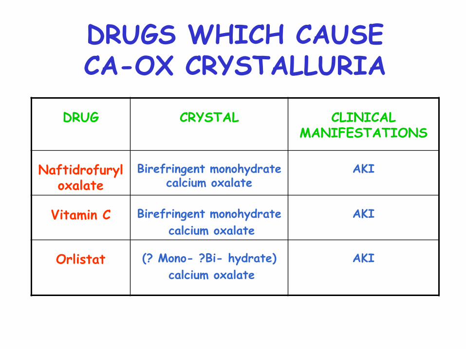
DRUGS WHICH CAUSE CA-OX CRYSTALLURIA
DRUG
CRYSTAL
CLINICAL MANIFESTATIONS
Naftidrofuryl oxalate
Birefringent monohydrate calcium oxalate
AKI
Vitamin C
Birefringent monohydrate
calcium oxalate
AKI
Orlistat
(? Mono- ?Bi- hydrate)
calcium oxalate
AKI
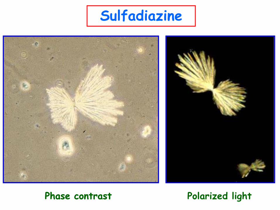
Sulfadiazine
Phase contrast Phase contrast Polarized light
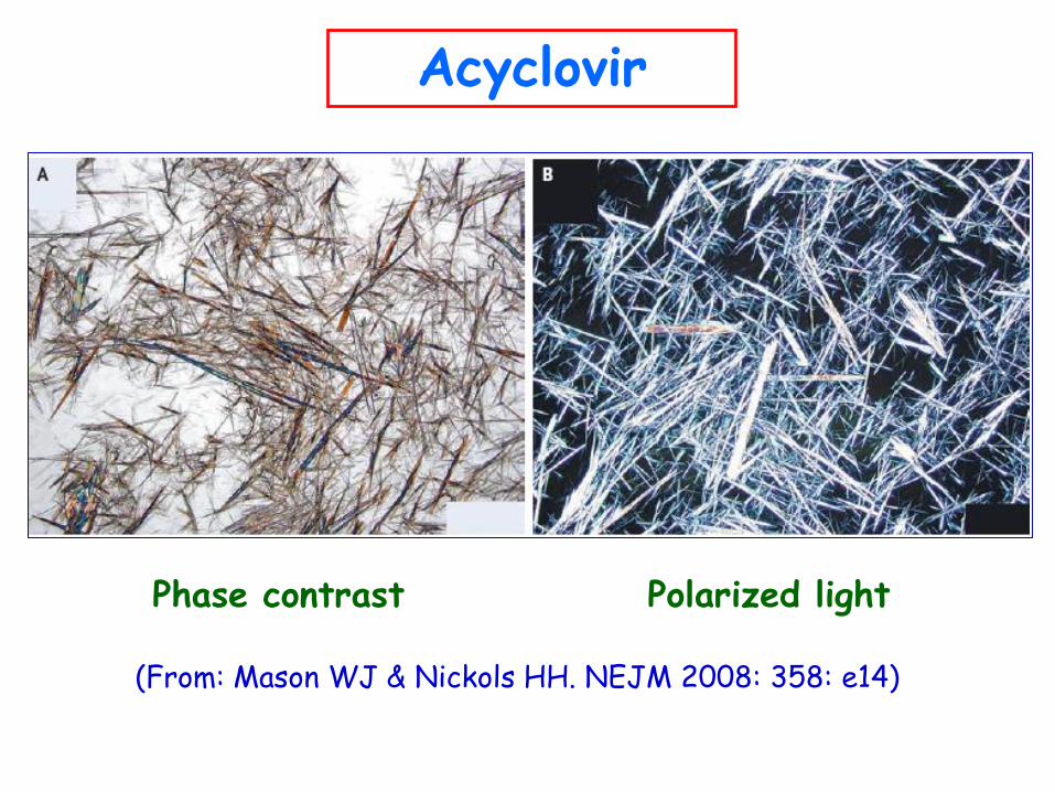
(From: Mason WJ & Nickols HH. NEJM 2008: 358: e14)
Acyclovir
Phase contrast Polarized light
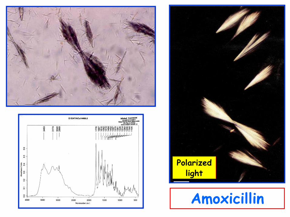
Amoxicillin
Polarized light
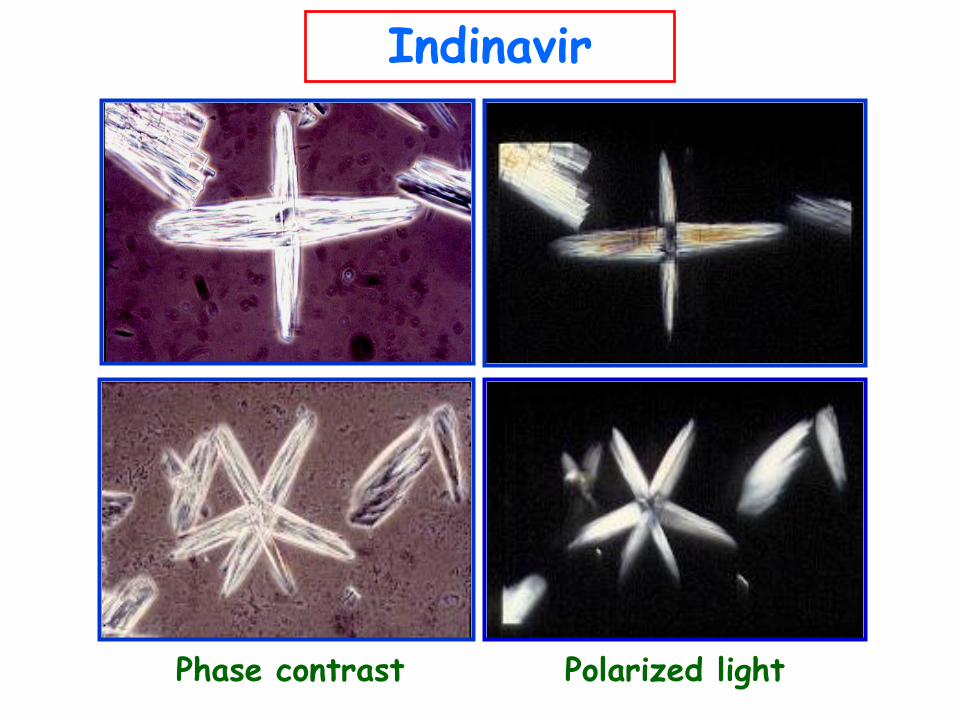
Indinavir
Polarized light Phase contrast
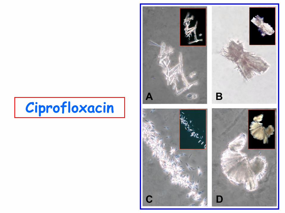
Ciprofloxacin
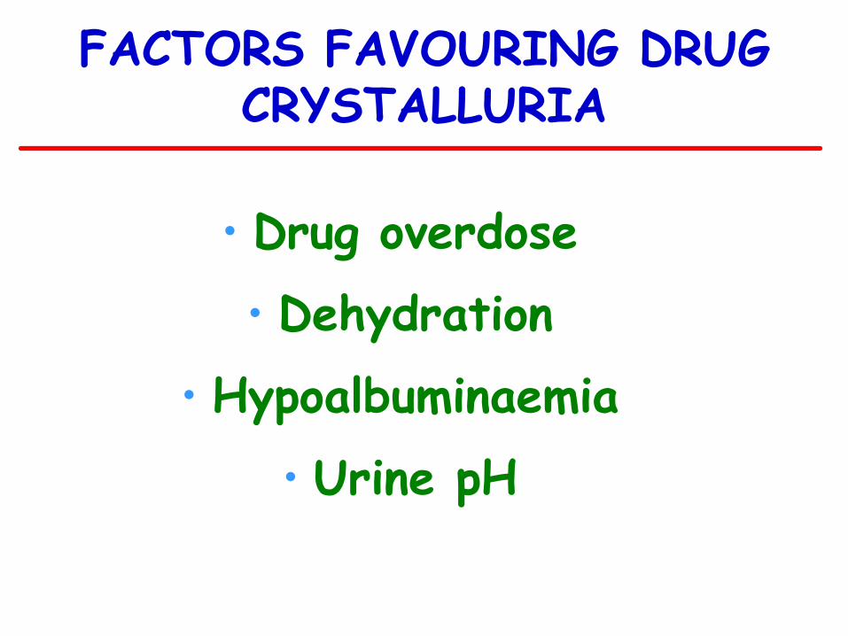
FACTORS FAVOURING DRUG CRYSTALLURIA
• Drug overdose
• Dehydration
• Hypoalbuminaemia
• Urine pH
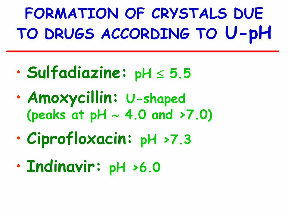
FORMATION OF CRYSTALS DUE
TO DRUGS ACCORDING TO U-pH
• Sulfadiazine: pH 5.5
• Amoxycillin: U-shaped (peaks at pH 4.0 and >7.0)
• Ciprofloxacin: pH >7.3
• Indinavir: pH >6.0
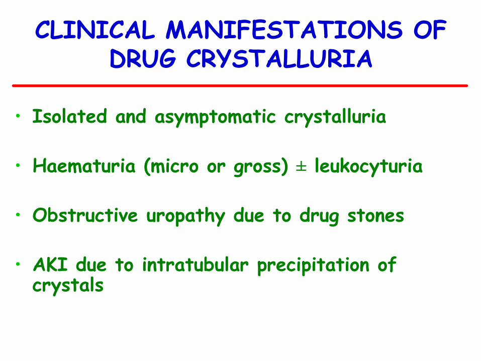
CLINICAL MANIFESTATIONS OF DRUG CRYSTALLURIA
• Isolated and asymptomatic crystalluria
• Haematuria (micro or gross) ± leukocyturia • Obstructive uropathy due to drug stones
• AKI due to intratubular precipitation of
crystals
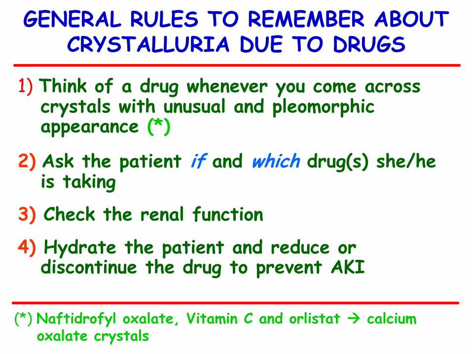
1) Think of a drug whenever you come across crystals with unusual and pleomorphic appearance (*)
2) Ask the patient if and which drug(s) she/he is taking
3) Check the renal function
4) Hydrate the patient and reduce or discontinue the drug to prevent AKI
GENERAL RULES TO REMEMBER ABOUT CRYSTALLURIA DUE TO DRUGS
(*) Naftidrofyl oxalate, Vitamin C and orlistat calcium oxalate crystals
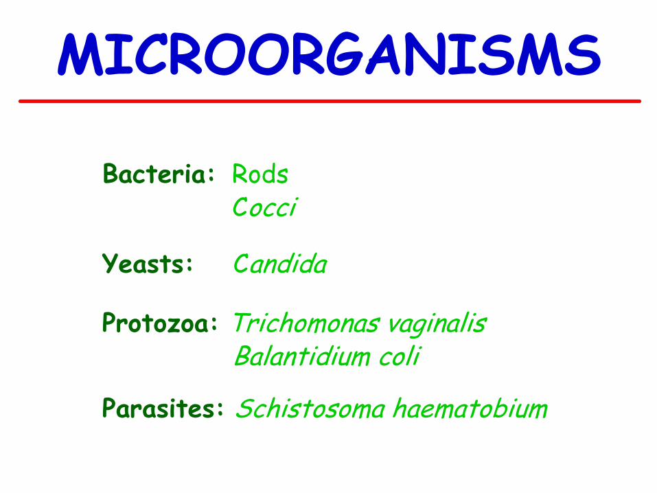
MICROORGANISMS
Bacteria: Rods Cocci
Yeasts: Candida
Protozoa: Trichomonas vaginalis Balantidium coli
Parasites: Schistosoma haematobium
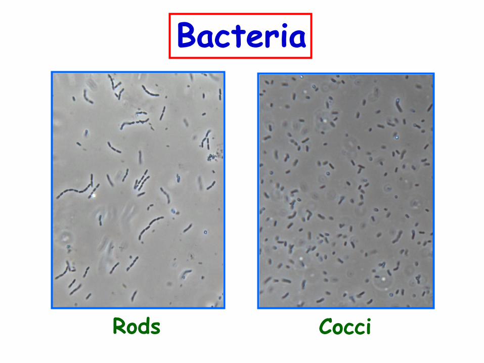
Bacteria
Rods Cocci
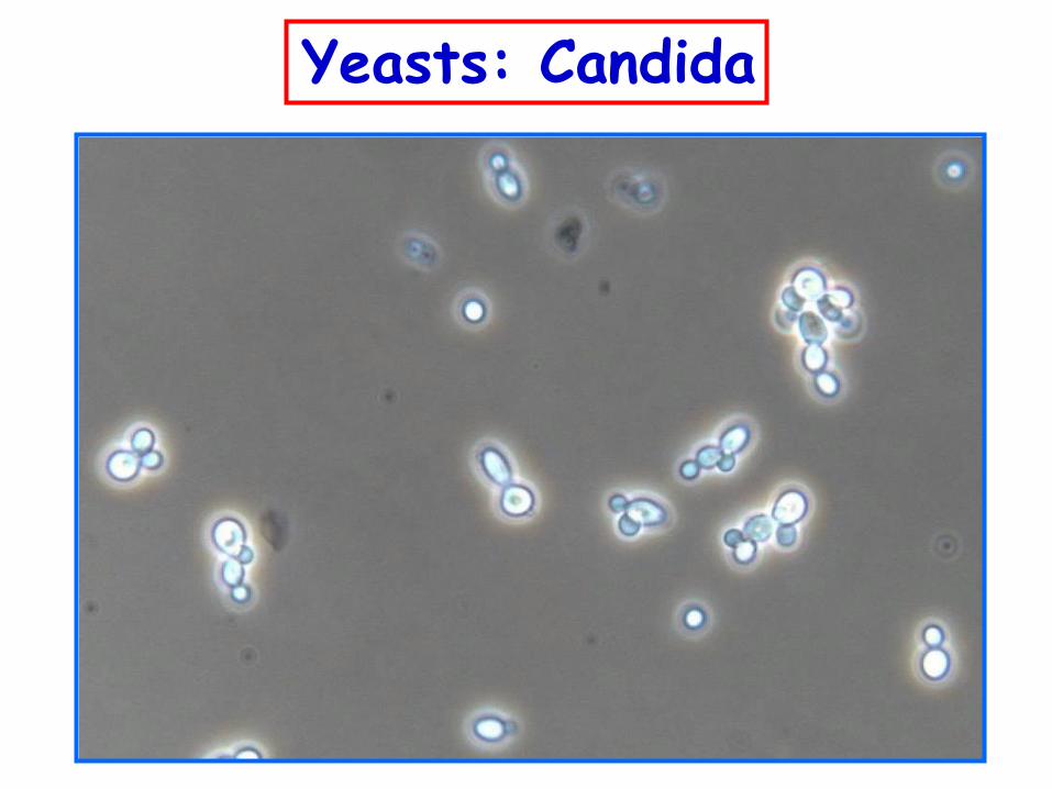
Yeasts: Candida
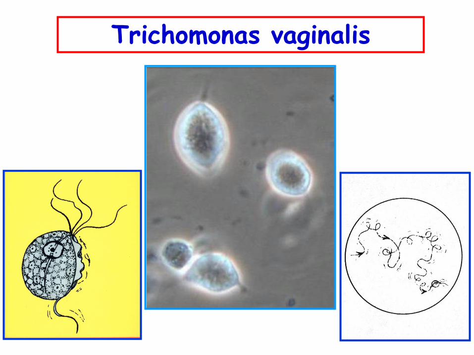
Trichomonas vaginalis
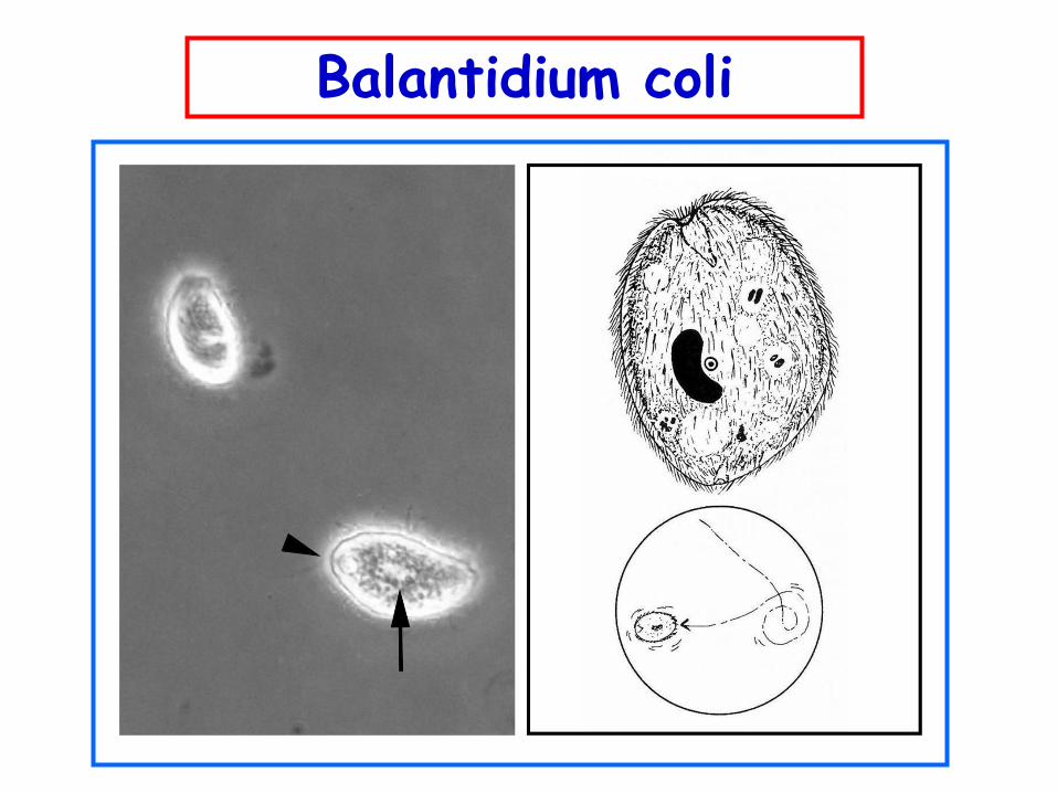
Balantidium coli
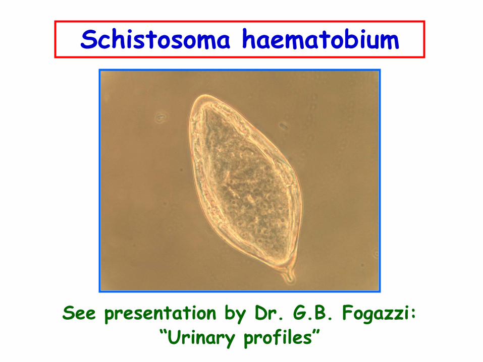
Schistosoma haematobium
See presentation by Dr. G.B. Fogazzi: “Urinary profiles”
CONTAMINANTS
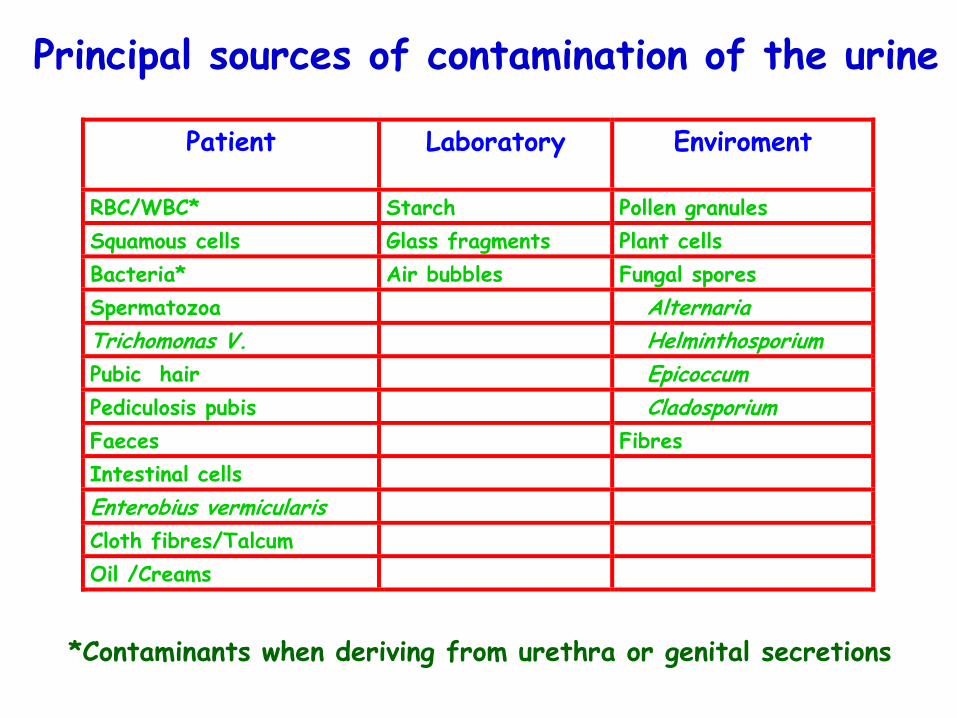
Principal sources of contamination of the urine
Patient Laboratory Enviroment
RBC/WBC* Starch Pollen granules
Squamous cells Glass fragments Plant cells
Bacteria* Air bubbles Fungal spores
Spermatozoa Alternaria
Trichomonas V. Helminthosporium
Pubic hair Epicoccum
Pediculosis pubis Cladosporium
Faeces Fibres
Intestinal cells
Enterobius vermicularis
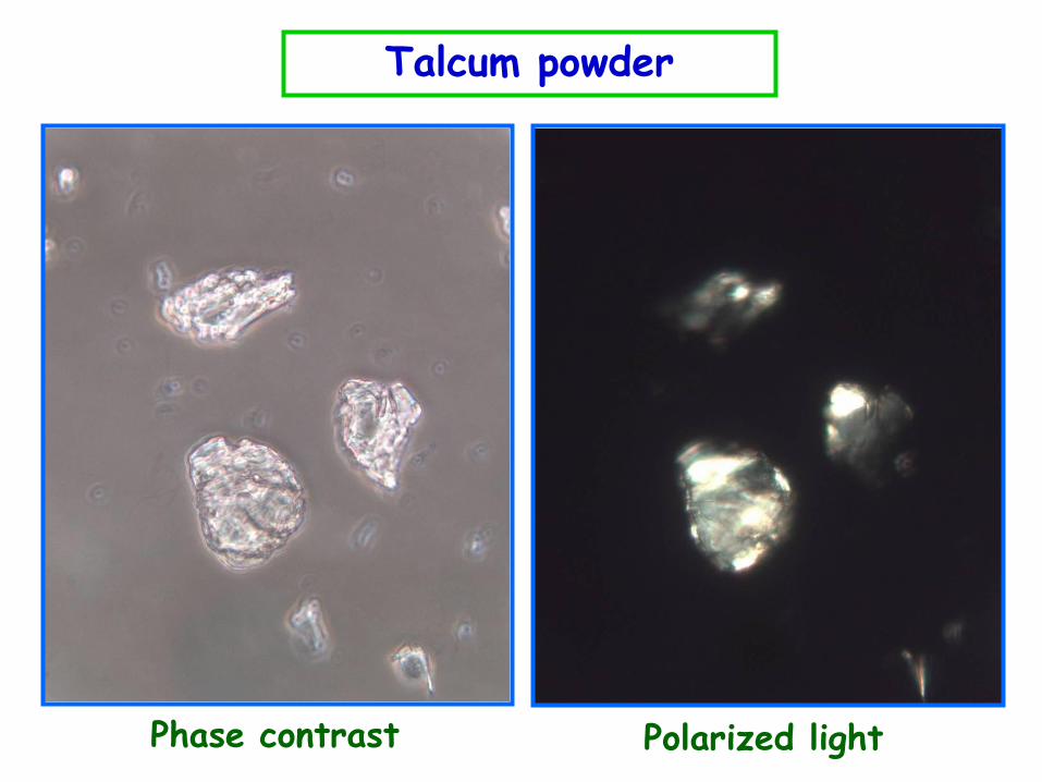
Cloth fibres/Talcum
Oil /Creams
*Contaminants when deriving from urethra or genital secretions
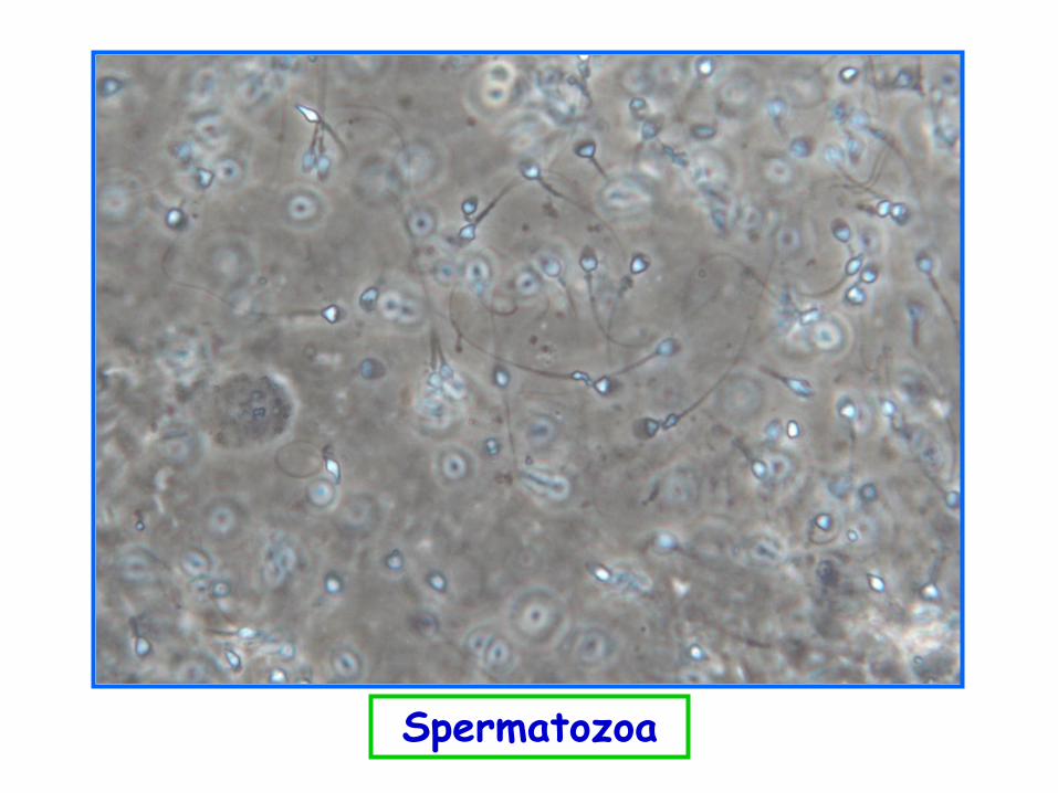
Spermatozoa
Phase contrast Polarized light
Talcum powder

Cloth fibre
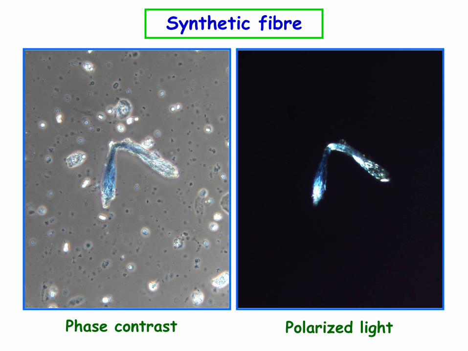
Synthetic fibre
Phase contrast Polarized light
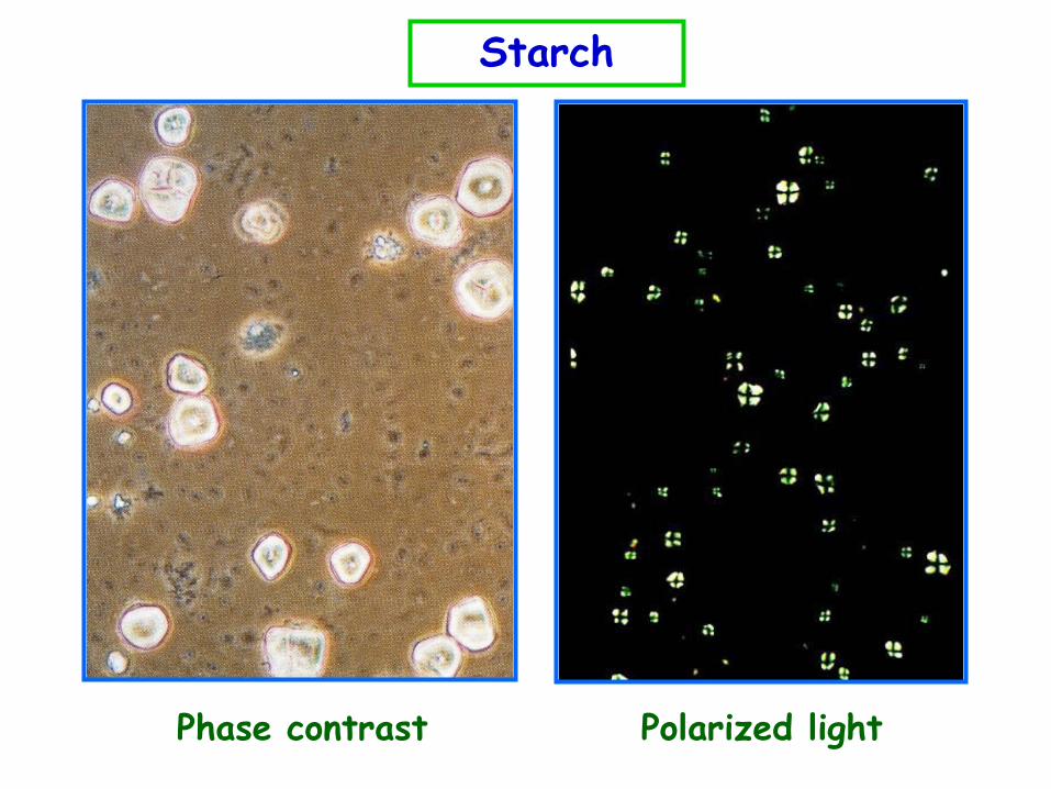
Starch
Polarized light Phase contrast

Glass fragment
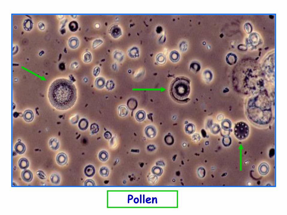
Pollen
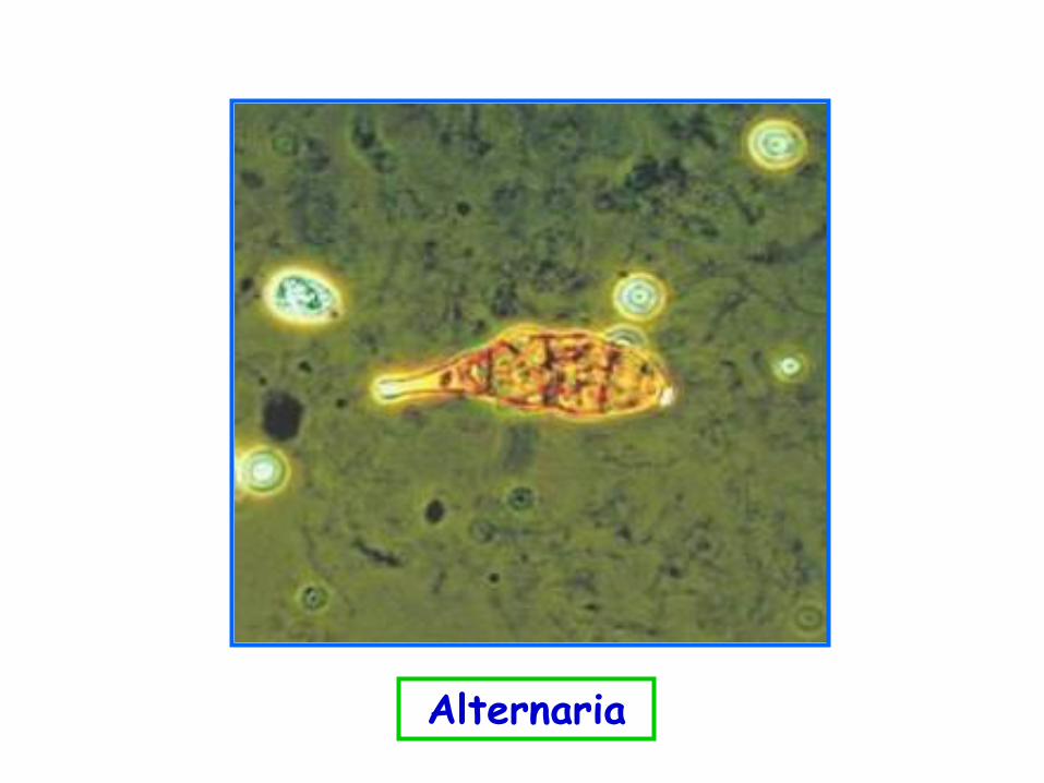
Alternaria
Thank you for your kind
attention


![Impact of enterococcal urinary tract infections in …men [1,4]. The infection may be restricted to lower urinary tract or can expand to upper urinary tract resulting to several clinical](https://img.pdfslide.us/doc/110x75/5e5f2365ac5ba200ef0f04fc/impact-of-enterococcal-urinary-tract-infections-in-men-14-the-infection-may.jpg)


![Clinical Anatomy of GIT and Urinary Tract [Autosaved]](https://img.pdfslide.us/doc/110x75/55cf9d72550346d033adaae3/clinical-anatomy-of-git-and-urinary-tract-autosaved.jpg)