Embed Size (px)
Citation preview
TH
EJ
OU
RN
AL
OF
CE
LL
BIO
LO
GY
©
The Rockefeller University Press $8.00The Journal of Cell Biology, Vol. 169, No. 1, April 11, 2005 105–116http://www.jcb.org/cgi/doi/10.1083/jcb.200408066
JCB: ARTICLE
JCB 105
The p38
�
/
�
MAPK functions as a molecular switch to activate the quiescent satellite cell
Nathan C. Jones,
1
Kristina J. Tyner,
3
Lisa Nibarger,
3
Heather M. Stanley,
3
Dawn D.W. Cornelison,
3
Yuri V. Fedorov,
2
and Bradley B. Olwin
3
1
Bayer Corporation, Research Triangle Park, NC 27709
2
Dharmacon Research, Lafayette, CO 80026
3
Department of Molecular, Cellular, and Developmental Biology, University of Colorado at Boulder, Boulder, CO 80309
omatic stem cells cycle slowly or remain quiescentuntil required for tissue repair and maintenance.Upon muscle injury, stem cells that lie between the
muscle fiber and basal lamina (satellite cells) are acti-vated, proliferate, and eventually differentiate to repairthe damaged muscle. Satellite cells in healthy muscle arequiescent, do not express MyoD family transcription fac-tors or cell cycle regulatory genes and are insulated fromthe surrounding environment. Here, we report that the
S
p38
�
/
�
family of mitogen-activated protein kinases (MAPKs)reversibly regulates the quiescent state of the skeletal mus-cle satellite cell. Inhibition of p38
�
/
�
MAPKs (a) promotesexit from the cell cycle, (b) prevents differentiation, and (c)insulates the cell from most external stimuli allowing thesatellite cell to maintain a quiescent state. Activation ofsatellite cells and p38
�
/
�
MAPKs occurs concomitantly,providing further support that these MAPKs function as amolecular switch for satellite cell activation.
Introduction
Maintenance and repair of skeletal muscle tissue is per-formed by a specialized somatic stem cell termed the satellitecell. Satellite cells, which comprise a small (1–6%) numberof the total myonuclei, are located between the basal laminaand the muscle fiber and can remain quiescent for an averageof 7 yr in adult humans (Schultz and McCormick, 1994;Seale and Rudnicki, 2000; Hawke and Garry, 2001). Thequiescent satellite cell expresses few gene products and doesnot express members of the MyoD family (Cornelison andWold, 1997; Cornelison et al., 2000). Activation of the satellitecell, a critical but poorly understood process, can be inducedby HGF or TNF
�
injection in vivo (Tatsumi et al., 1998; Li,2003) and is accompanied by a general initiation of genetranscription including induction of MyoD and myf-5, anincrease in cellular volume, and entry into the cell cycle(Cornelison and Wold, 1997; Cornelison et al., 2000). Quiescentsatellite cells express c-met (the HGF receptor), FGF receptors1 and 4, syndecan-3, and syndecan-4, all of which appear tobe involved in satellite cell activation and proliferation(Tatsumi et al., 1998; Flanagan-Steet et al., 2000; Cornelisonet al., 2001, 2004).
FGFs and HGF stimulate MAPK signaling cascades,which have been correlated with the regulation of proliferationand differentiation in many cell types, including skeletal myo-blasts. At least four MAPK families have been identified:the extracellular signal-regulated kinases (ERKs), the c-junNH
3
-terminal kinases/stress activated protein kinase, the p38MAPKs, and the ERK5 or big MAPKs (Lewis et al., 1998).The role of MAPK signaling cascades in myogenesis is contro-versial, as MAPK activation has been implicated in both posi-tive and negative regulation of myogenic differentiation. Thesediscrepancies may arise from the different origins of the linesused and the maintenance of these lines in culture. Dependingon cell type, activation of the MKK1/2–ERK1/2 pathway eitherpromotes (C3H10T1/2 cells; Gredinger et al., 1998) or inhibits(23A2, L6A1, and C2C12; Milasincic et al., 1996; Coolican etal., 1997; Weyman et al., 1997) myogenic differentiation. Wehave shown that activation of the Raf–MKK1/2–ERK1/2module is required for proliferation of the MM14 musclesatellite cell line, but is dispensable for FGF-dependent inhibitionof terminal differentiation (Jones et al., 2001). Additionally,activation of the MKK1/2–ERK1/2 pathway is not sufficient topromote proliferation of MM14 cells, suggesting that addi-tional FGF-dependent signaling pathways are required. Ectopicoverexpression of MKP-1, a negative regulator of MAPKs, inC2C12 myoblasts inhibits both proliferation and myoblast
N.C. Jones and K.J. Tyner contributed equally to this work.Correspondence to Bradley B. Olwin: [email protected] used in this paper: ERK, extracellular signal-regulated kinase.
on November 16, 2018jcb.rupress.org Downloaded from http://doi.org/10.1083/jcb.200408066Published Online: 11 April, 2005 | Supp Info:
JCB • VOLUME 169 • NUMBER 1 • 2005106
fusion without affecting expression of muscle-specific genes(Bennett and Tonks, 1997). Together, these results suggest thatdistinct subsets of MAPKs are likely to regulate proliferationand differentiation of skeletal myoblasts.
The p38
�
/
�
MAPKs, which are activated in responseto growth factor stimulation (Morooka and Nishida, 1998;Iwasaki et al., 1999; Maher, 1999) have been reported to berequired for late stages of myogenic differentiation of bothC2C12 and L6 myoblasts (Cuenda and Cohen, 1999; Zet-ser et al., 1999). We found that active p38
�
/
�
(pp38
�
/
�
)MAPKs present in proliferating satellite cells and MM14cells are localized to the nucleus, suggesting that these ki-nases may function before cell differentiation. Consistentwith this, we observed that inhibition of p38
�
/
�
MAPK ac-tivity prevented activation and proliferation of satellite cellscultured on intact myofibers. We also show that p38
�
/
�
MAPKs are required for MyoD induction in satellite cellsand for MM14 differentiation. Interestingly, inhibition ofthese MAPKs in either MM14 cells or satellite cells inducesa reversible quiescent state whereby the cells are unrespon-sive to external stimuli, similar to that observed for normaladult satellite cells in uninjured muscle tissue.
Results
Several MAPKs are expressed in proliferating and differentiated MM14 cells
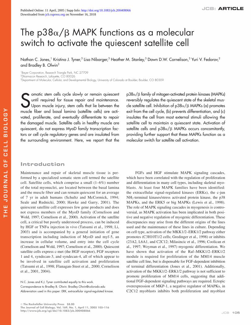
We have previously shown that ERK1/2 is required for prolif-eration but not differentiation in MM14 cells (Jones et al.,2001). To gain insight into which MAPKs may play additionalroles in MM14 cell proliferation and differentiation, we per-formed RT-PCR for MAPKs on MM14 cells at various timesafter induction of differentiation. Neither
erk1
,
2
nor
3
MAPKsappear to change expression levels upon MM14 cell differenti-ation (Fig. 1 A). Both ERK5 and
p38
�
appeared to decreaseduring differentiation, whereas
p38
�
/
�
decreased slightly (Fig.1 A). As a reference,
myoD
shows an increase in mRNA levelsfollowed by a decrease at 72 h.
fgfr-1
, which is lost during dif-ferentiation, also initially appears to increase and then declinesdramatically by 72 h of differentiation (Fig. 1 A). Although weexpected to observe
erk1/2
in proliferating cells, the presenceof p38
�
/
�
MAPK mRNA in proliferating cells was unex-pected, as the p38
�
/
�
MAPK kinases are reported to be bothsufficient and necessary to promote myogenesis (Cuenda andCohen, 1999; Zetser et al., 1999). We asked if the active phos-
Figure 1. p38�/� MAPK is present in proliferatingMM14 cells and is activated by FGF-2. (A) Expression ofMAPKs in proliferating and differentiated MM14 cells.FGF was removed from MM14 cell cultures at time 0 andthe expression of erk1, erk2, erk3, erk5, p38�/�, p38�,myoD, and fgfr-1 was determined by RT-PCR. Threesamples for each time point are shown, with increasingconcentrations of input cDNA for each indicated. A loadingcontrol (18S RNA) is also included. (B and C) Proliferat-ing MM14 cells were fixed and immunofluorescenceperformed with anti-pp38�/� antibodies. (C) Digitaldeconvolution analysis reveals that pp38 is present in thecell nuclei as identified by DAPI staining. (D) p38�/�MAPK is activated by FGF-2 but not 12-O-tetra-deca-noylphorbol-13-acetate (TPA) in MM14 cells. FGF-medi-ated phosphorylation of p38�/� MAPK is inhibited bySB203580 but not by MEK1/2 inhibitors. Cell extractswere subjected to Western analysis and probed usinganti-pp38�/� antibodies (top) then the blot stripped andreprobed with anti-p38�/� antibodies (bottom).
P38
�
/
�
MAPKS ACTIVATE SATELLITE CELLS • JONES ET AL.
107
phorylated p38
�
/
�
(pp38
�
/
�
) was present in MM14 cells andfound pp38
�
/
�
localized to the cell nucleus of proliferatingcells (Fig. 1, B and C). Because MM14 cells are exquisitelysensitive to FGF removal, which is sufficient to trigger differ-entiation from the G1 phase of the cell cycle, we asked if re-moval of FGF would promote p38
�
/
�
activation. Upon re-moval of FGF in the presence of 15% horse serum, we foundlittle if any detectable pp38
�
/
�
MAPK (Fig. 1 D, top). How-ever, FGF-2 addition to starved MM14 cells robustly stimu-lated p38
�
/
�
phosphorylation (Fig. 1 D). The activation ofthese MAPKs appears specific because it was blocked by incu-bation with the p38 inhibitor SB203580 but was insensitive toERK1/2 inhibitors (Fig. 1 D). The apparent activation of p38
�
/
�
MAPKs by FGF-2 is not due to changes in p38
�
/
�
protein lev-els as indicated by reprobing the Western blot with an antibodythat recognizes p38
�
/
�
(Fig. 1 D, bottom).
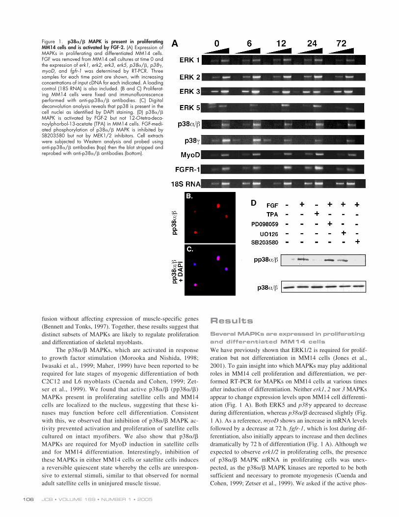
Active p38
�
/
�
is required for proliferation of MM14 cells
FGF-mediated activation of p38
�
/
�
MAPKs and their presencein the nucleus of proliferating myoblasts were unexpected, sug-gesting a role for these MAPKs in proliferating myoblasts. Weasked what effect inhibition of p38
�
/
�
MAPKs would have onproliferating MM14 cells. If synchronized by mitotic shake-offMM14 cells require FGF within 4-6 h to proceed through the
cell cycle (Clegg et al., 1987; Kudla et al., 1995) and thus wetested the effect of increasing doses of SB203580 on synchro-nized MM14 cells. Addition of SB203580 to proliferating cellsprevented DNA synthesis in a dose-dependent manner, reduc-ing DNA synthesis to control levels with 25
�
M SB203580(Fig. 2 A). To demonstrate that the SB203580-dependent inhi-bition of proliferation is specific for the p38
�
/
�
MAPKs, weattempted to rescue the SB203580 effects with an SB203580-resistant p38
�
(p38
�
TM
; Eyers et al., 1999). In the presenceof SB203580, MM14 cells expressing a control vector orwild-type p38
�
failed to proliferate (Fig. 2 B). In contrast,SB203580-treated cells expressing p38
�
TM
proliferated, dem-onstrating rescue by the drug-resistant p38
�
TM
(Fig. 2 C). To-gether, these data support the conclusion that p38
�
/
�
MAPKsare required for myoblast proliferation and that the phenotypeof SB203580-treated cells is specifically due to loss of p38
�
/
�
MAPK activity.
Active p38
�
/
�
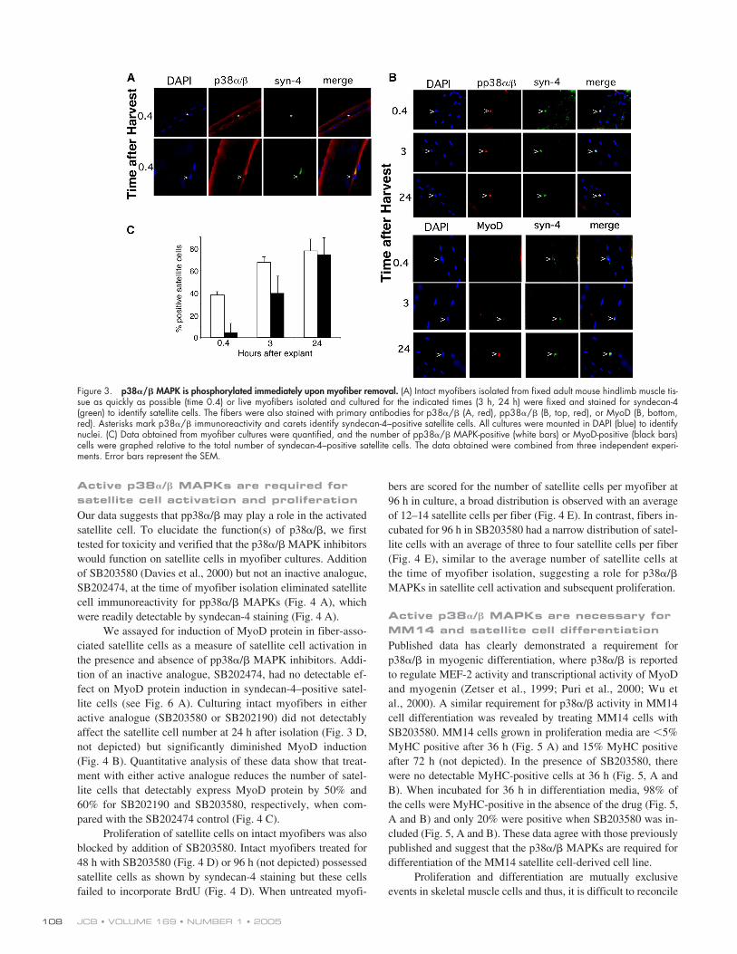
is present in recently activated satellite cellsA role for p38�/� MAPKs has not been demonstrated in pro-liferating myoblasts and thus might potentially reflect an arti-fact of the MM14 cell line. Therefore, we asked if p38�/�MAPKs were present in activated satellite cells cultured on in-tact myofibers. The p38�/� MAPKs appear present in freshlyisolated myofibers at the time of muscle harvest. The proteinappears localized in the cytoplasm of the myofibers adjacentto the myonuclei (Fig. 3 A, asterisk in myofiber panel). In ad-dition, immunoreactive p38�/� was present in quiescent skel-etal muscle satellite cells (Fig. 3 A, caret), readily identifiedby their syndecan-4 staining (Cornelison et al., 2001). In thesatellite cells, the p38�/� MAPKs appear to localize to the cy-toplasm (Fig. 3 A). If p38�/� MAPKs are required for satellitecell proliferation, pp38�/� should be present. When stainedwith an antibody that recognizes pp38�/� satellite cell nucleion myofibers fixed at the time of harvest and on myofibers af-ter 3 and 24 h of culture were pp38�/� positive (Fig. 3 B).When muscle tissue was removed, fixed, and myofibers teasedfrom the fixed muscle tissue, 40% of the satellite cells wereimmunoreactive for pp38�/� (Fig. 3, B and C). This numberincreases rapidly to �80% by 24 h in culture (Fig. 3 C) beforecell division at 36 h. The appearance of pp38�/� in muscletissue immediately upon harvest suggests that p38�/� phos-phorylation may rapidly occur upon satellite cell activation.Although we removed muscle tissue and fixed the tissueimmediately, a minimum of 15–20 min required to dissect themuscle is essentially a massive injury and likely results in sat-ellite cell activation. Thus, we expect isolation of muscle tis-sue to activate satellite cells. However, the induction of MyoDexpression, regarded as an early marker for satellite cell acti-vation was not detected until after 3 h of myofiber culture(Fig. 3 B). The frequency of pp38�/� immunoreactivity in sat-ellite cells is significantly higher than that of MyoD at 0.4 and3 h of myofiber culture (Fig. 3 C), revealing that p38�/�MAPKs are activated before MyoD accumulation in satellitecells (Fig. 3, B and C) and that both events occur before satel-lite cell duplication.
Figure 2. p38�/� MAPK activity is required for MM14 cell proliferation.(A) MM14 cells synchronized by mitotic shake-off were grown in thepresence or absence of added FGF-2 and treated with either DMSO (control)or increasing concentrations of SB203580, and DNA synthesis deter-mined by [3H]thymidine incorporation. (B) MM14 cell were cotransfectedwith expression vectors encoding �-galactosidase, p38�, or p38�TM andpcDNA as indicated to rescue SB203580 inhibition of p38�/� activity.Cells were incubated in either DMSO carrier (white bars) 20 �M (graybars) or 40 �M SB203580 (black bars) for 36 h then fixed, stained for�-galactosidase to identify transfected cells and scored. The data representtwo independent experiments performed in quadruplicate.
JCB • VOLUME 169 • NUMBER 1 • 2005108
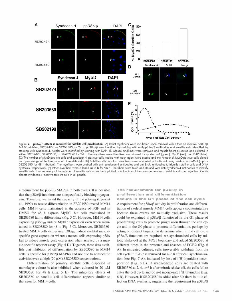
Active p38�/� MAPKs are required for satellite cell activation and proliferationOur data suggests that pp38�/� may play a role in the activatedsatellite cell. To elucidate the function(s) of p38�/�, we firsttested for toxicity and verified that the p38�/� MAPK inhibitorswould function on satellite cells in myofiber cultures. Additionof SB203580 (Davies et al., 2000) but not an inactive analogue,SB202474, at the time of myofiber isolation eliminated satellitecell immunoreactivity for pp38�/� MAPKs (Fig. 4 A), whichwere readily detectable by syndecan-4 staining (Fig. 4 A).
We assayed for induction of MyoD protein in fiber-asso-ciated satellite cells as a measure of satellite cell activation inthe presence and absence of pp38�/� MAPK inhibitors. Addi-tion of an inactive analogue, SB202474, had no detectable ef-fect on MyoD protein induction in syndecan-4–positive satel-lite cells (see Fig. 6 A). Culturing intact myofibers in eitheractive analogue (SB203580 or SB202190) did not detectablyaffect the satellite cell number at 24 h after isolation (Fig. 3 D,not depicted) but significantly diminished MyoD induction(Fig. 4 B). Quantitative analysis of these data show that treat-ment with either active analogue reduces the number of satel-lite cells that detectably express MyoD protein by 50% and60% for SB202190 and SB203580, respectively, when com-pared with the SB202474 control (Fig. 4 C).
Proliferation of satellite cells on intact myofibers was alsoblocked by addition of SB203580. Intact myofibers treated for48 h with SB203580 (Fig. 4 D) or 96 h (not depicted) possessedsatellite cells as shown by syndecan-4 staining but these cellsfailed to incorporate BrdU (Fig. 4 D). When untreated myofi-
bers are scored for the number of satellite cells per myofiber at96 h in culture, a broad distribution is observed with an averageof 12–14 satellite cells per fiber (Fig. 4 E). In contrast, fibers in-cubated for 96 h in SB203580 had a narrow distribution of satel-lite cells with an average of three to four satellite cells per fiber(Fig. 4 E), similar to the average number of satellite cells atthe time of myofiber isolation, suggesting a role for p38�/�MAPKs in satellite cell activation and subsequent proliferation.
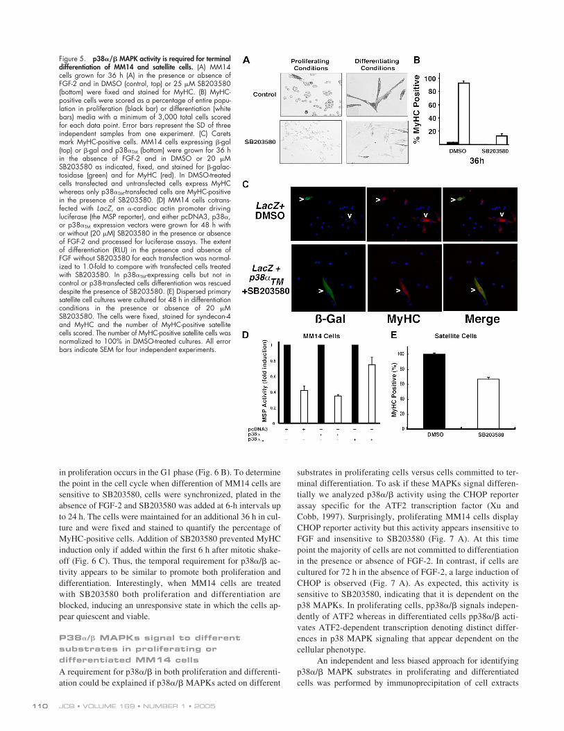
Active p38�/� MAPKs are necessary for MM14 and satellite cell differentiationPublished data has clearly demonstrated a requirement forp38�/� in myogenic differentiation, where p38�/� is reportedto regulate MEF-2 activity and transcriptional activity of MyoDand myogenin (Zetser et al., 1999; Puri et al., 2000; Wu etal., 2000). A similar requirement for p38�/� activity in MM14cell differentiation was revealed by treating MM14 cells withSB203580. MM14 cells grown in proliferation media are �5%MyHC positive after 36 h (Fig. 5 A) and 15% MyHC positiveafter 72 h (not depicted). In the presence of SB203580, therewere no detectable MyHC-positive cells at 36 h (Fig. 5, A andB). When incubated for 36 h in differentiation media, 98% ofthe cells were MyHC-positive in the absence of the drug (Fig. 5,A and B) and only 20% were positive when SB203580 was in-cluded (Fig. 5, A and B). These data agree with those previouslypublished and suggest that the p38�/� MAPKs are required fordifferentiation of the MM14 satellite cell-derived cell line.
Proliferation and differentiation are mutually exclusiveevents in skeletal muscle cells and thus, it is difficult to reconcile
Figure 3. p38�/� MAPK is phosphorylated immediately upon myofiber removal. (A) Intact myofibers isolated from fixed adult mouse hindlimb muscle tis-sue as quickly as possible (time 0.4) or live myofibers isolated and cultured for the indicated times (3 h, 24 h) were fixed and stained for syndecan-4(green) to identify satellite cells. The fibers were also stained with primary antibodies for p38�/� (A, red), pp38�/� (B, top, red), or MyoD (B, bottom,red). Asterisks mark p38�/� immunoreactivity and carets identify syndecan-4–positive satellite cells. All cultures were mounted in DAPI (blue) to identifynuclei. (C) Data obtained from myofiber cultures were quantified, and the number of pp38�/� MAPK-positive (white bars) or MyoD-positive (black bars)cells were graphed relative to the total number of syndecan-4–positive satellite cells. The data obtained were combined from three independent experi-ments. Error bars represent the SEM.
P38�/� MAPKS ACTIVATE SATELLITE CELLS • JONES ET AL. 109
a requirement for p38�/� MAPKs in both events. It is possiblethat the p38�/� inhibitors are nonspecifically blocking myogen-esis. Therefore, we tested the capacity of the p38�TM (Eyers etal., 1999) to rescue differentiation in SB203580-treated MM14cells. MM14 cells maintained in the absence of FGF and inDMSO for 48 h express MyHC, but cells maintained inSB203580 fail to differentiate (Fig. 5 C). However, MM14 cellsexpressing p38�TM induce MyHC expression even when main-tained in SB203580 for 48 h (Fig. 5 C). Moreover, SB203580-treated MM14 cells expressing p38�TM induce skeletal muscle-specific gene expression whereas treated cells expressing p38�
fail to induce muscle gene expression when assayed by a mus-cle-specific reporter assay (Fig. 5 D). Together, these data estab-lish that inhibition of differentiation by SB203580 in MM14cells is specific for p38�/� MAPKs and not due to nonspecificactivities even at high (20 �M) SB203580 concentrations.
Differentiation of primary satellite cells dispersed inmonolayer culture is also inhibited when cultured in 20 �MSB203580 for 48 h (Fig. 5 E). The inhibitory effects ofSB203580 on satellite cell differentiation appears similar tothat seen for MM14 cells.
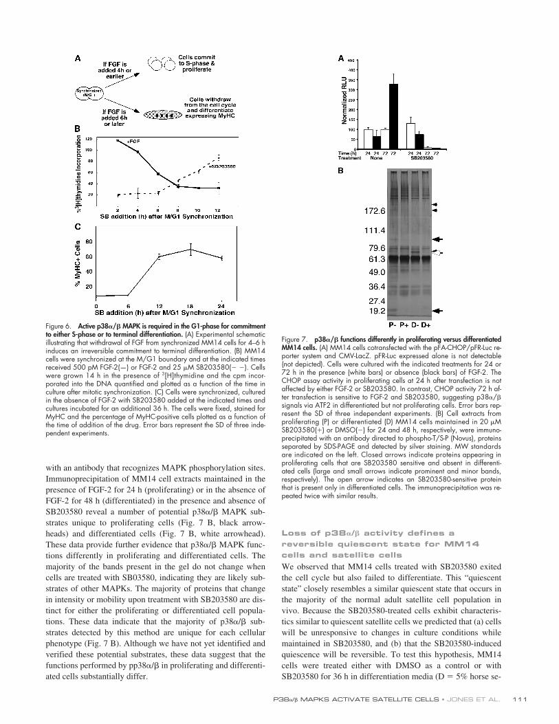
The requirement for p38�/� in proliferation and differentiation occurs in the G1 phase of the cell cycleA requirement for p38�/� activity in proliferation and differen-tiation of skeletal muscle MM14 cells appears counterintuitivebecause these events are mutually exclusive. These resultscould be explained if p38�/� functioned in the G1 phase ofproliferating cells to promote progression through the cell cy-cle and in the G0 phase to promote differentiation, perhaps byacting on distinct targets. To determine when in the cell cyclep38�/� functions are required, we synchronized cells by mi-totic shake-off at the M/G1 boundary and added SB203580 atdifferent times in the presence and absence of FGF-2 (Fig. 6A). In untreated cultures, cells irreversibly withdraw from thecell cycle if FGF-2 is removed for 4–6 h after cell synchroniza-tion (see Fig. 7 A), indicated by loss of [3H]thymidine incor-poration (Fig. 6 B). If synchronized cells are treated withSB203580 at 2, 4, or 6 h after mitotic shake-off, the cells fail toenter the cell cycle and do not incorporate [3H]thymidine (Fig.6 B). However, if SB203580 is added after 6 h there is little ef-fect on DNA synthesis, suggesting the requirement for p38�/�
Figure 4. p38�/� MAPK is required for satellite cell proliferation. (A) Intact myofibers were incubated upon removal with either an inactive p38�/�MAPK inhibitor, SB202474, or SB203580 for 24 h. pp38�/� was identified by staining with anti-pp38�/� antibodies and satellite cells identified bystaining with syndecan-4. Nuclei were identified by staining with DAPI. (B) Mouse hindlimbs were removed and muscle fibers dissected and cultured ineither SB202474, SB203580, or SB202190 for 24 h. The myofibers were then fixed and stained for syndecan-4 (green), MyoD (red), and DAPI (blue).(C) The number of MyoD-positive cells and syndecan-4–positive cells treated with each agent were scored and the number of MyoD-positive cells plottedas a percentage of the total number of satellite cells. (D) Satellite cells on intact myofibers were incubated in BrdU-containing medium in DMSO (top) orSB203580 for 48 h (bottom). The myofibers were probed with anti–syndecan-4 antibodies and anti-BrdU antibodies to identify satellite cells and DNAsynthesis, respectively. (E) Intact myofibers were cultured as in D for 96 h. The fibers were fixed and stained with anti–syndecan-4 antibodies to identifysatellite cells. The frequency of the number of satellite cells scored was plotted as a function of the average number of satellite cells per myofiber. Caretsdenote syndecan-4–positive satellite cells in all panels.
JCB • VOLUME 169 • NUMBER 1 • 2005110
in proliferation occurs in the G1 phase (Fig. 6 B). To determinethe point in the cell cycle when differention of MM14 cells aresensitive to SB203580, cells were synchronized, plated in theabsence of FGF-2 and SB203580 was added at 6-h intervals upto 24 h. The cells were maintained for an additional 36 h in cul-ture and were fixed and stained to quantify the percentage ofMyHC-positive cells. Addition of SB203580 prevented MyHCinduction only if added within the first 6 h after mitotic shake-off (Fig. 6 C). Thus, the temporal requirement for p38�/� ac-tivity appears to be similar to promote both proliferation anddifferentiation. Interestingly, when MM14 cells are treatedwith SB203580 both proliferation and differentiation areblocked, inducing an unresponsive state in which the cells ap-pear quiescent and viable.
P38�/� MAPKs signal to different substrates in proliferating or differentiated MM14 cellsA requirement for p38�/� in both proliferation and differenti-ation could be explained if p38�/� MAPKs acted on different
substrates in proliferating cells versus cells committed to ter-minal differentiation. To ask if these MAPKs signal differen-tially we analyzed p38�/� activity using the CHOP reporterassay specific for the ATF2 transcription factor (Xu andCobb, 1997). Surprisingly, proliferating MM14 cells displayCHOP reporter activity but this activity appears insensitive toFGF and insensitive to SB203580 (Fig. 7 A). At this timepoint the majority of cells are not committed to differentiationin the presence or absence of FGF-2. In contrast, if cells arecultured for 72 h in the absence of FGF-2, a large induction ofCHOP is observed (Fig. 7 A). As expected, this activity issensitive to SB203580, indicating that it is dependent on thep38 MAPKs. In proliferating cells, pp38�/� signals indepen-dently of ATF2 whereas in differentiated cells pp38�/� acti-vates ATF2-dependent transcription denoting distinct differ-ences in p38 MAPK signaling that appear dependent on thecellular phenotype.
An independent and less biased approach for identifyingp38�/� MAPK substrates in proliferating and differentiatedcells was performed by immunoprecipitation of cell extracts
Figure 5. p38�/� MAPK activity is required for terminaldifferentiation of MM14 and satellite cells. (A) MM14cells grown for 36 h (A) in the presence or absence ofFGF-2 and in DMSO (control, top) or 25 �M SB203580(bottom) were fixed and stained for MyHC. (B) MyHC-positive cells were scored as a percentage of entire popu-lation in proliferation (black bar) or differentiation (whitebars) media with a minimum of 3,000 total cells scoredfor each data point. Error bars represent the SD of threeindependent samples from one experiment. (C) Caretsmark MyHC-positive cells. MM14 cells expressing �-gal(top) or �-gal and p38�TM (bottom) were grown for 36 hin the absence of FGF-2 and in DMSO or 20 �MSB203580 as indicated, fixed, and stained for �-galac-tosidase (green) and for MyHC (red). In DMSO-treatedcells transfected and untransfected cells express MyHCwhereas only p38�TM-transfected cells are MyHC-positivein the presence of SB203580. (D) MM14 cells cotrans-fected with LacZ, an �-cardiac actin promoter drivingluciferase (the MSP reporter), and either pcDNA3, p38�,or p38�TM expression vectors were grown for 48 h withor without (20 �M) SB203580 in the presence or absenceof FGF-2 and processed for luciferase assays. The extentof differentiation (RLU) in the presence and absence ofFGF without SB203580 for each transfection was normal-ized to 1.0-fold to compare with transfected cells treatedwith SB203580. In p38�TM-expressing cells but not incontrol or p38-transfected cells differentiation was rescueddespite the presence of SB203580. (E) Dispersed primarysatellite cell cultures were cultured for 48 h in differentiationconditions in the presence or absence of 20 �MSB203580. The cells were fixed, stained for syndecan-4and MyHC and the number of MyHC-positive satellitecells scored. The number of MyHC-positive satellite cells wasnormalized to 100% in DMSO-treated cultures. All errorbars indicate SEM for four independent experiments.
P38�/� MAPKS ACTIVATE SATELLITE CELLS • JONES ET AL. 111
with an antibody that recognizes MAPK phosphorylation sites.Immunoprecipitation of MM14 cell extracts maintained in thepresence of FGF-2 for 24 h (proliferating) or in the absence ofFGF-2 for 48 h (differentiated) in the presence and absence ofSB203580 reveal a number of potential p38�/� MAPK sub-strates unique to proliferating cells (Fig. 7 B, black arrow-heads) and differentiated cells (Fig. 7 B, white arrowhead).These data provide further evidence that p38�/� MAPK func-tions differently in proliferating and differentiated cells. Themajority of the bands present in the gel do not change whencells are treated with SB03580, indicating they are likely sub-strates of other MAPKs. The majority of proteins that changein intensity or mobility upon treatment with SB203580 are dis-tinct for either the proliferating or differentiated cell popula-tions. These data indicate that the majority of p38�/� sub-strates detected by this method are unique for each cellularphenotype (Fig. 7 B). Although we have not yet identified andverified these potential substrates, these data suggest that thefunctions performed by pp38�/� in proliferating and differenti-ated cells substantially differ.
Loss of p38�/� activity defines a reversible quiescent state for MM14 cells and satellite cellsWe observed that MM14 cells treated with SB203580 exitedthe cell cycle but also failed to differentiate. This “quiescentstate” closely resembles a similar quiescent state that occurs inthe majority of the normal adult satellite cell population invivo. Because the SB203580-treated cells exhibit characteris-tics similar to quiescent satellite cells we predicted that (a) cellswill be unresponsive to changes in culture conditions whilemaintained in SB203580, and (b) that the SB203580-inducedquiescence will be reversible. To test this hypothesis, MM14cells were treated either with DMSO as a control or withSB203580 for 36 h in differentiation media (D � 5% horse se-
Figure 6. Active p38�/� MAPK is required in the G1-phase for commitmentto either S-phase or to terminal differentiation. (A) Experimental schematicillustrating that withdrawal of FGF from synchronized MM14 cells for 4–6 hinduces an irreversible commitment to terminal differentiation. (B) MM14cells were synchronized at the M/G1 boundary and at the indicated timesreceived 500 pM FGF-2(—) or FGF-2 and 25 �M SB203580(� �). Cellswere grown 14 h in the presence of 3[H]thymidine and the cpm incor-porated into the DNA quantified and plotted as a function of the time inculture after mitotic synchronization. (C) Cells were synchronized, culturedin the absence of FGF-2 with SB203580 added at the indicated times andcultures incubated for an additional 36 h. The cells were fixed, stained forMyHC and the percentage of MyHC-positive cells plotted as a function ofthe time of addition of the drug. Error bars represent the SD of three inde-pendent experiments.
Figure 7. p38�/� functions differently in proliferating versus differentiatedMM14 cells. (A) MM14 cells cotransfected with the pFA-CHOP/pFR-Luc re-porter system and CMV-LacZ. pFR-Luc expressed alone is not detectable(not depicted). Cells were cultured with the indicated treatments for 24 or72 h in the presence (white bars) or absence (black bars) of FGF-2. TheCHOP assay activity in proliferating cells at 24 h after transfection is notaffected by either FGF-2 or SB203580. In contrast, CHOP activity 72 h af-ter transfection is sensitive to FGF-2 and SB203580, suggesting p38�/�signals via ATF2 in differentiated but not proliferating cells. Error bars rep-resent the SD of three independent experiments. (B) Cell extracts fromproliferating (P) or differentiated (D) MM14 cells maintained in 20 �MSB203580(�) or DMSO(�) for 24 and 48 h, respectively, were immuno-precipitated with an antibody directed to phospho-T/S-P (Novus), proteinsseparated by SDS-PAGE and detected by silver staining. MW standardsare indicated on the left. Closed arrows indicate proteins appearing inproliferating cells that are SB203580 sensitive and absent in differenti-ated cells (large and small arrows indicate prominent and minor bands,respectively). The open arrow indicates an SB203580-sensitive proteinthat is present only in differentiated cells. The immunoprecipitation was re-peated twice with similar results.
JCB • VOLUME 169 • NUMBER 1 • 2005112
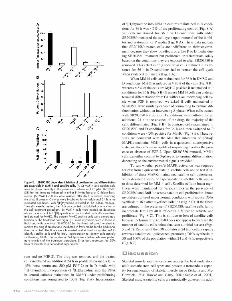
rum and no FGF-2). The drug was removed and the treatedcells incubated an additional 24 h in proliferation media (P �15% horse serum and 500 pM FGF-2) or in D media with3[H]thymidine. Incorporation of 3[H]thymidine into the DNAin control cultures maintained in DMSO under proliferatingconditions was normalized to 100% (Fig. 8 A). Incorporation
of 3[H]thymidine into DNA in cultures maintained in D condi-tions for 36 h was �5% of the proliferating control (Fig. 8 A)yet cells maintained for 36 h in D conditions with addedSB203580 reentered the cell cycle upon removal of the inhibi-tor and restoration of P media (Fig. 8 A). These data indicatethat SB203580-treated cells are indifferent to their environ-ment because they show no effects of either P or D media dur-ing SB203580 treatment but proliferate or differentiate solelybased on the conditions they are exposed to after SB203580 isremoved. This effect is drug specific as cells cultured in its ab-sence for 36 h in D conditions fail to reenter the cell cyclewhen switched to P media (Fig. 8 A).
When MM14 cells are maintained for 36 h in DMSO andD conditions, MyHC is induced in 95% of the cells (Fig. 8 B),whereas 5% of the cells are MyHC positive if maintained in Pconditions for 36 h (Fig. 8 B). Because MM14 cells can undergoterminal differentiation from G1 without an intervening cell cy-cle when FGF is removed, we asked if cells maintained inSB203580 were similarly capable of committing to terminal dif-ferentiation without an intervening S-phase. When cells treatedwith SB203580 for 36 h in D conditions were cultured for anadditional 24 h in the absence of the drug, the majority of thecells differentiated (Fig. 8 B). In contrast, cells maintained inSB203580 and D conditions for 36 h and then switched to Pconditions were �5% positive for MyHC (Fig. 8 B). These re-sults are consistent with the idea that inhibition of p38�/�MAPKs maintains MM14 cells in a quiescent, nonresponsivestate, and the cells are incapable of responding to either the pres-ence or absence of FGF-2. Upon SB203580 removal, MM14cells can either commit to S-phase or to terminal differentiation,depending on the environmental signals provided.
To test whether p38�/� MAPK activation was requiredfor exit from a quiescent state in satellite cells and to test if in-hibition of these MAPKs maintained satellite cell quiescence,we performed a series of experiments on satellite cells similarto those described for MM14 cells. Satellite cells on intact myo-fibers were maintained for various times in the presence ofSB203580 and BrdU to assess satellite cell proliferation. Intactmyofibers cultured under normal conditions will induce DNAsynthesis �36 h after myofiber isolation (Fig. 8 C). If the fibersare cultured in the presence of SB203580, satellite cells fail toincorporate BrdU by 48 h reflecting a failure to activate andproliferate (Fig. 8 C). This is not due to loss of satellite cellsbecause inclusion of SB203580 does not appear to decrease thenumber of satellite cells below that seen at initial harvest (Figs.5 and 7). Removal of the p38 inhibitor at 24 h of culture rapidlyreverses satellite cell quiescence, promoting DNA synthesis in50 and 100% of the population within 24 and 48 h, respectively(Fig. 8 C).
DiscussionSkeletal muscle satellite cells are among the best understoodadult somatic stem cell types and possess a tremendous capac-ity for regeneration of skeletal muscle tissue (Schultz and Mc-Cormick, 1994; Hawke and Garry, 2001; Seale et al., 2001).Skeletal muscle satellite cells are mitotically quiescent in adult
Figure 8. SB203580 dependent inhibition of proliferation and differentiationare reversible in MM14 and satellite cells. (A–C) MM14 and satellite cellswere incubated initially in the presence or absence of 25 �M SB203580(SB) for the times as indicated in either P (white bars) or D (black bars)media. (A) MM14 cultures were washed after 36 h in culture, removingthe drug, if present. Cultures were incubated for an additional 24 h in theindicated conditions with 3[H]thymidine included in the culture medium.The cells were harvested, the 3[H]cpm counted and plotted as a function ofthe cell treatment paradigm. (B) MM14 cells were treated as describedabove for A except that 3[H]thymidine was not added and cells were fixedand stained for MyHC. The percent MyHC-positive cells were plotted as afunction of the treatment paradigm. (C) Intact myofibers were cultured inBrdU and with or without SB203580 for the times indicated, washed toremove the drug if present and incubated in fresh media for the additionaltimes indicated. The fibers were harvested and stained for syndecan-4 toidentify satellite cells and for BrdU incorporation to identify cells activelysynthesizing DNA. The number of BrdU-positive satellite cells was plottedas a function of the treatment paradigm. Error bars represent the SEMfrom at least three independent experiments.

P38�/� MAPKS ACTIVATE SATELLITE CELLS • JONES ET AL. 113
uninjured skeletal muscle tissue and may remain quiescent foryears in humans (Schultz et al., 1978; Schultz and McCormick,1994). As such, satellite cells maintain a low metabolic profileand maintain gene expression for a small subset of growth-related genes that include FGF receptors 1 and 4 and c-met butno detectable expression of cell cycle–related genes or myo-genic transcription factors (Cornelison et al., 2000). Satellitecell activation occurs rapidly after muscle injury; once acti-vated, satellite cells induce expression of the myogenic regula-tory factors, commit to DNA synthesis and begin proliferatingin vivo (Schultz and Jaryszak, 1985; Schultz, 1996) and on in-tact myofibers (Bischoff, 1986; Yablonka-Reuveni and Rivera,1994; Cornelison and Wold, 1997). Removal of myofibersfrom intact skeletal muscle simulates an injury response, acti-vating satellite cells with an accompanying rapid increase incytoplasmic volume. Robust gene expression begins withthe initiation of myogenic regulatory factor expression whereMyoD is observed in 20–40% of the cells 3–6 h after isolationand 50–70% of the cells at 24 h. Proliferation is nearly syn-chronous for the first two divisions, occurring every 30–36 h(Bischoff, 1986; Cornelison and Wold, 1997).
Satellite cells within the muscle are both rare, comprisingonly 1–6% of the total muscle nuclei, and isolated; thus, little isknown regarding the intracellular pathways regulating their ac-tivation, proliferation, and differentiation. We have been inves-tigating the role(s) of MAPKs in skeletal muscle satellite cellsin the MM14 satellite cell line, explanted myofibers and in dis-persed cultures of satellite cells (Jones et al., 2001). For the p38MAPK family, work performed on either myogenic cell linesor in fibroblasts (10T1/2) cells converted to muscle by ectopicMyoD expression shows that p38�/� MAPKs promote myo-genesis, presumably via mechanisms that influence the tran-scriptional activation of the MyoD and MEF2 families (Cuendaand Cohen, 1999; Zetser et al., 1999; Puri et al., 2000; Wu etal., 2000; Xu et al., 2002). However, when examining the roleof p38�/� MAPKs in the context of a developing limb it wasobserved that myogenesis was significantly enhanced whenp38�/� MAPKs were inhibited, the opposite of what would bepredicted for the role of p38�/� from studies involving myo-genic cell lines (Weston et al., 2003). Explanations for thesedisparate observations have not yet been forthcoming.
The consequences of p38�/� inhibition are manifested asa failure to differentiate (a phenotype that is observed whenp38�/� is inhibited in the MM14, C2C12, and L6/L8 cell lines)and a failure to proliferate in both MM14 cells and satellitecells, a novel observation. The inhibition of proliferation and theblock to differentiation both occur when p38�/� activity is in-hibited in the G1 phase of the cell cycle. Targets of the p38�/�MAPKs that mediate these events are unidentified. Although asignificant body of data has shown that MEF2A and MEF2C aresubstrates of p38� and p38�2 (Ornatsky et al., 1999; Yang etal., 1999), satellite cells do not express detectable MEF2 tran-scripts until 96 h after myofiber isolation, suggesting that theseare unlikely p38�/� targets for satellite cell activation or prolif-eration. Other known substrates of p38�/� include transcriptionfactors (Max and ATF2), kinases (MAPKAP kinase 2 and 3),and phospholipase A2 (Lewis et al., 1998). In proliferating
MM14 cells, manipulation of p38�/� activity by FGF removalor by SB203580 addition has no significant effect on ATF2-dependent transcription. However, in differentiated cells, ATF2-dependent transcription is SB203580 and FGF-2 sensitive. Thissuggests that p38�/� MAPKs act upon different substrates inproliferating and differentiating cells. A similar observationwas noted for MM14 cells where differential activation ofthe ERK1/2 pathway occurred in cells committed to S-phaseas compared with cells committed to terminal differentiation(Campbell et al., 1995). These reports provide supporting evi-dence for differences in signaling pathways that are dependenton the phenotypic state of skeletal muscle cells. When furtheranalyzed for differences in substrate specificity, we found thatthe majority of p38�/� substrates were different in proliferatingversus differentiated cells. Thus, the observation that p38�/�MAPKs are required for both proliferation and differentiationcould be explained by differential substrate accessibility im-posed by commitment of the cell to S-phase or to terminal dif-ferentiation. A better understanding of the roles of p38�/�MAPKs in these events will require identification of substratesspecific for cells committed to proliferate and differentiate.
A requirement for p38�/� MAPKs in satellite cell prolif-eration appears similar to that observed in MM14 cells. How-ever, pp38�/� is detected in the cell nucleus of satellite cellswithin 20 min of isolation, suggesting that these MAPKs mayplay a role in signaling pathways that participate in activationof satellite cells. Consistent with this suggestion is the observa-tion that inhibition of p38�/� activity in satellite cells preventsMyoD induction and proliferation, both markers of satellite cellactivation. Although these data support a role for p38�/�MAPKs in satellite cell activation, the role of these kinases ap-pears more complicated. Our data suggest that p38�/� is criti-cal for balancing satellite cell activation and quiescence. Sup-porting this hypothesis is the observation that both culturedsatellite cells and MM14 cells fail to respond to changes in se-rum concentrations or FGF-2 when p38�/� is inhibited, indi-cating a general unresponsiveness to environmental stimuli.Importantly this unresponsive or quiescent state is reversible in
Figure 9. A model for the function of p38�/� MAPK in satellite cellactivation. Upon injury to the muscle tissue, the p38�/� MAPK is immediatelyactivated by as yet unidentified signals that may include signaling from eitherFGF receptor-1, FGF receptor-4, c-met, or the TNF�-receptor. Once activated,p38�/� MAPK activity is maintained to allow satellite cell proliferationand reparation of damaged tissue. Activated p38�/� is also required forcell differentiation, and acts on distinct substrates to promote proliferationand differentiation. The quiescent state is maintained by inhibition ofp38�/� MAPKs presumably via MAPK phosphatases.
JCB • VOLUME 169 • NUMBER 1 • 2005114
both MM14 and satellite cells. When SB203580 is removed,MM14 cells can either reenter the cell cycle and proliferate orexit the cell cycle and terminally differentiate upon addition ofP or D media, respectively. Satellite cells on intact myofibers,which do not undergo differentiation reenter the cell cycleupon SB203580 removal. Because the behavior of MM14 andsatellite cells in SB203580 resembles the quiescent state of sat-ellite cells in adult uninjured skeletal muscle tissue, we proposethat activation of p38�/� functions as an intracellular “molecu-lar switch” for satellite cell activation (Fig. 9).
In vivo, mechanical stress activates satellite cells via anHGF and NO-dependent mechanism (Tatsumi et al., 2002); ac-tivation also occurs upon in vivo administration of TNF� (Li,2003). It is noteworthy that p38�/� MAPKs are also activatedby mechanical stress (Cowan and Storey, 2003; Kumar et al.,2003; Wretman et al., 2001), HGF (Recio and Merlino, 2002),TNF� (Geng et al., 1996; Roulston et al., 1998) and FGFs. Cel-lular stress responses often lead to an inflammatory responseinvolving activation of p38�/� MAPKs in immune-responsivecells (Cowan and Storey, 2003; Kumar et al., 2003) and in skel-etal muscle tissue (Wretman et al., 2001), where a severestretch is capable of p38 activation. Our model (Fig. 9) for reg-ulation of satellite cell activation proposes that these physio-logical events activating satellite cells is mediated via activa-tion of p38�/� MAPKs. We further predict that the satellitecell quiescent state is maintained by inhibition of p38�/�MAPKs, presumably via a p38�/� MAPK phosphatase, per-haps MKP-1 (Bennett and Tonks, 1997).
We propose that activation of the skeletal muscle satellitecell, a well-studied adult somatic stem cell, is concomitant withthe activation of p38�/� MAPKs and suggest that these MAPKsfunction as a molecular switch determining the activation stateof the satellite cell. The universal response of the p38�/�MAPKs to stress suggests that similar mechanisms could be in-volved in the control of other somatic stem cell populations.
Materials and methodsCell cultureMM14 cells were grown on gelatin-coated plates in growth media consist-ing of Ham’s F10C or F12C media supplemented with 15% horse serumas previously described (Clegg et al., 1987). FGF-2 was added in in-creasing concentrations (from 0.3 to 2.5 nM) every 12 h, depending oncell density. Proliferation media was replaced every 24 h. Differentiationinducing culture media is comprised of Ham’s F10C or F12C media sup-plemented with 15% horse serum and no added FGF-2 unless otherwisenoted. Primary myofibers were isolated and cultured as described previ-ously (Cornelison et al., 2004).
Myofiber preparation, immunohistochemistry, and scoringMyofibers with their associated satellite cells were prepared as describedpreviously (Cornelison et al., 2004). In brief, muscle was dissected fromadult mouse hindlimbs and digested with collagenase type I (Worthington)to yield single intact myofibers. BrdU is routinely added to cultures to facil-itate cell cycle studies. Additional supplements to the medium included 50�M SB203580 in DMSO or the DMSO carrier as a control. At the desig-nated time points after harvest fibers were fixed and stained as describedabove. Primary antibodies and dilutions used also included rabbit affinitypurified polyclonal anti-p38 (C-20) (Santa Cruz Biotechnology, Inc.) at1:50, mouse monoclonal anti–phospho-p38 (New England BioLabs, Inc.)at 1:50, mouse monoclonal anti-BrdU (BMB) at 1:10, mouse monoclonalanti-MyoD (Novocastra) at 1:10, and mouse monoclonal anti-myogenin(F5D; Cusella-DeAngelis et al., 1992), neat, chicken anti–mouse synde-
can-4 (1:1,500). Secondary antibodies anti–rabbit, anti–mouse, and anti–chicken Alexa 488, anti–rabbit, anti–mouse Alexa 504, and anti–rat Cas-cade blue were purchased from Molecular Probes, Inc. and were used at1:500 unless otherwise indicated. Counts of resident satellite cells per myo-fiber were done by counting DAPI-stained myofiber nuclei (which can beidentified by their characteristic elongated shape) and DAPI-stained satel-lite cell nuclei coincident with syndecan-4–positive cell outlines. At least20 myofibers containing at least 5,000 myonuclei total were counted pertime point per condition. Counts of MyoD-positive satellite cells were doneby counting MyoD-positive DAPI-stained nuclei within syndecan-4–positivecell outlines and comparing to counts of total DAPI-stained nuclei withinsyndecan-4–positive cell outlines.
Primary satellite cell culturesPrimary satellite cells were isolated and cultured as described previously(Cornelison et al., 2004) on gelatin-coupled coverslips, incubated in dif-ferentiation media for 48 h (with 2.5% horse serum), and 20 �MSB20350 or DMSO fixed and stained with anti–syndecan-4 and MF20antibodies at 1:1,500 and neat, respectively. Secondary antibodies wereAlexa 488 and Alexa 504 at 1:500. Syndecan-4–positive cells werescored for MF-20 (MyHC) staining and plotted as a function of the synde-can-4–positive cells in the population.
Microscopy and image acquisitionAll microscope images were obtained on a microscope (model E800; Ni-kon) using a 60� plan Apo lens at RT using a Cooke Sensicam digitalcamera and Intelligent Imaging Innovations Slidebook software to acquireimages on a Macintosh computer. Fluorescence from labeled secondaryantibodies was subtracted from all fluorescent images. Images were ex-ported into Photoshop, if necessary the brightness and contrast was ad-justed to the entire image, the image cropped and individual color chan-nels extracted without color correction adjustments or gamma adjustments.
RT-PCR analysis of MAPK expressionMM14 cells were cultured on 100-mm plates under differentiating condi-tions (F10C supplemented with 1.5% horse serum) for 0, 6, 12, 24, and72 h (five plates at a density of 250,000 cells per plate for each treat-ment). Total RNA was isolated as previously described (Chomczynski andSacchi, 1987). 5 �g of total RNA was added to reverse transcriptasebuffer (GIBCO BRL) containing 0.025 Oligo (dT)12-18 (GIBCO BRL), 0.01 MDTT (GIBCO BRL), 0.5 mM dNTP mix (GIBCO BRL), and 200 U of Su-perscript II reverse transcriptase (GIBCO BRL) and incubated for 50 min at42�C. Nonreverse transcriptase controls were performed as describedabove with the exception of reverse transcriptase addition.
PCR amplification was performed by adding increasing concentra-tions of cDNA in 2 �l (1:100, 1:10 dilution, undiluted) to PCR buffer con-taining 0.25 mM each dNTP, 1.5 mM MgCl2, 0.5 �M each of forwardand reverse primers for ERK1/2, ERK3, ERK5, p38�/�, p38�, MyoD,FGFR-1, 18S RNA, and 5U of Taq polymerase (GIBCO BRL). Each reactionwas amplified for 35 cycles using the following parameters: denaturation94�C for 1 min, annealing at 50�C for 1 min, and elongation for 1 min. Af-ter amplification, each reaction was resolved on a 0.8% agarose gel con-taining ethidium bromide and visualized with a UV transilluminator.
Western analysisMM14 cells, at a density of 5 � 105 cells per 100-mm plate, were washedthree times with 5 ml of PBS (137 mM NaCl, 2.7 mM KCl, 4.3 mMNa2HPO4 •7H2O, 1.4 mM KH2PO4, pH 7.3) and then grown in growthmedia containing 1.5% horse serum in the absence of added FGF-2 for 0,6, 12, 24, and 72 h. Alternatively, MM14 cells were grown in differentia-tion media for 2 h, then DMSO, PD098059 (50 �M), U0126 (25 �M), orSB203580 (25 �M) was added and the cells grown for an additional 24 h.Cells were stimulated for 10 min with either 0.1 nM FGF-2 or 100 nM12-O-tetra-decanoylphorbol-13-acetate as indicated. Cell lysates were pre-pared as previously described. Protein concentrations were determined bybicinchonic acid protein assay (Pierce Chemical Co.). Extracted proteins(20 �g) were resolved by SDS-PAGE and electrophoretically transferred toImmobilon-P (Millipore) in 25 mM ethanolamine, 25 mM glycine, 20%methanol, pH 9.5. Nonspecific binding sites were blocked with 5% nonfatdried milk, 0.05% Tween 20 in PBS. p38�/� activity was determined byprobing blots with anti–phospho-p38�/� antibodies (New England Bio-labs, Inc.), and p38 expression was determined by probing with anti-p38(C20) antibodies (Santa Cruz Biotechnology, Inc.). Bound antibodies weredetected with either anti–rabbit IgG or anti–mouse IgG conjugated to HRP(Promega). Bound antibody complexes were visualized by RenaissanceWestern blot chemiluminescence reagent (Dupont).
P38�/� MAPKS ACTIVATE SATELLITE CELLS • JONES ET AL. 115
DNA synthesis assayDNA synthesis was assayed by [3H]thymidine incorporation. In brief,MM14 cells were synchronized by mitotic shake-off and plated in to 24-well plates at density 2,000 cells/well in the presence or absence of exog-enous FGF. The cells were grown with the addition of 7.5 �l of DMSO orincreasing concentrations of SB203580 for 8 h and then given 2 �Ci of[3H]thymidine (DuPont) and incubated for an additional 6 h. The amountof [3H]thymidine incorporated into DNA was determined by liquid scintil-lation counting as described previously.
For time course assays, MM14 cells were synchronized by mitoticshake-off and plated in growth media with 2 �Ci of [3H]thymidine (Du-pont) in the presence or absence of exogenous FGF-2,and either 25 �MSB203580 or 500 pM FGF-2 was added at increasing time intervals. Thecells were grown for a total of 14 h after plating. The amount of [3H]thymi-dine incorporated into DNA was determined by liquid scintillation count-ing as described previously. Thymidine incorporation for each samplewas normalized to the thymidine incorporation of MM14 cells grown en-tirely in proliferating conditions.
Additionally, MM14 cells were cultured in differentiation mediaand in the presence or absence of 25 �M SB203580 for 24 h. Cells werewashed three times with PBS and media replaced with either growth ordifferentiation culture media with 2 �Ci of [3H]thymidine (Dupont). Thecells were grown for 12 h and the amount of [3H]thymidine incorporatedinto DNA was determined by liquid scintillation counting. Thymidine incor-poration for each sample was normalized to the thymidine incorporationof MM14 cells grown entirely in proliferating conditions.
Analysis of myosin heavy chain expressionMM14 cells were plated onto 6-well plates at a cell density of 8,000cells/well in growth medium. After 6 h, either 2.5 �l of DMSO orSB203580 (50 �M) was added to the wells and cells were grown in thepresence or absence of FGF-2 as described above. At either 36 h or 72 hafter treatment, cells fixed and stained for myosin heavy chain expressionas described previously (Kudla et al., 1995). MHC-positive cells werescored as a percentage of entire cell population. A minimum of 1,000 to-tal cells was scored per plate. For time course assays, MM14 cells weresynchronized by mitotic shake-off and plated in differentiation media and25 �M SB203580 was added at increasing time intervals (0, 6, 12, 18,and 24 h) after plating. Cells were fixed 36 h after plating and stained forMHC expression. MHC-positive cells were scored as a percentage of en-tire cell population. A minimum of 1,000 total cells were scored per plate.Alternatively, MM14 cells were cultured in either proliferation media ordifferentiation media in the presence or absence of 25 �M SB203580.36 h after plating, cells were either fixed and stained for MHC expressionor washed three times in PBS (to remove residual SB203580) and mediareplenished with either proliferation or differentiation media.
ImmunoprecipitationMM14 cells (3 � 105 per plate, two plates each condition) in proliferationmedia were maintained in proliferation media or switched to differentia-tion media in the presence of either 20 �M SB203580 or DMSO. Prolif-erating cells were harvested 24 h later by centrifugation. AdditionalSB203580 or DMSO was added to differentiating cultures at 24 h andcells were harvested 24 h later by the same method. Cells were resus-pended in modified RIPA buffer, sonicated and 100 �g of protein wasprecleared with protein A–Sepharose beads before being incubated with3 �l of Novus Biologicals ab9344 overnight at 4�C, incubated 1 h at 4�Cwith 30 �l of prewashed protein A–Sepharose beads, washed and boiledin SDS-PAGE sample buffer for 10 min and 25 �l of supernatant fromeach sample separated on a 4–20% gradient gel. Proteins were visual-ized by silver staining.
Transient transfectionsClonal cell proliferation assay. MM14 cells were plated on 100-mm tissueculture plates at a density of 3,000 cells per plate and transfected as pre-viously described. Calcium phosphate-DNA precipitates were made asdescribed previously using 1 �g of a reporter construct containing the cy-tomegalovirus promoter driving the expression of �-galactosidase (CMV-LacZ) and 20 �g of either control DNA (pBSSK�) (Stratagene) caMKK3,or dnMKK3 as described previously (Fedorov et al., 2002) or with Lipo-fectamine 2000 as per manufacturer’s instructions transfected with 1 �gof CMV-LacZ, p38�, or p38�mut per well in a 6-well plate, transferred to100-cm plates 12 h later and plated at clonal density, then fixed 40 h af-ter plating and scored.
Muscle-specific reporter gene assay. MM14 cells were transfectedand assayed as previously described (Fedorov et al., 2002) or plated at
30,000 cells/well (on a 6-well) 12 h before transfection using the Lipo-fectamine 2000 kit. For transfections, 2 �g of plasmid (either p38�,p38�mut, or pDNA3) and 0.25 �g of LacZ and a muscle reporter (lu-ciferase driven by the �-cardiac actin promoter) were added per well. Allmuscle reporter experiments used 20 �M SB203580 or an equivalent vol-ume of DMSO in each well.
P38�/� MAPK reporter assayp38 MAPK activity was determined using Pathdetect CHOP reporting sys-tem (Stratagene) (Xu and Cobb, 1997). For this assay, MM14 cells wereplated on 6-well plates at a density of 104 cells/well and cotransfectedwith 2.5 �g pFR-Luc reporter vector, 500 ng pFA-CHOP vector, and 1 �gCMV-LacZ vector per well. The cells were harvested and assayed for lu-ciferase and �-galactosidase activities as described previously (Fedorov etal., 1998).
This work was supported by grants from the National Institutes of Health andMuscular Dystrophy Association (MDA) to B.B. Olwin (AR39467) and fromthe MDA to D.D.W. Cornelison.
Submitted: 11 August 2004Accepted: 25 February 2005
ReferencesBennett, A.M., and N.K. Tonks. 1997. Regulation of distinct stages of skeletal
muscle differentiation by mitogen-activated protein kinases. Science.278:1288–1291.
Bischoff, R. 1986. Proliferation of muscle satellite cells on intact myofibers inculture. Dev. Biol. 115:129–139.
Campbell, J.S., M.P. Wenderoth, S.D. Hauschka, and E.G. Krebs. 1995. Differ-ential activation of mitogen-activated protein kinase in response to basicfibroblast growth factor in skeletal muscle cells. Proc. Natl. Acad. Sci.USA. 92:870–874.
Chomczynski, P., and N. Sacchi. 1987. Single-step method of RNA isolation byacid guanidium thiocyanate-phenol-chloroform extraction. Anal. Bio-chem. 162:156–159.
Clegg, C.H., T.A. Linkhart, B.B. Olwin, and S.D. Hauschka. 1987. Growth fac-tor control of skeletal muscle differentiation: commitment to terminaldifferentiation occurs in G1 phase and is repressed by fibroblast growthfactor. J. Cell Biol. 105:949–956.
Coolican, S.A., D.S. Samuel, D.Z. Ewton, F.J. McWade, and J.R. Florini. 1997.The mitogenic and myogenic actions of insulin-like growth factors uti-lize distinct signaling pathways. J. Biol. Chem. 272:6653–6662.
Cornelison, D.D.W., and B.J. Wold. 1997. Single-cell analysis of regulatorygene expression in quiescent and activated mouse skeletal muscle satel-lite cells. Dev. Biol. 191:270–283.
Cornelison, D.D., B.B. Olwin, M.A. Rudnicki, and B.J. Wold. 2000. MyoD(�/�) satellite cells in single-fiber culture are differentiation defectiveand MRF4 deficient. Dev. Biol. 224:122–137.
Cornelison, D.D.W., M.S. Filla, H.M. Stanley, A.C. Rapraeger, and B.B. Olwin.2001. Syndecan-3 and syndecan-4 specifically mark skeletal muscle sat-ellite cells and are implicated in satellite cell maintenance and muscle re-generation. Dev. Biol. 239:79–94.
Cornelison, D.D., S. Wilcox-Adelman, P. Goetinck, H. Rauvala, A.C. Raprae-ger, and B.B. Olwin. 2004. Essential and nonredundant roles for synde-can-3 and syndecan-4 in skeletal muscle satellite cells. Genes Dev. 18:2231–2236.
Cowan, K.J., and K.B. Storey. 2003. Mitogen-activated protein kinases: newsignaling pathways functioning in cellular responses to environmentalstress. J. Exp. Biol. 206:1107–1115.
Cuenda, A., and P. Cohen. 1999. Stress-activated protein kinase-2/p38 and a ra-pamycin-sensitive pathway are required for C2C12 myogenesis. J. Biol.Chem. 274:4341–4346.
Cusella-DeAngelis, M.G., G. Lyons, C. Sonnino, L. DeAngelis, E. Vivarelli, K.Farmer, W.E. Wright, M. Molinaro, M. Bouche, M. Buckingham, and G.Cossu. 1992. MyoD, myogenin independent differentiation of primordialmyoblasts in mouse somites. J. Cell Biol. 116:1243–1255.
Davies, S.P., H. Reddy, M. Caivano, and P. Cohen. 2000. Specificity and mech-anism of action of some commonly used protein kinase inhibitors. Bio-chem. J. 351:95–105.
Eyers, P.A., P. van den IJssel, R.A. Quinlan, M. Goedert, and P. Cohen. 1999.Use of a drug-resistant mutant of stress-activated protein kinase 2a/p38to validate the in vivo specificity of SB 203580. FEBS Lett. 451:191–196.
Fedorov, Y.V., N.C. Jones, and B.B. Olwin. 1998. Regulation of myogenesis by
JCB • VOLUME 169 • NUMBER 1 • 2005116
fibroblast growth factors requires beta-gamma subunits of pertussistoxin-sensitive G proteins. Mol. Cell. Biol. 18:5780–5787.
Fedorov, Y.V., N.C. Jones, and B.B. Olwin. 2002. Atypical protein kinase cs arethe ras effectors that mediate repression of myogenic satellite cell differ-entiation. Mol. Cell. Biol. 22:1140–1149.
Flanagan-Steet, H., K. Hannon, M.J. McAvoy, R. Hullinger, and B.B. Olwin.2000. Loss of FGF Receptor 1 signaling reduces skeletal muscle massand disrupts myofiber organization in the developing limb. Dev. Biol.218:21–37.
Geng, Y., J. Valbracht, and M. Lotz. 1996. Selective activation of the mitogen-activated protein kinase subgroups c-Jun NH2 terminal kinase and p38by IL-1 and TNF in human articular chondrocytes. J. Clin. Invest. 98:2425–2430.
Gredinger, E., A.N. Gerber, Y. Tamir, S.J. Tapscott, and E. Bengal. 1998. Mito-gen-activated protein kinase pathway is involved in the differentiation ofmuscle cells. J. Biol. Chem. 273:10436–10444.
Hawke, T.J., and D.J. Garry. 2001. Myogenic satellite cells: physiology to mo-lecular biology. J. Appl. Physiol. 91:534–551.
Iwasaki, S., M. Iguchi, K. Watanabe, R. Hoshino, M. Tsujimoto, and M. Kohno.1999. Specific activation of the p38 mitogen-activated protein kinasesignaling pathway and induction of neurite outgrowth in PC12 cells bybone morphogenetic protein-2. J. Biol. Chem. 274:26503–26510.
Jones, N.C., Y.V. Fedorov, R.S. Rosenthal, and B.B. Olwin. 2001. ERK1/2 isrequired for myoblast proliferation but is dispensable for muscle geneexpression and cell fusion. J. Cell. Physiol. 186:104–115.
Kudla, A.J., M.L. John, D.F. Bowen-Pope, B. Rainish, and B.B. Olwin. 1995. Arequirement for fibroblast growth factor in regulation of skeletal musclegrowth and differentiation cannot be replaced by activation of platelet-derived growth factor signaling pathways. Mol. Cell. Biol. 15:3238–3246.
Kumar, S., J. Boehm, and J.C. Lee. 2003. p38 MAP kinases: key signalling mol-ecules as therapeutic targets for inflammatory diseases. Nat. Rev. DrugDiscov. 2:717–726.
Lewis, T.S., P.S. Shapiro, and N.G. Ahn. 1998. Signal transduction throughMAP kinase cascades. Adv. Cancer Res. 74:49–139.
Li, Y.P. 2003. TNF-alpha is a mitogen in skeletal muscle. Am. J. Physiol. CellPhysiol. 285:C370–C376.
Maher, P. 1999. p38 mitogen-activated protein kinase activation is required forfibroblast growth factor-2-stimulated cell proliferation but not differenti-ation. J. Biol. Chem. 274:17491–17498.
Milasincic, D.J., M.R. Calera, S.R. Farmer, and P.F. Pilch. 1996. Stimulation ofC2C12 myoblast growth by basic fibroblast growth factor and insulin-like growth factor 1 can occur via mitogen-activated protein kinase-depen-dent and -independent pathways. Mol. Cell. Biol. 16:5964–5973.
Morooka, T., and E. Nishida. 1998. Requirement of p38 mitogen-activated pro-tein kinase for neuronal differentiation in PC12 cells. J. Biol. Chem. 273:24285–24288.
Ornatsky, O.I., D.M. Cox, P. Tangirala, J.J. Andreucci, Z.A. Quinn, J.L. Wrana,R. Prywes, Y.T. Yu, and J.C. McDermott. 1999. Post-translational con-trol of the MEF2A transcriptional regulatory protein. Nucleic Acids Res.27:2646–2654.
Puri, P.L., Z. Wu, P. Zhang, L.D. Wood, K.S. Bhakta, J. Han, J.R. Feramisco,M. Karin, and J.Y. Wang. 2000. Induction of terminal differentiation byconstitutive activation of p38 MAP kinase in human rhabdomyosarcomacells. Genes Dev. 14:574–584.
Recio, J.A., and G. Merlino. 2002. Hepatocyte growth factor/scatter factor acti-vates proliferation in melanoma cells through p38 MAPK, ATF-2 andcyclin D1. Oncogene. 21:1000–1008.
Roulston, A., C. Reinhard, P. Amiri, and L.T. Williams. 1998. Early activationof c-Jun N-terminal kinase and p38 kinase regulate cell survival in re-sponse to tumor necrosis factor alpha. J. Biol. Chem. 273:10232–10239.
Schultz, E. 1996. Satellite cell proliferative compartments in growing skeletalmuscles. Dev. Biol. 175:84–94.
Schultz, E., and D.L. Jaryszak. 1985. Effects of skeletal muscle regeneration onthe proliferation potential of satellite cells. Mech. Ageing Dev. 30:63–72.
Schultz, E., M.C. Gibson, and T. Champion. 1978. Satellite cells are mitoticallyquiescent in mature mouse muscle: an EM and radioautographic study. J.Exp. Zool. 206:451–456.
Schultz, E., and K.M. McCormick. 1994. Skeletal muscle satellite cells. Rev.Physiol. Biochem. Pharmacol. 123:213–257.
Seale, P., and M.A. Rudnicki. 2000. A new look at the origin, function, and“stem-cell” status of muscle satellite cells. Dev. Biol. 218:115–124.
Seale, P., A. Asakura, and M.A. Rudnicki. 2001. The potential of muscle stemcells. Dev. Cell. 1:333–342.
Tatsumi, R., J.E. Anderson, C.J. Nevoret, O. Halevy, and R.E. Allen. 1998.HGF/SF is present in normal adult skeletal muscle and is capable of acti-
vating satellite cells. Dev. Biol. 194:114–128.
Tatsumi, R., A. Hattori, Y. Ikeuchi, J.E. Anderson, and R.E. Allen. 2002. Re-lease of hepatocyte growth factor from mechanically stretched skeletalmuscle satellite cells and role of pH and nitric oxide. Mol. Biol. Cell. 13:2909–2918.
Weston, A.D., A.V. Sampaio, A.G. Ridgeway, and T.M. Underhill. 2003. Inhi-bition of p38 MAPK signaling promotes late stages of myogenesis. J.Cell Sci. 116:2885–2893.
Weyman, C.M., M.B. Ramocki, E.J. Taparowsky, and A. Wolfman. 1997. Dis-tinct signaling pathways regulate transformation and inhibition of skele-tal muscle differentiation by oncogenic Ras. Oncogene. 14:697–704.
Wretman, C., A. Lionikas, U. Widegren, J. Lannergren, H. Westerblad, and J.Henriksson. 2001. Effects of concentric and eccentric contractions onphosphorylation of MAPK(erk1/2) and MAPK(p38) in isolated rat skele-tal muscle. J. Physiol. 535:155–164.
Wu, Z., P.J. Woodring, K.S. Bhakta, K. Tamura, F. Wen, J.R. Feramisco, M.Karin, J.Y. Wang, and P.L. Puri. 2000. p38 and extracellular signal-regu-lated kinases regulate the myogenic program at multiple steps. Mol. Cell.Biol. 20:3951–3964.
Xu, Q., L. Yu, L. Liu, C.F. Cheung, X. Li, S.P. Yee, X.J. Yang, and Z. Wu.2002. p38 Mitogen-activated protein kinase-, calcium-calmodulin-dependent protein kinase-, and calcineurin-mediated signaling pathwaystranscriptionally regulate myogenin expression. Mol. Biol. Cell. 13:1940–1952.
Xu, S., and M.H. Cobb. 1997. MEKK1 binds directly to the c-Jun N-terminalkinases/stress-activated protein kinases. J. Biol. Chem. 272:32056–32060.
Yablonka-Reuveni, Z., and A.J. Rivera. 1994. Temporal expression of regula-tory and structural muscle proteins during myogenesis of satellite cellson isolated adult rat fibers. Dev. Biol. 164:588–603.
Yang, S.H., A. Galanis, and A.D. Sharrocks. 1999. Targeting of p38 mitogen-activated protein kinases to MEF2 transcription factors. Mol. Cell. Biol.19:4028–4038.
Zetser, A., E. Gredinger, and E. Bengal. 1999. p38 mitogen-activated protein ki-nase pathway promotes skeletal muscle differentiation. Participation ofthe Mef2c transcription factor. J. Biol. Chem. 274:5193–5200.


![Bigendothelin-1 via p38-MAPK-dependent mechanism regulates ... · failure in humans [15] and animal models [16]. We have also observed increased phosphorylation of myocardial p38-MAPK](https://img.pdfslide.us/doc/110x75/5f0992017e708231d4277623/bigendothelin-1-via-p38-mapk-dependent-mechanism-regulates-failure-in-humans.jpg)


![Phase 1 and pharmacokinetic study of LY3007113, a p38 … · cytokines that promote angiogenesis [7 ]. As it relates to the tumor microenvironment, p38 MAPK controls the senescence-associated](https://img.pdfslide.us/doc/110x75/5af2e6f07f8b9a4d4d8b656b/phase-1-and-pharmacokinetic-study-of-ly3007113-a-p38-that-promote-angiogenesis.jpg)