Embed Size (px)
Citation preview

The Oxytocin Receptor System:Structure, Function, and Regulation
GERALD GIMPL AND FALK FAHRENHOLZ
Institut fur Biochemie, Johannes Gutenberg Universitat, Mainz, Germany
I. Introduction 630II. Oxytocin and Oxytocin-Like Peptides 630
A. Evolutionary aspects 630B. Gene structure 631C. Gene regulation 632
III. Oxytocin Receptors 634A. Gene structure and regulation 634B. Receptor structure 636C. Ligand binding characteristics 637D. Signal transduction and G protein coupling 641E. Receptor internalization and downregulation 642F. Effects of steroids 643
IV. The Peripheral Oxytocin System 645A. Female reproductive system 645B. Male reproductive tract 648C. Mammary tissues 649D. Kidney 650E. Heart and cardiovascular system 651F. Other localizations 652
V. The Central Oxytocin System 654A. Localization profile 654B. Hypothalamus-neurohypophysis 658C. Adenohypophysis 659D. Centrally mediated autonomic and somatic effects 659
VI. Central Behavioral Effects 662A. Sexual behavior 662B. Maternal behavior 664C. Social behavior 664D. Stress-related behavior 666E. Feeding and grooming 666F. Memory and learning 667G. Tolerance and dependence to opioids 667H. Central disorders in humans 667
VII. Concluding Remarks 668
Gimpl, Gerald, and Falk Fahrenholz. The Oxytocin Receptor System: Structure, Function, and Regulation.Physiol Rev 81: 629–683, 2001.—The neurohypophysial peptide oxytocin (OT) and OT-like hormones facilitatereproduction in all vertebrates at several levels. The major site of OT gene expression is the magnocellular neuronsof the hypothalamic paraventricular and supraoptic nuclei. In response to a variety of stimuli such as suckling,parturition, or certain kinds of stress, the processed OT peptide is released from the posterior pituitary into thesystemic circulation. Such stimuli also lead to an intranuclear release of OT. Moreover, oxytocinergic neuronsdisplay widespread projections throughout the central nervous system. However, OT is also synthesized in periph-eral tissues, e.g., uterus, placenta, amnion, corpus luteum, testis, and heart. The OT receptor is a typical class I Gprotein-coupled receptor that is primarily coupled via Gq proteins to phospholipase C-b. The high-affinity receptorstate requires both Mg21 and cholesterol, which probably function as allosteric modulators. The agonist-bindingregion of the receptor has been characterized by mutagenesis and molecular modeling and is different from theantagonist binding site. The function and physiological regulation of the OT system is strongly steroid dependent.
PHYSIOLOGICAL REVIEWS
Vol. 81, No. 2, April 2001Printed in U.S.A.
http://physrev.physiology.org 6290031-9333/01 $15.00 Copyright © 2001 the American Physiological Society

However, this is, unexpectedly, only partially reflected by the promoter sequences in the OT receptor gene. Theclassical actions of OT are stimulation of uterine smooth muscle contraction during labor and milk ejection duringlactation. While the essential role of OT for the milk let-down reflex has been confirmed in OT-deficient mice, OT’srole in parturition is obviously more complex. Before the onset of labor, uterine sensitivity to OT markedly increasesconcomitant with a strong upregulation of OT receptors in the myometrium and, to a lesser extent, in the deciduawhere OT stimulates the release of PGF2a. Experiments with transgenic mice suggest that OT acts as a luteotrophichormone opposing the luteolytic action of PGF2a. Thus, to initiate labor, it might be essential to generate sufficientPGF2a to overcome the luteotrophic action of OT in late gestation. OT also plays an important role in many otherreproduction-related functions, such as control of the estrous cycle length, follicle luteinization in the ovary, andovarian steroidogenesis. In the male, OT is a potent stimulator of spontaneous erections in rats and is involved inejaculation. OT receptors have also been identified in other tissues, including the kidney, heart, thymus, pancreas,and adipocytes. For example, in the rat, OT is a cardiovascular hormone acting in concert with atrial natriureticpeptide to induce natriuresis and kaliuresis. The central actions of OT range from the modulation of the neuroen-docrine reflexes to the establishment of complex social and bonding behaviors related to the reproduction and careof the offspring. OT exerts potent antistress effects that may facilitate pair bonds. Overall, the regulation by gonadaland adrenal steroids is one of the most remarkable features of the OT system and is, unfortunately, the leastunderstood. One has to conclude that the physiological regulation of the OT system will remain puzzling as long asthe molecular mechanisms of genomic and nongenomic actions of steroids have not been clarified.
I. INTRODUCTION
The neurohypophysial hormone oxytocin (OT) wasthe first peptide hormone to have its structure determinedand the first to be chemically synthesized in biologicallyactive form (143). It is named after the “quick birth”(vkyj 5 quick; tokoj 5 birth) which it causes due to itsuterotonic activity (124). OT was also found to be respon-sible for the milk-ejecting activity of the posterior pitu-itary gland (428). The structure of the OT gene was elu-cidated in 1984 (270), and the sequence of the OT receptorwas reported in 1992 (299).
OT is a very abundant neuropeptide. This becameobvious in a study where the most prevalent hypotha-lamic-specific mRNAs were analyzed. OT was found to bethe most abundant of 43 transcripts identified (202). To-day, we recognize that OT exerts a wide spectrum ofcentral and peripheral effects. The actions of OT rangefrom the modulation of neuroendocrine reflexes to theestablishment of complex social and bonding behaviorsrelated to the reproduction and care of the offspring.Overall, the cyclic nonapeptide OT and its structurallyrelated peptides facilitate the reproduction in all verte-brates at several levels.
In this review, we summarize the present knowledgeof the OT receptor system gained in the different fields ofresearch, thereby focusing mainly on the work over thepast decade. For more details to the different topics, thereader is referred to many excellent reviews that havebeen recently published (1, 42, 43, 155, 160, 161, 261, 265,266, 295, 298, 335, 369, 389, 413, 443, 572, 573, 621). In thefollowing two sections, we describe the structural fea-tures of the OT receptor system on the molecular level.OT has long been considered to be restricted to stimula-tion of uterine contractions during labor and milk ejection
during lactation. These classical functions of the OT sys-tem are treated in the first parts of section IV. The fact thatOT is found in equivalent concentrations in the neurohy-pophysis and plasma of both sexes suggests that OT hasfurther physiological functions. The expression of OT andits receptor has now been identified in a variety of periph-eral tissues. For example, evidence was provided that OTacts in concert with atrial natriuretic peptide (ANP) in thecontrol of body fluid and in cardiovascular homeostasis inthe rat. It is important to note that most of our knowledgeabout functions of OT derives from studies with rats.However, localization and expression patterns of OT re-ceptors show marked species differences, suggesting thatsome of the described OT activities may be species spe-cific. Over the past decade, particularly the central actionsof OT have been intensively studied revealing a profoundregulation by steroids. The regulation by steroids is in facta common theme throughout the review and is probablythe most remarkable feature of the OT receptor system.Finally, in the last section, we summarize the contribu-tions that have been reported on behavioral effects me-diated or modulated by OT. Ironically, the classical “oxy-tocic” function of OT is again open for discussion due tothe results with OT-deficient mice.
II. OXYTOCIN AND OXYTOCIN-LIKE PEPTIDES
A. Evolutionary Aspects
All neurohypophysial hormones are nonapeptideswith a disulfide bridge between Cys residues 1 and 6. Thisresults in a peptide constituted of a six-amino acid cyclicpart and a COOH-terminal a-amidated three-residue tail.Based on the amino acid at position 8, these peptides are
630 GERALD GIMPL AND FALK FAHRENHOLZ Volume 81

classified into vasopressin and OT families: the vasopres-sin family contains a basic amino acid (Lys, Arg), and theOT family contains a neutral amino acid at this position(Table 1). Isoleucine in position 3 is essential for stimu-lating OT receptors and Arg or Lys in position 8 for actingon vasopressin receptors. The difference in the polarity ofthese amino acid residues is believed to enable the vaso-pressin and OT peptides to interact with the respectivereceptors (42).
Virtually all vertebrate species possess an OT-likeand a vasopressin-like peptide. Bony fishes (Osteich-thyes), predecessors of the land vertebrates, possess iso-tocin and vasotocin. Thus two evolutionary molecularlineages have been proposed: an isotocin-mesotocin-OTline, associated with reproductive functions, and a vaso-tocin-vasopressin line involved in water homeostasis. Be-cause vasotocin has been found in the most primitivecyclostomes, the OT and vasopressin genes may havearose by duplication of a common ancestral gene after theradiation of cyclostomes. Based on calculations from thenucleotide level of OT and vasopressin gene, the ancestralgene encoding the precursor protein should be more than500 million years old. The exceptional structural stabilityof the nonapeptides during evolution suggests a strongselective pressure, e.g., by coevolution with the corre-sponding receptors and/or with specific processing en-zymes. A conspicuous diversity of OT-like peptides isfound in cartilaginous fishes (Chondrichthyes). These ma-rine fishes use urea rather than salts for osmoregulation.It was hypothesized that the OT-like hormones gained
their high diversity in Chondrichthyes as they have beenrelieved from the control of ionic homeostasis (1). Nota-bly, OT, the typical hormone of placental mammals, hasbeen identified in the Pacific ratfish, a Chondrichthyesspecies.
Mesotocin is the OT-like hormone found in mostterrestrial vertebrates from lungfishes to marsupials,which includes all nonmammalian tetrapods (amphibians,reptiles, and birds). Only two South American marsupialsexpress OT exclusively, whereas all other marsupialshave mesotocin. In the Northern brown bandicoot (Iso-
odon macrourus) (488) and the North American opossum(Didelphis virginiana) (102), OT is present together withmesotocin. Overall, mesotocin has the largest distributionin vertebrates after vasotocin found in all nonmammalianvertebrates and isotocin identified in bony fishes. Despitethis invariability, no clear physiological role has beenascribed to this peptide so far. It is unknown whether themarsupial species that are endowed with both OT andmesotocin have two distinct receptors. The earthwormEisenia foetida is the most primitive species from whichan OT-related peptide (annetocin) has been isolated(429). Injection of annetocin in the earthworm or inleechs results in induction of egg-laying behavior (430).
B. Gene Structure
In all species, OT and vasopressin genes are on thesame chromosomal locus but are transcribed in opposite
TABLE 1. Oxytocin and related peptides
1 2 3 4 5 6 7 8 9
Oxytocin Cys Tyr Ile Gln Asn Cys Pro Leu Gly(NH2) Placentals, some marsupials,ratfish (Hydrolagus colliei)
Mesotocin p p p p p p p Ile p Marsupials, nonmammaliantetrapods, lungfishes
Isotocin p p p Ser p p p Ile p OsteichthyesGlumitocin p p p Ser p p p Gln p Skates (Chondrichthyes)Valitocin p p p p p p p Val p Sharks (Chondrichthyes)Aspargtocin p p p Asn p p p p p Sharks (Chondrichthyes)Asvatocin p p p Asn p p p Val p Sharks (Chondrichthyes)Phasvatocin p p Phe Asn p p p Val p Sharks (Chondrichthyes)Cephalotocin p p Phe Arg p p p Ile p Octopus vulgaris (Molluscs)Annetocin p Phe Val Arg p p p Thr p Eisenia foetida (Annelids)
Vasotocin p p p p p p p Arg p Nonmammalian vertebrates,cyclostomes
Vasopressin p p Phe p p p p Arg p MammalsLysipressin p p Phe p p p p Lys p Pig, some marsupialsPhenypressin p Phe Phe p p p p Arg p Macropodids (Marsupials)Locupressin p Leu p Thr p p p Arg p Locusta migratoria
(Insects)Arg-conopressin p Ile p Arg p p p Arg p Conus geographicus
(Molluscs)Lys-conopressin p Phe p Arg p p p Lys p Lymnaea stagnalis
(Molluscs)
Asterisks indicate amino acid residues that are identical to the corresponding residues in the oxytocin sequence. [Modified from Acheret al. (1).]
April 2001 THE OXYTOCIN RECEPTOR SYSTEM 631

directions (Fig. 1). The intergenic distance between thesegenes range from 3 to 12 kb in mouse (224), human (500),and rat (391). This type of genomic arrangement couldresult from the duplication of a common ancestral gene,which was followed by inversion of one of the genes. Thehuman gene for OT-neurophysin I encoding the OT pre-propeptide is mapped to chromosome 20p13 (472) andconsists of three exons: the first exon encodes a translo-cator signal, the nonapeptide hormone, the tripeptide pro-cessing signal (GKR), and the first nine residues of neu-rophysin; the second exon encodes the central part ofneurophysin (residues 10–76); and the third exon encodesthe COOH-terminal region of neurophysin (residues 77–93/95).
The high homology of the OT-like precursor polypep-tides is well documented in the sequence of pre-proannetocin from Eisenia foetida, a primitive inverte-brate. It consists of a signal peptide, annetocin (flanked bya Gly COOH-terminal amidation signal and a Lys-Arg di-basic endoproteolytic sequence), and a neurophysin do-main. Notably, 14 cysteine residues that play a crucial rolein constructing the correct tertiary structure of a neuro-physin are completely conserved in the Eisenia neuro-physin domain (499).
The OT prepropeptide is subject to cleavage andother modifications as it is transported down the axon toterminals located in the posterior pituitary (74). The ma-ture peptide products, OT and its carrier molecule neuro-
physin, are stored in the axon terminals until neural in-puts elicit their release (475). The main function ofneurophysin, a small (93–95 residues) disulfide-rich pro-tein, appears to be related to the proper targeting, pack-aging, and storage of OT within the granula before releaseinto the bloodstream. OT is found in high concentrations(.0.1 M) in the neurosecretory granules of the posteriorpituitary complexed in a 1:1 ratio with neurophysin. Insuch complexes, OT-neurophysin dimers are the basicfunctional units as suggested by the crystal structure ofthe neurophysin-OT complex (486). Cys-1 and Tyr-2 in theOT molecule are the principal neurophysin binding resi-dues. In particular, the protonated a-amino group (Cys-1)in OT forms an essential contact site to neurophysin viaelectrostatic and multiple hydrogen bonding interactions.Due to its dependence on amino group protonation (pKa
;6.4), the binding strength between OT and neurophysinis much higher in an acidic compartment like the neuro-secretory granules (pH ;5.5). Conversely, the dissocia-tion of the complex is facilitated as the complex is re-leased from the neurosecretory granules and enters theplasma (pH 7.4).
C. Gene Regulation
Due to the lack of an appropriate cell culture system,the regulation of OT gene expression was studied in het-
FIG. 1. Organization of the oxytocin (OT) and vasopressin (VP) gene structure including schematic depiction of theputative cell-specific enhancers (open circle, enhancer of OT gene; shaded circle, enhancer of VP gene). [Modified fromGainer et al. (200).] A: details of the approximately 2160-bp region (composite hormone response element) of theupstream OT gene promoter conserved across five species including the sequences of the response elements estrogenresponse element (ERE), chicken ovalbumin upstream promoter transcription factor I (COUP-TF), and steroidogenicfactor-1 (SF-1) are indicated. [Modified from Ivell et al. (273a).] B: domain organization of preprooxytocin including theprocessing sites. The precursor is split into the indicated fragments by enzymatic cleavages, one involving a glycyl-lysyl-arginine (GKR) sequence and leaving a carboxamide group at the COOH-terminal end of OT. Signal, signal peptide.
632 GERALD GIMPL AND FALK FAHRENHOLZ Volume 81

erologous systems and in transgenic mice. The expressionpatterns found with bovine OT transgenes in mice sug-gested very complex mechanisms for the cell type-spe-cific expression of OT genes. A bovine OT transgeneconsisting of the OT structural gene flanked by 600 bp ofupstream and 1,900 bp of downstream sequences con-tained sufficient information to direct expression to mu-rine oxytocinergic magnocellular neurons, within which itwas subject to physiological regulation (240). However,enlargement of OT transgene constructs by addition of700 bp of contiguous downstream sequences repressedthe hypothalamic expression. Analysis of various geneconstructs in transgenic mice led to the proposal thatcell-specific enhancers for OT and vasopressin gene ex-pression are not located on the 59-upstream regions ofthese genes, but are present in the intergenic region 0.5–3kb downstream of the vasopressin gene (Fig. 1). So, con-structs containing genomic DNA from 0.5 to 9 kb 59-upstream of the OT and vasopressin genes but with noendogeneous 39-downstream sequences did not show sig-nificant expression in the hypothalamic magnocellularneurons (200).
The OT mRNA in the rat shows an increase in poly(A)tail length in response to the activation of the hypo-thalamoneurohypophysial system, e.g., during pregnancy,lactation, and dehydration. This could augment mRNAstability and may be an additional level of OT gene control(95, 623). Hexanucleotide AGGTCA motifs and variationsthereof are present in the proximal 59-flanking region ofcloned OT genes. This motif is part of binding sites for allmembers of the nuclear receptor superfamily, except theglucocorticoid, mineralocorticoid, progesterone, and an-drogen receptor. Various combinations of this motif exist,ranging from single hexanucleotides, direct or invertedrepeats with spacing varying from one to at least sixnucleotides (436). Thus potentially several members ofthe nuclear receptor family including many orphan recep-tors could interact with the OT gene and regulate itsexpression. The human and rat OT promoters could bestimulated by the ligand-activated estrogen receptorsERa and ERb, the thyroid hormone receptor THRa, andthe retinoic acid receptors RARa and RARb in a variety ofcells (3, 477, 478). However, it is important to note thatthese results were obtained from cotransfection experi-ments in cell lines, i.e., under nonphysiological circum-stances.
A highly conserved DNA element exists at ;160 nu-cleotides upstream from the transcriptional initiation site(Fig. 1). Deleting the region between 2172 and 2148resulted in complete loss of thyroid hormone responsive-ness and most of the responsiveness to estrogen andretinoic acid (4). This special “composite” hormone re-sponse element is composed of three TGACC motifs. Twoof them form an inverted repeat with a spacing of threenucleotides that differs in one nucleotide from the palin-
dromic, canonical estrogen response element (ERE) (79).The rat and human OT promoter shows a good homologywith the classic palindromic ERE. Accordingly, in a het-erologous transfection system, the rat and human but notthe bovine OT gene promoter could be stimulated byestradiol (477). In the appropriate cellular context, thestrongest activators were ERa and ERb. The compositehormone response element was suggested to synergizewith proximal elements for the estrogen responsivenessand was found to be essential for the positive regulationby retinoic acid (341, 478). However, estrogen receptorexpression was not detected in oxytocinergic cells of therat hypothalamus (37). Thus direct estrogen-dependentactivation may not regulate the OT gene expression inmagnocellular neurons in vivo. An estrogen responsive-ness was reported for a parvocellular OT-expressing cellgroup that contains ERb (112). Although THRs have beenlocalized in supraoptic nucleus (SON) and paraventricu-lar nucleus (PVN), only a small influence of thyroidhormones on OT gene expression was found in rats invivo (4).
The rat uterus displays a marked upregulation of OTgene expression before delivery. The main site of steroid-induced uterine OT gene expression was the endometrialepithelium. A strong increase in OT mRNA (;150-fold)preceded the increase in uterine OT binding sites thatoccurs very shortly before the onset of labor (326, 526).The estrogen-induced rise in uterine OT mRNA was prob-ably mediated via the common hormone response ele-ment in the OT gene promoter. The palindromic structureat the composite hormone response element at approxi-mately 2160 bp was identified as necessary and sufficientfor estrogen induction of the OT gene promoter. However,the high level of uterine OT mRNA at term was notachieved by any of the steroid treatment regimens testedso far (624).
Several investigations have focused on the role ofnuclear orphan receptors in the regulation of the OT gene.Unlike in the hypothalamus, the bovine OT gene could betissue-specifically expressed in the gonads by a minimalfunctional promoter contained within 600 bp of the tran-scription start site (15, 586). As mentioned above, thebovine OT gene is unresponsive to estradiol. Nuclearorphan receptors of corpus luteum granule cells havebeen identified to interact with the common hormoneresponse element (;2160 bp, Fig. 1): chicken ovalbuminupstream promoter transcription factor I (COUP-TFI) andsteroidogenic factor-1 (SF-1). The levels of these factorscould be responsible for the regulation of the endoge-neous OT gene in this tissue. SF-1 is a factor with consti-tutive activating properties on the OT gene (586). Theorphan COUP-TFI repressed the activation of the rat OTgene induced by retinoic acid, thyroid hormone, and es-trogens through competitive binding to the compositehormone response element (79). Another orphan receptor
April 2001 THE OXYTOCIN RECEPTOR SYSTEM 633

identified in the hypothalamus, testis receptor 4 (TR4),interacts, unlike all other nuclear receptors, at a regionfurther downstream (2112/277 bp) of the common re-sponse element in the OT gene (80).
The 59-flanked region of the rat OT gene also containsbinding sites for class III POU homeodomain proteins.Brn-2, a member of this family, is involved in the regula-tion of OT genes in magnocellular neurons. Brn-2 nullmice lack magnocellular vasopressin- and OT-expressingneurons in SON and PVN (225). Brn-2 is required for aspecific step in the developmental fate of magnocellularneurons. However, POU class III proteins did not displaya significant regulatory activity on the OT gene in heter-ologous expression systems (80).
Taken together, OT gene regulation in vivo appearsto be governed by multiple enhancers and repressorsinteracting in a complex yet ill-defined fashion.
III. OXYTOCIN RECEPTORS
A. Gene Structure and Regulation
Kimura et al. (299) first isolated and identified acDNA encoding the human OT receptor using an expres-sion cloning strategy. The encoded receptor is a 389-amino acid polypeptide with 7 transmembrane domainsand belongs to the class I G protein-coupled receptor
(GPCR) family. To date, the OT receptor encoding se-quences from pig (215), rat (489), sheep (480), bovine(44), mouse (315), and rhesus monkey (492) have alsobeen identified.
The human OT receptor mRNAs were found to be oftwo sizes, 3.6 kb in breast and 4.4 kb in ovary, endome-trium, and myometrium. The OT receptor gene is presentin single copy in the human genome and was mapped tothe gene locus 3p25–3p26.2 (254, 386, 519). The genespans 17 kb and contains 3 introns and 4 exons. Exons 1and 2 correspond to the 59-prime noncoding region. Ex-ons 3 and 4 encode the amino acids of the OT receptor.Intron 3, which is the largest at 12 kb, separates thecoding region immediately after the putative transmem-brane domain 6. Exon 4 contains the sequence encodingthe seventh transmembrane domain, the COOH terminus,and the entire 39-noncoding region, inluding the polyade-nylation signals (Fig. 2). Although many GPCRs have anintronless gene structure, the genes for some other mem-bers of the GPCR family including the human vasopressinV2 receptor (511) contain an intron at the same locationafter transmembrane domain 6. The transcription startsites lie 618 and 621 bp upstream of the initiation codon asdemonstrated by primer extension analysis. Nearby, aTATA-like motif and a potential SP-1 binding site is foundin the human OT receptor gene. The 59-flanking regionalso contains invert GATA-1 motifs, one c-Myb bindingsite, one AP-2 site, two AP-1 sites, but no complete ERE.
FIG. 2. Organization of the human OT re-ceptor gene including the localization of con-sensus sequences for transcription factors. Thehuman OT receptor gene consists of four exons.Exons 3 and 4 encode the amino acid sequencefor the OT receptor. The start (ATG) and stop(TGA) codons of the receptor cDNA are indi-cated. The DNA sequences encoding for trans-membrane regions I–VII are indicated by blackareas. [Modified from Inoue et al. (254).]
634 GERALD GIMPL AND FALK FAHRENHOLZ Volume 81

Instead, there were two half-palindromic 59-GGTCA-39motifs and one half-palindromic 59-TGACC-39 motif ofERE. Moreover, there were two nucleofactor interleu-kin-6 (NF IL-6) binding consensus sequences and twobinding site sequences for an acute phase reactant-re-sponsive element at the 59-flanking region (254).
In the OT receptor gene of the mouse, the promoterregion lacks an apparent TATA box but contains multipleputative interleukin-response elements, several half-palin-dromic motifs, and a classical ERE (315).
In case of the rat OT receptor gene expression atparturition, three transcripts (2.9, 4.8, and 6.7 kb) wereidentified that differ in the length of their 39-untranslatedregions (489). The promoter region of the rat OT receptorgene also contains multiple putative interleukin-responseelements, NF IL-6, and acute-phase response elements(APRE) (489). Further sequence analysis of 4 kb of the59-flanking DNA of the rat OT receptor gene revealed thepresence of a cAMP response element (CRE) as well asseveral other potential regulatory elements, includingAP-1, AP-2, AP-3, AP-4 sites, an ERE, and a half-steroidresponse element. A palindromic ERE was identified ;4kb 59 of the translational start site. An OT receptor re-porter construct of this promoter in the human cancerbreast cell line MCF-7 demonstrated pronounced induc-tion by both forskolin and phorbol ester, but contrary toin vivo findings, only a weak transcriptional response toestradiol (39). Constructs of the CRE and half-steroidresponse elements from the promoter of the rat OT re-ceptor gene function as active enhancers. This suggests apotential role for protein kinase A and C pathways in OTreceptor gene regulation. The protein kinase A pathway,for example, may be activated when forskolin treatmentpromotes upregulation of OT receptors in cultured rabbitamnion cells (235, 236). Protein kinase C may act toincrease fos/jun activity at AP-1 sites in response to phor-bol ester treatment (41). In a mammary tumor cell line(Hs578T) that expresses inducible, endogenous OT recep-tors, a DNA region containing an ets family target se-quence (59-GGA-39), and a CRE/AP-1-like motif was re-quired for both basal and serum-induced OT receptorgene expression. The ets factor GABPa/b slightly inducedOT receptor gene expression in this human breast cellline. The gene expression was markedly potentiated fol-lowing cotransfection with c-fos/c-jun (242).
APREs are typically found in genes for acute-phaseproteins such as a2-macroglobulin or T kininogen, whichare induced by infection or inflammation. The presence ofthese elements in the promoter region of the human andrat OT receptor gene suggests that the acute induction ofOT receptor expression could be a phenomenon similar tothe induction of acute-phase reponse genes. The deciduahas “macrophage-like” properties and functions. Possibly,inflammatory cytokines are able to induce labor and,
thereby, take usage of the transcriptional activation of theOT receptor gene.
However, the lack of classical EREs in a promoterdoes not exclude a potential direct effect of estrogens ongene expression, since the present half-palindromic EREmotifs can also act synergistically to mediate estrogenactivation as shown in the ovalbumin gene (284). Gonadalsteroids have an important influence on the uterine OTreceptor mRNA accumulation in vivo. Estrogens admin-istered to ovariectomized rats increased OT receptorbinding sites and increased OT receptor mRNA accumu-lation severalfold. Although progesterone leads to amarked decline of OT receptor binding sites, the mRNAlevels of OT receptor were nearly unchanged (622). Thisand several other findings (528) imply the involvement ofnongenomic effects of progesterone (see sect. IIIF).
The OT receptor gene is differentially expressed invarious tissues. In uterus or hypothalamus, the OT recep-tor regulation correlates with the pattern of sex steroids,in particular estradiol. As shown with knock-out mice,ERa is not necessary for basal OT receptor synthesis butis absolutely necessary for the induction of OT receptorbinding in the brain by estrogen (612). However, it isunclear whether OT receptor gene transcription is pre-dominantly regulated by estrogen. The continuous pres-ence of receptors in certain brain regions after gonadec-tomy suggests the existence of alternate mechanisms ofregulation. In this context, a study of the tammar wallaby,an Australian marsupial, is interesting. This species has atwin uterus attached to a double cervix so that eachuterus forms an independent environment. During preg-nancy, only one uterus becomes gravid, the other remainsempty and can thus be regarded as a natural control foruterine changes occurring during pregnancy. It wasshown that mesotocin receptor concentrations and theresponsiveness to mesotocin differed between the gravidand nongravid myometrium during pregnancy. This indi-cates that the stimulating agent for the mesotocin recep-tor is unlikely a circulating factor but rather a local factor,possibly of fetal or placental origin (438). Another well-studied system is the OT receptor gene expression inbovine endometrial cells. In vivo, bovine endometrial OTreceptors are upregulated in a cycle-dependent fashion.This regulation appears to be completely at the transcrip-tional level. Even if the receptors are downregulated invivo, they show upregulation when explanted and cul-tured in vitro (516). This indicates that the OT receptorregulation is partly due to gene suppression in vivo. De-spite the presence of steroid receptors in bovine endome-trial cells, the level of OT receptor mRNA could neither beaffected by progesterone or estradiol nor by a progester-one withdrawal protocol. The only factor that affected theOT receptor mRNA level was interferon-g. As in vivo, thiscytokine suppressed the OT receptor mRNA production(267).
April 2001 THE OXYTOCIN RECEPTOR SYSTEM 635

Nuclear protein binding and transfection experi-ments suggested that constitutive upregulation is a fea-ture of the OT receptor promoter (267). So, specific genesuppression is likely to play an important role for physi-ological control of the OT receptor expression. Of inter-est, a genomic element within the third intron of thehuman OT receptor gene was found to be associated withtranscriptional gene suppression. The intronic region washypermethylated in nonexpressing tissues, but relativelyhypomethylated in the myometrium of the cycle and atterm, when the OT receptor gene is upregulated (390).Taken together, it was concluded that sex steroids havean indirect effect on both the OT and OT receptor genes,possibly involving intermediate transcription factors orcofactors (273).
The transcriptional regulation of OT receptor showsspecies-specific differences. The brain OT receptor variesacross species in its distribution as well as in its regionalregulation by gonadal steroids (261, 262). For the OTreceptor as for many other genes, the DNA sequenceslocated in the 59-flanking region upstream from the codingregion are primarily responsible for conferring tissue-specific expression. Transgenic mice carrying 5 kb of the59-flanking region of the prairie vole OT receptor geneshowed the typical expression pattern of prairie vole OTreceptors in mice (616). However, the regulatory elementsthat confer the specific expression patterns for the OTreceptor gene in vivo are yet unknown.
One of the fundamental questions concerns the pos-sible existence of OT receptor subtypes (443, 572). Suchsubtypes have been suggested to be present, e.g., in therat uterus (101, 103), kidney (34), or brain (5, 132), toexplain differential pharmacological profiles or immuno-reactivity patterns. Application of polymerase chain reac-tion methods and Southern analysis in several tissuesknown to possess OT binding activity failed to identify agene encoding a further OT receptor subtype. However,the applied techniques only screen for genes with highhomology to the uterine-type OT receptor, and therefore,a putative further OT receptor with low homology to theuterine-type OT receptor would have been kept undetec-ted (43, 572).
B. Receptor Structure
The OT receptor is a typical member of the rhodop-sin-type (class I) GPCR family. The seven transmembranea-helices are most highly conserved among the GPCRfamily members. Conserved residues among the GPCRs(outlined in black in Figs. 3 and 4) may be involved in acommon mechanism for activation and signal transduc-tion to the G protein. On the basis of studies with modelGPCRs, it is assumed that the switching from the inactiveto the active conformation is associated with a change in
the relative orientation of transmembrane domains 3 and6, which then unmasks G protein binding sites. In theclass I GPCR family, an Asp in transmembrane domain 2(Asp-85 in human OT receptor, see Figs. 3–5) and a tri-peptide (E/D RY) at the interface of transmembrane 2 andthe first intracellular loop are believed to be important forreceptor activation (57). With respect to Asp-85, this wasconfirmed for the human OT receptor. When Asp-85 isexchanged by the residues Asn, Gln, or Ala, agonist bind-ing and signal transduction of the receptor becomes im-paired (166, 587). Mutations at the conserved tripeptidemotif DRY (DRC in case of the OT receptor) result in aneither inactive or a constitutively active OT receptor (seebelow) (166) (for an overview of the published mutagen-esis studies see Fig. 5 and Table 2).
The cysteine residues in the first and second extra-cellular loops are highly conserved within the GPCR fam-ily and are probably connected by a disulfide bridge. Twoother well-conserved Cys residues reside within theCOOH-terminal domain. Most likely, they are palmitoy-lated as demonstrated for the V2 receptor (491) and otherGPCRs and anchor the cytoplasmic tail in the lipid bi-layer. However, for the V2 receptor as well as for the ratOT receptor, elimination of palmitoylation sites by mu-tagenesis failed to produce significant alterations in re-ceptor function (505).
The OT receptor has two (mouse, rat) or three (hu-man, pig, sheep, rhesus monkey, bovine) potential N-glycosylation sites (N-X-S/T consensus motif) in its extra-cellular NH2-terminal domain. For the “core” OT receptor,a molecular mass of ;40–45 kDa can be calculated on thebasis of the amino acid sequence derived from the knowncDNA sequences of several species. In photoaffinity label-ing experiments using myometrial membranes obtainedfrom guinea pig during late pregnancy, a 68- to 80-kDaprotein was specifically labeled by a photoreactive OTantagonist (306) developed by Elands et al. (151). Degly-cosylation of the photolabeled receptor with endoglyco-sidase F gave rise to protein with 38–40 kDa (306). Sim-ilarly, in the same tissue, a 78-kDa protein was labeled bya photoreactive vasopressin analog (164). In contrast, inmembranes from rat mammary gland and rabbit amnioncells, photoreactive OT analogs specifically incorporatedinto a 65-kDa binding protein (237, 399). It is possible thatthe different molecular masses for the myometrial versusthe mammary gland and amnion OT receptor are due todifferential glycosylation patterns. With the assumption ofa mass of ;10 kDa for a typical glycosylation core, all ofthe potential glycosylation sites could be occupied byglycosylation moieties. Recombinant deglycosylation mu-tants of the human OT receptor have been created bysite-directed mutagenesis by exchanging Asp for Asn inpositions 8, 15, and 26. The deglycosylated receptors werehighly expressed in HeLa cells and showed unalteredreceptor binding characteristics (296). Thus the receptor
636 GERALD GIMPL AND FALK FAHRENHOLZ Volume 81

glycosylation appears not to be necessary for proper ex-pression and has no effect on the functional properties ofthe receptor. Similar findings have been reported forother GPCRs, e.g., the vasopressin V2 receptor (251).
C. Ligand Binding Characteristics
The high homology of the nonapeptides of the evo-lutionary line isotocin-mesotocin-OT (see sect. IIA and
FIG. 3. Primary sequence alignments of the human OT receptor (OTR), the human vasopressin 2 receptor (V2R), thehuman vasopressin 1A receptor (V1aR), and the human vasopressin 1b receptor (V1bR). The putative transmembrane helices1–7 are underlined (asterisks). The residues conservative within the subfamily (;25% of the whole sequence) are outlined ingray, while those conservative for the whole G protein-coupled receptor superfamily are outlined in black.
April 2001 THE OXYTOCIN RECEPTOR SYSTEM 637

Table 1) is also reflected in the high homology of thecorresponding receptors. Accordingly, the mammalianOT receptors share the highest degree of sequence simi-larity with the toad mesotocin receptor (70%) (6) and theisotocin receptor of teleost fish (66%) (229), whereas thesequence homologies with the vasopressin V1 (nearly50%) and V2 receptors (40%) are significantly lower.About 100 amino acids (; 25%) are invariant among the370–420 amino acids in the human receptors for vaso-pressin V2, V1a, V1b, and OT (see Figs. 3 and 4). Thehighest homology between the vasopressin/OT receptortypes is found in the extracellular loops and the trans-membrane helices. The NH2 terminus and the COOH ter-minus have lower similarities, and the intracellular loopsare the least of all conserved. Structural common featuresof the OT/vasopressin receptor family could play an im-portant role in ligand/receptor recognition, e.g., the se-quences FQVLPQ at the end of transmembrane domain 2,the sequences GPD (APD in mesotocin receptor) in thefirst extracellular loop, and DCWA (DCRA and DCWG in
mesotocin and isotocin receptor) and PWG in the secondextracellular loop.
For small molecules like catecholamines, the ligandsbind in a cavity between the a-helical segments formed bytransmembrane domains 3–6. Peptide ligands, on theother side, bind more superficially and also interact withextracellular loops and/or the NH2-terminal domain. Forthe binding of the peptides OT and arginine vasopressin(AVP), residues located in the transmembrane domains aswell as residues within extracellular domains are involvedin ligand binding. Because the OT/vasopressin peptides aswell as their receptors are well conserved, the ligandbinding interaction should consist of both common andselective contact sites. As derived from molecular model-ing in combination with mutagenesis studies, the agonistbinding site for the vasopressin/OT peptides was pro-posed to be located in a narrow cleft delimited by theringlike arrangement of the transmembrane domains. Anequivalent position was described for the binding of cat-ionic neurotransmitters. In the rat V1a receptor, the con-
FIG. 4. Schematic structure of the human OT receptor with amino acid residues shown in one-letter code. Theresidues are marked in the same manner as in Fig. 3, i.e., residues conservative within the OT/vasopressin receptorsubfamily are outlined in gray, and residues conservative for the whole G protein-coupled receptor superfamily areoutlined in black. The putative N-glycosylation (“Y”) and palmitoylation (at C346/C347) sites are marked.
638 GERALD GIMPL AND FALK FAHRENHOLZ Volume 81

served Gln residues in the transmembrane domains 2, 3, 4,and 6 and a Lys residue localized in transmembrane do-main 3 were replaced by Ala residues. All the receptormutants had a decreased affinity for the agonists vaso-pressin, OT, and vasotocin (see Table 2 and Fig. 5). Be-cause the corresponding Gln and Lys residues are highlyconserved, it was proposed that the agonist-binding
pocket is common to all the different subtypes of thisreceptor family (42, 398).
Photoaffinity labeling of the first extracellular loop ofthe bovine vasopressin V2 receptor using a photoreactivelysine vasopressin analog provided direct evidence for theinvolvement of the first extracellular loop in agonist bind-ing (305). In the first extracellular loop, the homologous
FIG. 5. Schematic model of the human OT receptor indicating amino acid residues that are putatively involved inligand-binding and associated signal transduction events (described in sect. III, B–E). The amino acid residues in graysolid circles are conserved (identical) between the OT receptors from different mammalian species (human, rhesusmonkey, pig, bovine, sheep, rat, and mouse). Residues in open circles show interspecies variation. At these positions, anamino acid substitution may be tolerated through mammalian evolution without influencing the functional properties ofthe receptor. Residues in black solid circles have been subjected to mutagenesis (see Table 2 and sect. III, B–E, for moredetails). The glutamine and lysine residues highly conserved within the vasopressin/OT receptor family may partly definean agonist-binding pocket that is common to all the different subtypes of this receptor family (42, 398). According to amolecular modeling approach (166), an OT docking site has been proposed (corresponding residues are marked byarrows). In the inactive receptor conformation, the highly conserved arginine (R137) may be constrained in a pocket thatis formed by polar residues (indicated by asterisks). After agonist binding, this arginine side chain may be shifted out ofthe “polar pocket,” thereby unmasking a G protein binding site. Receptor domains putatively interacting with OT, apeptide OT antagonist and Gqa are marked by lines (for details see sect. III, C and D).
April 2001 THE OXYTOCIN RECEPTOR SYSTEM 639

residues F103, Y115, and D115 in the human OT, V1a/V1b,and V2 receptor were found to be crucial for the deter-mination of the ligand selectivity (105, 562). For example,the mutation in the equivalent position (Y115F) of the ratV1a receptor led to a 19-fold increase of OT bindingcompared with the native receptor (105). Molecular mod-eling of ligand binding interaction to the V1a receptorsupported the view that the side chain of Arg-8 in AVPprojects outside the transmembrane core of the receptorand could interact with Tyr-115 located in the first extra-cellular loop. Arg-8 in AVP is known to be necessary forits high-affinity binding to the V1a receptor. When Tyr-115
in the V1a receptor is replaced by an Asp and a Phe, theamino acids naturally occurring in the V2 and in the OTreceptor subtypes, the agonist selectivity of the V1a re-ceptor switches accordingly (Table 2) (105). The corre-sponding residue also determined the agonist specificityof the bovine and pig V2 receptor (562). Thus this residuecertainly contributes to agonist selectivity. Additionally,by a peptide mimetic approach it was found that a syn-thetic dodecapeptide, which is homologous to the firstextracellular loop of the human OT receptor, inhibits thebinding of tritiated AVP to the human OT receptor (245).The second extracellular loop is also thought to be im-
TABLE 2. Mutagenesis of vasopressin/oxytocin receptors and its influence on oxytocin binding
Mutated Receptor/Cell System Mutation
Residue inhOTR*
ExpressionLevel Affinity for OT
ReferenceNos.
hOTR/HeLa N8D 8 .100% Unaltered 296hOTR/HeLa N15D 15 .100% Unaltered 296hOTR/HeLa N15/26D 15/26 Variable Unaltered 296hOTR/HeLa N8/26D 8/26 Variable Unaltered 296hOTR/COS-7 N57A 57 No binding, no signaling 166hOTR/COS-7 D85A 85 ,10% No binding, no signaling
(low affinity for AVP)166
hOTR/COS-7 D85N 85 No binding, no signaling 587hOTR/COS-7 D85E 85 ND Decreased 7-fold
(for OTA: increased 10-fold)587
rV1aR/COS-7 D97A 85 100% Decreased .12-fold 398rV1aR/COS-7 Q104A 92 100% Decreased .6-fold 398rV1aR/COS-7 Q108A 96 100% Decreased .12-fold 398pV2R/COS.M6 Y102F 103 200% Increased 2-fold 463pV2R/COS.M6 Y102D 103 ND Decreased .30-fold 562pV2R/COS.M6 Y102N 103 ND Decreased 3-fold 562bV2R/COS.M6 D103Y 103 50% Unaltered 562rV1aR/COS-7 Y115F 103 ND Increased 19-fold 105hV1aR/COS-7 Y115F 103 ND Increased 8-fold 105rV1aR/COS-7 Y115D 103 ND Unaltered 105rV1aR/COS-7 Y115L 103 ND Decreased 2.4-fold 105pV2R/COS.M6 R105Y 106 50% Decreased 1.4-fold †bV2R/COS.M6 R106L 106 60% Unaltered 562hOTR/COS-7 G107A 107 ND Unaltered 587rV1aR/COS-7 K128A 116 50% Decreased .6-fold 398rV1aR/COS-7 Q131A 119 100% Decreased .12-fold 398hOTR/COS-7 D136A 136 No binding, no signaling 166hOTR/COS-7 D136Q 136 No binding, no signaling 166hOTR/COS-7 R137A 137 10% Decreased 1.8-fold
(constitutively active)166
rV1aR/COS-7 Q185A 171 150% Decreased .12-fold 398pV2R/COS.M6 E197Q 193 50% Unaltered †rV1aR/COS-7 T223A 205 100% Increased 2.6-fold 398hOTR/COS-7 Y209F 209 50% Increased 2.5-fold
(for OTA: decreased 40-fold)106
hOTR/COS-7 Y209A 209 No binding, no signaling 106hOTR/COS-7 F284Y 284 85% Unaltered 106hOTR/COS-7 F284A 284 No binding, no signaling 106hOTR/COS-7 F293I 293 ND Unaltered 587rV1aR/COS-7 Q317A 295 50% Decreased .6-fold 398rOTR/CHO-K1 C351S 346 100% Unaltered 241rOTR/CHO-K1 C352S 347 100% Unaltered 241
The receptor mutants are characterized by the receptor type (for abbreviations, see legend to Fig. 3; species: r, rat; h, human; p, pig), the cellline in which the receptor has been expressed, and the name of the mutation using single-letter code for amino acids and numbering of residuesaccording to their primary sequence (e.g., in the mutant N15, 26D, the asparagines at positions 15 and 26 in the human OT receptor have beenmutated to aspartates). * The indicated residues represent the equivalent positions of the mutagenized amino acid within the human OT receptor(hOTR) (see Figs. 4 and 5, alignment in Fig. 3). The expression levels are given in percent of expression of the corresponding wild-type receptor.ND, not determined. † Postina and Fahrenholz, unpublished data.
640 GERALD GIMPL AND FALK FAHRENHOLZ Volume 81

portant for hormone binding of the human OT receptor,since it is conserved only within the nonapeptide receptorfamily (Fig. 5).
The OT receptor has a weak ligand selectivity profile:hormones with the same cyclic part and either Arg-8 (inarginine vasotocin) or Leu-8 (in OT) are bound with thesame affinity, whereby Ile-3 (in OT) in the cyclic hormonepart contributes more to affinity than Phe-3 (in oxypres-sin). This indicates that the cyclic part of OT is moreimportant in conferring binding selectivity for the OTreceptor compared with the linear tripeptidic part of thehormone. Using chimeric “gain in function” V2/OT recep-tor constructs, Postina et al. (463) demonstrated that theNH2 terminus and the first and second extracellular loopswere necessary for agonist binding and selectivity. Inparticular, the exchange of the NH2 terminus of the V2receptor for the corresponding first extracellular domainof the OT receptor resulted in a sixfold increase in bindingaffinity for OT. Presumably, the NH2 terminus of the OTreceptor takes part in hormone binding and probablyinteracts with the hydrophobic leucyl residue in position8 of the ligands. The NH2-terminal domain and the firstextracellular loop of the OT receptor are proposed tointeract with the linear COOH-terminal tripeptidic part ofOT, whereas the second extracellular loop of the OTreceptor could be identified to interact with the cyclichormone part (463) (Fig. 5).
Concerning the binding for OT and AVP, the OTreceptor is relatively unselective with only about 10-foldhigher affinity of the receptor for OT (297, 463). AVP actsas a partial agonist on the OT receptor. To elicit the sameresponse as induced by OT, ;100-fold higher concentra-tions of AVP are necessary (106, 297). However, AVPbecomes a full agonist when two aromatic residues of theOT receptor (Y209 and F284) are replaced by the residuesF and Y present at equivalent positions in the vasopressinreceptor subtypes (Table 2). These two residues aretherefore crucial for the response of the OT receptor tothe partial agonist AVP (106).
Chimeric constructs encoding parts of the whitesucker fish [Arg8]vasotocin receptor and parts of the iso-tocin receptor have shown that the NH2 terminus and aregion spanning the second extracellular loop and itsflanking transmembrane segments contribute to the affin-ity of the [Arg8]vasotocin receptor (230). For the isotocinreceptor from teleost fish, it has been shown that the sixthtransmembrane helix and/or the fourth extracellular do-main are involved in ligand binding (230).
Several studies indicate that the binding site of OTantagonists is different from the agonist binding site.Studies with chimeric receptors provided evidence thatthe binding site for the peptide OT antagonist (151) wasformed by the transmembrane helices 1, 2, and 7, with amajor contribution to binding affinity by the upper part ofhelix 7 (see Fig. 5). These regions did not participate in
OT binding (463). Most mutations affecting agonist bind-ing affinities (398) have little effect on antagonist bindingaffinities (42).
D. Signal Transduction and G Protein Coupling
GPCRs may also be constitutively active, in the ab-sence of any agonist. This was first shown for the b-ad-renergic receptor where mutations in the third intracellu-lar loop, or simply overexpression of the receptor,resulted in constitutive receptor activation. The human V2receptor mutant D136A (396) and the human OT receptormutant R137A (166) represent such constitutively activereceptors. Both positions are located within the con-served DRY motif (DRC in the OT receptor) at the cyto-plasmic side of transmembrane domain 3. The invariablyconserved Arg has been hypothesized to be constrained ina hydrophilic pocket formed by conserved polar residuesin transmembrane domains 1, 2, and 7 (see “polar pocket”site in Fig. 5) (166, 424). Receptor activation was sug-gested to involve protonation of the Asp in this motifcausing Arg to shift out of the polar pocket leading tocytoplasmic exposure of buried sequences in the secondand third intracellular loops. In accordance with this hy-pothesis, mutating Asp in this motif resulted in increasedagonist-independent activity of some receptors includingthe V2 receptor (208, 396). Although for the V2 receptor(487) as for some other receptors, mutations of the Argresidue within this motif result in uncoupled receptorforms, the human OT receptor mutant R137A possessesan increased basal activity (166). Thus, in this mutant,conformational constraints are released that normally sta-bilize the wild-type OT receptor in its inactive groundstate. Activation of the OT receptor might occur similarlyas proposed for the a1B-adrenergic receptor, i.e., by theopening of a solvent-exposed site in the cytosolic domainsthat has been hypothesized to be involved in G proteinrecognition (166, 503).
OT receptors are functionally coupled to Gq/11a classGTP binding proteins that stimulate together with Gbgthe activity of phospholipase C-b isoforms. This leads tothe generation of inositol trisphosphate and 1,2-diacyl-glycerol. Inositol trisphosphate triggers Ca21 release fromintracellular stores, whereas diacylglycerol stimulatesprotein kinase C, which phosphorylates unidentified tar-get proteins. Finally, in response to an increase of intra-cellular [Ca21], a variety of cellular events are initiated.For example, the forming Ca21-calmodulin complexestrigger activation of neuronal and endothelial isoforms ofnitric oxide (NO) synthase. NO in turn stimulates thesoluble guanylate cyclase to produce cGMP. In smoothmuscle cells, the Ca21-calmodulin system triggers theactivation of myosin light-chain kinase activity which ini-tiates smooth muscle contraction, e.g., in myometrial or
April 2001 THE OXYTOCIN RECEPTOR SYSTEM 641

mammary myoepithelial cells (495). In neurosecretorycells, rising Ca21 levels control cellular excitability, mod-ulate their firing patterns, and lead to transmitter release.Further Ca21-promoted processes include gene transcrip-tion and protein synthesis.
In most cell systems studied so far, OT-induced in-tracellular Ca21 increase is greater in the presence ofextracellular Ca21 than that in its absence. This suggeststhat OT has also effects on calcium influx through voltage-gated or receptor-coupled channels. The effect was nifed-ipine insensitive (495). OT was also shown to inhibitCa21/Mg21-ATPase activity in sarcolemmal membranesfrom the rat uterine myometrium (532). This could sustaintransient increases in intracellular Ca21 concentrationsand thereby prolong the effects of OT. In rat, guinea pig,and human myometrial cells, the OT-stimulated phospho-inositide hydrolysis was suggested to be mediated bypertussis toxin-sensitive and/or pertussis toxin-resistantG proteins (20, 359, 452). OT-stimulated GTPase andphospholipase C activities were attenuated by incubationwith an antibody directed against the COOH termini ofGqa and G11a in rat and human myometrial cells (314). Inhuman myometrium, the coupling of OT receptors to a;80-kDa G protein with transglutaminase activity, termedGh, was proposed from experiments with solubilized OTreceptor-G protein ternary complexes (38). Phospho-lipase C-d1 was suggested as the effector for this kind ofsignal transduction (435). In Chinese hamster ovary(CHO) cells expressing the rat OT receptor, OT stimu-lated increases in intracellular [Ca21], extracellular sig-nal-related kinase-2 (ERK-2) phosphorylation, and PGE2
synthesis (279, 539). OT also induces PGE2 synthesis inuterine endometrial and amnion cells. In cultured uterinemyometrial cells, OT caused tyrosine phosphorylation ofmitogen-activated protein (MAP) kinase through an islet-activating protein-sensitive G protein (421). Solubilizationexperiments in combination with pertussis toxin sensitiv-ity assays indicated that rat OT receptors can couple toboth Gq/11 and Gi proteins in transfected CHO cells as wellas in pregnant rat myometrium (539, 540).
Which are the receptor domains conferring G proteinspecificity? Functional analysis of V1a/V2 hybrid recep-tors demonstrated that the second intracellular loop ofthe V1a receptor and the third intracellular loop of the V2receptor each are required and sufficient for efficientcoupling to Gq/11 and Gs, respectively (344). Cytoplasmicloops 2 and 3 are also proposed to be implicated inreceptor G protein coupling for many other GPCRs. Incase of the OT receptor, this appears to be more complex.Several intracellular domains of the receptor could beinvolved in the specificity and/or efficacy of coupling toGq/11. This was concluded from the finding that variouscoexpressed intracellular receptor domains interferedwith the OT-stimulated inositol phosphate production(470, 495). Hoare et al. (241) provided evidence that prox-
imal parts of the COOH terminus of the rat OT receptorare required for coupling to Gq (Fig. 5). Whereas OTreceptors with COOH-terminal truncations of 22 and 39residues showed no effect on receptor function, the OTreceptor lacking 51 COOH-terminal residues revealed aninteresting phenotype: OT-induced intracellular [Ca21]transients could be produced, although the phosphoino-sitide pathway was apparently not activated. However, itremained unclear which signals could mediate this Ca21
release from intracellular stores. The D51 mutant receptorhad a reduced affinity for OT and was uncoupled from Gq-mediated pathways. A coupling of this receptor to Gi wasconcluded, since the OT-induced Ca21 transients weresensitive to pertussis toxin and to a Gbg sequestrant.Because the D39 mutant was still able to couple to bothGq/11 and Gi, the sequence comprising the residues 339–350 of the rat OT receptor is required for interaction withGq/11, but not Gi (241) (Fig. 5). Because of the high con-servation of the COOH terminus of the OT receptor be-tween various species, similar signal transduction mech-anisms may also occur for OT receptors from speciesother than rats. It is possible that the fidelity of receptorG protein interaction is decreased when OT receptors arestrongly upregulated, e.g., in myometrium near term.Moreover, it is known that phosphorylation of GPCRs notonly induce their desensitization but may also modifytheir coupling specificity (123).
E. Receptor Internalization and Downregulation
When receptors are persistently stimulated with ago-nists, they desensitize. This process can occur by numer-ous mechanisms operating at the transcriptional, transla-tional, and protein levels. Rapid, i.e., within seconds tominutes, homologous desensitization of GPCRs consistsof two steps, phosphorylation and subsequent arrestinbinding. The receptor uncouples from G proteins andundergoes endocytosis, internalization, or sequestration.Receptor sequestration is viewed as an early step in thedownregulation of receptors that occurs after prolonged(hours to days) agonist stimulation and that may eitherend in degradation within lysosomes or in recycling backto the plasma membrane. These processes have been beststudied for the adrenergic receptors (330). Birnbaumerand co-workers (52, 252, 253) have analyzed the internal-ization process for the vasopressin V1a and V2 receptorsin some detail.
Like most other GPCRs, OT receptors may undergorapid homologous desensitization following persistent ag-onist stimulation (162). Within 5–10 min after agoniststimulation, .60% of the human OT receptors expressedin HEK 293 fibroblasts were internalized (Gimpl and Fahr-enholz, unpublished data), similar to as found for thehuman V2 receptor expressed in the same system (450)
642 GERALD GIMPL AND FALK FAHRENHOLZ Volume 81

and in LLC-PK1 cells (276). Internalization of OT recep-tors occurs mainly by a clathrin-dependent pathway. Butwhen stably expressed in HEK 293 cells, a fraction of OTreceptors (10–15% of total) is localized in caveolae-likemembrane microdomains (210). The internalizationmechanism for this receptor population has not beenexamined. The internalized OT receptor is not recycledback to the cell surface (Gimpl and Fahrenholz, unpub-lished data). This indicates that the OT receptor behavesmore like the V2 receptor and unlike the V1a receptor thatrapidly recycles back to the cell surface (252). Usingmutagenesis experiments and chimeric receptor con-structs, Innamorati et al. (253) identified a serine cluster(Ser-362 to Ser-364) in the COOH-terminal tail of the V2receptor acting as a retention signal for the internalizedV2 receptor. The human OT receptor contains 17 potentialphosphorylation sites including two serine clusters in itsCOOH terminus. One may speculate that these clustersalso contribute to prevent the recycling of the internalizedOT receptor (Fig. 5).
Exposure of human myometrial cells to OT for up to20 h resulted in an almost 10-fold reduction in OT bindingcapacity (451). Although the total amount of OT receptorprotein appeared not to be affected by OT treatment forup to 48 h, the OT receptor mRNA was reduced, whichmay be due to transcriptional suppression and/or desta-bilization of mRNA (451). When HEK 293 cells expressingthe human OT receptor were treated for 18 h with high(mM) concentrations of OT, ;50% of the initial bindingcapacity remained at the cell surface (278). In WRK1 cells,OT was able to induce a desensitization of the vasopressin(VP) receptors when present for 18 h (89). Up- or down-regulation of receptors could also be affected by yetill-defined cross-talk mechanisms. Evidence for a cross-talk between the corticotropin-releasing hormone (CRH)and OT signal transduction pathways was provided inhuman myometrial cells at term (217). Furthermore, stim-ulation of b2-adrenergic receptors causes heterologousupregulation of OT receptors in the nonpregnant estro-gen-primed rat myometrium. In this system, a threefoldincrease in OT receptor mRNA, an ;100% rise in receptorbinding, and an augmented contractile response of iso-lated uterine strips to OT were observed (156).
F. Effects of Steroids
1. Cholesterol
Both solubilized and membrane-associated OT recep-tors require at least two essential components for high-affinity OT binding: divalent cations such as Mn21 orMg21 and cholesterol. Compared with many otherGPCRs, the GTP sensitivity of the agonist binding to theOT receptor is rather modest. All attempts to purify func-tional OT receptors have been unsuccessful to date. With
the use of 3-[(3-cholamidopropyl)dimethylammonio]-2-hydroxy-1-propanesulfonate (CHAPSO) as detergent, it ispossible to solubilize functional OT receptors from differ-ent sources (174, 234, 301, 527). However, a commonobservation is that following solubilization, OT receptorslose characteristic binding properties, the affinity for OTbecomes lower, and/or additional low-affinity state recep-tors appear in the extract. Unfortunately, low-affinity[e.g., dissociation constant (Kd) .10–50 nM] receptorpopulations cannot be characterized adequately by con-ventional radioligand binding assays. The necessity toseparate free from bound ligand concomitantly leads to adissociation of low-affinity ligand-receptor interactions.Solubilization with CHAPSO is known to lead to a sub-stantial cholesterol depletion of the soluble extract, andwe could demonstrate that substitution with cholesterolmarkedly enhanced the OT binding of soluble OT recep-tors (165, 301, 302). This became first evident in reconsti-tution of soluble OT receptors using liposomes of definedcomposition. A saturable high-affinity OT binding wasobtained only with liposomes that contained a criticalamount of cholesterol (301). Moreover, when OT recep-tors were expressed in insect cells, which naturally haveplasma membranes with low cholesterol content, the re-ceptors are mainly in a low-affinity state (Kd .100 nM).After addition of cholesterol to the culture medium, afraction of OT receptors is converted from a low- to ahigh-affinity state (Kd ;1 nM) (212). The low-affinity statewas identified as a physiological active receptor state, andthe conversion of the affinity states to each other is, atleast to a certain degree, reversible. The interaction ofcholesterol with OT receptors is of high specificity and isnot due to mere changes of membrane fluidity (209).Furthermore, cholesterol stabilizes both membrane-asso-ciated and solubilized OT receptors against thermal de-naturation (210). Taken together, our data suggest a directand cooperative molecular interaction of cholesterol withOT receptors. Cholesterol acts as an allosteric modulatorand stabilizes the receptor in a high-affinity state foragonists and antagonists. In many but not in all cell sys-tems, populations of high- and low-affinity OT receptorshave been observed (119, 135, 209, 456). This could reflectuneven cholesterol distributions within the plasma mem-brane of these cells. We would expect that high-affinitystate OT receptors are preferentially localized in choles-terol-rich subdomains of the plasma membrane. Likewise,receptor heterogeneity with respect to affinity statesshould be highest in cell systems with abundant choles-terol-rich domains such as rafts or caveolae structures,e.g., in myometrial cells at term (70). In fact, we recentlyprovided evidence for a partial enrichment of high-affinityOT receptors in cholesterol-rich plasma membrane do-mains in HEK 293 fibroblasts stably expressing the humanOT receptor (210, 211).
Divalent metal ions like Mg21 are long known to
April 2001 THE OXYTOCIN RECEPTOR SYSTEM 643
increase the response of target cells to OT and to shift thedose-response curve to the left. Thus addition of Mg21
was found to increase both the OT binding capacity andthe affinity state of the OT receptor (525). This is surpris-ingly similar to what cholesterol does. In addition, Mg21
has been proposed to display its effect on the OT receptorinteraction by influencing positive cooperativity (457).Mg21 increases the potency of OT analogs in stimulatinguterine contractions whereby the effects of Mg21 wereobserved to be inversely related to the potency of thepeptide. Relatively inactive peptides like 7-glycine OTbecame significantly more potent when the Mg21 concen-tration bathing the uterine smooth muscle in vivo wasincreased from 0 to 0.5 mM (530). Conclusively, choles-terol and Mg21 are essential allosteric modulators of the
OT receptor and may be involved in the regulation ofOT-mediated signaling functions (see Fig. 6).
Do these allosteric modulators play a role for theregulation of OT-related physiological processes? Somereports suggest that particularly in reproductive tissues,the cholesterol concentrations may be highly dynamic.With the use of freeze-fracture cytochemistry with thecholesterol-binding filipin, marked increases in choles-terol have been found in rat uterine epithelial cells atthe time of blastocyst implantation (400). In the humanplacental syncytiotrophoblast basal membrane, Sen etal. (512) observed a steady decrease in cholesterol-to-phospholipid ratio in correlation with an increase inmembrane fluidity during placental development. At termhowever, the cholesterol-to-phospholipids ratio in syncy-
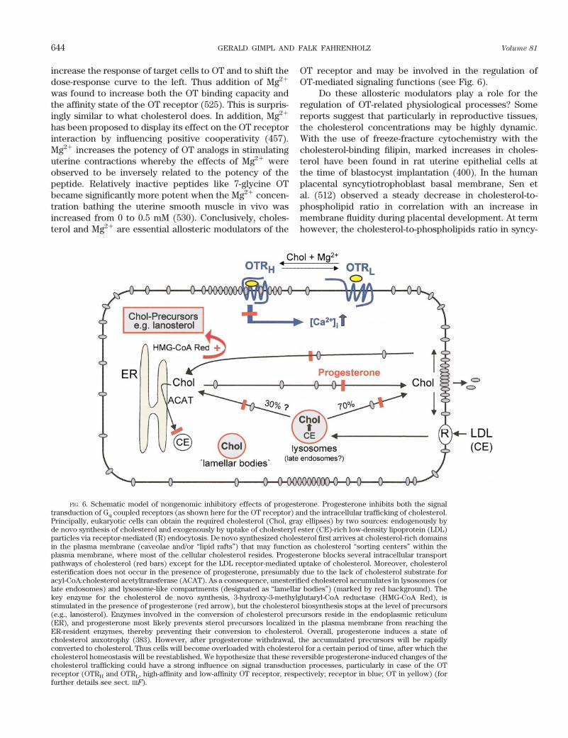
FIG. 6. Schematic model of nongenomic inhibitory effects of progesterone. Progesterone inhibits both the signaltransduction of Gq coupled receptors (as shown here for the OT receptor) and the intracellular trafficking of cholesterol.Principally, eukaryotic cells can obtain the required cholesterol (Chol, gray ellipses) by two sources: endogenously byde novo synthesis of cholesterol and exogenously by uptake of cholesteryl ester (CE)-rich low-density lipoprotein (LDL)particles via receptor-mediated (R) endocytosis. De novo synthesized cholesterol first arrives at cholesterol-rich domainsin the plasma membrane (caveolae and/or “lipid rafts”) that may function as cholesterol “sorting centers” within theplasma membrane, where most of the cellular cholesterol resides. Progesterone blocks several intracellular transportpathways of cholesterol (red bars) except for the LDL receptor-mediated uptake of cholesterol. Moreover, cholesterolesterification does not occur in the presence of progesterone, presumably due to the lack of cholesterol substrate foracyl-CoA:cholesterol acetyltransferase (ACAT). As a consequence, unesterified cholesterol accumulates in lysosomes (orlate endosomes) and lysosome-like compartments (designated as “lamellar bodies”) (marked by red background). Thekey enzyme for the cholesterol de novo synthesis, 3-hydroxy-3-methylglutaryl-CoA reductase (HMG-CoA Red), isstimulated in the presence of progesterone (red arrow), but the cholesterol biosynthesis stops at the level of precursors(e.g., lanosterol). Enzymes involved in the conversion of cholesterol precursors reside in the endoplasmic reticulum(ER), and progesterone most likely prevents sterol precursors localized in the plasma membrane from reaching theER-resident enzymes, thereby preventing their conversion to cholesterol. Overall, progesterone induces a state ofcholesterol auxotrophy (383). However, after progesterone withdrawal, the accumulated precursors will be rapidlyconverted to cholesterol. Thus cells will become overloaded with cholesterol for a certain period of time, after which thecholesterol homeostasis will be reestablished. We hypothesize that these reversible progesterone-induced changes of thecholesterol trafficking could have a strong influence on signal transduction processes, particularly in case of the OTreceptor (OTRH and OTRL, high-affinity and low-affinity OT receptor, respectively; receptor in blue; OT in yellow) (forfurther details see sect. IIIF).
644 GERALD GIMPL AND FALK FAHRENHOLZ Volume 81

tiotrophoblast membranes was found to be increasedcompared with the cholesterol-to-phospholipid ratio inearly placentas (365). Moreover, cholesterol-enrichedcaveolae structures are a conspicuous feature in the ratmyometrium at term (70). We have provided evidencethat cholesterol can modulate receptor function by bothchanges of the membrane fluidity and direct binding ef-fects, e.g., in case of the OT receptor (209). Plasma mem-branes with lowered cholesterol content showed a de-creased capacity (Bmax) of binding sites and/or adecreased affinity (Kd) of ligand-receptor binding. Inter-estingly, Lopez et al. (345) reported that pregnancy inhumans was associated with increases in both density andaffinity of OT receptors. To draw further conclusions,correlation studies are required using tissues in whichboth the membrane cholesterol content and the OT re-ceptor activity will be measured at the same time.
2. Progesterone
Progesterone is considered to be essential to main-tain the uterine quiescence. Grazzini et al. (218) recentlypostulated that progesterone specifically binds to the ratOT receptor with high affinity (Kd ;20 nM) and therebyinhibits the receptor function. In case of the human OTreceptor, a direct inhibitory interaction [inhibitory con-stant (Ki) ;30 nM] with a progesterone metabolite, 5b-pregnane-3,20-dione, has been reported by the same au-thors. They claimed that progesterone could act as anegative modulator of the OT receptor and thus offered aplausible mechanism of how progesterone could contrib-ute to uterine quiescence. However, these findings couldnot be reproduced in several other laboratories includingour own (81). Instead, we found that high concentrationsof progesterone (.10 mM) attenuated or blocked thesignaling of several GPCRs, including the OT receptor.The progesterone effects occurred within minutes, werereversible, and could not be blocked by a protein synthe-sis inhibitor (81). Overall, the action of progesterone wasmore cell type specific than receptor specific. The proges-terone doses that are required to affect the signalingfunction of receptors are much higher than the progester-one levels found in plasma or in nonsteroidogenic tissuessuch as the myometrium. In steroidogenic tissues, how-ever, huge amounts of progesterone have been measured.Near term, the human placenta secretes upward of 300 mgof progesterone daily. The progesterone content of thisorgan was shown to be 7 mg/g wet tissue (520). In humancorpus luteum, progesterone concentrations reachedpeak levels of ;25 mg/g tissue shortly after ovulation andin the early luteal phase (545). These values are within therange of the progesterone concentrations that were effec-tive in our study (81). Thus, in steroidogenic cells as wellas in their environment, progesterone might nongenomi-cally influence the signaling of receptors. The molecular
mechanisms underlying this progesterone action are notunderstood. A well-known progesterone binding proteinis the multidrug resistance P-glycoprotein (471). In addi-tion to their role in detoxification, P-glycoproteins areinvolved in intracellular cholesterol transport. It is knownthat progesterone markedly interferes with the intracel-lular transport (and metabolism?) of cholesterol (seemodel in Fig. 6). At concentrations in the micromolarrange, it inhibits both the cholesterol esterification andthe transport of cholesterol to and from the plasma mem-brane (343). In particular, progesterone reduces the cho-lesterol pool residing in caveolae (521). Paradoxically, atthe same time, progesterone stimulates the activity of3-hydroxy-3-methylglutaryl (HMG)-CoA reductase, thekey enzyme of de novo cholesterol biosynthesis. Hence,cholesterol precursors like lanosterol begin to enrich inthe membranes of the cell (383). As mentioned above, theOT receptor needs a cholesterol-rich microenvironmentto become stabilized in its high-affinity state (210). Be-cause the cholesterol precursors, particularly lanosterol,are completely inactive to support the OT receptor in itshigh-affinity state (209), the responsiveness of the OTsystem may not be fully operative during the continuouspresence of high progesterone concentrations. Accordingto this scenario, progesterone withdrawal would restorethe cholesterol transport so that the highly enrichedamounts of cholesterol precursors would now becomerapidly converted to cholesterol. This would lead to asudden rise of cholesterol and should push the respon-siveness to OT since low-affinity OT receptors could nowbe converted into their high-affinity state (302). Accordingto this postulated mechanism, progesterone could affectthe signaling of all those receptors that are functionallydependent on cholesterol. It is important to note that thenongenomic actions of progesterone including its influ-ence on the cholesterol transport require progesteroneconcentrations in the micromolar range. This suggeststhat the described effects may be limited to the steroido-genic tissues and to their environment. Most likely, pro-gesterone acts in these tissues via both genomic andnongenomic pathways (summarized in Fig. 6) togetherwith other steroids to control receptor activity.
IV. THE PERIPHERAL OXYTOCIN SYSTEM
A. Female Reproductive System
1. Uterus
The pregnant uterus is one of the traditional targetsof OT. OT is one of the most potent uterotonic agents andis clinically used to induce labor. Accordingly, the devel-opment of highly specific OT antagonists may be of ther-apeutic value for the prevention of preterm labor and theregulation of dysmenorrhea (358, 594).
April 2001 THE OXYTOCIN RECEPTOR SYSTEM 645

The OT gene was found to be expressed in the ratuterine epithelium at term. The estrogen-induced eleva-tion of OT mRNA levels was restricted to 3 days andreached peak levels that exceeded hypothalamic OTmRNA levels by a factor of 70 (327). In rats, OT geneexpression was shown to be present in placenta andamnion (328) and in humans in amnion, chorion, anddecidua (104). However, in most studies, significant in-creases of OT before the onset of labor have not beendetected, neither in maternal plasma nor in intrauterinetissues. On the other hand, some findings suggest thatthere is a relationship between the pattern of OT secre-tion and advancing pregnancy. In rhesus monkeys, it wasshown that maternal but not fetal OT concentrations werepositively correlated with nocturnal uterine activity andprogressively increased during late pregnancy and deliv-ery (239).
Around the onset of labor, uterine sensitivity to OTmarkedly increases. This is associated with both an up-regulation of OT receptor mRNA levels and a strong in-crease in the density of myometrial OT receptors, reach-ing a peak during early labor (188, 299). This has beendemonstrated both in the rat and in the human species, inwhich receptor levels rise during early labor to 200 timesthat in the nonpregnant state (189). Thus, at the onset oflabor, OT can stimulate uterine contractions at levels thatare ineffective in the nonpregnant state. After parturition,the concentrations of OT receptors rapidly decline. Inrats, the uterine OT receptor mRNA levels decreasedmore than sevenfold within 24 h (624). Possibly, thedownregulation of the OT receptors may be necessary toavoid unwanted contractile responses during lactationwhen OT levels are raised.
Gonadal steroids play an important role in the regu-lation of uterine OT receptors. In the days preceding birth,the ratio of plasma progesterone to estrogen falls. Thesechanges in the steroid concentrations may occur in mostmammals. At least in humans, progesterone withdrawalhas not been determined. The steep drop of circulatingprogesterone occurs after placental delivery. Alteration ofsex steroid metabolism in the fetoplacental unit appearsto occur in women and primates (374). As shown inbovine and in sheep, maturation of the fetal hypothalamusleads to an increased secretion of CRH, which in turnstimulates the pituitary to secrete ACTH (188). Subse-quently, ACTH stimulates the fetal adrenal to releasecortisol. Additionally, OT-induced contractures of themyometrium in late pregnancy could lead to temporarydecreases in blood flow and transient episodes of fetalhypoxia, which also may provoke a fetal stress response.Cortisol then increases the activity of the key enzymecyctochrome P-450c17a, which promotes placental preg-nenolone turnover into estrogens. In primates, CRH isadditionally synthesized by the placenta and stimulatesthe fetal adrenal to secrete dehydroepiandrosterone sul-
fate (DHEAS), the precursor of placental estrogen (374,522). In each case, the ratio of estrogen to progesteroneincreases in the maternal plasma concomitantly with in-creases in the synthesis of connexin-43, formation of gapjunctions, increased production of prostaglandins fromintrauterine tissues, and an upregulation of OT receptors(188, 388). Finally, the uterine quiescence that was main-tained by the high progesterone level ceases, and partu-rition can occur. Upregulation of OT receptors and anincreased expression of gap junctions also occur in thehours or days preceding human labor onset (191, 546).Obviously, estrogens and progesterone act in opposingfashion on the function, expression, and/or regulation ofOT receptors. The mechanisms for the sudden and some-times unpredictable responsiveness to OT of the myome-trium is still mysterious (298). As Kimura (293) pointedout, the reaction of the uterus to OT in the same patientcould vary from day to day. To induce labor at term, insome patients, OT is completely ineffective even at highdoses, whereas in others a minimal dose can induce hy-pertonus of the uterus (293). However, despite the strik-ing steroid dependence of the OT system, the promoter ofthe human OT receptor gene does not contain a classicalsteroid responsive element (see sect. IIIA). Several obser-vations indicated that progesterone promotes uterine re-laxation and inhibits the function of the OT receptorsystem by both genomic and nongenomic mechanisms.Even in the absence of protein synthesis, progesteroneinduces a reduction in uterine OT binding (528). More-over, progesterone-mediated downregulation of OT re-ceptors was not accompanied by a decrease in OT recep-tor gene expression (323). The mechanisms of howprogesterone acts nongenomically are still unknown butare of fundamental importance (see sect. IIIF).
The increase in OT receptors before labor is notconfined to the myometrium. It is also found in the de-cidua. During the course of parturition, the OT receptorgene expression in human chorio-decidual tissue wasfivefold increased (547). In the decidua, OT has a separateaction of stimulating the release of prostaglandin PGF2a.At the end of pregnancy, an increased secretion of PGF2a
drives luteolysis and thus leads to progesterone with-drawal and labor initiation in rodents. Mice lacking thegene encoding the PGF2a receptor developed normallybut were unable to deliver normal fetuses at term. Theseknock-out mice neither responded to OT nor did theyshow an induction of OT receptors. Furthermore, thenormal decline of serum progesterone concentrationsthat precedes parturition did not occur. Ovariectomy re-stored induction of OT receptors and permitted success-ful delivery in the PGF2a receptor-deficient mice. Thisindicates that PGF2a acts upstream of OT to induce lute-olysis, i.e., production of progesterone (543). The OTsystem may be regarded as a key regulator of parturition,with the induction of OT receptors as a trigger event.
646 GERALD GIMPL AND FALK FAHRENHOLZ Volume 81

However, in OT-deficient mice, parturition remained un-affected (416). This suggests that at least in mice, utero-tonins other than OT are (additionally?) operative as myo-metrial contractants to initiate labor. A recent study withmice deficient in both OT and cyclooxygenase-1 shedmore light for OT’s role at the onset of labor (219). Cy-clooxygenase-1 is involved in the synthesis of prostaglan-dins, and as expected, mice lacking this enzyme showedreduced levels of PGF2a, impaired luteolysis, a less pro-nounced fall in progesterone in late gestation, and finallya delayed initiation of labor. Surprisingly, mice deficientin both OT and cyclooxygenase-1 initiated labor at thenormal time. Thus OT and prostaglandins had apparentlyopposing actions on the onset of murine parturition: aluteotrophic function of OT versus a luteolytic action ofprostaglandins. So, OT stabilizes the progesterone synthe-sis before parturition. For the normal timing of labor itmight be essential to generate sufficient PGF2a to over-come the luteotrophic action of OT in late gestation.These findings might explain why the upregulation of OTreceptor occurs without an increased expression of OTshortly before the onset of labor. The induction of uterineOT receptor expression allows OT to act as a potentuterotonic agent, whereas an increase in OT expressioncould prolong gestation due its trophic effects on thecorpus luteum (219). Fuchs et al. (195) reported that OTmay also be involved in induction of cyclooxygenase-2expression and PGF2a release at term and during partu-rition in cows.
In ruminants, the endometrial OT receptor exhibitsa cycle-dependent regulation and plays a crucial role inreproduction (518). In the bovine endometrium, OTreceptor mRNA levels can exceed the levels in themyometrium at the same stage of the cycle (272).Within 2–3 days around estrus, the endometrial OTreceptors increase to a level similar to that observed atterm of pregnancy. For the remainder of the cycle, OTreceptor concentrations are almost undetectable. Pul-satile secretion of endometrial PGF2a is stimulated byOT during late diestrus in domestic ruminants and re-sults in regression of the corpus luteum leading to theonset of a new estrous cycle. Thus the uterus influencesthe length of the luteal phase via the expression of theendometrial OT receptor. The administration of an OTantagonist or the continuous administration of OT,which downregulates the OT receptor, delays the onsetof luteolysis and lengthens the cycle (178, 517). A steeprise in endometrial OT receptors also precedes theonset of labor in ruminants. In cows, the receptordensity increases almost 200-fold and remains at highlevels during labor (192). Suppression of the endome-trial OT receptor gene expression was shown to becaused by antiluteolytic proteins, e.g., interferon-g se-creted by the trophoblast of the developing blastocyste(267, 483). In primates, the endometrium is not essen-
tial for luteolysis, since hysterectomy does not abolishcyclic ovarian function. In humans, Northern blottingrevealed the presence of OT receptor mRNA in thepregnant myometrium, in the endometrium, and in theovary. In the nonpregnant human uterus, the OT recep-tor mRNA is mainly expressed in the glandular epithe-lial cells of the endometrium with highest expressionlevel occurring at ovulation (548). However, its physi-ological role is unclear.
One of the highest concentrations of OT receptors(;10 pmol/mg protein) has been measured in rabbit am-nion at term (234). At the end of gestation, rabbit amnionOT receptors increase more than 200-fold. This receptorupregulation is associated with the release of PGE2 fromcultured amnion cells and suggests an important role forthe amnion and OT in the initiation of labor in rabbits(234). In the same system, cortisol and cAMP caused amarked physiological upregulation of OT receptors aswell as an increased PGE2 response to OT. Addition ofcortisol to amnion cells increased OT-stimulated PGE2
release almost 100-fold, whereas the combination of for-skolin and cortisol increased the PGE2 response to OT;5,600 times (236). OT receptors in human amnion werealso associated with PGE2 release and were found to beupregulated in early and advanced labor, albeit to a muchlower degree (49, 393). In contrast, no OT binding siteswere observed in amniotic/chorionic or placental mem-branes from rats (328).
Many studies have supported the view that OT issynthesized in a paracrine system operating within humanintrauterine tissues (389). This includes the fetal mem-branes amnion and chorion and the maternal decidua.These tissue layers are continuously bathed in amnioticfluid and could transmit signals of maternal or fetal originto the myometrium. Because fetal membranes are capableof steroidogenesis, they may contribute to local changesin the estrogen-to-progesterone ratio. Although there isevidence that during late gestation OT gene expression isupregulated in intrauterine tissues, the OT peptide itselfwas not found at concentrations significantly higher thanin the circulating blood. This could be due to the highactivity of oxytocinases, but that still remains to be shown(388). The possibility of local, paracrine effects of OT wasalso suggested to be present within the intrauterine tis-sues of the rat and bovine (272, 327). In the pregnant cow,OT mRNA levels were found to be very low except for thecorpus luteum. After the onset of labor, both OT gene andOT peptide were expressed at significant levels in thecorpus luteum. This rescue of luteal OT at term could actto supplement the circulating hormone of pituitary origin(272). Thus OT may act primarily as a local mediator andnot as a circulating hormone during parturition. In anautocrine/paracrine system within the uterus, significantchanges of OT, prostaglandins, and sex steroids could
April 2001 THE OXYTOCIN RECEPTOR SYSTEM 647

occur without being reflected in the maternal circulation(389).
2. Ovary and corpus luteum
In several species, the ovary has been shown tocontain OT and may be a site of local OT production(271). In the marmoset monkey (Callithrix jacchus), invivo and in vitro studies suggested that OT is a follicularluteinization factor. After human chorionic gonadotropintreatment, almost all granulosa cell layers in antral folli-cles showed immunoreactivity for both OT and OT recep-tor. OT was produced only by granulosa cells derivedfrom preovulatory follicles, and after application of OT,only the granulosa cells cultured from preovulatory folli-cles elicited an increase in progesterone production (146,149). Functional OT receptors have been detected in bo-vine granulosa cells, suggesting that OT may be an auto-crine factor during follicular growth (423). OT also in-creased the rate of mouse blastocyst development andmight therefore play some role in the early stage of de-velopment of fertilized oocytes (199). Both OT and OTreceptor genes are expressed in human cumulus cellssurrounding the oocytes. Thus local OT may participate infertilization and early embryonic development in humans(198).
The interaction of neurohypophysial OT with endo-metrial OT receptors evokes the secretion of luteolyticpulses of uterine PGF2a. McCracken et al. (372) havepostulated the concept of a central OT pulse generatorthat functions as pacemaker for luteolysis. According tothis concept, the uterus transduces hypothalamic signalsin the form of episodic OT secretion, into luteolytic pulsesof uterine PGF2a. In ruminants, luteal OT is releasedsynchronously by uterine PGF2a pulses. Thus a positive-feedback loop is established that amplifies neural OTsignals. The onset of this feedback loop and episodicPGF2a secretion is controlled by the appearance of OTreceptors in the endometrium. During pregnancy, the de-veloping conceptuses must prevent endometrial PGF2a
from being released into the uterine vasculature to pre-vent corpus luteum regression and progesterone with-drawal. The continued progesterone secretion is requiredfor establishment and maintenance of pregnancy. Lutealcells produce OT, but they can also be target cells for OTaction. OT receptors have been characterized on bothsteroidogenic cell types of the corpus luteum, small andlarge cells (454). In several species, OT release was high-est in the young corpus luteum (148, 277). In the pig, thedensity of OT receptors was also highest during the earlyluteal phase, suggesting that autocrine/paracrine actionsof OT may occur primarily in the young corpus luteum(454). The effects of OT on cultivated luteal cells werealso strongly dependent on the age of the corpus luteum.In the pig, a function for OT in the ovary has been sug-
gested in both a luteotrophic and luteolytic context (604).Luteal OT and progesterone release occurs in tightly cou-pled pulses. In vivo, OT and PGF2a stimulate estradiol andprogesterone release, and estradiol itself further stimu-lates progesterone release. Thus there may be an intralu-teal circuit that involves paracrine effects of estradiol, OT,and PGF2a.
B. Male Reproductive Tract
1. Testis
In several species, a pulse of systemic OT, presum-ably of hypothalamic origin, appears to be associated withejaculation. The systemic hormone could act peripherallystimulating smooth muscle cells of the male reproductivetract, but could also reflect central effects in the brainmodulating sexual behavior (see sect. VIA).
OT has been identified in the testes from variousmammalian species, and a mesotocin-like peptide hasbeen demonstrated in the testes of birds (453) and mar-supials (47). There is evidence that OT is made locallywithin the testis, and possibly also the epididymis andprostate (265). Concerning the localization of the OTsystem in the male reproductive tract, species-specificdifferences are important to note. For example, mice donot have detectable levels of OT mRNA in their testes,whereas cattle have relatively high levels of testicular OTmRNA (16). In contrast to the eutherian mammals, in thetammar wallaby (Macropus eugenii), the mesotocin re-ceptor gene and protein, which are highly homologous tothe eutherian OT receptor, are expressed in the prostategland, but not in the testis (437). In the human, the com-plete OT system appears to be present in testis, epididy-mis, and prostate (182).
In the rat testis, OT concentrations are higher thanthose in the peripheral circulation. OT gene transcriptscould be detected using PCR analysis but not by Northernhybridization (180). Within the rat testes, OT is present inthe interstitial Leydig cells, the cells which provide themain source of testosterone in the male (412). TesticularOT levels are not constant but vary in dependence of thelevel of gonadotrophins and the activity of the seminifer-ous epithelium. OT production is increased in vitro byluteinizing hormone (LH), whereas testosterone itself hasno effect on the secretion of OT. Notably, OT was onlydetectable during active spermatogenesis (413). Primarilytwo functions have been ascribed to testicular OT,namely, the regulation of seminiferous tubule contractil-ity and the modulation of steroidogenesis. In the testis,the seminiferous tubules are surrounded by smooth mus-cle-like cells, the myoid cells. OT has been shown toenhance the contractility of the tubules; therefore, theresponsiveness to OT was higher at a certain stage of thespermatogenic cycle, around the time when the sperm are
648 GERALD GIMPL AND FALK FAHRENHOLZ Volume 81
shed into the lumen (413). Obviously, OT promotes thespermiation and the subsequent transport of the immotilespermatozoa to the epididymis. However, there is no clearevidence for the expression of OT receptors on myoidcells, the cells which are mainly responsible for the con-tractile activity of the tubules (246). Because Sertoli cellsexhibit the components of a local OT system under somecircumstances, they may also be involved in the contrac-tile activity of seminiferous tubules. On the other hand, itwas suggested that the contractility effects are mediatedby V1a receptors that are present in testes at severalfoldhigher densities compared with OT receptors (227).
Uterine-type OT receptors have been identified in theinterstitial spaces in the rat testis consistent with bindingto Leydig cells (45). Also in the male marmoset monkey(Callithrix jacchus), OT and its receptor have been de-tected predominantly within the Leydig cells (145). TheLeydig cells drive spermatogenesis via the secretion oftestosterone, which acts on the Sertoli and/or peritubularcells to create an environment that enables normal pro-gression of germ cells through the spermatogenic cycle.Daily subcutaneous injections of OT led to an increase inthe plasma and testicular levels of testosterone. In con-trast, continuous administration of the peptide into thetestis, whether by implants or in the transgenic mousewhich overexpresses the bovine OT gene in its testes,produced a decrease in testosterone but an increase indihydrotestosterone concentrations (15, 411). Although inthe testis the predominant androgen is testosterone, else-where in the male reproductive tract testosterone acts asa prohormone and is converted to its active metabolitedihydrotestosterone by the enzyme 5a-reductase. OT wasshown to increase the activity of 5a-reductase in bothtestis and epididymis and may thus have an autocrine/paracrine role modulating steroid metabolism in thesetissues (413).
2. Prostate gland
The prostate is an androgen-dependent gland. Tes-tosterone enters the cell and is converted to dihydrotes-tosterone, which then modulates growth and prostaticfunctions. OT is present in the prostate at concentrationshigher than in the plasma and can increase the restingtone of prostatic tissue from guinea pig, rat, dog, andhuman (58, 182, 265). OT also evoked contractile activityon mammalian prostates in vitro (58). Thus it was sug-gested that OT is involved in the contraction of the pros-tate and the resulting expulsion of prostatic secretions atejaculation (413).
OT may also be involved in the pathophysiology ofbenign prostatic hyperplasia (410). In humans, benignprostatic hyperplasia occurs spontaneously, is often asso-ciated with bladder outlet obstruction, and affects 50% ofmen over the age of 60 (51). However, obstructive symp-
toms may not only be caused by an enlargement of thegland but also by an increase in smooth muscle tone. So,in the nonoperative management of prostatic hyperplasia,a1-adrenergic blockers are presently used. Because OT iseven more potent at increasing smooth muscle tone thannorepinephrine, OT antagonists may be potentially usefulin the treatment of this disease (413).
OT can stimulate growth of the prostate in the rat,particularly the mitotic activity in the glandular epithe-lium (455). OT treatment elevated the testosterone levelsof plasma, testes, and prostate. Conversely, prostatic OTconcentrations are found to be decreased by testosteroneand increased after castration. In rats, OT treatment onlytransiently increased 5a-reductase activity in the prostate,consistent with the short-lived increase of dihydrotester-one in this tissue. These observations suggest that localfeedback mechanisms act to control prostatic levels ofdihydrotestosterone and prostatic growth (413). The find-ings in rats were somewhat different to those in dogs,which unlike rats can spontaneously develop prostatichyperplasia. Interestingly, old dogs with established pros-tatic hyperplasia showed raised OT levels accompaniedby elevated 5a-reductase activity within their prostatictissues. It is hypothesized that OT acts as a paracrinefactor to regulate cell growth via its influence on theenzyme 5a-reductase (410).
C. Mammary Tissues
1. Milk ejection
One of the classical roles assigned to OT is milkejection from the mammary gland. The secretion of themammary glands is triggered when the infant begins tosuck on the nipple. The stimulation of tactile receptors atthat site generates sensory impulses that are transmittedfrom the nipples to the spinal cord and then to the secre-tory oxytocinergic neurons in the hypothalamus. Theseneurons display a synchronized high-frequency burstingactivity, consisting of a brief (3–4 s) high-frequency dis-charge of action potentials recurring every 5–15 min.Each burst leads to massive release of OT into the blood-stream by which OT is carried to the lactating breasts.There it causes contraction of the myoepithelial cells inthe walls of the lactiferous ducts, sinuses, and breasttissue alveoli. In humans, within 30 s to 1 min after a babybegins to suckle the breast, milk begins to flow. Thisprocess is called milk ejection or milk let-down reflex andcontinues to function until weaning.
As mentioned above, central oxytocinergic neuronsare decisive components for the initiation and mainte-nance of successful lactation. So, the reflex that elicits OTrelease in women is activated before the tactile stimulusof suckling occurs and is related to such factors as thebaby crying (373). The essential role of OT for the milk
April 2001 THE OXYTOCIN RECEPTOR SYSTEM 649
let-down reflex has been confirmed in OT-deficient mice.In fact, the major deficit of these female OT knock-outmice was the failure to nurse their offspring (416). Inaddition, the presence of OT in conjunction with contin-ued milk removal was also required for postpartum alve-olar proliferation and mammary gland function. Alveolardensity and mammary epithelial cell differentiation atparturition were similar in wild-type and OT-deficientdams. However, within 12 h after parturition, ;2% of thealveolar cells in wild-type dams incorporated DNA andproliferated, whereas no proliferation was detected inOT-deficient dams. Continuous suckling of pups led to theexpansion of lobulo-alveolar units in wild-type but not inOT-deficient dams. Despite suckling and the presence ofsystemic lactogenic hormones, mammary tissue in OT-deficient dams partially involuted (581). Continuously el-evated OT concentrations such as those during infusionor during normal milking are also necessary for completemilk removal in diary cows (75).
Biochemical studies showed that the mammary OTreceptor is a 65-kDa protein (in rats), coupled to phos-phoinositide turnover, and is most likely the same as theuterine-type OT receptor (399, 448). In rats, Soloff andWieder (533) reported an ;100-fold increase in the num-ber of OT receptors per mammary gland between the firstday of pregnancy and late lactation. A very strong in-crease in OT binding sites toward term was also observedin porcine mammary tissues (351). In humans, however,Kimura et al. (294) did not find an elevated expressionlevel of the OT receptor mRNA during lactation. Asjudged by OT receptor immunoreactivity, unexpectedly,the ductal/glandular epithelium revealed a higher densityof OT receptors compared with myoepithelial cells inboth the lactating and nonlactating breast in humans andin the common marmoset (Callithrix jacchus) (294).
2. Breast cancer and tumor cells
Breast cancer is the leading cause of death in womenbetween ages 35 and 45, but it is most common in womenover age 50. It is unknown why mothers who breast-fedtheir babies have a 20% lower incidence of breast cancerafter menopause than mothers who did not. Murrell (403)proposed a hypothesis that partially links breast cancer tothe activation of the OT system. Accordingly, carcinogensin the breast may be generated by the action of superox-ide free radicals released when acinal gland distensioncauses microvessel ischemia. Thus inadequate nipple carein the at-risk years could lead to ductal obstruction pre-venting the elimination of carcinogens from the breast.The regular production of OT from nipple stimulationwould cause contraction of the myoepithelial cells, reliev-ing acinal gland distension and aiding the active elimina-tion of carcinogenic fluid from the breast. Thus OT pro-duction was suggested as a preventative factor in the
development of breast cancer both pre- and postmeno-pausally (403).
OT receptors have been described in a number ofhuman breast tumors and breast cell lines, e.g., the humantumor cell lines MCF-7 or Hs578T. Copland et al. (118)recently studied the behavior of OT receptors in depen-dence of culture conditions in Hs578 cells. Hs578 cellsresponded to OT by an increase of intracellular Ca21 andstimulation of ERK-2 phosphorylation and PGE2 synthe-sis. OT receptors in these cells were strongly downregu-lated by serum starvation. Conversely, restoration of se-rum and addition of dexamethasone (1 mM) increased theOT receptor levels by nearly 10-fold and also led to anelevation of the steady-state OT receptor mRNA level.These cells may be good models for future studies withrespect to OT receptor regulation in dependence of ste-roids (118).
Controversial results have been published concern-ing a proliferative action of OT on breast tumor cells. Itoet al. (263) found that OT had no effect on the growth ofdifferent breast cell lines cultured for 7 days. The OTreceptor was expressed in breast cancer derived not fromthe myoepithelium but from the glandular or ductal epi-thelium. According to Sapino et al. (497), OT exerts atrophic effect on myoepithelial cells in the mammarygland. In organotypic cultures of the mouse mammarygland, OT induced differentiation and proliferation ofmyoepithelial and, to a lesser extent, luminal epithelialcells. On the other hand, OT inhibited cell proliferationand tumor growth of rat and mouse mammary carcino-mas. In MCF7 cells for instance, OT inhibited estrogen-induced cell growth and enhanced the inhibitory effect oftamoxifen on cell proliferation. Antiproliferative effectsof OT have also been observed in various breast carci-noma cell lines as well as in human neuroblastoma andastrocytoma cells, and these effects were accompanied bythe activation of the cAMP/protein kinase A pathway (97,98). In vivo, OT reduced the growth of certain mammarytumors. With the use of immunohistochemistry and RT-PCR, OT receptors and its corresponding mRNA weredetected in normal and pathological breast tissues. Inter-estingly, the expression of OT receptors was positivelycorrelated with the progesterone level of these tissues(496). Further studies are required to clarify the role ofthe OT system concerning the long-term effects in mam-mary tissues and tumor cell lines derived from them.
D. Kidney
The kidney is one of the peripheral target tissues forneurohypophysial hormones that exercise control overhydromineral excretion. The hormones are released intothe blood by stimulations, such as hypovolemia or hyper-osmolarity, that generally activate the OT and AVP neu-
650 GERALD GIMPL AND FALK FAHRENHOLZ Volume 81

rons. When plasma sodium concentration exceeds 130mM, the levels of both hormones increase as an exponen-tial function of plasma sodium concentration (575). OT isa nonhypertensive natriuretic agent. It is involved in nor-mal osmolar regulation, which is presumably differentfrom the volume regulatory components of Na1 ho-meostasis. Acute administration of OT to conscious ratsproduced a modest increase in the glomerular filtrationrate and effective filtration fraction. The natriuretic effectof OT is mainly due to a reduction in tubular Na1 reab-sorption, probably in the terminal distal tubule or thecollecting duct (116, 247, 426).
Autoradiographical analysis showed the existenceand precise localization of OT receptors in the rat kidney.Interestingly, the distribution of OT binding sites under-goes reshaping during postnatal development as it wassimilarly observed in the rat brain during maturation (seesect. VA and Table 4) (504, 560). Specific OT binding siteshave been first detected at embryonic day 17 in thecortex. In the medulla, OT binding sites were first de-tected at embryonic day 19 when this region is forming.In the adult rat, OT binding sites were exclusively local-ized in the cortex, mainly concentrated in the maculadensa. In the inner medulla, the OT binding sites werefound on the loops of Henle of the juxtamedullarynephrons. Interestingly, at all stages examined, corticalOT-binding sites had a higher selectivity for OT versusvasopressin compared with the medullary sites. It wastherefore suggested that OT binding sites of the maculadensa and thin Henle’s loop could represent two subtypesof OT receptors (33, 34). The location profile of the re-ceptors suggests a possible role for OT in the regulation oftubuloglomerular feedback and solute transport.
Estrogen induced OT receptor gene expression in theouter medulla region and increased expression of OTreceptors in macula densa cells of ovariectomized femaleand adrenalectomized male rats (426). Experiments withthe antiestrogen tamoxifen suggested cell-specific regula-tion of OT receptor expression in macula densa and prox-imal tubule cells. Thus OT receptors may mediate estro-gen-induced alterations in renal fluid dynamics (426). OTreceptor mRNA levels have been measured in kidneys oflate-pregnant, peri-parturient, and lactating rats. Of inter-est, OT receptor transcripts could not be detected in renaltissues of peri-parturient females, at times when OT re-ceptor mRNA levels were highest in uterus. However, OTreceptor gene expression in macula densa cells graduallyreappeared and again achieved control levels by day 20
post partum (426). Breton et al. (67) observed that boththe OT receptor mRNA levels as well as the OT bindingcapacity of rat renal extracts were significantly lower atterm. Although in the rat kidney there was no indicationfor another than the uterine-type OT receptor gene ex-pression, the mechanisms controlling the expression of
OT receptor genes in uterus, pituitary, and kidney areobviously different (67).
OT injected intraperitoneally caused dose-dependentincreases in urinary osmolality, natriuresis, and kaliuresisin the rat. Moreover, the injection of OT evoked concom-itant release of ANP and natriuresis (222) (see sect. IVE).Soares et al. (523) recently provided evidence for thehypothesis that OT acts in concert with ANP to inducenatriuresis and kaliuresis via the common mediatorcGMP. In rats, the OT-induced natriuresis was causedmainly by decreased tubular Na1 reabsorption and wasaccompanied by increases in both urinary cGMP and NO3
2
excretion (523). Although the natriuretic action of ANP ismediated by activation of renal guanylyl cyclase A (GCA)receptors localized in glomeruli, their afferent and effer-ent arterioles, and the tubules, OT is hypothesized toinduce natriuresis by activation of renal NO synthaselocalized in macula densa cells (591). Presumably, bind-ing of OT to their receptors on NOergic cells in theproximal tubules and macula densa leads to an increaseof intracellular [Ca21], followed by Ca21/calmodulin-in-duced stimulation of NO synthase and activation of thesoluble guanylate cyclase by NO. The consequent releaseof cGMP could then mediate natriuresis and kaliuresis viaclosure of Na1 and K1 channels. In cortical collectingduct cells, it has been shown that the NO-inhibited Na1
transport is associated with increased cGMP content(538). Overall in rats, OT as well as ANP induce a con-comitant reduction of both extracellular and intracellularfluid volume in states of increased body fluid volume.
The signal transduction of the renal OT receptor hasbeen evaluated in various kidney epithelial cells in cul-ture, e.g., LLC-PK1. In these cells, OT induced stimulatedphosphoinositide hydrolysis and transiently increased cy-tosolic [Ca21]. OT was also able to stimulate the solubleguanylate cyclase and increased the intracellular level ofcGMP in LLC-PK1 cells (334, 534). Moreover, OT wasfound to stimulate the synthesis of prostaglandins in adose-dependent manner in kidney homogenates (100).
What is known about the OT effects on kidney func-tion in species other than rats? In rabbits, OT was shownto affect apical sodium conductance of the microperfusedcortical collecting duct through OT receptors distinctfrom the vasopressin receptors (255). The effects wereinhibited by the addition of an OT antagonist. Althoughthe natriuretic response to OT has also been described forconscious dogs, it probably does not occur in humans andnonhuman primates. Thus the contribution of OT to renalphysiology in primates including humans, if any, remainsuncertain (116).
E. Heart and Cardiovascular System
Peripherally injected OT decreases mean arterialpressure in rats (445, 449). Even in the absence of a
April 2001 THE OXYTOCIN RECEPTOR SYSTEM 651

central control mechanism, OT is able to reduce the heartrate and the force of atrial contractions in isolated atriafrom perfused rat hearts (167). An OT antagonist reversedthe bradycardia caused by OT. Moreover, administrationof OT at high concentrations (1 mM) leads to the stimu-lation of ANP release (221). An OT antagonist first inhib-ited this ANP release, and after prolonged perfusion, itfinally decreased the OT-induced ANP release below thatof control hearts. This suggested that intracardial OTstimulates ANP release in the heart. Gutkowska et al.(221) proposed that OT and ANP act in concert in thecontrol of body fluid and in cardiovascular homeostasis.In favor of this hypothesis, OT receptor transcripts andOT binding sites were shown to be present on atrial andventricular sections as detected by in situ hybridizationand autoradiograpy, respectively. Furthermore, the OTreceptor gene is expressed in all chambers of the ratheart, and the analysis of the RT-PCR products indicatedthe presence of the uterine-type OT receptors in the heart(221). Although the OT receptor mRNA levels in the atriawere found to be higher than in the ventricles, overall theOT receptor mRNA levels were calculated to be at least 10times lower than the OT receptor mRNA level present inthe uterus of a nonpregnant rat.
The rat heart was also shown to be a site of OTsynthesis. OT was detected in the effluent of isolatedheart perfusates as well as in the medium of culturedatrial myocytes. OT concentrations were found to behigher in the atria than in the ventricles. In the rightatrium, OT concentrations ;20-fold higher than those inthe rat uterus have been measured. In contrast, the OTmRNA level in the heart tissues was found to be lowerthan that in the rat uterus. This discrepancy arguesagainst the postulated abundant biosynthesis of cardiacOT (274). The relatively low concentrations of OT in theheart chambers and the high OT doses required for ANPrelease would be more compatible with paracrine or au-tocrine effects of cardiac OT, particularly in the rightatrium. Although the OT quantities released from theperfused heart are not sufficient to substantially changethe plasma concentrations of OT, they may contribute tothe natriuretic action via stimulation of ANP release(274). Presumably, blood volume expansion via barore-ceptor input to the brain causes the release of OT thatcirculates to the heart. OT-induced ANP release in theheart may be achieved after activation of OT receptorsand subsequent elevation of intracellular [Ca21], which inturn could stimulate exocytosis and ANP secretion (221,274). ANP then exerts a negative chrono- and inotropiceffect via activation of guanylyl cyclase and release ofcGMP. Finally, a rapid reduction in the effective circulat-ing blood volume is produced by an acute reduction incardiac output, coupled with ANP’s peripheral vasodilat-ing actions. The ANP released would also act on thekidneys to cause natriuresis, and ANP acts within thebrain to inhibit water and salt intake, leading to a gradual
recovery of circulating blood volume to normal (167).Since the plasma concentration of both OT and ANP werefound to be increased after parturition, the OT-stimulatedANP release might be at least partly responsible for themassive diuresis observed postpartum.
The effects of repeated subcutaneous OT injectionson blood pressure and heart rate were investigated inspontaneously hypertensive rats. Surprisingly, when OTwere given for 5 days, a sustained decrease in bloodpressure was observed in male but not female rats,whereas the heart rate was unaffected (446). Moreover,acute versus chronic OT treatments caused opposite ef-fects on blood pressure, and these effects were modifiedby female sex hormones (447).
It appears that the complete OT system is present inthe vasculature of the rat. The OT concentrations in theaorta and vena cava were reported to be even higher thanthose in the right atrium of the heart. Thus OT might playa direct role in volume and pressure regulation in a para-crine/autocrine manner (275). In the case of the umbilical-placental vasculature, OT was reported to be a far morepotent vasoconstrictor than vasopressin. OT was there-fore proposed to be involved in the closure of the umbil-ical vessels at birth (9). A uterine-type OT receptor couldalso mediate vasodilatory responses in human vascularendothelical cells (555).
OT-induced cardiac effects were also reported forsome other species. The cardiac effects of neurohypo-physial hormones were studied in the frog, Rana tigrina,and in the snake, Ptyas mucosa. In both species, OTproduced dose-related cardiac effects in isolated atrialpreparations. In the frog, vasotocin was the most potenthormone, whereas in the snake, OT produced the mostpotent dose-related positive inotropic changes (110). Fur-thermore, OT was found to modulate the intrathoracicsympathetic ganglionic neurons regulating the canineheart. When OT was injected into right atrial ganglionatedplexi, heart rate and atrial forces were reduced in 5 of 10dogs studied. However, no cardiac changes occurredwhen OT was injected into left atrial or ventricular gan-glionated plexi. In this study, the connectivity with thecentral nervous system (CNS) was found to be necessaryto elicit consistent OT-induced responses (32). AlthoughOT elicited neuronal and concomitant cardiovascular re-sponses in most dogs when administered adjacent tospontaneously active intrinsic cardiac neurons, OT injec-tion into the superior vena cava only evoked a slightsystemic hypotension in two of seven dogs (31).
F. Other Localizations
1. Thymus
The thymus is the primary lymphoid organ repon-sible for the selection of the peripheral T-cell repertoire.Neuropeptides and their receptors have been described in
652 GERALD GIMPL AND FALK FAHRENHOLZ Volume 81

the thymus, supporting the concept of a close dialoguebetween the neuroendocrine and the immune systems atthe level of early T-cell differentiation. The neurohypo-physial peptides have been shown to trigger thymocyteproliferation and could induce immune tolerance of thisconserved neuroendocrine family. OT has been identifiedin human thymus by immunoreactivity and at the tran-scriptional level. OT was present in the thymus in surpris-ingly large amounts and was found to be restricted tocertain subtypes of thymic epithelial cells (205, 392). Dueto its higher expression level in the thymic epithelium, OTwas proposed as the self-antigen of the neurohypophysialfamily (204). OT was found to be colocalized with thecytokeratin network of thymic epithelial cells and notwithin secretory granules. The peptide is therefore notsecreted but behaves like an antigen presented at theouter surface of the cell. Thymic OT also behaves as acryptocrine signal targeted at the epithelium membranefrom where it is able to interact with neurohypophysialpeptide receptors expressed by pre-T cells. OT receptorsare predominantly expressed by cytotoxic CD81 lympho-cytes, and they transduce signals via the phosphoinositidepathway. In pre-T cells, OT was found to induce thephosphorylation of focal adhesion kinase (360). Thus itwas suggested that OT actively intervenes in the programof T-cell differentiation both as a neuroendocrine self-antigen and as a promoter of T-cell focal adhesion follow-ing a cryptocrine pathway.
OT receptors detected in thymic membranes or onthymocytes revealed a ligand selectivity similar to that ofuterine OT receptors (153). Caldwell et al. (86) reportedthat sexual activity leads to decreases of OT receptordensities in the thymus of rats. In human thymus extracts,the content of the OT immunoreactivity declined withincreasing age, whereas in rat thymic extracts, it wasreported to increase during aging. The age-dependentchanges might be linked to thymic involution (381). Someimmune pathologies in humans may be explained by thy-mic OT being involved in T-cell-positive selection andactivation.
2. Fat cells
In adipocytes, OT has a so-called insulin-like activityin that it stimulates glucose oxidation and lipogenesis andincreases pyruvate dehydrogenase activity (223). Treat-ment of rat adipocytes with lipolytic stimuli such as glu-cagon or isoprenaline increased the conversion of cholineinto phosphatidylcholine, and this lipolytic effect wasantagonized by OT (285). In rat adipocytes, OT at a con-centration of 1 nM markedly increased phosphoinositidebreakdown (159). OT also elicited a transient increase inintracellular [Ca21] and stimulated the protein kinase Cactivity (144, 510). Moreover, human fat cells possess aplasma membrane-bound H2O2-generating system that issensitive to extracellular stimuli. OT as well as insulin
were able to activate this system. It was suggested thatthe H2O2 produced might participate in the regulation offat cell differentiation and/or maintenance of the differ-entiated state (312). Populations of low- and high-affinityOT receptors have been described in rat adipocytes (61).
3. Pancreas
OT and AVP have been identified in human and ratpancreatic extracts at higher concentrations than thosefound in the peripheral plasma (10). However, a localsynthesis of these peptides within this organ has not yetbeen established. According to most studies in severalspecies, the neurohypophysial hormones induce the re-lease of glucagon and, to a lesser extent, insulin from thepancreas. During in situ perfusion of the rat pancreas withOT, a marked stimulation of glucagon release and a mod-est stimulation of insulin release were observed (142).With the use of isolated islets from rat pancreas, OTelicited a stimulation of glucagon release but failed toinfluence insulin release in a culture medium with lowglucose content. The glucagonotrophic action of OT wasgreatly diminished in the presence of a higher concentra-tion of glucose (141). Similar findings were reported instudies with conscious dogs (141, 578). With the use of themicrodialysis technique, OT administered directly in thepancreas of the rat stimulates the release of both insulinand glucagon. OT binding sites were identified in theperiphery of the islets of Langerhans, corresponding tothe localization of the glucagon-producing a-cells (535).In isolated mouse islets, OT produced an increase insomatostatin, glucagon, and insulin release, and it ampli-fied the glucose-induced insulin release probably via stim-ulation of the phosphoinositide metabolism (201). In hu-mans, OT evoked a rapid surge in plasma glucose andglucagon levels followed by a later increase in plasmainsulin and epinephrine levels. The effects of OT onplasma glucagon and epinephrine levels were potentiatedby hypoglycemia. OT was also found to potentiate glu-cose-induced insulin secretion (434). In contrast, Page etal. (432) observed no effects of OT on the decline orrecovery of blood glucose concentrations or on theplasma glucagon response to insulin-induced hypoglyce-mia in humans.
Impaired glucose tolerance and hyperinsulinemia arecommon features of obesity. Interestingly, the plasmalevels of OT were found to be fourfold higher in male andfemale obese subjects compared with control subjects(536). OT rises in hypoglycemia, and this response ispartially inhibited by dexamethasone (108). The OT rise inresponse to insulin-induced hypoglycemia was reduced inobese men. Pretreatment with the opioid antagonist nal-oxone enhanced the OT response to hypoglycemia inobese men and suggested an abnormal activity of endog-enous opioids in obesity (115). In women, unlike men,
April 2001 THE OXYTOCIN RECEPTOR SYSTEM 653

endogenous opioids did not modulate OT release duringinsulin-induced hypoglycemia (282).
A pancreatic OT receptor was cloned from the ratinsulinoma cell line RINm5F (279). CHO cells that havebeen transfected with this receptor responded to OT withan increase in cytosolic Ca21 concentration and inositolphosphate levels as well as arachidonic acid release andPGE2 synthesis. Amino acid sequence homology, bindingspecificity, and the ligand-activated signal transductionpathways suggested that the pancreatic OT receptors ex-pressed in RINm5F cells are indistinguishable from theuterine-type OT receptors that have been described inother tissues (279).
4. Adrenal gland
Ang and Jenkins (17) first identified immunoreactiveOT and AVP in human and rat adrenal glands. OT pre-dominanted over AVP, and both peptides occurred atconcentrations far greater than those found in plasma.Immunohistochemical studies in adrenal glands from rat,cow, hamster, and guinea pig showed that OT was local-ized in both the cortex and medulla in all these species. Inthe cortex, the OT immunoreactivity was most intense inthe zona glomerulosa, whereas in the medulla, the OTstaining was higher but revealed more species variation(231). Bovine adrenal medulla membranes revealed high-affinity low-capacity OT binding sites (420). OT bindingwas also detected on primary monolayer cultures of bo-vine adrenal chromaffin cells. However, the capacity ofthese sites was near the detection limit, and OT concen-trations in the micromolar range were necessary to stim-ulate the phosphoinositide pathway (550). From the fewstudies that have addressed the role of OT in the adrenalgland, no clear picture has emerged. Perfusion of theisolated rat adrenal gland with OT at 100 nM inhibited theacetylcholine-stimulated aldosterone secretion (462),whereas smaller doses of OT given as a bolus were able tostimulate aldosterone secretion in the intact perfused ratadrenal gland, but not in superfused adrenal cells (238).Legros et al. (331) hypothesized that OT acts also at theadrenal gland level to decrease cortisol release and/orsynthesis in humans. In addition, a proliferative effect ofOT was suggested by the observation that the number ofchromaffin cells in the adrenal medulla of rats was in-creased in response to OT (461).
The list of peripheral tissues containing OT receptorsis certainly not complete. Recently, functional OT recep-tors have also been discovered in primary cultures ofhuman osteoblasts and in a human epithelial osteosar-coma cell line (Saos-2) (117). Further studies are requiredto show whether OT is a bone anabolic agent as suggestedby the authors.
Table 3 gives an overview of the expression patternof the OT system in different species.
V. THE CENTRAL OXYTOCIN SYSTEM
A. Localization Profile
1. Localization of OT
In the CNS, the OT gene is primarily expressed inmagnocellular neurons in the hypothalamic PVN andSON. Action potentials in these neurosecretory cells trig-ger the release of OT from their axon terminals in theneurohypophysis (464). In the PVN, two populations ofOT-staining neurons have been identified: magnocellularneurons that terminate in the neurohypophysis and par-vocellular neurons that terminate elsewhere in the CNS.Only a small fraction (0.2%) of the OT neurons werecalculated to possess axon collaterals to both the neuro-hypophysis and the extrahypothalamic areas. Althoughfew OT perikarya were observed other than in the mag-nocellular nuclei, OT fibers and endings have been de-scribed in various brain areas in the rat: the dorsomedialhypothalamic nucleus, several thalamic nuclei, the dorsaland ventral hippocampus, subiculum, entorhinal cortex,medial and lateral septal nuclei, amygdala, olfactorybulbs, mesencephalic central gray nucleus, substantianigra, locus coeruleus, raphe nucleus, the nucleus of thesolitary tract, and the dorsal motor nucleus of the vagusnerve. OT fibers also run toward the pineal gland and thecerebellum, with most of them continuing toward thespinal cord. A few OT fibers end on the portal capillariesin the median eminence (see references in Refs. 78, 309,476, 501, 524).
OT concentrations in the extracellular fluid of theSON were calculated to be .100- to 1,000-fold higher thanthe basal OT concentration in plasma, i.e., more than 1–10nM. High-frequency electrical discharges of OT neuronsas they occur, e.g., during the milk ejection reflex, mightrelease even higher local OT concentrations (321).
Plasma OT does not readily cross the blood-brainbarrier, and there is no relationship between the releaseof OT into the blood by the neurohypophysis and thevariations in OT concentrations in the cerebrospinal fluid(CSF). Peripheral stimulations such as suckling or vaginaldilation that elicit large increases in plasma OT may ormay not change the concentration of OT in the CSF. Asshown in rats, electrical stimulation of the neurohypoph-ysis only evokes the release of OT into the blood, whereasstimulation of the PVN elicits a release of OT into theblood and into the CSF (228). After hypophysectomy, OTdisappears from the blood, whereas its concentration in-creases in the CSF (139). The OT in the CSF is probablyderived from neurons that extend to the third ventricle,the limbic system, the brain stem, and the spinal cord. Inthe CSF, OT is normally present at concentrations of10–50 pM, and its half-life is much longer (28 min) than inthe blood (1–2 min) (283, 384). In humans and in mon-
654 GERALD GIMPL AND FALK FAHRENHOLZ Volume 81

TABLE 3. Expression of oxytocin and oxytocin receptors in the peripheral system
Tissue Species OT GeneOT
BindingOTR
mRNA Reference Nos.
Female reproductive systemMyometrium* Hum bov she 1 1 44, 191, 299, 482, 529, 531
Rat ND 1 1 7, 120, 194, 323, 624Rab gp 1 164, 479, 531Pig 1 1 63, 351
Endometrium† Hum 1 1 299Rat 1 1 ND 101, 323, 327, 624Bov she 1 1 44, 480, 482, 529Pig 1 1 63, 351, 387, 388Rab 1 355
Amnion Hum 1 1 1 104Rat 1 ND 328Bov 1 ND 193, 272Rab 1 234
Chorion Hum 1 1 1 49, 104, 547Rat 1 ND 328
Decidua Hum 1 1 1 104, 190, 547Rat gp rab 1 190, 196, 479, 547, 550
Ovary Hum mar 1 1 1 147, 187, 198, 269, 299Mou 1 1 199Bov 1 1 268, 271, 187Gp pos 1 46, 619
Corpus luteum‡ Hum pig mac 1 1 126, 150, 269, 291, 292, 454, 585Bov 1 1 ND 173, 187, 272, 584She 1 1 513, 554, 583Rat bab goa 1 197, 243, 269
Male reproductive systemTestis Hum rat 1 1 45, 180, 269, 414
Mar 1 1 145Mac 1 (1) 182Mou (1) 16, 415Bov she ban 1 16, 47, 207, 563Pig 1 354
Epididymis Hum 1 (1) 182Mar (1) (1) 145Wal 1 1 437Pig 1 354Rat 1 226
Prostate gland Hum 1 (1) 58, 182Mar ND (1) 145Mar mac (1) 145, 182Wal 1 1 437Bov ND 502Rat gp dog (1) 58
Mammary gland* Hum (1) 1 293, 294Rat mar bov pig 1 294, 351, 399, 531, 533, 620
Kidney Rat 1 1 33, 34, 67, 353, 427, 504, 551Rab (1) 14, 255Pos 1 46
Heart Rat 1 1 1 221, 274Vascular endothelium Hum 1 1 555Thymus Hum 1 1 205, 206
Rat 1 153Mou 1 203
Adipocytes Rat 1 36, 61, 62, 144Pancreas Hum (1) 10
Rat (1) 1 10, 535, 608Adrenal gland Hum rat ham gp 1 17, 180, 231, 419, 473
Bov 1 1 420, 550Osteoblasts Hum 1 1 117
Cell linesOsteosarcoma Saos-2 Hum 1 1 117Myometrial PHM1-41 Hum 1 138Pancreatic RINm5F Rat 1 279Gonadotrope-derived aT3-1 Mou 1 161Mammary tumor Hs578T Hum 1 242Breast cancer lines (e.g., MCF7) Hum (1) 1 40, 168, 263, 496, 549Vascular endothelial ECV304 Hum 1 555Vascular smooth muscle (HVSMC) Hum 1 607Sweat gland NCL-SG3 Hum (1) 481Immature thymic pre-T RL12-NP Mou 1 360Kidney epithelial LLC-PK1 Pig 1 1 215
1, Detectable signal; (1), weak or unclear signal (e.g., based on poorly characterized antireceptor antibodies) and/or contradictory reports have been published(for details, see the references); ND, not detected. Indicated species are as follows: bab, baboon; ban, bandicoot; bov, bovine; goa, goat; gp, guinea pig; ham, hamster;hum, human; mac, macaque monkey; mar, marmoset monkey; mou, mouse; pos, brushtail possum; rab, rabbit; she, sheep; wal, wallaby. * Oxytocin (OT) receptors arepresent in myometrial and mammary myoepithelial cells of all mammalians. † OT receptors are probably present in endometrium of all ruminants. ‡ OT has been foundto be synthesized in the luteal cells of all species so far studied.
April 2001 THE OXYTOCIN RECEPTOR SYSTEM 655

keys, a circadian rhythm in the OT concentrations in theCSF has been found with peak values at midday. No suchcircadian rhythms have been observed in the CSF of rats,cats, guinea pigs, or goats. Circadian rhythms have neverbeen observed in plasma OT concentrations (12).
Further complicating factors are the presence of anOT-like peptide and the appearance of conversion prod-ucts of OT that possibly exert their effects exclusively onthe brain, e.g., to influence learning and memory pro-cesses. Moreover, OT fragments such as OT-(1O6) orOT-(7O9) could cross the blood-brain barrier more easily(132).
2. Localization of OT receptors
Experiments with primary cell cultures showed thatOT receptors are localized both on hypothalamic neuronsand astrocytes (136). Concerning the regional distributionof OT binding sites in the brain, marked species differ-ences have been observed. In rats, OT receptors are abun-dantly present in several brain regions, i.e., some corticalareas, the olfactory system, the basal ganglia, the limbicsystem, the thalamus, the hypothalamus, the brain stem,and the spinal cord (see Table 4). In the adult rat, a highdensity of OT receptors is found in the dorsal peduncularcortex, the anterior olfactory nucleus, the islands ofCalleja and ventral pallidum cell groups, the limbic sys-tem (bed nucleus of the stria terminalis, central amygda-loid nucleus, ventral subiculum), and the hypothalamicventromedial nucleus (43, 561). OT receptor mRNA wasdetected in brain areas mostly coinciding with the occur-rence of OT binding sites (610). OT receptors were de-tectable in all spinal segments, but in low amounts andrestricted to the superficial layers of the dorsal horn(557). No major differences in receptor distribution wereobserved between male and female brains. Notably, thedistribution pattern of OT binding sites is markedly dif-ferent from that of binding sites for AVP. Wheneverpresent in the same area, OT and AVP binding sites werelocated in different parts of the area.
The distribution and numbers of OT binding sitesundergo major changes during development. As shown byTribollet et al. (561), only a fraction of OT receptors isconstantly present during the development. Some OT re-ceptors are only transiently expressed on neurons, e.g.,during infancy or during maturation (Table 4). In maleand in female rats, two critical periods during the devel-opment were recognized: the third postnatal week, whichprecedes weaning, and puberty. For example, in the infantbrain, the cingulate and retrosplenial cortex as well as thesubstantia gelatinosa of the spinal cord contained thehighest densities of OT binding sites, whereas low orundetectable numbers of OT receptors were observed inthese areas in the adult rat brain. Conversely, OT recep-tors in some other areas were abundant in the adult brain
but undetectable before puberty, e.g., in the olfactorytubercle (Calleja islands and ventral pallidum cell groups)and in the hypothalamic ventromedial nucleus (561) (Ta-ble 4). During aging, however, the number of OT bindingsites decreased again in the latter areas. This was proba-bly mediated by the markedly lower level of plasma tes-tosterone in aged rats. In fact, the expression of OTreceptors in the olfactory tubercle and in the ventrome-dial hypothalamic nucleus was shown to be dependent ongonadal steroids, and testosterone treatment of aging ratscould restore normal adult levels of OT receptors in theseareas (35).
As already mentioned, there is a high diversity of OTreceptor distribution between different species. For ex-ample, the ventral subiculum in the hippocampus con-tains high densities of OT binding sites in the rat, whereasin the guinea pig, the hamster, the rabbit, and the mar-morset, no OT binding sites were detected in this area. Inthe rabbit, no OT receptors were found in the hypotha-lamic ventromedial nucleus. In monogamous versus po-lygamous voles, OT receptor distribution was shown toreflect social organization (258). In human brains, denseOT receptor binding sites were visualized in the parscompacta of the substantia nigra unlike in several otherspecies examined so far. Thus, in humans, nigrostriataldopamine neurons could be a target for OT, and the OTsystem may be involved in motor and other basal ganglia-related functions. Strong OT binding intensity was alsoobserved in the basal nucleus of Meynert, but OT bindingsites were lacking in the hippocampus, amygdala, ento-rhinal cortex, and olfactory bulb of human brains (346)(Table 4).
In a few brain areas such as the ventral hippocampusand the bed nucleus of the stria terminalis (BNST), thereactivity of neurons to OT could be related with OT axonendings and OT receptors. The distribution of OT bindingsites in the rat spinal cord was shown to coincide withthat of the OT innervation, suggesting that OT is involvedin sensory and autonomic functions (474). In most otherbrain areas, it was not possible to identify a clear rela-tionship between the presence of the whole OT systemand the physiological data. So, in male rats, Hosono et al.(244) identified a functional OT receptor system in thesubfornical organ, where in binding studies the expres-sion is almost neglected. Marked receptor-peptide mis-matches exist in some brain regions, for example, in theamygdala, where very few OT afferents but highest OTbinding activities have been found. On the other hand, onehas to recognize that OT binding sites in regions with highOT concentrations may be downregulated to a level thatmay not be detectable by autoradiography using radioli-gands. In the lactating rat, for instance, OT binding siteshave been found to be nearly undetectable in den PVNand SON. As soon as 5–20 min after intracerebroventric-ular injection of an OT antagonist, a strong increase of OT
656 GERALD GIMPL AND FALK FAHRENHOLZ Volume 81

TABLE 4. Distribution of oxytocin receptors in the central nervous system
Brain Regions
RatHuman
mRNAOT binding
(infant)*OT binding
(adult)* OT binding
Olfactory systemOlfactory bulb 1 ? ? NDAnterior olfactory nucleus 111 11 11 ?Olfactory tubercle 111 ? 11 ?Islands of Calleja ND ND 111 1Piriform cortex 11 ? ? ?Entorhinal/perirhinal area 1 1 1 ND
Cortical areasPeduncular cortex ? 11 111 (1)Insular cortex ? 1 1 ?Cingulate cortex 1 111 ND ?Retrosplenial cortex ? 111 ND ?Frontal cortex 11 ? (1) NDTemporal cortex (1) ? 1 NDTaenia tecta 111 ? (1) ?Diagonal band of Broca 1 ? ? 1Basal nucleus of Meynert ND ND ND 111
Basal gangliaCaudoputamen 111 111 11 NDVentral pallidum cell groups 11 ND 111 11Globus pallidus ND 111 ND 11Nucleus accumbens 1 ? 1 ND
Limbic systemLateral septal nucleus 1 1 1 111Bed nucleus of stria terminalis (BNST) 111 11 111 NDAmygdaloid-hippocampal area 111 1 1 NDCentral amygdaloid nucleus 111 11 111 NDMedial amygdaloid nucleus 11 1 1 NDBasolateral amygdaloid nucleus 111 1 1 NDParasubiculum and presubiculum ND 11 11 NDDorsal subiculum 111 111 (1) NDVentral subiculum 111 1 111 ND
Thalamus and hypothalamusAnteroventral thalamic nucleus ND 1 ND NDParaventricular thalamic nucleus 11 11 1 1Ventromedial hypothalamic nucleusb 111 ND 11 NDAnterior medial preoptic area 111 ND ND 11Supraoptic nucleus (SON) 111 ND (1) NDParaventricular nucleus (PVN) 11 ND (1) NDMedial tuberal nucleus ND 11 11 1Posterior hypothalamic area 1 ND ND 11Supramammillary nucleus 11 1 1 NDLateral mammillary nucleus ND 111 1 11Medial mammillary nucleus ND 111 ND 1
Brain stemSubstantia nigra pars compacta 11 ND ND 111Ventral and dorsal tegmental area 11 ND ND NDCentral gray 1 ND ND 1Dorsal raphe nucleus 1 ND ND 1Reticular nuclei 1 ND ND NDMedial vestibular nucleus 1 ND ND NDHypoglossus nucleus 11 ND ND 11Nucleus of the solitary tract ND ND (1) 111Dorsal motor nucleus of the vagus nerve 111 1 1 1Inferior olive nucleus ND 1 1 (1)Substantia gelatinosa of trigeminal nucleus 1 111 1 111
Pituitary gland ND 1 1 ND
Expression levels of the OT receptors are indicated by the following symbols: 1, low; 11, moderate; 111, high; ND, not detectable; (1),at the detection limit and/or not observed by all investigators; ?, not recorded. The data are derived from References 5, 152, 184–186, 303, 311, 346,347, 514, 556–561, 609, 610. * The relative densities of OT binding sites in adult (postnatal day 90) versus infant (postnatal day 10) rat brain hasbeen reported by Tribollet et al. (561). † OT receptor density is strongly regulated by estrogen.
April 2001 THE OXYTOCIN RECEPTOR SYSTEM 657

binding sites was noticed in the magnocellular nuclei. Inthis case, the presence of the OT antagonist may preventthe downregulation of OT receptors induced by the highconcentrations of OT that are released into the magno-cellular nuclei during lactation (183).
In the brain, estrogen has only a modest influence onthe synthesis of OT, but it has a pronounced effect on theregulation of the OT receptor. OT receptors (but not AVPreceptors) are regulated by gonadal steroids in the ratbrain in a complex fashion (368, 556). Castration andinhibition of aromatase activity reduced, whereas estra-diol and testosterone increased OT binding, particularlyin regions of the brain assumed to be involved in repro-ductive functions, such as the ventrolateral part of thehypothalamic ventromedial nucleus (VMN) and the is-lands of Calleja and neighboring cell groups (556). Estro-gen treatment increased the affinity of OT receptors in themedial preoptic area-anterior hypothalamus (88) and in-creased both the density and the area of OT binding in therat VMN (114). Progesterone caused a further increase inreceptor binding and was required for a maximal exten-sion of the area covered by OT receptors (114, 509). Inanother study, chronic progesterone treatment increasedbasal OT receptor density in the limbic structures, de-creased it in the ventromedial nucleus, and preventedestrogen-induced increases in ligand binding in all areasstudied with the exception of the medial preoptic area(439). Glucocorticoids (340) have also been reported tomodulate cerebral OT receptors (439, 509). The brain OTreceptor varies across species (compare rat versus humanin Table 4) not only in its distribution but also in itsregional regulation by gonadal steroids. For example, es-trogen increased OT receptor binding in the rat brain butreduced OT receptor binding in the homologous regionsof the mouse brain (262).
B. Hypothalamus-Neurohypophysis
The magnocellular neurons also release OT and VPfrom their perikarya, dendrites, and/or axon collaterals(335, 467). Although the amount of release is small com-pared with the amount released from the neurohypophy-sis, the concentration of OT and VP in the extracellularfluid of the SON resulting from this somatodendritic re-lease has been calculated to be 100- to 1,000-fold higherthan the basal plasma concentration (319). Intranuclearrelease of these peptides occurs in response to a widevariety of stimuli, including suckling; parturation; hemor-rhage; certain kinds of stress such as fever, physical re-straint, and pain; mating and territorial marking behav-iors; dehydration; administration of hypertonic solutions;and a range of pharmacological stimuli (318, 349, 476).The magnocellular neurons display characteristic activitypatterns that are associated with particular secretion pat-
terns for the peptide (475). For example, OT neuronsrespond to such stimuli as hyperosmolarity with smallincreases in spontaneous firing rate, whereas during lac-tation the same cells display explosive synchronizedbursts of activity associated with a pulsatile release of OTinto the circulation to cause contraction of mammarysmooth muscle and milk let-down (475). PVN neurons canbe identified as either AVP or OT secreting on the basis oftheir spontaneous discharge patterns. Interestingly, inmost cases, central release patterns of OT are accompa-nied by peripheral ones, whereas release of AVP is not. Inthe rat, OT is released into the plasma without AVP, e.g.,by suckling, parturition, stress, and nausea (603).
OT is required for successfull milk ejection in re-sponse to suckling as confirmed by the phenotype of OTknock-out mice (see sect. IVC). All offspring of these micedied shortly after birth because of the dam’s inability tonurse. Postpartum injections of OT to the OT-deficientmothers restored milk ejection and rescued the offspring(416). OT released into the SON probably plays an essen-tial role in the milk-ejection reflex, facilitating suckling-induced electrical activation of OT neurons (395). So, theinjection of an OT antagonist into the SON prevented theOT-induced facilitation and completely interrupted themilk-ejection reflex (317). During parturition and suck-ling, OT is released within the SON (394, 408) and appar-ently excites via a short positive-feedback loop the samecells by which it is produced and secreted (406, 407). Thisautoexcitatory mechanism leads to further amplificationof local and/or neurohypophysial OT release and ensuresthe synchrony of firing activity among oxytocinergic neu-rons. The underlying molecular mechanism of this puta-tive autoexcitation is unclear. It was suggested that OTdepresses the synaptic GABAergic input from perinuclearinterneurons projecting into the SON (77). In addition tothe magnocellular nuclei, the BNST may participate in thecontrol of neuroendocrine responses during lactation(250). Injection of OT into the BNST increases the fre-quency of milk ejections, and electrophysiological record-ings showed increased activity of BNST neurons coinci-dent with this facilitatory effect (316).
All the physiological situations during which largeamounts of OT are released into the blood are character-ized by ultrastructural changes in the magnocellular nu-clei, e.g., reduced astrocytic coverage of oxytocinergicsomata and dendrites, increases in GABAergic synapses,increases in the juxtaposition of the membranes of theperikarya and of the dendrites between adjacents neu-rons, and increases in the contact area between neurose-cretory terminals and the perivascular space. These mor-phological changes are reversible with cessation ofstimulation, affect exclusively oxytocinergic neurons, andmay serve to facilitate and maintain the characteristicsynchronized electrical activity of these neurons at milkejection (552, 553). Neuronal network rearrangement may
658 GERALD GIMPL AND FALK FAHRENHOLZ Volume 81

also occur after behavioral experience. So, in maternallyexperienced ewes, parturition-induced increases in theexpression of OT receptor mRNA in the PVN and theislands of Calleja were potentiated (69).
C. Adenohypophysis
It has been suggested that some hypothalamic OTreaches the anterior pituitary lobe via the hypothalamo-pituitary portal vasculature. OT might thus be able toinfluence anterior pituitary hormones as a hypothalamicregulating factor. OT is present in nerve terminals in themedian eminence. It was found to be released into theportal vessels, and specific OT receptors are present inthe rat adenohypopophysis (18, 494). Another pathwayfor OT delivery to the adenohypophysis might be the shortportal vessels connecting the posterior and anterior lobes.OT may participate in the physiological regulation of theadenohypophysial hormones prolactin (433), ACTH (332),and the gonadotropins (484).
There was a long controversy on whether OT re-leased in response to suckling was responsible for theconcomitant secretion of prolactin from the adenohy-pophysis. During suckling and under stress, both hor-mones are quantitatively predominant among the factorsreleased. OT could only act as prolactin releasing factorwhen the dopamine levels are low, e.g., during the briefperiods of dopamine withdrawal that characterize theonset of prolactin secretion under various physiologicalstimuli (494). With the use of a cell-specific targetingapproach, it was shown that a subpopulation of lac-totrophs respond to OT (493). Breton et al. (68) demon-strated that the pituitary OT receptor gene expression isrestricted to lactotrophs and dramatically increases at theend of gestation or after estrogen treatment. These find-ings question a direct function of OT on pituitary cellsother than lactotrophs and suggest that OT might exert itsfull potential as a physiological prolactin-releasing factoronly toward the end of gestation (68).
The major endocrine response to stress is via activa-tion of the hypothalamic-pituitary-adrenal axis. ACTH se-cretion from the anterior pituitary is primarily regulatedby CRH and AVP synthesized in neurons of the PVN.Unlike ACTH, plasma OT does not increase in response toall kinds of stress. In rats, OT can potentiate the release ofACTH induced by CRH. OT was also reported to stimulateACTH secretion from corticotrophs in fetal and adultsheep (363). CRH is responsible for the immediate secre-tion of ACTH in response to stress. However, when theCRH levels are decreased following prolonged stress, thepersistent level of OT in the median eminence couldbecome important for the delayed ACTH response and forthe generation of pulsatile ACTH secretory bursts (65).
In contrast, OT infusion into human volunteers actu-
ally inhibited the plasma ACTH responses to CRH (433).Suckling and breast stimulation in humans produced anincrease in plasma OT and a decrease in plasma ACTHlevel. The observed negative correlation of both hor-mones indicates an inhibitory influence of OT on ACTH/cortisol secretion under a certain physiological conditionin humans (11, 107). Conclusively, OT might controlACTH release under some physiological conditions in aspecies-specific manner.
Gonadotropes in the adenohypophysis synthesizeand secrete the two gonadotropin hormones, luteinizinghormone (LH) and follicle-stimulating hormone (FSH).Although gonadotropin-releasing hormone (GnRH) is be-lieved to be the primary secretagogue for LH, OT has alsobeen shown to stimulate LH release. OT administered toproestrous rats caused advancement of the LH surge andearlier ovulation. In addition, OT antagonists inhibited thepeak of LH at proestrous (484). On the other hand, OTreceptors have not been detected on gonadotropes butwere identified on a gonadotrope-derived cell line (162).Indirect effects of OT on LH release have also been dis-cussed (160). OT has been observed to synergisticallyenhance GnRH-stimulated LH release. OT may sensitizethe pituitary before full GnRH stimulation. In human fe-males, preovulatory OT administration promoted the on-set of the mid-cycle LH surge (248). Overall, the physio-logical connection between OT and LH release has yet tobe definitively established (160).
D. Centrally Mediated Autonomic and
Somatic Effects
1. Cardiovascular regulation
In the rat, OT-containing axons terminate in severalbrain stem nuclei known to be involved in cardiovascularcontrol. High concentrations of OT are found in the nu-cleus of the solitary tract, which receives sensory inputsfrom the viscera, including baroreceptors of the cardio-vascular system. Nevertheless, microinjection of OT intothe nucleus tractus solitarius did not change the meanarterial pressure or the heart rate in the rat (567). Incontrast, injection of OT into the dorsal motor nucleus ofthe vagus reduced the heart rate, and this effect waseliminated by an OT antagonist (485). In humans and rats,the bolus intravenous administration of OT is often asso-ciated with a decrease in blood pressure (445).
A role of central OT with respect to cardiovascularcontrol has been established in many studies. Centralpretreatment of rats with an OT antisense oligode-oxynucleotide attenuated the mean arterial pressure andheart rate responses induced by substance P. This sug-gests that OT neurons in the PVN mediate the increases inblood pressure and heart rate induced by stimulation ofsubstance P receptors in the forebrain (357). An OT anti-
April 2001 THE OXYTOCIN RECEPTOR SYSTEM 659

sense oligonucleotide injected into the PVN abolished thetachycardia produced by shaker stress in rats, indicatingthat OT may act as mediator of stress-induced tachycardia(397). In addition, neuropeptide Y and peptide YY, whichare central and peripheral modulators of cardiovascularand neuroendocrine functions, triggered OT release fromperfused isolated terminals of the rat neurohypophysis(515). There is further evidence for an interaction be-tween the central oxytocinergic and vasopressinergic sys-tems in cardiovascular control. So, central injection ofAVP (1–10 pmol) increased the mean arterial pressure andheart rate, and both responses were found to be enhancedin rats pretreated with OT (465). Recent findings sug-gested a putative interaction of OT with the ANPergicsystem. Although atrial stretch releases ANP from cardiacmyocytes, the response to acute blood volume expansionwas found to be markedly reduced after elimination ofneural control (19). Volume expansion distends barore-ceptors in the right atria, carotid-aortic sinuses, and kid-ney and in turn alters afferent input to the brain stem andthe hypothalamus. Because axons of the ANP neuronscontained only low amounts of ANP (1,000 times less thanin right atrium of the heart), it was hypothesized thathypothalamic ANP neurons cause release of OT, whichcirculates to the right atrium to stimulate ANP release.Because ANP has a negative ino- and chronotropic effectin the atrium, this would produce an acute reduction incardiac output and, coupled with peripheral vasodilatingactions, causes a rapid reduction in effective circulatingblood volume (220).
2. Analgesia
Analgesic effects of OT have been reported in mostbut not all studies in mice, rats, dogs, and humans (73, 85,121, 307). Certain stimulations such as vaginal dilation lednot only to a rise in plasma OT concentrations but also toan increase of the pain threshold (121). Dogs with neckand back pain caused by spinal cord compression hadsignificantly more OT in their CSF than clinically normaldogs (73). Analgesia was observed in rats after injectingOT into the lateral ventricles (307). On the other hand,systemic OT did not produce analgesia in rats. Thus it wasconcluded that the observed increase in response latencyin the hot-plate test may result from the sedative andvasoconstrictive effects of OT. Because the OT antagonistdid not alter response latency on the hot-plate test, itseems unclear whether endogenous OT exerts a toniceffect on the pain threshold in rats (605).
In humans, intrathecal injection of OT was effectivein treating low back pain for up to 5 h. An OT antagonistand the opiate receptor-blocker naloxone could reverseOT-induced analgesia. OT also increased b-endorphin andL-encephalin contents in the spinal cord, whereas an OTantagonist caused a decrease in the concentration of
these opioids. Moreover, OT levels were elevated in theCSF of patients with chronic low back pain, perhaps acompensatory response to the painful condition (606). Ina clinical case study, a high concentration (300 mg) of OTinjected intravenously was reported to evoke strong an-algesic effects lasting more than 70 min in a patient withintractable cancer pain at a time when opiates were nolonger effective (352, 476).
3. Motor activity
Centrally administered OT can induce or modify sev-eral forms of behavior together with the associated motorsequences. OT increased general motor activity, and OTantisera decreased this hyperactivity and seizures in acomplementary fashion. In this context, OT may possiblyact at the spinal level (59). OT changed the spontaneousmotor activity in female rats in strong dependence onsteroid hormones (444). Treatment with low OT doses ledto a decrease in peripheral locomotor activity, whereasincreasing doses of OT provoked sedative effects as indi-cated by a suppression of locomotor activity and rearing.A maximal effect was obtained within 1 h and thereafter,the behavior gradually returned to normal within 24 h(566). During static muscle contraction, blood pressureand heart rate reflexly increase as shown in anesthetizedcats. In this respect, OT may participate to regulate car-diovascular responses elicited by contraction of skeletalmuscle (338, 476).
4. Thermoregulation
In contrast to centrally administered AVP, for whichantipyretic actions have been well documented, OTevoked, if at all, mostly weak antipyretic effects at higherconcentrations (404). In rabbits, intracerebroventricularadministered OT produced small but long-lasting hyper-thermias that did not exceed 0.4°C (342). Intracisternallyinjected OT to adult male mice significantly increasedcolonic temperatures, with the maximal rise in tempera-ture occurring 30 min after administration of the peptide.OT also antagonized the hypothermia produced by otherpeptides such as bombesin or neurotensin (362, 476).Conversely, whenever the body temperature was elevatedin urethan-anesthetized rats, e.g., in response to prosta-glandin fever, OT was released into the ventral septalarea, but not into the hippocampus (320). OT could alsomodulate the antipyretic action of AVP in rats (466).Possibly, the effects of OT with respect to thermoregula-tion may be physiologically significant during parturitionand lactation.
5. Gastric motility
In conjunction with the natriuretic effects of circu-lating OT, Verbalis et al. (573) suggested the concept of a
660 GERALD GIMPL AND FALK FAHRENHOLZ Volume 81

coordinated central and peripheral OT secretion (603) asa mechanism for regulating body solute homeostasis inrats. The following findings support this concept. OT in-hibits food intake in fasted rats and NaCl intake in hypo-volemic rats. Inhibition of food and/or NaCl intake stim-ulates expression of c-fos in parvocellular andmagnocellular OT neurons and indicates simultaneousactivation of centrally projecting and pituitary-projectingOT neurons. Selective inactivation of brain cells contain-ing OT-receptive elements leads to a disinhibition of NaClintake similar to that produced by OT antagonists (573).In particular, gastric motility is inhibited by microinjec-tion of OT into the dorsal motor nucleus or by intracere-broventricular administration of OT in rats. An OT antag-onist administered intracerebroventricularly alonecaused an increase in baseline gastric motility (175). Inaddition, stimulation of gastric vagal afferents by sys-temic administration of cholecystokinin (CCK) inhibitedgastric motility, reduced food intake, and stimulated pi-tuitary secretion of OT in rats. McCann and Rogers (366)provided evidence that central OTergic neurons influencegastric motility and secretion by increasing the excitabil-ity of brain stem vagal neurons, i.e., sensory neurons inthe nucleus tractus solitarius and motor neurons in thedorsal motor nucleus, which are both related to gastricfunction. According to Esplugues et al. (157), the inhibi-tory effect of OT on the distension-stimulated gastric acidsecretion in the rat perfused stomach is mediated by anervous reflex involving a neuronal pathway that includesNO synthesis in the dorsal motor nucleus of the vagus.
For OT to have physiological relevance for mainte-nance of solute homeostasis, a coactivation of magnocel-lular and parvocellular OTergic systems has been postu-lated: central, i.e., parvocellular OT as inhibitor of soluteingestion, and peripheral, i.e., pituitary secreted OT toenhance solute excretion via renal sodium excretion.However, pituitary OT secretion from magnocellular neu-rons is not always linked to decreased gastric motility inrats. For example, nipple attachment and sucking bypups, a well-known stimulus for neurohypophysial secre-tion of OT, did not decrease gastric motility in lactatingrats (232). In view of the fact that OT promotes reproduc-tive behaviors, it appears quite plausible that OT simulta-neously acts as inhibitor for food intake, since both be-havioral responses would conflict with each other.However, some findings do not fully support this conceptor need to be clarified. First, central administration of anOT antagonist did not block the effects of CCK and LiCl toinhibit gastric motility. This suggests that the parvocellu-lar OT projections modulate the activity of intrinsic brainstem reflex arcs and do not exert a direct control overvagal efferent outputs (175). Second, OT receptors andmRNA localizations have been reported to occur predom-inantly on vagal neurons of the dorsal motor nucleus,which are generally excitatory to gastric motility, whereas
OT is known to inhibit motility via neurons of the nucleustractus solitarius. Finally, marked interspecies differ-ences have been reported with respect to OT’s relevancefor maintenance of solute homeostasis. Whereas in ratsnausea-producing agents and CCK each stimulated OTrather than AVP release, in humans and monkeys vaso-pressin but not OT secretion was provoked in response tothese agents (385, 577). In humans, nausea is a powerfulstimulator of VP release induced by centrally acting emet-ics such as apomorphine, whereas OT is only weaklystimulated by apomorphine. In the rat, the converse istrue. OT is stimulated by emetic agents, and VP showslittle change. Therefore, we have little evidence that OT isinvolved in the physiology of emesis in humans. Intracis-ternal injection of OT also failed to induce emesis in dogs(337).
6. Hydromineral regulation and osmoregulation
In rats, the concerted release of OT and AVP plays akey role in osmoregulation through the effects of natri-uresis and diuresis. Osmoreceptors are located in sys-temic viscera and in central structures that lack the blood-brain barrier. The nucleus tractus solitarius and lateralparabrachial nucleus receive neural signals from barore-ceptors and are responsible for inhibiting the ingestion offluids under conditions of increased volume and pressureand for stimulating thirst under conditions of hypovole-mia and hypotension. Osmosensitive neurons combinewith endogenously generated osmoreceptor potentials tomodulate the firing rate of magnocellular neurons. Inmagnocellular neurons isolated from the SON of the adultrat, stretch-inactivated cationic channels transduce os-motically evoked changes in cell volume into functionallyrelevant changes in membrane potential (64, 281). Asjudged from the distribution of Fos immunoreactivity, afraction of oxytocinergic neurons in the subnuclei ofthe PVN were found to be activated during the satietyprocess of sodium appetite (181). Blackburn et al. (54)provided evidence that salt appetite is regulated by bothsodium- and osmolality-sensing mechanisms in rats. Us-ing a selective receptor inactivation approach, these au-thors concluded that in contrast to the central ANPreceptors that mediate both sodium- and osmolality-in-duced inhibition of NaCl ingestion, the central OT recep-tors primarily mediate osmolality-induced inhibition ofNaCl ingestion (54).
In anesthetized rats, electrical stimulation of renalafferents provided evidence that sensory informationoriginating in renal receptors changes the activity of oxy-tocinergic neurons in the PVN. Thus renal receptors couldcontribute to the hypothalamic control of AVP and OTrelease into the circulation (113) (see sect. IVD). In hu-mans, there is neither an OT response to changes in
April 2001 THE OXYTOCIN RECEPTOR SYSTEM 661

plasma osmolality nor is there a natriuretic response tolarge doses of OT (418).
VI. CENTRAL BEHAVIORAL EFFECTS
A. Sexual Behavior
The “classical” peripheral target tissues for OT,uterus and mammary glands, are both organs linked toreproduction. There is also evidence that OT homologs infish, amphibians, and reptiles as well as in molluscs andannelids are important in the control of reproductivebehaviors. In the mollusc Lymnaea stagnalis, the cono-pressin gene encoding the putative ancestral receptor tothe VP/OT receptor family is expressed in neurons thatcontrol male sexual behavior, and its gene products arepresent in the penis nerve and the vas deferens (569).Thus a phylogenetically old connection may exist be-tween systems controlling reproductive hormone releaseand reproductive behaviors. Moreover, the neurohypoph-ysial release of OT into the circulation is most efficientlyprovoked by various kinds of stimulations of genitals andthe breast in different mammalian species (94, 369).
Unlike humans, the majority of animals breed onlyduring certain times of the year, and the expression ofsexual behavior is under strict endocrine regulation. Maleand female animals mate only when circulating steroidhormone levels are at appropriate concentrations, and toinfluence behavior, the steroid hormones must pro-foundly affect the neurotransmission in the brain. In con-trast, humans and other primates display a phenomenoncalled “concealed ovulation” that may have played a rolein the evolution of social structures (369).
1. Female sexual behavior in animals
In the female rat, the sexual behavior is generallydivided into proceptive (soliciting) and receptive (lordo-sis) responses, both of which are under the control ofgonadal steroids. Many studies have shown that intrace-rebral infusion of OT into the hypothalamus facilitatesboth aspects of the sexual behavior in estrogen-primedfemale rats (82, 256). In many but not in all studies,progesterone administration was necessary for the OT-induced facilitation of lordosis behavior (29, 87, 216, 508).In particular, prolonged (3 days) priming with estradiolinduced lordosis, even in the absence of progesterone(87). Furthermore, the frequency and/or duration of lor-dosis was differentially affected, dependent on the site ofOT administration. It was therefore suggested that OTmay control different aspects of lordosis (506). Surpris-ingly, low concentrations of OT could even suppress lor-dosis when infused into the lateral ventricle of female rats(507).
On the other hand, several experiments with OTantagonists confirmed that endogenous OT is essential forthe expression of sexual behavior. So, infusion of an OTantagonist into the medial preoptic area before progester-one inhibited sexual receptivity and increased rejection infemale rats (84). Additionally, in nonovariectomized fe-male rats in spontaneous behavioral estrus, intracerebro-ventricular injection of OT increased lordosis quotientand lordosis duration, whereas an OT antagonist pre-vented this OT-induced effect (50). Witt and Insel (601)observed that significant OT antagonist effects were ab-sent in females primed with estradiol alone, suggestingthat the OT antagonist attenuates progesterone facilita-tion of female sexual behavior. Surprisingly, these behav-ioral effects of OT antagonist administration did not ap-pear immediately, but emerged only when antagonist wasgiven with progesterone 4–6 h before behavioral testing(601). Two other observations support further evidencefor the involvement of endogenous OT in the expressionof female sexual behavior. First, the immunoreactivity ofthe immediate early gene product Fos was induced in OTneurons after sexual activity in female rats (176). Second,intrahypothalamic infusions of antisense oligonucleotidesdirected against the OT receptor mRNA reduced lordosisbehavior as well as OTR in the VMN of estrogen-primedrats (370).
How could OT act to induce the expression of lordo-sis? Vincent and Etgen (579) showed that steroid primingpromotes an OT-induced norepinephrine release in theventromedial hypothalamus of female rats. This occursmost probably by a peripheral mechanism, e.g., vagino-cervical contraction, leading to increased excitability ofventromedial hypothalamus neuronal activity and the ex-pression of lordosis (158, 579).
With respect to OT effects on sexual behavior, onlyfew studies were performed in species other than rats (formore details, see Table 5). In ovariectomized hamsters(Mesocricetus auratus) primed with estradiol, microin-jection of OT into the medial preoptic area-anterior hypo-thalamus or the ventromedial hypothalamus induced sex-ual receptivity, whereas injection of a selective OTantagonist reduced the levels of sexual receptivity (589).OT injection into the hypothalamus also increased ultra-sonic vocalizations that female hamsters use to alert andattract potential mates (179).
2. Male sexual behavior
Male sexual behaviors and performances have alsobeen mostly studied in rats and are measured, e.g., bychanges in the frequency or durations of mounts, penileerections, intromissions, ejaculations, and yawnings.Yawning is considered to be a phylogenetically old, ste-reotyped event that occurs alone or associated with
662 GERALD GIMPL AND FALK FAHRENHOLZ Volume 81

stretching and/or penile erection under different condi-tions (22).
OT was found to be one of the most potent agents toinduce penile erection in rat, rabbits, and monkeys (21).Melis et al. (378) reported that low doses of OT ($3 ng)can elicit penile erection and yawning in rats followingsite-specific injection into the PVN, i.e., the nucleus whichcontains cell bodies of the majority of oxytocinergic neu-rons projecting to extrahypothalamic brain areas. BothOT effects were prevented by intracerebroventricular in-jection of OT antagonists as well as by electric lesions ofthe PVN (23, 379). According to Argiolas and Melis (22),the oxytocinergic neurons facilitate yawning by releasingOT at sites distant from the PVN, i.e., the hippocampus,the pons, and/or the medulla oblongata when activated bythe D1/D2 dopamine agonist apomorphine, N-methyl-D-aspartate, or OT itself. This activation was antagonized byopioid peptides that prevent the yawning response (22).Central administration of OT also significantly reducedthe latency to achieve ejaculation and shortened the post-ejaculatory interval. This effect was more pronouncedin aged male rats and might be related to decreases incirculating testosterone (441). Apomorphine- and OT-induced penile erection and yawning were endocrine de-pendent and are differentially modulated by sexual ste-roids (380).
It was hypothesized that OT induces sexual behav-ioral responses by increasing NO synthase activity in thecell bodies of oxytocinergic neurons projecting to extra-hypothalamic brain areas (22), e.g., via intracellular[Ca21] increase and stimulation of the Ca21/calmodulin-dependent NO synthase (66). In agreement with this sug-gested mechanism, OT-induced penile erections were pre-vented by PVN injections of v-conotoxin, a blocker of
N-type voltage-dependent calcium channels (542). ThePVN is one of the richest brain areas containing NOsynthase (580), and NO is a well-known mediator of pe-nile erection, both peripherally and centrally. Accord-ingly, injections of NO donors into the PVN induced pe-nile erections, and this response was prevented by theintracerebroventricular administration of an OT antago-nist (377). Conclusively, OT may induce male sexual be-haviors by activating NO synthase in the PVN. The con-comitant production of NO could cause the activation ofoxytocinergic neurons projecting to extrahypothalamicbrain areas such as the hippocampus and/or spinal cordand control the male sexual function.
However, not all studies are in favor of a centralfacilitatory effect of OT on male sexual activity. OT pro-duced an immediate cessation in sexual activity of maleprairie voles and remained so for at least 24 h (356). Inanother study, centrally injected OT prolonged thepostejaculatory refractory periods (537). These and someother findings are consistent with the hypothesis that OTcould also play a role in sexual satiety. OT-immunoreac-tive neurons in the parvocellular subnuclei of the PVNproject to a sexually dimorphic motor nucleus in thelower spinal cord which innervates the striated bulbocav-ernosus muscle. This muscle is responsible for penilereflexes, and it was suggested that this pathway is in-volved in the integration of penile reflexes with otheraspects of male copulatory behavior that are under hypo-thalamic control (2) (Table 5).
3. Sexual behavior in humans
It is well documented that levels of circulating OTincrease during sexual stimulation and arousal and peak
TABLE 5. Actions of oxytocin on behaviors in different species
Behaviors
Species
Reference Nos.Rat Mouse Prairie vole Sheep Human
Maternal behavior 1 (1) ? 1 ? 163, 256, 289, 367, 440, 441, 599, 616Female sexual behavior 1 7 1 or 2 2 1 13, 87, 122, 256, 288, 416, 599, 601Male sexual behavior 1 or 2 7 2 ? 1 21, 24, 90–92, 286, 356, 416, 537, 599Female affiliative behavior ? 7 1 1 ? 256, 286, 416, 593Male affiliative behavior 1 7 1 or 7 ? ? 111, 256, 416, 602, 613(Auto)grooming 1 1 (1) ? (1) 83, 127, 140, 324, 356, 375, 568, 600, 602Social memory 1 1 1 1 ? 94, 125, 137, 154, 256, 286, 459, 598Male aggression ? 1 1 or 7 ? ? 130, 286, 356, 597, 613Female aggression 1 or 2 ? 2 ? ? 172, 213, 286, 600Nociception 2 2 ? ? 2 27, 350, 606Anxiety 2 2 ? ? ? 368, 371, 566, 595Feeding 2 ? ? ? ? 25, 26, 382, 574, 576Memory and learning 2 1 or 2 ? ? 2 56, 60, 76, 133, 134, 155, 170, 310, 431Tolerance to opiates 2 2 ? ? ? 313, 498
The effects of oxytocin on behaviors are indicated as follows: 1, increase; 7, no effect; 2, decrease; ?, unknown. Symbols in parenthesesindicate a weak effect or a slight tendency to a decrease (2) or increase (1) (for details, see the indicated references and additional referencesin the text). [Modified from Kendrick (286).]
April 2001 THE OXYTOCIN RECEPTOR SYSTEM 663

during orgasm in both men and women (90, 92, 401).Murphy et al. (402) measured plasma OT and AVP con-centrations in men during sexual arousal and ejaculationand found that plasma AVP but not OT significantly in-creased during arousal. However, at ejaculation, meanplasma OT rose about fivefold and fell back to basalconcentrations within 30 min, while AVP had alreadyreturned to basal levels at the time of ejaculation andremained stable thereafter (402). Men who took the opi-oid antagonist naloxone before self-stimulation had re-ductions in both OT secretion and the degree of arousaland orgasm. A recent study confirmed that also in womenpeak levels of serum OT were measured at or shortly afterorgasm (55). Carmichael et al. (91) reported that theintensity of muscular contractions during orgasm in bothmen and women were highly correlated with OT plasmalevel. This suggests that some of OT’s effects may berelated to its ability to stimulate the contraction ofsmooth muscles in the genital-pelvic area. Enhanced sex-ual arousal and orgasm intensity was reported in a womanduring intranasal administration of OT. This responsecould be elicited only while she was taking daily doses ofan oral contraceptive with estrogenic and progestogenicactions and might be caused through direct effects onsexual organs or sensory nerve sensitivity (13). Overall,beyond its peripheral effects on reproductive organs, OTmight affect or sensitize cerebral neurons responsible forthe cognitive feelings of orgasm and could thus serve as aphysiological substrate for both sexual behavior and per-formances in humans as well as in animals (Table 5).
B. Maternal Behavior
The development of maternal care has been wellstudied in rats. Nulliparous females display little interestin infants and when presented with foster young willeither avoid or cannibalize them. At parturition, however,a dramatic shift in motivation occurs and maternal behav-iors such as nest building and retrieval of pups becameestablished. Pedersen and Prange (442) first demon-strated that injection of OT into the lateral ventricles ofnulliparous ovariectomized rats induces maternal behav-ior. The rapid onset of maternal behavior in response toOT has been confirmed in several studies (256, 287, 405,476, 592). However, it is important to note that OT iseffective only to initiate the maternal behavior, but not forthe performance of maternal behavior per se. So, whenthe females become maternal or enter into estrus, an OTantagonist had no effect (601). Moreover, steroid primingwas found to be essential for the initiation of maternalbehaviors in all cases so far studied. Subcutaneous injec-tion of the NO donor sodium nitroprusside was shown toprolong parturition and to inhibit maternal behavior inrats. Because OT was able to restore intrapartum mater-
nal behavior, NO was suggested to interfere with theinitiation of maternal behavior via blocking or diminish-ing the release of OT (422). Genital stimulations that areknown to activate the oxytocinergic system could alsoprovoke maternal behaviors as shown in ewes and rats.Genital stimulations may also influence olfactory functionthrough the activation of afferent noradrenergic pathwaysin the olfactory bulbs. The olfactory bulb is in fact re-garded as a critical site where OT may induce onset ofmaternal behavior. OT originating in the hypothalamicPVN possibly acts there to decrease olfactory processing(618). This is in line with earlier findings according towhich anosmia favors the establishment of maternal be-havior (582). However, the female gets a variety of sen-sory stimulations from contact with the pups so that theneural circuits responsible for postpartum maternal be-havior are rather complex. In addition, recent studiesdemonstrate that the paternal genome has aquired theability to regulate maternal behavior by a epigeneticmechanism called genomic imprinting, which results inthe parent-of-origin-specific expression of only one alleleof a gene. A striking impairment of maternal behavior wasobserved in mice in which neurally expressed imprintedgenes of paternal origin (e.g., Peg3 or Mest) were mutatedor deficient (329, 339). One hypothesis to explain thisphenomenon is that the paternal interest is best served byprolonged care and feeding of his progeny through ma-ternal lactation.
The role of OT for maternal behavior became ques-tionable by studies with transgenic mice, since femalemice genetically deficient in OT displayed an apparentlynormal maternal behavior (416). This clearly demon-strates that OT itself is not essential for maternal behav-iors in mice. Of course, this does not rule out that otherOT-like ligands are present to activate the OT receptors orthat in other species than mice, OT is essential to developnormal maternal behaviors.
In humans, OT-related maternal behaviors have notbeen the subject of any systematic studies so far. Inearlier reports it was shown that breast-feeding within 1 hof birth, when OT levels are very high, supports a long-lasting mother-infant bond and has a beneficial effect onthe development of the child (290).
C. Social Behavior
Parental care, nursing, social interaction, pair bond-ing, and mutual defense are prototypical mammalian spe-cies behaviors and have been important for the success-full evolution of mammals. Love and social attachmentsfunction to facilitate reproduction, provide a sense ofsafety, and reduce anxiety or stress. These social behav-iors are clearly opposed to the ancient self-preservativebehaviors. When these balanced social interactions are
664 GERALD GIMPL AND FALK FAHRENHOLZ Volume 81

disturbed, e.g., by a stressor, the self-preservative, fight-flight response pattern takes priority (233). Recent studiesin rodents suggest that the neurohypophysial hormones inconcert with steroids are key components in the centralmediation of complex social behaviors, including affilia-tion, parental care, sexual behavior, mate guarding, andterritorial aggression (256, 441, 613). OT has been re-ported to mediate aggressive and affiliative behaviors inseveral species (see Table 5). Moreover, disruption of theOT gene in mice showed reduced aggression behaviors insome tests (130, 617).
Social behaviors have been intensively studied inNorth American species within the genus Microtus
(voles), which exhibit diverse forms of social organiza-tion ranging from minimally parental and promiscous(e.g., montane vole, M. montanus) to biparental andmonogamous (e.g., prairie vole, M. ochrogaster) socialstructures (93). The highly social prairie voles usuallyform long-term monogamous relationships as a conse-quence of mating and thereby develop a variety ofaffiliative behaviors, such as parental care, increasedphysical contacts with each other, and the attacking ofadult intruders as a putative mate guarding response.Because vaginocervical stimulation concomitant withthe intense mating of the prairie voles activates theoxytocinergic system, OT was suggested as a goodcandidate for the facilitation of pair bonding in prairievoles. Moreover, in females that did not mate, intrace-rebroventricular but not peripheral administration ofOT facilitated the formation of a pair bond (593). Con-versely, intracerebroventricular injection of an OT an-tagonist before mating prevented partner preferenceformation in female prairie voles (593). This suggeststhat OT, released in response to mating behavior, issufficient and necessary for the female to form a pairbond to her mate. In males, however, AVP, but not OT,was shown to be critically involved in partner prefer-ence and selective aggression (596). In contrast, neitherpeptide appears to induce pair bonding in the promis-cuos montane voles. Whereas OT had little effect on thesocial behavior of montane voles, the same dose of AVPthat induced aggression in the prairie vole increasedself-grooming in the montane vole (597, 614, 615). Sincethe Microtus species share similar OT and AVP immu-noreactive patterns but respond differently to exoge-nous peptide, the neuroendocrine differences betweenmonogamous and polygamous species probably resideon the corresponding receptor molecules. In addition,the neuroanatomical distribution of OT and AVP recep-tors in several Microtus species suggested that thepattern of OT and AVP receptor binding is associatedwith social organization (613). High OT receptor den-sities were found in the prelimbic cortex and nucleusaccumbens of prairie voles, but not montane voles.Because these brain areas are involved in the mesolim-
bic dopamine reward pathway, it was hypothesized thatin the female prairie voles OT released in response tomating activates this reward pathway and may condi-tion the female to the odor of her mate (613). Further-more, in the montane vole, which shows little affiliativebehavior except during the postpartum period, brainOT receptor distribution changed within 24 h of partu-rition, concurrent with the onset of maternal behavior.This suggests that variable expression of the OT recep-tor in brain could be an important mechanism in evo-lution of species-typical differences in social bondingand affiliative behavior (258).
The complexity of the neuropeptide’s actions on so-cial behavior is illustrated by studies in the squirrel mon-key, in which the effects of exogeneous OT depended onthe prior social status of individuals. Dominant malesresponded to OT administration with increased sexualand aggressive behavior, whereas subordinate males dis-played more associative behaviors. The differential be-haviors might be due to the higher serum testosteronelevels in dominant animals (260).
Transgenic mice created with the 59-flanking regionof the prairie vole OT receptor gene demonstrated thatsequencing in this region influences the pattern of expres-sion within the brain. Promoter sequences of the prairievole OT receptor and V1a receptor genes and the resultingspecies-specific pattern of regional expression provide apotential molecular mechanism for the evolution of pair-bonding behaviors and a cellular basis for monogamy(259). Recently, Young et al. (611) reported that an AVP-induced increase in affiliative behaviors could be ob-served in mice that were transgenic for the prairie voleV1a receptor gene.
Interactions among OT, AVP, and glucocorticoidscould provide substrates for dynamic changes in socialbehaviors, including those required in the developmentand expression of monogamy. Results from research withvoles suggest that the behaviors characteristics of monog-amy may be modified by hormones during developmentand may be regulated by different mechanisms in malesand females (94). Nonnoxious sensory stimulation asso-ciated with friendly social interaction induces a responsepattern involving sedation, relaxation, and decreasedsympathoadrenal activity. It is suggested that OT releasedfrom parvocellular neurons in the PVN in response tononnoxious stimulation integrates this response patternat the hypothalamic level. The health-promoting aspect offriendly and supportive relationships might be a conse-quence of repetitive exposure to nonnoxious sensorystimulation (564). On the other hand, OT and AVP couldalso be involved in the pathophysiology of clinical disor-ders, such as autism, which is characterized by an inabil-ity to form normal social attachments (257).
April 2001 THE OXYTOCIN RECEPTOR SYSTEM 665

D. Stress-Related Behavior
In both male and female rats, OT exerts potent anti-stress effects such as decreases in blood pressure, corti-costerone/cortisone level, and increases in insulin andCCK level. After repeated OT treatment, weight gain couldbe promoted, and the healing rate of wounds increased(565). Furthermore, acute exposure of rats to immobili-zation stress resulted in an increase in OT mRNA level(280). OT secretion was also increased by forced swim-ming in virgin and early pregnant rats (409). In addition,microdialysis experiments on male rats showed specificincreases in both central and plasma OT in response toforced swimming (603) and shaker stress (417). Thus thestimulated release of OT could function to facilitate theactivation of the hypothalamic-pituitary-adrenal axis andincrease glucocorticoid release. In contrast to most otherOT-induced behavioral and physiological effects, theantistress effects could not be blocked by OT antagonists,suggesting that yet unidentified OT receptors may exist(565).
It is also well documented that stress-induced centralrelease of OT can ameliorate the stress-associated symp-toms such as anxiety. So, OT has anxiolytic properties inestrogen-treated females in mice and in rats (371) (Table5) (595). Estrogen-induced increases in OT binding den-sity in the lateral septum may contribute to the facilitationof social interactions (371). Like many other anxiolyticcompounds, OT acts as an antidepressant as shown intwo animal models of depression (30). Moreover, theantinociceptive action of OT is consistent with its antide-pressant action (Table 5). Some of these effects may bemediated by an influence of OT on dopaminergic neuro-transmission in limbic brain regions (368).
Because stress and anxiety can impair maternal care-taking and reduce milk ejection, reduced stress respon-siveness during lactation is adaptive for both mother andinfant. Accordingly, lactating women had reduced hor-monal responses to exercise stress when compared withpostpartum women who bottle-feed their infants (8). Inaddition, women with panic disorder can experience arelief of symptoms during lactation (300). Because OTlevels increase in blood and CSF after nonnoxious sen-sory stimulation such as touch, light pressure, and warmtemperature in both female and male rats, OT could notonly be responsible for the antistress effects occurringduring lactation but may also be involved in health-pro-moting effects of relationships, social contacts, and net-works (564).
Some of the OT-induced effects may be explainableby the anxiolytic actions of OT (368) (Table 5). For ex-ample, OT acting as an anxiolytic may reduce the inhibi-tion inherent in social encounters. Also, behavioral testsin the laboratory frequently involve the exposure of theanimal to a novel environment, combined with exposure
to an unfamiliar conspecific. These stimuli are likely toinduce a stress response, and perhaps this anxiety isreduced by OT. So, a unifying principal in OT action in thebrain may be to facilitate social encounters by reducingthe associated anxiety (368). However, more studies arenecessary to clearly distinguish between physiologicaland pharmacological effects.
E. Feeding and Grooming
OT acts as a “satiety hormone” in animals since bothperipherally and centrally administered OT reduces feed-ing. In addition, food and anorexia-inducing agents, suchas CCK, lead to pituitary OT secretion and subsequentlyto reduced food intake. This suggests that both nauseaand satiety activate a common hypothalamic oxytociner-gic pathway that controls the inhibition of digestion (425).Arletti et al. (25) reported that OT, whether given intra-peritoneally or intracerebroventricularly, reduced foodconsumption and the time spent eating, and it increasedthe latency to the first meal in fasted rats. Pretreatmentwith an OT antagonist completely prevented the feedinginhibitory effect of OT, and per se increased food intake.In particular, hyperosmolality is a very potent stimulus forOT release (541), and central OT was observed to mediateosmolality-related inhibition of salt appetite but was notessential for Na1-sensing mechanism in salt appetite inrats (53). While Arletti et al. (26) reported that OT directlyinhibited both food and water intake in rats, Olsen et al.(425) observed a relatively small effect of OT on waterintake when given in doses that significantly inhibitedfood intake. How could OT mediate its influence on theingestive behaviors? PVN neurons in general, and OTprojections in particular, could either act to modulate theactivity of intrinsic brain stem reflex arcs or exert a directcontrol over vagal efferents that project to the gut andinhibit the gastric motility.
In mice, central administration of OT elicits dramaticbehavioral excitation such as stress-induced escape,scratching, and grooming, particularly a pronounced self-grooming (376). Slow infusion of OT into the PVN alsoinitiates self-grooming in rats (568). OT activates severalaspects of grooming, preferentially genital grooming (570)(Table 5). The ability of OT to facilitate grooming involvesactivation of dopamine D1 receptor-mediated neurotrans-mission in the mesolimbic pathway as shown with knock-out mice lacking the D1 dopamine receptor. However,neurotransmitters other than dopamine (e.g., endorphins)may also play a supplementary role in neuropeptide-en-hanced grooming in mice (140). Grooming is a behavioralresponse to stressors, and the induction of groomingmight be part of a homeostatic mechanism for reducinganxiety and facilitation of pair bonds, whether betweenmother and infant or between mating partners, by in-creased transfer of odors between individuals (94).
666 GERALD GIMPL AND FALK FAHRENHOLZ Volume 81

F. Memory and Learning
OT and AVP are considered to play a pivotal role invarious aspects of learned behavior (131). Overall, theconcept emerged that AVP reinforces memory, whereasOT has just the opposite effect, namely, the attenuation oflearning processes and memory (Table 5). In particular,OT was shown to facilitate the extinction of avoidancereaction (134, 249) and to attenuate the storage of verbalmemory (76). In cultured neurons, OT at concentrationsover 1 mM reduces the activity of NMDA receptors, thusimpairing one of the major substrates for the induction oflearning and memory (96). Regarding its impairing effectson some memory-related tasks, OT could possibly beinvolved in the forgetting of delivery pain in mothers(161). On the other hand, studies on rodents indicatedthat socially relevant behaviors are controlled in a morecomplex way by both AVP and OT released intracere-brally and argued against a simplistic view with respect tothe memory-attenuating effects of OT (155). In this con-text, reports on the social memory in rats provide a goodexample. Social recognition can be measured in rats asthe specific decrease in social investigation of a juvenileconspecies during the second encounter of the same in-dividual. This social memory is based on the olfactorycharacteristics of the conspecies and is short lasting (,40min). OT had an impairing effect on the social memory inmale rats (125). In another study, intraperitoneally in-jected OT to male rats significantly improved social mem-ory (28). Depending on the dose, social memory wasshown to be attenuated or facilitated by OT and deriva-tives as reported by Popik et al. (458). Interestingly, theactive moieties for attenuation and facilitation of socialmemory reside in different parts of the OT molecule, e.g.,the 5–7 and 8–9 region, respectively (460). However, thewide variety of observed effects has also led to the sug-gestion that OT has a more general effect on the corticalarousal rather than a specific effect limited to a certainstage of information processing (169).
G. Tolerance and Dependence to Opioids
Drug tolerance, dependence, and addiction may in-volve neuroadaptive mechanisms related to learning andmemory at cellular and systems levels. Chronic morphineadministration alters the brain OT system, which suggeststhat OT might contribute to the behavioral, emotional,and neuroendocrine responses to opioids (322). More-over, OT has been shown to inhibit the development oftolerance to morphine and to attenuate various symptomsof morphine withdrawal in mice (Table 5). OT also de-creased cocaine-induced locomotor hyperactivity and ste-reotyped behavior in rodents (308). Dopaminergic neuro-transmission and central OT receptors located in limbic
and basal forebrain structures are probably responsiblefor mediating these various effects of OT in the opiate-and cocaine-addicted organism.
The influence of opioids on the secretory activity ofOT neurons in the SON was studied in rats in more detail.The brain stem noradrenergic system that is activated,e.g., by peripheral administration of CCK, represents amajor excitatory input to OT neurons (348). Morphineinhibits the OT neurons presynaptically via inhibition ofthe afferent noradrenergic input. When the opiate antag-onist naloxone is administered locally in the SON, theinhibitory effect of morphine on CCK-stimulated norepi-nephrine release is reversed, and the firing rate of OT cellsmarkedly increases (71). However, noradrenergic sys-tems are not essential for the expression of morphinewithdrawal excitation, since chronic neurotoxic destruc-tion of the noradrenergic inputs to the hypothalamus didnot affect the magnitude of withdrawal-induced OT secre-tion (72). Opioid receptors on OT neurons and/or on theirafferent input may be activated by the endogeneous opi-oids pro-dynorphin and pro-enkephalin and are involvedto restrain the secretory activity of OT cells (490). Inter-estingly, the efficiency of the endogeneous opioid re-straint changes during pregnancy in rats (336). Therefore,Russell et al. (490) have speculated that central opioidmechanisms could control OT neurons during parturitionand can interrupt established parturition by inhibiting OTneuron-firing rate in disadvantageous environmental cir-cumstances.
H. Central Disorders in Humans
1. Obsessive-compulsive disorder
Obsessive-compulsive disorder (OCD) includes cog-nitive and behavioral symptoms that are related to OT-associated behaviors. A possible role for OT in the neu-robiology of a subtype of the OCD was suggested by theelevated CSF levels of OT and by the correlation betweenCSF OT levels and the severity of the disorder (325).Excessive grooming is often a component of OCD, andcentral OT administration induces high levels of self-grooming in animals. Periods of increased gonadal steroidsecretion such as puberty and pregnancy are associatedwith a heightened risk for onset of OCD. Perhaps, gonadalsteroids activate OT receptor during these periods. How-ever, the results of another study do not support thehypothesis that OT might be a potential anticompulsiveagent (129).
2. Eating disorders: anorexia and
Prader-Willi syndrome
The OT neurons of the PVN are good candidates forplaying a physiological role in ingestive behavior as “sa-
April 2001 THE OXYTOCIN RECEPTOR SYSTEM 667

tiety neurons” in the human hypothalamus. In humans, OTappears not to be released in response to a meal. Inwomen with anorexia nervosa, CSF levels of OT are re-duced during the starvation phase of the illness and returnto normal as the patients increase their food intake (128).Both hypoglycemia- and estrogen-induced OT increaseswere lower in underweight anorectic patients comparedwith normal controls. Anorectic subjects regained normalOT responsiveness to both stimuli after complete weightrecovery (109). Reduced brain and plasma levels of OTduring the starvation phase of the illness might contributeto the symptom profile of anxiety, loss of libido, amenor-rhea, and increased activation of the hypothalamic-pitu-itary-adrenal hormonal stress axis.
In Prader-Willi syndrome, a genetic disorder charac-terized by gross obesity, insatiable hunger, hypotonia,hypogonadism, mental retardation, and a high risk forOCD, a 42% decrease in the number of hypothalamic OTneurons was observed. Moreover, there was a reductionin total cell number in the PVN, and the volume of thePVN-containing OT-expressing neurons was decreased by54% (544). The lower number of OT neurons in the hypo-thalamic PVN is presumed to be the basis of the insatiablehunger and obesity of patients with the syndrome. Fur-ther evidence for hypothalamic and oxytocinergic dys-function in Prader-Willi syndrome was provided by thefinding that CSF OT was nearly twofold higher in patientswith Prader-Willi syndrome compared with control sub-jects (361).
3. Depression and schizophrenia
In postmortem samples from patients with depres-sion, a 23% increase in the number of OT-immunoreactiveneurons in the PVN of the hypothalamus was found (469).The putative higher rate of oxytocinergic activity in thesepatients is possibly associated with activation of the hy-pothalamic-pituitary-adrenal axis in these patients, sinceOT is known to potentiate the effects of CRH.
Increased concentrations of OT were observed in theCSF of schizophrenic patients, and they were higher inpatients receiving neuroleptic treatment (48). However,Glovinsky et al. (214) found no changes of the OT con-centration in the CSF of schizophrenic patients eitherwith or without neuroleptic medication. In another study,the basal level of OT-neurophysin was found to be three-fold higher in the plasma of schizophrenics (333). So,impairments in the function of the OT system could playa role in the social deficits of schizophrenic patients, andOT is a candidate novel endogenous antipsychotic (171).
4. Neurodegenerative diseases
OT injected into the hippocampus is reported to in-terfere with the formation of memory in experimentalanimals. In humans, high density of OT binding sites was
observed in the nucleus basalis of Meynert, which is themajor nucleus for cortical cholinergic neurons and degen-erates in Alzheimer’s disease (346). It has been reportedthat OT binding site density in the basal nuclei of Meynertdecreases in the brains of patients suffering from Alzhei-mer-type senile dementia (186). OT concentration wasfound to be increased 33% in the hippocampus and tem-poral cortex of Alzheimer brains but was normal in allother regions examined (364). In two other sets of pa-tients with Alzheimer dementia, the number of hypotha-lamic OT-expressing cells remained unaltered with aging(177, 590). A slightly decreased number of OT-immunore-active neurons was reported in the PVN of the hypothal-amus in Parkinson’s disease (468). However, more studiesare needed to evaluate the role of OT for neurodegenera-tive disorders.
VII. CONCLUDING REMARKS
In the past decade, the OT receptor structure hasbeen elucidated, and considerable advances have beenmade in understanding the structure and function of theOT receptor system that has now been detected in manydifferent tissues. Is there a further OT receptor subtype assuggested by some findings? It is certainly too early toexclude this possibility. On the other hand, many of theunexplained observations in the OT receptor field mayresult from complex cross-talks to poorly defined signal-ing cascades, interactions with receptor modulators likeMg21 and cholesterol, and/or regulation by steroids viagenomic and nongenomic pathways. Particularly, thefunctional dependence on steroids, a characteristic fea-ture of the OT receptor system, is among the least under-stood. This is not surprising in view of the multiple targetsof steroids and the novel facets of steroid receptor ac-tions. For example, recent observations suggest that sev-eral steroid receptors can be activated by various agentsin the absence of cognate hormone (99). To clarify theunderlying mechanisms for both genomic and non-genomic steroid actions will therefore be of fundamentalimportance to understand the physiological regulation ofthe OT receptor system.
To some extent, most of the different actions of OTcan be integrated in a concept according to which OTsupports and facilitates the reproduction at several levels.In social living species like humans, affiliative behaviorsare an essential component of reproduction. It will betherefore interesting to see whether in humans, misloca-tions of the OT receptor, naturally occurring mutations, orpolymorphisms in the OT receptor gene can be correlatedwith (gender-specific?) physiological or behavioral defi-cits. In view of the widespread OT-related actions, OTantagonists may not only be regarded as promising can-didates to prevent preterm labor and dysmenorrhea, but
668 GERALD GIMPL AND FALK FAHRENHOLZ Volume 81

may together with OT agonists also prove useful for treat-ment of psychiatric illnesses such as anxiety, drug abuse,sexual dysfunctions, eating disorders, or autism.
The work in the authors’ laboratory was supported byDeutsche Forschungsgemeinschaft Grant SFB 474 (to F. Fahr-enholz) and Habilitationsstipendium Gi-201/2–3 (to G. Gimpl).
Address for reprint requests and other correspondence:F. Fahrenholz and/or G. Gimpl, Institut fur Biochemie, Bech-erweg 30, D-55099 Mainz, Germany (E-mails: F. Fahrenholz,[email protected]; G. Gimpl, [email protected]).
REFERENCES
1. ACHER R, CHAUVET J, AND CHAUVET MT. Man and the chimaera.Selective versus neutral oxytocin evolution. Adv Exp Med Biol 395:615–627, 1995.
2. ACKERMAN AE, LANGE GM, AND CLEMENS LG. Effects of paraventricu-lar lesions on sex behavior and seminal emission in male rats.Physiol Behav 63: 49–53, 1997.
3. ADAN RA, COX JJ, BEISCHLAG TV, AND BURBACH JP. A compositehormone response element mediates the transactivation of the ratoxytocin gene by different classes of nuclear hormone receptors.Mol Endocrinol 7: 47–57, 1993.
4. ADAN RA, COX JJ, VAN KATS JP, AND BURBACH JP. Thyroid hormoneregulates the oxytocin gene. J Biol Chem 267: 3771–3777, 1992.
5. ADAN RA, VAN LEEUWEN FW, SONNEMANS MA, BROUNS M, HOFFMAN G,VERBALIS JG, AND BURBACH JP. Rat oxytocin receptor in brain,pituitary, mammary gland, and uterus: partial sequence and immu-nocytochemical localization. Endocrinology 136: 4022–4028, 1995.
6. AKHUNDOVA A, GETMANOVA E, GORBULEV V, CARNAZZI E, EGGENA P, AND
FAHRENHOLZ F. Cloning and functional characterization of the am-phibian mesotocin receptor, a member of the oxytocin/vasopressinreceptor superfamily. Eur J Biochem 237: 759–767, 1996.
7. ALEXANDROVA M AND SOLOFF MS. Oxytocin receptors and parturition.I. Control of oxytocin receptor concentration in the rat myome-trium at term. Endocrinology 106: 730–735, 1980.
8. ALTEMUS M, DEUSTER PA, GALLIVEN E, CARTER CS, AND GOLD PW.Suppression of hypothalmic-pituitary-adrenal axis responses tostress in lactating women. J Clin Endocrinol Metab 80: 2954–2959,1995.
9. ALTURA BM AND ALTURA BT. Actions of vasopressin, oxytocin, andsynthetic analogs on vascular smooth muscle. Federation Proc 43:80–86, 1984.
10. AMICO JA, FINN FM, AND HALDAR J. Oxytocin and vasopressin arepresent in human and rat pancreas. Am J Med Sci 296: 303–307,1988.
11. AMICO JA, JOHNSTON JM, AND VAGNUCCI AH. Suckling-induced atten-uation of plasma cortisol concentrations in postpartum lactatingwomen. Endocr Res 20: 79–87, 1994.
12. AMICO JA, TENICELA R, JOHNSTON J, AND ROBINSON AG. A time-depen-dent peak of oxytocin exists in cerebrospinal fluid but not inplasma of humans. J Clin Endocrinol Metab 57: 947–951, 1983.
13. ANDERSON HM AND DENNERSTEIN L. Increased female sexual re-sponse after oxytocin. BMJ 309: 929, 1994.
14. ANDO Y AND ASANO Y. Functional evidence for an apical V1 receptorin rabbit cortical collecting duct. Am J Physiol Renal Fluid Elec-
trolyte Physiol 264: F467–F471, 1993.15. ANG HL, IVELL R, WALTHER N, NICHOLSON H, UNGEFROREN H, MILLAR
M, CARTER D, AND MURPHY D. Over-expression of oxytocin in thetestes of a transgenic mouse model. J Endocrinol 140: 53–62, 1994.
16. ANG HL, UNGEFROREN H, DE BREE F, FOO NC, CARTER D, BURBACH JP,IVELL R, AND MURPHY D. Testicular oxytocin gene expression inseminiferous tubules of cattle and transgenic mice. Endocrinology
128: 2110–2117, 1991.17. ANG VT AND JENKINS JS. Neurohypophysial hormones in the adrenal
medulla. J Clin Endocrinol Metab 58: 688–691, 1984.18. ANTONI FA. Oxytocin receptors in rat adenohypophysis: evidence
from radioligand binding studies. Endocrinology 119: 2393–2395,1986.
19. ANTUNES RJ, RAMALHO MJ, REIS LC, MENANI JV, TURRIN MQ, GUT-KOWSKA J, AND MCCANN SM. Lesions of the hypothalamus and pitu-itary inhibit volume-expansion-induced release of atrial natriureticpeptide. Proc Natl Acad Sci USA 88: 2956–2960, 1991.
20. ANWER K AND SANBORN BM. Changes in intracellular free calcium inisolated myometrial cells: role of extracellular and intracellularcalcium and possible involvement of guanine nucleotide-sensitiveproteins. Endocrinology 124: 17–23, 1989.
21. ARGIOLAS A. Oxytocin stimulation of penile erection. Pharmacology,site, and mechanism of action. Ann NY Acad Sci 652: 194–203,1992.
22. ARGIOLAS A AND MELIS MR. The neuropharmacology of yawning. Eur
J Pharmacol 343: 1–16, 1998.23. ARGIOLAS A, MELIS MR, MAURI A, AND GESSA GL. Paraventricular
nucleus lesion prevents yawning and penile erection induced byapomorphine and oxytocin but not by ACTH in rats. Brain Res 421:349–352, 1987.
24. ARLETTI R, BAZZANI C, CASTELLI M, AND BERTOLINI A. Oxytocin im-proves male copulatory performance in rats. Horm Behav 19: 14–20, 1985.
25. ARLETTI R, BENELLI A, AND BERTOLINI A. Influence of oxytocin onfeeding behavior in the rat. Peptides 10: 89–93, 1989.
26. ARLETTI R, BENELLI A, AND BERTOLINI A. Oxytocin inhibits food andfluid intake in rats. Physiol Behav 48: 825–830, 1990.
27. ARLETTI R, BENELLI A, AND BERTOLINI A. Influence of oxytocin onnociception and morphine antinociception. Neuropeptides 24: 125–129, 1993.
28. ARLETTI R, BENELLI A, POGGIOLI R, LUPPI P, MENOZZI B, AND BERTOLINI
A. Aged rats are still responsive to the antidepressant and memory-improving effects of oxytocin. Neuropeptides 29: 177–182, 1995.
29. ARLETTI R AND BERTOLINI A. Oxytocin stimulates lordosis behavior infemale rats. Neuropeptides 6: 247–253, 1985.
30. ARLETTI R AND BERTOLINI A. Oxytocin acts as an antidepressant intwo animal models of depression. Life Sci 41: 1725–1730, 1987.
31. ARMOUR JA AND KLASSEN GA. Oxytocin modulation of intrathoracicsympathetic ganglionic neurons regulating the canine heart. Pep-
tides 11: 533–537, 1990.32. ARMOUR JA, YUAN BX, AND BUTLER CK. Cardiac responses elicited by
peptides administered to canine intrinsic cardiac neurons. Peptides
11: 753–761, 1990.33. ARPIN BM, WALTISPERGER E, FREUND MM, AND STOECKEL ME. In the
rat kidney the specific oxytocin binding sites have different selec-tivity for vasopressin in the cortex and the medulla. Adv Exp Med
Biol 395: 351–352, 1995.34. ARPIN BM, WALTISPERGER E, FREUND MM, AND STOECKEL ME. Two
oxytocin-binding site subtypes in rat kidney: pharmacological char-acterization, ontogeny and localization by in vitro and in vivoautoradiography. J Endocrinol 153: 49–59, 1997.
35. ARSENIJEVIC Y, DREIFUSS JJ, VALLET P, MARGUERAT A, AND TRIBOLLET E.Reduced binding of oxytocin in the rat brain during aging. Brain
Res 698: 275–279, 1995.36. AUGERT G AND EXTON JH. Insulin and oxytocin effects on phospho-
inositide metabolism in adipocytes. J Biol Chem 263: 3600–3609,1988.
37. AXELSON JF AND VAN LEEUWEN FW. Differential location of estrogenreceptors in various vasopressin synthesizing nuclei of rat brain.J Neuroendocrinol 2: 209–216, 1990.
38. BAEK KJ, KWON NS, LEE HS, KIM MS, MURALIDHAR P, AND IM MJ.Oxytocin receptor couples to the 80 kDa Gha family protein inhuman myometrium. Biochem J 315: 739–744, 1996.
39. BALE TL AND DORSA DM. Cloning, novel promoter sequence, andestrogen regulation of a rat oxytocin receptor gene. Endocrinology
138: 1151–1158, 1997.40. BALE TL AND DORSA DM. NGF, cyclic AMP, and phorbol esters
regulate oxytocin receptor gene transcription in SK-N-SH andMCF7 cells. Brain Res 53: 130–137, 1998.
41. BALE TL AND DORSA DM. Transcriptional regulation of the oxytocinreceptor gene. Adv Exp Med Biol 449: 307–315, 1998.
42. BARBERIS C, MOUILLAC B, AND DURROUX T. Structural bases of vaso-pressin/oxytocin receptor function. J Endocrinol 156: 223–229,1998.
43. BARBERIS C AND TRIBOLLET E. Vasopressin and oxytocin receptors inthe central nervous system. Crit Rev Neurobiol 10: 119–154, 1996.
April 2001 THE OXYTOCIN RECEPTOR SYSTEM 669
44. BATHGATE R, RUST W, BALVERS M, HARTUNG S, MORLEY S, AND IVELL R.Structure and expression of the bovine oxytocin receptor gene.DNA Cell Biol 14: 1037–1048, 1995.
45. BATHGATE RA AND SERNIA C. Characterization and localization ofoxytocin receptors in the rat testis. J Endocrinol 141: 343–352,1994.
46. BATHGATE RA AND SERNIA C. Characterization of vasopressin andoxytocin receptors in an Australian marsupial. J Endocrinol 144:19–29, 1995.
47. BATHGATE RA, SERNIA C, AND GEMMELL RT. Arginine vasopressin- andoxytocin-like peptides in the testis of two Australian marsupials.Peptides 14: 701–705, 1993.
48. BECKMANN H, LANG RE, AND GATTAZ WF. Vasopressin-oxytocin incerebrospinal fluid of schizophrenic patients and normal controls.Psychoneuroendocrinology 10: 187–191, 1985.
49. BENEDETTO MT, DE CICCO F, ROSSIELLO F, NICOSIA AL, LUPI G, AND
DELL’ACQUA S. Oxytocin receptor in human fetal membranes at termand during labor. J Steroid Biochem 35: 205–208, 1990.
50. BENELLI A, POGGIOLI R, LUPPI P, RUINI L, BERTOLINI A, AND ARLETTI R.Oxytocin enhances, and oxytocin antagonism decreases, sexualreceptivity in intact female rats. Neuropeptides 27: 245–250, 1994.
51. BERRY SJ, COFFEY DS, WALSH PC, AND EWING LL. The development ofhuman benign prostatic hyperplasia with age. J Urol 132: 474–479,1984.
52. BIRNBAUMER M, ANTARAMIAN A, THEMMEN AP, AND GILBERT S. Desen-sitization of the human V2 vasopressin receptor. Homologous ef-fects in the absence of heterologous desensitization. J Biol Chem
267: 11783–11788, 1992.53. BLACKBURN RE, SAMSON WK, FULTON RJ, STRICKER EM, AND VERBALIS
JG. Central oxytocin inhibition of salt appetite in rats: evidence fordifferential sensing of plasma sodium and osmolality. Proc Natl
Acad Sci USA 90: 10380–10384, 1993.54. BLACKBURN RE, SAMSON WK, FULTON RJ, STRICKER EM, AND VERBALIS
JG. Central oxytocin and ANP receptors mediate osmotic inhibitionof salt appetite in rats. Am J Physiol Regulatory Integrative Comp
Physiol 269: R245–R251, 1995.55. BLAICHER W, GRUBER D, BIEGLMAYER C, BLAICHER AM, KNOGLER W,
AND HUBER JC. The role of oxytocin in relation to female sexualarousal. Gynecol Obstet Invest 47: 125–126, 1999.
56. BOCCIA MM, KOPF SR, AND BARATTI CM. Effects of a single adminis-tration of oxytocin or vasopressin and their interactions with twoselective receptor antagonists on memory storage in mice. Neuro-
biol Learn Mem 69: 136–146, 1998.57. BOCKAERT J AND PIN JP. Molecular tinkering of G protein-coupled
receptors: an evolutionary success. EMBO J 18: 1723–1729, 1999.58. BODANSZKY M, SHARAF H, ROY JB, AND SAID SI. Contractile activity of
vasotocin, oxytocin, and vasopressin on mammalian prostate. Eur
J Pharmacol 216: 311–313, 1992.59. BODNAR RJ, NILAVER G, WALLACE MM, BADILLO MD, AND ZIMMERMAN
EA. Pain threshold changes in rats following central injection ofbeta-endorphin, Met-enkephalin, vasopressin or oxytocin antisera.Int J Neurosci 24: 149–160, 1984.
60. BOHUS B, KOVACS GL, AND DE WIED D. Oxytocin, vasopressin andmemory: opposite effects on consolidation and retrieval processes.Brain Res 157: 414–417, 1978.
61. BOLAND D AND GOREN HJ. Binding and structural properties ofoxytocin receptors in isolated rat epididymal adipocytes. Regul
Pept 18: 7–18, 1987.62. BONNE D AND COHEN P. Characterization of oxytocin receptors on
isolated rat fat cells. Eur J Biochem 56: 295–303, 1975.63. BOULTON MI, MCGRATH TJ, GOODE JA, BROAD KD, AND GILBERT CL.
Changes in content of mRNA encoding oxytocin in the pig uterusduring the oestrous cycle, pregnancy, at parturition and in lacta-tional anoestrus. J Reprod Fertil 108: 219–227, 1996.
64. BOURQUE CW AND OLIET SH. Osmoreceptors in the central nervoussystem. Annu Rev Physiol 59: 601–619, 1997.
65. BOYLE LL, BROWNFIELD MS, LENT SJ, GOODMAN B, VO HILL H, LITWIN J,AND CARNES M. Intensive venous sampling of adrenocorticotropichormone in rats with sham or paraventricular nucleus lesions. J
Endocrinol 153: 159–167, 1997.66. BREDT DS AND SNYDER SH. Isolation of nitric oxide synthetase, a
calmodulin-requiring enzyme. Proc Natl Acad Sci USA 87: 682–685,1990.
67. BRETON C, NECULCEA J, AND ZINGG HH. Renal oxytocin receptormessenger ribonucleic acid: characterization and regulation duringpregnancy and in response to ovarian steroid treatment. Endocri-
nology 137: 2711–2717, 1996.68. BRETON C, PECHOUX C, MOREL G, AND ZINGG HH. Oxytocin receptor
messenger ribonucleic acid: characterization, regulation, and cel-lular localization in the rat pituitary gland. Endocrinology 136:2928–2936, 1995.
69. BROAD KD, LEVY F, EVANS G, KIMURA T, KEVERNE EB, AND KENDRICK
KM. Previous maternal experience potentiates the effect of partu-rition on oxytocin receptor mRNA expression in the paraventricu-lar nucleus. Eur J Neurosci 11: 3725–3737, 1999.
70. BRODERICK R AND BRODERICK KA. Uterine Function. Molecular and
Cellular Aspects. New York: Plenum, 1990, p. 1–33.71. BROWN CH, MUNRO G, MURPHY NP, LENG G, AND RUSSELL JA. Activa-
tion of oxytocin neurones by systemic cholecystokinin is un-changed by morphine dependence or withdrawal excitation in therat. J Physiol (Lond) 496: 787–794, 1996.
72. BROWN CH, MURPHY NP, MUNRO G, LUDWIG M, BULL PM, LENG G, AND
RUSSELL JA. Interruption of central noradrenergic pathways andmorphine withdrawal excitation of oxytocin neurones in the rat.J Physiol (Lond) 507: 831–842, 1998.
73. BROWN DC AND PERKOWSKI S. Oxytocin content of the cerebrospinalfluid of dogs and its relationship to pain induced by spinal cordcompression. Vet Surg 27: 607–611, 1998.
74. BROWNSTEIN MJ, RUSSELL JT, AND GAINER H. Synthesis, transport, andrelease of posterior pituitary hormones. Science 207: 373–378, 1980.
75. BRUCKMAIER RM, SCHAMS D, AND BLUM JW. Continuously elevatedconcentrations of oxytocin during milking are necessary for com-plete milk removal in dairy cows. J Dairy Res 61: 323–334, 1994.
76. BRUINS J, HIJMAN R, AND VAN REE JM. Effect of a single dose ofdes-glycinamide-[Arg8]vasopressin or oxytocin on cognitive pro-cesses in young healthy subjects. Peptides 13: 461–468, 1992.
77. BRUSSAARD AB, KITS KS, AND DE-VLIEGER TA. Postsynaptic mecha-nism of depression of GABAergic synapses by oxytocin in thesupraoptic nucleus of immature rat. J Physiol (Lond) 497: 495–507,1996.
78. BUIJS RM, DEVRIES GJ, AND VAN LEEUWEN FW. The distribution andsynaptic release of oxytocin in the central nervous system. In:Oxytocin: Clinical and Laboratory Studies, edited by Amico JAand Robinson AG. Amsterdam: Excerpta Med, 1985, p. 77–86.
79. BURBACH JP, LOPES-DA SS, COX JJ, ADAN RA, COONEY AJ, TSAI MJ, AND
TSAI SY. Repression of estrogen-dependent stimulation of the oxy-tocin gene by chicken ovalbumin upstream promoter transcriptionfactor I. J. Biol Chem 269: 15046–15053, 1994.
80. BURBACH JP, VAN SHAIK H, S. LOPES DA SILVA C, ASBREUK HJ, AND
SCHMIDT MP. Hypothalamic transcription factors and the regulationof the hypothalamo-neurohypophysial hormone. Adv Exp Med Biol
449: 29–37, 1998.81. BURGER K, FAHRENHOLZ F, AND GIMPL G. Non-genomic effects of
progesterone on the signaling function of G protein-coupled recep-tors. FEBS Lett 464: 25–29, 1999.
82. CALDWELL JD. Central oxytocin and female sexual behavior. Ann
NY Acad Sci 652: 166–179, 1992.83. CALDWELL JD, DRAGO F, PRANGE AJ, AND PEDERSEN CA. A comparison
of grooming behavior potencies of neurohypophysial nonapep-tides. Regul Pept 14: 261–271, 1986.
84. CALDWELL JD, JOHNS JM, FAGGIN BM, SENGER MA, AND PEDERSEN CA.Infusion of an oxytocin antagonist into the medial preoptic areaprior to progesterone inhibits sexual receptivity and increasesrejection in female rats. Horm Behav 28: 288–302, 1994.
85. CALDWELL JD, MASON GA, STANLEY DA, JERDACK G, HRUBY VJ, HILL P,PRANGE AJ, AND PEDERSEN CA. Effects of nonapeptide antagonistson oxytocin- and arginine-vasopressin-induced analgesia in mice.Regul Pept 18: 233–241, 1987.
86. CALDWELL JD, MUSIOL IM, WALKER CH, PEDERSEN CA, AND MASON GA.Effects of mating and estrogen on thymic oxytocin receptors. Ann
NY Acad Sci 689: 573–577, 1993.87. CALDWELL JD, PRANGE AJ, AND PEDERSEN CA. Oxytocin facilitates the
sexual receptivity of estrogen-treated female rats. Neuropeptides 7:175–189, 1986.
88. CALDWELL JD, WALKER CH, PEDERSEN CA, BARAKAT AS, AND MASON
670 GERALD GIMPL AND FALK FAHRENHOLZ Volume 81

GA. Estrogen increases affinity of oxytocin receptors in the medialpreoptic area-anterior hypothalamus. Peptides 15: 1079–1084, 1994.
89. CANTAU B, GUILLON G, ALAOUI MF, CHICOT D, BALESTRE MN, AND
DEVILLIERS G. Evidence of two steps in the homologous desensiti-zation of vasopressin-sensitive phospholipase C in WRK1 cells.Uncoupling and loss of vasopressin receptors. J Biol Chem 263:10443–10450, 1988.
90. CARMICHAEL MS, HUMBERT R, DIXEN J, PALMISANO G, GREENLEAF W,AND DAVIDSON JM. Plasma oxytocin increases in the human sexualresponse. J Clin Endocrinol Metab 64: 27–31, 1987.
91. CARMICHAEL MS, WARBURTON VL, DIXEN J, AND DAVIDSON JM. Rela-tionships among cardiovascular, muscular, and oxytocin responsesduring human sexual activity. Arch Sex Behav 23: 59–79, 1994.
92. CARTER CS. Oxytocin and sexual behavior. Neurosci Biobehav Rev
16: 131–144, 1992.93. CARTER CS. Neuroendocrine perspectives on social attachment and
love. Psychoneuroendocrinology 23: 779–818, 1998.94. CARTER CS, DEVRIES AC, AND GETZ LL. Physiological substrates of
mammalian monogamy: the prairie vole model. Neurosci Biobehav
Rev 19: 303–314, 1995.95. CARTER DA AND MURPHY D. Rapid changes in poly(A) tail length of
vasopressin and oxytocin mRNAs form a common early compo-nent of neurohypophysial peptide gene activation following phys-iological stimulation. Neuroendocrinology 53: 1–6, 1991.
96. CARUSO S, AGNELLO C, CAMPO MG, AND NICOLETTI F. Oxytocin re-duces the activity of N-methyl-D-aspartate receptors in culturedneurons. J Endocrinol Invest 16: 921–924, 1993.
97. CASSONI P, SAPINO A, FORTUNATI N, MUNARON L, CHINI B, AND BUSSO-LATI G. Oxytocin inhibits the proliferation of MDA-MB231 humanbreast-cancer cells via cyclic adenosine monophosphate and pro-tein kinase A. Int J Cancer 72: 340–344, 1997.
98. CASSONI P, SAPINO A, STELLA A, FORTUNATI N, AND BUSSOLATI G.Presence and significance of oxytocin receptors in human neuro-blastomas and glial tumors. Int J Cancer 77: 695–700, 1998.
99. CENNI B AND PICARD D. Ligand-independent activation of steroidreceptors: new roles for old players. TEM 10: 41–46, 1999.
100. CHAN WY. Renal prostaglandins and natriuretic action of oxytocinand vasopressin in rats. J Pharmacol Exp Ther 246: 603–609, 1988.
101. CHAN WY, CHEN DL, AND MANNING M. Oxytocin receptor subtypes inthe pregnant rat myometrium and decidua: pharmacological differ-entiations. Endocrinology 132: 1381–1386, 1993.
102. CHAUVET J, HURPET D, COLNE T, MICHEL G, CHAUVET MT, AND ACHER
R. Neurohypophysial hormones as evolutionary tracers: identifica-tion of oxytocin, lysine vasopressin, and arginine vasopressin intwo South American opossums (Didelphis marsupialis and Phi-
lander opossum). Gen Comp Endocrinol 57: 320–328, 1985.103. CHEN DL, CHAN WY, AND MANNING M. Agonist and antagonist spec-
ificities of decidual prostaglandin-releasing oxytocin receptors andmyometrial uterotonic oxytocin receptors in pregnant rats. J Re-
prod Fertil 102: 337–343, 1994.104. CHIBBAR R, MILLER FD, AND MITCHELL BF. Synthesis of oxytocin in
amnion, chorion, and decidua may influence the timing of humanparturition. J Clin Invest 91: 185–192, 1993.
105. CHINI B, MOUILLAC B, ALA Y, BALESTRE MN, TRUMPP KS, HOFLACK J,ELANDS J, HIBERT M, MANNING M, JARD S, AND BARBERIS C. Tyr115 isthe key residue for determining agonist selectivity in the V1avasopressin receptor. EMBO J 14: 2176–2182, 1995.
106. CHINI B, MOUILLAC B, BALESTRE MN, TRUMPP KS, HOFLACK J, HIBERT
M, ANDRIOLO M, PUPIER S, JARD S, AND BARBERIS C. Two aromaticresidues regulate the response of the human oxytocin receptor tothe partial agonist arginine vasopressin. FEBS Lett 397: 201–206,1996.
107. CHIODERA P, SALVARANI C, BACCHI MA, SPALLANZANI R, CIGARINI C,ALBONI A, GARDINI E, AND COIRO V. Relationship between plasmaprofiles of oxytocin and adrenocorticotropic hormone during suck-ling or breast stimulation in women. Horm Res 35: 119–123, 1991.
108. CHIODERA P, VOLPI R, CAIAZZA A, DAVOLI C, MARCHESI C, PAPADIA C,CAPRETTI L, BOCCHI R, AND COIRO V. Inhibitory effect of dexameth-asone on the oxytocin response to insulin-induced hypoglycemia innormal men. J Endocrinol Invest 15: 459–463, 1992.
109. CHIODERA P, VOLPI R, CAPRETTI L, MARCHESI C, D’AMATO L, DE FERRI
A, BIANCONI L, AND COIRO V. Effect of estrogen or insulin-induced
hypoglycemia on plasma oxytocin levels in bulimia and anorexianervosa. Metabolism 40: 1226–1230, 1991.
110. CHIU KW, LEE YC, AND PANG PK. Neurohypophysial hormones andcardiac activity in the frog, Rana tigrina, and in the snake, Ptyas
mucosa. Gen Comp Endocrinol 78: 150–154, 1990.111. CHO MM, DEVRIES AC, WILLIAMS JR, AND CARTER CS. The effects of
oxytocin and vasopressin on partner preferences in male and fe-male prairie voles (Microtus ochrogaster). Behav Neurosci 113:1071–1079, 1999.
112. CHUNG SK, MCCABE JT, AND PFAFF DW. Estrogen influences onoxytocin mRNA expression in preoptic and anterior hypothalamicregions studied by in situ hybridization. J Comp Neurol 307: 281–295, 1991.
113. CIRIELLO J. Afferent renal inputs to paraventricular nucleus vaso-pressin and oxytocin neurosecretory neurons. Am J Physiol Reg-
ulatory Integrative Comp Physiol 275: R1745–R1754, 1998.114. COIRINI H, SCHUMACHER M, FLANAGAN LM, AND MCEWEN BS. Transport
of estrogen-induced oxytocin receptors in the ventromedial hypo-thalamus. J Neurosci 11: 3317–3324, 1991.
115. COIRO V, CAPRETTI L, SPERONI G, CASTELLI A, BIANCONI L, CAVAZZINI U,MARCATO A, VOLPI R, AND CHIODERA P. Increase by naloxone ofarginine vasopressin and oxytocin responses to insulin-inducedhypoglycemia in obese men. J Endocrinol Invest 13: 757–763, 1990.
116. CONRAD KP, GELLAI M, NORTH WG, AND VALTIN H. Influence ofoxytocin on renal hemodynamics and sodium excretion. Ann NY
Acad Sci 689: 346–362, 1993.117. COPLAND JA, IVES KL, SIMMONS DJ, AND SOLOFF MS. Functional oxy-
tocin receptors discovered in human osteoblasts. Endocrinology
140: 4371–4374, 1999.118. COPLAND JA, JENG YJ, STRAKOVA Z, IVES KL, HELLMICH MR, AND SOLOFF
MS. Demonstration of functional oxytocin receptors in humanbreast Hs578T cells and their up-regulation through a protein ki-nase C-dependent pathway. Endocrinology 140: 2258–2267, 1999.
119. CRANKSHAW D, GASPAR V, AND PLISKA V. Multiple [3H]-oxytocin bind-ing sites in rat myometrial plasma membranes. J Recept Res 10:269–285, 1990.
120. CRANKSHAW DJ, BRANDA LA, MATLIB MA, AND DANIEL EE. Localizationof the oxytocin receptor in the plasma membrane of rat myome-trium. Eur J Biochem 86: 481–486, 1978.
121. CROWLEY WR, RODRIGUEZ-SIERRA JF, AND KOMISARUK BR. Analgesiainduced by vaginal stimulation in rats is apparently independent ofa morphine-sensitive process. Psychopharmacology 54: 223–225,1977.
122. CUSHING BS AND CARTER CS. Prior exposure to oxytocin mimics theeffects of social contact and facilitates sexual behaviour in females.J Neuroendocrinol 11: 765–769, 1999.
123. DAAKA Y, LUTTRELL LM, AND LEFKOWITZ RJ. Switching of the couplingof the beta2-adrenergic receptor to different G proteins by proteinkinase A. Nature 390: 88–91, 1997.
124. DALE HH. On some physiological actions of ergot. J Physiol (Lond)
34: 163–206, 1906.125. DANTZER R, BLUTHE RM, KOOB GF, AND LE MOAL M. Modulation of
social memory in male rats by neurohypophysial peptides. Psycho-
pharmacology 91: 363–368, 1987.126. DAWOOD MY AND KHAN DF. Human ovarian oxytocin: its source and
relationship to steroid hormones. Am J Obstet Gynecol 154: 756–763, 1986.
127. DELANOY RL, DUNN AJ, AND TINTNER R. Behavioral responses tointracerebroventricularly administered neurohypophysial peptidesin mice. Horm Behav 11: 348–362, 1978.
128. DEMITRACK MA, LESEM MD, LISTWAK SJ, BRANDT HA, JIMERSON DC,AND GOLD PW. CSF oxytocin in anorexia nervosa and bulimianervosa: clinical and pathophysiologic considerations. Am J Psy-
chiatry 147: 882–886, 1990.129. DEN BOER JA AND WESTENBERG HG. Oxytocin in obsessive compul-
sive disorder. Peptides 13: 1083–1085, 1992.130. DEVRIES AC, YOUNG WS, AND NELSON RJ. Reduced aggressive behav-
iour in mice with targeted disruption of the oxytocin gene. J Neu-
roendocrinol 9: 363–368, 1997.131. DE WIED D. The influence of the posterior and intermediate lobe of
the pituitary and pituitary peptides on the maintenance of a con-ditioned avoidance response in rats. Int J Neuropharmacol 4:157–167, 1965.
April 2001 THE OXYTOCIN RECEPTOR SYSTEM 671

132. DE WIED D, DIAMANT M, AND FODOR M. Central nervous systemeffects of the neurohypophysial hormones and related peptides.Front Neuroendocrinol 14: 251–302, 1993.
133. DE WIED D, ELANDS J, AND KOVACS G. Interactive effects of neuro-hypophysial neuropeptides with receptor antagonists on passiveavoidance behavior: mediation by a cerebral neurohypophysialhormone receptor? Proc Natl Acad Sci USA 88: 1494–1498, 1991.
134. DE WIED D, GAFFORI O, BURBACH JP, KOVACS GL, AND VAN REE JM.Structure activity relationship studies with C-terminal fragments ofvasopressin and oxytocin on avoidance behaviors of rats. J Phar-
macol Exp Ther 241: 268–274, 1987.135. DI-SCALA GD AND STROSSER MT. Oxytocin receptors on cultured
astroglial cells. Regulation by a guanine-nucleotide-binding proteinand effect of Mg21. Biochem J 284: 499–505, 1992.
136. DI-SCALA GD AND STROSSER MT. Downregulation of the oxytocinreceptor on cultured astroglial cells. Am J Physiol Cell Physiol 268:C413–C418, 1995.
137. DLUZEN DE, MURAOKA S, ENGELMANN M, AND LANDGRAF R. The effectsof infusion of arginine vasopressin, oxytocin, or their antagonistsinto the olfactory bulb upon social recognition responses in malerats. Peptides 19: 999–1005, 1998.
138. DODGE KL AND SANBORN BM. Evidence for inhibition by proteinkinase A of receptor/Ga(q)/phospholipase C (PLC) coupling by amechanism not involving PLCb2. Endocrinology 139: 2265–2271,1998.
139. DOGTEROM J, VAN-WIMERSMA GT, AND SWABB DF. Evidence for therelease of vasopressin and oxytocin into cerebrospinal fluid: mea-surements in plasma and CSF of intact and hypophysectomizedrats. Neuroendocrinology 24: 108–118, 1977.
140. DRAGO F, CONTARINO A, AND BUSA L. The expression of neuropep-tide-induced excessive grooming behavior in dopamine D1 and D2receptor-deficient mice. Eur J Pharmacol 365: 125–131, 1999.
141. DUNNING BE, MOLTZ JH, AND FAWCETT CP. Actions of neurohypo-physial peptides on pancreatic hormone release. Am J Physiol
Endocrinol Metab 246: E108–E114, 1984a.142. DUNNING BE, MOLTZ JH, AND FAWCETT CP. Modulation of insulin and
glucagon secretion from the perfused rat pancreas by the neuro-hypophysial hormones and by desamino-D-arginine vasopressin(DDAVP). Peptides 5: 871–875, 1984b.
143. DU VIGNEAUD V, RESSLER C, AND TRIPPETT S. The sequence of aminoacids in oxytocin, with a proposal for the structure of oxytocin.J Biol Chem 205: 949–957, 1953.
144. EGAN JJ, SALTIS J, WEK SA, SIMPSON IA, AND LONDOS C. Insulin,oxytocin, and vasopressin stimulate protein kinase C activity inadipocyte plasma membranes. Proc Natl Acad Sci USA 87: 1052–1056, 1990.
145. EINSPANIER A AND IVELL R. Oxytocin and oxytocin receptor expres-sion in reproductive tissues of the male marmoset monkey. Biol
Reprod 56: 416–422, 1997.146. EINSPANIER A, IVELL R, AND HODGES JK. Oxytocin: a follicular luteini-
sation factor in the marmoset monkey. Adv Exp Med Biol 395:517–522, 1995.
147. EINSPANIER A, IVELL R, RUNE G, AND HODGES JK. Oxytocin geneexpression and oxytocin immunoactivity in the ovary of the com-mon marmoset monkey (Callithrix jacchus). Biol Reprod 50:1216–1222, 1994.
148. EINSPANIER A, JARRY H, PITZEL L, HOLTZ W, AND WUTTKE W. Determi-nation of secretion rates of estradiol, progesterone, oxytocin, andangiotensin II from tertiary follicles and freshly formed corporalutea in freely moving sows. Endocrinology 129: 3403–3409, 1991.
149. EINSPANIER A, JURDZINSKI A, AND HODGES JK. A local oxytocin systemis part of the luteinization process in the preovulatory follicle of themarmoset monkey (Callithrix jacchus). Biol Reprod 57: 16–26,1997.
150. EINSPANIER R, PITZEL L, WUTTKE W, HAGENDORFF G, PREUSS KD,KARDALINOU E, AND SCHEIT KH. Demonstration of mRNAs for oxy-tocin and prolactin in porcine granulosa and luteal cells. Effects ofthese hormones on progesterone secretion in vitro. FEBS Lett 204:37–40, 1986.
151. ELANDS J, BARBERIS C, JARD S, TRIBOLLET E, DREIFUSS JJ, BANKOWSKI
K, MANNING M, AND SAWYER WH. 125I-labelled d(CH2)5[Tyr(Me)2,Thr4,Tyr-NH2(9)]OVT: a selective oxytocin receptor ligand. Eur
J Pharmacol 147: 197–207, 1988.
152. ELANDS J, BEETSMA A, BARBERIS C, AND DE KLOET ER. Topography ofthe oxytocin receptor system in rat brain: an autoradiographicalstudy with a selective radioiodinated oxytocin antagonist. J Chem
Neuroanat 1: 293–302, 1988.153. ELANDS J, RESINK A, AND DE KLOET ER. Neurohypophysial hormone
receptors in the rat thymus, spleen, and lymphocytes. Endocrinol-
ogy 126: 2703–2710, 1990.154. ENGELMANN M, EBNER K, WOTJAK CT, AND LANDGRAF R. Endogenous
oxytocin is involved in short-term olfactory memory in female rats.Behav Brain Res 90: 89–94, 1998.
155. ENGELMANN M, WOTJAK CT, NEUMANN I, LUDWIG M, AND LANDGRAF R.Behavioral consequences of intracerebral vasopressin and oxyto-cin: focus on learning and memory. Neurosci Biobehav Rev 20:341–358, 1996.
156. ENGSTROM T, BRATHOLM P, VILHARDT H, AND CHRISTENSEN NJ. b2-Adrenoceptor desensitization in non-pregnant estrogen-primed ratmyometrium involves modulation of oxytocin receptor gene ex-pression. J Mol Endocrinol 20: 261–270, 1998.
157. ESPLUGUES JV, BARRACHINA MD, BELTRAN B, CALATAYUD S, WHITTLE
BJ, AND MONCADA S. Inhibition of gastric acid secretion by stress: aprotective reflex mediated by cerebral nitric oxide. Proc Natl Acad
Sci USA 93: 14839–14844, 1996.158. ETGEN AM. Oxytocin does not act locally in the hypothalamus to
elicit norepinephrine release. Brain Res 703: 242–244, 1995.159. ETINDI R AND FAIN JN. Insulin does not activate a phosphoinositide-
specific phospholipase C in adipocytes. Mol Cell Endocrinol 67:149–153, 1989.
160. EVANS JJ. Oxytocin and the control of LH. J Endocrinol 151: 169–174, 1996.
161. EVANS JJ. Oxytocin in the human-regulation of derivations anddestinations. Eur J Endocrinol 137: 559–571, 1997.
162. EVANS JJ, FORREST OW, AND MCARDLE CA. Oxytocin receptor-medi-ated activation of phosphoinositidase C and elevation of cytosoliccalcium in the gonadotrope-derived alphaT3–1 cell line. Endocri-
nology 138: 2049–2055, 1997.163. FAHRBACH SE, MORRELL JI, AND PFAFF DW. Possible role for endog-
enous oxytocin in estrogen-facilitated maternal behavior in rats.Neuroendocrinology 40: 526–532, 1985.
164. FAHRENHOLZ F, HACKENBERG M, AND MUELLER M. Identification of amyometrial oxytocin-receptor protein. Eur J Biochem 174: 81–85,1988.
165. FAHRENHOLZ F, KLEIN U, AND GIMPL G. Conversion of the myometrialoxytocin receptor from low to high affinity state by cholesterol.Adv Exp Med Biol 395: 311–319, 1995.
166. FANELLI F, BARBIER P, ZANCHETTA D, DE BENEDETTI PG, AND CHINI B.Activation mechanism of human oxytocin receptor: a combinedstudy of experimental and computer-simulated mutagenesis. Mol
Pharmacol 56: 214–225, 1999.167. FAVARETTO AL, BALLEJO GO, ALBUQUERQUE AW, GUTKOWSKA J, AN-
TUNES RJ, AND MCCANN SM. Oxytocin releases atrial natriureticpeptide from rat atria in vitro that exerts negative inotropic andchronotropic action. Peptides 18: 1377–1381, 1997.
168. FAY MJ, DU J, LONGO KA, AND NORTH WG. Oxytocin does not inducea rise in intracellular free calcium in human breast cancer cells. Res
Commun Mol Pathol Pharmacol 103: 115–128, 1999.169. FEHM WG AND BORN J. Behavioral effects of neurohypophysial
peptides in healthy volunteers: 10 years of research. Peptides 12:1399–1406, 1991.
170. FEHM WG, BORN J, VOIGT KH, AND FEHM HL. Human memory andneurohypophysial hormones: opposite effects of vasopressin andoxytocin. Psychoneuroendocrinology 9: 285–292, 1984.
171. FEIFEL D AND REZA T. Oxytocin modulates psychotomimetic-in-duced deficits in sensorimotor gating. Psychopharmacology 141:93–98, 1999.
172. FERRIS CF, FOOTE KB, MELTSER HM, PLENBY MG, SMITH KL, AND INSEL
TR. Oxytocin in the amygdala facilitates maternal aggression. Ann
NY Acad Sci 652: 456–457, 1992.173. FIELDS PA, ELDRIDGE RK, FUCHS AR, ROBERTS RF, AND FIELDS MJ.
Human placental and bovine corpora luteal oxytocin. Endocrinol-
ogy 112: 1544–1546, 1983.174. FLANAGAN CL, MOGA CN, AND FLUHARTY SJ. Solubilization of oxytocin
receptors in porcine renal LLC-PK1 cell membranes. Peptides 17:251–256, 1996.
672 GERALD GIMPL AND FALK FAHRENHOLZ Volume 81

175. FLANAGAN LM, DOHANICS J, VERBALIS JG, AND STRICKER EM. Gastricmotility and food intake in rats after lesions of hypothalamicparaventricular nucleus. Am J Physiol Regulatory Integrative
Comp Physiol 263: R39–R44, 1992.176. FLANAGAN LM, PFAUS JG, PFAFF DW, AND MCEWEN BS. Induction of
FOS immunoreactivity in oxytocin neurons after sexual activity infemale rats. Neuroendocrinology 58: 352–358, 1993.
177. FLIERS E, SWAAB DF, POOL CW, AND VERWER RW. The vasopressinand oxytocin neurons in the human supraoptic and paraventricularnucleus; changes with aging and in senile dementia. Brain Res 342:45–53, 1985.
178. FLINT AP, RILEY PR, KALUZ S, STEWART HJ, AND ABAYASEKARA DR. Thesheep endometrial oxytocin receptor. Adv Exp Med Biol 395: 281–294, 1995.
179. FLOODY OR, COOPER TT, AND ALBERS HE. Injection of oxytocin intothe medial preoptic-anterior hypothalamus increases ultrasoundproduction by female hamsters. Peptides 19: 833–839, 1998.
180. FOO NC, CARTER D, MURPHY D, AND IVELL R. Vasopressin and oxyto-cin gene expression in rat testis. Endocrinology 128: 2118–2128,1991.
181. FRANCHINI LF AND VIVAS L. Distribution of Fos immunoreactivity inrat brain after sodium consumption induced by peritoneal dialysis.Am J Physiol Regulatory Integrative Comp Physiol 276: R1180–R1187, 1999.
182. FRAYNE J AND NICHOLSON HD. Localization of oxytocin receptors inthe human and macaque monkey male reproductive tracts: evi-dence for a physiological role of oxytocin in the male. Mol Hum
Reprod 4: 527–532, 1998.183. FREUND MM AND STOECKEL ME. Somatodendritic autoreceptors on
oxytocin neurones. Adv Exp Med Biol 395: 185–194, 1995.184. FREUND MM, STOECKEL ME, AND KLEIN MJ. Oxytocin receptors on
oxytocin neurones: histoautoradiographic detection in the lactatingrat. J Physiol (Lond) 480: 155–161, 1994.
185. FREUND MM, STOECKEL ME, PALACIOS JM, PAZOS A, REICHHART JM,PORTE A, AND RICHARD P. Pharmacological characteristics and ana-tomical distribution of [3H]oxytocin-binding sites in the Wistar ratbrain studied by autoradiography. Neuroscience 20: 599–614, 1987.
186. FREUND-MERCIER MJ, PALACIOS JM, RIGO M, WIEDERHOLD KH, AND
STOECKEL ME. Autoradiographic study of oxytocin specific bindingsites in the human brain: characterization, distribution and modi-fications in some neurodegenerative diseases. Eur J Neurosci
Suppl 2: 123, 1989.187. FUCHS AR, BEHRENS O, HELMER H, VANGSTED A, IVANISEVIC M, GRIFO J,
BARROS C, AND FIELDS M. Oxytocin and vasopressin binding sites inhuman and bovine ovaries. Am J Obstet Gynecol 163: 1961–1967,1990.
188. FUCHS AR, FIELDS MJ, FREIDMAN S, SHEMESH M, AND IVELL R. Oxytocinand the timing of parturition. Influence of oxytocin receptor geneexpression, oxytocin secretion, and oxytocin-induced prostaglan-din F2a and E2 release. Adv Exp Med Biol 395: 405–420, 1995.
189. FUCHS AR AND FUCHS F. Endocrinology of human parturition: areview. Br J Obstet Gynecol 91: 948–967, 1984.
190. FUCHS AR, FUCHS F, HUSSLEIN P, AND SOLOFF MS. Oxytocin receptorsin the human uterus during pregnancy and parturition. Am J Obstet
Gynecol 150: 734–741, 1984.191. FUCHS AR, FUCHS F, HUSSLEIN P, SOLOFF MS, AND FERNSTROM MJ.
Oxytocin receptors and human parturition: a dual role for oxytocinin the initiation of labor. Science 215: 1396–1398, 1982.
192. FUCHS AR, HELMER H, BEHRENS O, LIU HC, ANTONIAN L, CHANG SM,AND FIELDS MJ. Oxytocin and bovine parturition: a steep rise inendometrial oxytocin receptors precedes onset of labor. Biol Re-
prod 47: 937–944, 1992.193. FUCHS AR, HELMER H, CHANG SM, AND FIELDS MJ. Concentration of
oxytocin receptors in the placenta and fetal membranes of cowsduring pregnancy and labour. J Reprod Fertil 96: 775–783, 1992.
194. FUCHS AR, PERIYASAMY S, ALEXANDROVA M, AND SOLOFF MS. Correla-tion between oxytocin receptor concentration and responsivenessto oxytocin in pregnant rat myometrium: effects of ovarian ste-roids. Endocrinology 113: 742–749, 1983.
195. FUCHS AR, RUST W, AND FIELDS MJ. Accumulation of cyclooxygen-ase-2 gene transcripts in uterine tissues of pregnant and parturientcows: stimulation by oxytocin. Biol Reprod 60: 341–348, 1999.
196. FUCHS AR, VANGSTED A, IVANISEVIC M, AND DEMAREST K. Oxytocin
antagonist (dTVT) and oxytocin receptors in myometrium anddecidua. Am J Perinatol 6: 205–208, 1989.
197. FULLER PJ, CLEMENTS JA, TREGEAR GW, NIKOLAIDIS I, WHITFELD PL,AND FUNDER JW. Vasopressin-neurophysin II gene expression in theovary: studies in Sprague-Dawley, Long-Evans and Brattleboro rats.J Endocrinol 105: 317–321, 1985.
198. FURUYA K, MIZUMOTO Y, MAKIMURA N, MITSUI C, MURAKAMI M, TO-KUOKA S, ISHIKAWA N, IMAIZUMI E, KATAYAMA E, SEKI K, NAGATA I, AND
IVELL R. Gene expressions of oxytocin and oxytocin receptor incumulus cells of human ovary. Horm Res 44 Suppl 2: 47–49, 1995.
199. FURUYA K, MIZUMOTO Y, MAKIMURA N, MITSUI C, MURAKAMI M, TO-KUOKA S, ISHIKAWA N, NAGATA I, KIMURA T, AND IVELL R. A novelbiological aspect of ovarian oxytocin: gene expression of oxytocinand oxytocin receptor in cumulus/luteal cells and the effect ofoxytocin on embryogenesis in fertilized oocytes. Adv Exp Med Biol
395: 523–528, 1995.200. GAINER H. Cell-specific gene expression in oxytocin and vasopres-
sin magnocellular neurons. Adv Exp Med Biol 449: 15–27, 1998.201. GAO ZY, DREWS G, AND HENQUIN JC. Mechanisms of the stimulation
of insulin release by oxytocin in normal mouse islets. Biochem J
276: 169–174, 1991.202. GAUTVIK KM, DE LECEA L, GAUTVIK VT, DANIELSON PE, TRANQUE P,
DOPAZO A, BLOOM FE, AND SUTCLIFFE JG. Overview of the mostprevalent hypothalamus-specific mRNAs, as identified by direc-tional tag PCR subtraction. Proc Natl Acad Sci USA 93: 8733–8738,1996.
203. GEENEN V, DEFRESNE MP, ROBERT F, LEGROS JJ, FRANCHIMONT P, AND
BONIVER J. The neurohormonal thymic microenvironment: immu-nocytochemical evidence that thymic nurse cells are neuroendo-crine cells. Neuroendocrinology 47: 365–368, 1988.
204. GEENEN V, KECHA O, BRILOT F, CHARLET RC, AND MARTENS H. Thethymic repertoire of neuroendocrine-related self antigens: biologi-cal role in T-cell selection and pharmacological implications. Neu-
roimmunomodulation 6: 115–125, 1999.205. GEENEN V, LEGROS JJ, FRANCHIMONT P, BAUDRIHAYE M, DEFRESNE MP,
AND BONIVER J. The neuroendocrine thymus: coexistence of oxyto-cin and neurophysin in the human thymus. Science 232: 508–511,1986.
206. GEENEN V, LEGROS JJ, FRANCHIMONT P, DEFRESNE MP, BONIVER J, IVELL
R, AND RICHTER D. The thymus as a neuroendocrine organ. Synthe-sis of vasopressin and oxytocin in human thymic epithelium. Ann
NY Acad Sci 496: 56–66, 1987.207. GEMMELL RT AND SERNIA C. Immunocytochemical location of oxy-
tocin and mesotocin within the hypothalamus of two Australianmarsupials, the bandicoot Isoodon macrourus and the brushtailpossum Trichosurus vulpecula. Gen Comp Endocrinol 75: 96–102,1989.
208. GETHER U AND KOBILKA BK. G protein-coupled receptors. II. Mech-anism of agonist activation. J Biol Chem 273: 17979–17982, 1998.
209. GIMPL G, BURGER K, AND FAHRENHOLZ F. Cholesterol as modulator ofreceptor function. Biochemistry 36: 10959–10974, 1997.
210. GIMPL G, BURGER K, POLITOWSKA E, CIARKOWSKI J, AND FAHRENHOLZ F.Oxytocin receptors and cholesterol: interaction and regulation.Exp Physiol 85: 41–50, 2000.
211. GIMPL G AND FAHRENHOLZ F. The human oxytocin receptor in cho-lesterol-rich vs. cholesterol-poor microdomains of the plasmamembrane. Eur J Biochem 267: 2483–2497, 2000.
212. GIMPL G, KLEIN U, REILAENDER H, AND FAHRENHOLZ F. Expression ofthe human oxytocin receptor in baculovirus-infected insect cells:high-affinity binding is induced by a cholesterol-cyclodextrin com-plex. Biochemistry 34: 13794–13801, 1995.
213. GIOVENARDI M, PADOIN MJ, CADORE LP, AND LUCION AB. Hypothalamicparaventricular nucleus modulates maternal aggression in rats:effects of ibotenic acid lesion and oxytocin antisense. Physiol
Behav 63: 351–359, 1998.214. GLOVINSKY D, KALOGERAS KT, KIRCH DG, SUDDATH R, AND WYATT RJ.
Cerebrospinal fluid oxytocin concentration in schizophrenic pa-tients does not differ from control subjects and is not changed byneuroleptic medication. Schizophr Res 11: 273–276, 1994.
215. GORBULEV V, BUCHNER H, AKHUNDOVA A, AND FAHRENHOLZ F. Molec-ular cloning and functional characterization of V2 [8-lysine] vaso-pressin and oxytocin receptors from a pig kidney cell line. Eur
J Biochem 215: 1–7, 1993.
April 2001 THE OXYTOCIN RECEPTOR SYSTEM 673

216. GORZALKA BB AND LESTER GL. Oxytocin-induced facilitation of lor-dosis behaviour in rats is progesterone-dependent. Neuropeptides
10: 55–65, 1987.217. GRAMMATOPOULOS DK AND HILLHOUSE EW. Activation of protein ki-
nase C by oxytocin inhibits the biological activity of the humanmyometrial corticotropin-releasing hormone receptor at term. En-
docrinology 140: 585–594, 1999.218. GRAZZINI E, GUILLON G, MOUILLAC B, AND ZINGG HH. Inhibition of
oxytocin receptor function by direct binding of progesterone. Na-
ture 392: 509–512, 1998.219. GROSS GA, IMAMURA T, LUEDKE C, VOGT SK, OLSON LM, NELSON DM,
SADOVSKY Y, AND MUGLIA LJ. Opposing actions of prostaglandins andoxytocin determine the onset of murine labor. Proc Natl Acad Sci
USA 95: 11875–11879, 1998.220. GUTKOWSKA J, ANTUNES RJ, AND MCCANN SM. Atrial natriuretic pep-
tide in brain and pituitary gland. Physiol Rev 77: 465–515, 1997.221. GUTKOWSKA J, JANKOWSKI M, LAMBERT C, MUKADDAM DS, ZINGG HH,
AND MCCANN SM. Oxytocin releases atrial natriuretic peptide bycombining with oxytocin receptors in the heart. Proc Natl Acad Sci
USA 94: 11704–11709, 1997.222. HAANWINCKEL MA, ELIAS LK, FAVARETTO AL, GUTKOWSKA J, MCCANN
SM, AND ANTUNES RJ. Oxytocin mediates atrial natriuretic peptiderelease and natriuresis after volume expansion in the rat. Proc Natl
Acad Sci USA 92: 7902–7906, 1995.223. HANIF K, LEDERIS K, HOLLENBERG MD, AND GOREN HJ. Inability of
oxytocin to activate pyruvate dehydrogenase in the Brattleboro rat.Science 216: 1010–1012, 1982.
224. HARA Y, BATTEY J, AND GAINER H. Structure of mouse vasopressinand oxytocin genes. Brain Res Mol Brain Res 8: 319–324, 1990.
225. HARA Y, ROVESCALLI AC, KIM Y, AND NIRENBERG M. Structure andevolution of four POU domain genes expressed in mouse brain.Proc Natl Acad Sci USA 89: 3280–3284, 1992.
226. HARRIS GC, FRAYNE J, AND NICHOLSON HD. Epididymal oxytocin inthe rat: its origin and regulation. Int J Androl 19: 278–286, 1996.
227. HARRIS GC AND NICHOLSON HD. Stage-related differences in rat sem-iniferous tubule contractility in vitro and their response to oxyto-cin. J Endocrinol 157: 251–257, 1998.
228. HARRIS MC, JONES PM, AND ROBINSON IC. Differences in the releaseof oxytocin into the blood and cerebrospinal fluid following hypo-thalamic and pituitary stimulation in rats. J Physiol (Lond) 320:109–110, 1981.
229. HAUSMANN H, MEYERHOF W, ZWIERS H, LEDERIS K, AND RICHTER D.Teleost isotocin receptor: structure, functional expression, mRNAdistribution and phylogeny. FEBS Lett 370: 227–230, 1995.
230. HAUSMANN H, RICHTERS A, KREIENKAMP HJ, MEYERHOF W, MATTES H,LEDERIS K, ZWIERS H, AND RICHTER D. Mutational analysis and mo-lecular modeling of the nonapeptide hormone binding domains ofthe [Arg8]vasotocin receptor. Proc Natl Acad Sci USA 93: 6907–6912, 1996.
231. HAWTHORN J, NUSSEY SS, HENDERSON JR, AND JENKINS JS. Immuno-histochemical localization of oxytocin and vasopressin in the ad-renal glands of rat, cow, hamster and guinea pig. Cell Tissue Res
250: 1–6, 1987.232. HELMREICH DL, THIELS E, SVED AF, VERBALIS JG, AND STRICKER EM.
Effect of suckling on gastric motility in lactating rats. Am J Physiol
Regulatory Integrative Comp Physiol 261: R38–R43, 1991.233. HENRY JP AND WANG S. Effects of early stress on adult affiliative
behavior. Psychoneuroendocrinology 23: 863–875, 1998.234. HINKO A AND SOLOFF MS. Characterization of oxytocin receptors in
rabbit amnion involved in the production of prostaglandin E2.Endocrinology 130: 3547–3553, 1992.
235. HINKO A AND SOLOFF MS. Up-regulation of oxytocin receptors inrabbit amnion by adenosine 39,59-monophosphate. Endocrinology
132: 126–132, 1993.236. HINKO A AND SOLOFF MS. Up-regulation of oxytocin receptors in
rabbit amnion by glucocorticoids: potentiation by cyclic adenosine39,59-monophosphate. Endocrinology 133: 1511–1519, 1993.
237. HINKO A, SOLOFF MS, AND POTIER M. Molecular size characterizationof oxytocin receptors in rabbit amnion. Endocrinology 130: 3554–3559, 1992.
238. HINSON JP, VINSON GP, PORTER ID, AND WHITEHOUSE BJ. Oxytocin andarginine vasopressin stimulate steroid secretion by the isolatedperfused rat adrenal gland. Neuropeptides 10: 1–7, 1987.
239. HIRST JJ, HALUSKA GJ, COOK MJ, AND NOVY MJ. Plasma oxytocin andnocturnal uterine activity: maternal but not fetal concentrationsincrease progressively during late pregnancy and delivery in rhesusmonkeys. Am J Obstet Gynecol 169: 415–422, 1993.
240. HO MY, CARTER DA, ANG HL, AND MURPHY D. Bovine oxytocintransgenes in mice. Hypothalamic expression, physiological regu-lation, and interactions with the vasopressin gene. J Biol Chem 270:27199–27205, 1995.
241. HOARE S, COPLAND JA, STRAKOVA Z, IVES K, JENG YJ, HELLMICH MR,AND SOLOFF MS. The proximal portion of the COOH terminus of theoxytocin receptor is required for coupling to Gq, but not Gi. J Biol
Chem 274: 28682–28689, 1999.242. HOARE S, COPLAND JA, WOOD TG, JENG YJ, IZBAN MG, AND SOLOFF MS.
Identification of a GABP alpha/beta binding site involved in theinduction of oxytocin receptor gene expression in human breastcells, potentiation by c-Fos/c-Jun. Endocrinology 140: 2268–2279,1999.
243. HOMEIDA AM. Evidence for the presence of oxytocin in the corpusluteum of the goat. Br J Pharmacol 87: 673–676, 1986.
244. HOSONO T, SCHMID HA, KANOSUE K, AND SIMON E. Neuronal actions ofoxytocin on the subfornical organ of male rats. Am J Physiol
Endocrinol Metab 276: E1004–E1008, 1999.245. HOWL J, PARSLOW RA, AND WHEATLEY M. Defining the ligand-binding
site for vasopressin receptors: a peptide mimetic approach. Bio-
chem Soc Trans 23: 103–108, 1995.246. HOWL J, RUDGE SA, LAVIS RA, DAVIES AR, PARSLOW RA, HUGHES PJ,
KIRK CJ, MICHELL RH, AND WHEATLEY M. Rat testicular myoid cellsexpress vasopressin receptors: receptor structure, signal transduc-tion, and developmental regulation. Endocrinology 136: 2206–2213,1995.
247. HUANG W, LEE SL, ARNASON SS, AND SJOQUIST M. Dehydration natri-uresis in male rats is mediated by oxytocin. Am J Physiol Regula-
tory Integrative Comp Physiol 270: R427–R433, 1996.248. HULL ML, REID RA, EVANS JJ, BENNY PS, AND AICKIN DR. Pre-ovula-
tory oxytocin administration promotes the onset of the luteinizinghormone surge in human females. Hum Reprod 10: 2266–2269,1995.
249. IBRAGIMOV RS. Influence of neurohypophysial peptides on the for-mation of active avoidance conditioned reflex behavior. Neurosci
Behav Physiol 20: 189–193, 1990.250. INGRAM CD, ADAMS TS, JIANG QB, TERENZI MG, LAMBERT RC, WAKER-
LEY JB, AND MOOS F. Mortyn Jones Memorial Lecture. Limbic re-gions mediating central actions of oxytocin on the milk-ejectionreflex in the rat. J Neuroendocrinol 7: 1–13, 1995.
251. INNAMORATI G, SADEGHI H, AND BIRNBAUMER M. A fully active nong-lycosylated V2 vasopressin receptor. Mol Pharmacol 50: 467–473,1996.
252. INNAMORATI G, SADEGHI H, AND BIRNBAUMER M. Transient phosphor-ylation of the V1a vasopressin receptor. J Biol Chem 273: 7155–7161, 1998.
253. INNAMORATI G, SADEGHI HM, TRAN NT, AND BIRNBAUMER M. A serinecluster prevents recycling of the V2 vasopressin receptor. Proc Natl
Acad Sci USA 95: 2222–2226, 1998.254. INOUE T, KIMURA T, AZUMA C, INAZAWA J, TAKEMURA M, KIKUCHI T,
KUBOTA Y, OGITA K, AND SAJI F. Structural organization of the humanoxytocin receptor gene. J Biol Chem 269: 32451–32456, 1994.
255. INOUE T, NARUSE M, NAKAYAMA M, KUROKAWA K, AND SATO T. Oxytocinaffects apical sodium conductance in rabbit cortical collectingduct. Am J Physiol Renal Fluid Electrolyte Physiol 265: F487–F503, 1993.
256. INSEL TR. Oxytocin, a neuropeptide for affiliation: evidence frombehavioral, receptor autoradiographic, and comparative studies.Psychoneuroendocrinology 17: 3–35, 1992.
257. INSEL TR, O’BRIEN DJ, AND LECKMAN JF. Oxytocin, vasopressin, andautism: is there a connection? Biol Psychiatry 45: 145–157, 1999.
258. INSEL TR AND SHAPIRO LE. Oxytocin receptor distribution reflectssocial organization in monogamous and polygamous voles. Proc
Natl Acad Sci USA 89: 5981–5985, 1992.259. INSEL TR, WINSLOW JT, WANG Z, AND YOUNG LJ. Oxytocin, vasopres-
sin, and the neuroendocrine basis of pair bond formation. Adv Exp
Med Biol 449: 215–224, 1998.260. INSEL TR, WINSLOW JT, WANG ZX, YOUNG L, AND HULIHAN TJ. Oxytocin
674 GERALD GIMPL AND FALK FAHRENHOLZ Volume 81

and the molecular basis of monogamy. Adv Exp Med Biol 395:227–234, 1995.
261. INSEL TR, YOUNG L, AND WANG Z. Central oxytocin and reproductivebehaviours. Rev Reprod 2: 28–37, 1997.
262. INSEL TR, YOUNG L, WITT DM, AND CREWS D. Gonadal steroids haveparadoxical effects on brain oxytocin receptors. J Neuroendocri-
nol 5: 619–628, 1993.263. ITO Y, KOBAYASHI T, KIMURA T, MATSUURA N, WAKASUGI E, TAKEDA T,
SHIMANO T, KUBOTA Y, NOBUNAGA T, MAKINO Y, AZUMA C, SAJI F, AND
MONDEN M. Investigation of the oxytocin receptor expression inhuman breast cancer tissue using newly established monoclonalantibodies. Endocrinology 137: 773–779, 1996.
265. IVELL R, BALVERS M, RUST W, BATHGATE R, AND EINSPANIER A. Oxyto-cin and male reproductive function. Adv Exp Med Biol 424: 253–264, 1997.
266. IVELL R, BATHGATE R, KIMURA T, AND PARRY L. Molecular biology ofthe oxytocin receptor: a comparative approach. Biochem Soc
Trans 25: 1058–1066, 1997.267. IVELL R, BATHGATE RA, WALTHER N, AND KIMURA T. The molecular
basis of oxytocin and oxytocin receptor gene expression in repro-ductive tissues. Adv Exp Med Biol 449: 297–306, 1998.
268. IVELL R, BRACKETT KH, FIELDS MJ, AND RICHTER D. Ovulation triggersoxytocin gene expression in the bovine ovary. FEBS Lett 190:263–267, 1985.
269. IVELL R, FURUYA K, BRACKMANN B, DAWOOD Y, AND KHAN DF. Expres-sion of the oxytocin and vasopressin genes in human and baboongonadal tissues. Endocrinology 127: 2990–2996, 1990.
270. IVELL R AND RICHTER D. Structure and comparison of the oxytocinand vasopressin genes from rat. Proc Natl Acad Sci USA 81: 2006–2010, 1984.
271. IVELL R AND RICHTER D. The gene for the hypothalamic peptidehormone oxytocin is highly expressed in the bovine corpus luteum:biosynthesis, structure and sequence analysis. EMBO J 3: 2351–2354, 1984.
272. IVELL R, RUST W, EINSPANIER A, HARTUNG S, FIELDS M, AND FUCHS AR.Oxytocin and oxytocin receptor gene expression in the reproduc-tive tract of the pregnant cow: rescue of luteal oxytocin productionat term. Biol Reprod 53: 553–560, 1995.
273. IVELL R AND WALTHER N. The role of sex steroids in the oxytocinhormone system. Mol Cell Endocrinol 151: 95–101, 1999.
273a.IVELL R, WALTHER N, WEHRENBERG U, MCARDLE C, AND UNGEFROREN
H. The regulation of neurohypophysial peptide gene expression ingonadal tissues. Regul Pept 45: 263–267, 1993.
274. JANKOWSKI M, HAJJAR F, KAWAS SA, MUKADDAM DS, HOFFMAN G,MCCANN SM, AND GUTKOWSKA J. Rat heart: a site of oxytocin pro-duction and action. Proc Natl Acad Sci USA 95: 14558–14563, 1998.
275. JANKOWSKI M, WANG D, HAJJAR F, MUKADDAM-DAHER S, MCCANN SM,AND GUTKOWSKA J. Oxytocin and its receptors are synthesized in therat vasculature. Proc Natl Acad Sci USA 97: 6207–6211, 2000.
276. JANS DA, JANS P, LUZIUS H, AND FAHRENHOLZ F. Monensin-resistantLLC-PK1 cell mutants are affected in recycling of the adenylatecyclase-stimulating vasopressin V2-receptor. Mol Cell Endocrinol
81: 165–174, 1991.277. JARRY H, EINSPANIER A, KANNGIESSER L, DIETRICH M, PITZEL L, HOLTZ
W, AND WUTTKE W. Release and effects of oxytocin on estradiol andprogesterone secretion in porcine corpora lutea as measured by anin vivo microdialysis system. Endocrinology 126: 2350–2358, 1990.
278. JASPER JR, HARRELL CM, O’BRIEN JA, AND PETTIBONE DJ. Character-ization of the human oxytocin receptor stably expressed in 293human embryonic kidney cells. Life Sci 57: 2253–2261, 1995.
279. JENG YJ, LOLAIT SJ, STRAKOVA Z, CHEN C, COPLAND JA, MELLMAN D,HELLMICH MR, AND SOLOFF MS. Molecular cloning and functionalcharacterization of the oxytocin receptor from a rat pancreatic cellline (RINm5F). Neuropeptides 30: 557–565, 1996.
280. JEZOVA D, SKULTETYOVA I, TOKAREV DI, BAKOS P, AND VIGAS M. Vaso-pressin and oxytocin in stress. Ann NY Acad Sci 771: 192–203, 1995.
281. JOHNSON AK AND THUNHORST RL. The neuroendocrinology of thirstand salt appetite: visceral sensory signals and mechanisms of cen-tral integration. Front Neuroendocrinol 18: 292–353, 1997.
282. JOHNSON MR, BOWER M, SECKL JR, AND LIGHTMAN SL. Neurohypo-physial secretion to insulin-induced hypoglycemia and its regula-tion by endogenous opioids in women. Acta Endocrinol Copenh
122: 467–471, 1990.
283. JONES PM AND ROBINSON IC. Differential clearance of neurophysinand neurohypophysial peptides from the cerebrospinal fluid inconscious guinea pigs. Neuroendocrinology 34: 297–302, 1982.
284. KATO S, TORA L, YAMAUCHI J, MASUSHIGE S, BELLARD M, AND CHAMBON
P. A far upstream estrogen response element of the ovalbumin genecontains several half-palindromic 59-TGACC-39 motifs acting syn-ergistically. Cell 68: 731–742, 1992.
285. KELLY KL, GUTIERREZ G, AND MARTIN A. Hormonal regulation ofphosphatidylcholine synthesis by reversible modulation of cyti-dylyltransferase. Biochem J 255: 693–698, 1988.
286. KENDRICK KM. Oxytocin, motherhood and bonding. Exp Physiol 85Suppl: 111S–124S, 2000.
287. KENDRICK KM, DA COSTA AP, BROAD KD, OHKURA S, GUEVARA R, LEVY
F, AND KEVERNE EB. Neural control of maternal behaviour andolfactory recognition of offspring. Brain Res Bull 44: 383–395,1997.
288. KENDRICK KM, FABRE NC, BLACHE D, GOODE JA, AND BROAD KD. Therole of oxytocin release in the mediobasal hypothalamus of thesheep in relation to female sexual receptivity. J Neuroendocrinol 5:13–21, 1993.
289. KENDRICK KM, KEVERNE EB, AND BALDWIN BA. Intracerebroventricu-lar oxytocin stimulates maternal behaviour in the sheep. Neuroen-
docrinology 46: 56–61, 1987.290. KENNELL JH, JERAULD R, WOLFE H, CHESLER D, KREGER NC, MCALPINE
W, STEFFA M, AND KLAUS MH. Maternal behavior one year after earlyand extended post-partum contact. Dev Med Child Neurol 16: 172–179, 1974.
291. KHAN DF, KANU EJ, AND DAWOOD MY. Baboon corpus luteum: pres-ence of oxytocin receptors. Biol Reprod 49: 262–266, 1993.
292. KHAN DF, MARUT EL, AND DAWOOD MY. Oxytocin in the corpusluteum of the cynomolgus monkey (Macaca fascicularis). Endo-
crinology 115: 570–574, 1984.293. KIMURA T. Investigation of the oxytocin receptor at the molecular
level. Adv Exp Med Biol 395: 259–268, 1995.294. KIMURA T, ITO Y, EINSPANIER A, TOHYA K, NOBUNAGA T, TOKUGAWA Y,
TAKEMURA M, KUBOTA Y, IVELL R, MATSUURA N, SAJI F, AND MURATA Y.Expression and immunolocalization of the oxytocin receptor inhuman lactating and non-lactating mammary glands. Hum Reprod
13: 2645–2653, 1998.295. KIMURA T AND IVELL R. The oxytocin receptor. Results Prob Cell
Differ 26: 135–168, 1999.296. KIMURA T, MAKINO Y, BATHGATE R, IVELL R, NOBUNAGA T, KUBOTA Y,
KUMAZAWA I, SAJI F, MURATA Y, NISHIHARA T, HASHIMOTO M, AND
KINOSHITA M. The role of N-terminal glycosylation in the humanoxytocin receptor. Mol Hum Reprod 3: 957–963, 1997.
297. KIMURA T, MAKINO Y, SAJI F, TAKEMURA M, INOUE T, KIKUCHI T, KUBOTA
Y, AZUMA C, NOBUNAGA T, TOKUGAWA Y, AND TANIZAWA O. Molecularcharacterization of a cloned human oxytocin receptor. Eur J En-
docrinol 131: 385–390, 1994.298. KIMURA T AND SAJI F. Molecular endocrinology of the oxytocin
receptor. Endocr J 42: 607–615, 1995.299. KIMURA T, TANIZAWA O, MORI K, BROWNSTEIN MJ, AND OKAYAMA H.
Structure and expression of a human oxytocin receptor. Nature
356: 526–529, 1992.300. KLEIN DF, SKROBOLA AM, AND GARFINKEL RS. Preliminary look at the
effects of pregnancy on the course of panic disorder. Anxiety 1:227–232, 1995.
301. KLEIN U AND FAHRENHOLZ F. Reconstitution of the myometrial oxy-tocin receptor into proteoliposomes. Dependence of oxytocin bind-ing on cholesterol. Eur J Biochem 220: 559–567, 1994.
302. KLEIN U, GIMPL G, AND FAHRENHOLZ F. Alteration of the myometrialplasma membrane cholesterol content with beta-cyclodextrin mod-ulates the binding affinity of the oxytocin receptor. Biochemistry
34: 13784–13793, 1995.303. KLEIN U, JURZAK M, GERSTBERGER R, AND FAHRENHOLZ F. A new
tritiated oxytocin receptor radioligand—synthesis and applicationfor localization of central oxytocin receptors. Peptides 16: 851–857,1995.
304. KNICKERBOCKER JJ, SAWYER HR, AMANN RP, TEKPETEY FR, AND NIS-WENDER GD. Evidence for the presence of oxytocin in the ovineepididymis. Biol Reprod 39: 391–397, 1988.
305. KOJRO E, EICH P, GIMPL G, AND FAHRENHOLZ F. Direct identification of
April 2001 THE OXYTOCIN RECEPTOR SYSTEM 675

an extracellular agonist binding site in the renal V2 vasopressinreceptor. Biochemistry 32: 13537–13544, 1993.
306. KOJRO E, HACKENBERG M, ZSIGO J, AND FAHRENHOLZ F. Identificationand enzymatic deglycosylation of the myometrial oxytocin recep-tor using a radioiodinated photoreactive antagonist. J Biol Chem
266: 21416–21421, 1991.307. KORDOWER JH AND BODNAR RJ. Vasopressin analgesia: specificity of
action and non-opioid effects. Peptides 5: 747–756, 1984.308. KOVACS GL, SARNYAI Z, AND SZABO G. Oxytocin and addiction: a
review. Psychoneuroendocrinology 23: 945–962, 1998.309. KOZLOWSKI GP AND NILAVER G. Localization of neurohypophysial
hormones in the mammalian brain. In: Neuropeptides and Behav-
ior. The Neurohypophysial Hormones. Oxford, UK: Pergamon,1986, p. 23–38.
310. KREJCI I, KUPKOVA B, AND DLABAC A. Passive avoidance behavior:opposite effects of oxytocin analogs with agonist and antagonistproperties. Regul Pept 2: 285–291, 1981.
311. KREMARIK P, FREUND MM, AND STOECKEL ME. Histoautoradiographicdetection of oxytocin- and vasopressin-binding sites in the telen-cephalon of the rat. J Comp Neurol 333: 343–359, 1993.
312. KRIEGER BH AND KATHER H. The stimulus-sensitive H2O2-generatingsystem present in human fat-cell plasma membranes is multirecep-tor-linked and under antagonistic control by hormones and cyto-kines. Biochem J 307: 543–548, 1995.
313. KRIVAN M, SZABO G, SARNYAI Z, KOVACS GL, AND TELEGDY G. Oxytocinblocks the development of heroin-fentanyl cross-tolerance in mice.Pharmacol Biochem Behav 52: 591–594, 1995.
314. KU CY, QIAN A, WEN Y, ANWER K, AND SANBORN BM. Oxytocinstimulates myometrial guanosine triphosphatase and phospho-lipase-C activities via coupling to Ga q/11. Endocrinology 136:1509–1515, 1995.
315. KUBOTA Y, KIMURA T, HASHIMOTO K, TOKUGAWA Y, NOBUNAGA K, AZUMA
C, SAJI F, AND MURATA Y. Structure and expression of the mouseoxytocin receptor gene. Mol Cell Endocrinol 124: 25–32, 1996.
316. LAMBERT RC, MOOS FC, INGRAM CD, WAKERLEY JB, KREMARIK P,GUERNE Y, AND RICHARD P. Electrical activity of neurons in theventrolateral septum and bed nuclei of the stria terminalis insuckled rats: statistical analysis gives evidence for sensitivity tooxytocin and for relation to the milk-ejection reflex. Neuroscience
54: 361–376, 1993.317. LAMBERT RC, MOOS FC, AND RICHARD P. Action of endogenous oxy-
tocin within the paraventricular or supraoptic nuclei: a powerfullink in the regulation of the bursting pattern of oxytocin neuronsduring the milk-ejection reflex in rats. Neuroscience 57: 1027–1038,1993.
318. LANDGRAF R. Mortyn Jones Memorial Lecture. Intracerebrally re-leased vasopressin and oxytocin: measurement, mechanisms andbehavioural consequences. J Neuroendocrinol 7: 243–253, 1995.
319. LANDGRAF R AND LUDWIG M. Vasopressin release within the supraop-tic and paraventricular nuclei of the rat brain: osmotic stimulationvia microdialysis. Brain Res 558: 191–196, 1991.
320. LANDGRAF R, MALKINSON TJ, VEALE WL, LEDERIS K, AND PITTMAN QJ.Vasopressin and oxytocin in rat brain in response to prostaglandinfever. Am J Physiol Regulatory Integrative Comp Physiol 259:R1056–R1062, 1990.
321. LANDGRAF R, NEUMANN I, RUSSELL JA, AND PITTMAN QJ. Push-pullperfusion and microdialysis studies of central oxytocin and vaso-pressin release in freely moving rats during pregnancy, parturition,and lactation. Ann NY Acad Sci 652: 326–339, 1992.
322. LAORDEN ML, MILANES MV, CHAPLEUR CM, AND BURLET A. Changes inhypothalamic oxytocin levels during morphine tolerance. Neu-
ropeptides 31: 143–146, 1997.323. LARCHER A, NECULCEA J, BRETON C, ARSLAN A, ROZEN F, RUSSO C, AND
ZINGG HH. Oxytocin receptor gene expression in the rat uterusduring pregnancy and the estrous cycle and in response to gonadalsteroid treatment. Endocrinology 136: 5350–5356, 1995.
324. LECKMAN JF, GOODMAN WK, NORTH WG, CHAPPELL PB, PRICE LH,PAULS DL, ANDERSON GM, RIDDLE MA, MCDOUGLE AND BARR LC. Therole of central oxytocin in obsessive compulsive disorder andrelated normal behavior. Psychoneuroendocrinology 19: 723–749,1994.
325. LECKMAN JF, GOODMAN WK, NORTH WG, CHAPPELL PB, PRICE LH,PAULS DL, ANDERSON GM, RIDDLE MA, MCSWIGGAN HM, AND MC-
DOUGLE CJ. Elevated cerebrospinal fluid levels of oxytocin in ob-sessive-compulsive disorder Comparison with Tourette’s syndromeand healthy controls. Arch Gen Psychiatry 51: 782–792, 1994.
326. LEFEBVRE DL, FAROOKHI R, GIAID A, NECULCEA J, AND ZINGG HH.Uterine oxytocin gene expression. II. Induction by exogenous ste-roid administration. Endocrinology 134: 2562–2566, 1994.
327. LEFEBVRE DL, GIAID A, BENNETT H, LARIVIERE R, AND ZINGG HH.Oxytocin gene expression in rat uterus. Science 256: 1553–1555,1992.
328. LEFEBVRE DL, LARIVIERE R, AND ZINGG HH. Rat amnion: a novel siteof oxytocin production. Biol Reprod 48: 632–639, 1993.
329. LEFEBVRE L, VIVILLE S, BARTON SC, ISHINO F, KEVERNE EB, AND SURANI
MA. Abnormal maternal behaviour and growth retardation associ-ated with loss of the imprinted gene Mest. Nat Genet 20: 163–169,1998.
330. LEFKOWITZ RJ. G protein-coupled receptors. III. New roles for re-ceptor kinases and beta-arrestins in receptor signaling and desen-sitization. J Biol Chem 273: 18677–18680, 1998.
331. LEGROS JJ, CHIODERA P, AND GEENEN V. Inhibitory action of exoge-nous oxytocin on plasma cortisol in normal human subjects: evi-dence of action at the adrenal level. Neuroendocrinology 48: 204–206, 1988.
332. LEGROS JJ, CHIODERA P, GEENEN V, SMITZ S, AND VON FRENCKELL R.Dose-response relationship between plasma oxytocin and cortisoland adrenocorticotropin concentrations during oxytocin infusionin normal men. J Clin Endocrinol Metab 58: 105–109, 1984.
333. LEGROS JJ, GAZZOTTI C, CARVELLI T, FRANCHIMONT P, TIMSIT BM, VON
FRENCKELL R, AND ANSSEAU M. Apomorphine stimulation of vaso-pressin- and oxytocin-neurophysins. Evidence for increased oxyto-cinergic and decreased vasopressinergic function in schizophren-ics. Psychoneuroendocrinology 17: 611–617, 1992.
334. LEITMAN DC, AGNOST VL, CATALANO RM, SCHRODER H, WALDMAN SA,BENNETT BM, TUAN JJ, AND MURAD F. Atrial natriuretic peptide,oxytocin, and vasopressin increase guanosine 39,59-monophos-phate in LLC-PK1 kidney epithelial cells. Endocrinology 122: 1478–1485, 1988.
335. LENG G, BROWN CH, AND RUSSELL JA. Physiological pathways regu-lating the activity of magnocellular neurosecretory cells. Prog Neu-
robiol 57: 625–655, 1999.336. LENG G, DYE S, AND BICKNELL RJ. Kappa-opioid restraint of oxytocin
secretion: plasticity through pregnancy. Neuroendocrinology 66:378–383, 1997.
337. LENZ HJ, KLAPDOR R, HESTER SE, WEBB VJ, GALYEAN RF, RIVIER JE,AND BROWN MR. Inhibition of gastric acid secretion by brain pep-tides in the dog. Role of the autonomic nervous system and gastrin.
Gasteroenterology 91: 905–912, 1986.338. LI J, HAND GA, POTTS JT, AND MITCHELL JH. Identification of hypo-
thalamic vasopressin and oxytocin neurons activated during theexercise pressor reflex in cats. Brain Res 752: 45–51, 1997.
339. LI L, KEVERNE EB, APARICIO SA, ISHINO F, BARTON SC, AND SURANI MA.Regulation of maternal behavior and offspring growth by paternallyexpressed Peg3. Science 284: 330–333, 1999.
340. LIBERZON I AND YOUNG EA. Effects of stress and glucocorticoids onCNS oxytocin receptor binding. Psychoneuroendocrinology 22:411–422, 1997.
341. LIPKIN SM, NELSON CA, GLASS CK, AND ROSENFELD MG. A negativeretinoic acid response element in the rat oxytocin promoter re-stricts transcriptional stimulation by heterologous transactivationdomains. Proc Natl Acad Sci USA 89: 1209–1213, 1992.
342. LIPTON JM AND GLYN JR. Central administration of peptides altersthermoregulation in the rabbit. Peptides 1: 15–18, 1980.
343. LISCUM R AND MUNN NJ. Intracellular cholesterol transport. Biochim
Biophys Acta 1438: 19–37, 1999.344. LIU J AND WESS J. Different single receptor domains determine the
distinct G protein coupling profiles of members of the vasopressinreceptor family. J Biol Chem 271: 8772–8778, 1996.
345. LOPEZ BA, RIVERA J, EUROPE FG, PHANEUF S, AND ASBOTH G. Partu-rition: activation of stimulatory pathways or loss of uterine quies-cence? Adv Exp Med Biol 395: 435–451, 1995.
346. LOUP F, TRIBOLLET E, DUBOIS DM, AND DREIFUSS JJ. Localization ofhigh-affinity binding sites for oxytocin and vasopressin in the hu-man brain. An autoradiographic study. Brain Res 555: 220–232,1991.
676 GERALD GIMPL AND FALK FAHRENHOLZ Volume 81

347. LOUP F, TRIBOLLET E, DUBOIS DM, PIZZOLATO G, AND DREIFUSS JJ.Localization of oxytocin binding sites in the human brainstem andupper spinal cord: an autoradiographic study. Brain Res 500: 223–230, 1989.
348. LUCKMAN SM, HAMAMURA M, ANTONIJEVIC I, DYE S, AND LENG G.Involvement of cholecystokinin receptor types in pathways con-trolling oxytocin secretion. Br J Pharmacol 110: 378–384, 1993.
349. LUDWIG M. Functional role of intrahypothalamic release of oxytocinand vasopressin: consequences and controversies. Am J Physiol
Endocrinol Metab 268: E537–E545, 1995.350. LUNDEBERG T, UVNAS MK, AGREN G, AND BRUZELIUS G. Anti-nocicep-
tive effects of oxytocin in rats and mice. Neurosci Lett 170: 153–157, 1994.
351. LUNDIN SS, KREIDER DL, RORIE RW, HARDESTY D, MITCHELL MD, AND
KOIKE TI. Characterization of porcine endometrial, myometrial, andmammary oxytocin binding sites during gestation and labor. Biol
Reprod 55: 575–581, 1996.352. MADRAZO I, FRANCO BR, LEON MV, AND MENA I. Intraventricular
somatostatin-14, arginine vasopressin, and oxytocin: analgesic ef-fect in a patient with intractable cancer pain. Appl Neurophysiol
50: 427–431, 1987.353. MAEDA Y, HAN JS, GIBSON CC, AND KNEPPER MA. Vasopressin and
oxytocin receptors coupled to Ca21 mobilization in rat inner med-ullary collecting duct. Am J Physiol Renal Fluid Electrolyte
Physiol 265: F15–F25, 1993.354. MAGGI M, MALOZOWSKI S, KASSIS S, GUARDABASSO V, AND RODBARD D.
Identification and characterization of two classes of receptors foroxytocin and vasopressin in porcine tunica albuginea, epididymis,and vas deferens. Endocrinology 120: 986–994, 1987.
355. MAGGI M, PERI A, GIANNINI S, FANTONI G, GUARDABASSO V, AND SERIO
M. Oxytocin and V1 vasopressin receptors in rabbit endometriumduring pregnancy. J Reprod Fertil 91: 575–581, 1991.
356. MAHALATI K, OKANOYA K, WITT DM, AND CARTER CS. Oxytocin inhibitsmale sexual behavior in prairie voles. Pharmacol Biochem Behav
39: 219–222, 1991.357. MAIER T, DAI WJ, CSIKOS T, JIRIKOWSKI GF, UNGER T, AND CULMAN J.
Oxytocin pathways mediate the cardiovascular and behavioral re-sponses to substance P in the rat brain. Hypertension 31: 480–486,1998.
358. MANNING M, CHENG LL, KLIS WA, STOEV S, PRZYBYLSKI J, BANKOWSKI K,SAWYER WH, BARBERIS C, AND CHAN WY. Advances in the design ofselective antagonists, potential tocolytics, and radioiodinated li-gands for oxytocin receptors. Adv Exp Med Biol 395: 559–583,1995.
359. MARC S, LEIBER D, AND HARBON S. Fluoroaluminates mimic musca-rinic- and oxytocin-receptor-mediated generation of inositol phos-phates and contraction in the intact guinea-pig myometrium. Rolefor a pertussis/cholera-toxin-insensitive G protein. Biochem J 255:705–713, 1988.
360. MARTENS H, KECHA O, CHARLET RC, DEFRESNE MP, AND GEENEN V.Neurohypophysial peptides stimulate the phosphorylation of pre-Tcell focal adhesion kinases. Neuroendocrinology 67: 282–289, 1998.
361. MARTIN A, STATE M, ANDERSON GM, KAYE WM, HANCHETT JM, MCCO-NAHA CW, NORTH WG, AND FLECKMAN J. Cerebrospinal fluid levels ofoxytocin in Prader-Willi syndrome: a preliminary report. Biol Psy-
chiatry 44: 1349–1352, 1998.362. MASON GA, CALDWELL JD, STANLEY DA, HATLEY OL, PRANGE AJ, AND
PEDERSEN CA. Interactive effects of intracisternal oxytocin andother centrally active substances on colonic temperatures of mice.Regul Pept 14: 253–260, 1986.
363. MATTHEWS SG. Hypothalamic oxytocin in the developing ovine fe-tus: interaction with pituitary-adrenocortical function. Brain Res
820: 92–100, 1999.364. MAZUREK MF, BEAL MF, BIRD ED, AND MARTIN JB. Oxytocin in
Alzheimer’s disease: postmortem brain levels. Neurology 37: 1001–1003, 1987.
365. MAZZANTI L, STAFFOLANI R, CESTER N, ROMANINI C, PUGNALONI A,BELMONTE MM, SALVOLINI E, BRUNELLI MA, AND BIAGINI G. A biochem-ical-morphological study on microvillus plasma membrane devel-opment. Biochim Biophys Acta 1192: 101–106, 1994.
366. MCCANN MJ AND ROGERS RC. Oxytocin excites gastric-related neu-rones in rat dorsal vagal complex. J Physiol (Lond) 428: 95–108,1990.
367. MCCARTHY MM. Oxytocin inhibits infanticide in female house mice(Mus domesticus). Horm Behav 24: 365–375, 1990.
368. MCCARTHY MM. Estrogen modulation of oxytocin and its relation tobehavior. Adv Exp Med Biol 395: 235–245, 1995.
369. MCCARTHY MM AND ALTEMUS DM. Central nervous system actions ofoxytocin and modulation of behavior in humans. Mol Med Today 3:269–275, 1997.
370. MCCARTHY MM, KLEOPOULOS SP, MOBBS CV, AND PFAFF DW. Infusionof antisense oligodeoxynucleotides to the oxytocin receptor in theventromedial hypothalamus reduces estrogen-induced sexual re-ceptivity and oxytocin receptor binding in the female rat. Neuroen-
docrinology 59: 432–440, 1994.371. MCCARTHY MM, MCDONALD CH, BROOKS PJ, AND GOLDMAN D. An
anxiolytic action of oxytocin is enhanced by estrogen in the mouse.Physiol Behav 60: 1209–1215, 1996.
372. MCCRACKEN JA, CUSTER EE, AND LAMSA JC. Luteolysis: a neuroen-docrine-mediated event. Physiol Rev 79: 263–323, 1999.
373. MCNEILLY AS, ROBINSON IC, HOUSTON MJ, AND HOWIE PW. Release ofoxytocin and prolactin in response to suckling. Br Med J Clin Res
Ed 286: 257–259, 1983.374. MECENAS CA, GIUSSANI DA, OWINY JR, JENKINS SL, WU WX, HONNEBIER
BO, LOCKWOOD CJ, KONG L, GULLER S, AND NATHANIELSZ PW. Produc-tion of premature delivery in pregnant rhesus monkeys by andro-stenedione infusion. Nat Med 2: 443–448, 1996.
375. MEISENBERG G AND SIMMONS WH. Behavioral effects of intracere-broventricularly administered neurohypophysial hormone analogsin mice. Pharmacol Biochem Behav 16: 819–825, 1982.
376. MEISENBERG G AND SIMMONS WH. Minireview. Peptides and theblood-brain barrier. Life Sci 32: 2611–2623, 1983.
377. MELIS MR AND ARGIOLAS A. Role of central nitric oxide in the controlof penile erection and yawning. Prog Neuropsychopharmacol Biol
Psychiatry 21: 899–922, 1997.378. MELIS MR, ARGIOLAS A, AND GESSA GL. Oxytocin-induced penile
erection and yawning: site of action in the brain. Brain Res 398:259–265, 1986.
379. MELIS MR, ARGIOLAS A, AND GESSA GL. Evidence that apomorphineinduces penile erection and yawning by releasing oxytocin in thecentral nervous system. Eur J Pharmacol 164: 565–570, 1989.
380. MELIS MR, MAURI A, AND ARGIOLAS A. Apomorphine- and oxytocin-induced penile erection and yawning in intact and castrated malerats: effect of sexual steroids. Neuroendocrinology 59: 349–354,1994.
381. MELIS MR, MAURI A, AND ARGIOLAS A. Opposite changes in thecontent of oxytocin- and vasopressin-like immunoreactive peptidesin the rat thymus during aging. Regul Pept 59: 335–340, 1995.
382. MERALI Z AND PLAMONDON H. Pharmaco-ontogenic modulation offeeding by oxytocin, bombesin, and their antagonists. Peptides 17:1119–1126, 1996.
383. METHERALL JE, WAUGH K, AND LI H. Progesterone inhibits choles-terol biosynthesis in cultured cells. Accumulation of cholesterolprecursors. J Biol Chem 271: 2627–2633, 1996.
384. MEYER C, FREUND MM, GUERNE Y, AND RICHARD P. Relationshipbetween oxytocin release and amplitude of oxytocin cell neurose-cretory bursts during suckling in the rat. J Endocrinol 114: 263–270, 1987.
385. MIASKIEWICZ SL, STRICKER EM, AND VERBALIS JG. Neurohypophysialsecretion in response to cholecystokinin but not meal-inducedgastric distention in humans. J Clin Endocrinol Metab 68: 837–843,1989.
386. MICHELINI S, URBANEK M, DEAN M, AND GOLDMAN D. Polymorphismand genetic mapping of the human oxytocin receptor gene onchromosome 3. Am J Med Genet 60: 183–187, 1995.
387. MIRANDO MA, PRINCE BC, TYSSELING KA, CARNAHAN KG, LUDWIG TE,HOAGLAND TA, AND CRAIN RC. A proposed role for oxytocin inregulation of endometrial prostaglandin F2a secretion during lute-olysis in swine. Adv Exp Med Biol 395: 421–433, 1995.
388. MITCHELL BF AND CHIBBAR R. Synthesis and metabolism of oxytocinin late gestation in human decidua. Adv Exp Med Biol 395: 365–380,1995.
389. MITCHELL BF, FANG X, AND WONG S. Oxytocin: a paracrine hormonein the regulation of parturition? Rev Reprod 3: 113–122, 1998.
390. MIZUMOTO Y, KIMURA T, AND IVELL R. A genomic element within thethird intron of the human oxytocin receptor gene may be involved
April 2001 THE OXYTOCIN RECEPTOR SYSTEM 677
in transcriptional suppression. Mol Cell Endocrinol 135: 129–138,1997.
391. MOHR E, BAHNSEN U, KIESSLING C, AND RICHTER D. Expression of thevasopressin and oxytocin genes in rats occurs in mutually exclu-sive sets of hypothalamic neurons. FEBS Lett 242: 144–148, 1988.
392. MOLL UM, LANE BL, ROBERT F, GEENEN V, AND LEGROS JJ. Theneuroendocrine thymus. Abundant occurrence of oxytocin-, vaso-pressin-, and neurophysin-like peptides in epithelial cells. Histo-
chemistry 89: 385–390, 1988.393. MOORE JJ, DUBYAK GR, MOORE RM, AND VANDER KD. Oxytocin acti-
vates the inositol-phospholipid-protein kinase-C system and stim-ulates prostaglandin production in human amnion cells. Endocri-
nology 123: 1771–1777, 1988.394. MOOS F, POULAIN DA, RODRIGUEZ F, GUERNE Y, VINCENT JD, AND
RICHARD P. Release of oxytocin within the supraoptic nucleus dur-ing the milk ejection reflex in rats. Exp Brain Res 76: 593–602,1989.
395. MOOS F AND RICHARD P. Paraventricular and supraoptic burstingoxytocin cells in rat are locally regulated by oxytocin and function-ally related. J Physiol (Lond) 408: 1–18, 1989.
396. MORIN D, COTTE N, BALESTRE MN, MOUILLAC B, MANNING M, BRETON
C, AND BARBERIS C. The D136A mutation of the V2 vasopressinreceptor induces a constitutive activity which permits discrimina-tion between antagonists with partial agonist and inverse agonistactivities. FEBS Lett 441: 470–475, 1998.
397. MORRIS M, CALLAHAN MF, LI P, AND LUCION AB. Central oxytocinmediates stress-induced tachycardia. J Neuroendocrinol 7: 455–459, 1995.
398. MOUILLAC B, CHINI B, BALESTRE MN, ELANDS J, TRUMPP KS, HOFLACK J,HIBERT M, JARD S, AND BARBERIS C. The binding site of neuropeptidevasopressin V1a receptor. Evidence for a major localization withintransmembrane regions. J Biol Chem 270: 25771–25777, 1995.
399. MUELLER M, SOLOFF MS, AND FAHRENHOLZ F. Photoaffinity labelling ofthe oxytocin receptor in plasma membranes from rat mammarygland. FEBS Lett 242: 333–336, 1989.
400. MURPHY CR AND DWARTE DM. Increase in cholesterol in the apicalplasma membrane of uterine epithelial cells during early pregnancyin the rat. Acta Anat Basel 128: 76–79, 1987.
401. MURPHY MR, CHECKLEY SA, SECKL JR, AND LIGHTMAN SL. Naloxoneinhibits oxytocin release at orgasm in man. J Clin Endocrinol
Metab 71: 1056–1058, 1990.402. MURPHY MR, SECKL JR, BURTON S, CHECKLEY SA, AND LIGHTMAN SL.
Changes in oxytocin and vasopressin secretion during sexual ac-tivity in men. J Clin Endocrinol Metab 65: 738–741, 1987.
403. MURRELL TG. The potential for oxytocin (OT) to prevent breastcancer: a hypothesis. Breast Cancer Res Treat 35: 225–229, 1995.
404. NAYLOR AM, RUWE WD, AND VEALE WL. Antipyretic action of cen-trally administered arginine vasopressin but not oxytocin in the cat.Brain Res 385: 156–160, 1986.
405. NELSON EE AND PANKSEPP J. Brain substrates of infant-mother at-tachment: contributions of opioids, oxytocin, and norepinephrine.Neurosci Biobehav Rev 22: 437–452, 1998.
406. NEUMANN I, DOUGLAS AJ, PITTMAN QJ, RUSSELL JA, AND LANDGRAF R.Oxytocin released within the supraoptic nucleus of the rat brain bypositive feedback action is involved in parturition-related events.J Neuroendocrinol 8: 227–233, 1996.
407. NEUMANN I, KOEHLER E, LANDGRAF R, AND SUMMY LJ. An oxytocinreceptor antagonist infused into the supraoptic nucleus attenuatesintranuclear and peripheral release of oxytocin during suckling inconscious rats. Endocrinology 134: 141–148, 1994.
408. NEUMANN I, RUSSELL JA, AND LANDGRAF R. Oxytocin and vasopressinrelease within the supraoptic and paraventricular nuclei of preg-nant, parturient and lactating rats: a microdialysis study. Neuro-
science 53: 65–75, 1993.409. NEUMANN ID, JOHNSTONE HA, HATZINGER M, LIEBSCH G, SHIPSTON M,
RUSSELL JA, LANDGRAF R, AND DOUGLAS AJ. Attenuated neuroendo-crine responses to emotional and physical stressors in pregnantrats involve adenohypophysial changes. J Physiol (Lond) 508:289–300, 1998.
410. NICHOLSON HD. Oxytocin: a paracrine regulator of prostatic func-tion. Rev Reprod 1: 69–72, 1996.
411. NICHOLSON HD, GULDENAAR SE, BOER GJ, AND PICKERING BT. Testic-
ular oxytocin: effects of intratesticular oxytocin in the rat. J En-
docrinol 130: 231–238, 1991.412. NICHOLSON HD AND HARDY MP. Luteinizing hormone differentially
regulates the secretion of testicular oxytocin and testosterone bypurified adult rat Leydig cells in vitro. Endocrinology 130: 671–677,1992.
413. NICHOLSON HD AND JENKIN L. Oxytocin and prostatic function. Adv
Exp Med Biol 395: 529–538, 1995.414. NICHOLSON HD, SWANN RW, BURFORD GD, WATHES DC, PORTER DG,
AND PICKERING BT. Identification of oxytocin and vasopressin in thetestis and in adrenal tissue. Regul Pept 8: 141–146, 1984.
415. NICHOLSON HD, WORLEY RT, CHARLTON HM, AND PICKERING BT. LHand testosterone cause the development of seminiferous tubulecontractile activity and the appearance of testicular oxytocin inhypogonadal mice. J Endocrinol 110: 159–167, 1986.
416. NISHIMORI K, YOUNG LJ, GUO Q, WANG Z, INSEL TR, AND MATZUK MM.Oxytocin is required for nursing but is not essential for parturitionor reproductive behavior. Proc Natl Acad Sci USA 93: 11699–11704,1996.
417. NISHIOKA T, ANSELMO FJ, LI P, CALLAHAN MF, AND MORRIS M. Stressincreases oxytocin release within the hypothalamic paraventricularnucleus. Brain Res 781: 56–60, 1998.
418. NUSSEY SS, ANG VT, FINER N, AND JENKINS JS. Responses of neuro-hypophysial peptides to hypertonic saline and insulin-induced hy-poglycaemia in man. Clin Endocrinol Oxf 24: 97–105, 1986.
419. NUSSEY SS, ANG VT, JENKINS JS, CHOWDREY HS, AND BISSET GW.Brattleboro rat adrenal contains vasopressin. Nature 310: 64–66,1984.
420. NUSSEY SS, PRYSOR JR, TAYLOR A, ANG VT, AND JENKINS JS. Argininevasopressin and oxytocin in the bovine adrenal gland. J Endocrinol
115: 141–149, 1987.421. OHMICHI M, KOIKE K, NOHARA A, KANDA Y, SAKAMOTO Y, ZHANG ZX,
HIROTA K, AND MIYAKE A. Oxytocin stimulates mitogen-activatedprotein kinase activity in cultured human puerperal uterine myo-metrial cells. Endocrinology 136: 2082–2087, 1995.
422. OKERE CO, HIGUCHI T, KABA H, RUSSELL JA, OKUTANI F, TAKAHASHI S,AND MURATA T. Nitric oxide prolongs parturition and inhibits ma-ternal behavior in rats. Neuroreport 7: 1695–1699, 1996.
423. OKUDA K, UENOYAMA Y, FUJITA Y, IGA K, SAKAMOTO K, AND KIMURA T.Functional oxytocin receptors in bovine granulosa cells. Biol Re-
prod 56: 625–631, 1997.424. OLIVEIRA L, PAIVA AC, SANDER C, AND VRIEND G. A common step for
signal transduction in G protein-coupled receptors. Trends Phar-
macol Sci 13: 170–172, 1994.425. OLSON BR, DRUTAROSKY MD, STRICKER EM, AND VERBALIS JG. Brain
oxytocin receptor antagonism blunts the effects of anorexigenictreatments in rats: evidence for central oxytocin inhibition of foodintake. Endocrinology 129: 785–791, 1991.
426. OSTROWSKI NL AND LOLAIT SJ. Oxytocin receptor gene expression infemale rat kidney. The effect of estrogen. Adv Exp Med Biol 395:329–340, 1995.
427. OSTROWSKI NL, YOUNG WS, AND LOLAIT SJ. Estrogen increases renaloxytocin receptor gene expression. Endocrinology 136: 1801–1804,1995.
428. OTT J AND SCOTT JC. The galactogogue action of the thymus andcorpus luteum. Proc Soc Exp Biol 8: 49, 1910.
429. OUMI T, UKENA K, MATSUSHIMA O, IKEDA T, FUJITA T, MINAKATA H, AND
NOMOTO K. Annetocin: an oxytocin-related peptide isolated fromthe earthworm, Eisenia foetida. Biochem Biophys Res Commun
198: 393–399, 1994.430. OUMI T, UKENA K, MATSUSHIMA O, IKEDA T, FUJITA T, MINAKATA H, AND
NOMOTO K. Annetocin, an annelid oxytocin-related peptide, inducesegg-laying behavior in the earthworm, Eisenia foetida. J Exp Zool
276: 151–156, 1996.431. PABAN V, ALESCIO LB, DEVIGNE C, AND SOUMIREU MB. The behavioral
effect of vasopressin in the ventral hippocampus is antagonized byan oxytocin receptor antagonist. Eur J Pharmacol 361: 165–173,1998.
432. PAGE SR, ANG VT, JACKSON R, AND NUSSEY SS. The effect of oxytocinon the plasma glucagon response to insulin-induced hypoglycaemiain man. Diabetes Metab 16: 248–251, 1990.
433. PAGE SR, ANG VT, JACKSON R, WHITE A, NUSSEY SS, AND JENKINS JS.
678 GERALD GIMPL AND FALK FAHRENHOLZ Volume 81

The effect of oxytocin infusion on adenohypophysial function inman. Clin Endocrinol Oxf 32: 307–313, 1990.
434. PAOLISSO G, SGAMBATO S, PASSARIELLO N, TORELLA R, GIUGLIANO D,MIGNANO S, VARRICCHIO M, AND D’ONOFRIO F. Pharmacological dosesof oxytocin affect plasma hormone levels modulating glucose ho-meostasis in normal man. Horm Res 30: 10–16, 1988.
435. PARK ES, WON JH, HAN KJ, SUH PG, RYU SH, LEE HS, YUN HY, KWON
NS, AND BAEK KJ. Phospholipase C-delta1 and oxytocin receptorsignalling: evidence of its role as an effector. Biochem J 331:283–289, 1998.
436. PARKER MG, ARBUCKLE N, DAUVOIS S, DANIELIAN P, AND WHITE R.Structure and function of the estrogen receptor. Ann NY Acad Sci
684: 119–126, 1993.437. PARRY LJ AND BATHGATE RA. Mesotocin receptor gene and protein
expression in the prostate gland, but not testis, of the tammarwallaby, Macropus eugenii. Biol Reprod 59: 1101–1107, 1998.
438. PARRY LJ, BATHGATE RA, SHAW G, RENFREE MB, AND IVELL R. Evi-dence for a local fetal influence on myometrial oxytocin receptorsduring pregnancy in the tammar wallaby (Macropus eugenii). Biol
Reprod 56: 200–207, 1997.439. PATCHEV VK, SCHLOSSER SF, HASSAN AH, AND ALMEIDA OF. Oxytocin
binding sites in rat limbic and hypothalamic structures: site-specificmodulation by adrenal and gonadal steroids. Neuroscience 57:537–543, 1993.
440. PEDERSEN CA, ASCHER JA, MONROE YL, AND PRANGE AJ. Oxytocininduces maternal behavior in virgin female rats. Science 216: 648–650, 1982.
441. PEDERSEN CA, CALDWELL JD, PETERSON G, WALKER CH, AND MASON
GA. Oxytocin activation of maternal behavior in the rat. Ann NY
Acad Sci 652: 58–69, 1992.442. PEDERSEN CA AND PRANGE AJ. Induction of maternal behavior in
virgin rats after intracerebroventricular administration of oxytocin.Proc Natl Acad Sci USA 76: 6661–6665, 1979.
443. PETER J, BURBACH H, ADAN RA, LOLAIT SJ, VAN LEEUWEN FW, MEZEY
E, PALKOVITS M, AND BARBERIS C. Molecular neurobiology and phar-macology of the vasopressin/oxytocin receptor family. Cell Mol
Neurobiol 15: 573–595, 1995.444. PETERSSON M, AHLENIUS S, WIBERG U, ALSTER P, AND UVNAS MK.
Steroid dependent effects of oxytocin on spontaneous motor ac-tivity in female rats. Brain Res Bull 45: 301–305, 1998.
445. PETERSSON M, ALSTER P, LUNDEBERG T, AND UVNAS MK. Oxytocincauses a long-term decrease of blood pressure in female and malerats. Physiol Behav 60: 1311–1315, 1996.
446. PETERSSON M, LUNDEBERG T, AND UVNAS MK. Oxytocin decreasesblood pressure in male but not in female spontaneously hyperten-sive rats. J Auton Nerv Syst 66: 15–18, 1997.
447. PETERSSON M, LUNDEBERG T, AND UVNAS MK. Short-term increase andlong-term decrease of blood pressure in response to oxytocin-potentiating effect of female steroid hormones. J Cardiovasc Phar-
macol 33: 102–108, 1999.448. PETTIBONE DJ, WOYDEN CJ, AND TOTARO JA. Identification of func-
tional oxytocin receptors in lactating rat mammary gland in vitro.Eur J Pharmacol 188: 235–241, 1990.
449. PETTY MA, LANG RE, UNGER T, AND GANTEN D. The cardiovasculareffects of oxytocin in conscious male rats. Eur J Pharmacol 112:203–210, 1985.
450. PFEIFFER R, KIRSCH J, AND FAHRENHOLZ F. Agonist and antagonist-dependent internalization of the human vasopressin V2 receptor.Exp Cell Res 244: 327–339, 1998.
451. PHANEUF S, ASBOTH G, CARRASCO MP, EUROPE FG, SAJI F, KIMURA T,HARRIS A, AND LOPEZ BA. The desensitization of oxytocin receptorsin human myometrial cells is accompanied by down-regulation ofoxytocin receptor messenger RNA. J Endocrinol 154: 7–18, 1997.
452. PHANEUF S, EUROPE FG, VARNEY M, MACKENZIE IZ, WATSON SP, AND
LOPEZ BA. Oxytocin-stimulated phosphoinositide hydrolysis in hu-man myometrial cells: involvement of pertussis toxin-sensitive and-insensitive G-proteins. J Endocrinol 136: 497–509, 1993.
453. PICKERING BT, BIRKETT SD, GULDENAAR SE, NICHOLSON HD, WORLEY
RT, AND YAVACHEV L. Oxytocin in the testis: what, where, and why?Ann NY Acad Sci 564: 198–209, 1989.
454. PITZEL L, JARRY H, AND WUTTKE W. Demonstration of oxytocinreceptors in porcine corpora lutea: effects of the cycle stage and
the distribution on small and large luteal cells. Biol Reprod 48:640–646, 1993.
455. PLECAS B, POPOVIC A, JOVOVIC D, AND HRISTIC M. Mitotic activity andcell deletion in ventral prostate epithelium of intact and castratedoxytocin-treated rats. J Endocrinol Invest 15: 249–253, 1992.
456. PLISKA V, HEINIGER J, MULLER LA, PLISKA P, AND EKBERG B. Binding ofoxytocin to uterine cells in vitro. Occurrence of several binding sitepopulations and reidentification of oxytocin receptors. J Biol
Chem 261: 16984–16989, 1986.457. PLISKA V AND KOHLHAUF AH. Effect of Mg21 on the binding of
oxytocin to sheep myometrial cells. Biochem J 277: 97–101, 1991.458. POPIK P AND VAN REE JM. Neurohypophysial peptides and social
recognition in rats. Prog Brain Res 119: 415–436, 1998.459. POPIK P, VETULANI J, AND VAN REE JM. Low doses of oxytocin
facilitate social recognition in rats. Psychopharmacology 106: 71–74, 1992.
460. POPIK P, VETULANI J, AND VAN REE JM. Facilitation and attenuation ofsocial recognition in rats by different oxytocin-related peptides.Eur J Pharmacol 308: 113–116, 1996.
461. POPOVIC A, PLECAS B, UGRESIC N, AND GLAVASKI A. Altered gonadalhormone level and constant light-induced stress interfere with theresponse of the adrenal medulla to oxytocin. Braz J Med Biol Res
29: 273–280, 1996.462. PORTER ID, WHITEHOUSE BJ, TAYLOR AH, AND NUSSEY SS. Effect of
arginine vasopressin and oxytocin on acetylcholine-stimulation ofcorticosteroid and catecholamine secretion from the rat adrenalgland perfused in situ. Neuropeptides 12: 265–271, 1988.
463. POSTINA R, KOJRO E, AND FAHRENHOLZ F. Separate agonist and pep-tide antagonist binding sites of the oxytocin receptor defined bytheir transfer into the V2 vasopressin receptor. J Biol Chem 271:31593–31601, 1996.
464. POULAIN DA AND WAKERLEY JB. Electrophysiology of hypothalamicmagnocellular neurones secreting oxytocin and vasopressin. Neu-
roscience 7: 773–808, 1982.465. POULIN P, KOMULAINEN A, TAKAHASHI Y, AND PITTMAN QJ. Enhanced
pressor responses to ICV vasopressin after pretreatment with oxy-tocin. Am J Physiol Regulatory Integrative Comp Physiol 266:R592–R598, 1994.
466. POULIN P AND PITTMAN QJ. Possible involvement of brain oxytocin inmodulating vasopressin antipyretic action. Am J Physiol Regula-
tory Integrative Comp Physiol 265: R151–R156, 1993.467. POW DV AND MORRIS JF. Differential distribution of acetylcholines-
terase activity among vasopressin- and oxytocin-containing su-praoptic magnocellular neurons. Neuroscience 28: 109–119, 1989.
468. PURBA JS, HOFMAN MA, AND SWAAB DF. Decreased number of oxy-tocin-immunoreactive neurons in the paraventricular nucleus ofthe hypothalamus in Parkinson’s disease. Neurology 44: 84–89,1994.
469. PURBA JS, HOOGENDIJK WJ, HOFMAN MA, AND SWAAB DF. Increasednumber of vasopressin- and oxytocin-expressing neurons in theparaventricular nucleus of the hypothalamus in depression. Arch
Gen Psychiatry 53: 137–143, 1996.470. QIAN A, WANG W, AND SANBORN BM. Evidence for the involvement of
several intracellular domains in the coupling of oxytocin receptorto G a(q/11). Cell Signal 10: 101–105, 1998.
471. QIAN XD AND BECK WT. Progesterone photoaffinity labels P-glyco-protein in multidrug-resistant human leukemic lymphoblasts.J Biol Chem 265: 18753–18756, 1990.
472. RAO VV, LOFFLER C, BATTEY J, AND HANSMANN I. The human gene foroxytocin-neurophysin I (OXT) is physically mapped to chromo-some 20p13 by in situ hybridization. Cytogenet Cell Genet 61:271–273, 1992.
473. REHBEIN M, HILLERS M, MOHR E, IVELL R, MORLEY S, SCHMALE H, AND
RICHTER D. The neurohypophysial hormones vasopressin and oxy-tocin. Precursor structure, synthesis and regulation. Biol Chem
Hoppe-Seyler 367: 695–704, 1986.474. REITER MK, KREMARIK P, FREUND MM, STOECKEL ME, DESAULLES E,
AND FELTZ P. Localization of oxytocin binding sites in the thoracicand upper lumbar spinal cord of the adult and postnatal rat: ahistoautoradiographic study. Eur J Neurosci 6: 98–104, 1994.
475. RENAUD LP AND BOURQUE CW. Neurophysiology and neuropharma-cology of hypothalamic magnocellular neurons secreting vasopres-sin and oxytocin. Prog Neurobiol 36: 131–169, 1991.
April 2001 THE OXYTOCIN RECEPTOR SYSTEM 679
476. RICHARD P, MOOS F, AND FREUND MM. Central effects of oxytocin.Physiol Rev 71: 331–370, 1991.
477. RICHARD S AND ZINGG HH. The human oxytocin gene promoter isregulated by estrogens. J Biol Chem 265: 6098–6103, 1990.
478. RICHARD S AND ZINGG HH. Identification of a retinoic acid responseelement in the human oxytocin promoter. J Biol Chem 266: 21428–21433, 1991.
479. RIEMER RK, GOLDFIEN AC, GOLDFIEN A, AND ROBERTS JM. Rabbituterine oxytocin receptors and in vitro contractile response: abruptchanges at term and the role of eicosanoids. Endocrinology 119:699–709, 1986.
480. RILEY PR, FLINT AP, ABAYASEKARA DR, AND STEWART HJ. Structureand expression of an ovine endometrial oxytocin receptor cDNA. J
Mol Endocrinol 15: 195–202, 1995.481. RING A AND MORK AC. Electrophysiological responses to oxytocin
and ATP in monolayers of a human sweat gland cell line. Biochem
Biophys Res Commun 234: 30–34, 1997.482. ROBERTS JS, MCCRACKEN JA, GAVAGAN JE, AND SOLOFF MS. Oxytocin-
stimulated release of prostaglandin F2a from ovine endometrium invitro: correlation with estrous cycle and oxytocin-receptor binding.Endocrinology 99: 1107–1114, 1976.
483. ROBERTS RM, CROSS JC, AND LEAMAN DW. Interferons as hormones ofpregnancy. Endocr Rev 13: 432–452, 1992.
484. ROBINSON G AND EVANS JJ. Oxytocin has a role in gonadotrophinregulation in rats. J Endocrinol 125: 425–432, 1990.
485. ROGERS RC AND HERMANN GE. Dorsal medullary oxytocin, vasopres-sin, oxytocin antagonist, and TRH effects on gastric acid secretionand heart rate. Peptides 6: 1143–1148, 1985.
486. ROSE JP, WU CK, HSIAO CD, BRESLOW E, AND WANG BC. Crystalstructure of the neurophysin-oxytocin complex. Nat Struct Biol 3:163–169, 1996.
487. ROSENTHAL W, ANTARAMIAN A, GILBERT S, AND BIRNBAUMER M. Neph-rogenic diabetes insipidus. A V2 vasopressin receptor unable tostimulate adenylyl cyclase. J Biol Chem 268: 13030–13033, 1993.
488. ROUILLE Y, CHAUVET MT, CHAUVET J, AND ACHER R. Dual duplicationof neurohypophysial hormones in an Australian marsupial: meso-tocin, oxytocin, lysine vasopressin and arginine vasopressin in asingle gland of the northern bandicoot (Isoodon macrourus). Bio-
chem Biophys Res Commun 154: 346–350, 1988.489. ROZEN F, RUSSO C, BANVILLE D, AND ZINGG HH. Structure, character-
ization, and expression of the rat oxytocin receptor gene. Proc Natl
Acad Sci USA 92: 200–204, 1995.490. RUSSELL JA, LENG G, AND BICKNELL RJ. Opioid tolerance and depen-
dence in the magnocellular oxytocin system: a physiological mech-anism? Exp Physiol 80: 307–340, 1995.
491. SADEGHI HM, INNAMORATI G, DAGARAG M, AND BIRNBAUMER M. Palmi-toylation of the V2 vasopressin receptor. Mol Pharmacol 52: 21–29,1997.
492. SALVATORE CA, WOYDEN CJ, GUIDOTTI MT, PETTIBONE DJ, AND JACOB-SON MA. Cloning and expression of the rhesus monkey oxytocinreceptor. J Recept Signal Transduct Res 18: 15–24, 1998.
493. SAMSON WK, ALEXANDER BD, SKALA KD, HUANG FL, AND FULTON RJ.Ricin-cytotoxin conjugate administration reveals a physiologicallyrelevant role for oxytocin in the control of gonadotropin secretion.Ann NY Acad Sci 652: 411–422, 1992.
494. SAMSON WK AND SCHELL DA. Oxytocin and the anterior pituitarygland. Adv Exp Med Biol 395: 355–364, 1995.
495. SANBORN BM, DODGE K, MONGA M, QIAN A, WANG W, AND YUE C.Molecular mechanisms regulating the effects of oxytocin on myo-metrial intracellular calcium. Adv Exp Med Biol 449: 277–286, 1998.
496. SAPINO A, CASSONI P, STELLA A, AND BUSSOLATI G. Oxytocin receptorwithin the breast: biological function and distribution. Anticancer
Res 18: 2181–2186, 1998.497. SAPINO A, MACRI L, TONDA L, AND BUSSOLATI G. Oxytocin enhances
myoepithelial cell differentiation and proliferation in the mousemammary gland. Endocrinology 133: 838–842, 1993.
498. SARNYAI Z AND KOVACS GL. Role of oxytocin in the neuroadaptationto drugs of abuse. Psychoneuroendocrinology 19: 85–117, 1994.
499. SATAKE H, TAKUWA K, MINAKATA H, AND MATSUSHIMA O. Evidence forconservation of the vasopressin/oxytocin superfamily in Annelida.J Biol Chem 274: 5605–5611, 1999.
500. SAUSVILLE E, CARNEY D, AND BATTEY J. The human vasopressin gene
is linked to the oxytocin gene and is selectively expressed in acultured lung cancer cell line. J Biol Chem 260: 10236–10241, 1985.
501. SAWCHENKO PE AND SWANSON LW. Relationship of oxytocin path-ways to the control of neuroendocrine and autonomic function. In:Oxytocin: Clinical and Laboratory Studies. Amsterdam: ExcerptaMedica, 1985, p. 87–103.
502. SCHAMS D, KRUIP TA, AND KOLL R. Oxytocin determination in steroidproducing tissues and in vitro production in ovarian follicles. Acta
Endocrinol Copenh 109: 530–536, 1985.503. SCHEER A, FANELLI F, COSTA T, DE BENEDETTI PG, AND COTECCHIA S.
Constitutively active mutants of the alpha 1B-adrenergic receptor:role of highly conserved polar amino acids in receptor activation.EMBO J 15: 3566–3578, 1996.
504. SCHMIDT A, JARD S, DREIFUSS JJ, AND TRIBOLLET E. Oxytocin receptorsin rat kidney during development. Am J Physiol Renal Fluid Elec-
trolyte Physiol 259: F872–F881, 1990.505. SCHUELEIN R, LIEBENHOFF U, MULLER H, BIRNBAUMER M, AND
ROSENTHAL W. Properties of the human arginine vasopressin V2receptor after site-directed mutagenesis of its putative palmitoyl-ation site. Biochem J 313: 611–616, 1996.
506. SCHULZE HG AND GORZALKA BB. Oxytocin effects on lordosis fre-quency and lordosis duration following infusion into the medialpre-optic area and ventromedial hypothalamus of female rats. Neu-
ropeptides 18: 99–106, 1991.507. SCHULZE HG AND GORZALKA BB. Low concentrations of oxytocin
suppress lordosis when infused into the lateral ventricle of femalerats. Endocr Regul 26: 23–27, 1992.
508. SCHUMACHER M, COIRINI H, FRANKFURT M, AND MCEWEN BS. Localizedactions of progesterone in hypothalamus involve oxytocin. Proc
Natl Acad Sci USA 86: 6798–6801, 1989.509. SCHUMACHER M, COIRINI H, PFAFF DW, AND MCEWEN BS. Behavioral
effects of progesterone associated with rapid modulation of oxy-tocin receptors. Science 250: 691–694, 1990.
510. SCHWARTZ Y, GOODMAN HM, AND YAMAGUCHI H. Refractoriness togrowth hormone is associated with increased intracellular calciumin rat adipocytes. Proc Natl Acad Sci USA 88: 6790–6794, 1991.
511. SEIBOLD A, BRABET P, ROSENTHAL W, AND BIRNBAUMER M. Structureand chromosomal localization of the human antidiuretic hormonereceptor gene. Am J Hum Genet 51: 1078–1083, 1992.
512. SEN A, GHOSH PK, AND MUKHERJEA M. Changes in lipid compositionand fluidity of human placental basal membrane and modulation ofbilayer protein functions with progress of gestation. Mol Cell Bio-
chem 187: 183–190, 1998.513. SERNIA C, GEMMELL RT, AND THOMAS WG. Oxytocin receptors in the
ovine corpus luteum. J Endocrinol 121: 117–123, 1989.514. SHAPIRO LE AND INSEL TR. Ontogeny of oxytocin receptors in rat
forebrain: a quantitative study. Synapse 4: 259–266, 1989.515. SHEIKH SP, FELDTHUS N, ORKILD H, GOKE R, MCGREGOR GP, TURNER D,
MOLLER M, AND STUENKEL EL. Neuropeptide Y2 receptors on nerveendings from the rat neurohypophysis regulate vasopressin andoxytocin release. Neuroscience 82: 107–115, 1998.
516. SHELDRICK EL, FLICK SH, AND DOS-SANTOS CG. Oxytocin receptorbinding activity in cultured ovine endometrium. J Reprod Fertil 98:521–528, 1993.
517. SILVIA WJ, LEE JS, TRAMMELL DS, HAYES SH, LOWBERGER LL, AND
BROCKMAN JA. Cellular mechanisms mediating the stimulation ofovine endometrial secretion of prostaglandin F2 alpha in responseto oxytocin: role of phospholipase C and diacylglycerol. J Endo-
crinol 141: 481–490, 1994.518. SILVIA WJ, LEWIS GS, MCCRACKEN JA, THATCHER WW, AND WILSON L.
Hormonal regulation of uterine secretion of prostaglandin F2 alphaduring luteolysis in ruminants. Biol Reprod 45: 655–663, 1991.
519. SIMMONS-CF J, CLANCY TE, QUAN R, AND KNOLL JH. The oxytocinreceptor gene (OXTR) localizes to human chromosome 3p25 byfluorescence in situ hybridization and PCR analysis of somatic cellhybrids. Genomics 26: 623–625, 1995.
520. SIMPSON ER AND BURKHART MF. Acyl CoA:cholesterol acyl trans-ferase activity in human placental microsomes: inhibition by pro-gesterone. Arch Biochem Biophys 200: 79–85, 1980.
521. SMART EJ, YING Y, DONZELL WC, AND ANDERSON RG. A role forcaveolin in transport of cholesterol from endoplasmic reticulum toplasma membrane. J Biol Chem 271: 29427–29435, 1996.
522. SMITH R, WICKINGS EJ, BOWMAN ME, BELLEOUD A, DUBREUIL G, DAVIES
680 GERALD GIMPL AND FALK FAHRENHOLZ Volume 81

JJ, AND MADSEN G. Corticotropin-releasing hormone in chimpanzeeand gorilla pregnancies. J Clin Endocrinol Metab 84: 2820–2825,1999.
523. SOARES TJ, COIMBRA TM, MARTINS AR, PEREIRA AG, CARNIO EC,BRANCO LG, ALBUQUERQUE AW, DE NUCCI G, FAVARETTO AL, GUT-KOWSKA J, MCCANN SM, AND ANTUNES RJ. Atrial natriuretic peptideand oxytocin induce natriuresis by release of cGMP. Proc Natl
Acad Sci USA 96: 278–283, 1999.524. SOFRONIEW MV. Vasopressin, oxytocin and their related neuro-
physins. In: Handbook of Chemical Neuroanantomy: GABA and
Neuropeptides in the CNS. Amsterdam: Elsevier, 1985, vol. 4, p.93–165.
525. SOLOFF MS. Uterine Function. Molecular and Cellular Aspects.New York: Plenum, 1990, p. 373–392.
526. SOLOFF MS, ALEXANDROVA M, AND FERNSTROM MJ. Oxytocin recep-tors: triggers for parturition and lactation? Science 204: 1313–1315,1979.
527. SOLOFF MS AND FERNSTROM MA. Solubilization and properties ofoxytocin receptors from rat mammary gland. Endocrinology 120:2474–2482, 1987.
528. SOLOFF MS, FERNSTROM MA, PERIYASAMY S, SOLOFF S, BALDWIN S, AND
WIEDER M. Regulation of oxytocin receptor concentration in ratuterine explants by estrogen and progesterone. Can J Biochem Cell
Biol 61: 625–630, 1983.529. SOLOFF MS AND FIELDS MJ. Changes in uterine oxytocin receptor
concentrations throughout the estrous cycle of the cow. Biol Re-
prod 40: 283–287, 1989.530. SOLOFF MS AND GRZONKA Z. Binding studies with rat myometrial and
mammary gland membranes on effects of manganese on relativeaffinities of receptors for oxytocin analogs. Endocrinology 119:1564–1569, 1986.
531. SOLOFF MS, SCHROEDER BT, CHAKRABORTY J, AND PEARLMUTTER AF.Characterization of oxytocin receptors in the uterus and mammarygland. Federation Proc 36: 1861–1866, 1977.
532. SOLOFF MS AND SWEET P. Oxytocin inhibition of (Ca21 1 Mg21)-ATPase activity in rat myometrial plasma membranes. J Biol Chem
257: 10687–10693, 1982.533. SOLOFF MS AND WIEDER MH. Oxytocin receptors in rat involuting
mammary gland. Can J Biochem Cell Biol 61: 631–635, 1983.534. STASSEN FL, HECKMAN G, SCHMIDT D, PAPADOPOULOS MT, NAMBI P,
SARAU H, AIYAR N, GELLAI M, AND KINTER L. Oxytocin induces atransient increase in cytosolic free [Ca21] in renal tubular epithelialcells: evidence for oxytocin receptors on LLC-PK1 cells. Mol Phar-
macol 33: 218–224, 1988.535. STOCK S, FASTBOM J, BJORKSTRAND E, UNGERSTEDT U, AND UVNAS MK.
Effects of oxytocin on in vivo release of insulin and glucagonstudied by microdialysis in the rat pancreas and autoradiographicevidence for [3H]oxytocin binding sites within the islets of Langer-hans. Regul Pept 30: 1–13, 1990.
536. STOCK S, GRANSTROM L, BACKMAN L, MATTHIESEN AS, AND UVNAS MK.Elevated plasma levels of oxytocin in obese subjects before andafter gastric banding. Int J Obes 13: 213–222, 1989.
537. STONEHAM MD, EVERITT BJ, HANSEN S, LIGHTMAN SL, AND TODD K.Oxytocin and sexual behaviour in the male rat and rabbit. J Endo-
crinol 107: 97–106, 1985.538. STOOS BA, CARRETERO OA, FARHY RD, SCICLI G, AND GARVIN JL.
Endothelium-derived relaxing factor inhibits transport and in-creases cGMP content in cultured mouse cortical collecting ductcells. J Clin Invest 89: 761–765, 1992.
539. STRAKOVA Z, COPLAND JA, LOLAIT SJ, AND SOLOFF MS. Erk2 mediatesoxytocin-stimulated PGE2 synthesis. Am J Physiol Endocrinol
Metab 274: E634–E641, 1998.540. STRAKOVA Z AND SOLOFF MS. Coupling of oxytocin receptor to G
proteins in rat myometrium during labor: Gi receptor interaction.Am J Physiol Endocrinol Metab 272: E870–E876, 1997.
541. STRICKER EM AND VERBALIS JG. Interaction of osmotic and volumestimuli in regulation of neurohypophysial secretion in rats. Am J
Physiol Regulatory Integrative Comp Physiol 250: R267–R275,1986.
542. SUCCU S, SPANO MS, MELIS MR, AND ARGIOLAS A. Different effects ofomega-conotoxin on penile erection, yawning and paraventricularnitric oxide in male rats. Eur J Pharmacol 359: 19–26, 1998.
543. SUGIMOTO Y, YAMASAKI A, SEGI E, TSUBOI K, AZE Y, NISHIMURA T, OIDA
H, YOSHIDA N, TANAKA T, KATSUYAMA M, HASUMOTO K, MURATA T,HIRATA M, USHIKUBI F, NEGISHI M, ICHIKAWA A, AND NARUMIYA S.Failure of parturition in mice lacking the prostaglandin F receptor.Science 277: 681–683, 1997.
544. SWAAB DF, PURBA JS, AND HOFMAN MA. Alterations in the hypotha-lamic paraventricular nucleus and its oxytocin neurons (putativesatiety cells) in Prader-Willi syndrome: a study of five cases. J Clin
Endocrinol Metab 80: 573–579, 1995.545. SWANSTON IA, MCNATTY KP, AND BAIRD DT. Concentration of pros-
taglandin F2a and steroids in the human corpus luteum. J Endo-
crinol 73: 115–122, 1977.546. TABB T, THILANDER G, GROVER A, HERTZBERG E, AND GARFIELD R. An
immunochemical and immunocytologic study of the increase inmyometrial gap junctions (and connexin 43) in rats and humansduring pregnancy. Am J Obstet Gynecol 167: 559–567, 1992.
547. TAKEMURA M, KIMURA T, NOMURA S, MAKINO Y, INOUE T, KIKUCHI T,KUBOTA Y, TOKUGAWA Y, NOBUNAGA T, KAMIURA S, ONOVE H, AZUMA C,SAJI F, KITAMURA Y, AND TANIZAWA O. Expression and localization ofhuman oxytocin receptor mRNA and its protein in chorion anddecidua during parturition. J Clin Invest 93: 2319–2323, 1994.
548. TAKEMURA M, NOMURA S, KIMURA T, INOUE T, ONOUE H, AZUMA C, SAJI
F, KITAMURA Y, AND TANIZAWA O. Expression and localization ofoxytocin receptor gene in human uterine endometrium in relationto the menstrual cycle. Endocrinology 132: 1830–1835, 1993.
549. TAYLOR AH, ANG VT, JENKINS JS, SILVERLIGHT JJ, COOMBES RC, AND
LUQMANI YA. Interaction of vasopressin and oxytocin with humanbreast carcinoma cells. Cancer Res 50: 7882–7886, 1990.
550. TAYLOR AH, WHITLEY GS, AND NUSSEY SS. The interaction of argininevasopressin and oxytocin with bovine adrenal medulla cells. J
Endocrinol 121: 133–139, 1989.551. TEITELBAUM I. Vasopressin-stimulated phosphoinositide hydrolysis
in cultured rat inner medullary collecting duct cells is mediated bythe oxytocin receptor. J Clin Invest 87: 2122–2126, 1991.
552. THEODOSIS DT, EL MM, GIES U, AND POULAIN DA. Physiologically-linked structural plasticity of inhibitory and excitatory synapticinputs to oxytocin neurons. Adv Exp Med Biol 395: 155–171, 1995.
553. THEODOSIS DT, MONTAGNESE C, RODRIGUEZ F, VINCENT JD, AND POU-LAIN DA. Oxytocin induces morphological plasticity in the adulthypothalamo-neurohypophysial system. Nature 322: 738–740, 1986.
554. THEODOSIS DT, WOODING FB, SHELDRICK EL, AND FLINT AP. Ultrastruc-tural localisation of oxytocin and neurophysin in the ovine corpusluteum. Cell Tissue Res 243: 129–135, 1986.
555. THIBONNIER M, CONARTY DM, PRESTON JA, PLESNICHER CL, DWEIK RA,AND ERZURUM SC. Human vascular endothelial cells express oxyto-cin receptors. Endocrinology 140: 1301–1309, 1999.
556. TRIBOLLET E, AUDIGIER S, DUBOIS DM, AND DREIFUSS JJ. Gonadalsteroids regulate oxytocin receptors but not vasopressin receptorsin the brain of male and female rats. An autoradiographical study.
Brain Res 511: 129–140, 1990.557. TRIBOLLET E, BARBERIS C, AND ARSENIJEVIC Y. Distribution of vaso-
pressin and oxytocin receptors in the rat spinal cord: sex-relateddifferences and effect of castration in pudendal motor nuclei.Neuroscience 78: 499–509, 1997.
558. TRIBOLLET E, BARBERIS C, DUBOIS DM, AND DREIFUSS JJ. Localizationand characterization of binding sites for vasopressin and oxytocinin the brain of the guinea pig. Brain Res 589: 15–23, 1992.
559. TRIBOLLET E, BARBERIS C, JARD S, DUBOIS DM, AND DREIFUSS JJ.Localization and pharmacological characterization of high affinitybinding sites for vasopressin and oxytocin in the rat brain by lightmicroscopic autoradiography. Brain Res 442: 105–118, 1988.
560. TRIBOLLET E, CHARPAK S, SCHMIDT A, DUBOIS DM, AND DREIFUSS JJ.Appearance and transient expression of oxytocin receptors in fetal,infant, and peripubertal rat brain studied by autoradiography andelectrophysiology. J Neurosci 9: 1764–1773, 1989.
561. TRIBOLLET E, DUBOIS DM, DREIFUSS JJ, BARBERIS C, AND JARD S.Oxytocin receptors in the central nervous system. Distribution,development, and species differences. Ann NY Acad Sci 652: 29–38, 1992.
562. UFER E, POSTINA R, GORBULEV V, AND FAHRENHOLZ F. An extracellularresidue determines the agonist specificity of V2 vasopressin recep-tors. FEBS Lett 362: 19–23, 1995.
563. UNGEFROREN H, DAVIDOFF M, AND IVELL R. Post-transcriptional block
April 2001 THE OXYTOCIN RECEPTOR SYSTEM 681

in oxytocin gene expression within the seminiferous tubules of thebovine testis. J Endocrinol 140: 63–72, 1994.
564. UVNAS MK. Physiological and endocrine effects of social contact.Ann NY Acad Sci 807: 146–163, 1997.
565. UVNAS MK. Oxytocin may mediate the benefits of positive socialinteraction and emotions. Psychoneuroendocrinology 23: 819–835,1998.
566. UVNAS MK, AHLENIUS S, HILLEGAART V, AND ALSTER P. High doses ofoxytocin cause sedation and low doses cause an anxiolytic-likeeffect in male rats. Pharmacol Biochem Behav 49: 101–106, 1994.
567. VALLEJO M, CARTER DA, AND LIGHTMAN SL. Haemodynamic effects ofarginine-vasopressin microinjections into the nucleus tractus soli-tarius: a comparative study of vasopressin, a selective vasopressinreceptor agonist and antagonist, and oxytocin. Neurosci Lett 52:247–252, 1984.
568. VAN ERP AM, KRUK MR, SEMPLE DM, AND VERBEET DW. Initiation ofself-grooming in resting rats by local PVH infusion of oxytocin butnot a-MSH. Brain Res 607: 108–112, 1993.
569. VAN KESTEREN RE, TENSEN CP, SMIT AB, VAN MINNEN J, KOLAKOWSKI
LF, MEYERHOF W, RICHTER D, VAN HEERIKHUIZEN H, VREUGDENHIL E,AND GERAERTS WP. Co-evolution of ligand-receptor pairs in thevasopressin/oxytocin superfamily of bioactive peptides. J Biol
Chem 271: 3619–3626, 1996.570. VAN-WIMERSMA-GREIDANUS TB, KROODSMA JM, POT ML, STEVENS M,
AND MAIGRET C. Neurohypophysial hormones and excessive groom-ing behaviour. Eur J Pharmacol 187: 1–8, 1990.
571. VEERAMACHANENI DN AND AMANN RP. Oxytocin in the ovine ductuliefferentes and caput epididymidis: immunolocalization and endo-cytosis from the luminal fluid. Endocrinology 126: 1156–1164, 1990.
572. VERBALIS JG. The brain oxytocin receptor(s)? Front Neuroendocri-
nol 20: 146–156, 1999.573. VERBALIS JG, BLACKBURN RE, HOFFMAN GE, AND STRICKER EM. Estab-
lishing behavioral and physiological functions of central oxytocin:insights from studies of oxytocin and ingestive behaviors. Adv Exp
Med Biol 395: 209–225, 1995.574. VERBALIS JG, BLACKBURN RE, OLSON BR, AND STRICKER EM. Central
oxytocin inhibition of food and salt ingestion: a mechanism forintake regulation of solute homeostasis. Regul Pept 45: 149–154,1993.
575. VERBALIS JG AND DOHANICS J. Vasopressin and oxytocin secretion inchronically hyposmolar rats. Am J Physiol Regulatory Integrative
Comp Physiol 261: R1028–R1038, 1991.576. VERBALIS JG, MCHALE CM, GARDINER TW, AND STRICKER EM. Oxytocin
and vasopressin secretion in response to stimuli producing learnedtaste aversions in rats. Behav Neurosci 100: 466–475, 1986.
577. VERBALIS JG, RICHARDSON DW, AND STRICKER EM. Vasopressin releasein response to nausea-producing agents and cholecystokinin inmonkeys. Am J Physiol Regulatory Integrative Comp Physiol 252:R749–R753, 1987.
578. VILHARDT H, KRARUP T, HOLST JJ, AND BIE P. The mechanism of theeffect of oxytocin on plasma concentrations of glucose, insulin andglucagon in conscious dogs. J Endocrinol 108: 293–298, 1986.
579. VINCENT PA AND ETGEN AM. Steroid priming promotes oxytocin-induced norepinephrine release in the ventromedial hypothalamusof female rats. Brain Res 620: 189–194, 1993.
580. VINCENT SR AND KIMURA H. Histochemical mapping of nitric oxidesynthase in the rat brain. Neuroscience 46: 755–784, 1992.
581. WAGNER KU, YOUNG WS, LIU X, GINNS EI, LI M, FURTH PA, AND
HENNIGHAUSEN L. Oxytocin and milk removal are required for post-partum mammary-gland development. Genes Funct 1: 233–244,1997.
582. WAMBOLDT MZ AND INSEL TR. The ability of oxytocin to induce shortlatency maternal behavior is dependent on peripheral anosmia.Behav Neurosci 101: 439–441, 1987.
583. WATHES DC AND SWANN RW. Is oxytocin an ovarian hormone?Nature 297: 225–227, 1982.
584. WATHES DC, SWANN RW, BIRKETT SD, PORTER DG, AND PICKERING BT.Characterization of oxytocin, vasopressin, and neurophysin fromthe bovine corpus luteum. Endocrinology 113: 693–698, 1983.
585. WATHES DC, SWANN RW, PICKERING BT, PORTER DG, HULL MG, AND
DRIFE JO. Neurohypophysial hormones in the human ovary. Lancet
2: 410–412, 1982.
586. WEHRENBERG U, IVELL R, JANSEN M, VON GOEDECKE S, AND WALTHER N.Two orphan receptors binding to a common site are involved in theregulation of the oxytocin gene in the bovine ovary. Proc Natl Acad
Sci USA 91: 1440–1444, 1994.587. WHEATLEY M, HAWTIN SR, AND YARWOOD NJ. Structure/function stud-
ies on receptors for vasopressin and oxytocin. Adv Exp Med Biol
449: 363–365, 1998.588. WHITEAKER SS, MIRANDO MA, BECKER WC, AND HOSTETLER CE. De-
tection of functional oxytocin receptors on endometrium of pigs.Biol Reprod 51: 92–98, 1994.
589. WHITMAN DC AND ALBERS HE. Role of oxytocin in the hypothalamicregulation of sexual receptivity in hamsters. Brain Res 680: 73–79,1995.
590. WIERDA M, GOUDSMIT E, VAN DER WOUDE PF, PURBA JS, HOFMAN MA,BOGTE H, AND SWAAB DF. Oxytocin cell number in the humanparaventricular nucleus remains constant with aging and in Alzhei-mer’s disease. Neurobiol Aging 12: 511–516, 1991.
591. WILCOX CS, WELCH WJ, MURAD F, GROSS SS, TAYLOR G, LEVI R, AND
SCHMIDT HH. Nitric oxide synthase in macula densa regulates glo-merular capillary pressure. Proc Natl Acad Sci USA 89: 11993–11997, 1992.
592. WILLIAMS GL AND GRIFFITH MK. Maternal behavior and neuroendo-crine regulation of suckling-mediated anovulation in cows.J Physiol Pharmacol 43: 165–177, 1992.
593. WILLIAMS JR, INSEL TR, HARBAUGH CR, AND CARTER CS. Oxytocinadministered centrally facilitates formation of a partner preferencein female prairie voles (Microtus ochrogaster). J Neuroendocrinol
6: 247–250, 1994.594. WILLIAMS PD, BOCK MG, EVANS BE, FREIDINGER RM, AND PETTIBONE
DJ. Progress in the development of oxytocin antagonists for use inpreterm labor. Adv Exp Med Biol 449: 473–479, 1998.
595. WINDLE RJ, SHANKS N, LIGHTMAN SL, AND INGRAM CD. Central oxyto-cin administration reduces stress-induced corticosterone releaseand anxiety behavior in rats. Endocrinology 138: 2829–2834, 1997.
596. WINSLOW JT, HASTINGS N, CARTER CS, HARBAUGH CR, AND INSEL TR. Arole for central vasopressin in pair bonding in monogamous prairievoles. Nature 365: 545–548, 1993.
597. WINSLOW JT, SHAPIRO L, CARTER CS, AND INSEL TR. Oxytocin andcomplex social behavior: species comparisons. Psychopharmacol
Bull 29: 409–414, 1993.598. WINSLOW JT, YOUNG LJ, HEARN E, GUO Q, MATZUK MM, AND INSEL TR.
Impaired social recognition by oxytocin peptide null mutant mice.Soc Neurosci Abstr 24: 202, 1998.
599. WITT DM. Oxytocin and rodent sociosexual responses: from behav-ior to gene expression. Neurosci Biobehav Rev 19: 315–324, 1995.
600. WITT DM, CARTER CS, AND WALTON DM. Central and peripheraleffects of oxytocin administration in prairie voles (Microtus ochro-
gaster). Pharmacol Biochem Behav 37: 63–69, 1990.601. WITT DM AND INSEL TR. A selective oxytocin antagonist attenuates
progesterone facilitation of female sexual behavior. Endocrinology
128: 3269–3276, 1991.602. WITT DM, WINSLOW JT, AND INSEL TR. Enhanced social interactions
in rats following chronic, centrally infused oxytocin. Pharmacol
Biochem Behav 43: 855–861, 1992.603. WOTJAK CT, GANSTER J, KOHL G, HOLSBOER F, LANDGRAF R, AND
ENGELMANN M. Dissociated central and peripheral release of vaso-pressin, but not oxytocin, in response to repeated swim stress: newinsights into the secretory capacities of peptidergic neurons. Neu-
roscience 85: 1209–1222, 1998.604. WUTTKE W, JARRY H, KNOKE I, PITZEL L, AND SPIESS S. Luteotropic and
luteolytic effects of oxytocin in the porcine corpus luteum. Adv
Exp Med Biol 395: 495–506, 1995.605. XU XJ AND WIESENFELD HZ. Is systemically administered oxytocin an
analgesic in rats? Pain 57: 193–196, 1994.606. YANG J. Intrathecal administration of oxytocin induces analgesia in
low back pain involving the endogenous opiate peptide system.Spine 19: 867–871, 1994.
607. YAZAWA H, HIRASAWA A, HORIE K, SAITA Y, IIDA E, HONDA K, AND
TSUJIMOTO G. Oxytocin receptors expressed and coupled to Ca21
signalling in a human vascular smooth muscle cell line. Br J Phar-
macol 117: 799–804, 1996.
682 GERALD GIMPL AND FALK FAHRENHOLZ Volume 81

608. YIBCHOK-ANUN S, CHENG H, HEINE PA, AND HSU WH. Characterizationof receptors mediating AVP- and OT-induced glucagon release fromthe rat pancreas. Am J Physiol Endocrinol Metab 277: E56–E62,1999.
609. YOSHIMURA R, KIMURA T, WATANABE D, AND KIYAMA H. Differentialexpression of oxytocin receptor mRNA in the developing rat brain.Neurosci Res 24: 291–304, 1996.
610. YOSHIMURA R, KIYAMA H, KIMURA T, ARAKI T, MAENO H, TANIZAWA O,AND TOHYAMA M. Localization of oxytocin receptor messenger ribo-nucleic acid in the rat brain. Endocrinology 133: 1239–1246, 1993.
611. YOUNG LJ, NILSEN R, WAYMIRE KG, MACGREGOR GR, AND INSEL TR.Increased affiliative response to vasopressin in mice expressing theV1a receptor from a monogamous vole. Nature 400: 766–768, 1999.
612. YOUNG LJ, WANG Z, DONALDSON R, AND RISSMAN EF. Estrogen recep-tor alpha is essential for induction of oxytocin receptor by estro-gen. Neuroreport 9: 933–936, 1998.
613. YOUNG LJ, WANG Z, AND INSEL TR. Neuroendocrine bases of monog-amy. Trends Neurosci 21: 71–75, 1998.
614. YOUNG LJ, WINSLOW JT, NILSEN R, AND INSEL TR. Species differencesin V1a receptor gene expression in monogamous and nonmonoga-mous voles: behavioral consequences. Behav Neurosci 111: 599–605, 1997.
615. YOUNG LJ, WINSLOW JT, NILSEN R, AND INSEL TR. Species differencesin V1a receptor gene expression in monogamous and nonmonoga-mous voles: behavioral consequences. Behav Neurosci 111: 599–605, 1997.
616. YOUNG LJ, WINSLOW JT, WANG Z, GINGRICH B, GUO Q, MATZUK MM, AND
INSEL TR. Gene targeting approaches to neuroendocrinology: oxy-
tocin, maternal behavior, and affiliation. Horm Behav 31: 221–231,1997.
617. YOUNG WS, SHEPARD E, DEVRIES AC, ZIMMER A, LAMARCA ME, GINNS
EI, AMICO J, NELSON RJ, HENNIGHAUSEN L, AND WAGNER KU. Targetedreduction of oxytocin expression provides insights into its physio-logical roles. Adv Exp Med Biol 449: 231–240, 1998.
618. YU GZ, KABA H, OKUTANI F, TAKAHASHI S, AND HIGUCHI T. The olfac-tory bulb: a critical site of action for oxytocin in the induction ofmaternal behaviour in the rat. Neuroscience 72: 1083–1088, 1996.
619. ZHANG L, DREIFUSS JJ, DUBOIS DM, AND TRIBOLLET E. Autoradio-graphical localization of oxytocin-binding sites in the guinea-pigovary at different stages of the oestrous cycle. J Endocrinol 131:421–426, 1991.
620. ZHAO X AND GOREWIT RC. Oxytocin receptors in bovine mammarytissue. J Recept Res 7: 729–741, 1987.
621. ZINGG HH. Vasopressin and oxytocin receptors. Baillieres Clin
Endocrinol Metab 10: 75–96, 1996.622. ZINGG HH, GRAZZINI E, BRETON C, LARCHER A, ROZEN F, RUSSO C,
GUILLON G, AND MOUILLAC B. Genomic and non-genomic mecha-nisms of oxytocin receptor regulation. Adv Exp Med Biol 449:287–295, 1998.
623. ZINGG HH AND LEFEBVRE DL. Oxytocin mRNA: increase of polyade-nylate tail size during pregnancy and lactation. Mol Cell Endocrinol
65: 59–62, 1989.624. ZINGG HH, ROZEN F, BRETON C, LARCHER A, NECULCEA J, CHU K, RUSSO
C, AND ARSLAN A. Gonadal steroid regulation of oxytocin and oxy-tocin receptor gene expression. Adv Exp Med Biol 395: 395–404,1995.
April 2001 THE OXYTOCIN RECEPTOR SYSTEM 683