Embed Size (px)
Citation preview

Biology of Human Tumors
The Ovarian Cancer Chemokine Landscape IsConducive to Homing of Vaccine-Primed andCD3/CD28–Costimulated T Cells Prepared forAdoptive TherapyEmese Zsiros1,2,3, Priyanka Duttagupta1, Denarda Dangaj1,4, Hongzhe Li5, Renee Frank6,Thomas Garrabrant1, Ian S. Hagemann6, Bruce L. Levine6, Carl H. June6, Lin Zhang1,Ena Wang7,8, Francesco M. Marincola7,8, Davide Bedognetti7,8, Daniel J. Powell Jr1,Janos Tanyi1, Michael D. Feldman6, Lana E. Kandalaft1,4,9, and George Coukos1,4,9
Abstract
Purpose: Chemokines are implicated in T-cell trafficking. Wemapped the chemokine landscape in advanced stage ovariancancer and characterized the expression of cognate receptors inautologous dendritic cell (DC)–vaccine primed T cells in thecontext of cell-based immunotherapy.
Experimental Design: The expression of all known humanchemokines in patients with primary ovarian cancer wasanalyzed on two independent microarray datasets and val-idated on tissue microarray. Peripheral blood T cells fromfive HLA-A2 patients with recurrent ovarian cancer, whopreviously received autologous tumor DC vaccine, under-went CD3/CD28 costimulation and expansion ex vivo.Tumor-specific T cells were identified by HER2/neu pentamerstaining and were evaluated for the expression and function-ality of chemokine receptors important for homing toovarian cancer.
Results: The chemokine landscape of ovarian cancer is hetero-geneous with high expression of known lymphocyte-recruitingchemokines (CCL2, CCL4, and CCL5) in tumors with intrae-pithelial T cells, whereas CXCL10, CXCL12, and CXCL16 areexpressed quasi-universally, including in tumors lacking tumor-infiltrating T cells. DC-vaccine primed T cells were found toexpress the cognate receptors for the above chemokines. Ex vivoCD3/CD28 costimulation and expansion of vaccine-primedTcells upregulated CXCR3 andCXCR4, and enhanced theirmigra-tion toward universally expressed chemokines in ovarian cancer.
Conclusions: DC-primed tumor-specific T cells are armedwith the appropriate receptors to migrate toward universalovarian cancer chemokines, and these receptors are furtherupregulated by ex vivo CD3/CD28 costimulation, which renderT cells more fit for migrating toward these chemokines. ClinCancer Res; 21(12); 2840–50. �2015 AACR.
IntroductionDespite therapeutic advances in the treatment of ovarian can-
cer, survival of patients with late-stage disease remains low.Increased infiltration of cytotoxic T cells in tumor islets correlateswith significantly longer survival (1), while increased numbers of
immunosuppressive cells, such as CD4þCD25þFoxP3þ regulato-ry T cells (Treg) or B7-H4–expressing tumormacrophages, predictpoor survival (2, 3). Our group has focused on autologous wholetumor lysate DC–based immune therapy strategies for patientswith recurrent ovarian cancer. In a recent pilot clinical trial(UPCC-11807), patients showed clinical benefit from a person-alized vaccine manufactured with freeze–thawed lysate of autol-ogous tumor cells pulsed on autologous DCs after they werepretreated with systemic anti-VEGF antibody bevacizumab andoral metronomic cyclophosphamide (4). In addition to clinicalbenefit, in 4 out of 6 patients, a significant increase in circulatingtumor-reactive T cellswasdetected after vaccination. Furthermore,vaccine-primed T cells expanded efficiently in response to CD3/CD28 bead stimulation ex vivo while retaining tumor-reactivespecificities. Following completion of this clinical trial, patientswho had not progressed but had residual measurable disease,received an infusion of 5 � 109 of autologous vaccine-primed, exvivo CD3/CD28–costimulated peripheral blood T cells. Impor-tantly, tumor-reactive T cells reconstituted effectively in vivo afteradoptive transfer and resulted in complete response in 1 patientand stable disease in another (4).
Following further optimization of the DC vaccine (5), we nextopened a clinical trial for recurrent stage III/IV ovarian cancer(UPCC-19809, NCT01132014; ref. 6). In this trial, subjects are
1Ovarian Cancer Research Center, University of Pennsylvania, Phila-delphia, Pennsylvania. 2Department of Gynecologic Oncology, Ros-well Park Cancer Institute, Buffalo, New York. 3Center for Immuno-therapy, Roswell Park Cancer Institute, Buffalo, New York. 4LudwigCancer Research Center, University of Lausanne, Lausanne, Switzer-land. 5DepartmentofEpidemiology&Biostatistics,UniversityofPenn-sylvania, Philadelphia, Pennsylvania. 6Department of Pathology &Laboratory Medicine, University of Pennsylvania, Philadelphia, Penn-sylvania. 7National Institutes of Health, Bethesda, Maryland. 8SidraMedical and Research Centre, Doha,Qatar. 9Department of Oncology,University Hospital of Lausanne, Lausanne, Switzerland.
Note: Supplementary data for this article are available at Clinical CancerResearch Online (http://clincancerres.aacrjournals.org/).
Corresponding Author: George Coukos, University Hospital of Lausanne(CHUV), Rue du Bugnon 46, Lausanne, BH09-701, Switzerland. Phone: 215-898-4352; Fax: 215-573-7627; E-mail: [email protected]
doi: 10.1158/1078-0432.CCR-14-2777
�2015 American Association for Cancer Research.
ClinicalCancerResearch
Clin Cancer Res; 21(12) June 15, 20152840
on August 21, 2018. © 2015 American Association for Cancer Research. clincancerres.aacrjournals.org Downloaded from
Published OnlineFirst February 23, 2015; DOI: 10.1158/1078-0432.CCR-14-2777
vaccinated five times intranodally with autologous DCs loadedwith HOCl-oxidized autologous tumor lysate in combinationwith bevacizumab, low-dose cyclophosphamide, and therapeuticdose acetylsalicylic acid (ASA) to inhibit tumor VEGF, attenuateTregs, and suppress tumor prostaglandin production, respective-ly. Preliminary results show that vaccination produces clinicalbenefit, which correlateswith the inductionof antitumor immuneresponse (7). Following DC vaccination, patients undergo aphe-resis to collect vaccine-primed T cells, retaining the option toenroll in a subsequent adoptive T-cell therapy study (UPCC-26810, NCT01312376) using ex vivo CD3/CD28–costimulatedautologous vaccine-primed T cells, in an attempt to boost theefficacy of the autologous cancer vaccine.
Successful immunotherapy depends on the ability of T cells tohome into tumors. Infiltration of tumors by T cells is a complexmultistep process involving adhesive interactions with vascularcells andmigration within the stroma,much of which is regulatedby chemotactic gradients (8, 9). Chemokines are structurallysimilar chemotactic cytokines, with overlapping receptor speci-ficity and functions (10–12) andhave amultifaceted role in tumorbiology (13–16). The chemokine landscape of the tumor micro-environment may differ significantly among tumors, and canaffect immune cell composition, tumor growth, and metastasis(17). Because leukocyte infiltration into tumors is controlledby chemokine gradients in the tumor microenvironment andcognate chemokine receptors expressed on immune cells (13),(18–20), decreased expression of appropriate chemokines cancontribute to a lack of effector T-cell infiltration and resistance toimmunotherapy (21). Thus, successful immunotherapy shouldachieve an optimal match between the chemokine landscape oftargeted tumors and the chemokine receptor repertoire expressedby the elicited effector T cells.
The heterogeneity of tumors with respect to their chemokineexpression represents a major challenge to overcome. For exam-
ple, although tumors with preexisting intraepithelial T-cell infil-trate exhibit a microenvironment that is conducive to T-cellaccumulation, tumors lacking T cells at the steady state could beresistant to immunotherapy. Considering that vascular normal-ization and reduction of Tregs would be two important maneu-vers to enhance T-cell homing in tumors and the impact ofimmunotherapy, we have designed a clinical trial that combinesthe optimized DC vaccine with low-dose cyclophosphamide andbevacizumab (4, 6). However, it remains uncertain whether thechemokines expressed by these tumors can pair with the chemo-kine receptors expressed by tumor-reactive T cells generated byimmunotherapy and thus remains a potentially important issuein the design of effective immunotherapeutic strategies.
This study aimed to map the chemokine microenvironment inadvanced-stage papillary serous ovarian cancer to understand therequirements for chemokine receptor expression by tumor-reac-tive T cells. We also sought to characterize the chemokine recep-tors expressed by peripheral blood T lymphocytes after DCvaccination and after ex vivo CD3/CD28 costimulation in prep-aration for adoptive transfer. We found that ovarian cancers,including tumors lacking intraepithelial T cells, express specificlymphocyte attracting chemokines, such as CXCL10 andCXCL12,which are quasi-universally prevalent across patients and acrosstumor sites within patients. Importantly, vaccine-primed tumor-specific peripheral blood T cells expressed functional cognatereceptors for these chemokines, which enabled them to migratetoward these common ovarian cancer chemokines. Furthermore,CD3/CD28 costimulation of vaccine-primed T cells significantlyenhanced the expression of the relevant chemokine receptors andaugmented the migration of vaccine-induced tumor-specific Tcells to the common ovarian cancer chemokines.
Materials and MethodsGene expression analysis
To analyze chemokine expression patterns in ovarian cancer,we performed gene expression profiling on 63 stage III–IV pap-illary serous primary (but not metastatic) ovarian cancer samplesresected during debulking at the University of Turin (Turin, Italy).The clinicopathologic characteristics of patients in this cohort arepresented in Table 1. All patients received standard platinum-taxane–based chemotherapy. Gene-expression profiling was per-formed using Affymetrix GeneChip Human Gene ST 1.0 Arrays(GSE62873) as previously described (22). Publicly availableAffymetrix array expression dataset (GSE9891) covering 222matching patients with papillary serous ovarian cancer (withsimilar clinicopathologic characteristics; Table 1) from the Aus-tralianOvarianCancer Studywas used as a validation cohort (23).
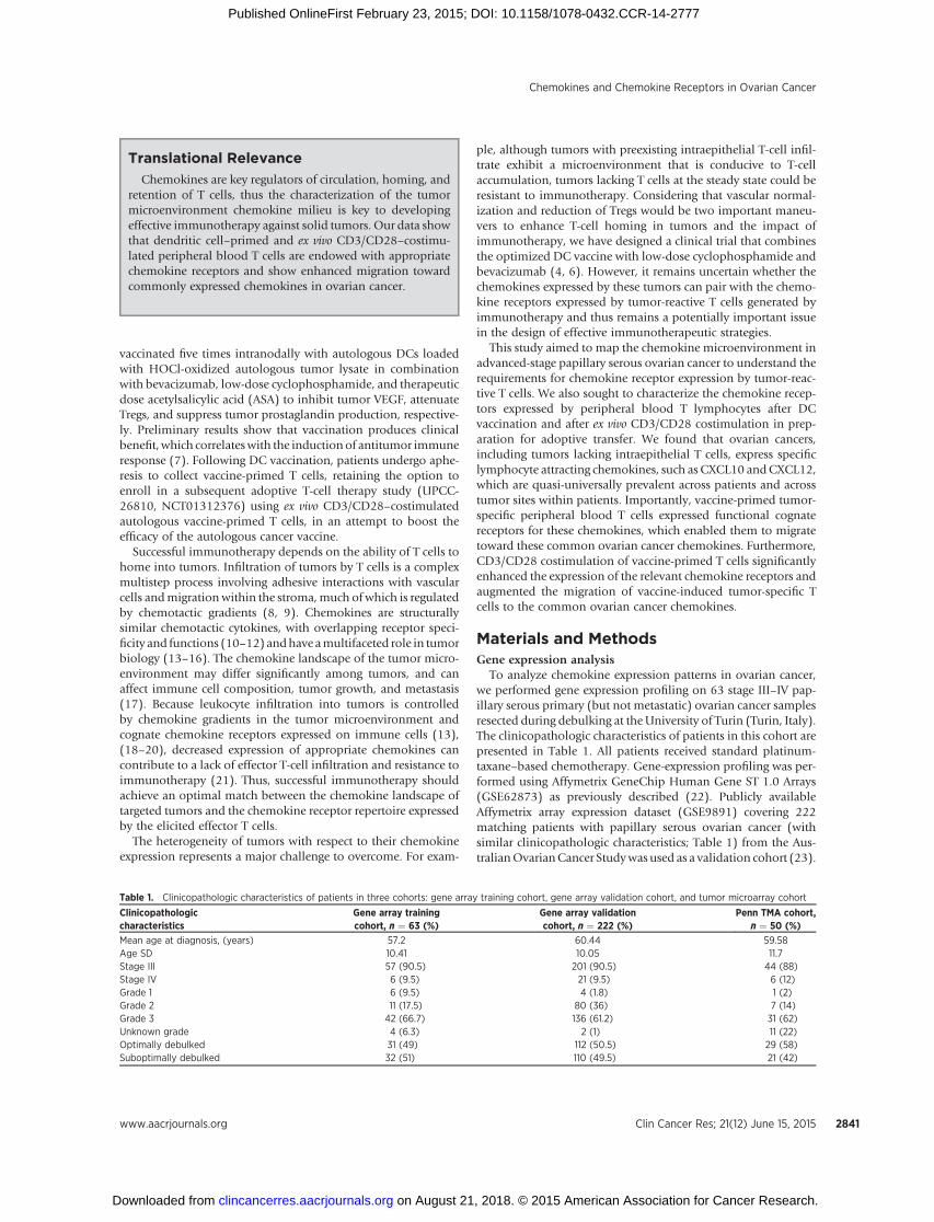
Table 1. Clinicopathologic characteristics of patients in three cohorts: gene array training cohort, gene array validation cohort, and tumor microarray cohort
Clinicopathologiccharacteristics
Gene array trainingcohort, n ¼ 63 (%)
Gene array validationcohort, n ¼ 222 (%)
Penn TMA cohort,n ¼ 50 (%)
Mean age at diagnosis, (years) 57.2 60.44 59.58Age SD 10.41 10.05 11.7Stage III 57 (90.5) 201 (90.5) 44 (88)Stage IV 6 (9.5) 21 (9.5) 6 (12)Grade 1 6 (9.5) 4 (1.8) 1 (2)Grade 2 11 (17.5) 80 (36) 7 (14)Grade 3 42 (66.7) 136 (61.2) 31 (62)Unknown grade 4 (6.3) 2 (1) 11 (22)Optimally debulked 31 (49) 112 (50.5) 29 (58)Suboptimally debulked 32 (51) 110 (49.5) 21 (42)
Translational Relevance
Chemokines are key regulators of circulation, homing, andretention of T cells, thus the characterization of the tumormicroenvironment chemokine milieu is key to developingeffective immunotherapy against solid tumors. Our data showthat dendritic cell–primed and ex vivo CD3/CD28–costimu-lated peripheral blood T cells are endowed with appropriatechemokine receptors and show enhanced migration towardcommonly expressed chemokines in ovarian cancer.
Chemokines and Chemokine Receptors in Ovarian Cancer
www.aacrjournals.org Clin Cancer Res; 21(12) June 15, 2015 2841
on August 21, 2018. © 2015 American Association for Cancer Research. clincancerres.aacrjournals.org Downloaded from
Published OnlineFirst February 23, 2015; DOI: 10.1158/1078-0432.CCR-14-2777
Following normalization, technical outlier samples were identi-fied and excluded.
Gene expression analysis was also performed on 13 primaryand established human ovarian cancer cell lines using AffymetrixGeneChip Human Gene ST 1.0 Arrays (GSE63553). OV7M andOV95 primary ovarian cancer cell lines were provided by Dr.Richard Carroll at the University of Pennsylvania (UPENN; Phi-ladelphia, PA) andwere derived frompatientswith ovarian canceras described previously (24). All other cell lines (A2008, OAW42,OVCAR2, OVCAR3, OVCAR4, OVCAR5, OVCAR8, OVCAR10,PE01, PE04, and SKOV3) were obtained from theOvarian CancerResearch Center cell bank at UPENN with documented and citedorigin, thus were not reauthenticated at the time of the study (25).The detailed origin and characteristics of the cell lines are listed inSupplementary Table S1.
Tissue microarrays constructionA tissue microarray (TMA) was constructed at the Department
of Pathology at UPENN from 50 treatment-na€�ve patients withstage IIIC or IV papillary serous ovarian cancer who underwentprimary resection at UPENN between 2005 and 2008, and whoseclinical characteristics were similar to the two cohorts of patientsused in the microarray data analysis (Table 1). Both primarytumor samples and matched metastatic deposits from the samepatients were included in the TMA, as previously reported (26).Briefly, a total of 207 tumor sites were represented on the arraywith a mean of 3.8 sites per patient, including one or two ovariansites and one to seven metastatic sites. TMA blocks were selectedby a trainedpathologist. For each block, triplicate 0.6-mmcores oftumor were placed on a TMA slide using a manual arrayer.
Immunohistochemistry and TMA scoringCommercially available chemokine antibodies were validated
and titrated on positive (i.e., lymph node, prostate, lung, or coloncancer) and negative control tissues (i.e., cerebellum, stomach, ortestis)—as recommendedby themanufacturers anddescribed in theliterature—before immunohistochemical analysis of the TMA sam-ples. TMA immunostaining was performed with the followingantibody clones: CCL2 (HPA019163; Sigma-Aldrich), CCL4(1738-1; Epitomics), CCL5 (AF-278-NA; R&D Systems), CCL28(MAB7171; R&D Systems), CXCL10 (ab9807; Abcam), CXCL12(SC-28876; Santa Cruz Biotechnology), CXCL16 (ab101404;Abcam), CX3CL1 (HPA040361; Sigma-Aldrich). Lymphocyteswerestained for CD3 (0452; Dakocytomation), CD8 (C8/144B, M7103;Dakocytomation), and FoxP3 (206D, 320102; BioLegend).
Tumor cores were imaged using Aperio ImageScope and wereindependently scored at �20 magnification by two experiencedpathology observers (R. Frank and M.D. Feldman) who wereblinded to clinical and pathologic parameters. An H-score wascalculated by using intensity (score of 3: strongly staining, score of2: moderately staining, score of 1: weakly staining, score of 0: nostaining) � percentage of tumor tissue stained (score of 1: 0%–
25%, score of 2: 26%–50%, score of 3: 51%–75%, and score of 4:76%–100%) for each core (maximum score was 12). Whenmultiple fields were available for review, the median of theirscores was recorded for that core. In the few cases where there wasa discrepancy, a review was performed and a consensus reached.The immunostained microarrays were also scored for tumor-infiltrating lymphocytes (TIL) by an experienced pathologist(I.S. Hagemann). Lymphocytes infiltrating the tumor islets (i.e.,the malignant epithelial compartment) and stromal lymphocytes
(all other lymphocytes) were graded according to the followingquantitative criteria: 0 – absent; 1 – rare [1–10/400� high-powerfield (hpf)]; 2 –moderate (11–20/hpf); 3 – numerous (>20/hpf).The lymphocyte density in partial/incomplete fields was normal-ized on the basis of a visual assessment of the percentage thatconsisted of tumor epithelium or stroma.
Procurement of T cells from ovarian cancer subjectsundergoing vaccination (UPCC-19809) followed by adoptiveT-cell transfer (UPCC-26810)
We analyzed T cells from five HLA-A�0201 subjects with recur-rent ovarian cancer, who were enrolled in a phase I clinicalprotocol at UPENN UPCC-19809 (NCT01132014) and admin-istered therapeutic vaccination using autologous DCs loaded invitro with oxidized autologous tumor lysate. Vaccine-primedperipheral blood T cells were obtained in all subjects as part ofstudy procedures through apheresis 10 to 15 days after the fourthor fifth vaccination. An approximately 15 L apheresis was per-formed at the Apheresis Unit of the Hospital of the UPENN(HUP). Cells were transferred fresh to the Clinical Cell andVaccine Production Facility (CVPF) at HUP where they werewashed to remove plasma, platelets, and red blood cell contam-ination using the Baxter CytoMate or Haemonetics CellSaver5with X-VIVO 15 based media (Lonza). T cells were isolated bycounterflow centrifugal elutriation (Terumo ElutraTM Cell Sep-aration System) to eliminatemonocyte contamination. Cellswerethen counted and cryopreserved in liquid nitrogen vapor phase incryopreservation media containing 10% DMSO.
Following completion of vaccination, the same subjects asabove were enrolled in a follow-on phase I clinical protocol atUPENN (UPCC-26810, NCT01312376) administering adoptivetransfer of ex vivoCD3/CD28–costimulated vaccine-primed autol-ogous peripheral blood T cells. Elutriated vaccine-primed T cellswere thawed and seeded into gas-permeable flasks in X-VIVO 15media supplemented with 5% pooled human AB serum. Anti-CD3/anti-CD28 antibody-coated Dynal microbeads were addedat a ratio of 3:1 beads to cell. To maintain appropriate T-celldensity, freshmediawith low level of IL2 (100 IU/mL)were addedthroughout the 11-day expansion. Cells were then harvested foradoptive transfer, while aliquots were cryopreserved aftermicrobead removal. For this study, we analyzed freshly thawedelutriated T cells from the apheresis product (vaccine-primed Tcells) as well as freshly thawed CD3/CD28–costimulated T cells.
Chemotaxis assaysT cells number and viability was assessed usingGuava Viacount
reagent followed by quantitative capillary flow cytometry (Milli-pore Guava). Cells were rested in fresh media (RPMI-1640;Mediatech) supplemented with 10% FBS, 100 IU/mL of penicil-lin, 100mg/mLof streptomycin, and 20 IU/mLof IL2 for 24hoursbefore the chemotaxis assay. Following assay optimization andthe manufacturers' guidelines, the following chemokines wereplated either alone or in combinations on the bottom of Corningplate: 10 ng/mL of CCL28 (SRP3112; Sigma-Aldrich), 50 ng/mLofCXCL10 (300-12; Peprotech), and100ng/mLofCXCL12 (300-28A; Peprotech). Vaccine-primed and CD3/CD28-costimulatedT cells were seeded at a concentration of 3–5� 105 cells at the toptranswell migration chamber (Corning HTS Transwell-96, 5-mmpore polycarbonate membrane). The average number of liveloaded CD3þ cells was determined by cell counter for eachpatient. After 3 hours of migration, the total number of migrated
Zsiros et al.
Clin Cancer Res; 21(12) June 15, 2015 Clinical Cancer Research2842
on August 21, 2018. © 2015 American Association for Cancer Research. clincancerres.aacrjournals.org Downloaded from
Published OnlineFirst February 23, 2015; DOI: 10.1158/1078-0432.CCR-14-2777
CD3þwas counted, and these cells were further analyzed for CD4,CD8, and/orHER-2/neu pentamer staining by flow cytometry. Allexperiments were performed in duplicates and repeated in threeindependent experiments. Results of migration were reported asthe average number of migrated CD4þ-, CD8þ-, or HER2/neu–specific T cells divided by the average loaded live CD3þ cells forthat patient and then multiplied by 100.
ToblockCXCL10-andCXCL12-mediatedmigration, T cellswereprestained with anti-CXCR3 (MAB160; R&D Systems) and anti-CXCR4 antibodies (MAB170; R&DSystems), respectively. ToblockCCL28-mediatedmigration, anti-CCL28 antibody (MAB717; R&DSystems)was directly added to the chemokine containing chamberand incubated at 37�C for an hour.
Statistical analysisStatistical analysis of gene expression and the significance of
associations between categorical variables were performed usingPearson's c2-tests in R. Heatmaps and cluster analysis of the eightmost highly expressed chemokines in ovarian cancer TMAs weredone using R. A paired t-test was used to compare chemokinereceptor expression and migration data. P values <0.05 wereconsidered statistically significant.
ResultsSpecific chemokine genes are expressed by advanced ovariancancer
To determine the chemokines that are important in ovariancancer for T-cell homing, the expression of all known humanchemokines was analyzed on a gene expression array dataset from63 patients with primary ovarian cancer from the University ofTurin, and validated on a publicly available microarray datasetfrom 222 ovarian cancer samples from the Australian OvarianCancer study. Most chemokines were expressed at low levels onaverage in ovarian cancer, although there was significant hetero-geneity in the expression levels among patients for approximatelyhalf of the chemokine genes (Fig. 1A–D). The five most highlyexpressed chemokines in both the training and validationdatasetswere CCL2, CCL5, CXCL10, CXCL12, and CXCL16.
We further validated the chemokines expressed by ovariancancer cells by examining the gene expression data of 13 estab-lished ovarian cancer cell lines with papillary serous origin. Wefound that few were constitutively expressed in vitro, includingCXCL16, CXCL1, CX3CL1, CCL17, and CCL28.
Among the chemokines most highly expressed in ovariancancer in vivo, we found high constitutive expression of onlyCXCL16 in cancer cell lines in vitro, while CXCL10, CXCL12,CCL2, and CCL5 were not expressed or were expressed at lowlevels (Fig. 1E and F). Thus, ovarian tumors express specificchemokines, and their expression seems to be associated with invivo tumor conditions rather than being a constitutive feature ofovarian cancer cells.
Ovarian cancers express quasi-universal chemokinesNext, we validated the chemokine expression by immunohis-
tochemistry using commercially available antibodies on an ovar-ian serous carcinoma TMA from an independent cohort of 50patients fromUPENN(Table 1). This patient cohortwas similar tothe two previous cohorts of patients analyzed by Affymetrixarrays: themost important clinical factors associatedwith survival(age, stage of disease, tumor grade, and ratio of optimally/sub-
optimally debulked patients) were not statistically differentamong the three cohorts, and all cohorts received standardadjuvant chemotherapy that reflected the best current practice.All samples were collected during primary surgery from chemo-therapy-na€�ve patients. We selected for validation the chemokinegeneswith themost high and/or prevalent expression in anyof thethree above microarray datasets, as well as chemokines withexpression level showing large variation across patients. This finallist included nine chemokines: CCL2, CCL4, CCL5, CCL28,CXCL1, CXCL10, CXCL12, CXCL16, and CX3CL1. With theexception of CXCL1 (where staining with all the commerciallyavailable antibodies appeared to be nonspecific), staining of theselected chemokines was successfully titrated and validated onpositive and negative control tissues before immunohistochem-ical analysis of the TMAsamples. To associate the expressionof theabove chemokines with the presence of effector cytotoxic lym-phocytes (CTL) and Tregs at the time of diagnosis, TMA sampleswere also stained for CD3, CD8, and FoxP3 (Fig. 2A). Medianexpression levels of chemokines and T cells were correlated atprimary and metastatic sites (Fig. 2E).
Immunohistochemistry confirmed the expression of all abovechemokines at the protein level in ovarian cancer (Fig. 2B).Importantly, expression of all chemokines was confirmed withintumor islets in association with the tumor cells. In addition,expression of the same chemokines was found at both primaryand metastatic sites, indicating high degree of similarities amongsites with no statistical staining differences for any of thesechemokines (Fig. 2C).
However, there was heterogeneity of chemokine expressionamong samples. This heterogeneitywasmostly restricted toCCL2,CCL4, CCL5, CCL28, and CX3CL1, which showed no expressionin a proportion of patients (Fig. 2C). On the other hand, we couldidentify a group of chemokines that appeared to be quasi-uni-versally expressed in ovarian cancer (CXCL10, CXCL12, andCXCL16; Fig. 2C). On the basis of the median expression, thecalculated mean H-score for chemokine staining was the highestfor CXCL10, CXCL12, and CXCL16, demonstrating that thesechemokines showed the strongest staining and stained the highestpercentage of ovarian cancer tissue (Fig. 2D).
Tumors with intraepithelial T cells expressed many of the priormentioned variable chemokines: CCL2, CCL4, CCL5, CCL28, andCX3CL1, and in addition, they expressed most of the quasi-uni-versal chemokines:CXCL10,CXCL12,andCXCL16.Theexpressionof CCL4 and CCL5 showed the strongest correlation with thepresence of tumor-infiltrating CD8þ cells, whereas CCL4, CX3CL1,and CXCL12 expression showed correlation with increased num-bers of FoxP3þ cells in tumor islets (correlation, 0.4–0.5) in theprimary tumor tissues (Fig. 2E). In addition, CCL28 and CXCL16were correlated with FoxP3þ cells present in the stromal tumortissue. These data are in agreement with previous observations(2, 27, 28). Importantly, the above correlations were substantiallyreduced or lost in the metastatic tumor sites (Fig. 2E).
Vaccine-primed and CD3/CD28-costimulated T cells used inadoptive therapy express appropriate receptors for ovariancancer chemokines
On the basis of the above data, we hypothesized that ovariancancers express ligands for the following chemokine receptors:CCR1, CCR2, CCR3, CCR4, CCR5, CCR10, CXCR3, CXCR4,CXCR6, and CX3CR1 (Fig. 3A). We thus asked whether our twoimmunotherapy protocols, that is, the whole tumor lysate-pulsed
Chemokines and Chemokine Receptors in Ovarian Cancer
www.aacrjournals.org Clin Cancer Res; 21(12) June 15, 2015 2843
on August 21, 2018. © 2015 American Association for Cancer Research. clincancerres.aacrjournals.org Downloaded from
Published OnlineFirst February 23, 2015; DOI: 10.1158/1078-0432.CCR-14-2777
Figure 1.Results of gene expression analysis of the top 35 chemokines expressed in ovarian cancer. Gene expression analysis of chemokines in the training cohort (A)and in the validation cohort (B), showing the median expression and the four quartiles with the outliers presented as individual circles. Heatmap generatedfrom the training cohort (C; n ¼ 63) and the validation cohort (D; n ¼ 222), showing the individual patients' chemokine expression level for the top 20 most highlyexpressed chemokines from both datasets. Color code shows the expression level compared with the mean gene expression, where the white color showsthe mean expression level for all genes. E, results of gene expression analysis on the top 35 chemokines in 13 papillary serous ovarian cancer cell lines, showingthe median expression and the four quartiles with the outliers presented as individual circles. F, heatmap generated from the 13 ovarian cancer cell linesshowing each cell line separately with the expression level for all tested chemokines.
Zsiros et al.
Clin Cancer Res; 21(12) June 15, 2015 Clinical Cancer Research2844
on August 21, 2018. © 2015 American Association for Cancer Research. clincancerres.aacrjournals.org Downloaded from
Published OnlineFirst February 23, 2015; DOI: 10.1158/1078-0432.CCR-14-2777
DC vaccine approach (UPCC-19809 protocol) and the followingadoptive transfer of ex vivo CD3/CD28–costimulated vaccine-primed peripheral blood T cells (UPCC-26810 protocol), gener-ate T cells expressing the appropriate chemokine receptors forbeing attracted by the ovarian cancer chemokine microenviron-ment. In protocolUPCC-19809, vaccine-primedperipheral bloodT cells were collected through apheresis and elutriation followingfour to five vaccinations. A portion of these vaccine-primed cellswas then subjected to ex vivo costimulation and expansion usinganti-CD3/anti-CD28 Ab-coated beads, in preparation for adop-tive transfer to the same patient. As expected, CD3/CD28 costi-mulation resulted in preferential expansion of CD4þ cells com-pared with the apheresis samples (79%, SD¼ 13.7 vs. 59%, SD¼21.4 of total CD3þ cells, respectively, not shown). We analyzedthe expression of the above chemokine receptors by flow cyto-metry in matched samples of vaccine-primed peripheral bloodCD4þ and CD8þ cells T cells collected at the completion of
vaccination and following ex vivo expansion with CD3/CD28-coated beads from five HLA-A�0201 subjects (Fig. 3B andC). Figures 3D and 3E shows representative examples of FACSanalysis and staining specificity.
In peripheral blood CD4þ T cells collected following vaccina-tion the most commonly expressed chemokine receptors wereCCR10 (CCL27 and CCL28 receptor), expressed on 48.5% (SD¼17.1) of cells; CXCR3 (CXCL9-11 receptor), expressed on 19.8%(SD ¼ 17.5) of cells; CXCR4 (CXCL12 receptor), expressed on8.5% (SD¼ 5.4) of cells; and CCR4 (CCL2, CCL4, CCL5, CCL17,andCCL22 receptor), expressed on 19.4% (SD¼ 7.7) of cells (Fig.3C). A lowproportion [5.5%(SD¼4.4)] of vaccine-primedCD4þ
cells also expressed CCR5 (CCL3, CCL4, and CCL5 receptor).Vaccine-primed CD8þ cells similarly expressed CCR10 (77.3%;SD ¼ 12.7), CXCR3 (21.8%; SD ¼ 8.2), CXCR4 (9.3%; SD ¼13.7), and CCR5 (13.3%; SD ¼ 11.4; Fig. 3D). CCR1, CCR2,CCR3, CXCR2, CXCR6, and CX3CXR1 receptors were all
Figure 2.Correlation of chemokine expressionand infiltration of CD8þ and FoxP3þ
T cells in ovarian cancer. A,representative images of staining forCD8 andFoxP3�20. B, representativeimages of papillary serous ovariancancer specimens that stainedstrongly positive for chemokineexpression �20. C, heterogeneityheatmap demonstrating the medianH-score calculated for every singleevaluable tumor core. The first tworows represent tumor samples fromthe primary disease site (ovaries),while the last four rows representtumor samples from availablemetastatic sites. Missing values arecoded with white. D, heatmapdemonstrating the average medianH-scores from all sites (ovaries andmetastasis combined) calculated forevery patient sample on TMA. E,heatmap showing the correlationbetween chemokine expression andCD8þ and FoxP3þ cells at the primaryand metastatic site.
Chemokines and Chemokine Receptors in Ovarian Cancer
www.aacrjournals.org Clin Cancer Res; 21(12) June 15, 2015 2845
on August 21, 2018. © 2015 American Association for Cancer Research. clincancerres.aacrjournals.org Downloaded from
Published OnlineFirst February 23, 2015; DOI: 10.1158/1078-0432.CCR-14-2777
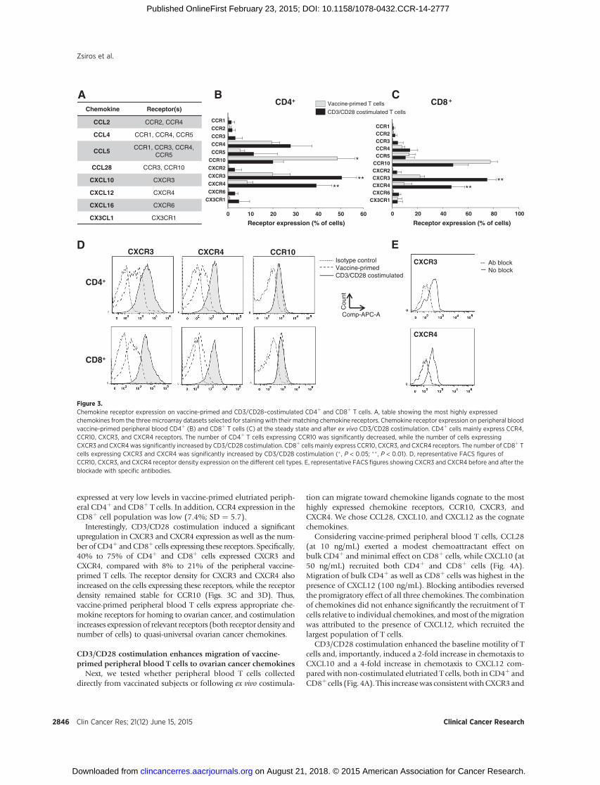
expressed at very low levels in vaccine-primed elutriated periph-eral CD4þ and CD8þ T cells. In addition, CCR4 expression in theCD8þ cell population was low (7.4%; SD ¼ 5.7).
Interestingly, CD3/CD28 costimulation induced a significantupregulation in CXCR3 and CXCR4 expression as well as the num-ber of CD4þ and CD8þ cells expressing these receptors. Specifically,40% to 75% of CD4þ and CD8þ cells expressed CXCR3 andCXCR4, compared with 8% to 21% of the peripheral vaccine-primed T cells. The receptor density for CXCR3 and CXCR4 alsoincreased on the cells expressing these receptors, while the receptordensity remained stable for CCR10 (Figs. 3C and 3D). Thus,vaccine-primed peripheral blood T cells express appropriate che-mokine receptors for homing to ovarian cancer, and costimulationincreases expression of relevant receptors (both receptor density andnumber of cells) to quasi-universal ovarian cancer chemokines.
CD3/CD28 costimulation enhances migration of vaccine-primed peripheral blood T cells to ovarian cancer chemokines
Next, we tested whether peripheral blood T cells collecteddirectly from vaccinated subjects or following ex vivo costimula-
tion can migrate toward chemokine ligands cognate to the mosthighly expressed chemokine receptors, CCR10, CXCR3, andCXCR4. We chose CCL28, CXCL10, and CXCL12 as the cognatechemokines.
Considering vaccine-primed peripheral blood T cells, CCL28(at 10 ng/mL) exerted a modest chemoattractant effect onbulk CD4þ and minimal effect on CD8þ cells, while CXCL10 (at50 ng/mL) recruited both CD4þ and CD8þ cells (Fig. 4A).Migration of bulk CD4þ as well as CD8þ cells was highest in thepresence of CXCL12 (100 ng/mL). Blocking antibodies reversedthe promigratory effect of all three chemokines. The combinationof chemokines did not enhance significantly the recruitment of Tcells relative to individual chemokines, andmost of themigrationwas attributed to the presence of CXCL12, which recruited thelargest population of T cells.
CD3/CD28 costimulation enhanced the baseline motility of Tcells and, importantly, induced a 2-fold increase in chemotaxis toCXCL10 and a 4-fold increase in chemotaxis to CXCL12 com-pared with non-costimulated elutriated T cells, both in CD4þ andCD8þ cells (Fig. 4A). This increasewas consistentwithCXCR3and
Chemokine Receptor(s)
CCL2 CCR2, CCR4
CCL4 CCR1, CCR4, CCR5
CCL5CCR1, CCR3, CCR4,
CCR5
CCL28 CCR3, CCR10
CXCL10 CXCR3
CXCL12 CXCR4
CXCL16 CXCR6
CX3CL1 CX3CR1
Isotype controlVaccine-primed CD3/CD28 costimulated
CXCR3
CXCR4
Ab blockNo block
Cou
nt
Comp-APC-A
ED
0 20 40 60 80 100
CX3CR1CXCR6CXCR4CXCR3CXCR2CCR10CCR5CCR4CCR3CCR2CCR1
Receptor expression (% of cells)0 10 20 30 40 50 60
CX3CR1
CXCR6
CXCR4
CXCR3
CXCR2
CCR10
CCR5
CCR4
CCR3
CCR2
CCR1
Receptor expression (% of cells)
CD4+ +CD8
******
**
*
Vaccine-primed T cells
CD3/CD28 costimulated T cells
CD4+
CD8+
CXCR3 CXCR4 CCR10
A B C
Figure 3.Chemokine receptor expression on vaccine-primed and CD3/CD28–costimulated CD4þ and CD8þ T cells. A, table showing the most highly expressedchemokines from the three microarray datasets selected for staining with their matching chemokine receptors. Chemokine receptor expression on peripheral bloodvaccine-primed peripheral blood CD4þ (B) and CD8þ T cells (C) at the steady state and after ex vivo CD3/CD28 costimulation. CD4þ cells mainly express CCR4,CCR10, CXCR3, and CXCR4 receptors. The number of CD4þ T cells expressing CCR10 was significantly decreased, while the number of cells expressingCXCR3 and CXCR4was significantly increased by CD3/CD28 costimulation. CD8þ cells mainly express CCR10, CXCR3, and CXCR4 receptors. The number of CD8þ Tcells expressing CXCR3 and CXCR4 was significantly increased by CD3/CD28 costimulation (� , P < 0.05; �� , P < 0.01). D, representative FACS figures ofCCR10, CXCR3, and CXCR4 receptor density expression on the different cell types. E, representative FACS figures showing CXCR3 and CXCR4 before and after theblockade with specific antibodies.
Zsiros et al.
Clin Cancer Res; 21(12) June 15, 2015 Clinical Cancer Research2846
on August 21, 2018. © 2015 American Association for Cancer Research. clincancerres.aacrjournals.org Downloaded from
Published OnlineFirst February 23, 2015; DOI: 10.1158/1078-0432.CCR-14-2777
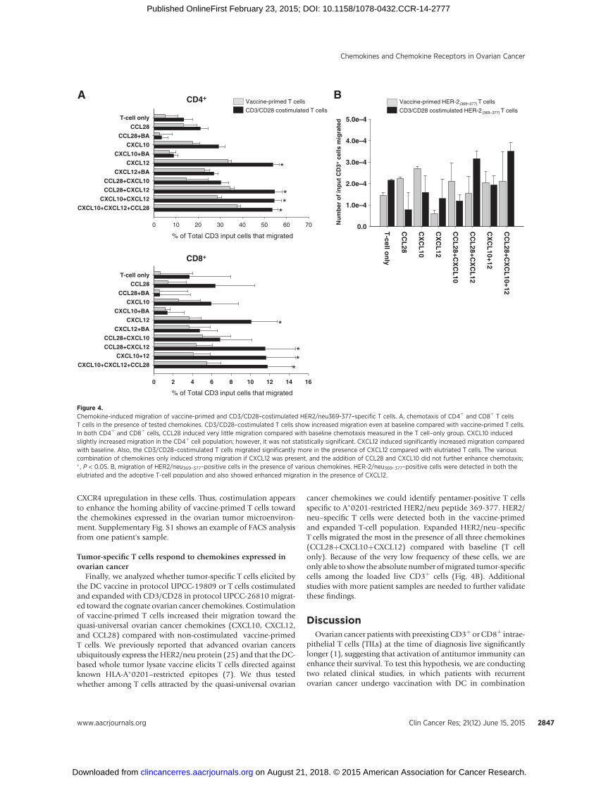
CXCR4 upregulation in these cells. Thus, costimulation appearsto enhance the homing ability of vaccine-primed T cells towardthe chemokines expressed in the ovarian tumor microenviron-ment. Supplementary Fig. S1 shows an example of FACS analysisfrom one patient's sample.
Tumor-specific T cells respond to chemokines expressed inovarian cancer
Finally, we analyzed whether tumor-specific T cells elicited bythe DC vaccine in protocol UPCC-19809 or T cells costimulatedand expanded with CD3/CD28 in protocol UPCC-26810 migrat-ed toward the cognate ovarian cancer chemokines. Costimulationof vaccine-primed T cells increased their migration toward thequasi-universal ovarian cancer chemokines (CXCL10, CXCL12,and CCL28) compared with non-costimulated vaccine-primedT cells. We previously reported that advanced ovarian cancersubiquitously express theHER2/neu protein (25) and that the DC-based whole tumor lysate vaccine elicits T cells directed againstknown HLA-A�0201–restricted epitopes (7). We thus testedwhether among T cells attracted by the quasi-universal ovarian
cancer chemokines we could identify pentamer-positive T cellsspecific to A�0201-restricted HER2/neu peptide 369-377. HER2/neu–specific T cells were detected both in the vaccine-primedand expanded T-cell population. Expanded HER2/neu–specificT cells migrated the most in the presence of all three chemokines(CCL28þCXCL10þCXCL12) compared with baseline (T cellonly). Because of the very low frequency of these cells, we areonly able to show the absolute number ofmigrated tumor-specificcells among the loaded live CD3þ cells (Fig. 4B). Additionalstudies with more patient samples are needed to further validatethese findings.
DiscussionOvarian cancer patients with preexisting CD3þ or CD8þ intrae-
pithelial T cells (TILs) at the time of diagnosis live significantlylonger (1), suggesting that activation of antitumor immunity canenhance their survival. To test this hypothesis, we are conductingtwo related clinical studies, in which patients with recurrentovarian cancer undergo vaccination with DC in combination
A
% of Total CD3 input cells that migrated
0 10 20 30 40 50 60 70
CXCL10+CXCL12+CCL28
CXCL10+CXCL12
CCL28+CXCL12
CCL28+CXCL10
CXCL12+BA
CXCL12
CXCL10+BA
CXCL10
CCL28+BA
CCL28
T-cell only
CD4+
*
* *
*
% of Total CD3 input cells that migrated
0 2 4 6 8 10 12 14 16
CXCL10+CXCL12+CCL28
CXCL10+12
CCL28+CXCL12
CCL28+CXCL10
CXCL12+BA
CXCL12
CXCL10+BA
CXCL10
CCL28+BA
CCL28
T-cell only
CD8+
* *
*
*
Vaccine-primed T cells
CD3/CD28 costimulated T cells
T-cell o
nly
CC
L28
CX
CL
10
CX
CL
12
CC
L28+C
XC
L10
CC
L28+C
XC
L12
CX
CL
10+12
CC
L28+C
XC
L10+12
Nu
mb
er o
f in
pu
t C
D3+
cel
ls m
igra
ted
0.0
1.0e–4
2.0e–4
3.0e–4
4.0e–4
5.0e–4
B Vaccine-primed HER-2 (369–377) T cells
CD3/CD28 costimulated HER-2 (369–377) T cells
Figure 4.Chemokine-induced migration of vaccine-primed and CD3/CD28–costimulated HER2/neu369-377–specific T cells. A, chemotaxis of CD4þ and CD8þ T cellsT cells in the presence of tested chemokines. CD3/CD28–costimulated T cells show increased migration even at baseline compared with vaccine-primed T cells.In both CD4þ and CD8þ cells, CCL28 induced very little migration compared with baseline chemotaxis measured in the T cell–only group. CXCL10 inducedslightly increased migration in the CD4þ cell population; however, it was not statistically significant. CXCL12 induced significantly increased migration comparedwith baseline. Also, the CD3/CD28–costimulated T cells migrated significantly more in the presence of CXCL12 compared with elutriated T cells. The variouscombination of chemokines only induced strong migration if CXCL12 was present, and the addition of CCL28 and CXCL10 did not further enhance chemotaxis;� , P < 0.05. B, migration of HER2/neu369–377–positive cells in the presence of various chemokines. HER-2/neu369–377–positive cells were detected in both theelutriated and the adoptive T-cell population and also showed enhanced migration in the presence of CXCL12.
Chemokines and Chemokine Receptors in Ovarian Cancer
www.aacrjournals.org Clin Cancer Res; 21(12) June 15, 2015 2847
on August 21, 2018. © 2015 American Association for Cancer Research. clincancerres.aacrjournals.org Downloaded from
Published OnlineFirst February 23, 2015; DOI: 10.1158/1078-0432.CCR-14-2777
with bevacizumab and low-dose cyclophosphamide, followed byadoptive transfer of CD3/CD28–costimulated vaccine-primed Tcells. Although in patients with preexisting TILs, it is intuitive thatlymphocytes elicited by the vaccine or transferred adoptivelyshould be able to home to tumors, such assumption may notbe justified in patients lacking preexisting T cells, because theirtumor at the steady state may not have the necessary conditionsfor lymphocyte homing. As chemokines can regulate immune celltrafficking, we sought to understand the chemokine microenvi-ronment of ovarian cancer and test whether this environment cansupport the homing of effector lymphocytes generated with cell-based immunotherapy.
Although the overall ovarian cancer chemokine landscape wasfound to be quite heterogeneous with high expression of knownlymphocyte-recruiting chemokines in tumors with TILs, few che-mokines, such as CXCL10, CXCL12, and CXCL16, were expressedquasi-universally, including in tumors with T cells in the stromabut lacking TILs. We also observed high expression of CXCL1mRNA in all three gene expression datasets, however, could notvalidate its presence at the protein level due to the lack of reliablecommercial antibodies. CXCL1 is a highly proangiogenic ELRþ
chemokine (29), which mainly recruits neutrophils and thuscould be less relevant with respect to T-cell homing. Importantly,and with relevance to immune therapy, CXCL10, CXCL12, andCXCL16, are important lymphocyte chemoattractants. Theirexpression was localized to the epithelial component of thetumor, that is, in associationwith tumor cells, and itwas preservedacross all metastatic sites, suggesting that T cells elicited byimmunotherapy can home to the tumor cell compartment andpotentially to all metastatic deposits.
The expression of T cell–recruiting chemokines by cancer cellscould appear paradoxical. For example, although tumor-derivedCXCL12 has been reported to attract T cells (30, 31), it also exertsimportant tumor-promoting functions. In fact, CXCL12 andCXCL16 are overexpressed in epithelial ovarian carcinomas(32, 33) and other solid tumors (34, 35). CXCL12 is a powerfulactivator of the MAPK cascade in ovarian cancer cells. Cross-talkbetween the CXCL12/CXCR4 and the EGFR pathways promotesovarian cell proliferation (36) and CXCL12/CXCR4 expressionis associated with peritoneal metastasis and ascites formation(36, 37). High CXCL16 expression has also been correlated withpoor prognosis in colorectal and prostate cancer (38, 39). UnlikeCXCL10 and CXCL12, which were expressed only in vivo, CXCL16seems constitutively activated in ovarian cancer cells, as it was alsodetected inprimary and established cell lines in vitro. This could bedriven, in part, by the commonly activated Akt/mTOR pathway(40, 41).
Although CXCL10, CXCL12, and CXCL16 are known to recruitT cells, their expression did not correlate with T-cell infiltration inovarian tumor islets at the time of diagnosis. In fact, approxi-mately half of the tumors did not exhibit intraepithelial T cells, inagreement with our previous observations (1), despite expressingone or more of these chemokines. This could raise the possibilitythat such chemokines in fact donot contribute to recruiting T cells.For example, tumors can secrete an antagonistic N-terminallycleaved CXCL10 variant, which leads to early impairment of theimmune response (42). CXCL12 expression by tumor fibroblastswas recently associated with lack of T cells in a pancreatic ade-nocarcinoma model, while the use of a CXCR4 small-moleculeinhibitor ameliorated T-cell homing to tumors and tumorimmune attack (43). It was hypothesized that CXCL12 bound
on tumor cells through CXCR4 could directly eliminate tumor-reactive T cells. CXCL12 can also recruit suppressive plasmacytoidDCs to ovarian cancer (44) andwe found a correlation of CXCL12expression with the presence of Tregs. These are indirect CXCL12-dependent mechanisms, which could drive peripheral tolerancethrough deregulation of tumor antigen presentation or attenua-tion of tumor-reactive T cells. Furthermore, CXCR4 ligation maydownregulate tumor cell MHC-I expression (45), leading to theinability of T cells to recognize the tumor and engraft in tumorislets (46).
Additional mechanisms to prevent T-cell homing and engraft-ment are likely to operate in these tumors. For example, we havedescribed the tumor endothelial barrier, which prevents T-cellextravasation in tumors, in part, due to endothelin B receptor–mediated downregulation of endothelial ICAM-1 expression(47), and through the upregulation of death-inducing moleculeson surface endothelium such as Fas ligand (48). In addition,tumor-derived soluble factors and surface-inhibitory ligands canattenuate T-cell function, further preventing their engraftment(49). Successful immunotherapy must address these multiplemechanisms to maximize clinical efficacy.
A small fraction of peripheral blood CD4þ and CD8þ T cellsfrom patients with ovarian cancer vaccinated with DC vaccineexpressed chemokine receptors to thequasi-universal chemokinesCXCL10 (CXCR3) and CXCL12 (CXCR4) and a higher fraction ofperipheral blood CD4þ and CD8þ T cells expressed CCR10, thereceptor for CCL28. In addition, a small fraction expressed CCR4and CCR5, the receptors for heterogeneous ovarian cancer che-mokines such as CCL2, CCL4, and CCL5. Thus, peripheral bloodCD4þ and CD8þ T cells have the potential ability to traffic toovarian cancer sites, which was corroborated by chemotaxisexperiments, demonstrating migration toward recombinantCCL28, CXCL10, or CXCL12 alone or combined with the above.Among vaccine-primed T cells migrating toward the above cyto-kines, we found CD8þ T cells specific to the HER-2/neu peptide369–377, which as we demonstrated previously, are undetectableat baseline and are specifically induced by DC-lysate vaccine(4, 7). Importantly, CD3/CD28 costimulation induced a signif-icant upregulation of CXCR3 and CXCR4 in both CD4þ andCD8þ T cells from vaccine-primed peripheral blood T cells. Webelieve this enhanced migration in the CD3/CD28–costimulatedcells was mainly attributed to upregulated CXCR3 and CXCR4receptors; however, it is known that activated T lymphocytesundergo a metabolic reprogramming with increased aerobicglycolysis and comparatively low rates of oxidative phosphory-lation, factors that could also contribute to migration (50).
In summary, we demonstrate that ovarian cancers express avariety of lymphocyte recruiting chemokines. Tumors with pre-existing intraepithelial T cells express high levels of CCL4, CCL5,or CCL8, which can recruit vaccine-primed T cells expressingCCR5 or CCR10. The expression of CCL4 and CCL5, which bindto CCR1, CCR3, CCR5, and CCR4, showed the strongest corre-lation with the presence of tumor-infiltrating CD8þ T cells. Onecould hypothesize that upregulation of CCR5, CCR4, and CCR3in ex vivo CD3/CD28-activated vaccine-primed T cells couldimprove tumor homing in cancers expressing CCL4 and CCL5.In addition, these tumors also express one or more of the quasi-universal chemokines CXCL10 and CXCL12, which could recruitrelevant vaccine-primed tumor-reactive T cells expressing CXCR3or CXCR4. Given the ability of these tumors to recruit T cells atbaseline, and based on their chemokine repertoire, these tumors
Zsiros et al.
Clin Cancer Res; 21(12) June 15, 2015 Clinical Cancer Research2848
on August 21, 2018. © 2015 American Association for Cancer Research. clincancerres.aacrjournals.org Downloaded from
Published OnlineFirst February 23, 2015; DOI: 10.1158/1078-0432.CCR-14-2777
are predictably readily infiltrated by vaccine-primed or ex vivocostimulated T cells. On the other hand, tumors lacking preexist-ing T cells were found to express one or both of the quasi-universallymphocyte recruiting chemokines CXCL10 and CXCL12. T cellselicited by DC vaccine expressed the appropriate receptors tohome to these chemokines, and ex vivo CD3/CD28 costimulationfurther enhanced expression of these chemokine receptors andmigration toward these chemokines. Further work is required toassess whether CXCL12 or CXCL10 can promote or rather sup-press recruitment of T cells in tumors lacking intraepithelialT cells, but additional factors could certainly prevent T-cellhoming and function in these tumors. In our clinical trial design,the addition of vascular normalizationwith bevacizumab and theattenuation of Treg with low-dose cyclophosphamide are addres-sing two such important factors. Immunotherapy approaches thatcan expand the available pool of tumor-reactive T cells, such asvaccines and adoptive transfer of vaccine-primed T cells, couldobviate the lack of intraepithelial T cells in many ovarian cancers.The ongoing studies with the tumor lysate-pulsed DC vaccine andadoptive transfer of vaccine-primed costimulated T cells willprovide us an opportunity to test this hypothesis.
Disclosure of Potential Conflicts of InterestC.H. June has ownership interest in IP rights and patents licensed to U.S.
Government. No potential conflicts of interest were disclosed by the otherauthors.
Authors' ContributionsConception and design: E. Zsiros, P. Duttagupta, B.L. Levine, F.M. Marincola,J. Tanyi, G. Coukos
Development of methodology: E. Zsiros, P. Duttagupta, C.H. June, F.M.Marincola, D.J. Powell Jr, M.D. Feldman, G. CoukosAcquisition of data (provided animals, acquired and managed patients,provided facilities, etc.): E. Zsiros, P. Duttagupta, R. Frank, T. Garrabrant,I.S. Hagemann, B.L. Levine, L. Zhang, E. Wang, F.M. Marincola, D. Bedognetti,J. Tanyi, M.D. Feldman, L.E. Kandalaft, G. CoukosAnalysis and interpretation of data (e.g., statistical analysis, biostatistics,computational analysis): E. Zsiros, P. Duttagupta, H. Li, R. Frank, E.Wang, F.M.Marincola, D. Bedognetti, D.J. Powell Jr, M.D. FeldmanWriting, review, and/or revision of themanuscript: E. Zsiros, P. Duttagupta, D.Dangaj, H. Li, R. Frank, I.S. Hagemann, B.L. Levine, C.H. June, F.M. Marincola,D. Bedognetti, D.J. Powell Jr, J. Tanyi, M.D. Feldman, L.E. Kandalaft, G. CoukosAdministrative, technical, or material support (i.e., reporting or organizingdata, constructing databases): E. Zsiros, I.S. Hagemann, D.J. Powell JrStudy supervision: E. Zsiros, L.E. Kandalaft, G. Coukos
AcknowledgmentsThe authors thankDr. KathleenMontone, Li-PingWang, and Amy Ziober for
their assistancewith validating andperforming the chemokine staining onTMA.
Grant SupportThis study was supported by NCI P01-CA83638 SPORE in Ovarian Cancer,
R01 FD003520, and the Ovarian Cancer Research Fund (to G. Coukos) and theCA127334 (to H. Li). The study was also supported by the Conquer CancerFoundation of the American Society of Clinical Oncology (Young InvestigatorAward granted to Davide Bedognetti).
The costs of publication of this articlewere defrayed inpart by the payment ofpage charges. This article must therefore be hereby marked advertisement inaccordance with 18 U.S.C. Section 1734 solely to indicate this fact.
Received December 6, 2014; revised January 31, 2015; accepted February 6,2015; published OnlineFirst February 23, 2015.
References1. Zhang L, Conejo-Garcia JR, Katsaros D, Gimotty PA,MassobrioM, Regnani
G, et al. Intratumoral T cells, recurrence, and survival in epithelial ovariancancer. N Engl J Med 2003;348:203–13.
2. Curiel TJ, Coukos G, Zou L, Alvarez X, Cheng P, Mottram P, et al. Specificrecruitment of regulatory T cells in ovarian carcinoma fosters immuneprivilege and predicts reduced survival. Nat Med 2004;10:942–9.
3. Kryczek I, Wei S, Zhu G, Myers L, Mottram P, Cheng P, et al. Relationshipbetween B7-H4, regulatory T cells, and patient outcome in human ovariancarcinoma. Cancer Res 2007;67:8900–5.
4. Kandalaft LE, Powell DJ Jr, Chiang CL, Tanyi J, Kim S, Bosch M, et al.Autologous lysate-pulsed dendritic cell vaccination followed by adoptivetransfer of vaccine-primed ex vivo co-stimulated T cells in recurrent ovariancancer. Oncoimmunology 2013;2:e22664.
5. Chiang CL, Hagemann AR, Leskowitz R, Mick R, Garrabrant T, CzernieckiBJ, et al. Day-4 myeloid dendritic cells pulsed with whole tumor lysate arehighly immunogenic and elicit potent anti-tumor responses. PLoS ONE2011;6:e28732.
6. Kandalaft LE, Chiang CL, Tanyi J, Motz G, Balint K, Mick R, et al. A phase Ivaccine trial using dendritic cells pulsedwith autologous oxidized lysate forrecurrent ovarian cancer. J Transl Med 2013;11:149.
7. Chiang CL, Kandalaft LE, Tanyi J, Hagemann AR, Motz GT, Svoronos N,et al. A dendritic cell vaccine pulsed with autologous hypochlorous acid-oxidized ovarian cancer lysate primes effective broad antitumor immunity:from bench to bedside. Clin Cancer Res 2013;19:4801–15.
8. Springer TA. Traffic signals for lymphocyte recirculation and leukocyteemigration: the multistep paradigm. Cell 1994;76:301–14.
9. Mukai S, Kjaergaard J, Shu S, Plautz GE. Infiltration of tumors by system-ically transferred tumor-reactive T lymphocytes is required for antitumorefficacy. Cancer Res 1999;59:5245–9.
10. Zlotnik A, YoshieO.Chemokines: a new classification systemand their rolein immunity. Immunity 2000;12:121–7.
11. Baggiolini M, Dewald B, Moser B. Human chemokines: an update. AnnuRev Immunol 1997;15:675–705.
12. Luster AD. Chemokines—chemotactic cytokines that mediate inflamma-tion. N Engl J Med 1998;338:436–45.
13. Kulbe H, Chakravarty P, Leinster DA, Charles KA, Kwong J, Thompson RG,et al. A dynamic inflammatory cytokine network in the human ovariancancer microenvironment. Cancer Res 2012;72:66–75.
14. Vandercappellen J, Van Damme J, Struyf S. The role of CXC chemokinesand their receptors in cancer. Cancer Lett 2008;267:226–44.
15. Wang JM, Deng X, Gong W, Su S. Chemokines and their role in tumorgrowth and metastasis. J Immunol Methods 1998;220:1–17.
16. Zlotnik A, Yoshie O. The chemokine superfamily revisited. Immunity2012;36:705–16.
17. Balkwill FR. The chemokine system and cancer. J Pathol 2012;226:148–57.18. Owen JL, Criscitiello MF, Libreros S, Garcia-Areas R, Guthrie K, Torroella-
Kouri M, et al. Expression of the inflammatory chemokines CCL2, CCL5and CXCL2 and the receptors CCR1–3 and CXCR2 in T lymphocytes frommammary tumor-bearing mice. Cell Immunol 2011;270:172–82.
19. Hwang TL, Lee LY,WangCC, Liang Y, Huang SF,WuCM. CCL7 andCCL21overexpression in gastric cancer is associated with lymph node metastasisand poor prognosis. World J Gastroenterol 2012;18:1249–56.
20. Vinader V, Afarinkia K. The emerging role of CXC chemokines and theirreceptors in cancer. Future Med Chem 2012;4:853–67.
21. Oh SM,OhK, LeeDS. Intratumoral administration of secondary lymphoidchemokine and unmethylated cytosine-phosphorothioate-guanine oligo-deoxynucleotide synergistically inhibits tumor growth in vivo. J KoreanMed Sci 2011;26:1270–6.
22. Liu Q, Tomei S, Ascierto ML, De Giorgi V, Bedognetti D, Dai C, et al.Melanoma NOS1 expression promotes dysfunctional IFN signaling. J ClinInvest 2014;124:2147–59.
23. Tothill RW, Tinker AV, George J, Brown R, Fox SB, Lade S, et al. Novelmolecular subtypes of serous and endometrioid ovarian cancer linked toclinical outcome. Clin Cancer Res 2008;14:5198–208.
24. Bertozzi CC, Chang CY, Jairaj S, Shan X, Huang J, Weber BL, et al. Multipleinitial culture conditions enhance the establishment of cell lines from
www.aacrjournals.org Clin Cancer Res; 21(12) June 15, 2015 2849
Chemokines and Chemokine Receptors in Ovarian Cancer
on August 21, 2018. © 2015 American Association for Cancer Research. clincancerres.aacrjournals.org Downloaded from
Published OnlineFirst February 23, 2015; DOI: 10.1158/1078-0432.CCR-14-2777
primary ovarian cancer specimens. In Vitro Cell Dev Biol Anim2006;42:58–62.
25. Lanitis E, Dangaj D, Hagemann IS, Song DG, Best A, SandaltzopoulosR, et al. Primary human ovarian epithelial cancer cells broadly expressHER2 at immunologically-detectable levels. PLoS ONE 2012;7:e49829.
26. HagemannAR,Hagemann IS, CadungogM,HwangWT, Patel P, Lal P, et al.Tissue-based immune monitoring II: multiple tumor sites reveal immu-nologic homogeneity in serous ovarian carcinoma. Cancer Biol Ther2011;12:367–77.
27. Facciabene A, Peng X, Hagemann IS, Balint K, Barchetti A, Wang LP, et al.Tumour hypoxia promotes tolerance and angiogenesis via CCL28 and T(reg) cells. Nature 2011;475:226–30.
28. Facciabene A, Santoro S, Coukos G. Know thy enemy: why are tumor-infiltrating regulatory T cells so deleterious? Oncoimmunology 2012;1:575–7.
29. Kiefer F, Siekmann AF. The role of chemokines and their receptors inangiogenesis. Cell Mol Life Sci 2011;68:2811–30.
30. Dunussi-Joannopoulos K, Zuberek K, Runyon K, Hawley RG, Wong A,Erickson J, et al. Efficacious immunomodulatory activity of the chemokinestromal cell-derived factor 1 (SDF-1): local secretion of SDF-1 at the tumorsite serves as T-cell chemoattractant and mediates T-cell-dependent anti-tumor responses. Blood 2002;100:1551–8.
31. Franciszkiewicz K, Boutet M, Gauthier L, Vergnon I, Peeters K, Duc O, et al.Synaptic release of CCL5 storage vesicles triggers CXCR4 surface expressionpromoting CTL migration in response to CXCL12. J Immunol 2014;193:4952–61.
32. Guo L, Cui ZM, Zhang J, Huang Y. Chemokine axes CXCL12/CXCR4 andCXCL16/CXCR6 correlate with lymph node metastasis in epithelial ovar-ian carcinoma. Chinese J Cancer 2011;30:336–43.
33. Son DS, Parl AK, Rice VM, Khabele D. Keratinocyte chemoattractant (KC)/human growth-regulated oncogene (GRO) chemokines and pro-inflam-matory chemokine networks in mouse and human ovarian epithelialcancer cells. Cancer Biol Ther 2007;6:1302–12.
34. Phillips RJ, Burdick MD, Lutz M, Belperio JA, Keane MP, Strieter RM. Thestromal derived factor-1/CXCL12-CXC chemokine receptor 4 biologicalaxis in non-small cell lung cancer metastases. Am J Respir Crit Care Med2003;167:1676–86.
35. Zeelenberg IS, Ruuls-Van Stalle L, Roos E. The chemokine receptor CXCR4is required for outgrowth of colon carcinomamicrometastases. Cancer Res2003;63:3833–9.
36. Jiang YP,WuXH, Shi B,WuWX, YinGR. Expression of chemokine CXCL12and its receptor CXCR4 in human epithelial ovarian cancer: an indepen-dent prognostic factor for tumor progression. Gynecol Oncol 2006;103:226–33.
37. Machelon V,Gaudin F, Camilleri-Broet S, Nasreddine S, Bouchet-Delbos L,Pujade-Lauraine E, et al. CXCL12 expression by healthy and malignantovarian epithelial cells. BMC Cancer 2011;11:97.
38. Hershberger PM, Peddibhotla S, Sugarman E,Maloney P, KeyD, Suyama E,et al. Probing the CXCR6/CXCL16 axis: targeting prevention of prostatecancer metastasis. Probe Reports from the NIH Molecular Libraries Pro-gram. Bethesda (MD); 2010.
39. Matsushita K, Toiyama Y, Tanaka K, Saigusa S, Hiro J, Uchida K, et al.Soluble CXCL16 in preoperative serum is a novel prognostic marker andpredicts recurrence of liver metastases in colorectal cancer patients. AnnSurg Oncol 2012;19:S518–27.
40. Deng L, Chen N, Li Y, Zheng H, Lei Q. CXCR6/CXCL16 functions as aregulator in metastasis and progression of cancer. Biochim Biophys Acta2010;1806:42–9.
41. Huang J, Zhang L, Greshock J, Colligon TA,Wang Y,Ward R, et al. Frequentgenetic abnormalities of the PI3K/AKT pathway in primary ovarian cancerpredict patient outcome. Genes Chromosomes Cancer 2011;50:606–18.
42. Rainczuk A, Rao JR, Gathercole JL, Fairweather NJ, Chu S, Masadah R, et al.Evidence for the antagonistic form of CXC-motif chemokine CXCL10 inserous epithelial ovarian tumours. Int J Cancer 2013;134:530–41.
43. Feig C, Jones JO, Kraman M, Wells RJ, Deonarine A, Chan DS, et al.Targeting CXCL12 from FAP-expressing carcinoma-associated fibroblastssynergizes with anti-PD-L1 immunotherapy in pancreatic cancer. Proc NatlAcad Sci U S A 2013;110:20212–7.
44. ZouW,MachelonV,Coulomb-L'HerminA, Borvak J,NomeF, Isaeva T, et al.Stromal-derived factor-1 inhuman tumors recruits andalters the functionofplasmacytoid precursor dendritic cells. Nat Med 2001;7:1339–46.
45. Wang Z, Zhang L,QiaoA,Watson K, Zhang J, FanGH. Activation of CXCR4triggers ubiquitination and down-regulation of major histocompatibilitycomplex class I (MHC-I) on epithelioid carcinomaHeLa cells. J Biol Chem2008;283:3951–9.
46. Han LY, Fletcher MS, Urbauer DL, Mueller P, Landen CN, Kamat AA, et al.HLA class I antigen processing machinery component expression andintratumoral T-Cell infiltrate as independent prognosticmarkers in ovariancarcinoma. Clin Cancer Res 2008;14:3372–9.
47. Buckanovich RJ, Facciabene A, Kim S, Benencia F, Sasaroli D, Balint K, et al.Endothelin B receptor mediates the endothelial barrier to T cell homing totumors and disables immune therapy. Nat Med 2008;14:28–36.
48. MotzGT, Santoro SP,Wang LP,Garrabrant T, Lastra RR,Hagemann IS, et al.Tumor endothelium FasL establishes a selective immune barrier promot-ing tolerance in tumors. Nat Med 2014;20:607–15.
49. Motz GT, Coukos G. Deciphering and reversing tumor immune suppres-sion. Immunity 2013;39:61–73.
50. Gaber T, Strehl C, Sawitzki B, Hoff P, Buttgereit F. Cellular Energy Metab-olism in T-Lymphocytes. Int Rev Immunol 2015;34:34–49.
Clin Cancer Res; 21(12) June 15, 2015 Clinical Cancer Research2850
Zsiros et al.
on August 21, 2018. © 2015 American Association for Cancer Research. clincancerres.aacrjournals.org Downloaded from
Published OnlineFirst February 23, 2015; DOI: 10.1158/1078-0432.CCR-14-2777
2015;21:2840-2850. Published OnlineFirst February 23, 2015.Clin Cancer Res Emese Zsiros, Priyanka Duttagupta, Denarda Dangaj, et al. Prepared for Adoptive Therapy
Costimulated T Cells−Homing of Vaccine-Primed and CD3/CD28 The Ovarian Cancer Chemokine Landscape Is Conducive to
Updated version
10.1158/1078-0432.CCR-14-2777doi:
Access the most recent version of this article at:
Material
Supplementary
http://clincancerres.aacrjournals.org/content/suppl/2015/02/25/1078-0432.CCR-14-2777.DC1
Access the most recent supplemental material at:
Cited articles
http://clincancerres.aacrjournals.org/content/21/12/2840.full#ref-list-1
This article cites 49 articles, 11 of which you can access for free at:
E-mail alerts related to this article or journal.Sign up to receive free email-alerts
Subscriptions
Reprints and
To order reprints of this article or to subscribe to the journal, contact the AACR Publications Department at
Permissions
Rightslink site. Click on "Request Permissions" which will take you to the Copyright Clearance Center's (CCC)
.http://clincancerres.aacrjournals.org/content/21/12/2840To request permission to re-use all or part of this article, use this link
on August 21, 2018. © 2015 American Association for Cancer Research. clincancerres.aacrjournals.org Downloaded from
Published OnlineFirst February 23, 2015; DOI: 10.1158/1078-0432.CCR-14-2777