Embed Size (px)
Citation preview
Ma and Li BMC Oral Health (2021) 21:109 https://doi.org/10.1186/s12903-021-01474-7
RESEARCH ARTICLE
The optimal orthodontic displacement of clear aligner for mild, moderate and severe periodontal conditions: an in vitro study in a periodontally compromised individual using the finite element modelYanning Ma and Song Li*
Abstract
Background: Pathologic tooth migration (PTM) is a common complication of mild to severe periodontitis and proper orthodontic treatment is helpful to alleviate periodontal diseases. The goal of this study is to explore an opti-mal orthodontic displacement of clear aligner using a three-dimensional (3D) finite element model (FEM).
Methods: The cone beam computed tomography (CBCT) data of a patient received invisible orthodontics without diabetes and other systemic diseases were collected. Based on the new classification scheme for periodontal diseases in 2017 (stage I: mild periodontitis, [M1]; stage II: moderate periodontitis, [M2]; stage III: severe periodontitis, [M3]), 3D-FEMs of mandible were established using MIMICS 10.0 and ABAQUS 6.5 softwares. The 3D stress distribution diagrams and stress value of the teeth (left lower incisor, left lower central incisor, right lower lateral incisor, and right lower central incisor) under three different periodontal conditions (M1, M2, and M3) with axial inclination 90° and 100° were obtained by ABAQUS 6.5.
Results: The stress of anterior teeth was concentrated in the teeth neck, and became greater when the periodontal condition was worse. The stress value of anterior teeth and the strain at the top of the alveolar crest are greater as the displacement increasing. The stress value of anterior teeth and the strain at the top of the alveolar crest in axial inclination 100° are relatively great compared to those of axial inclination 90°. For patients with excessively inclined anterior teeth (such as 100°), the optimal orthodontic displacement is 0.18 mm. In order to ensure that alveolar ridge crest is not deformed, the displacement is less than 0.18 mm (strain for 0.165 mm), 0.15 mm (strain for 0.167 mm) and 0.10 mm (strain for 0.117 mm) respectively when alveolar bone is normal, resorption 1/3 or 1/3–1/2.
Conclusions: The optimal orthodontic displacement for patients (M1, M2, and M3) with excessively inclined anterior teeth (axial inclination 100°) is 0.18 mm. To avoid the strain at the top of the alveolar crest, the optimal displacements for M1, M2 and M3 periodontal disease patients are less than 0.18 mm, 0.15 mm and 0.10 mm, respectively.
Keywords: Clear aligner, Anterior teeth, Alveolar ridge crest, Displacement
© The Author(s) 2021. Open Access This article is licensed under a Creative Commons Attribution 4.0 International License, which permits use, sharing, adaptation, distribution and reproduction in any medium or format, as long as you give appropriate credit to the original author(s) and the source, provide a link to the Creative Commons licence, and indicate if changes were made. The images or other third party material in this article are included in the article’s Creative Commons licence, unless indicated otherwise in a credit line to the material. If material is not included in the article’s Creative Commons licence and your intended use is not permitted by statutory regulation or exceeds the permitted use, you will need to obtain permission directly from the copyright holder. To view a copy of this licence, visit http://creat iveco mmons .org/licen ses/by/4.0/. The Creative Commons Public Domain Dedication waiver (http://creat iveco mmons .org/publi cdoma in/zero/1.0/) applies to the data made available in this article, unless otherwise stated in a credit line to the data.
BackgroundPathologic tooth migration (PTM) is a common compli-cation of mild to severe periodontitis and manifests as the inclination, elongation and fan-out of the anterior teeth [1]. The incidence of periodontal diseases among adults
Open Access
*Correspondence: [email protected] of Orthodontics, School of Stomatology, Capital Medical University, No. 4, Tian Tan Xi Li, Beijing 100050, China
Page 2 of 8Ma and Li BMC Oral Health (2021) 21:109
is as high as 76%-92% [2]. In adult patients, the speed of alveolar bone resorption under pressure is greater than that of traction and hyperplasia [3]. The impedance center of the tooth moves to the root and the torque is increased, which further aggravates the destruction of periodontal tissues [4]. Therefore, adult patients with periodontitis should be treated with light force to main-tain the health and stability of the periodontal environ-ment [5]. A 12-year follow-up experiment has shown that orthodontic treatment is no longer a contraindication for severe periodontal diseases [6]. Therefore, a compre-hensive and complete orthodontic treatment should be carried out, otherwise accelerating periodontal inflam-mation and bone destruction.
Recent years, more attentions have been paid to the effects of clear aligner on periodontal diseases due to its aesthetics and comfort on wearers [7]. As a new type of orthodontic treatment, clear aligner can cover all the teeth and keratinized gingiva without obvious damage to the periodontal tissues [8]. Besides, the use of removable orthodontic appliance is conducive to the maintenance of oral hygiene and minimizes the risk of periodontal complication [9, 10]. There are significant differences between the use of fixed appliance and clear aligner. The fixed appliance exerts continuous light force by bonding the brackets on the tooth surface and ligating the arch wire [11], while the clear aligner applies a kind of instan-taneous stress, which is about 50 to 500 times than that of fixed orthodontic loading [11, 12]. However, whether the instantaneous higher-stress has a certain impact on the periodontal tissues, or whether there is a gap in the efficiency of tooth movement due to different periodontal conditions still need to be explored. Additionally, among the various types of movement of teeth such as intrusion, extrusion, rotation, tipping, and alignment movements, intrusion movement is easier to cause tooth loosening or root absorption [13]. Therefore, it is very crucial to inves-tigate intrusion movement in the treatment of periodon-tal diseases [13, 14].
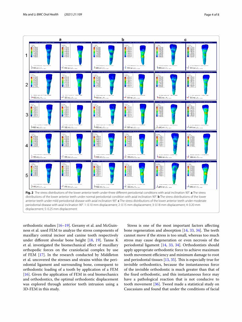
The finite element model (FEM) is an engineering tech-nique used to calculate stress and deformation devel-oped on a geometric solid submitted to external forces [15–19]. Recent years, FEM has been widely applied in different dental fields, from fixtures to the simulation of dental movements to assess the stresses generated within the different tissue structures, such as alveolar bone, per-iodontal ligament, and teeth [20–23]. For instance, Crimi et al. used FEM to analyze the changes in the buccal cor-tical bone in patients undergoing orthodontics surgeries and indicated that there is no direct proportionality rela-tionship between the extent of bone apposition/reabsorp-tion and dental movement [24]. Interestingly, Cervino et al. introduced a new prosthodontic technology named
Digital Smile Design, which is used in combination with FEM to improve the quality of the rehabilitations [25]. In addition, with the use of FEM, it is possible to deter-mine loading and displacement patterns according to the appliance used [26, 27]. But relevant FEM researches on the optimal displacement of clear aligner under different periodontal-statuses are relatively rare.
In this study, we investigated the effects of anterior teeth intrusion on different periodontal conditions (stage I: mild periodontitis, [M1]; stage II: moderate periodon-titis, [M2]; stage III: severe periodontitis, [M3]) using a 3D-FEM, and explored the optimal displacement of teeth when anterior teeth were intruded using clear aligner. This study is aimed to provide a direction for the use of clear aligner in clinic.
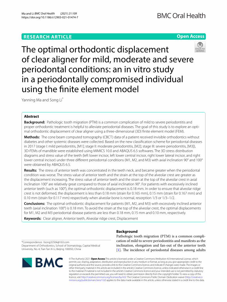
MethodsEstablishment of mandible modelThe Cone Beam Computed Tomography (CBCT) data (HiRes3D-Plus; Largev, Beijing, China) of a patient (female, 35 years old) with mild periodontitis received invisible orthodontics were collected. The exclusion criteria for the patient included diabetes and/or other systemic diseases that may cause progressive periodon-tal injury. Afterwards, based on the new classification scheme for periodontal diseases in 2017 (M1: stage I, normal alveolar bone with mild periodontitis; M2: stage II, alveolar bone resorption 1/3 with moderate periodon-titis; M3: alveolar bone resorption 1/3–1/2 with severe periodontitis) [28, 29], 3D-FEMs of mandible (Fig. 1) were established using MIMICS 10.0 (Materi-alise, Leu-ven, Belgium) and ABAQUS 6.5 (ABAQUS. Inc, USA) softwares as previous studies described [17–19]. Though the finite element software ABAQUS 6.5, we calculated the stress of each part in the models, and obtained the 3D stress distribution diagrams and stress value of the teeth.
Simulation of anterior teeth intrusionTo simulate of anterior teeth intrusion (left lower incisor, left lower central incisor, right lower lateral incisor, and right lower central incisor), the displacement of the teeth (0.10 mm, 0.15 mm, 0.18 mm, 0.20 mm and 0.25 mm, respectively) was preseted [14] and was used to analyze the change of stress value with two different axial inclina-tions of anterior teeth (90° and 100°). The stress value of normal periodontal-condition was utilized as a control.
Structure parameters and calculation of stressAn elastic-lineal behavior and standard Young’s modulus (alveolar bone: 1.40 × 10³ MPa; tooth: 1.96 × 10⁴ MPa; clear aligner: 528 MPa; vertical rectangular-attach-ment: 1.25 × 10⁴ MPa) and Poisson’s ratio (alveo-lar bone: 0.30; tooth: 0.30; clear aligner: 0.36; vertical
Page 3 of 8Ma and Li BMC Oral Health (2021) 21:109
rectangular-attachment: 0.36) of the model were consid-ered for all calculations as the previous studies described [30, 31]. The Von Mises comprehensive equivalent stress was selected as the main indicator to measure the stress level. Though the finite element software ABAQUS 6.5, we calculated the stress of each part in the models, and obtained the 3D stress distribution diagrams and stress value of the teeth.
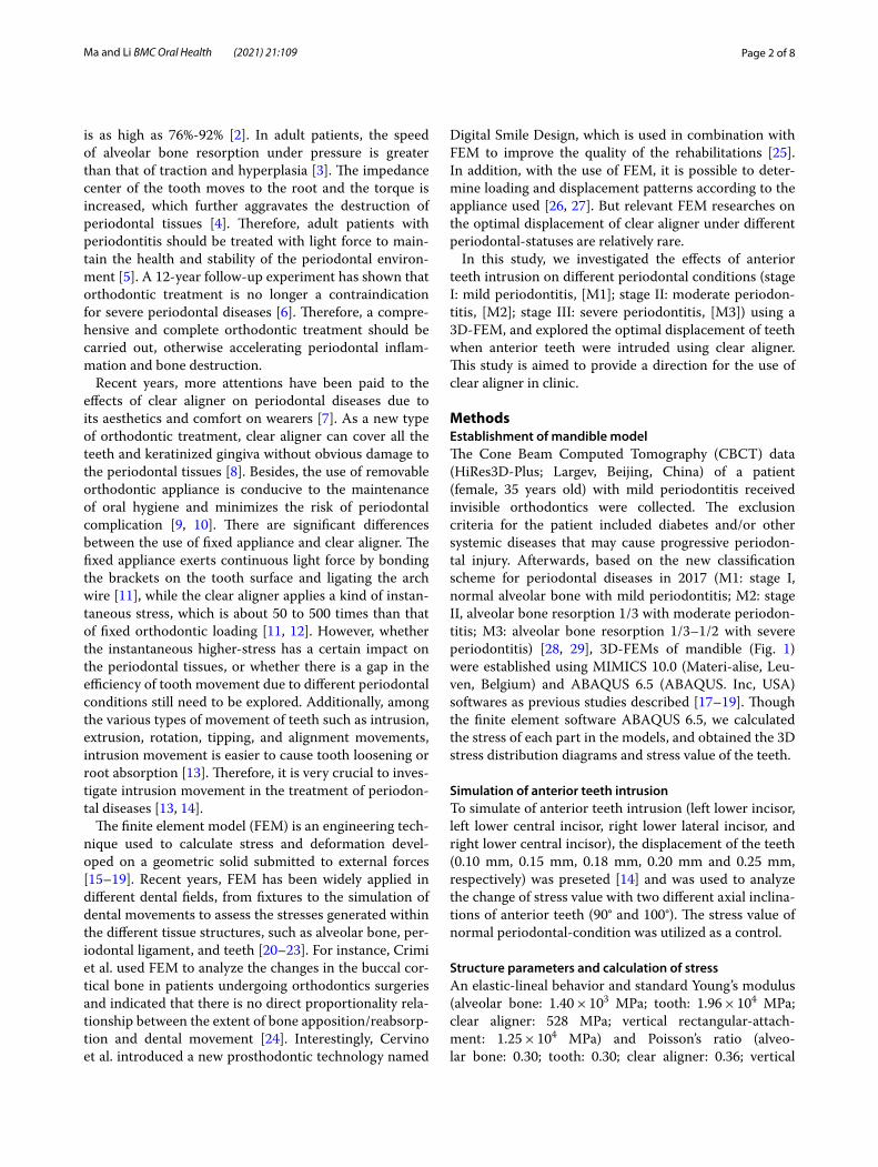
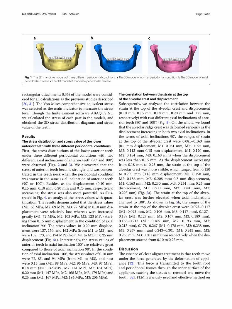
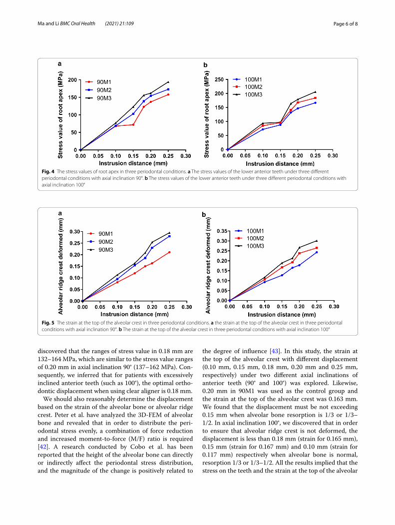
ResultsThe stress distribution and stress value of the lower anterior teeth with three different periodontal conditionsFirst, the stress distributions of the lower anterior teeth under three different periodontal conditions with two different axial inclinations of anterior teeth (90° and 100°) were observed (Figs. 2 and 3). We discovered that the stress of anterior teeth became stronger and was concen-trated in the teeth neck when the periodontal condition was worse in the same axial inclination of anterior teeth (90° or 100°). Besides, as the displacement (0.10 mm, 0.15 mm, 0.18 mm, 0.20 mm and 0.25 mm, respectively) increasing, the stress was also more powerful. As illus-trated in Fig. 4, we analyzed the stress values with quan-tification. The results demonstrated that the stress values (M1: 68 MPa, M2: 69 MPa, M3: 77 MPa) in 0.10 mm dis-placement were relatively low, whereas were increased greatly (M1: 72 MPa, M2: 103 MPa, M3: 123 MPa) start-ing from 0.15 mm displacement in the condition of axial inclination 90°. The stress values in 0.20 mm displace-ment were 137, 154, and 162 MPa (from M1 to M3), and were 158, 173, and 194 MPa (from M1 to M3) in 0.25 mm displacement (Fig. 4a). Interestingly, the stress values of anterior teeth in axial inclination 100° are relatively great compared to those of axial inclination 90°. In the condi-tion of axial inclination 100°, the stress values of 0.10 mm were 72, 85, and 94 MPa (from M1 to M3), and next were 0.15 mm (M1: 88 MPa, M2: 96 MPa, M3: 97 MPa), 0.18 mm (M1: 132 MPa, M2: 141 MPa, M3: 164 MPa), 0.20 mm (M1: 147 MPa, M2: 168 MPa, M3: 179 MPa) and 0.25 mm (M1: 167 MPa, M2: 184 MPa, M3: 206 MPa).
The correlation between the strain at the top of the alveolar crest and displacementSubsequently, we analyzed the correlation between the strain at the top of the alveolar crest and displacement (0.10 mm, 0.15 mm, 0.18 mm, 0.20 mm and 0.25 mm, respectively) with two different axial inclinations of ante-rior teeth (90° and 100°) (Fig. 5). On the whole, we found that the alveolar ridge crest was deformed seriously as the displacement increasing in both two axial inclinations. In the terms of axial inclinations 90°, the ranges of strain at the top of the alveolar crest were 0.081–0.163 mm (0.1 mm displacement, M1: 0.081 mm, M2: 0.095 mm, M3: 0.113 mm; 0.15 mm displacement, M1: 0.120 mm, M2: 0.154 mm, M3: 0.163 mm) when the displacement was less than 0.15 mm. As the displacement increasing from 0.18 mm to 0.25 mm, the strain at the top of the alveolar crest was more visible, which ranged from 0.150 to 0.295 mm (0.18 mm displacement, M1: 0.150 mm, M2: 0.186 mm, M3: 0.208 mm; 0.2 mm displacement, M1: 0.163 mm, M2: 0.230 mm, M3: 0.254 mm; 0.25 mm displacement, M1: 0.211 mm, M2: 0.280 mm, M3: 0.295 mm) (Fig. 5a). The strain at the top of the alveo-lar crest was further elevated when axial inclinations changed to 100°. As shown in Fig. 5b, the ranges of the strain at the top of the alveolar crest were 0.093–0.117 (M1: 0.093 mm, M2: 0.106 mm, M3: 0.117 mm), 0.127–0.189 (M1: 0.127 mm, M2: 0.167 mm, M3: 0.189 mm), 0.165–0.213 (M1: 0.165 mm, M2: 0.193 mm, M3: 0.213 mm), 0.178–0.267 (M1: 0.178 mm, M2: 0.238 mm, M3: 0.267 mm), and 0.243–0.301 (M1: 0.243 mm, M2: 0.265 mm, M3: 0.301 mm) mm respectively when the dis-placement started from 0.10 to 0.25 mm.
DiscussionThe essence of clear aligner treatment is that teeth move under the force generated by the deformation of appli-ance [32]. This force is transmitted to the tooth root and periodontal tissues through the inner surface of the appliance, causing the tissues to remodel and move the tooth [32]. FEM is a widely used and effective method on
Fig. 1 The 3D mandible models of three different periodontal conditions. a The 3D model of normal periodontal condition. b The 3D model of mild periodontal disease. c The 3D model of moderate periodontal disease
Page 4 of 8Ma and Li BMC Oral Health (2021) 21:109
orthodontic studies [16–19]. Geramy et al. and McGuin-ness et al. used FEM to analyze the stress components of maxillary central incisor and canine tooth respectively under different alveolar bone height [18, 19]. Tanne K et al. investigated the biomechanical effect of maxillary orthopedic forces on the craniofacial complex by use of FEM [17]. In the research conducted by Middleton et al. uncovered the stresses and strains within the peri-odontal ligament and surrounding bone, consequent to orthodontic loading of a tooth by application of a FEM [16]. Given the application of FEM in oral biomechanics and orthodontics, the optimal orthodontic displacement was explored through anterior teeth intrusion using a 3D-FEM in this study.
Stress is one of the most important factors affecting bone regeneration and absorption [14, 33, 34]. The teeth cannot move if the stress is too small, whereas too much stress may cause degeneration or even necrosis of the periodontal ligament [14, 33, 34]. Orthodontists should apply appropriate orthodontic force to achieve maximum tooth movement efficiency and minimum damage to root and periodontal tissues [13, 35]. This is especially true for invisible orthodontics, because the instantaneous force of the invisible orthodontic is much greater than that of the fixed orthodontic, and this instantaneous force may have a pathological reaction that is not conducive to tooth movement [36]. Tweed made a statistical study on Caucasians and found that under the conditions of facial
Fig. 2 The stress distributions of the lower anterior teeth under three different periodontal conditions with axial inclination 90°. a The stress distributions of the lower anterior teeth under normal periodontal condition with axial inclination 90°. b The stress distributions of the lower anterior teeth under mild periodontal disease with axial inclination 90°. c The stress distributions of the lower anterior teeth under moderate periodontal disease with axial inclination 90°. 1: 0.10 mm displacement; 2: 0.15 mm displacement; 3: 0.18 mm displacement; 4: 0.20 mm displacement; 5: 0.25 mm displacement
Page 5 of 8Ma and Li BMC Oral Health (2021) 21:109
coordination, stable dental arch and good masticatory function, the axial inclination is 90° [37]. The orthodontic treatment mainly depends on changing the position and inclination of the lower incisor. When the axial inclina-tion is greater than 90° (such as 100°), the lower anterior teeth presents a status of lip inclination and the lower lip was protruding, which needs orthodontic treatment [37]. In this study, we found that as the periodontal conditions becoming worse (M1 to M3), the maximum stress val-ues of the anterior teeth change accordingly. Meanwhile, the larger of the axial inclination, the greater of stress in anterior teeth is. We conjectured that the oversize axial inclination may increase the possibility of root damage. Besides, inadequate orthodontic treatment induces an
injury of the periodontium that is clinically observed as root resorption and/or alveolar bone loss [38, 39].There-fore, it was necessary to reduce the displacement of tooth when designing tooth movement. Recent studies have demonstrated that in the process of invisible orthodon-tic, the displacement of each tooth is 0.20–0.40 mm [40, 41]. More importantly, the default displacement of differ-ent brands of clear aligners is usually 0.20–0.30 mm per step [40, 41]. Therefore, 0.20 mm displacement in axial inclination 90° was also considered as a control among the five displacements in this study. When the axial incli-nation was increased, the maximum stress value of ante-rior teeth should not exceed the stress value when the displacement was 0.20 mm. In axial inclination 100°, we
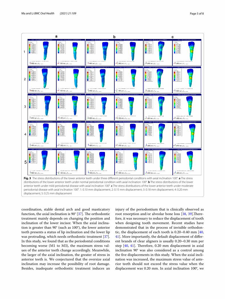
Fig. 3 The stress distributions of the lower anterior teeth under three different periodontal conditions with axial inclination 100°. a The stress distributions of the lower anterior teeth under normal periodontal condition with axial inclination 100°. b The stress distributions of the lower anterior teeth under mild periodontal disease with axial inclination 100°. c The stress distributions of the lower anterior teeth under moderate periodontal disease with axial inclination 100°. 1: 0.10 mm displacement; 2: 0.15 mm displacement; 3: 0.18 mm displacement; 4: 0.20 mm displacement; 5: 0.25 mm displacement
Page 6 of 8Ma and Li BMC Oral Health (2021) 21:109
discovered that the ranges of stress value in 0.18 mm are 132–164 MPa, which are similar to the stress value ranges of 0.20 mm in axial inclination 90° (137–162 MPa). Con-sequently, we inferred that for patients with excessively inclined anterior teeth (such as 100°), the optimal ortho-dontic displacement when using clear aligner is 0.18 mm.
We should also reasonably determine the displacement based on the strain of the alveolar bone or alveolar ridge crest. Peter et al. have analyzed the 3D-FEM of alveolar bone and revealed that in order to distribute the peri-odontal stress evenly, a combination of force reduction and increased moment-to-force (M/F) ratio is required [42]. A research conducted by Cobo et al. has been reported that the height of the alveolar bone can directly or indirectly affect the periodontal stress distribution, and the magnitude of the change is positively related to
the degree of influence [43]. In this study, the strain at the top of the alveolar crest with different displacement (0.10 mm, 0.15 mm, 0.18 mm, 0.20 mm and 0.25 mm, respectively) under two different axial inclinations of anterior teeth (90° and 100°) was explored. Likewise, 0.20 mm in 90M1 was used as the control group and the strain at the top of the alveolar crest was 0.163 mm. We found that the displacement must be not exceeding 0.15 mm when alveolar bone resorption is 1/3 or 1/3–1/2. In axial inclination 100°, we discovered that in order to ensure that alveolar ridge crest is not deformed, the displacement is less than 0.18 mm (strain for 0.165 mm), 0.15 mm (strain for 0.167 mm) and 0.10 mm (strain for 0.117 mm) respectively when alveolar bone is normal, resorption 1/3 or 1/3–1/2. All the results implied that the stress on the teeth and the strain at the top of the alveolar
Fig. 4 The stress values of root apex in three periodontal conditions. a The stress values of the lower anterior teeth under three different periodontal conditions with axial inclination 90°. b The stress values of the lower anterior teeth under three different periodontal conditions with axial inclination 100°
Fig. 5 The strain at the top of the alveolar crest in three periodontal conditions. a the strain at the top of the alveolar crest in three periodontal conditions with axial inclination 90°. b The strain at the top of the alveolar crest in three periodontal conditions with axial inclination 100°
Page 7 of 8Ma and Li BMC Oral Health (2021) 21:109
crest should be taken into account at the same time when presetting displacement in orthodontic treatment.
However, there are some limitations in this study. First, this study is focused on four of the anterior teeth, and posterior tooth movement remains unclear. Second, tooth movements are influenced by the patient’s age, per-iodontal support, root length, and bone density, and large sample size is necessary. Third, tooth movement is also affected by periodontal tissue remodeling, action time, strength attenuation, and oral and maxillofacial muscle occlusion. The results of preliminary finite element need to be used in clinical practice to verify its efficacy.
ConclusionsIn summary, we explored the optimal orthodontic dis-placement to minimize the adverse effects of the clear aligner on periodontal tissues under laboratory condi-tions. However, the structure of oral cavity is extremely complex and there may be other factors to affect the treatment effect of clear aligner. Even so, we also hope our findings will improve orthodontic treatment with clear aligners.
AbbreviationsCBCT: cone beam computed tomography; PTM: pathologic tooth migration.
AcknowledgementsNot applicable.
Authors’ contributionsYNM: conception, design and analysis of data, performed the data analyses and wrote the manuscript; SL: contributed to the conception of the study and revised the manuscript; All authors have read and approved the manuscript.
FundingNot applicable.
Availability of data and materialsThe datasets used and/or analysed during the current study are available from the corresponding author on reasonable request.
Ethics approval and consent to participateThis study was approved by the ethics committee of School of Stomatology, Capital Medical University. (No. CMUSH-IRB-KJ-PJ-2020-11) Written informed consent was obtained from all subjects.
Consent to publishNot applicable.
Competing interestsThe authors declare that they have no competing interests.
Received: 4 October 2020 Accepted: 28 February 2021
References 1. Zafar K, Nazeer MR, Ghafoor R. Interdisciplinary management of gingival
recession and pathologic teeth migration-revisiting dental aesthetics. J Pak Med Assoc. 2019;69:1385–9.
2. Brown LF, Ford PJ, Symons AL. Periodontal disease and the special needs patient. Periodontol. 2000;2017(74):182–93.
3. Gkantidis N, Christou P, Topouzelis N. The orthodontic-periodontic inter-relationship in integrated treatment challenges: a systematic review. J Oral Rehabil. 2010;37:377–90.
4. Santosh TS, Srikanth K, Haritha D, Reddy ML, Parmar R. Need for speed in orthodontics: a review of noninvasive methods to accelerate the ortho-dontic tooth movement. Int J Oral Care Res. 2020;8:48.
5. Gyawali R, Bhattarai B. Orthodontic management in aggressive periodon-titis. Int Sch Res Notices. 2017;2017:8098154.
6. Re S, Corrente G, Abundo R, Cardaropoli D. Orthodontic treatment in periodontally compromised patients: 12-year report. Int J Periodontics Restorative Dent. 2000;20:31–9.
7. Tamer I, Oztas E, Marsan G. Orthodontic treatment with clear aligners and the scientific reality behind their marketing: a literature review. Turk J Orthod. 2019;32:241–6.
8. Seo JH, Eghan-Acquah E, Kim MS, Lee JH, Jeong YH, Jung TG, et al. Com-parative analysis of stress in the periodontal ligament and center of rota-tion in the tooth after orthodontic treatment depending on clear aligner thickness-finite element analysis study. Materials (Basel). 2021;14:324.
9. Levrini L, Mangano A, Montanari P, Margherini S, Caprioglio A, Abbate GM. Periodontal health status in patients treated with the Invisalign((R)) system and fixed orthodontic appliances: A 3 months clinical and micro-biological evaluation. Eur J Dent. 2015;9:404–10.
10. Rossini G, Parrini S, Castroflorio T, Deregibus A, Debernardi CL. Periodontal health during clear aligners treatment: a systematic review. Eur J Orthod. 2015;37:539–43.
11. Ke Y, Zhu Y, Zhu M. A comparison of treatment effectiveness between clear aligner and fixed appliance therapies. BMC Oral Health. 2019;19:24.
12. Zheng M, Liu R, Ni Z, Yu Z. Efficiency, effectiveness and treatment stability of clear aligners: a systematic review and meta-analysis. Orthodontics Craniofacial Res. 2017;20:127–33.
13. Rossini G, Parrini S, Castroflorio T, Deregibus A, Debernardi CL. Efficacy of clear aligners in controlling orthodontic tooth movement: a systematic review. Angle Orthod. 2015;85:881–9.
14. Liu Y, Hu W. Force changes associated with different intrusion strategies for deep-bite correction by clear aligners. Angle Orthod. 2018;88:771–8.
15. Korioth TW, Versluis A. Modeling the mechanical behavior of the jaws and their related structures by finite element (FE) analysis. Crit Rev Oral Biol Med. 1997;8:90–104.
16. Middleton J, Jones M, Wilson A. The role of the periodontal ligament in bone modeling: the initial development of a time-dependent finite ele-ment model. Am J Orthod Dentofacial Orthop. 1996;109:155–62.
17. Tanne K, Hiraga J, Kakiuchi K, Yamagata Y, Sakuda M. Biomechanical effect of anteriorly directed extraoral forces on the craniofacial complex: a study using the finite element method. Am J Orthod Dentofacial Orthop. 1989;95:200–7.
18. McGuinness NJ, Wilson AN, Jones ML, Middleton J. A stress analysis of the periodontal ligament under various orthodontic loadings. Eur J Orthod. 1991;13:231–42.
19. Geramy A. Initial stress produced in the periodontal membrane by orthodontic loads in the presence of varying loss of alveolar bone: a three-dimensional finite element analysis. Eur J Orthod. 2002;24:21–33.
20. Hayashi K, Araki Y, Uechi J, Ohno H, Mizoguchi I. A novel method for the three-dimensional (3-D) analysis of orthodontic tooth movement-calculation of rotation about and translation along the finite helical axis. J Biomech. 2002;35:45–51.
21. Jones ML, Hickman J, Middleton J, Knox J, Volp C. A validated finite element method study of orthodontic tooth movement in the human subject. J Orthod. 2001;28:29–38.
22. Lauritano F, Runci M, Cervino G, Fiorillo L, Bramanti E, Cicciu M. Three-dimensional evaluation of different prosthesis retention systems using finite element analysis and the Von Mises stress test. Minerva Stomatol. 2016;65:353–67.
23. Cervino G, Romeo U, Lauritano F, Bramanti E, Fiorillo L, D’Amico C, et al. Fem and Von Mises analysis of OSSTEM ((R)) dental implant structural components: evaluation of different direction dynamic loads. Open Dent J. 2018;12:219–29.
24. Crimi S, Defila L, Nanni M, Cicciu M, Fiorillo L, Cervino G, et al. Three-dimensional evaluation on cortical bone during orthodontic surgical treatment. J Craniofac Surg. 2020;31:1637–46.
Page 8 of 8Ma and Li BMC Oral Health (2021) 21:109
• fast, convenient online submission
•
thorough peer review by experienced researchers in your field
• rapid publication on acceptance
• support for research data, including large and complex data types
•
gold Open Access which fosters wider collaboration and increased citations
maximum visibility for your research: over 100M website views per year •
At BMC, research is always in progress.
Learn more biomedcentral.com/submissions
Ready to submit your researchReady to submit your research ? Choose BMC and benefit from: ? Choose BMC and benefit from:
25. Cervino G, Fiorillo L, Arzukanyan AV, Spagnuolo G, Cicciu M. Dental restorative digital workflow: digital smile design from aesthetic to func-tion. Dent J (Basel). 2019;7:30.
26. Cattaneo PM, Dalstra M, Melsen B. The finite element method: a tool to study orthodontic tooth movement. J Dent Res. 2005;84:428–33.
27. McGuinness N, Wilson AN, Jones M, Middleton J, Robertson NR. Stresses induced by edgewise appliances in the periodontal ligament—a finite element study. Angle Orthod. 1992;62:15–22.
28. Highfield J. Diagnosis and classification of periodontal disease. Aust Dent J. 2009;54(Suppl 1):S11-26.
29. Caton JG, Armitage G, Berglundh T, Chapple ILC, Jepsen S, Kornman KS, et al. A new classification scheme for periodontal and peri-implant diseases and conditions—introduction and key changes from the 1999 classification. J Clin Periodontol. 2018;45(Suppl 20):S1–8.
30. Gomez JP, Pena FM, Martinez V, Giraldo DC, Cardona CI. Initial force sys-tems during bodily tooth movement with plastic aligners and composite attachments: a three-dimensional finite element analysis. Angle Orthod. 2015;85:454–60.
31. Liu YF, Wang R, Baur DA, Jiang XF. A finite element analysis of the stress distribution to the mandible from impact forces with various orientations of third molars. J Zhejiang Univ Sci B. 2018;19:38–48.
32. Wolfram H, Antonia Z, Henning D, Julia FF, Susanne FZ, Rudolf G, et al. Torquing an upper central incisor with aligners–acting forces and biome-chanical principles. Eur J Orthod. 2010;32:607–13.
33. Singh A, Gill G, Kaur H, Amhmed M, Jakhu H. Role of osteopontin in bone remodeling and orthodontic tooth movement: a review. Prog Orthod. 2018;19:18.
34. Cortona A, Rossini G, Parrini S, Deregibus A, Castroflorio T. Clear aligner orthodontic therapy of rotated mandibular round-shaped teeth: a finite element study. Angle Orthod. 2020;90:247–54.
35. Hennessy J, Al-Awadhi EA. Clear aligners generations and orthodontic tooth movement. J Orthod. 2016;43:68–76.
36. Li X, Ren C, Wang Z, Zhao P, Wang H, Bai Y. Changes in force associated with the amount of aligner activation and lingual bodily movement of the maxillary central incisor. Korean J Orthod. 2016;46:65–72.
37. Tweed CH. Clinical orthodontics. Clin. Orthodontics. 1966;1:940. 38. Hollender L, Ronnerman A, Thilander B. Root resorption, marginal bone
support and clinical crown length in orthodontically treated patients. Eur J Orthod. 1980;2:197–205.
39. Reed BE, Polson AM, Subtelny JD. Long-term periodontal status of teeth moved into extraction sites. Am J Orthod. 1985;88:203–8.
40. Kravitz ND, Kusnoto B, BeGole E, Obrez A, Agran B. How well does Invisalign work? A prospective clinical study evaluating the efficacy of tooth movement with Invisalign. Am J Orthod Dentofacial Orthop. 2009;135:27–35.
41. Boyd RL, Miller RJ, Vlaskalic V. The Invisalign system in adult orthodontics: mild crowding and space closure cases. J Clin Orthod. 2000;34:203–12.
42. Jeon PD, Turley PK, Ting K. Three-dimensional finite element analysis of stress in the periodontal ligament of the maxillary first molar with simu-lated bone loss. Am J Orthod Dentofacial Orthop. 2001;119:498–504.
43. Cobo J, Arguelles J, Puente M, Vijande M. Dentoalveolar stress from bodily tooth movement at different levels of bone loss. Am J Orthod Dentofacial Orthop. 1996;110:256–62.
Publisher’s NoteSpringer Nature remains neutral with regard to jurisdictional claims in pub-lished maps and institutional affiliations.