Embed Size (px)
Citation preview

Volume 10, Issue 21 n December 21, 2015 www.usuhs.edu
Learning to Care for Those in Harm’s Way
The Official USU Newsletter
2 the pulse December 21, 2015
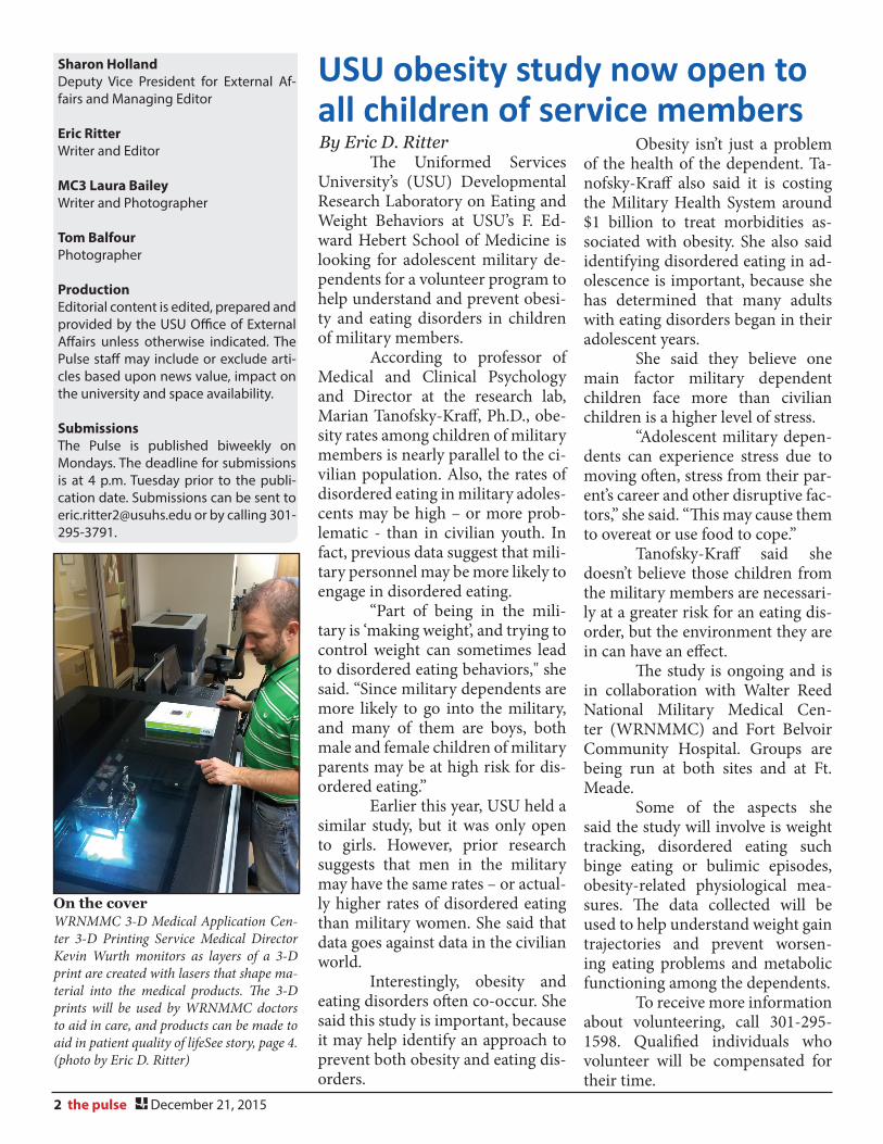
On the coverWRNMMC 3-D Medical Application Cen-ter 3-D Printing Service Medical Director Kevin Wurth monitors as layers of a 3-D print are created with lasers that shape ma-terial into the medical products. The 3-D prints will be used by WRNMMC doctors to aid in care, and products can be made to aid in patient quality of lifeSee story, page 4. (photo by Eric D. Ritter)
Sharon HollandDeputy Vice President for External Af-fairs and Managing Editor
Eric RitterWriter and Editor
MC3 Laura BaileyWriter and Photographer
Tom BalfourPhotographer ProductionEditorial content is edited, prepared and provided by the USU Office of External Affairs unless otherwise indicated. The Pulse staff may include or exclude arti-cles based upon news value, impact on the university and space availability.
SubmissionsThe Pulse is published biweekly on Mondays. The deadline for submissions is at 4 p.m. Tuesday prior to the publi-cation date. Submissions can be sent to [email protected] or by calling 301-295-3791.
By Eric D. Ritter
USU obesity study now open to all children of service members The Uniformed Services University’s (USU) Developmental Research Laboratory on Eating and Weight Behaviors at USU’s F. Ed-ward Hebert School of Medicine is looking for adolescent military de-pendents for a volunteer program to help understand and prevent obesi-ty and eating disorders in children of military members. According to professor of Medical and Clinical Psychology and Director at the research lab, Marian Tanofsky-Kraff, Ph.D., obe-sity rates among children of military members is nearly parallel to the ci-vilian population. Also, the rates of disordered eating in military adoles-cents may be high – or more prob-lematic - than in civilian youth. In fact, previous data suggest that mili-tary personnel may be more likely to engage in disordered eating. “Part of being in the mili-tary is ‘making weight’, and trying to control weight can sometimes lead to disordered eating behaviors," she said. “Since military dependents are more likely to go into the military, and many of them are boys, both male and female children of military parents may be at high risk for dis-ordered eating.” Earlier this year, USU held a similar study, but it was only open to girls. However, prior research suggests that men in the military may have the same rates – or actual-ly higher rates of disordered eating than military women. She said that data goes against data in the civilian world. Interestingly, obesity and eating disorders often co-occur. She said this study is important, because it may help identify an approach to prevent both obesity and eating dis-orders.
Obesity isn’t just a problem of the health of the dependent. Ta-nofsky-Kraff also said it is costing the Military Health System around $1 billion to treat morbidities as-sociated with obesity. She also said identifying disordered eating in ad-olescence is important, because she has determined that many adults with eating disorders began in their adolescent years. She said they believe one main factor military dependent children face more than civilian children is a higher level of stress. “Adolescent military depen-dents can experience stress due to moving often, stress from their par-ent’s career and other disruptive fac-tors,” she said. “This may cause them to overeat or use food to cope.” Tanofsky-Kraff said she doesn’t believe those children from the military members are necessari-ly at a greater risk for an eating dis-order, but the environment they are in can have an effect. The study is ongoing and is in collaboration with Walter Reed National Military Medical Cen-ter (WRNMMC) and Fort Belvoir Community Hospital. Groups are being run at both sites and at Ft. Meade. Some of the aspects she said the study will involve is weight tracking, disordered eating such binge eating or bulimic episodes, obesity-related physiological mea-sures. The data collected will be used to help understand weight gain trajectories and prevent worsen-ing eating problems and metabolic functioning among the dependents. To receive more information about volunteering, call 301-295-1598. Qualified individuals who volunteer will be compensated for their time.

3 the pulse December 21 2015
Avatars to offer new realism in training Members of the class of 2018--military medical students at the F. Edward Hébert School of Medicine (SOM) at the Uniformed Services University of the Health Sciences (USU)--will be the first to use a new simulated technology in January 2016. Since 2001, USU’s SOM stu-dents going through their Psychia-try clerkships in the National Cap-ital Area have come weekly to the Val G. Hemming Simulation Center (SIMCEN) to train with paid actors who portray patients (standardized patients) visiting a simulated clinic. Standardized patients are the “gold standard” of simulated training, be-cause they allow students to practice their clinical skills in a realistic and controlled environment with real people. However, paid actors can be costly, said Air Force Col. (Dr.) Rita L. DuBoyce, the principal investiga-tor of the Virtual Patient (VP) proj-ect and senior director of clinical education at USU’s SIMCEN. Pa-tient simulation is about to get even better for USU SOM students with the launch of VP, a computer pro-gram developed by the University of Southern California (USC) Institute for Creative Technologies (ICT). Using virtual human tech-nology to create life-like avatars, the VP will train USU clinicians in interpersonal areas such as rapport, interviewing and diagnosis, much like standardized patients do, but without scripts to memorize, coach-ing for the actors and expensive hourly wages. In addition to cutting costs, other benefits include ease of access and variety. The future VP system will be delivered over the web and mo-
bile devices such as tablets for easy and continuous training, according to the USC ICT website. Virtual pa-tients will be available 24/7 and can portray a multitude of conditions that might be difficult for actors to represent or repeat with success. The fact students will be able to access the program easily is a big benefit, because it means more prac-tice for the students, said Duboyce. “We want them to make all their mistakes here while they’re practicing so they can safely learn from them and avoid making those same mistakes with real world pa-tients,” she said. “I think it could work really well for remedial in-struction as well – for those students who may be struggling and need the extra practice. Also, having an array of characters available from elderly and young patients in different gen-ders and cultures is really great.” This technology is the wave of the future, said Duboyce. But even though VP is geared toward today’s generation of young, tech-savvy
medical students, VP by no means replaces standardized patients. The two methods will work together and provide a more well-rounded learning experience for SOM stu-dents and ultimately give real-world patients a more prepared doctor in their time of need. “I think our students are really going to like using the pro-gram,” she added. “A lot of time has gone into the preparations for this launch – more than a year from start to finish, which included writing the grant proposal, waiting for the approval and funding, then author-ing the program to depict certain scenarios for the students. I’m really excited to see how the students do with the addition of Virtual Patient. We plan to launch the program to our first batch of SOM students sometime in January 2016. The class of 2018 will be USU’s pioneers for the program, but I think in a fun way. I’m confident the program and the students will do well.”
By MC3 Laura Bailey
Air Force Col. (Dr.) Rita L. DuBoyce, the principal investigator of the Vir-tual Patient (VP) project and senior director of clinical education at USU’s SIMCEN monitors a simulation program used to aid in more realistic training for students. (Photo by MC3 Laura Bailey)
4 the pulse December 21, 2015
By Eric D. Ritter
USU, WRNMMC 3-D printing offers better quality of life for some patients
See 3-D, Page 5
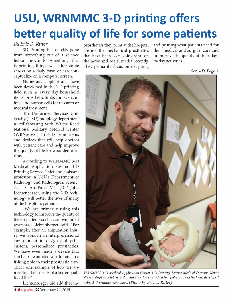
3D Printing has quickly gone from something out of a science fiction movie to something that is printing things we either come across on a daily basis or can con-ceptualize on a computer screen. Numerous applications have been developed in the 3-D printing field such as every day household items, prosthetic limbs and even an-imal and human cells for research or medical treatment. The Uniformed Services Uni-versity (USU) radiology department is collaborating with Walter Reed National Military Medical Center (WRNMMC) to 3-D print items and devices that will help doctors with patient care and help improve the quality of life for wounded war-riors. According to WRNMMC 3-D Medical Application Center 3-D Printing Service Chief and assistant professor in USU's Department of Radiology and Radiological Scienc-es, U.S. Air Force Maj. (Dr.) John Lichtenberger, using the 3-D tech-nology will better the lives of many of the hospital’s patients. “We are primarily using this technology to improve the quality of life for patients such as our wounded warriors,” Lichtenberger said. “For example, after an amputation inju-ry, we work in an interprofessional environment to design and print custom, personalized prosthetics. We have even made a device that can help a wounded warrior attach a fishing pole to their prosthetic arm. That’s one example of how we are meeting their needs of a better qual-ity of life.” Lichtenberger did add that the
prosthetics they print at the hospital are not the mechanical prosthetics that have been seen going viral on the news and social media recently. They primarily focus on designing
and printing what patients need for their medical and surgical care and to improve the quality of their day-to-day activities.
WRNMMC 3-D Medical Application Center 3-D Printing Service Medical Director, Kevin Wurth, displays a fabricated metal plate to be attached to a patient's skull that was developed using 3-D printing technology. (Photo by Eric D. Ritter)

5 the pulse December 21 2015
“Sometimes a whole prosthet-ic limb is not what they want,” he said. “For example, someone who lost their legs may not want to put on full-sized prosthetic legs to go just a few feet or when they need more mobility such as working on a car. We can print out titanium plate prosthetics that can be comfortably and easily attached to the residual limbs to protect them. In addition to that, we also create a lot of cranial and oral implants for patients who require surgical implants. Those prints are used in the medical care of patients and help the patients be-come more functional.” Lichtenberger said many of the computer models used for the 3-D prints are designed by graphic art-ists who add a great deal of expertise and aesthetic to the products. How-
ever, the graphic artists there aren’t just trained graphic artists. They are also trained CT techs. “They bring a lot of image-pro-cessing knowledge to what we are doing,” he said. “They have an in-timate knowledge of the layers and angles of the images and then trans-fer that into the computer model that will be used in the print.” The other major significance of being able to print a model based on MRI and CT scans is that it can print specific areas of the body without the need for a surgeon to perform an operation in order to physically see the area they need to see. It can print out specific vessels, muscles and so forth. “It really does minimize op-erating time,” he said. “It helps the medical team understand the pa-tient before the surgery begins. In
fact, they may even be able to see anatomic relationships they may not have anticipated like a vessel near a tumor that they should be aware of and will know to address it during surgery. Whenever we can assist with surgical planning and mini-mize surgical time like this, it will benefit the patient.” 3-D printing has actually been around since the mid-1980s. How-ever, its purpose was more to create machine parts in the industrial sec-tor. The medical applications in 3-D printing have only recently taken off. Lichtenberger said the hospital doesn’t yet have the ability to print with cells to make human tissue, also known as bioprinting. Howev-er, it is something they plan to add in the near future. The three printer types Licht-enberger said they use at the hos-pital are the Electron Beam Melter (EBM), which melts metal powder layer by layer with an electron beam to fabricate a print, Fused Depo-sition Modeling, which lays down material like acrylonitrile butadiene styrene (ABS) plastic in layers until the print is finished, and Stereoli-thography which can lay down clear material like plastics and produce larger prints such as a human pelvis. There’s also a major time ben-efit to these 3-D prints. Previously, for many prosthetic pieces, for ex-ample, molds needed to be made, and then the molds would be sent off to be developed at an off-site manufacturer. The 3-D-printing ability has eliminated that step and can print out a product for a patient that day instead of waiting. “This is ultimately all about personalized patient care,” he said. “This is giving the patient the prod-ucts they need to live a better life, and it provides doctors with a qual-ity and accurate product that assists them in better medical care.”
3-D from Page 4
WRNMMC 3-D Medical Application Center 3-D Printing Service Chief US Air Force Maj. (Dr.) John Lichtenberger displays how the 3-D printer can print out specific body parts like blood vessels taken from CT or MRI scans of a patient to help doctors understand affected areas before surgeries. (Photo by Eric D. Ritter)
6 the pulse December 21, 2015
WAVE, from Page 6
See WAVE, Page 7
by MC3 Laura Bailey
WAVE simulator provides realistic field medical scenarios at Val G. Hemming Simulation Center CH47 Chinook helicopters land under fire in the Khyber Pass, Afghanistan to a mass casualty air disaster. Two wounded airmen lay on the ground nearby. One is un-conscious and bleeding heavily from the knee where his leg has been blown off and the other, with visible wounds to the abdomen and chest, is in a state of panic. Over the sounds of bomb blasts, rapid gunfire and sirens, her desperate screams for help call com-bat medics into action. Staying close to the ground in a military crawl, they make their way through blasts,
shooting debris, clouds of smoke and heavy enemy gun fire, to reach the wounded. The medics are on a danger-ous rescue mission: save lives even while putting their own lives on the line. One medic says to the wound-ed, “We’ve got you. You’re going to make it.” He and his team quickly apply bandages and tourniquets. Stop the bleeding. Check. Monitor vital signs. Check. Working diligently through the distracting sounds of a most hostile environ-ment – the medics remain unde-terred.
Like a dream that ends upon waking, the completed training sce-nario comes to a halt. Bright lights come on inside the Wide Area Vir-tual Environment (WAVE) at the Val G. Hemming Simulation Cen-ter (SIMCEN) at the Uniformed Services University of the Health Sciences (USU). The wounded role-player jumps to her feet com-pletely unharmed. Sound effects dissipate into silence and a smoke machine exhales its final plume as SIMCEN staff enter the scene to reset the mannequin for the next
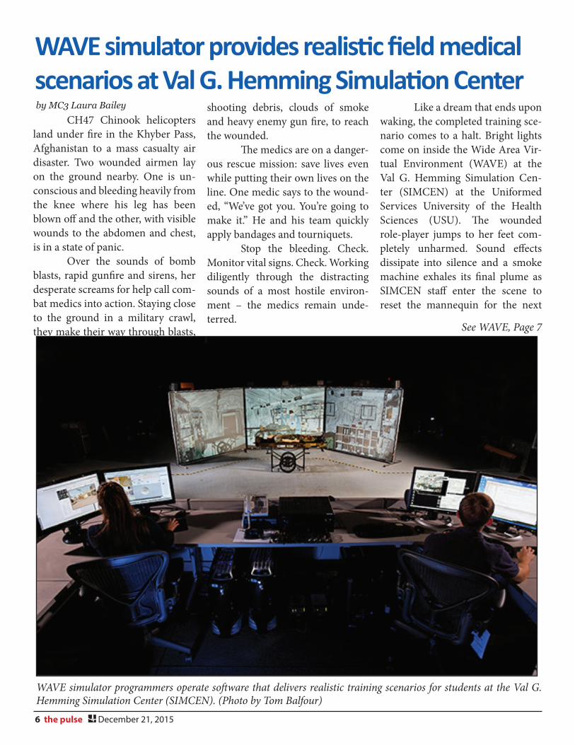
WAVE simulator programmers operate software that delivers realistic training scenarios for students at the Val G. Hemming Simulation Center (SIMCEN). (Photo by Tom Balfour)

7 the pulse December 21 2015
WAVE, from Page 6
by Sharon Holland
training group. “Last year, this center sup-ported about 36,000 active duty training hours,” said Gil Muniz, the deputy director of the center. “We support the military’s medical stu-dents at USU, as well as medics from the military and civilian first re-sponders. Keeping all of this as close to reality as possible is key. And with our trainers right there, we can give immediate feedback and make sure when these medics hit the real reali-ty, they’ll be ready.” Unlike simulators that use computer monitors or head-mount-ed displays, the 8,000-square-feet virtual space created by the WAVE allows up to 18 team members to
interact with each other and real equipment, and gives instructors the opportunity to teach and assess teamwork skills. Stereoscopic im-ages are displayed on the screens with paired DLP projectors while users wear lightweight stereoscopic glasses to view the scene. The WAVE boasts two pods 20 feet in diameter, eighteen 9-feet tall and 12-feet wide movie screens surrounding the two pods, 144 image projectors, a 5.1-channel sound system that gen-erates a “soundscape” to enhance the realism of the experience with flying bullets, screams and explo-sions, and smoke generators and de-bris scattered across the floor to add to the mayhem. One pod simulates the aftermath of the first scenario such as a firefight. Students evacu-
ate patients along the corridor to the second pod where a second scenar-io such as helicopter transport plays. Meanwhile the first pod is being re-staged as a field hospital. Thus train-ing can be sustained in real time for extended periods. “The WAVE is the world’s largest virtual environment for medical training,” said Dr. Alan Liu, director of the Virtual Medical En-vironments Laboratory and creator of WAVE. “It’s a room with 24 3-D screens that puts users inside a vir-tual reality game, surrounding and immersing medics into any kind of environment – from downtown Iraq in the middle of a firefight to inside a cargo aircraft. “There’s nothing else like this in the world.”
Fourteen members of the F. Edward Hébert School of Medicine class of 2018 competed against 108 other candidates for the Expert Field Medical Badge, Dec. 15-18, 2015, at Fort Irwin, California. When it was all over, half of them walked away with coveted badge. The soldiers spent 72 hours on the vast training base located in the Mojave desert vying for the badge, which is considered one of the Army’s most difficult and prestigious to earn. According to the Army Medical Department (AMEDD) website, “the EFMB test is the utmost challenge to the pro-fessional competence and physical
endurance of the soldier medic. It is the most sought after peacetime award in the AMEDD, and while the Combat Medical Badge is the ‘portrait of courage’ in wartime, the Expert Field Medical Badge is un-doubtedly the ‘portrait of excellence’ in the army all of the time.” Over the last 50 years, members of the U.S. Army, Air Force and Navy medical departments, along with their coun-terparts from allied nations, have had the opportunity to compete in this grueling competition, in the hopes of earning the EFMB. Candidates completed a se-ries of training lanes where they were tested, while under “enemy” fire, on combat care and various military survival skills. Candidates
also took a comprehensive written exam on the skills they learned, completed day and night land nav-igation, and if they made it to the end, finished a 12-mile road march in full gear, within three hours. The pass rate for the badge typically falls between 15 and 20 percent. “The members of my class achieved an outstanding 50 per-cent pass rate,” said Army 2nd Lt. Andrew Jacobson, president of the SoM Class of 2018. This year’s USU EFMB re-cipients include Army 2nd Lts. John Blickle, Rachel Bridwell, Charles Llewellyn, Thomas Peterson, Oriana Ellis, Kimberly Gerling, and Kaoru Song.
USU students compete for expert field medical badge
8 the pulse December 21, 2015
Final Frame
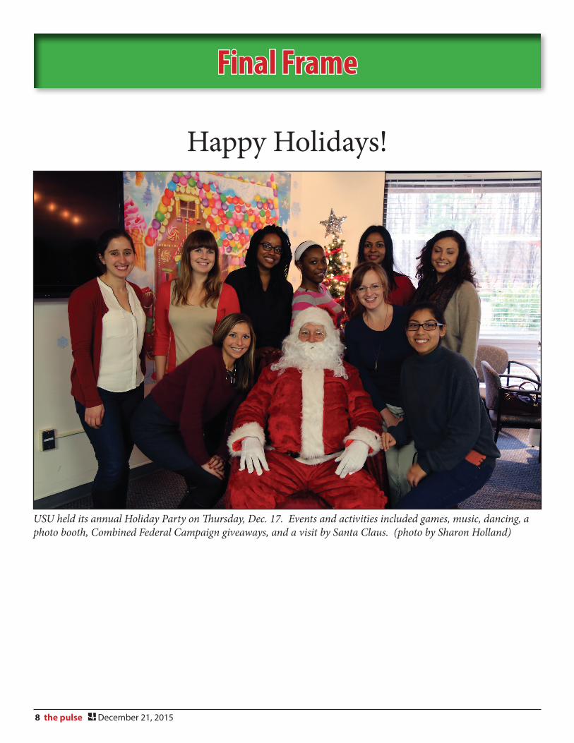
USU held its annual Holiday Party on Thursday, Dec. 17. Events and activities included games, music, dancing, a photo booth, Combined Federal Campaign giveaways, and a visit by Santa Claus. (photo by Sharon Holland)
Happy Holidays!