Embed Size (px)
Citation preview

THE OCULAR MANIFESTATIONS OFSICKLE-CELL DISEASE: A PREVALENCEAND NATURAL HISTORY STUDY*
BY John G. Clarkson, MD
INTRODUCTION
THE SICKLE HEMOGLOBINOPATHIES RESULT FROM AN ABNORMALITY IN
the beta chain of the hemoglobin molecule. The chief manifestations arechronic hemolytic anemia and vaso-occlusive crises that produce severepain as well as long-term and widespread organ damage. There areseveral clinically important hemoglobin variants that constitute the sicklesyndromes.About 8% of black Americans are heterozygous for hemoglobin S and
have sickle cell trait. These individuals usually have about 35% to 40%hemoglobin S and 55% to 60% hemoglobin A; they do not have increasedmorbidity or mortality.1
Approximately 0. 15% ofblack children in the United States have homo-zygous hemoglobin S (SS disease).1 They suffer from a severe hemolyticanemia with hematocrit values between 18% and 30%. Symptoms do notusually develop until after the age of 6 months, when fetal hemoglobin(hemoglobin F) has been replaced by hemoglobin S. Delayed growth anddevelopment and increased susceptibility to infection are the primaryconstitutional manifestations. They prevalence among adults is muchlower because patients with sickle cell anemia have a decreased lifeexpectancy. The increased morbidity and mortality in homozygous sicklecell disease is due primarily to recurrent vaso-occlusive episodes. Themost common clinical event is the so-called painful crisis.2 These eventsusually appear suddenly and affect various parts of the body, particularlythe abdomen, chest, and joints. Painful crises are frequently preceded bya viral or bacterial infection but may occur in association with a change in
*From the Department of Ophthalmology, Bascom Palmer Eye Institute, University ofMiami School of Medicine, Miami. This study was supported in part by ComprehensiveSickle Cell Center grant H-580654-1979-88 from the National Heart, Lung and BloodInstitute of the National Institutes of Health, Bethesda.
TR. AM. OPHTH. Soc. vol. LXXXX, 1992

Clarkson
temperature, most often due to cold. Reduced oxygen concentrationresults in a change in shape of the red blood cells containing hemoglobinS from a biconcave disc into an elongated crescent shape or "sickle"-shaped cell. As the red blood cell sickles, it becomes rigid and mayobstruct capillary flow. Such capillary obstruction can lead to furthertissue hypoxia and increased tendency for the red cells to sickle. Almostany organ can be affected by the vaso-occlusive phenomenon, but it mostfrequently affects the lungs, kidneys, liver, skeleton, and skin.1There are several double heterozygous states included in the sickle
syndromes. The two most common are sickle cell-hemoglobin C (SC)disease and sickle beta-thalassemia (sickle 13-thalassemia). In SC disease,about 50% of the hemoglobin is hemoglobin S and the remaining 50% ishemoglobin C. The gene frequency for hemoglobin C is only about onefourth of that for hemoglobin S. However, the prevalence of SC diseaseamong adults is almost as high as that of SS disease because of theincreased mortality associated with SS disease and the relatively normallife expectancy for persons with SC disease. 1 The presence ofhemoglobinC reduces the risk of sickling as compared with SS disease. However,persons with SC disease usually have a mild to moderate hemolyticanemia and may occasionally have painful crises or organ infarcts. Sickle13-thalassemia is commonly encountered in people from Mediterraneancountries and from Central Africa. These individuals show from 60% to90% hemoglobin S and from 10% to 30% hemoglobin F In some forms ofsickle ,B-thalassemia, hemoglobin A represents 10% to 30%.1The diagnosis of one of the sickle hemoglobinopathies depends upon
the demonstration of sickling of red cells under reduced oxygen condi-tions. In the sickle prep test, sickled cells may be seen microscopicallyafter the addition of an oxygen consuming reagent such as metabisulfite.However, this test does not distinguish between sickle trait, SS disease,SC disease, or sickle thalassemia. Hemoglobin electrophoresis is neces-sary to establish a definitive diagnosis.1"3The sickle syndromes have the highest incidence in black Africans and
African-Americans but are also found in people from Mediterraneancountries (Greece, Italy, Israel) as well as Saudi Arabia and India. Africanshave known of the disease for generations, and it has been traced back asfar as 1670 in Ghana.2 It was first reported in the United States in 1910 byHerrick.4 In 1949, Neel5 demonstrated that sickle cell anemia was trans-mitted as a recessive gene, and the same year Pauling and associates3noted that sickle hemoglobin and normal hemoglobin demonstrated adifferent electrophoretic mobility.
Abnormalities in the ocular fundus in sickle cell anemia were first
482
Sickle Cell Retinopathy
described in 1930 by Cook,6 who noted fresh hemorrhages in the retina ina patient who died of subarachnoid hemorrhage. In 1952, Edington andSarkies7 reported two patients with sickle cell anemia who had retinalaneurysms and vitreous hemorrhage. In 1966, Welch and Goldberg8reported the ocular findings in 55 patients with SS disease, 34 patientswith sickle trait, and 22 cases of SC disease, and compared these findingswith those in 38 patients with normal hemoglobin. These investigatorswere the first to note background changes such as the "black sunburstspot," "refractive and lipid deposits," and "peripheral arterial oblitera-tion." They described the proliferative retinopathy as the "sea fan sign,"because it resembled Gorgoniafalbeloum, known as a sea fan, which theyfound primarily in patients with hemoglobin SC disease. Fluoresceinangiography confirmed peripheral vaso-occlusive disease. They found nosignificant fundus abnormality in the patients with sickle trait, and al-though they observed evidence ofproliferative disease in patients with SSdisease, they noted more severe proliferative disease in patients withhemoglobin SC disease.
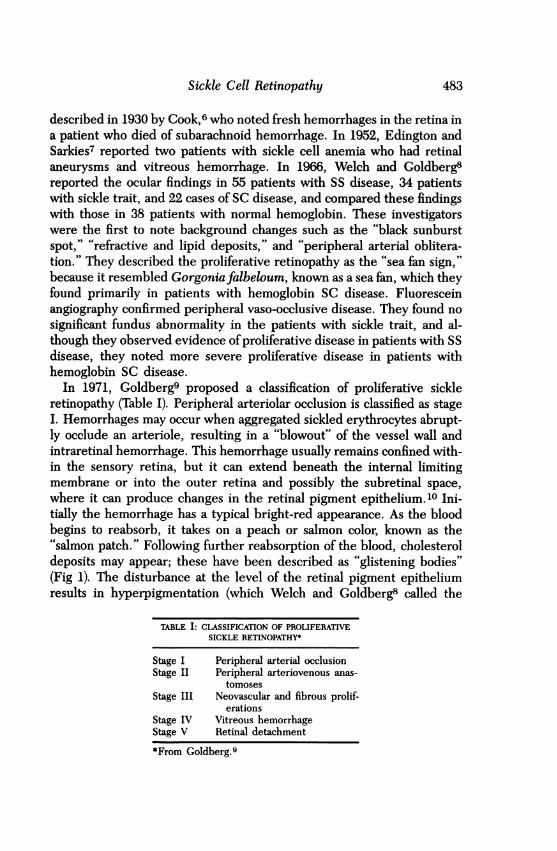
In 1971, Goldberg9 proposed a classification of proliferative sickleretinopathy (Table I). Peripheral arteriolar occlusion is classified as stageI. Hemorrhages may occur when aggregated sickled erythrocytes abrupt-ly occlude an arteriole, resulting in a "blowout" of the vessel wall andintraretinal hemorrhage. This hemorrhage usually remains confined with-in the sensory retina, but it can extend beneath the internal limitingmembrane or into the outer retina and possibly the subretinal space,where it can produce changes in the retinal pigment epithelium.10 Ini-tially the hemorrhage has a typical bright-red appearance. As the bloodbegins to reabsorb, it takes on a peach or salmon color, known as the"salmon patch." Following further reabsorption of the blood, cholesteroldeposits may appear; these have been described as "glistening bodies"(Fig 1). The disturbance at the level of the retinal pigment epitheliumresults in hyperpigmentation (which Welch and Goldberg8 called the
TABLE I: CLASSIFICATION OF PROLIFERATIVESICKLE RETINOPATHY*
Stage I Peripheral arterial occlusionStage II Peripheral arteriovenous anas-
tomosesStage III Neovascular and fibrous prolif-
erationsStage IV Vitreous hemorrhageStage V Retinal detachment
*From Goldberg.9
483
Clarkson
FIGURE 1Stage I proliferative sickle retinopathy. A: Partially resorbed intraretinal hemorrhage tem-poral to macula in right eye of 17-year-old black male with sickle cell-hemoglobin S disease.B: Iridescent spot in superior midperiphery in left eye of 27-year-old black male with sickle
cell-hemoglobin S disease.
484
.. .....
.....
Sickle Cell Retinopathy
"black sunburst spot"). Although these changes typically occur in themidperiphery, they may occur more posteriorly in the fundus as well.
Stage II is represented by peripheral arteriovenous anastomoses, whichoccur subsequent to the peripheral occlusion. Fluorescein angiographyconfirms the presence of retinal capillary nonperfusion peripheral to thearteriovenous anastomoses. It is at this site that stage III, proliferativeneovascularization, develops. Goldberg9 defined stage IV as vitreoushemorrhage usually due to proliferative retinopathy, and stage V as anyretinal detachment.
Other ocular changes may occur in the sickle hemoglobinopathies. 11-17Patonl8 described a unique segmentation ofconjunctival vessels occurringprimarily in patients with homozygous sickle-cell disease, which he calledthe "conjunctival sign." This segmentation of capillary flow is often mini-mal, and it is best observed on the inferior bulbar conjunctiva. Similarsegmentation of the blood column has been observed on the optic nervehead of patients with one of the sickle hemoglobinopathies and is calledthe "disc sign."19 Iris infarcts may also occur. Additional fundus findingshave been described, including epiretinal membrane formation, angioidstreaks, macular hole, ischemic optic neuropathy, central and branchretinal artery occlusion, and central retinal vein occlusion.11-17,19The prevalence of the ocular findings in patients with SS disease, SC
disease and sickle thalassemia disease has been reported by severalinvestigators. However, only Condon and Seijeantl6,17,20 reviewed a groupof patients unselected on the basis of ocular complications. The naturalhistory of the eye findings, particularly peripheral retinal vascular dis-ease, and the development of proliferative sickle retinopathy has beenreported by Goldberg,9 Condon and Serjeant,20 and Raichand and associ-ates.2' In these studies it was anticipated that proliferative lesions wouldnot regress. Condon and Serjeant20 were the first to notice that sponta-neous regression of proliferative lesions could occur. They20 and others22subsequently confirmed that regression due to autoinfarction does occurin a large number of proliferative lesions. Welch and Goldberg8 firstproposed that photocoagulation in patients with proliferative retinopathy,particularly in hemoglobin SC disease, might be indicated to reduce therisk ofvitreous hemorrhage and/or retinal detachment. The demonstratedbenefit of laser photocoagulation for proliferative retinopathy due todiabetes, branch vein occlusion, and cryotherapy for stage III retinopathyof prematurity would appear to indicate that some form of treatmentshould be considered for eyes with proliferative sickle retinopathy.23-25However, Goldbaum and associates26 observed retinal detachment in twoof nine patients receiving cryotherapy for proliferative sickle retinopathy
485

Clarkson
using a multiple freeze-thaw technique. Also, in over 90% of eyes, cho-roidal neovascularization developed when photocoagulation treatmentwas applied to the feeder vessel in a clinical trial reported by Condon andSerjeant.27 The significant complications after cryotherapy and long-termcomplications of photocoagulation cast doubt on the effectiveness of thesetreatments and stress the importance of understanding the natural historyof proliferative sickle retinopathy. Previous natural history studies werelimited by patient selection and length of follow-up. More recent reportshave specifically evaluated the long-term visual effects of proliferativeretinopathy in untreated and treated sickle cell patients.28-30
To gain a better understanding of the natural history of proliferativesickle retinopathy, a group of sickle cell patients were contacted to reportfor a complete eye examination. Patients were selected only on the basisof having one of the sickle hemoglobinopathies. A subset of this originalgroup, including all previously untreated patients who demonstratedevidence of proliferative retinopathy (stage III or greater) at the initialevaluation, was followed prospectively for an average of 6.7 years todetermine the long-term natural history.
METHODS AND MATERIALS
In 1979, 200 patients followed at the Sickle Cell Center, Miami, FL, wererandomly selected to have eye examinations. Eighteen patients under 5years ofage were excluded from this study. Of the remaining 182 patients,150 reported for the examinations. Table II summarizes the demographicsand prevalence of the subgroups of sickle-cell disease in these patients.Twenty-four of the 27 patients with stage III proliferative sickle retinopa-thy (neovascular proliferative disease based on the Goldberg classifica-tion9) from the prevalence study returned for periodic follow-up examina-tions between 1979 and 1989. Fundus findings were documented withlarge retinal drawings and fundus photographs. Twenty-three of the 24patients (38 of 39 eyes) with stage III proliferative retinopathy were
TABLE II: DEMOGRAPHICS OF 150 SICKLE CELL PATIENTS IN PREVALENCE STUDY
HEMOGLOBIN TYPE
SS SC S THAL TOTAL
No. of patients 109 29 12 150Median age (yr) 20 24 20 20Age range (yr) 5-66 7-58 6-52 5-66Sex (male/female) 51/58 18/11 4/8 73/77
SS, sickle S disease; SC, sickle C disease; S thal, sickle thalassemia disease.
486
Sickle Cell Retinopathy
followed for a minimum of 2 years (mean, 75 months; range, 31 to 118months). One patient with sickle thalassemia who had mild proliferativeretinopathy in one eye was lost to follow-up. Three patients were ex-cluded from the natural history study because they had received someform of prophylactic treatment, either laser photocoagulation or cryother-apy, in both eyes before the initial examination. One eye of each of twopatients from this group required surgical intervention during the courseof the study. A pars plana vitrectomy was performed on a patient with adense, nonclearing vitreous hemorrhage; in another eye a retinal detach-ment associated with a macular hole was repaired through vitreous sur-gery. Data collection for natural history on these patients was terminatedwhen surgical intervention occurred.
In addition to the patients with proliferative disease, a group of 76patients from the prevalence study, unselected on the basis of ocularfindings, with either normal findings on examination or early peripheralvascular occlusive disease returned for periodic follow-up examinations.Sixty-two of the 76 patients (82%) had a minimum of 2 years' follow-up(average, 83 months; range, 28 to 123 months). One patient died beforeadequate follow-up was obtained. Thirteen were lost to follow-up. There-fore, a total of 85 patients were examined in the natural history portion ofthe study and were followed for a mean of 80 months (range, 28 to 123months) (Table III).The type ofhemoglobin was determined by the Sickle Cell Center with
use of cellulose acetate electrophoresis. Results were verified by theCenters for Disease Control, Atlanta, with use of both cellulose acetateand citrate agar electrophoresis and were confirmed by family studies andclinical data. The ocular examination at each visit included best correctedvisual acuity, slit-lamp examination of the anterior segment, and fundusexamination with a binocular indirect ophthalmoscope. The Hruby andfundus contact lenses were used to further evaluate macular and opticnerve changes, and the Goldmann 3-mirror contact lens and, in laterexaminations, the Volk 90-diopter lens were used to supplement examina-tion of the peripheral retina. Color photographs and fluorescein angio-grams were obtained in cases with suspected macular disease and/or pro-liferative sickle retinopathy. Stage III disease was defined by ophthalmo-scopically visible neovascular or fibrous lesions. The hemoglobin type wasnot made known to the examiner until the conclusion of the study. Allpatients in the follow-up study were examined annually, and most wereexamined every 6 months.
487

Clarkson
c"')
coc.
0o
co)0c C
zH
0~~ ~ ~ ~ ~ 0z~~~~~~~~~~~"
Z
--4 co
00kf)it,'-4,-4
(2 :§
0~~~~~
0 0
-A
488
I
Sickle Cell Retinopathy
RESULTS
PREVALENCE STUDY
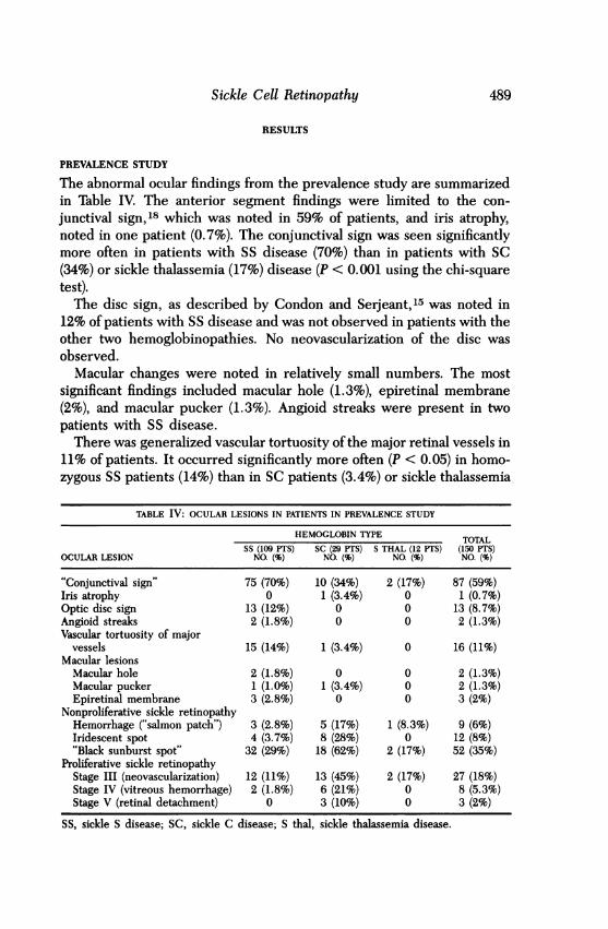
The abnormal ocular findings from the prevalence study are summarizedin Table IV. The anterior segment findings were limited to the con-
junctival sign,18 which was noted in 59% of patients, and iris atrophy,noted in one patient (0.7%). The conjunctival sign was seen significantlymore often in patients with SS disease (70%) than in patients with SC(34%) or sickle thalassemia (17%) disease (P < 0.001 using the chi-squaretest).The disc sign, as described by Condon and Serjeant,15 was noted in
12% of patients with SS disease and was not observed in patients with theother two hemoglobinopathies. No neovascularization of the disc was
observed.Macular changes were noted in relatively small numbers. The most
significant findings included macular hole (1.3%), epiretinal membrane(2%), and macular pucker (1.3%). Angioid streaks were present in twopatients with SS disease.There was generalized vascular tortuosity of the major retinal vessels in
11% of patients. It occurred significantly more often (P < 0.05) in homo-zygous SS patients (14%) than in SC patients (3.4%) or sickle thalassemia
TABLE IV: OCULAR LESIONS IN PATIENTS IN PREVALENCE STUDY
HEMOGLOBIN TYPE TOTALSS (109 PTS) SC (29 PTS) S THAL (12 PTS) (150 PTS)
OCULAR LESION NO. (%) NO. (%) NO. (%) NO. (%)
"Conjunctival sign" 75 (70%) 10 (34%) 2 (17%) 87 (59%)Iris atrophy 0 1 (3.4%) 0 1 (0.7%)Optic disc sign 13 (12%) 0 0 13 (8.7%)Angioid streaks 2 (1.8%) 0 0 2 (1.3%)Vascular tortuosity of major
vessels 15 (14%) 1 (3.4%) 0 16 (11%)Macular lesions
Macular hole 2 (1.8%) 0 0 2 (1.3%)Macular pucker 1 (1.0%) 1 (3.4%) 0 2 (1.3%)Epiretinal membrane 3 (2.8%) 0 0 3 (2%)
Nonproliferative sickle retinopathyHemorrhage ("salmon patch") 3 (2.8%) 5 (17%) 1 (8.3%) 9 (6%)Iridescent spot 4 (3.7%) 8 (28%) 0 12 (8%)"Black sunburst spot" 32 (29%) 18 (62%) 2 (17%) 52 (35%)
Proliferative sickle retinopathyStage III (neovascularization) 12 (11%) 13 (45%) 2 (17%) 27 (18%)Stage IV (vitreous hemorrhage) 2 (1.8%) 6 (21%) 0 8 (5.3%)Stage V (retinal detachment) 0 3 (10%) 0 3 (2%)
SS, sickle S disease; SC, sickle C disease; S thal, sickle thalassemia disease.
489

Clarkson
patients (0%). Localized vascular tortuosity was noted in an additional 2%of patients.
Nonproliferative sickle retinopathy changes (retinal hemorrhage, iri-descent spots, and black sunburst spots) were found in patients with eachof the different hemoglobinopathies and in conjunction with all stages ofproliferative sickle retinopathy. Retinal hemorrhages and their sequelae,iridescent spots, were present more often in SC patients (17% and 28%,respectively) than in SS patients (2.8% and 3.7%) and sickle thalassemiapatients (8.3% and 0%). Black sunburst spots were also found morefrequently in SC patients than in SS or sickle thalassemia patients. Theincreased incidence of the findings in SC patients was statistically signifi-cant for iridescent spots (P < 0.001) and black sunbursts (P < 0.002). Onlyblack sunburst spots were significantly associated with patients who hadstage III proliferative retinopathy (P < 0.02 using the chi-square test).The prevalence of proliferative sickle retinopathy is also listed in Table
IV. Proliferative changes (stage III) occurred in 18% of all patients. Thisincluded 45% of patients with SC disease, 11% of SS patients, and 17% ofsickle thalassemia patients. Vitreous hemorrhage (stage IV) occurred in21% of SC patients and in 1.8% of SS patients. Retinal detachment (stageV) occurred in 10% of SC patients. None of the 12 sickle thalassemiapatients manifested vitreous hemorrhage or retinal detachment. Therewas an increased incidence of proliferative retinopathy in patients overthe age of 40 in all three hemoglobinopathies (86% in SC, 39% in SS, and100% in sickle thalassemia).
Visual acuity of less than 20/30 occurred in five eyes of the 27 patientswith proliferative retinopathy. Three patients with SC disease had aretinal detachment caused by proliferative sickle retinopathy. Two ofthese had no light perception following unsuccessful repair. Two patientswith SS disease had decreased central vision caused by a macular hole inone case and by a macular epiretinal membrane following cryotherapy forstage III disease in the other.Four of the 123 patients with normal findings on peripheral examina-
tion or stage I or II disease had visual acuity of less than 20/30. Decreasedvision was attributed to a cerebral vascular accident, traumatic cataract,and optic atrophy in three patients. The fourth patient had SS disease, aunilateral pigmented macular chorioretinal scar resembling a black sun-burst, and a past history of blunt ocular injury.
NATURAL HISTORY STUDY
Eighty-five patients from the prevalence study were followed for anaverage of 6.7 years. This group consisted of the 23 patients with untreat-
490

Sickle Cell Retinopathy 491
ed stage III proliferative retinopathy and 62 patients with either normalfindings on peripheral examination or stage I or II proliferative retinopa-thy. Changes in the prevalence of ocular fundus findings, not includingthe proliferative neovascular changes that occurred during follow-up, aresummarized in Table V.
Angioid StreaksTwo patients (1.3%) manifested angioid streaks at the time of the initialexamination. Angioid streaks developed in five additional patients overthe course of the study, giving a final incidence of 7.2%. The majority ofpatients (4 of 6) with angioid streaks were over age 50. The difference inincidence in patients over age 50, compared with those under age 50, wasstatistically significant (P < 0.01 using Fisher's exact test). None of theseeyes manifested clinical evidence of choroidal neovascularization.
Macular ChangesThe macular changes noted either initially or during follow-up are listedin Table V. The percentage of patients with epiretinal membranes increasedfrom 1.2% (1) to 4.7% (4), although only one patient had visual acuity ofless than 20/30. This was an SS patient in whom a macular puckerdeveloped during follow-up. A macular hole developed in one patient.
TABLE V: CHANGE IN PREVALENCE OF NONPROLIFERATIVE OCULAR FUNDUSLESIONS DURING NATURAL HISTORY FOLLOW-UP IN 85 PATIENTS
NO. OF INITLkL FINALOCULAR LESION PATIENTS PREVALENCE PREVALENCE
Angioid streaks 1 (1%) 6 (7%)Macular hole 1 (1%) 2 (2%)Epiretinal membrane 1 (1%) 4 (5%)Retinal hemorrhage
SS 59 2 (3%) 2 (3%)SC 23 5 (22%) 3 (13%)S thal 3 0 0Total 85 7 (8%) 5 (6%)
Iridescent spotSS 59 2 (3%) 5 (8%)SC 23 8 (35%) 5 (22%)S thal 3 0 0Total 85 10 (12%) 10 (12%)
"Black sunburst spot"SS 59 20 (34%) 26 (44%)SC 23 14 (60%) 19 (82%)S thal 3 1 1Total 85 35 (41%) 46 (54%)
SS, sickle S disease; SC, sickle C disease; S thal, sickle thalassemiadisease.
Clarkson
This patient had stage III proliferative retinopathy with normal vision atthe time of entry into the study. Five years later a vitelliform macularlesion associated with cuticular drusen was noted on fluorescein angiog-raphy. Over a 2-year period the yellow lesion resolved, leaving mildpigmentary changes and a visual acuity of 20/50. This patient returned 1year later with a macular hole and a secondary retinal detachment. Thedetachment was successfully repaired, and the patient has maintained20/200 vision for 1 year.
Nonproliferative Sickle RetinopathyTIhe initial and final percentages of nonproliferative changes (retinal hem-orrhages, iridescent spots, and black sunbursts) are listed in Table V. Thepercentage of patients with retinal hemorrhages and iridescent spotsremained basically unchanged during follow-up. None of the nonprolifer-ative changes was significantly associated with age or proliferative reti-nopathy. However, these findings were detected more frequently inpatients with SC disease. The number of patients with black sunburstspots increased from 35 to 46. Nearly every patient with SC (19 of 23) hadat least one black sunburst spot in one or both eyes, but less than half(44%) of patients with SS disease had black sunburst spots at the end ofthe study. The black sunburst spots were seen more frequently in thesuperotemporal quadrant (46%), followed by the inferotemporal (25%),superonasal (17%), and inferonasal (13%) quadrants.
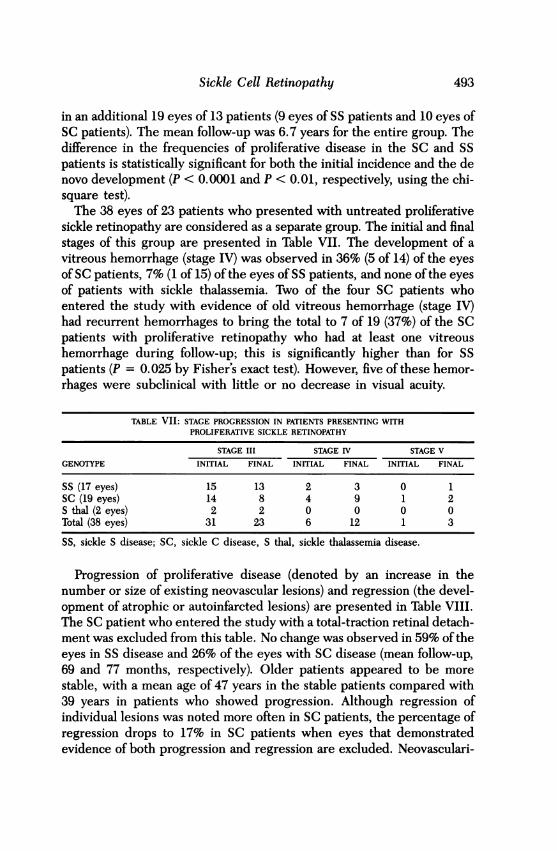
Proliferative Sickle RetinopathyThe change in prevalence of proliferative sickle retinopathy in the differ-ent hemoglobin types is summarized in Table VI. Since all untreatedpatients with stage III disease or worse were included in the naturalhistory study, the initial incidence of proliferative disease was determinedfrom the prevalence study. During follow-up, stage III lesions developed
TABLE VI: CHANGE IN PREVALENCE OF PROLIFERATIVE SICKLE RETINOPATHY(STAGE III OR HIGHER) DURING FOLLOW-UP IN 82 PATIENTS*
INITIAL DEVELOPEDGENOTYPE EXAMINATION STAGE III, IV, OR V FINAL
SS 59 Patients 11 (19%) 7 18 (31%)117 Eyes 17 (15%) 9 26 (22%)
SC 23 Patients 11 (48%) 6 17 (74%)45 Eyes 19 (42%) 10 29 (64%)
*Patients with sickle thalassemia included in natural history study are notincluded. None of those patients had new proliferative lesions develop duringfollow-up.SS, sickle S disease; SC, sickle C disease.
492
Sickle Cell Retinopathy
in an additional 19 eyes of 13 patients (9 eyes of SS patients and 10 eyes ofSC patients). The mean follow-up was 6.7 years for the entire group. Thedifference in the frequencies of proliferative disease in the SC and SSpatients is statistically significant for both the initial incidence and the denovo development (P < 0.0001 and P < 0.01, respectively, using the chi-square test).The 38 eyes of 23 patients who presented with untreated proliferative
sickle retinopathy are considered as a separate group. The initial and finalstages of this group are presented in Table VII. The development of avitreous hemorrhage (stage IV) was observed in 36% (5 of 14) of the eyesof SC patients, 7% (1 of 15) of the eyes of SS patients, and none of the eyesof patients with sickle thalassemia. Two of the four SC patients whoentered the study with evidence of old vitreous hemorrhage (stage IV)had recurrent hemorrhages to bring the total to 7 of 19 (37%) of the SCpatients with proliferative retinopathy who had at least one vitreoushemorrhage during follow-up; this is significantly higher than for SSpatients (P = 0.025 by Fisher's exact test). However, five of these hemor-rhages were subelinical with little or no decrease in visual acuity.
TABLE VII: STAGE PROGRESSION IN PATIENTS PRESENTING WITHPROLIFERATIVE SICKLE RETINOPATHY
STAGE III STAGE IV STAGE V
GENOTYPE INITIAL FINAL INITIAL FINAL INITIAL FINAL
SS (17 eyes) 15 13 2 3 0 1SC (19 eyes) 14 8 4 9 1 2S thal (2 eyes) 2 2 0 0 0 0Total (38 eyes) 31 23 6 12 1 3
SS, sickle S disease; SC, sickle C disease, S thal, sickle thalassemia disease.
Progression of proliferative disease (denoted by an increase in thenumber or size of existing neovascular lesions) and regression (the devel-opment of atrophic or autoinfarcted lesions) are presented in Table VIII.The SC patient who entered the study with a total-traction retinal detach-ment was excluded from this table. No change was observed in 59% of theeyes in SS disease and 26% of the eyes with SC disease (mean follow-up,69 and 77 months, respectively). Older patients appeared to be morestable, with a mean age of 47 years in the stable patients compared with39 years in patients who showed progression. Although regression ofindividual lesions was noted more often in SC patients, the percentage ofregression drops to 17% in SC patients when eyes that demonstratedevidence of both progression and regression are excluded. Neovasculari-
493
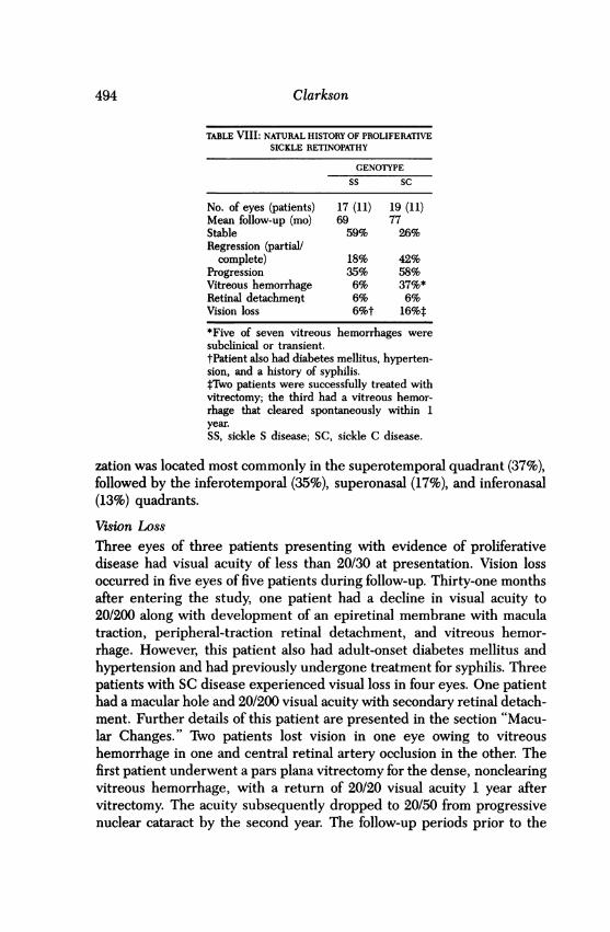
Clarkson
TABLE VIII: NATURAL HISTORY OF PROLIFERATIVESICKLE RETINOPATHY
GENOTYPE
SS SC
No. of eyes (patients) 17 (11) 19 (11)Mean follow-up (mo) 69 77Stable 59% 26%Regression (partial/
complete) 18% 42%Progression 35% 58%Vitreous hemorrhage 6% 37%*Retinal detachment 6% 6%Vision loss 6%t 16%t*Five of seven vitreous hemorrhages weresubclinical or transient.tPatient also had diabetes mellitus, hyperten-sion, and a history of syphilis.*Two patients were successfully treated withvitrectomy; the third had a vitreous hemor-rhage that cleared spontaneously within 1year.SS, sickle S disease; SC, sickle C disease.
zation was located most commonly in the superotemporal quadrant (37%),followed by the inferotemporal (35%), superonasal (17%), and inferonasal(13%) quadrants.
Vision LossThree eyes of three patients presenting with evidence of proliferativedisease had visual acuity of less than 20/30 at presentation. Vision lossoccurred in five eyes of five patients during follow-up. Thirty-one monthsafter entering the study, one patient had a decline in visual acuity to20/200 along with development of an epiretinal membrane with maculatraction, peripheral-traction retinal detachment, and vitreous hemor-rhage. However, this patient also had adult-onset diabetes mellitus andhypertension and had previously undergone treatment for syphilis. Threepatients with SC disease experienced visual loss in four eyes. One patienthad a macular hole and 20/200 visual acuity with secondary retinal detach-ment. Further details of this patient are presented in the section "Macu-lar Changes." Two patients lost vision in one eye owing to vitreoushemorrhage in one and central retinal artery occlusion in the other. Thefirst patient underwent a pars plana vitrectomy for the dense, nonclearingvitreous hemorrhage, with a return of 20/20 visual acuity 1 year aftervitrectomy. The acuity subsequently dropped to 20/50 from progressivenuclear cataract by the second year. The follow-up periods prior to the
494
Sickle Cell Retinopathy
reported vision loss for the three patients were 118, 49, and 27 months,respectively. One other patient with SC disease had transient vision losssecondary to vitreous hemorrhage, but vision returned to 20/20 within 2weeks and remained normal for an additional 36 months of follow-up.
In the 19 eyes in which proliferative retinopathy developed de novo,only one patient had associated visual acuity loss. An 11-year-old patientwith SC disease presented with nonproliferative disease in his right eyeand stage III disease in his left eye. One year after he entered the study,stage III disease developed in the right eye. A retinal detachment devel-oped in the right eye 41/2 years later. The patient underwent an unsuc-cessful attempt at retinal reattachment with a scleral buckling procedureand lost all light perception. This was the youngest patient in the study inwhom proliferative sickle retinopathy developed and the only patientwith permanent central vision loss directly attributed to proliferativesickle retinopathy. His untreated left eye maintained 20/20 acuity after 9years of follow-up.
DISCUSSION
ANGIOID STREAKS
The association between angioid streaks and sickle cell disease has beenwell documented.31-35 The incidence of angioid streaks in an unselectedpopulation of sickle cell patients has been estimated to be 1% to 2%,although a definite age relationship has been observed.34,35Condon andSerjeant34 reported that 22% of 60 homozygous patients over 40 years ofage had angioid streaks, compared with only 2% of 150 younger Jamaicanpatients. The present study confirms those findings. Angioid streaks wereobserved initially in two patients (1.3%) and developed in five patients,aged 25 to 68, during follow-up (Fig 2). Streaks were found in both SS andSC patients, and the overall incidence was 7.2% for all patients and 27%for patients over the age of 50. No relationship was found between angioidstreaks and the type of hemoglobinopathy or the severity of sickle reti-nopathy. No associated vision loss caused by complications of angioidstreaks occurred in these patients; this is consistent with the benignclinical course of angioid streaks in sickle patients described in previousstudies.34,35
PROLIFERATIVE SICKLE RETINOPATHY
Retinal neovascularization was observed more frequently in the temporalquadrants than in the nasal quadrants. This relationship has been noted in
495

Clarkson
FIGURE 2Homozygous hemoglobin S disease in 17-year-old black male. A: Right eye at first examina-tion, January 1981. Visual acuity was 20/20 in both eyes, and there was no evidence of anyfundus abnormality. B: Left eye, January 1981. C: Right eye, September 1982. Early angioidstreak is visible at 12 o'clock position, just superior to disc. D: Left eye, September 1982,continues to appear normal. E: Right eye, January 1989. F: Left eye, January 1989. Botheyes had 20/20 vision. Evidence of peripheral vascular occlusion with arteriovenous anasto-moses and peripheral "sunburst spots" is seen. Peripapillary angioid streaks were present in
both eyes.
496
Sickle Cell Retinopathy
the past, with the superotemporal quadrant considered the most commonlocation for early neovascular proliferation. This was true for SC patientsin the present study, but not for 55 patients in whom the inferotemporalquadrant was more frequently involved. This predilection for the tempo-ral quadrants early in the development of stage III proliferative sickleretinopathy was quite dramatic. Over 93% (54 of 58 eyes) had at leastsome, if not all, of the neovascularization located temporally when retinalneovascularization was first observed.
Other investigators8"1516 have documented that proliferative retinopa-thy is more common in patients with SC disease than in patients with SSdisease. Condon and Serjeant16 examined 76 SS and 70 SC randomlyselected patients and found a prevalence of 32.8% for SC patients (laterrevised to 37%27) and only 2.6% for SS patients. The present studydemonstrates a greater frequency of proliferative retinopathy for both SCdisease (45%) and SS disease (11%). Condon and Serjeant found that theincidence of proliferative disease increases with age, with the maximumincidence between ages 15 and 29. The median age of the patients in thisstudy (26 years) is probably older than that in the studies by Condon andSerjeant, in which 58 of76 patients with SS disease'5 and 47 of 70 patientswith SC diseasel6 were all under age 30. Although patients with SSdisease in the Jamaican study'5 were referred from a sickle clinic, thosewith SC disease in Jamaica'6 were solicited through letters and homevisitation. Also, the patients who were referred from the Sickle CellCenter in the present study may have had more severe systemic diseasethan the Jamaican patients.The increased severity of proliferative disease in SC patients as com-
pared with SS patients has been observed previously,8'20'27 and thedifference is striking in this natural history study. In SC patients therewas a significant increase in frequency of de novo development of neo-vascularization (Table VII) (26% in SC compared to 12% in SS), anincrease in number and size of proliferative lesions (58% in SC comparedwith 35% of SS eyes), and a progression to higher stages of the disease(Table VIII). Vitreous hemorrhage developed or recurred in 37% of theeyes of patients with stage III or IV proliferative sickle retinopathy at thebeginning ofthe study, compared, with only 6% ofthe eyes of SS patients.However, in spite of the increasing development of neovascular lesionsand/or vitreous hemorrhage in these eyes, visual function remained sur-prisingly stable. Many eyes demonstrated gradual regression of neo-vascularization with little or no change in central vision, although someevidence of "activity," such as minimal vitreous bleeding or angiographicevidence of leakage of fluorescein may have been present (Figs 3 and 4).
497

Clarkson
FIGURE 3Homozygous hemoglobin S disease in 25-year-old male who presented with stage IIIproliferative retinopathy and 20/20 visual acuity in both eyes. Autoinfarction of proliferativelesion in left eye is evident. Patient maintained 20/20 visual acuity in both eyes with morethan 10 years of follow-up. A: Color fundus photograph of proliferative lesion in temporalperiphery, left eye. B: Fluorescein angiogram of same area, showing marked vascularactivity at time of initial visit. C: Color fundus photograph of same lesion 5 years later. D:Fluorescein angiogram ofsame area, demonstrating nearly complete autoinfarction ofprolif-
erative lesion.
TREATMENT
Treatment has been advocated on the basis of the impression that thenatural history of stage III proliferative sickle retinopathy will lead to lossof vision. Various treatment modalities have been utilized and have shownsome degree of success; these include diathermy,36 cryotherapy,37,38 treat-ment of feeder vessels with xenon and argon laser photocoagulation,30,38-40and indirect argon laser treatment to the nonperfused peripheral reti-na.41-44 Some form of argon laser treatment has been widely recom-mended; cryotherapy has been limited to those cases with cloudy media.The direct treatment of feeder vessels, although successful in causingregression of neovascularization, has been questioned because of thedevelopment of choroidal neovascularization or choriovitreal neovascular-ization in 94% of eyes followed for a mean of 11.2 years.28 Vitreoushemorrhage, vitreoretinal traction, and retinal detachment have also
498

Sickle Cell Retinopathy
FIGURE 4Sickle cell-hemoglobin C disease in 21-year-old black male. A: Fundus photograph taken inMay 1983 shows prominent epiretinal membrane covering disc and most of macular area,right eye. Visual acuity was 20/30. B: Fundus photograph taken 34 months later (March1986) confirms spontaneous retraction of epiretinal membrane, no change in visual acuity.C: Fundus photograph of nasal periphery taken in May 1983 shows evidence of proliferativeretinopathy, primarily fibrous in nature. Smaller frond shows definite vascular component.D: Fundus photograph ofsame area taken in March 1986 documents no significant change in
peripheral fibrovascular lesions. Note pigmented sunburst spot.
been noted as complications of this form of treatment.2728 The long-termfollow-up of 29 of the original 44 patients (20 with SC, 4 with SS, and 5with sickle ,B-thalassemia) from the randomized clinical trial of feedervessel photocoagulation revealed the treated group had a high rate ofcomplications (32%); these included 2 eyes with retinal detachment, 1with retinal tear, and 5 with choroidal neovascularization.45 However,none of these complications appeared to have a long-term adverse out-come. The control group experienced an increased number of transientvitreous hemorrhages, but there was no significant difference in the finalvisual acuity between the treated and control eyes.A comparison of the results of this natural history study of patients with
proliferative sickle retinopathy (Table VIII) with two studies42 43 recom-mending prophylactic scatter laser treatment (Table IX) reveals no indica-tion for prophylactic treatment in patients with SS disease. Though the
499
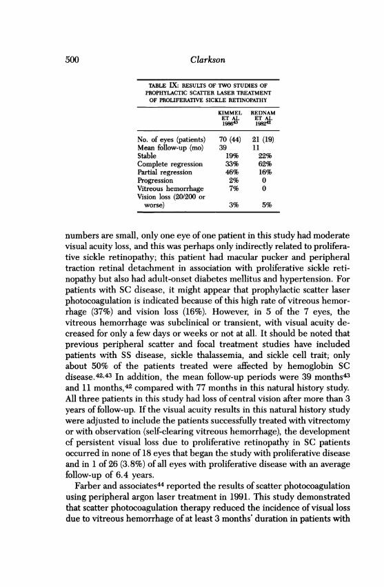
Clarkson
TABLE IX: RESULTS OF TWO STUDIES OFPROPHYLACTIC SCATTER LASER TREATMENTOF PROLIFERATIVE SICKLE RETINOPATHY
KIMMEL REDNAMET AL ET AL198643 198242
No. of eyes (patients) 70 (44) 21 (19)Mean follow-up (mo) 39 11Stable 19% 22%Complete regression 33% 62%Partial regression 46% 16%Progression 2% 0Vitreous hemorrhage 7% 0Vision loss (20/200 or
worse) 3% 5%
numbers are small, only one eye of one patient in this study had moderatevisual acuity loss, and this was perhaps only indirectly related to prolifera-tive sickle retinopathy; this patient had macular pucker and peripheraltraction retinal detachment in association with proliferative sickle reti-nopathy but also had adult-onset diabetes mellitus and hypertension. Forpatients with SC disease, it might appear that prophylactic scatter laserphotocoagulation is indicated because of this high rate of vitreous hemor-rhage (37%) and vision loss (16%). However, in 5 of the 7 eyes, thevitreous hemorrhage was subclinical or transient, with visual acuity de-creased for only a few days or weeks or not at all. It should be noted thatprevious peripheral scatter and focal treatment studies have includedpatients with SS disease, sickle thalassemia, and sickle cell trait; onlyabout 50% of the patients treated were affected by hemoglobin SCdisease.42'43 In addition, the mean follow-up periods were 39 months43and 11 months,42 compared with 77 months in this natural history study.All three patients in this study had loss of central vision after more than 3years of follow-up. If the visual acuity results in this natural history studywere adjusted to include the patients successfully treated with vitrectomyor with observation (self-clearing vitreous hemorrhage), the developmentcf persistent visual loss due to proliferative retinopathy in SC patientsoccurred in none of 18 eyes that began the study with proliferative diseaseand in 1 of 26 (3.8%) of all eyes with proliferative disease with an averagefollow-up of 6.4 years.
Farber and associates4 reported the results of scatter photocoagulationusing peripheral argon laser treatment in 1991. This study demonstratedthat scatter photocoagulation therapy reduced the incidence of visual lossdue to vitreous hemorrhage of at least 3 months' duration in patients with
500
Sickle Cell Retinopathy
proliferative sickle retinopathy. However, this study did not show definiteevidence that the incidence of retinal detachment, the primary cause ofblindness in patients with proliferative sickle retinopathy, was reducedthrough peripheral scatter photocoagulation. There were no complica-tions reported from the peripheral scatter photocoagulation. The averagelength of follow-up was 47 months for treated eyes. Although theseinvestigators had reported previously that the extent of stage III retinopa-thy was probably an important risk factor for vision-threatening complica-tions, only 11 of the treated eyes had 600 or greater (2 clock hours or more)of fibrovascular proliferation. Therefore, 88 of the 99 treated eyes had lessthan 2 clock hours of neovascularization.
Farber and associates4 recommend peripheral scatter laser treatmenton the basis of a reduction of "ocular events" in the treated eyes. How-ever, ifpermanent reduction in vision is considered, five control eyes andthree treated eyes lost vision due to proliferative sickle retinopathyduring follow-up. This is similar to the loss of vision found in this naturalhistory study (Table VIII), and the difference between treated and un-treated eyes is not significant.
SUMMARY
Prophylactic photocoagulation may have a role in the treatment of prolif-erative sickle retinopathy in selected patients with. SC disease, but noneof the studies reported to date have established that such treatment ofthese eyes improves the long-term visual outcome as compared with thenatural history as documented in this study. The similar visual outcomesin the eyes reviewed in this natural history study as compared with thosethat have been treated with photocoagulation should not be unexpected,because there is greater predilection for spontaneous involution or autoin-farction of the neovascular tissue in SC disease as opposed to the neo-vascularization that develops in other retinal vascular disease.A controlled, multicenter clinical trial designed to study those eyes at
greatest risk should be considered. Such a trial should specify patient age(15 to 30 years), hemoglobin type (SC disease), and a minimum thresholdof active proliferative disease (600 or greater). Such criteria have beensuggested by others.29'40 By comparing the outcome of eligible eyesrandomly assigned to treatment or observation and followed for an ex-tended period, it will be possible to learn whether laser photocoagulationoffers a better prognosis than the natural history of proliferative sickleretinopathy. There is no question that in some eyes with proliferativesickle retinopathy, nonclearing vitreous hemorrhage and/or retinal de-
501

502 Clarkson
tachment will develop. Clear definition of the risk factors leading to theseadvanced stages, however, is lacking, and the value of treatment is uncer-tain.
ACKNOWLEDGMENT
The author wishes to thank John Leon, MD, and Astrid Mack, PhD, fortheir assistance, and to acknowledge the technical assistance of Bill Feuer,MA.
REFERENCES
1. Bunn HF: Disorders of hemoglobin, in E Braunwald, KJ Isselbacher, RG Petersdorf etal (eds): Harrison's Principles of Internal Medicine. 11 ed. New York, McGraw-Hill,1987, pp 1518-1523
2. Konotey-Ahulu FID: The sickle cell diseases: Clinical manifestations including the"sickle crisis." Arch Intern Med 1974; 133:611-619.
3. Pauling L, Itano HA, Singer SJ: Sickle cell anemia, a molecular disease. Science 1949;110:543-548.
4. Herrick JB: Peculiar elongated and sickle-shaped red blood corpuscles in a case ofsevere anemia. Arch Intern Med 1910; 6:517-521.
5. Neel JV: The inheritance of sickle cell anemia. Science 1949; 110:64-66.6. Cook WC: A case of sickle cell anemia with associated subarachnoid hemorrhage. J Med
1930; 11:541.7. Edington GM, Sarkies JWR: Two cases of sickle-cell anaemia associated with retinal
microaneurysms. Trans R Soc Trop Med Hyg 1952; 46:59-628. Welch RB, Goldberg MF: Sickle-cell hemoglobin and its relation to fundus abnormality.
Arch Ophthalmol 1966; 75:353-362.9. Goldberg MF: Natural history of untreated proliferative sickle retinopathy. Arch Oph-
thalmol 1971; 85:428-437.10. Romayananda N, Goldberg MF, Green WR: Histopathology of sickle cell retinopathy.
Trans Am Acad Ophthalmol Otolaryngol 1973; 77:0P652-OP676.11. Nagpal KC, Goldberg MF, Rabb MF: Ocular manifestations of sickle hemoglobinopa-
thies. Surv Ophthalmol 1977; 21:391-411.12. Paton D: Angioid streaks and sickle cell anemia: A report of two cases. Arch Ohthalmol
1959; 62:852-858.13. Cohen SB, Fletcher ME, Goldberg MF, et al: Diagnosis and management of ocular
complications of sickle hemoglobinopathies: Part II. Ophthalmic Surg 1986; 17:110-116.14. Lieb WA, Geeraets WJ, Guerry D III: Sickle-cell retinopathy: Ocular and systemic
manifestations of sickle-cell disease. Acta Ophthalmol (Suppl) 1959; 58:1-45.15. Condon PI, Serjeant GR: Ocular findings in homozygous sickle cell anemia in Jamaica.
Am J Ophthalmol 1972; 73:533-543.16. : Ocular findings in hemoglobin SC disease in Jamaica. Am J Ophthalmol 1972;
74:921-931.17. : Ocular findings in sickle-cell thalassemia in Jamaica. Am J Ophthalmol 1972;
7:1105-1109.18. Paton D: The conjunctival sign of sickle cell-disease. Arch Ophthalmol 1961; 66:90-94.19. Serjeant GR: The Clinical Features of Sickle Cell Disease. New York, Elsevier, 1974.20. Condon PI, Serjeant GR: Behaviour of untreated proliferative sickle retinopathy. BrJ
Ophthalmol 1980; 64:404-411.
Sickle Cell Retinopathy 503
21. Raichand M, Goldberg MF, Nagpal KC, et al: Evolution of neovascularization in sicklecell retinopathy: A prospective fluorescein angiographic study. Arch Ophthalmol 1977;95:1543-1552.
22. Nagpal KC, Patrianakos D, Asdourian GK, et al: Spontaneous regression (autoinfarc-tion) of proliferative sickle retinopathy. Am J Ophthalmol 1975; 80:885-892.
23. Diabetic Retinopathy Study Research Group: Photocoagulation treatment of prolifera-tive diabetic retinopathy: The second report of diabetic retinopathy study findings.Ophthalmology 1978; 85:82-106.
24. Branch Vein Occlusion Study Group: Argon laser scatter photocoagulation for preven-tion of neovascularization and vitreous hemorrhage in branch vein occlusion: A ran-domized clinical trial. Arch Ophthalmol 1986; 104:34-41.
25. Cryotherapy for Retinopathy of Prematurity Cooperative Group: Multicenter trial ofcryotherapy for retinopathy of prematurity: Preliminary results. Arch Ophthalmol 1988;106:471-479.
26. Goldbaum MH, Fletcher RC, Jampol LM, et al: Cryotherapy of proliferative sickleretinopathy: II. Triple freeze-thaw cycle. Br J Ophthalmol 1979; 63:97-101.
27. Condon PI, Serjeant GR: Photocoagulation in proliferative sickle retinopathy: Results ofa 5-year study. Br J Ophthalmol 1980; 64:832-840.
28. Fox PD, Acheson RW, Serjeant GR: Outcome of iatrogenic choroidal neovascularisationin sickle cell disease. Br J Ophthalmol 1990; 74:417-420.
29. Moriarty BJ, Acheson RW, Condon PI, et al: Patterns of visual loss in untreated sicklecell retinopathy. Eye 1988; 2:330-335.
30. Jacobson MS, Gagliano DA, Cohen SB, et al: A randomized clinical trial offeeder vesselphotocoagulation of proliferative sickle retinopathy: A ten-year follow-up. Abstract.Ophthalmology (Suppl) 1989; 96:113.
31. Stevens TS, Busse B, Lee C-B, et al: Sickling hemoglobinopathies: Macular andperimacular vascular abnormalities. Arch Ophthalmol 1974; 92:455-463.
32. Geeraets WJ, Gu-erry D III: Angioid streaks and sickle-cell disease. Am J Ophthalmol1960; 49:450470.
33. Nagpal KC, Asdourian G, Goldbaum M, et al: Angioid streaks and sickle haemoglobin-opathies. Br J Ophthalmol 1976; 60:31-34.
34. Condon PI, Serjeant GR: Ocular findings in elderly cases of homozygous sickle-celldisease in Jamaica. Br J Ophthalmoll976; 60:361-364.
35. Clarkson JG, Altman RD: Angioid streaks. Surv Ophthalmol 1982; 26:235-246.36. Condon PI, Serjeant GR: Photocoagulation and diathermy in the treatment of prolifera-
tive sickle retinopathy. Br J Ophthalmol 1974; 58:650-662.37. Lee C-B, Woolf MB, Galinos SO, et al: Cryotherapy of proliferative sickle retinopathy:
Part I. Single freeze-thaw cycle. Ann Ophthalmol 1975; 7:1299-1308.38. Goldberg MF: Treatment of proliferative sickle retinopathy. Trans Am Acad Ophthal-
mol Otolaryngol 1971; 75:532-556.39. Jampol LM, Condon P, Farber M, et al: A randomized clinical trial of feeder vessel
photocoagulation of proliferative sickle cell retinopathy: I. Preliminary results. Oph-thalmology 1983; 90:540-545.
40. Condon P, Jampol LM, Farber MD, et al: A randomized clinical trial of feeder vesselphotocoagulation of proliferative sickle cell retinopathy: II. Update and analysis of riskfactors. Ophthalmology 1984; 91:1496-1498.
41. Cruess AF, Stephens RF, Magargal LE, et al: Peripheral circumferential retinal scatterphotocoagulation for treatment of proliferative sickle retinopathy. Ophthalmology 1983;90:272-277.
42. Rednam KRV, Jampol LM, Goldberg MF: Scatter retinal photocoagulation for prolifera-tive sickle cell retinopathy. Am J Ophthalmol 1982; 93:594-599.
43. Kimmel AS, Magargal LE, Stephens RF, et al: Peripheral circumferential retinal scatterphotocoagulation for the treatment of proliferative sickle retinopathy: A an update.Ophthalmology 1986; 93:1429-1432.

504 Clarkson
44. Farber MD, Jampol LM, Fox P, et al: A randomized clinical trial of scatter photocoagula-tion of proliferative sickle retinopathy. Arch Ophthalmol 1991; 109:363-367.
45. Jacobson MS, Gagliano DA, Cohen SB, et al: A randomized clinical trial offeeder vesselphotocoagulation of sickle cell retinopathy: A long-term follow-up. Ophthalmology1991; 98:581-585.