Embed Size (px)
Citation preview

The Novel Marker, DOG1, Is Expressed Ubiquitouslyin Gastrointestinal Stromal Tumors Irrespective of KITor PDGFRA Mutation Status
Robert B. West,* Christopher L. Corless,†
Xin Chen,‡ Brian P. Rubin,§
Subbaya Subramanian,* Kelli Montgomery,*Shirley Zhu,* Catherine A. Ball,¶
Torsten O. Nielsen,� Rajiv Patel,**John R. Goldblum,** Patrick O. Brown,‡§§
Michael C. Heinrich,†† and Matt van de Rijn*From the Departments of Pathology,* Genetics,¶ and
Biochemistry,‡ and Howard Hughes Medical Institute,§§ Stanford
University Medical Center, Stanford, California; the Departments
of Pathology † and Medicine,†† Oregon Health and Science
University Cancer Institute and Portland Veteran’s
Administration Medical Center, Portland, Oregon; the
Department of Anatomical Pathology,§ University of Washington
Medical Center, Seattle, Washington; the Department of
Pathology and Genetic Pathology Evaluation Centre,� Vancouver
General Hospital, Vancouver, British Columbia; and the
Department of Anatomic Pathology,** Cleveland Clinic
Foundation, Cleveland, Ohio
We recently characterized gene expression patternsin gastrointestinal stromal tumors (GISTs) usingcDNA microarrays, and found that the gene FLJ10261(DOG1 , discovered on GIST-1), encoding a hypothet-ical protein, was specifically expressed in GISTs. Theimmunoreactivity of a rabbit antiserum to syntheticDOG1 peptides was assessed on two soft tissue tumormicroarrays. The tissue microarrays included 587 softtissue tumors, with 149 GISTs, including 127 GISTcases for which the KIT and PDGFRA mutation statuswas known. Immunoreactivity for DOG1 was found in136 of 139 (97.8%) of scorable GISTs. All seven GISTcases with a PDGFRA mutation were DOG1-positive,while most of these failed to react for KIT. The immu-nohistochemical findings were confirmed with in situhybridization probes for DOG1 , KIT , and PDGFRA.Other neoplasms in the differential diagnosis of GIST,including desmoid fibromatosis (0 of 17) and Schwan-noma (0 of 3), were immunonegative for DOG1. Only4 of 438 non-GIST cases were immunoreactive forDOG1. DOG1, a protein of unknown function, is ex-pressed strongly on the cell surface of GISTs and israrely expressed in other soft tissue tumors. Reactiv-ity for DOG1 may aid in the diagnosis of GISTs, in-cluding PDGFRA mutants that fail to express KIT an-
tigen, and lead to appropriate treatment withimatinib mesylate, an inhibitor of the KIT tyrosinekinase. (Am J Pathol 2004, 165:107–113)
Gastrointestinal stromal tumors occur in the wall of thebowel and have been proposed to arise from the inter-stitial cells of Cajal. The differential diagnosis of thesetumors includes desmoid fibromatosis, Schwannoma,leiomyosarcoma, and, in some cases, high-grade sarco-mas.1 Accurate diagnosis of gastrointestinal stromal tu-mor (GIST) is important, because imatinib mesylate hasbeen shown to significantly inhibit these tumors presum-ably through inhibition of the KIT tyrosine kinase receptor,which is highly expressed in these tumors.2–5 As a result,the diagnosis of GIST relies heavily on KIT immunoreac-tivity. Current recommendations in the literature empha-size a diffuse, strong KIT immunoreactivity for the diag-nosis of GIST.6 CD34 immunostaining can also aid in thediagnosis, but a subset of cases is immunonegative whilemany other types of sarcomas are immunoreactive forthis marker.7–10 In the vast majority of GISTs, high levelsof KIT expression are accompanied by a KIT gene mu-tation in exons 9, 11, 13, or 17.11,12
Recently, a subset of GISTs have been found to havePDGFRA mutations rather than KIT mutations.13,14 Pa-tients with GISTs containing mutations in PDGFRA maystill benefit from imatinib therapy, but these tumors oftenfail to react with antibodies against KIT and hence mayremain undiagnosed as GIST.2 In addition, some GISTswith KIT mutations may have low KIT expression by immu-nohistochemistry yet will still respond to imatinib therapy.15
Although much work has been done on the biology ofGISTs and KIT, additional insight has recently beengained through gene microarray studies.16–18 Thesestudies have identified a number of genes whose expres-sion is relatively increased compared to other soft tissuetumors. This includes genes known to be involved with
Supported by the National Institutes of Health (grants CA85129 andCA84967) and the Howard Hughes Medical Institute.
Accepted for publication March 12, 2004.
P.O.B. is an Associate Investigator of the Howard Hughes MedicalInstitute.
Address reprint requests to Matt van de Rijn: Department of Pathology,Stanford University Medical Center, 300 Pasteur Dr., Stanford, CA, 94305.E-mail: [email protected].
American Journal of Pathology, Vol. 165, No. 1, July 2004
Copyright © American Society for Investigative Pathology
107
GISTs, such as KIT and CD34, but also includes a num-ber of genes that have not been well characterized. Wehave generated an antiserum against one GIST-specificgene, encoding for the hypothetical protein FLJ10261,which we have named “Discovered on GIST 1” (DOG1).Using immunohistochemistry with this antiserum and insitu hybridization with DOG1-specific probes, we showthat DOG1 is highly expressed not only in typical GISTsbut also in KIT-mutation-negative GISTs.
Materials and Methods
Tissue Microarray (TMA)
The studies described here were performed with theapproval of the Institutional Review Board at StanfordUniversity Hospital. Two TMAs were used for this study.The first TMA contained 460 different soft tissue tumorsfrom 421 patients, with each tumor represented by twocores. The samples were distributed over two arrayblocks that were constructed using a technique previ-ously described19 with a tissue arrayer from BeecherInstruments, Silver Spring, MD. Cores (0.6 mm) weretaken from paraffin-embedded soft tissue tumors ar-chived from the Stanford University Medical Center be-tween 1995 and 2001. This array has also been used forcharacterization of apolipoprotein D expression.20 Thesecond TMA used GISTs that were obtained from thepathology archives of Oregon Health and Science Uni-versity Hospital, the Portland VA Medical Center, and theKaiser Permanente Northwest Regional Laboratory. Thissingle-block array consisted of 0.6-mm cores from forma-lin-fixed, paraffin-embedded tumor assembled using asemiautomated tissue arrayer.21 There was one core foreach tumor, and all of the GISTs on this TMA were ana-lyzed for mutations in exons 9, 11, 13, and 17 of the KITgene using a combination of denaturing high pressureliquid chromatography and direct sequencing, as previ-ously described.13,22 KIT wild-type tumors included onthe array were also screened for mutations in exons 12and 18 of the PDGFRA gene.13
Antibody Generation
The cDNA-derived protein sequence of DOG1 showed nosignificant homology with other genes, including the KITgene. A rabbit polyclonal antibody was raised by inject-ing three peptides derived from the gene sequence (Ap-plied Genomics Inc., Hunstville, AL). These peptideshave no sequence homology to KIT. The peptides weresynthesized by standard FMOC chemistry: peptide 1,EEAVKDHPRAEYEARVLEKSLK; peptide 2, DHEECVKR-KQRYEVDYNLE; peptide 3, KEKVLMVELFMREEQDK.The peptides were conjugated to keyhole limpet hemo-cyanin and injected into two out-bred rabbits. The serum(S284) was harvested after the rabbits demonstrated asignificant anti-peptide titer. Affinity-purified antibodieswere obtained by passing the antiserum over an affinitycolumn conjugated with the three peptides; bound anti-bodies were eluted with a pH gradient.
Immunohistochemistry
Primary antibodies were directed toward DOG1 (S284,rabbit polyclonal, 1:50; Applied Genomics Inc.) and KIT(rabbit polyclonal, 1:50; DAKO, Carpinteria, CA). Serialsections of 4 �m were cut from the tissue array blocks,deparaffinized in xylene, and hydrated in a graded seriesof alcohol. Staining was then performed using the EnVi-sion� anti-rabbit system (DAKO).
In Situ Hybridization
In situ hybridization of TMA sections was performedbased on a protocol published previously.23,24 Briefly,digoxigenin-labeled sense and anti-sense RNA probesare generated by polymerase chain reaction amplifica-tion of 400- to 600-bp products with the T7 promoterincorporated into the primers. In vitro transcription wasperformed with a digoxigenin RNA-labeling kit and T7polymerase according to the manufacturer’s protocol(Roche Diagnostics, Indianapolis, IN). Sections (5 �mthick) cut from the paraffin blocks, deparaffinized in xy-lene, were hydrated in graded concentrations of ethanolfor 5 minutes each. Sections were then incubated with1% hydrogen peroxide, followed by digestion in 10 �g/mlof proteinase K at 37°C for 30 minutes. Sections werehybridized overnight at 55°C with either sense or anti-sense riboprobes at 200 ng/ml dilution in mRNA hybrid-ization buffer (DAKO). The following day, sections werewashed in 2� standard saline citrate and incubated with1:35 dilution of RNase A cocktail (Ambion, Austin, TX) in2� standard saline citrate for 30 minutes at 37°C. Next,sections were stringently washed in 2� standard salinecitrate/50% formamide twice, followed by one wash at0.08� standard saline citrate at 50°C. Biotin-blockingreagents (DAKO) were applied to the section to block theendogenous biotin. For signal amplification, a horserad-ish peroxidase-conjugated rabbit anti-digoxigenin anti-body (DAKO) was used to catalyze the deposition ofbiotinyl tyramide, followed by secondary streptavidincomplex (GenPoint kit, DAKO). The final signal was de-veloped with diaminobenzidine (GenPoint kit, DAKO),and the tissues were counterstained in hematoxylin for 15seconds.
Scoring of Immunohistochemistry and in SituHybridization
Cores were scored as follows. A score of 0 was given forabsent or insignificant staining: less than 5% tumor cellswith light brown staining. A score of 1 was given forunscorable cores. A score of 2 was given for light brownstain in greater than 5% of tumor cells or dark brown stainin less than 50% of tumor cells. A score of 3 was given fordark brown staining in greater that 50% tumor cells.Nontumor cells and cells of unknown origin were notscored. The cores were independently reviewed by twopathologists (RBW and MvdR) and disagreements werereviewed together to achieve a consensus score.
108 West et alAJP July 2004, Vol. 165, No. 1
Digital Image Collection and Data Analysis
To aid in the analysis of numerous tissue cores stained byimmunohistochemistry and in situ hybridization, digital im-ages were collected using the BLISS instrument(Bacuslabs, Lombard IL; http://bacuslabs.com). Scoringresults were combined using Deconvoluter and repre-sented in Treeview,25 as shown on the accompanying web-site (http://microarray-pubs.stanford.edu/tma_portal/dog1/),where more than 4000 digital images are available.
Results
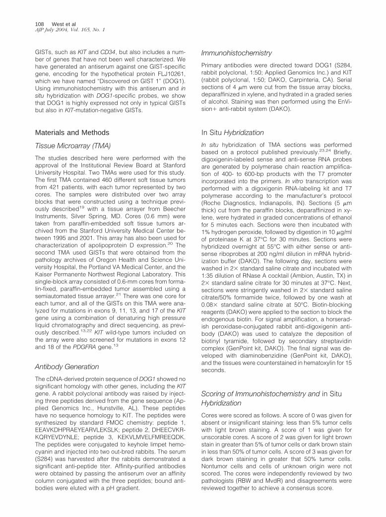
Previously, we examined the gene expression profile ofGISTs using cDNA microarrays and identified a numberof genes, in addition to the KIT gene, that demonstrateda specific pattern of elevated mRNA expression inGISTs.18 Figure 1 shows the relative level of mRNA ex-pression for one of these genes, DOG1 (FLJ10261), com-pared with KIT in a variety of soft tissue tumors, includingthose in the differential diagnosis of GIST. Searchesfailed to show any sequence similarity between the geneson either the DNA or protein level.
A rabbit antiserum was generated against syntheticpeptides derived from the putative coding sequence ofDOG1. Antiserum immunoreactivity was characterized ontwo separate TMAs containing soft tissue tumors. The firstTMA contained 460 different soft tissue tumor samplesrepresenting more than 50 different diagnostic entities.20
This array included 22 KIT-immunoreactive GISTs. Thesecond TMA included 127 GIST cases for which the KITand PDGFRA mutation status was previously determined.On this TMA there were 102 cases with an activatingmutation in KIT, 8 cases with a mutation in PDGFRA, and17 cases that were wild type for both kinases but never-theless had clinical, histological, and immunophenotypicfeatures typical for GIST.
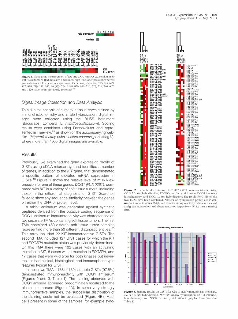
In these two TMAs, 136 of 139 scorable GISTs (97.8%)demonstrated immunoreactivity with DOG1 antiserum(Figures 2 and 3, Table 1). The staining observed withDOG1 antisera appeared predominately localized to theplasma membrane (Figure 4A). In some very stronglyimmunoreactive samples, the subcellular distribution ofthe staining could not be evaluated (Figure 4B). Mastcells present in some of the samples, for example syno-
Figure 2. Hierarchical clustering of CD117 (KIT) immunohistochemistry,CD117 in situ hybridization, PDGFRA in situ hybridization, DOG1 immuno-histochemistry, and DOG1 in situ hybridization. The results for GISTs on thetwo TMAs have been combined. Antisera or hybridization probes are in col-umns, tumors in rows. Bright red denotes strong reactivity, whereas dark redand green indicate low and absent reactivity, respectively. White means missingdata.
Figure 3. Staining results on GISTs for CD117 (KIT) immunohistochemistry,CD117 in situ hybridization, PDGFRA in situ hybridization, DOG1 immuno-histochemistry, and DOG1 in situ hybridization in graphic form (see alsoTable 1).
Figure 1. Gene array measurement of KIT and DOG1 mRNA expression in 30soft tissue tumors. Red indicates a relatively high level of expression whereasgreen denotes a low level of expression. Gene array data for STTs 524, 629,417, 418, 219, 111, 656, 94, 335, 794, 1148, 850, 616, 710, 523, 526, 740, 607,and 1220 have been previously reported.18
DOG1 Expression in GISTs 109AJP July 2004, Vol. 165, No. 1

vial sarcoma, were strongly immunoreactive as well (Fig-ure 4C), whereas the same samples showed only weakstaining in the mast cells with KIT antibodies. We con-firmed these results with in situ hybridization studies (Fig-ures 5 and 6). Interestingly, DOG1 antisera stained alleight scorable PDGFRA-mutant GISTs (one case fromfirst TMA and seven cases from second TMA), whereasthe KIT antibody staining was weak in three of thesecases and negative in the remaining five. These findingswere further extended by in situ hybridization with PDG-FRA (Figure 6). PDGFRA expression was predominately,but not exclusively, present in the PDGFRA-mutantGISTs. Five of six (83%) scorable PDGFRA-mutant GISTs
were positive for PDGFRA in situ hybridization (Figures 2and 3, Table 1). In contrast, only 10 of 70 (14%) KIT-mutant and KIT-wild-type GISTs were positive for PDG-FRA in situ hybridization. Correlation of KIT in situ hybrid-ization with KIT immunohistochemistry was good, with thein situ hybridization signal detectable in almost all immu-nopositive cases (Figure 2). However, a difference wasseen in the PDGFRA-mutant GISTs with regard to KITexpression. Three cases were immunopositive for KIT,but only one case was positive by KIT in situ hybridization.Hierarchical clustering analysis of immunohistochemistryand in situ hybridization data were performed as previ-ously described.25 Among these parameters—KIT immu-nohistochemistry, KIT in situ hybridization, DOG1 immu-nohistochemistry, DOG1 in situ hybridization, andPDGFRA in situ hybridization—the most distinguishingfeature was PDGFRA in situ hybridization positivity (Figure2), with overexpression of PDGFRA by PDFGRA in situ
Table 1. Staining Results for CD117 IHC, CD117 ISH, PDGFRA ISH, DOG1 IHC, and DOG1 ISH in Tabular Form (see alsoFigure 3)
Mutation status CD117 CD117 ISH PDGFRA ISH DOG1 DOG1 ISH
wt 14 10 9 14 3 Total scorables14 9 1 14 3 Total positive
100 90 11 100 100 % positiveKIT ex 9 9 7 7 9 6 Total scorables
9 6 2 8 5 Total positive100 86 29 89 83 % positive
KIT ex 11 86 57 51 81 39 Total scorables82 47 6 81 38 Total positive95 82 12 100 97 % positive
KIT ex 13 3 3 2 3 2 Total scorables3 2 1 3 2 Total positive
100 67 50 100 100 % positiveKIT ex 17 1 1 1 1 0 Total scorables
1 1 0 1 0 Total positive100 100 0 100 NA % positive
PDGFRA 8 7 6 8 7 Total scorables3 1 5 8 5 Total positive
37.5 14 83 100 71 % positiveUnknown 23 23 21 23 23 Total scorables
22 21 8 21 22 Total positive96 91 38 91 96 % positive
IHC, immunohistochemistry; ISH, in situ hybridization.
Figure 4. Immunohistochemical staining with anti-DOG1 serum (S284) andKIT on two GISTs [TMA 822 (A) and 3688 (B)] and a synovial sarcoma [TMA856 (C)].
Figure 5. In situ hybridization of a GIST and leiomyosarcoma with anti-sense probes to DOG1 and KIT on a GIST and a leiomyosarcoma (LMS). Thecorresponding negative control sense probes are included in the inset in thetop right corner of the GIST sample.
110 West et alAJP July 2004, Vol. 165, No. 1

hybridization seen in only in a small subset of GISTs.Images of all cores from both TMAs were digitally cap-tured and are available at the accompanying website(http://microarray-pubs.stanford.edu/tma�portal/dog1/).
From the 460 tumor samples that were not classified asGIST in the first TMA, only four cases that were nothistologically and immunophenotypically consistent withGIST were immunoreactive with DOG1 antiserum: onesynovial sarcoma (1 of 20 � 5%), one (1 of 40 � 2.5%)leiomyosarcoma, one (1 of 4 � 25%) fibrosarcoma, and(1 of 9 � 11%) one Ewing’s sarcoma/PNET. Of the 40leiomyosarcomas, 17 originated in the abdomen andnone of these were DOG1 immunoreactive. Other tumorsin the GIST differential diagnosis failed to stain with theDOG1 antisera. These include desmoid fibromatosis (17cases) and Schwannoma (3 cases). Parenthetically, un-der the staining conditions used, none of the fibromatosiscases were positive for KIT by immunohistochemistry orin situ hybridization. One leiomyosarcoma was positive forKIT immunohistochemistry only (TMA 3725). Interest-ingly, the staining was exclusively in a diffuse nuclearpattern. This tumor was negative for DOG1 by both im-munohistochemistry and in situ hybridization and for KITin situ hybridization.
Seven cases in the first TMA, not counted among the22 unequivocal GISTs, showed histological features in-determinate between GIST and smooth muscle tumor. Allof these tumors were located in the wall of the stomach orintestine, with four tumors from the stomach, one from theduodenum, one from the gastro-esophageal junction,and one from the rectum. All seven cases were negativefor KIT by immunohistochemistry and thus might not beconsidered GISTs according to current recommenda-tions.6 However, four of the seven cases were positive byKIT in situ hybridization, while DOG1 immunoreactivitywas seen in two cases, and all seven cases were positivefor DOG1 by in situ hybridization. Furthermore, two cases(TMAs 863 and 3696) were positive for PDGFRA in situhybridization. Subsequent sequence analysis of cases863 and 3696 revealed a point mutation and a deletion inexon 18 of PDGFRA, respectively. To date, such muta-tions have only been described in GISTs. We concludethat the seven KIT immunonegative cases with morpho-logical features between GIST and smooth muscle tumoractually represent GISTs.
We also stained a TMA containing a spectrum of nor-mal tissues with the DOG1 antiserum (data not shown).We observed staining in the epithelium of breast, pros-
Figure 6. In situ hybridization of KIT, DOG1, and PDGFRA with GISTs. A: GIST with mutation in KIT shows positive in situ hybridization for KIT and DOG1 butnot PDGFRA. B: GIST with mutation in PDGFRA shows positive in situ hybridization for DOG1 and PDGFRA but not for KIT. C: Negative control leiomyosarcoma.
DOG1 Expression in GISTs 111AJP July 2004, Vol. 165, No. 1

tate, salivary gland, liver, stomach, testis, pancreas, andgallbladder. The pattern of DOG1 immunostaining of theinterstitial cells of Cajal was similar to KIT. In addition,DOG1 antiserum reacted with a number of tumor cores ina carcinoma array, including some that did not stain withKIT antiserum (data not shown).
Discussion
GISTs have a high rate of local recurrence.1 Imatinib, asmall molecule inhibitor of several type III receptor ty-rosine kinases, including KIT and PDGFRA, has demon-strated promise in controlling GIST growth.3–5 The major-ity of GISTs (80 to 85%) harbor oncogenic mutations ofKIT, and for this reason KIT has been regarded as theprimary target for imatinib therapy. Indeed, initial trials ofimatinib were limited to KIT-immunoreactive GISTs. Re-cently it was discovered that a subset of GISTs (5 to 7%)has activating mutations of PDGFRA.13,14 Most of thesetumors are weak or negative in immunostaining for KIT,which may lead to underdiagnosis and possible withhold-ing of imatinib therapy. Furthermore, identification ofPDGFRA-mutant GISTs requires molecular analysis, a la-borious process that is not ideal for application in aroutine clinical setting.
In this article, we demonstrate that a novel gene,DOG1, identified in a DNA microarray analysis of geneexpression patterns as associated with GIST, is highlyexpressed in both KIT- and PDGFRA-mutant GISTs. Ex-pression of DOG1 in GISTs was demonstrated both byimmunodetection of the protein and by in situ hybridiza-tion. DOG1 immunoreactivity was assessed on two softtissue tumor microarrays representing 587 soft tissuetumors, including 149 GISTs. Of scorable GISTs 97.8%demonstrated immunoreactivity with DOG1 antisera.Only four KIT-negative, non-GIST soft tissue tumors wereDOG1 immunoreactive. Several GISTs with mutations inthe PDGRFA gene were found to react only by in situhybridization for DOG1 and to be negative for DOG1 byimmunohistochemistry. Future studies are necessary todetermine whether monoclonal antibodies against puri-fied DOG1 might yield tools with sensitivity similar to thatseen with in situ hybridization probes. We also confirmPDGFRA expression in a subset of GISTs using in situhybridization. PDGRFA expression and KIT expressionare not mutually exclusive. A subset of KIT-mutatedGISTs expresses PDGRFA in addition to KIT while asubset of PDGRFA-mutated tumors also expresses KIT.These data were seen with both immunohistochemicaland in situ hybridization techniques.
In addition to the marked similarity in reactivity forDOG1 protein on non-GIST sarcomas, DOG1 protein canalso be seen in a subset of melanomas and germ celltumors as has been described for KIT (West et al, inpreparation). Furthermore just as seen with the KIT mol-ecule, a variety of carcinomas also express DOG1. Thesetumors mostly overlap with the KIT-positive tumors. Al-though within the field of soft tissue tumors DOG1 expres-sion seems quite specific for GIST, in a differential diag-nostic setting DOG1 reactivity does not exclude carci-
nomas. Therefore additional markers such as keratinstains should be performed when the differential diagno-sis includes carcinoma.
We also demonstrated the feasibility of assessing GISTmarkers by in situ hybridization on paraffin-embeddedtissue. Correlation between immunohistochemistry and insitu hybridization for DOG1 on GISTs was excellent. In thecase of KIT, the correlation was not as strong because ofrelatively weak or absent in situ hybridization signals insome CD117-positive GISTs. It is likely that this reflectslower sensitivity of the KIT in situ hybridization assay,although cross-reactivity of the CD117 antibody to an-other epitope on GISTs has not been excluded. In situhybridization for PDGFRA proved to be valuable in iden-tifying KIT-negative GISTs, although DOG1 immunohisto-chemistry was equally sensitive for these cases. Overall,we have found that in situ hybridization techniques arecomplementary to immunohistochemistry tests in theevaluation of GISTs.
DOG1 has been recently identified as a gene in theCCND1-EMS1 locus on human chromosome 11q13,which is amplified in esophageal cancer, bladder tumors,and breast cancer.26 Human DOG1 protein showed89.8% total amino acid identity with mouse DOG1 pro-tein, and also 58.4%, 38.3%, and 38.6% identity withhuman C12orf3, C11orf25, and FLJ34272/BAC03704proteins, respectively. Sequence analysis predicts thepresence of eight transmembrane-spanning segments.This correlates with our observations of the immunohis-tochemical localization to the cell membrane. DOG1 maybe part of an as yet unclassified ion transporter family.
Because the biological function is unknown, it is un-clear why DOG1 is so widely expressed in GISTs. Twobroad possibilities exist. It may be that the protein has arole in receptor kinase type III signal transduction path-ways. On the other hand, DOG1 may be a fortuitousmarker of the GIST phenotype, with no direct connectionto the KIT and PDGFRA signaling pathways. The findingthat mast cells are also immunoreactive for DOG1 tendsto favor the former possibility.
In summary, we demonstrate that detection of a novelgene, DOG1, identifies the vast majority of both KIT- andPDGFRA-mutated GISTs. This may be of clinical value inidentifying candidates for Gleevec therapy. As a cellmembrane-associated protein, with markedly elevatedexpression in GISTs, DOG1 may also be a potential ther-apeutic target.
References
1. Weiss SW, Goldblum JR: Soft Tissue Tumors. St Louis, Mosby, 20012. Heinrich MC, Corless C, Demetri GD, Blanke C, von Mehren M,
Joensuu H, McGreevey L, Chen CJ, Van den Abbeele A, Druker B,Kiese B, Eisenberg B, Roberts P, Singer S, Fletcher CD, Silberman S,Dimitrijevic S, Fletcher JA: Kinase mutations and imatinib response inpatients with metastatic gastrointestinal stromal tumor. J Clin Oncol2003, 21:4342–4349
3. van Oosterom AT, Judson I, Verweij J, Stroobants S, Donato di PaolaE, Dimitrijevic S, Martens M, Webb A, Sciot R, Van Glabbeke M,Silberman S, Nielsen OS, European Organization for Research andTreatment of Cancer Soft Tissue and Bone Sarcoma Group: Safety
112 West et alAJP July 2004, Vol. 165, No. 1
and efficacy of imatinib (STI571) in metastatic gastrointestinal stromaltumours: a phase I study. Lancet 2001, 358:1421–1423
4. Demetri G, von Mehren M, Blanke C, Van den Abbeele A, EisenbergB, Roberts P, Heinrich M, Tuveson D, Singer S, Janicek M, Fletcher J,Silverman S, Silberman S, Capdeville R, Kiese B, Peng B, DimitrijevicS, Druker B, Corless C, Fletcher C, Joensuu H: Efficacy and safety ofimatinib mesylate in advanced gastrointestinal stromal tumors. N EnglJ Med 2002, 347:472–480
5. Joensuu H, Roberts P, Sarlomo-Rikala M, Andersson L, TervahartialaP, Tuveson D, Silberman S, Capdeville R, Dimitrijevic S, Druker B,Demetri G: Effect of the tyrosine kinase inhibitor STI571 in a patientwith a metastatic gastrointestinal stromal tumor. N Engl J Med 2001,344:1052–1056
6. Fletcher CD, Berman JJ, Corless C, Gorstein F, Lasota J, Longley BJ,Miettinen M, O’Leary TJ, Remotti H, Rubin BP, Shmookler B, SobinLH, Weiss SW: Diagnosis of gastrointestinal stromal tumors: a con-sensus approach. Hum Pathol 2002, 33:459–465
7. van de Rijn M, Rouse RV: CD34: a review. Appl Immunohistochem1994, 2:71–80
8. van de Rijn M, Hendrickson MR, Rouse RV: CD34 expression bygastrointestinal tract stromal tumors. Hum Pathol 1994, 25:766–771
9. Yantiss RK, Spiro IJ, Compton CC, Rosenberg AE: Gastrointestinalstromal tumor versus intra-abdominal fibromatosis of the bowel wall:a clinically important differential diagnosis. Am J Surg Pathol 2000,24:947–957
10. Smithey BE, Pappo AS, Hill DA: C-kit expression in pediatric solidtumors: a comparative immunohistochemical study. Am J Surg Pathol2002, 26:486–492
11. Hirota S, Nishida T, Isozaki K, Taniguchi M, Nakamura J, Okazaki T,Kitamura Y: Gain-of-function mutation at the extracellular domain ofKIT in gastrointestinal stromal tumours. J Pathol 2001, 193:505–510
12. Rubin B, Singer S, Tsao C, Duensing A, Lux M, Ruiz R, Hibbard M,Chen C, Xiao S, Tuveson D, Demetri G, Fletcher C, Fletcher J: KITactivation is a ubiquitous feature of gastrointestinal stromal tumors.Cancer Res 2001, 61:8118–8121
13. Heinrich MC, Corless CL, Duensing A, McGreevey L, Chen CJ,Joseph N, Singer S, Griffith DJ, Haley A, Town A, Demetri GD,Fletcher CD, Fletcher JA: PDGFRA activating mutations in gastroin-testinal stromal tumors. Science 2003, 299:708–710
14. Hirota S, Ohashi A, Nishida T, Isozaki K, Kinoshita K, Shinomura Y,Kitamura Y: Gain-of-function mutations of platelet-derived growth fac-tor receptor alpha gene in gastrointestinal stromal tumors. Gastroen-terology 2003, 125:660–667
15. Bauer S, Corless C, Heinrich M, Dirsch O, Antoch G, Kanja J, SeeberS, Schutte J: Response to imatinib mesylate of a gastrointestinal
stromal tumor with very low expression of KIT. Cancer ChemotherPharmacol 2003, 51:261–265
16. Allander SV, Nupponen NN, Ringner M, Hostetter G, Maher GW,Goldberger N, Chen Y, Carpten J, Elkahloun AG, Meltzer PS: Gas-trointestinal stromal tumors with KIT mutations exhibit a remarkablyhomogeneous gene expression profile. Cancer Res 2001, 61:8624–8628
17. Khan J, Wei JS, Ringner M, Saal LH, Ladanyi M, Westermann F,Berthold F, Schwab M, Antonescu CR, Peterson C, Meltzer PS: Clas-sification and diagnostic prediction of cancers using gene expressionprofiling and artificial neural networks. Nat Med 2001, 7:673–679
18. Nielsen TO, West RB, Linn SC, Alter O, Knowling MA, O’Connell JX,Zhu S, Fero M, Sherlock G, Pollack JR, Brown PO, Botstein D, van deRijn M: Molecular characterisation of soft tissue tumours: a geneexpression study. Lancet 2002, 359:1301–1307
19. Kononen J, Bubendorf L, Kallioniemi A, Barlund M, Schraml P, Leigh-ton S, Torhorst J, Mihatsch MJ, Sauter G, Kallioniemi OP: Tissuemicroarrays for high-throughput molecular profiling of tumor speci-mens. Nat Med 1998, 4:844–847
20. West RB, Harvell J, Linn S, Lui C, Prapong W, Hernandez-BoussardT, Montgomery K, Nielsen TO, Rubin BP, Patel R, Goldblum JR,Brown P, van de Rijn M: Apo D in soft tissue tumors: a novel markerfor dermatofibrosarcoma protuberans. Am J Surg Pathol (In press)
21. Torhorst J, Bucher C, Kononen J, Haas P, Zuber M, Kochli O, MrossF, Dieterich H, Moch H, Mihatsch M, Kallioniemi O, Sauter G: Tissuemicroarrays for rapid linking of molecular changes to clinical end-points. Am J Pathol 2001, 159:2249–2256
22. Corless C, McGreevey L, Haley A, Town A, Heinrich M: KIT mutationsare common in incidental gastrointestinal stromal tumors one centi-meter or less in size. Am J Pathol 2002, 160:1567–1572
23. St. Croix B, Rago C, Velculescu V, Traverso G, Romans K, Montgom-ery E, Lal A, Riggins G, Lengauer C, Vogelstein B, Kinzler K: Genesexpressed in human tumor endothelium. Science 2000, 289:1197–1202
24. Iacobuzio-Donahue CA, Ryu B, Hruban RH, Kern SE: Exploring thehost desmoplastic response to pancreatic carcinoma: gene expres-sion of stromal and neoplastic cells at the site of primary invasion.Am J Pathol 2002, 160:91–99
25. Liu CL, Prapong W, Natkunam Y, Alizadeh A, Montgomery K, GilksCB, van de Rijn M: Software tools for high-throughput analysis andarchiving of immunohistochemistry staining data obtained with tissuemicroarrays. Am J Pathol 2002, 161:1557–1565
26. Katoh M, Katoh M: FLJ10261 gene, located within the CCND1-EMS1locus on human chromosome 11q13, encodes the eight-transmem-brane protein homologous to C12orf3, C11orf25 and FLJ34272 geneproducts. Int J Oncol 2003, 22:1375–1381
DOG1 Expression in GISTs 113AJP July 2004, Vol. 165, No. 1