Embed Size (px)
Citation preview

The NFL-TBS.40-63 Anti-Glioblastoma Peptide DisruptsMicrotubule and Mitochondrial Networks in the T98GGlioma Cell LineRomain Rivalin1,2., Claire Lepinoux-Chambaud1,2., Joel Eyer1,2, Frederique Savagner1,2,3*
1 Universite d’Angers, Angers, France, 2 Laboratoire Neurobiologie & Transgenese, LNBT, UPRES EA-3143, Universite d’Angers, Batiment IBS-IRIS, Angers, France, 3 CHU
Angers, Laboratoire de Biochimie, Angers, France
Abstract
Despite aggressive therapies, including combinations of surgery, radiotherapy and chemotherapy, glioblastoma remains ahighly aggressive brain cancer with the worst prognosis of any central nervous system disease. We have previouslyidentified a neurofilament-derived cell-penetrating peptide, NFL-TBS.40-63, that specifically enters by endocytosis inglioblastoma cells, where it induces microtubule destruction and inhibits cell proliferation. Here, we explore the impact ofNFL-TBS.40-63 peptide on the mitochondrial network and its functions by using global cell respiration, quantitative PCRanalysis of the main actors directing mitochondrial biogenesis, western blot analysis of the oxidative phosphorylation(OXPHOS) subunits and confocal microscopy. We show that the internalized peptide disturbs mitochondrial andmicrotubule networks, interferes with mitochondrial dynamics and induces a rapid depletion of global cell respiration. Thiseffect may be related to reduced expression of the NRF-1 transcription factor and of specific miRNAs, which may impactmitochondrial biogenesis, in regard to default mitochondrial mobility.
Citation: Rivalin R, Lepinoux-Chambaud C, Eyer J, Savagner F (2014) The NFL-TBS.40-63 Anti-Glioblastoma Peptide Disrupts Microtubule and MitochondrialNetworks in the T98G Glioma Cell Line. PLoS ONE 9(6): e98473. doi:10.1371/journal.pone.0098473
Editor: Peter Canoll, Columbia University, United States of America
Received December 5, 2013; Accepted May 2, 2014; Published June 4, 2014
Copyright: � 2014 Rivalin et al. This is an open-access article distributed under the terms of the Creative Commons Attribution License, which permitsunrestricted use, distribution, and reproduction in any medium, provided the original author and source are credited.
Funding: This work was supported by grants from CIMATH-2 (ciblage moleculaire et applications therapeutiques) Region Pays de la Loire, Ligue Contre le Cancerand L’INSERM. The funders had no role in study design, data collection and analysis, decision to publish, or preparation of the manuscript.
Competing Interests: The authors have declared that no competing interests exist.
* E-mail: [email protected]
. These authors contributed equally to this work.
Introduction
Glioblastoma is a highly aggressive brain cancer that has been
designated as grade IV, according to the World Health
Organization [1]. It represents an extremely invasive form of
glioma and has the worst prognosis of any central nervous system
disease. Despite aggressive therapies that include combinations of
surgery, radiotherapy and chemotherapy, the median post-
diagnostic survival period is approximately one year [2]. Many
aspects of glioblastoma contribute to its poor prognosis, including
the invasive nature of these abnormal cells [3] and the extreme
heterogeneity of this cancer [4]. The lack of specificity for the
current treatments and their side effects imply the need to develop
new therapeutic strategies that target tumor cells [5].
Microtubule-targeting agents (MTAs) represent an important
class of drugs used in the treatment of cancers. Microtubules are
ubiquitous cellular polymers composed of heterodimers of a- and
b-tubulin subunits [6]. They play major roles in several cellular
functions, including intracellular transport, maintenance of cell
architecture, cell signaling and mitosis. MTAs exert their anti-
tumoral activity by altering microtubule polymerization and
dynamics, which causes growth arrest in mitosis and subsequent
cell death by apoptosis [7]. Proteins that compose the intermediate
filaments are able to bind free unpolymerized tubulin onto specific
sites named tubulin-binding sites (TBS) and thus can affect
microtubule polymerization in vitro and in vivo [8]. A peptide
derived from the light neurofilament subunit (NFL) that corre-
sponds to the sequence of TBS (NFL-TBS.40-63) can enter
specifically into glioblastoma cells by endocytosis [9], where it
disrupts the microtubule network and induces cell death by
apoptosis [10].
Recent studies have confirmed that interactions between
intermediate filaments, notably NFL or Vimentin, and key
molecules responsible for the plasticity of the mitochondrial
network, including Mitofusin-1 and -2 (MFN1 and 2, respectively)
or Dynamin, are necessary to maintain organelle integrity and to
allow mitochondrial motility [11–13]. The fission process ensures
mitochondrial structural quality by removing damaged mitochon-
dria through mitophagy and facilitating apoptosis in conditions of
cellular stress [14,15]. The fusion process could be divided into
transient and complete fusion [16]. Contrary to complete fusion,
the transient process is essential for promoting mitochondrial
metabolism and motility by interplaying with the cytoskeletal
anchorage.
A close relationship has been demonstrated between the
oxidative phosphorylation (OXPHOS) process and mitochondrial
network organization, which is controlled by the balance between
fusion and fission events [17]. The failure of mitochondrial fusion–
fission dynamics has been involved in the pathogenesis of several
neurodegenerative diseases and cancers [18,19]. Mitochondrial
biogenesis is dependant of transcription factors such as nuclear
respiratory factors and estrogen-related receptors that coordinate
PLOS ONE | www.plosone.org 1 June 2014 | Volume 9 | Issue 6 | e98473

the synthesis of OXPHOS complex subunits encoded by the
nuclear and mitochondrial genomes. The transcriptional efficiency
of these factors is controlled by coactivators from the peroxisome
proliferator-activated receptor c coactivator-1 (PGC-1) family, i.e.,
PGC-1a, PGC-1b and the PGC-1-related coactivator (PRC), that
integrates mitochondrial biogenesis and function to various
environmental signals [20]. We previously showed that the
ubiquitous PRC member was able to control mitochondrial fission
by modulating the Fission-1 (FIS1) expression level in cancer cells,
in addition to its effect on mitochondrial biogenesis [21].
Numerous studies have highlighted selected miRNAs related to
glioma pathogenesis [22]. Some of them have potential applica-
tions as novel diagnostic and prognostic indicators. Thus, the re-
expression of miR-34a encoded at Chr1p36.22, a region deleted in
many glioblastomas, could be associated with reduced tumor
proliferation, cell migration and invasion [23]. Conversely, miR-
21 has been identified as an anti-apoptotic factor and presents a
significant up-regulation in glioblastoma, while its inhibition
induced apoptosis in glioblastoma cells in vitro and in vivo [24,25].
This miRNA is involved in the down regulation of the tumor
suppressor gene PTEN, in caspase 3/7 activation and confers a
drug resistance to cancer cells [26]. Moreover, an over-expression
of miR-221 has been linked with increased cellular proliferation
and an over-expression of the c-KIT gene [27,28]. These miRNAs
have also recently been related to a pool of miRNAs called
mitomiRs, which are associated with the mitochondrial compart-
ment [29]. Their role in the control of mitochondrial functions
and cell redox status is now established [30,31].
In this study, we focused on the role for MTAs in the OXPHOS
process and the dynamics of mitochondrial networks. For this
purpose, we used the T98G cellular model of human glioblastoma,
in which we have previously demonstrated the incorporation and
cytoskeleton effect for 10 mM NFL-TBS.40-63 peptide [8–10,32].
Materials and Methods
Cell lines and peptide designHuman T98G glioblastoma cells and mouse NIH-3T3 embry-
onic fibroblast cells were obtained from ATCC (Manassas, VA,
USA). They were grown in T75 flasks at 37 uC under 5% CO2, in
Dulbecco’s modified Eagle’s medium with 1 g/l glucose, which
was supplemented with 5% L-glutamine, 10% fetal calf serum
(both from Lonza, Walkersville, MD, USA) and 5% (penicillin/
streptomycin (Sigma, Saint-Quentin Fallavier, France). A biotiny-
lated peptide corresponding to NFL (NFL-TBS.40-63: YSSY-
SAPVSSSLSVRRSYSSSSGS) and a similarly labeled scrambled
peptide (NFL-SCR: SLGSPSSSVRASYSSSRSYVYSSS) were
synthesized (with more than 95% purity) by MilleGen (Toulouse,
France) and dissolved in water at 1 mM concentration. They were
used at final concentrations of 2, 5 and 10 mM for 6 h at 37 uC to
evaluate their cellular effects.
Quantitative mRNA and miRNA analysesT98G cells were tested for peptide effects on independent
triplicate cell cultures. Cells were harvested following a trypsin-
EDTA 16 (Sigma) treatment for 5–10 min and centrifugation.
Total RNA was extracted from cells pellets using the RNeasyHMini Kit (Qiagen, Valencia, CA, USA) according to the
manufacturer’s recommendations. Reverse transcription was
performed on 1 mg of total RNA with the Advantage RT-for-
PCR kit (Clontech, Palo Alto, CA, USA), following the
manufacturer’s recommendations. Real-time quantification was
performed on a 96-well plate using the Power SYBRH Green
Master mix and the ABI 7900 apparatus (Applied Biosystems by
Life technologies, Grand Island, NY, USA). Ten genes were tested
for quantitative expression: MFN2 and FIS1 for the mitochondrial
fusion and fission process, PRC/PPRC1, PGC-1a/PPARGCA1,
NRF-1, CYCS (Cytochrome c) and TFAM (Mitochondrial
transcription factor A) for mitochondrial biogenesis and its
functions, PTEN, NAIP and FGFR3 as genes directly targeted
by miR-21, 221 and 100, respectively [33,34]. The data were
normalized to b-globin, and the relative expression level of specific
mRNA was calculated with the usual 2–DDCt method. The
sequences of primers used in this study are referenced in Table 1.
For miRNA analyses, cDNA was first produced using the VILO
miRNA cDNA Synthesis Kit (Invitrogen, Carlsbad, CA, USA)
according to the manufacturer’s protocol. Using Express SYBR
GreenER qPCR SuperMix (Invitrogen), real-time PCR was
carried out on a ABI 7900 apparatus (Applied Biosystems by Life
technologies) using a universal primer and forward primers specific
to each miRNA, according to the NCode miRNA Database
(Table 1). For each sample, three independent reverse transcrip-
tion reactions were performed, and each reaction was assayed in
duplicate by real-time PCR. MiRNA levels were normalized to U5
snRNA, a snoRNA (small nucleolar RNA), which has been
established as the most stably expressed reference gene [35].
Western blot analysesT98G and NIH-3T3 cells were incubated with 10 mM of NFL-
TBS.40-63 or NFL-SCR. After 6 hours of treatment, the cells
were rinsed in phosphate-buffered saline 1X (PBS 1X; Sigma-
Aldrich, St. Louis, MO, USA), trypsinized and collected in
centrifuge tubes. The cells were lyzed and the protein concentra-
tions were measured using a protein assay (Thermo Scientific,
Waltham, MA, USA). 20 mg of the proteins were separated by
SDS-PAGE, transferred to poly (vinylidene difluoride) membranes
(Hybond-P, Amersham, Buckinghamshire, UK) and incubated
either with dilutions of the following monoclonal antibodies: 1/
10000 anti-a2tubulin (Abcam, Cambridge, UK), 1/2000 anti-
complex-IV, subunit IV (COX4, MS408, Mitosciences, Eugene,
OR, USA) and 1/2000 anti-complex-II, subunit Ip (SDHB,
MS203, Mitosciences), or with dilutions of the following polyclonal
antibodies (All from Abcam): 1/1000 anti b-actin (Ab-8229), 1/
5000 anti NRF-1 (Ab-86516) and 1/2000 anti-PGC-1a (Ab-
54481). After several washes in TBS-Tween (with 0.1% Tween-
20), the membranes were incubated with an appropriate
chemiluminescent-labeled, horseradish peroxidase-conjugated sec-
ondary antibody (Jackson ImmunoResearch, WestGrove, PA,
USA). The blots were developed using the enhanced chemilumi-
nescence method (ECLplus, Amersham). Signal quantification was
performed via non-saturating picture scanning using a Gel Doc
1000 Molecular Analyst apparatus (Biorad, Hercules, CA, USA).
Mitochondrial oxygen consumption analysesNIH-3T3 and T98G cells were seeded in Seahorse XF-24 plates
(from Seahorse Bioscience, North Billerica, MA, USA). The cells
were tested with 2, 5, or 10 mM of NFL-TBS and compared to
10 mM NFL-SCR. After 6 h of treatment, the cells were changed
to an unbuffered DMEM (DMEM base medium supplemented
with 17.5 mM glucose, 1 mM sodium pyruvate, 31 mM NaCl and
2 mM Glutamine, pH 7.4) and incubated at 37 uC in a non-CO2
incubator for 1 h. Three baseline measurements of oxygen
consumption rate were collected on the XF 24 Seahorse apparatus
using the XF cell Mito Stress kit (#101706 from Seahorse
Bioscience). After the Seahorse analysis, the cells were lyzed, and
the protein concentration was measured using a protein assay
(Thermo Scientific, Waltham, MA, USA). The oxygen consump-
Mitochondrial Plasticity Affected by MTA
PLOS ONE | www.plosone.org 2 June 2014 | Volume 9 | Issue 6 | e98473

tion of each well was normalized according to the total protein
amount (pmol/min/mg).
Confocal microscopyTo study the molecular impact of the NFL-TBS.40-63 peptide
on T98G cells, we first used mitochondria (Mitotracker Red CMX
ROS) and microtubule markers (Alexa647-labeled anti-tubulin
antibody, both from Life Technologies, Carlsbad, CA, USA) on
fixed cells. The cells were seeded in 24 well plates (26104 cells/
well) containing coverslips. After 48 h, the cells were incubated in
10 mM biotinylated peptides (NFL-TBS.40-63 or NFL-SCR) for
6 h at 37uC. After washing with the media, the cells were
incubated for 15 min at 37 uC with 100 nM mitotracker Red
CMX ROS diluted in the media. After washing with PBS, the cells
were fixed in 4% paraformaldehyde in PBS for 10 min. Following
three washes in PBS, the cells were incubated for 10 min in a
permeabilization solution (Pipes 0.1 M, EGTA 1 mM, MgCl2
0.1 M, 4% PEG 8000, 0.5% triton X-100, in PBS, pH 7.4). They
were washed three times in PBS before incubation in a blocking
solution (5% bovine serum albumin) for 15 min. Permeabilized
cells were incubated overnight with a mouse anti-a tubulin
antibody (Sigma) at 1/500. Then, tubulin and biotinylated
peptides were localized using Alexa 647 nm anti-mouse antibody
and streptavidin Alexa 488 nm (Life Technologies) respectively, at
1/200 for 1 h. After washing by PBS, cell nuclei were stained by
3 mM 4,6-diamidino-2-phenylindole (DAPI, Sigma) for 5 min.
Finally, coverslips were mounted with an anti-fading mounting
medium (Prolong, Life Technologies) and observed with a
confocal Nikon A1RSI instrument. The images were analyzed
using Nikon NIS-element software.
To study dynamics of the mitochondrial network, we have
realized live cell imaging after 15 hours of cell treatment by 10 mM
FITC-NFL-TBS.40-63 or FITC-scrambled peptide in Labtek
four-chambered coverglasses (Nalge Nunc International), followed
by 10 mM mitotracker Red CMX Ros treatment during 15
minutes. After washing in PBS, cells were rapidly placed in culture
media according to recommended protocol [36]. Mitochondrial
imaging was acquired within 3 minutes with 5 seconds intervals
using confocal Nikon A1RSI instrument. Minimum laser power
was used to minimize photo bleaching. Image analysis was done
using the tracking suite of Metamorph software application
module (Molecular Devices, Sunnylade, CA, USA).
To determine the migration ability of T98G cells treated by
NFL-TBS 40-63 peptide or scramble, we have used a transwell
migration assay as previously described [10].
Statistical analysesAll experiments were repeated at least three times. The data are
represented as mean values 6 standard deviation (SD), with N
representing the number of experiments. The statistical signifi-
cance of the variations observed was assessed using the Mann-
Whitney test. The differences were considered significant at P,
0.05. All analyses were performed using Prism3.00 (GraphPad
software, San Diego, CA).
Results
The NFL-TBS.40-63 peptide regulates the number ofmitochondria and their function in human glioblastomaT98G cell line
We investigated the impact of a 6 hours-incubation in various
NFL-TBS.40-63 peptide concentrations (2 to 10 mM) on mito-
chondrial respiratory function and biogenesis from T98G cells and
NIH3T3 control cells [8,9]. A mitostress test was performed to
investigate the main parameters of the OXPHOS process,
including basal respiration rate, ATP turnover, proton leak and
maximal oxygen consumption rate. Mitochondrial respiration is
divided into two fractions. The oligomycin insensitive fraction
Table 1. Primers used for mRNA and miRNA quantification.
PRC/PPRC1 For: 59-CACTGGTTGACCCTGTTCCT-39 Rev: 59-GTGTTTCAGGGCTTCTCTGC-39
miR-125a-3p
59-GGTGAGGTTCTTGGGAGCC-3’
CYCS For: 59-CCAGTGCCACACCGTTGAA-39 Rev: 59-TCCCCAGATGATGCCTTTGTT-39
miR-181b 59-ATTCATTGTTGTCGGTGGGT-3’
PGC1a/PPARGCA1 For: 5’-ACTCAAGTGGTGCAGTGACC-3’ Rev: 5’-CTGGGTACTGAGACCACTGC-3’
miR-107 59-GTCGTGAGCAGCATTGTACAG-3’
NRF-1 For: 5’-GGAGTGATGTCCGCACAGAA-3’ Rev: 5’-CGCTGTTAAGCGCCATAGTG-3’
miR-30a 59-TGTAAACATCCTCGACTGGAAG-3’
TFAM For: 5’-CCGAGGTGGTTTTCATCTGT-3’ Rev: 5’-CAGGAAGTTCCCTCCAACGC -3’
miR-146b 59-TGAGAACTGAATTCCATAGGCT-3’
FIS1 For: 5’- GGAGGAACAGCGGGATTACGT-3’ Rev: 5’-CTTCATGGCCTTGTCAATGAGC-3’
miR-96 59-TTTGGCACTAGCACATTTTTGCT-3’
MFN2 For: 5’- GAAGAACAGGTTCTGGACGTC-3’ Rev: 5’-CCTCATGGCCATCTGTGCCC-3’
miR-221 59-ACCTGGCATACAATGTAGATT-3’
PTEN For:59- CGGCAGCATCAAATGTTTCAG-39 Rev: 59-AACTGGCAGGTAGAAGGCAAC-39
miR-885 59-AGGCAGCGGGGTGTAGTGGATA-3’
NAIP For: 59-TAGACTTGCGTCCTTCAGGAA-39 Rev: 59-CTGCAACTCCCACAGCTGATT-39
miR-218 59-CGTTGTGCTTGATCTAACCATGT-3’
FGFR3 For: 59-ACCTTCAAGCAGCTGGTGGA-39 Rev: 59-CTAGGGACCCCTCACATTGT-39
miR-100 59-GCCCAAGCTTGTATCTATAGGTAT-3’
b-Globin For: 59-CAACTTCATCCACGTTCACC-39 Rev: 59-ACACAACTGTGTTCACTAG-39
miR-31 59-AGGCAAGATGCTGGCATAGCT-3’
U5snRNA 59-AAATTGGAACGATACAGAGAAG-39 miR-21 59-CGGTAGCTTATCAGACTGATGTTG-3’
doi:10.1371/journal.pone.0098473.t001
Mitochondrial Plasticity Affected by MTA
PLOS ONE | www.plosone.org 3 June 2014 | Volume 9 | Issue 6 | e98473

corresponds to non-phosphorylating respiration and is recorded
after the inhibition of ATP synthase with 1 mM oligomycin. The
oligomycin-sensitive fraction represents the phosphorylating res-
piration and is the fraction used for ATP synthesis, which is
calculated by subtracting the nonphosphorylating respiration rate
from the basal respiration rate. The NFL-TBS.40-63 peptide
significantly affected both oligomycin-sensitive and insensitive
fractions at 10 mM (Figure 1A). The global oxygen consumption
rate was reduced at all concentrations, with specific and significant
decreases in the oligomycin-insensitive fractions of approximately
20% at the 2 mM and 5 mM concentrations. However, no defect in
the fraction used for ATP synthesis (oligomycin-sensitive fraction)
could be noticed at these lowest concentrations, contrary to that
observed for 10 mM peptide. Conversely, for the NIH-3T3 cells,
none of the two oligomycin fractions seemed to be significantly
affected by the peptide even if a tendency to decrease could be
noticed at 10 mM peptide for the oligomycin-sensitive fraction. To
explore the impact of 10 mM NFL-TBS.40-63 treatment on
mitochondrial biogenesis, we have investigated the protein
expression level of mitochondrial complex subunits using Western
blot analysis (Figure 1B). Our results show a significant reduction
in SDHB (complex II subunit) by 30% and of COX4 (complex IV
Figure 1. Action of the NFL-TBS.40-63 peptide on mitochondrial number and functions in T98G cells and NIH 3T3 control cells. 1A:The relative oxygen consumption was measured using a mitostress kit and a Seahorse XF-24 apparatus (from Seahorse Bioscience, North Billerica, MA,USA). The oligomycin-insensitive fraction represents non-phosphorylating respiration, which was recorded after the inhibition of ATP synthase witholigomycin. The oligomycin-sensitive fraction represents the phosphorylating respiration, i.e., the fraction used for ATP synthesis. Results areexpressed relative to oxygen consumption of scramble treated cells used as control (pmol/min/mg protein). 1B: The protein expression ofmitochondrial subunit IV of complex IV (COX4, MS408, Mitosciences) and subunit Ip of complex II (SDHB, MS203, Mitosciences) were measured byWestern blot analysis after a 6-hour exposure to 10 mM NFL-TBS.40-63 peptide and normalized to the a-tubulin level (65 KDa; Abcam, Cambridge,UK). The protein expression for the peptide-treated samples was expressed relative to that of the scramble-treated samples. S: Scramble; P: NFL-TBS.40-63 peptide. Results are expressed relative to protein expression ratio of scramble treated cells used as control. The values represent theaverage 6 SD for three separate determinations (N = 3). *: P,0.05 versus control.doi:10.1371/journal.pone.0098473.g001
Mitochondrial Plasticity Affected by MTA
PLOS ONE | www.plosone.org 4 June 2014 | Volume 9 | Issue 6 | e98473

subunit) by 20%, respectively, in T98G cells, when compared to
the scrambled peptide. In NIH-3T3 cells, no such reduction was
observed, in accordance with our previous results for non-
permeant NIH-3T3 cells for this peptide [8]. These results
indicate a primary impact of the peptide on mitochondrial
functions at low concentrations (2 and 5 mM), which had no
detectable effect on the microtubule network. However, at 10 mM
of peptide, both networks were affected [8].
The NFL-TBS.40-63 peptide rapidly affects the regulatoryfactors of the mitochondrial biogenesis in the T98G cellline
The expression of five essential genes that control mitochondrial
biogenesis and function -PRC (PPRC1) and PGC-1a
(PPARGC1A) coactivators, NRF-1 transcription factor, mitochon-
drial transcription factor TFAM and a component of the
respiratory chain (CYCS) - were investigated in T98G cells. We
observed that reduced mitochondrial oxygen consumption at
10 mM NFL-TBS.40-63 peptide was associated with a significantly
reduced expression of NRF-1 and its target gene CYCS by 20%
and 50%, respectively (Figure 2A). However, no significant
changes in the expression levels of PRC, PGC-1a or TFAM
could be observed after 6 hours of NFL-TBS.40-63 treatment.
These results indicate that NFL-TBS.40-63 peptide has a rapid
impact on the transcriptional machinery regulating the mitochon-
drial biogenesis. This regulation is independent of the expression
level for PRC and PGC-1a and could be related to the post-
transcriptional regulation of NRF-1 expression.
Figure 2. Expression analysis of the genes involved in the control of mitochondrial biogenesis in T98G cells treated by 10 mM ofNFL-TBS.40-63 peptide. 2A: Quantitative PCR analysis: PRC (PPRC1) and PGC-1a (PPARGC1A) coactivators, NRF-1 transcription factor,mitochondrial transcription factor TFAM and a component of the respiratory chain, Cytochrome c (CYCS), were measured. The data are expressed inrelative units (mRNA expression of a specific gene normalized to b-globin mRNA expression) and expressed relative to the control, which wasassigned a unit value. The values are the average 6 SD for three separate determinations (N = 3). *: P,0.05 versus control. 2B: Western blot analysis:The protein expression of PGC-1a (105KDa, Ab- 54481, Abcam) and NRF-1 (54 KDa, Ab-86516, Abcam) were measured by Western blot analysis after a6-hours exposure to 10 mM NFL-TBS.40-63 peptide and normalized to the b-actin level (45 KDa; Abcam). Results are expressed relative to proteinexpression ratio of scramble treated cells which was assigned a unit value. The values are the average 6 SD (N = 3). *: P,0.05 versus control.doi:10.1371/journal.pone.0098473.g002
Mitochondrial Plasticity Affected by MTA
PLOS ONE | www.plosone.org 5 June 2014 | Volume 9 | Issue 6 | e98473

We confirmed, at the protein level, the decrease in NRF-1
expression compared to PGC-1a following the 10 mM peptide
treatment (Figure 2B).
The NFL-TBS.40-63 peptide alters microtubules andmitochondrial organization at 10 mM Concentration
Previously, we have shown that T98G human glioblastoma cells
internalized the NFL-TBS.40-63 peptide at a 10 mM concentra-
tion, which induces the disruption of their microtubule network
[8]. Consequently, tubulin is aggregated around the nucleus, while
cells lose their extensions and become spherical. Using markers of
both mitochondrial and microtubule networks, in association with
a marked peptide, confocal microscopy showed that the peptide
entered in T98G and accumulated in a polarized manner
(Figure 3A and Figure S1 in File S1 for unmerged images). This
was related with a reduction in microtubule and mitochondrial
density where the peptide accumulated. It was also convincing for
dividing cells where the basis of the midbody was enriched with
peptide and mitochondria but completely devoid of microtubule
(Figure 3B). The NFL-TBS.40-63 peptide was also able to
surround microtubule tips and should limit filipodia formation
(Figure 3C), in accordance with our previous results showing the
reduction of cell motility at a low peptide concentration [10]. We
observed that the peptide was able to co-localize with both the
microtubules and the mitochondria to modify the architecture of
their networks contrary to that observed in untreated cells (Figure 4
and Figure S4 in File S1). We also observed that mitochondria
could be structured independently of the microtubule network
with co-localized mitochondrial network and long chains of NFL-
TBS.40-63 peptide sequences (Figure 4, yellow arrows).
The NFL-TBS.40-63 peptide reduces mitochondrialdynamics
We explored the impact of NFL-TBS.40-63 on the mitochon-
drial fission–fusion balance using the master regulator of
mitochondrial dynamics, MFN2, which is responsible for mito-
chondrial multiplication and FIS1, which is involved in mito-
chondrial fission (Figure 5A). Here, we showed that, even if both
factors presented a significant decrease in expression level, the
FIS1/MFN2 ratio, which refers to the balance between dynamic
events, was conserved. Rather, this conserved modeling balance
involved differences mainly in mitochondrial motility resulting
from abnormal cytoskeletal anchorage. Thus, we have observed a
decrease in mitochondrial motility (mean speed motility 7-times
slower) using mitochondrial network imaging as well as decrease in
cell motility (53% mean decrease in cell migration) using the
transwell assay, in peptide-treated cells compared to scramble-
treated (Figures S2 and S3 in File S1, respectively).
The NFL-TBS.40-63 peptide induces post-transcriptionalregulation
We have analyzed the expression of relevant miRNAs
associated with mitochondria, drug resistance, or both and found
that a 6-hours treatment with 10 mM of NFL-TBS.40-63 peptide
induced a significant change in expression for four miRNA: miR-
96, miR-218, miR-21 and miR-221 (Figure 5B). Two of these
miRNA (miR-21 and miR-221) were found to be overexpressed in
many cancer cells, including glioblastoma, as they had anti-
apoptotic and anti-mitophagic effects. We have confirmed the
effect of miR-21 and miR-221 by measuring up regulation of
PTEN and NAIP genes, directly targeted by these miRNA,
compared to unchanged expression level for FGFR3 gene targeted
by miR-100 (Figure 5C).
Discussion
A close interaction between mitochondria and the cytoskeleton
is essential to ensure the proper distribution of mitochondria
within a cell. Recent studies have highlighted interactions between
intermediate filaments, notably NFL or Vimentin and the key
molecules necessary for the maintenance of organelle integrity and
Figure 3. Effects of 10 mM of NFL-TBS.40-63 peptide onmitochondrial and microtubule networks in human T98Gglioblastoma cells. 3A: NFL peptide accumulates within the cell ina polarized manner, limiting the density of the microtubules andmitochondrial networks. 3B: NFL peptide accumulates at the basis ofthe midbody and excludes the microtubule network. 3C: The peptidesurrounds the microtubules’ tips and limits filopodia formation.Microtubules were detected using an Alexa647-labeled, anti-a-tubulinantibody (purple); biotinylated NFL-TBS.40-63 peptide was labeled withstreptavidin Alexa488 (green), the nuclei with diamidino phenylindole(DAPI; blue) and mitochondria with a mitotracker (RedCMX ROS). Thecells were examined with a Nikon A1RSI confocal microscope and theimages were analyzed with Nikon NIS-element software. The red barsare the measuring scale.doi:10.1371/journal.pone.0098473.g003
Mitochondrial Plasticity Affected by MTA
PLOS ONE | www.plosone.org 6 June 2014 | Volume 9 | Issue 6 | e98473
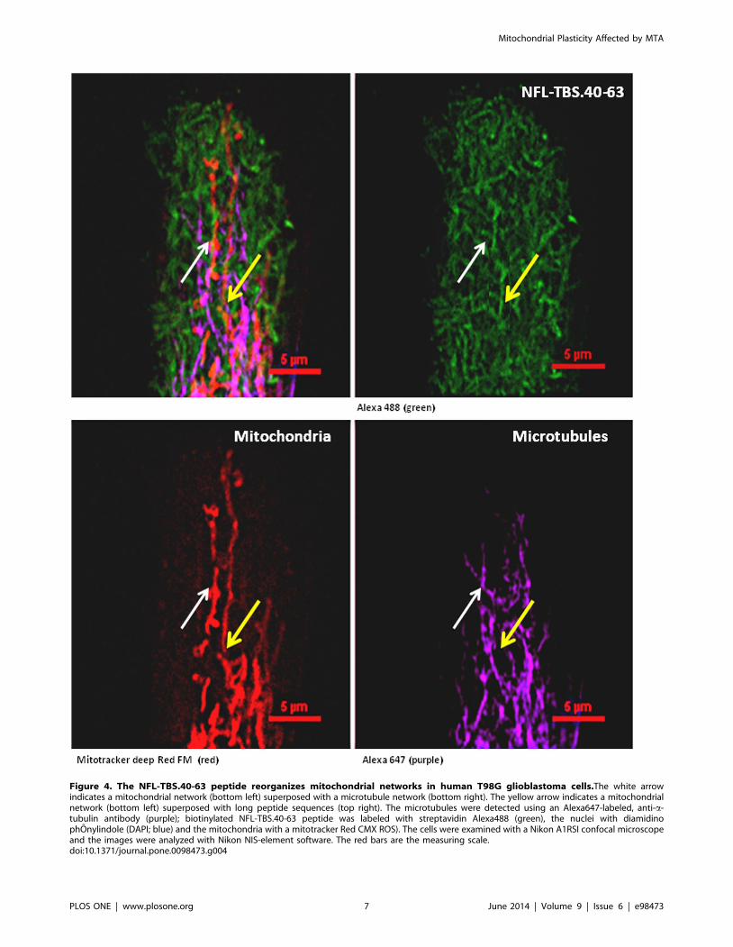
Figure 4. The NFL-TBS.40-63 peptide reorganizes mitochondrial networks in human T98G glioblastoma cells.The white arrowindicates a mitochondrial network (bottom left) superposed with a microtubule network (bottom right). The yellow arrow indicates a mitochondrialnetwork (bottom left) superposed with long peptide sequences (top right). The microtubules were detected using an Alexa647-labeled, anti-a-tubulin antibody (purple); biotinylated NFL-TBS.40-63 peptide was labeled with streptavidin Alexa488 (green), the nuclei with diamidinophOnylindole (DAPI; blue) and the mitochondria with a mitotracker Red CMX ROS). The cells were examined with a Nikon A1RSI confocal microscopeand the images were analyzed with Nikon NIS-element software. The red bars are the measuring scale.doi:10.1371/journal.pone.0098473.g004
Mitochondrial Plasticity Affected by MTA
PLOS ONE | www.plosone.org 7 June 2014 | Volume 9 | Issue 6 | e98473

Figure 5. Quantitative PCR analysis of mitochondrial fission/fusion actors and relevant differentially-expressed miRNA-mRNAcomplexes in human T98G glioblastoma cells. 5A: Quantitative PCR analysis of mitochondrial fission/fusion actors (FIS1 and MFN2) in T98Gcells. The data are expressed in units (mRNA expression of a specific gene normalized to b-globin mRNA expression) that are relative to the control,which was assigned a unit value. 5B: Quantitative PCR analysis of relevant differentially expressed miRNA in T98G cells. The data are expressed in units(miRNA expression relative to U5 snRNA) that are relative to the control, which was assigned a unit value. 5C: Quantitative PCR analysis of PTEN andNAIP mRNA, directly targeted by miR-21 and miR-221, respectively. FGFR3 expression was used as negative control of miR-100, which expression level
Mitochondrial Plasticity Affected by MTA
PLOS ONE | www.plosone.org 8 June 2014 | Volume 9 | Issue 6 | e98473

mitochondrial motility [11–13]. The NFL-TBS.40-63 peptide is
able to alter microtubule formation when it is internalized by
T98G glioblastoma cells and inhibits their proliferation [8]. In this
study, we have evaluated the effect of NFL-TBS.40-63 peptide
internalization on mitochondrial biogenesis and function.
Our observations revealed a negative impact on T98G cell
respiration after 6 hours of NFL-TBS.40-63 peptide treatment (2
to 10 mM). This action on mitochondrial function, at lower
concentrations than those necessary to disturb the cytoskeleton
(10 mM), could be related to a primary modification of the
mitochondrial motility. It has been shown that peptides derived
from the N-terminal domain of intermediate filaments, like
desmin, vimentin and keratin, can interact with the unpolymerized
tubulin [8]. A recent study demonstrated that the N-terminal
domain of vimentin (residues 41–94) can also directly bind
mitochondria and serve as an adaptor between actin microfila-
ments and mitochondria [13]. We suggest that the primary action
of NFL-TBS.40-63 leads to sequential organization of the peptide,
which disturbs the cytoskeleton and reorganizes the mitochondrial
network. This should be related to the conserved FIS1/MFN2
ratio we observed at higher peptide concentrations. At a 10 mM
peptide treatment, we also revealed a colocalization of NFL-
TBS.40-63 with mitochondria and a specific accumulation at the
microtubules’ extremities, which may limit membrane ruffling, as
previously reported [37,38].
This study revealed that the NFL-TBS.40-63 peptide provokes a
redistribution of mitochondria throughout the cytoplasm. Mito-
chondria were able to reorganize along the peptide from end to
end, in order to form a polarized but less dense network and
reduce cell respiration. Mitochondria and autophagy are linked to
homeostatic elements that act in response to changes in the cellular
environment, such as energy, nutrients and stress. Thus, defects in
plasticity could simultaneously impair autophagy, which may
result in increased risk for various human diseases [39,40]. The
peptide treatment induces an inhibition of FIS1 and MFN2 gene
expression. As has been shown, deregulation of mitochondrial
fusion or fission is associated with alterations in the organization of
the mitochondrial network and with the inhibition of energy
metabolism [41]. Alterations in energetic metabolism cause defects
in respiratory chain subunits and may lead to mitochondrial
network fragmentation [42]. Western blotting analyses indicated
that decreases in the OXPHOS process were also related to a
decrease in mitochondrial biogenesis when using 10 mM of
NFLTBS.40-63 peptide, regarding protein levels for two subunits
of the respiratory chain complexes (SDHB and COX4) and for
transcription factor NRF-1. This rapid reduction of mitochondria
after 6 hours of peptide treatment may be related to the induction
of mitophagy. Thus, the PGC-1a/PRC pathway, which is related
to the transcriptional regulation of mitochondrial biogenesis, was
not affected after 6 hours of treatment, while NRF-1 and CYCS
were repressed; this suggests a lack of extra-cellular signal
regulation or a delayed PGC-adaptive response to energy
depletion. Moreover, this could suggest a rapid regulation of
mitophagy/biogenesis balance through post-transcriptional path-
ways, as recently reported [43,44].
We found that the expression of two relevant miRNAs-miR-21
and miR-221-was altered by a 6-hours treatment with the NFL-
TBS.40-63 peptide, compared to the scrambled control. These
miRNAs are referred to as oncomirs, as they have anti-apoptotic
and proliferative effects [25,26,28,45]. In human tumors, miR-21
down-regulates the expression of PTEN, which is involved in
mitophagy through its negative regulation of PINK1 (PTEN-
induced kinase 1) [34]. Up-regulation of miR-221 has also been
correlated with down-regulation of one of its targets, NAIP (NLR-
family, apoptosis inhibitory protein), which is involved in
neurodegeneration and apoptosis regulation [33]. On the
contrary, over-expression of miR-218 and miR-96 were associated
with apoptosis induction through targeting the PINK1/NF-KB
pathway and FOXO1, respectively [46,47]. The inverse functions
of these miRNAs on apoptosis or mitophagy should be considered,
depending on their half-time. Thus, miR-21 is considered to be
one of the most long-lived miRNAs [48].
In conclusion, we showed that a 6-hours treatment with 10 mM
of NFL-TBS.40-63 peptide induces a direct reduction in the global
respiration of cells that have internalized the peptide. This effect
can be related to a reduction of the main transcription factor
involved in mitochondrial biogenesis and in the OXPHOS
process. The treatment is efficient in reducing the expression of
miRNAs that are involved in glioblastoma pathogenesis and MTA
resistance. To our knowledge, this is the first report showing the
role played by this peptide in the regulation of global respiration
for glioblastoma cells. Our results show that disrupting a
microtubule network affects the mitochondrial network distribu-
tion, thus confirming the close relationship between these two
networks.
Supporting Information
File S1
(PDF)
Acknowledgments
We are grateful to Philippe Hulin and Kevin Nedellec for technical help.
Author Contributions
Conceived and designed the experiments: FS JE. Performed the
experiments: RR CLC. Analyzed the data: FS JE RR CLC. Contributed
reagents/materials/analysis tools: FS JE. Wrote the paper: RR FS.
References
1. Louis DN, Ohgaki H, Wiestler OD, Cavenee WK, Burger PC, et al. (2007) The
2007 WHO classification of tumours of the central nervous system. Acta
Neuropathol 114: 97–109.
2. Stupp R, Mason WP, van den Bent MJ, Weller M, Fisher B, et al. (2005)
Radiotherapy plus concomitant and adjuvant temozolomide for glioblastoma.
N Engl J Med 352: 987–996.
3. Monticone M, Daga A, Candiani S, Romeo F, Mirisola V, et al. (2012)
Identification of a novel set of genes reflecting different in vivo invasive patterns
of human GBM cells. BMC Cancer 12: 358.
4. Ohgaki H, Kleihues P (2005) Epidemiology and etiology of gliomas. Acta
Neuropathol 109: 93–108.
5. Stupp R, Hegi ME, Mason WP, van den Bent MJ, Taphoorn MJ, et al. (2009)
Effects of radiotherapy with concomitant and adjuvant temozolomide versus
was unchanged by peptide treatment. The data are expressed in units (mRNA expression of a specific gene normalized to b-globin mRNA expression)that are relative to the control, which was assigned a unit value. The values are the average 6 SD for three separate determinations (N = 3). *: P,0.05versus control.doi:10.1371/journal.pone.0098473.g005
Mitochondrial Plasticity Affected by MTA
PLOS ONE | www.plosone.org 9 June 2014 | Volume 9 | Issue 6 | e98473
radiotherapy alone on survival in glioblastoma in a randomised phase III study:
5-year analysis of the EORTC-NCIC trial. Lancet Oncol 10: 459–466.6. Nogales E (2000) Structural insights into microtubule function. Annu Rev
Biochem 69: 277–302.
7. Dumontet C, Jordan MA (2010) Microtubule-binding agents: a dynamic field ofcancer therapeutics. Nat Rev Drug Discov 9: 790–803.
8. Bocquet A, Berges R, Frank R, Robert P, Peterson AC, et al. (2009)Neurofilaments bind tubulin and modulate its polymerization. J Neurosci 29:
11043–11054.
9. Lepinoux-Chambaud C, Eyer J (2013) The NFL-TBS.40-63 anti-glioblastomapeptide enters selectively in glioma cells by endocytosis. Int J Pharm 454: 738–
747.10. Berges R, Balzeau J, Peterson AC, Eyer J (2012) A tubulin binding peptide
targets glioma cells disrupting their microtubules, blocking migration, andinducing apoptosis. Mol Ther 20: 1367–1377.
11. Cagalinec M, Safiulina D, Liiv M, Liiv J, Choubey V, et al. (2013) Principles of
the mitochondrial fusion and fission cycle in neurons. J Cell Sci 126: 2187–2197.12. Gentil BJ, Minotti S, Beange M, Baloh RH, Julien JP, et al. (2012) Normal role
of the low-molecular-weight neurofilament protein in mitochondrial dynamicsand disruption in Charcot-Marie-Tooth disease. FASEB J 26: 1194–1203.
13. Nekrasova OE, Mendez MG, Chernoivanenko IS, Tyurin-Kuzmin PA,
Kuczmarski ER, et al. (2011) Vimentin intermediate filaments modulate themotility of mitochondria. Mol Biol Cell 22: 2282–2289.
14. Gomes LC, Di BG, Scorrano L (2011) During autophagy mitochondriaelongate, are spared from degradation and sustain cell viability. Nat Cell Biol 13:
589–598.15. Suen DF, Norris KL, Youle RJ (2008) Mitochondrial dynamics and apoptosis.
Genes Dev 22: 1577–1590.
16. Liu X, Weaver D, Shirihai O, Hajnoczky G (2009) Mitochondrial ’kiss-and-run’:interplay between mitochondrial motility and fusion-fission dynamics. EMBO J
28: 3074–3089.17. Hollenbeck PJ, Saxton WM (2005) The axonal transport of mitochondria. J Cell
Sci 118: 5411–5419.
18. Grandemange S, Herzig S, Martinou JC (2009) Mitochondrial dynamics andcancer. Semin Cancer Biol 19: 50–56.
19. Westermann B (2010) Mitochondrial fusion and fission in cell life and death. NatRev Mol Cell Biol 11: 872–884.
20. Scarpulla RC (2008) Transcriptional paradigms in mammalian mitochondrialbiogenesis and function. Physiol Rev 88: 611–638.
21. Raharijaona M, Le Pennec S, Poirier J, Mirebeau-Prunier D, Rouxel C, et al.
(2009) PGC-1-related coactivator modulates mitochondrial-nuclear crosstalkthrough endogenous nitric oxide in a cellular model of oncocytic thyroid
tumours. PLoS One 4: e7964.22. Tivnan A, McDonald KL (2013) Current progress for the use of miRNAs in
glioblastoma treatment. Mol Neurobiol 48: 757–768.
23. Li WB, Ma MW, Dong LJ, Wang F, Chen LX, et al. (2011) MicroRNA-34atargets notch1 and inhibits cell proliferation in glioblastoma multiforme. Cancer
Biol Ther 12: 477–483.24. Corsten MF, Miranda R, Kasmieh R, Krichevsky AM, Weissleder R, et al.
(2007) MicroRNA-21 knockdown disrupts glioma growth in vivo and displayssynergistic cytotoxicity with neural precursor cell delivered S-TRAIL in human
gliomas. Cancer Res 67: 8994–9000.
25. Ren Y, Zhou X, Mei M, Yuan XB, Han L, et al. (2010) MicroRNA-21 inhibitorsensitizes human glioblastoma cells U251 (PTEN-mutant) and LN229 (PTEN-
wild type) to taxol. BMC Cancer 10: 27.26. Costa PM, Cardoso AL, Nobrega C, Pereira de Almeida LF, Bruce JN, et al.
(2013) MicroRNA-21 silencing enhances the cytotoxic effect of the antiangio-
genic drug sunitinib in glioblastoma. Hum Mol Genet 22: 904–918.27. Hao J, Zhang C, Zhang A, Wang K, Jia Z, et al. (2012) miR-221/222 is the
regulator of Cx43 expression in human glioblastoma cells. Oncol Rep 27: 1504–1510.
28. Kim HJ, Kim YH, Lee DS, Chung JK, Kim S (2008) In vivo imaging of
functional targeting of miR-221 in papillary thyroid carcinoma. J Nucl Med 49:
1686–1693.
29. Barrey E, Saint-Auret G, Bonnamy B, Damas D, Boyer O, et al. (2011) Pre-
microRNA and mature microRNA in human mitochondria. PLoS One 6:
e20220.
30. Ninio-Many L, Grossman H, Shomron N, Chuderland D, Shalgi R (2013)
microRNA-125a-3p reduces cell proliferation and migration by targeting Fyn.
J Cell Sci 126: 2867–2876.
31. Tomasetti M, Neuzil J, Dong L (2014) MicroRNAs as regulators of
mitochondrial function: Role in cancer suppression. Biochim Biophys Acta
1840: 1441–1453.
32. Balzeau J, Pinier M, Berges R, Saulnier P, Benoit JP, et al. (2013) The effect of
functionalizing lipid nanocapsules with NFL-TBS.40-63 peptide on their uptake
by glioblastoma cells. Biomaterials 34: 3381–3389.
33. Lukiw WJ, Cui JG, Li YY, Culicchia F (2009) Up-regulation of micro-RNA-221
(miRNA-221; chr Xp11.3) and caspase-3 accompanies down-regulation of the
survivin-1 homolog BIRC1 (NAIP) in glioblastoma multiforme (GBM).
J Neurooncol 91: 27–32.
34. Zhang JG, Wang JJ, Zhao F, Liu Q, Jiang K, et al. (2010) MicroRNA-21 (miR-
21) represses tumor suppressor PTEN and promotes growth and invasion in
non-small cell lung cancer (NSCLC). Clin Chim Acta 411: 846–852.
35. Galiveti CR, Rozhdestvensky TS, Brosius J, Lehrach H, Konthur Z (2010)
Application of housekeeping npcRNAs for quantitative expression analysis of
human transcriptome by real-time PCR. RNA 16: 450–461.
36. Mitra K, Lippincott-Schwartz J (2010) Analysis of mitochondrial dynamics and
functions using imaging approaches. Curr Protoc Cell Biol Chapter 4: Unit-21.
37. Rinnerthaler G, Geiger B, Small JV (1988) Contact formation during fibroblast
locomotion: involvement of membrane ruffles and microtubules. J Cell Biol 106:
747–760.
38. Schober JM, Komarova YA, Chaga OY, Akhmanova A, Borisy GG (2007)
Microtubule-targeting-dependent reorganization of filopodia. J Cell Sci 120:
1235–1244.
39. Kondo-Okamoto N, Noda NN, Suzuki SW, Nakatogawa H, Takahashi I, et al.
(2012) Autophagy-related protein 32 acts as autophagic degron and directly
initiates mitophagy. J Biol Chem 287: 10631–10638.
40. Okamoto K, Kondo-Okamoto N, Ohsumi Y (2009) Mitochondria-anchored
receptor Atg32 mediates degradation of mitochondria via selective autophagy.
Dev Cell 17: 87–97.
41. Pich S, Bach D, Briones P, Liesa M, Camps M, et al. (2005) The Charcot-Marie-
Tooth type 2A gene product, Mfn2, up-regulates fuel oxidation through
expression of OXPHOS system. Hum Mol Genet 14: 1405–1415.
42. Benard G, Faustin B, Passerieux E, Galinier A, Rocher C, et al. (2006)
Physiological diversity of mitochondrial oxidative phosphorylation. Am J Physiol
Cell Physiol 291: C1172–C1182.
43. Kubli DA, Gustafsson AB (2012) Mitochondria and mitophagy: the yin and yang
of cell death control. Circ Res 111: 1208–1221.
44. Li P, Jiao J, Gao G, Prabhakar BS (2012) Control of mitochondrial activity by
miRNAs. J Cell Biochem 113: 1104–1110.
45. Karsy M, Arslan E, Moy F (2012) Current Progress on Understanding
MicroRNAs in Glioblastoma Multiforme. Genes Cancer 3: 3–15.
46. Guo Y, Liu H, Zhang H, Shang C, Song Y (2012) miR-96 regulates FOXO1-
mediated cell apoptosis in bladder cancer. Oncol Lett 4: 561–565.
47. Xia H, Yan Y, Hu M, Wang Y, Wang Y, et al. (2013) MiR-218 sensitizes glioma
cells to apoptosis and inhibits tumorigenicity by regulating ECOP-mediated
suppression of NF-kappaB activity. Neuro Oncol 15: 413–422.
48. Gantier MP, McCoy CE, Rusinova I, Saulep D, Wang D, et al. (2011) Analysis
of microRNA turnover in mammalian cells following Dicer1 ablation. Nucleic
Acids Res 39: 5692–5703.
Mitochondrial Plasticity Affected by MTA
PLOS ONE | www.plosone.org 10 June 2014 | Volume 9 | Issue 6 | e98473